Note: Descriptions are shown in the official language in which they were submitted.
CA 02461611 2004-03-19
WO 03/027224 PCT/US02/28755
1
OPTOINJECTION METHODS
Background of the Invention
This invention relates to methods for cell
manipulation and more specifically to methods for
transiently permeabilizing a cell so that a variety of
exogenous materials, such as expressible foreign DNA, can
be loaded into the cell.
Previous loading methods have included chemical
treatments, microinjection, electroporation and particle
bombardment. However, these techniques can be time-
consuming and suffer from low yields or poor cell
survival. Another technique termed "optoporation" has
used light directed toward cells and the surrounding
media to induce shock waves, thereby causing small holes
to form temporarily in the surface of nearby cells,
allowing materials to.non-specifically enter cells in the
area. Another technique termed "optoinjection" also uses
light, but directs the light to specific cells.
Nevertheless, previous light-based implementations
techniques have suffered from the same disadvantages as
other loading techniques.
Thus, there is a need for a method for rapid
and efficient loading of a variety of exogenous molecules
into cells, with high cell survival rates. The present
invention satisfies this need and provides related
advantages as well.
CA 02461611 2004-03-19
WO 03/027224 PCT/US02/28755
2
Summary of the Invention
The present invention provides optoinjection
methods for transiently permeabili~ing a target cell. In
the general method, the steps are (a) illuminating a
population of cells contained in a frame; (b) detecting
at least one property of light directed from the frame;
(c) locating a target cell by the property of light; and
(d) irradiating the target cell with a pulse of
radiation.
In particular embodiments, a static
representation is obtained when the population of cells
is substantially stationary; the cells are illuminated
through a lens having a numerical aperture of at most
0.5; the pulse of radiation has a diameter of at least
10 microns at the point of contact with the target cell;
or the resulting pulse of radiation delivers at most
1 ~.ZJ/~.tm~ . As a result, the method provides rapid and
efficient loading of a variety of exogenous molecules
into cells, with high cell survival rates.
Brief Description of The Drawings
Figure 1 is a perspective view of one
embodiment of a cell treatment apparatus and illustrates
the outer design of the housing and display.
Figure 2 is a perspective view of one
embodiment of a cell treatment apparatus with the outer
housing removed and the inner components illustrated.
CA 02461611 2004-03-19
WO 03/027224 PCT/US02/28755
3
Figure 3 is a block diagram of the optical
subassembly design within one embodiment of a cell
treatment apparatus.
Figure 4 is a perspective view of one
embodiment of an optical subassembly within one
embodiment of a cell treatment apparatus.
Figure 5 is a side view of one embodiment of an
optical subassembly that illustrates the arrangement of
the scanning lens and the movable stage.
Figure 6 is a bottom perspective view of one
embodiment of an optical subassembly.
Figure 7 is a top perspective view of the
movable stage of the cell treatment apparatus.
Figure 8 shows cells under broad-spectrum light
(8A), cells showing loading of Texas-Red-Dextran (8B) and
nonviable cells (8C).
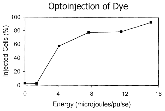
Figure 9 illustrates that the efficiency of
optoinjection is energy dose-dependent
Figure 10 compares expression of a plasmid in
optoinjected cells (10B) compared to control cells
without optoinjection (10A).
Detailed Description
A method and apparatus is described for
selectively identifying, and individually targeting with
an energy beam, specific cells within a cell population
for the purpose of inducing a response in the targeted
cells. The population of cells can be a mixed population
CA 02461611 2004-03-19
WO 03/027224 PCT/US02/28755
4
or homogenous in origin. The responses of any of the
embodiments of the methods and apparatuses of the
invention can be lethal or non-lethal. Examples of such
responses are set forth above and throughout this
disclosure. The cells targeted can be labeled as is
often the case when the specimen is a mixed population.
On the other hand, when the specimen is homogenous, the
targeted cells can be those individual cells that are
being interrogated or intersected by the illumination
source or the energy beam, in order to study the response
of the cell. For instance, such responses include the
morphological or physiological characteristics of the
cell. Generally, the method first employs a label that
acts as a marker to identify and locate individual cells
of a first population of cells within a cell mixture that
is comprised of the first population of cells and a
second population of cells. The cells targeted by the
apparatus and methods herein are those that are
selectively labeled, in the case of a mixed population of
cells, or the ones undergoing interrogation or
intersection by the illumination source or energy beam.
The chosen label can be any that substantially
identifies and distinguishes the first population of
cells from the second population of cells. For example,
monoclonal antibodies that are directly or indirectly
tagged with a fluorochrome can be used as specific
labels. Other examples of cell surface binding labels
include non-antibody proteins, lectins, carbohydrates, or
short peptides with selective cell binding capacity.
Membrane intercalating dyes, such as PKH-2 and PKH-26,
could also serve as a useful distinguishing label
indicating mitotic history of a cell. Many membrane-
CA 02461611 2004-03-19
WO 03/027224 PCT/US02/28755
permeable reagents are also available to distinguish
living cells from one another based upon selected
criteria. For example, phalloidin indicates membrane
integrity, tetramethyl rhodamine methyl ester (TMRM)
5 indicates mitochondrial transmembrane potential,
monochlorobimane indicates glutathione reductive stage,
carboxymethyl fluorescein diacetate (CMFDA) indicates
thiol activity, carboxyfluorescein diacetate indicates
intracellular pH, fura-2 indicates intracellular
Ca2+level, and 5,5',6,6'-tetrachloro-1,1',3,3'-
tetraethylbenzimidazolo carbocyanine iodide (JC-1)
indicates membrane potential. Cell viability can be
assessed by the use of fluorescent SYTO 13 or YO PRO
reagents. Similarly, a fluorescently-tagged genetic
probe (DNA or RNA) could be used to label cells which
carry a gene of interest, or express a gene of interest.
Further, cell cycle status could be assessed through the
use of Hoechst 33342 dye to label existing DNA combined
with. bromodeoxyuridine (BrdU) to label newly synthesized
DNA.
It should be noted that if no specific label is
available for cells of the first population, the method
can be implemented in an inverse fashion by utilizing a
specific label for cells of the second population. For
example, in hematopoietic cell populations, the CD34 or
ACC-133 cell markers can be used to label only the
primitive hematopoietic cells, but not the other cells
within the mixture. In this embodiment, cells of the
first population are identified by the absence of the
label, and are thereby targeted by the energy beam.
After cells of the first population are
identified, an energy beam, such as from a laser,
CA 02461611 2004-03-19
WO 03/027224 PCT/US02/28755
6
collimated or focused non-laser light, RF energy,
accelerated particle, focused ultrasonic energy, electron
beam, or other radiation beam, is used to deliver a
targeted dose of energy that induces the pre-determined
response in each of the cells of the first population,
without substantially affecting cells of the second
population.
One such pre-determined response is
photobleaching. In photobleaching, a label in the form
of a dye, such as rhodamine 123, GFP, fluorescein
isothiocyanate (FITC), or phycoerythrin, is added to the
specimen before the instant methods are commenced. After
the population of cells has time to interact with. the
dye, the energy beam is used to bleach a region of
individual cells in the population. Such. photobleaching
studies can be used to study the motility, replenishment,
dynamics and the like of cellular components and
processes.
Another response is internal molecular
uncaging. In such a process, the specimen is combined
with a caged molecule prior to the commencement of the
instant methods. Such caged molecules include the (3-2,6-
dinitrobenzyl ester of L-aspartic acid or the 1-(2-
nitrophenyl)ethyl ether of 8-hydroxylpyrene-1,3,6-tris-
sulfonic acid. Similarly, caging groups including
alphacarboxyl-2-nitrobenzyl (CNB) and 5-carboxylmethoxy-
2-nitrobenzyl (CMNB) can be linked to biologically active
molecules as ethers, thioethers, esters, amines, or
similar functional groups. The term "internal molecular
uncaging" refers to the fact that the molecular uncaging
takes place on the surface or within the cell. Such
CA 02461611 2004-03-19
WO 03/027224 PCT/US02/28755
7
uncaging experiments study rapid molecular processes such
as cell membrane permeability and cellular signaling.
Yet another response is external molecular
uncaging. This uses approximately the same process as
internal molecular caging. However, in external
molecular uncaging, the uncaged molecule, is not attached
to or incorporated into the targeted cells. Instead, the
responses of the surrounding targeted cells to the caged
and uncaged variants of the molecule are imaged by the
instant apparatus and methods.
Figure 1 is an illustration of one embodiment
of a cell treatment apparatus 10. The cell treatment
apparatus 10 includes a housing 15 that stores the inner
components of the apparatus. The housing includes laser
safety interlocks to ensure safety of the user, and also
limits interference by external influences (e. g., ambient
light, dust, etc.). Located on the upper portion of the
housing 15 is a display unit 20 for displaying captured
images of cell populations during treatment. These
images are captured by a camera, as will be discussed
more specifically below. A keyboard 25 and mouse 30 are
used to input data and control the apparatus 10. An
access door 35 provides access to a movable stage that
holds a specimen container of cells undergoing treatment.
An interior view of the apparatus 10 is
provided in Figure 2. As illustrated, the apparatus 10
provides an upper tray 200 and lower tray 210 that hold
the interior components of the apparatus. The upper tray
200 includes a pair of intake filters 215A,B that filter
ambient air being drawn into the interior of the
apparatus 10. Below the access door 35 is the optical
CA 02461611 2004-03-19
WO 03/027224 PCT/US02/28755
8
subassembly (not shown). The optical subassembly is
mounted to the upper tray 200 and is discussed in detail
with regard to Figures 3 to 6.
On the lower tray 210 is a computer 225 which
stores the software programs, commands and instructions
that run the apparatus 10. In addition, the computer 225
provides control signals to the treatment apparatus
through electrical signal connections for steering the
laser to the appropriate spot on the specimen in order to
treat the cells.
As illustrated, a series of power supplies
230A,B,C provide power to the various electrical
components within the apparatus 10. In addition, an
uninterruptable power supply 235 is incorporated to allow
the apparatus to continue functioning through short
external power interruptions.
Figure 3 provides a layout of one embodiment of
an optical subassembly design 300 within an embodiment of
a cell treatment apparatus 10. As illustrated, an
illumination laser 305 provides a directed laser output
that is used to excite a particular label that is
attached to targeted cells within the specimen. In this
embodiment, the illumination laser emits light at a
wavelength of 532 nm. Once the illumination laser has
generated a light beam, the light passes into a shutter
310 which controls the pulse length of the laser light.
After the illumination laser light passes
through the shutter 310, it enters a ball lens 315 where
it is focused into a SMA fiber optic connector 320.
After the illumination laser beam has entered the fiber
optic connector 320, it is transmitted through a fiber
CA 02461611 2004-03-19
WO 03/027224 PCT/US02/28755
9
optic cable 325 to an outlet 330. By passing the
illumination beam through the fiber optic cable 325, the
illumination laser 305 can be positioned anywhere within
the treatment apparatus and thus is not limited to only
being positioned within a direct light pathway to the
optical components. In one embodiment, the fiber optic
cable 325 is connected to a vibrating motor 327 for the
purpose of mode scrambling and generating a more uniform
illumination spot.
After the light passes through the outlet 330,
it is directed into a series of condensing lenses in
order to focus the beam to the proper diameter for
illuminating one frame of cells. As used herein, one
frame of cells is defined as the portion of the
biological specimen that is captured within one frame
image captured by the camera. This is described more
specifically below.
Accordingly, the illumination laser beam passes
through a first condenser lens 335. In one embodiment,
this first lens has a focal length of 4.6 mm. The light
beam then passes through a second condenser lens 340
which, in one embodiment, provides a 100 mm focal length.
Finally, the light beam passes into a third condenser
lens 345, which preferably provides a 200 mm focal
length. While the present invention has been described
using specific condenser lenses, it should be apparent
that other similar lens configurations that focus the
illumination laser beam to an advantageous diameter would
function similarly. Thus, this invention is not limited
to the specific implementation of ariy particular
condenser lens system.
CA 02461611 2004-03-19
WO 03/027224 PCT/US02/28755
Once the illumination laser beam passes through
the third condenser lens 345, it enters a cube beam
splatter 350 that is designed to transmit the 532 nm
wavelength of light emanating from the illumination
5 laser. Preferably, the cube beam splatter 350 is a 25.4
mm square cube (Melles-Griot, Irvine, Calif.). However,
other sizes are anticipated to function similarly. In
addition, a number of plate beam splatters or pellicle
beam splatters could be used in place of the cube beam
10 splatter 350 with no appreciable change in function.
Once the illumination laser light has been
transmitted through the cube beam splatter 350, it
reaches a long wave pass mirror 355 that reflects the 532
nm illumination laser light to a set of galvanometer
mirrors 360 that steer the illumination laser light under
computer control to a scanning lens (Special Optics,
Wharton, N.J.) 365, which directs the illumination laser
light to the specimen (not shown). The galvanometer
mirrors are controlled so that the illumination laser
light is directed at the proper cell population (i.e.
frame of cells) for imaging. The "scanning lens"
described in this embodiment of the invention includes a
refractive lens. It should be noted that the term
"scanning lens" as used in the present invention
includes, but is not limited to, a system of one or more
refractive or reflective optical elements used alone or
in combination. Further, the "scanning lens" may include
a system of one or more diffractive elements used in
combination with one or more refractive and/or reflective
optical elements. One skilled in the art will know how
to design a "scanning lens" system in order to illuminate
the proper cell population.
CA 02461611 2004-03-19
WO 03/027224 PCT/US02/28755
11
The light from the illumination laser is of a
wavelength that is useful for illuminating the specimen.
In this embodiment, energy from a continuous wave 532 nm
Nd:YAG frequency-doubled laser (B&W Tek, Newark, Del.)
reflects off the long wave pass mirror (Custom
Scientific, Phoenix, Ariz.) and excites fluorescent tags
in the specimen. In one embodiment, the fluorescent tag
is phycoerythrin. Alternatively, Alexa 532 (Molecular
Probes, Eugene, Oregon) can be used. Phycoerythrin and
Alexa 532 have emission spectra with peaks near 580 nm,
so that the emitted fluorescent light from the specimen
is transmitted via the long wave pass mirror to be
directed into the camera. The use of the filter in front
of the camera blocks light that is not within the
wavelength range of interest, thereby reducing the amount
of background light entering the camera.
It is generally known that many other devices
could be used in this manner to illuminate the specimen,
including, but not limited to, an arc lamp (e. g.,
mercury, xenon, etc.) with or without filters, a light-
emitting diode (LED), other types of lasers; etc.
Advantages of this particular laser include high
intensity, relatively efficient use of energy, compact
size, and minimal heat generation. It is also generally
known that other fluorochromes with different excitation
and emission spectra could be used in such an apparatus
with the appropriate selection of illumination source,
filters, and long and/or short wave pass mirrors. For
example, allophycocyanin (APC) could be excited with a
633 nm HeNe illumination laser, and fluoroisothiocyanate
(FITC) could be excited with a 488 nm Argon illumination
laser. One skilled in the art could propose many other
CA 02461611 2004-03-19
WO 03/027224 PCT/US02/28755
12
optical layouts with various components in order to
achieve the objective of this invention.
In addition to the illumination laser 305, an
optional treatment laser 400 is present to irradiate the
targeted cells once they have been identified by image
analysis. Of course, in one embodiment, the treatment
induces necrosis of targeted cells within the cell
population. As shown, the treatment laser 400 outputs an
energy beam of 523 nm that passes through a shutter 410.
Although the exemplary laser outputs an energy beam
having a 523 nm wavelength, other sources that generate
energy at other wavelengths are also within the scope of
the present invention.
Once the treatment laser energy beam passes
through the shutter 410, it enters a beam expander
(Special Optics, Wharton, N.J.) 415 which adjusts the
diameter of the energy beam to an appropriate size at the
plane of the specimen. Following the beam expander 415
is a half-wave plate 420 which controls the polarization
of the beam. The treatment laser energy beam is then
reflected off a mirror 425 and enters the cube beam
splitter 350. The treatment laser energy beam is
reflected by 90 degrees in the cube beam splitter 350,
such that it is aligned with the exit pathway of the
illumination laser light beam. Thus, the treatment laser
energy beam and the illumination laser light beam both
exit the cube beam splitter 350 along the same light
path. From the cube beam splitter 350, the treatment
laser beam reflects off the long wave pass mirror 355, is
steered by the galvanometers 360, thereafter contacts the
scanning lens 365, and finally is focused upon a targeted
cell within the specimen. Again, the "scanning lens"
CA 02461611 2004-03-19
WO 03/027224 PCT/US02/28755
13
described in this embodiment includes a refractive lens.
As previously mentioned, the term "scanning lens"
includes, but is not limited to, a system of one or more
refractive or reflective optical elements used alone or
in combination. Further, the "scanning lens" may include
one or more diffractive elements used in combination. with
one or more refractive and/or reflective elements. One
skilled in the art will know how to design a "scanning
lens" system in order to focus upon the targeted cell
within the specimen.
It should be noted that a small fraction of the
illumination laser light beam passes through the long
wave pass mirror 355 and enters a power meter sensor
(Gentec, Palo Alto, Calif.) 445. The fraction of the
beam entering the power sensor 445 is used to calculate
the level of power emanating from the illumination laser
305. In an analogous fashion, a small fraction of the
treatment laser energy beam passes through the cube beam
splitter 350 and enters a second power meter sensor
(Gentec, Palo Alto, Calif.) 446. The fraction of the
beam entering the power sensor 446 is used to calculate
the level of power emanating from the treatment laser
400. The power meter sensors are electrically linked to
the computer system so that instructions/commands within
the computer system capture the power measurement and
determine the amount of energy that was emitted.
The energy beam from the treatment laser is of
a wavelength that is useful for achieving a response in
the cells. In the example shown, a pulsed 523 nm Nd:YLF
frequency-doubled laser is used to heat a localized
volume containing the targeted cell, such that it is
induced to die within a pre-determined period of time.
CA 02461611 2004-03-19
WO 03/027224 PCT/US02/28755
14
The mechanism of death is dependent upon the actual
temperature achieved in the cell, as reviewed by Niem~,
M. H., Laser-tissue interactions: Fundamentals and
Applications (Springer-Verlag, Berlin 1996).
A Nd:YLF frequency-doubled, solid-state laser
(Spectra-Physics, Mountain View, Calif.) is used because
of its stability, high repetition rate of firing, and
long time of maintenance-free service. However, most
cell culture fluids and cells are relatively transparent
to light in this green wavelength, and therefore a very
high fluence of energy would be required to achieve cell
death. To significantly reduce the amount of energy
required, and therefore the cost and sire of the
treatment laser, a dye is purposefully added to the
specimen to efficiently absorb the energy of the
treatment laser in the specimen. In the example shown,
the non-toxic dye FD&C red #40 (allura red) is used to
absorb the 523 nm energy from the treatment laser, but
one skilled in the art could identify other laser/dye
combinations that would result in efficient absorption of
energy by the specimen. For example, a 633 nm HeNe
laser's energy would be efficiently absorbed by FD&C
green #3 (fast green FCF), a 488 nm Argon laser's energy
would be efficiently absorbed by FD&C yellow #5 (sunset
yellow FCF), and a 1064 nm Nd:YAG laser's energy would be
efficiently absorbed by Filtron (Gentex, ~eeland, Mich.)
infrared absorbing dye. Through the use of an energy
absorbing dye, the amount of energy required to kill a
targeted cell can be reduced since more of the treatment '
laser energy is absorbed in the presence of such a dye.
Another method of achieving thermal killing of
cells without the addition of a dye involves the use of
CA 02461611 2004-03-19
WO 03/027224 PCT/US02/28755
an ultraviolet laser. Energy from a 355 nm Nd:YAG
frequency-tripled laser will be absorbed by nucleic acids
and proteins within the cell, resulting in thermal
heating and death. Yet another method of achieving
5 thermal killing of cells without the addition of a dye
involves the use of a near-infrared laser. Energy from a
2100 nm Ho:YAG laser or a 2940 nm Er:YAG laser will be
absorbed by water within the cell, resulting in thermal
heating and death.
10 Although this embodiment describes the killing
of cells via thermal heating by the energy beam, one
skilled in the art would recognize that other responses
can also be induced in the cells by an energy beam,
including photomechanical disruption, photodissociation,
15 photoablation, and photochemical reactions, as reviewed
by Niemz (Niemz, supra). For example, a photosensitive
substance (e. g., hematoporphyrin derivative, tin-
etiopurpurin, lutetium texaphyrin) (Oleinick and Evans,
The photobiology of photodynamic therapy: Cellular
targets and mechanisms, Rad. Res. 150: 5146-5156 (1998))
within the cell mixture could be specifically activated
in targeted cells by irradiation. Additionally, a small,
transient pore could be made in the cell membrane
(Palumbo et al., Targeted gene transfer in eukaryotic
cells by dye-assisted laser optoporation, J. Photochem.
Photobiol. 36:41-46 (1996)) to allow the entry of genetic
or other material. Further, specific molecules in or on
the cell, such as proteins or genetic material, could be
inactivated by the directed energy beam (Grate and
Wilson, Laser-mediated, site-specific inactivation of RNA
transcripts, PNAS 96:6131-6136 (1999); Jay, D. G.,
Selective destruction of protein function by chromophore-
CA 02461611 2004-03-19
WO 03/027224 PCT/US02/28755
16
assisted laser inactivation, PNAS 85:5454-5458 (1988)).
Also, photobleaching can be utilised to measure
intracellular movements such as the diffusion of proteins
in membranes and the movements of microtubules during
mitosis (Ladha et al., J. Cell Sci.,110(9):1041 (1997);
Centonze and Borisy, J. Cell Sci. 100 (part 1):205
(1991); White and Stelzer, Trends Cell Biol. 9(2):61-5
(1999); Meyvis, et al., Pharm. Res. 16(8):1153-62 (1999).
Further, photolysis or uncaging, including multiphoton
uncaging, of caged compounds can be utilised to control
the release, with temporal and spatial resolution, of
biologically active products or other products of
interest (Theriot and Mitchison, J. Cell Biol. 119:367
(1992); Denk, PNAS 91(14):6629 (1994)). These mechanisms
of inducing a response in a targeted cell via the use of
electromagnetic radiation directed at specific targeted
cells are also intended to be incorporated into the
present invention.
In addition to the illumination laser 305 and
treatment laser 400, the apparatus includes a camera 450
that captures images (i.e. frames) of the cell
populations. As illustrated in Figure 3, the camera 450
is focused through a lens 455 and filter 460 in order to
accurately record an image of the cells without capturing
stray background images. A stop 462 is positioned
between the filter 460 and mirror 355 in order to
eliminate light that may enter the camera from angles not
associated with the image from the specimen. The filter
460 is chosen to only allow passage of light within a
certain wavelength range. This wavelength range includes
light that is emitted from the targeted cells upon
CA 02461611 2004-03-19
WO 03/027224 PCT/US02/28755
17
excitation by the illumination laser 305, as well as
light from a back-light source 475.
The back-light source 475 is located above the
specimen to provide back-illumination of the specimen at
a wavelength different from that provided by the
illumination laser 305. This LED generates light at 590
nm, such that it can be transmitted through the long wave
pass mirror to be directed into the camera. This back-
illumination is useful for imaging cells when there are
no fluorescent targets within the frame being imaged. An
example of the utility of this back-light is its use in
attaining proper focus of the system, even when there are
only unstained, non-fluorescent cells in the frame. In
one embodiment, the back-light is mounted on the
underside of the access door 35 (Figure 2).
Thus, as discussed above, the only light
returned to the camera is from wavelengths that are of
interest in the specimen. Other wavelengths of light do
not pass through the filter 460, and thus do not become
recorded by the camera 450. This provides a more
reliable mechanism for capturing images of only those
cells of interest. It is readily apparent to one skilled
in the art that the single filter 460 could be replaced
by a movable filter wheel that would allow different
filters to be moved in and out of the optical pathway.
In such an embodiment, images of different wavelengths of
light could be captured at different times during cell
processing, allowing the use of multiple cell labels.
It should be noted that in this embodiment, the
camera is a charge-coupled device (CCD) and transmits
images back to the computer system for processing. As
CA 02461611 2004-03-19
WO 03/027224 PCT/US02/28755
18
will be described below, the computer system determines
the coordinates of the targeted cells in the specimen by
reference to the image captured by the CCD camera.
Referring now to Figure 4, a perspective view
of an embodiment of an optical subassembly is
illustrated. As illustrated, the illumination laser 305
sends a light beam through the shutter 310 and ball lens
315 to the SMA fiber optic connector 320. The light
passes through. the fiber optic cable 325 and through the
output 330 into the condenser lenses 335, 340 and 345.
The light then enters the cube beam splitter 350 and is
transmitted to the long wave pass mirror 355. From the
long wave pass mirror 355, the light beam enters the
computer-controlled galvanometers 360 and is then steered
to the proper frame of cells in the specimen from the
scanning lens 365.
As also illustrated in the perspective drawing
of Figure 4, the treatment laser 400 transmits energy
through the shutter 410 and into the beam expander 415.
Energy from the treatment laser 400 passes through the
beam expander 415 and passes through the half-wave plate
420 before hitting the fold mirror 425, entering the cube
beam splitter 350 where it is reflected 90 degrees to the
long wave pass mirror 355, from which it is reflected
into the computer controlled galvanometer mirrors 360.
After being steered by the galvanometer mirrors 360 to
the scanning lens 365, the laser energy beam strikes the
proper location within the cell population in order to
induce a response in a particular targeted cell.
In order to accommodate a very large surface
area of specimen to treat, the apparatus includes a
CA 02461611 2004-03-19
WO 03/027224 PCT/US02/28755
19
movable stage that mechanically moves the specimen
container with respect to the scanning lens. Thus, once
a specific sub-population (i.e. field) of cells within
the scanning lens field-of-view has been treated, the
movable stage brings another sub-population of cells
within the scanning lens field-of-view. As illustrated
in Figure 5, a computer-controlled movable stage 500
holds a specimen container (not shown) to be processed.
The movable stage 500 is moved by computer-controlled
servo motors along two axes so that the specimen
container can be moved relative to the optical components
of the instrument. The stage movement along a defined
path is coordinated with other operations of the
apparatus. In addition, specific coordinates can be
saved and recalled to allow return of the movable stage
to positions of interest. Encoders on the x and y
movement provide closed-loop feedback control on stage
position.
The flat-field (F-theta) scanning lens 365 is
mounted below the movable stage. The scanning lens
field-of-.view comprises the portion of the specimen that
is presently positioned above the scanning lens by the
movable stage 500. The lens 365 is mounted to a stepper
motor that allows the lens 365 to be automatically raised
and lowered (along the z-axis) for the purpose of
focusing the system.
As illustrated in Figures 4 to 6, below the
scanning lens 365 are the galvanometer-controlled
steering mirrors 360 that deflect electromagnetic energy
along two perpendicular axes. Behind the steering
mirrors is the long wave pass mirror 355 that reflects
electromagnetic energy of a wavelength shorter than 545
CA 02461611 2004-03-19
WO 03/027224 PCT/US02/28755
nm. Wavelengths longer than 545 nm are passed through
the long wave pass mirror, directed through the filter
460, coupling lens 455, and into the CCD camera, thereby
producing an image of the appropriate size on the CCD
5 sensor of the camera 450 (see Figures 3 and 4). The
magnification defined by the combination of the scanning
lens 365 and coupling lens 455 is chosen to reliably
detect single cells while maximizing the area viewed in
one frame by the camera. Although a CCD camera (DVC,
10 Austin, Tex.) is illustrated in this embodiment, the
camera can be any type of detector or image gathering
equipment known to those skilled in the art. The optical
subassembly of the apparatus is preferably mounted on a
vibration-isolated platform to provide stability during
15 operation as illustrated in Figures 2 and 5.
Referring now to Figure 7, a top view of the
movable stage 500 is illustrated. As shown, a specimen
container is mounted in the movable stage 500. The
specimen container 505 rests on an upper axis nest plate
20 510 that is designed to move in the forward/backward
direction with respect to the movable stage 500. A
stepper motor (not shown) is connected to the upper axis
nest plate 510 and computer system so that commands from
the computer cause forward/backward movement of the
specimen container 505.
The movable stage 500 is also connected to a
timing belt 515 that provides side-to-side movement of
the movable stage 500 along a pair of bearing tracks
525A,B. The timing belt 515 attaches to a pulley (not
shown) housed under a pulley cover 530. The pulley is
connected to a stepper motor 535 that drives the timing
belt 515 to result in side-to-side movement of the
CA 02461611 2004-03-19
WO 03/027224 PCT/US02/28755
21
movable stage 500. The stepper motor 535 is electrically
connected to the computer system so that commands within
the computer system result in side-to-side movement of
the movable stage 500. A travel limit sensor 540
connects to the computer system and causes an alert if
the movable stage travels beyond a predetermined lateral
distance.
A pair of accelerometers 545A,B is preferably
incorporated on this platform to register any excessive
bumps or vibrations that may interfere with the apparatus
operation. In addition, a two-axis inclinometer 550 is
preferably incorporated on the movable stage to ensure
that the specimen container is level, thereby reducing
the possibility of gravity-induced motion in the specimen
container.
The specimen chamber has a fan with ductwork to
eliminate condensation on the specimen container, and a
thermocouple to determine whether the specimen chamber is
within an acceptable temperature range. Additional fans
are provided to expel the heat generated by the
electronic components, and appropriate filters are used
on the air intakes 215A,B.
The computer system 225 controls the operation
and synchronization of the various pieces of electronic
hardware described above. The computer system can be any
commercially available computer that can interface with
the hardware. One example of such a computer system is
an Intel Pentium II, III or IV-based computer running the
Microsoft WINDOWS NT operating system. Software is used
to communicate with the various devices, and control the
operation in the manner that is described below.
CA 02461611 2004-03-19
WO 03/027224 PCT/US02/28755
22
When the apparatus is first initialized, the
computer loads files from the hard drive into RAM for
proper initialization of the apparatus. A number of
built-in tests are automatically performed to ensure the
apparatus is operating properly, and calibration routines
are executed to calibrate the apparatus. Upon successful
completion of these routines, the user is prompted to
enter information via the keyboard and mouse regarding
the procedure that is to be performed. Once the required
information is entered, the user is prompted to open the
access door 35 and load a specimen onto the movable
stage.
Once a specimen is in place on the movable
stage and the door is closed, the computer passes a
signal to the stage to move into a home position. The
fan is initialized to begin warming and defogging of the
specimen. During this time, cells within the specimen
are allowed to settle to the bottom surface. In
addition, during this time, the apparatus may run
commands that ensure that the specimen is properly
loaded, and is within the focal range of the system
optics. For example, specific markings on the specimen
container can be located and focused on by the system to
ensure that the scanning lens has been properly focused
on the bottom of the specimen container. Such markings
could also be used by the instrument to identify the
container, its contents, and even the procedure to be
performed. After a suitable time, the computer turns off
the fan to prevent excess vibrations during treatment,
and cell processing begins.
First, the computer instructs the movable stage
to be positioned over the scanning lens so that the first
CA 02461611 2004-03-19
WO 03/027224 PCT/US02/28755
23
area (i.e. field) of the specimen to be treated is
directly in the scanning lens field-of-view. The
galvanometer mirrors are instructed to move such that the
center frame within the field-of-view is imaged in the
camera. As discussed below, the field imaged by the
scanning lens is separated into a plurality of frames.
Each frame is the proper size so that the cells within
the frame are effectively imaged by the camera.
The back-light 475 is then activated in order
to illuminate the field-of-view so that it can be brought
into focus by the scanning lens. Once the scanning lens
has been properly focused upon the specimen, the computer
system divides the field-of-view into a plurality of
frames so that each frame is analyzed separately by the
camera. This methodology allows the apparatus to process
a plurality of frames within a large field-of-view
without moving the mechanical stage. Because the
galvanometers can move from one frame to the next very
rapidly compared to the mechanical steps involved in
moving the stage, this method results is an extremely
fast and efficient apparatus.
Other means of ensuring that the specimen is in
focus are also available. For example, a laser
proximeter (Cooke Corp., Auburn, Mich.) could rapidly
determine the distance between the scanning lens and the
sample, and adjust the scanning lens position
accordingly. Ultrasonic proximeters are also available,
and would achieve the same objective. One skilled in the
art could propose other means of ensuring that the
specimen is in focus above the scanning lens.
CA 02461611 2004-03-19
WO 03/027224 PCT/US02/28755
24
In one preferred embodiment, the apparatus
described herein processes at least 1, 2, 3, 4, 5, 6, 7,
or 14 square centimeters of a biological specimen per
minute. In another embodiment, the apparatus described
herein processes at least 0.25, 0.5, l, 2, 3, 4 or 8
million cells of a biological specimen per minute. In
one other embodiment, the apparatus can preferably induce
a response in targeted cells at a rate of 50, 100, 150,
200, 250, 300, 350, 400 or 800 cells per second.
Initially, an image of the frame at the center
of the field-of-view is captured by the camera and stored
to a memory in the computer. Instructions in the
computer analyze the focus of the specimen by looking at
the size of, number of, and other object features in the
image. If necessary, the computer instructs the z-axis
motor attached to the scanning lens to raise or lower in
order to achieve the best focus. The apparatus may
iteratively analyze the image at several z-positions
until the best focus is achieved. The galvanometer-
controlled mirrors are then instructed to image a first
frame, within the field-of-view, in the camera. For
example, the entire field-of-view might be divided into
4, 9, 12, 18, 24 or more separate frames that will be
individually captured by the camera. Once the
galvanometer mirrors are pointed to the first frame in
the field-of-view, the shutter in front of the
illumination laser is opened to illuminate the first
frame through the galvanometer mirrors and scanning lens.
The camera captures an image of any fluorescent emission
from the specimen in the first frame of cells. Once the
image has been acquired, the shutter in front of the
CA 02461611 2004-03-19
WO 03/027224 PCT/US02/28755
illumination laser is closed and a software program
(Epic, Buffalo Grove, Ill.) within the computer processes
the image.
The power sensor 445 discussed above detects
5 the level of light that was emitted by the illumination
laser, thereby allowing the computer to calculate if it
was adequate to illuminate the frame of cells. If not,
another illumination and image capture sequence is
performed. Repeated failure to sufficiently illuminate
10 the specimen will result in an error condition that is
communicated to the operator.
Shuttering of illumination light reduces
undesirable heating and photobleaching of the specimen
and provides a more repeatable fluorescent signal. An
15 image analysis algorithm is run to locate the x-y
centroid coordinates of all targeted cells in the frame
by reference to features in the captured image. If there
are targets in the image, the computer calculates the
two-dimensional coordinates of all target locations in
2Q relation to the movable stage position and field-of-view,
and then positions the galvanometer-controlled mirrors to
point to the location of the first target in the first
frame of cells. It should be noted that only a single
frame of cells within the field-of-view has been captured
25 and analyzed at this point. Thus, there should be a
relatively small number of identified targets within this
sub-population of the specimen. Moreover, because the
camera is pointed to a smaller population of cells, a
higher magnification is used so that each target is
imaged by many pixels within the CCD camera.
CA 02461611 2004-03-19
WO 03/027224 PCT/US02/28755
26
Once the computer system has positioned the
galvanometer controlled mirrors to point to the location
of the first targeted cell within the first frame of
cells, the treatment laser is fired for a brief interval
so that the first targeted cell is given an appropriate
dose of energy. The power sensor 446 discussed above
detects the level of energy that was emitted by the
treatment laser, thereby allowing the computer to
calculate if it was adequate to induce a response in the
targeted cell. If not sufficient, the treatment laser is
fired at the same target again. If repeated shots do not
deliver the required energy dose, an error condition is
communicated to the operator. These targeting, firing,
and sensing steps are repeated by the computer for all
targets identified in the captured frame.
Once all of the targets have been irradiated
with the treatment laser in the first frame of cells, the
mirrors are then positioned to the second frame of cells
in the field-of-view, and the processing repeats at the
point of frame illumination and camera imaging. This
processing continues for all frames within the field-of-
view above the scanning lens. T~lhen all of these frames
have been processed, the computer instructs the movable
stage to move to the next field-of-view in the specimen,
and the process repeats at the back-light illumination
and auto-focus step. Frames and fields-of-view are
appropriately overlapped to reduce the possibility of
inadvertently missing areas of the specimen. Once the
specimen has been fully processed, the operator is
signaled to remove the specimen, and the apparatus is
immediately ready for the next specimen.
CA 02461611 2004-03-19
WO 03/027224 PCT/US02/28755
27
Although the text above describes the analysis
of fluorescent images for locating targets, one can
easily imagine that the non-fluorescent back-light LED
illumination images will be useful for locating other
types of targets as well, even if they are unlabeled.
The advantage of using the galvanometer mirrors
to control the imaging of successive frames and the
irradiation of successive targets is significant. One
brand of galvanometer is the Cambridge Technology, Inc.
model number 6860 (Cambridge, Mass.). This galvanometer
can reposition very accurately within a few milliseconds,
making the processing of large areas and many targets
possible within a reasonable amount of time. In
contrast, the movable stage is relatively slow, and is
therefore used only to move specified areas of the
specimen into the scanning lens field-of-view. Error
signals continuously generated by the galvanometer
control boards are monitored by the computer to ensure
that the mirrors are in position and stable before an
image is captured, or before a target is fired upon, in a
closed-loop fashion.
In the context of the present invention, the
term "specimen" has a broad meaning. It is intended to
encompass any type of biological sample placed within the
apparatus. The specimen may be enclosed by, or
associated with, a container to maintain the sterility
and viability of the cells. Further, the specimen may
incorporate, or be associated with, a cooling apparatus
to keep it above or below ambient temperature during
operation of the methods described herein. The specimen
container, if one is used, must be compatible with the
use of the illumination laser, back-light illuminator,
CA 02461611 2004-03-19
WO 03/027224 PCT/US02/28755
28
and treatment laser, such that it transmits adequate
energy without being substantially damaged itself.
Of course, many variations of the above-
described embodiment are possible, including alternative
methods for illuminating, imaging, and targeting the
cells. For example, movement of the specimen relative to
the scanning lens could be achieved by keeping the
specimen substantially stationary while the scanning lens
is moved. Steering of the illumination beam, images, and
energy beam could be achieved through any controllable
reflective or diffractive device, including prisms,
piezo-electric tilt platforms, or acousto-optic
deflectors. Additionally, the apparatus can
image/process from either below or above the specimen.
Because the apparatus is focused through a movable
scanning lens, the illumination and energy beams can be
directed to different focal planes along the z-axis.
Thus, portions of the specimen that are located at
different vertical heights can be specifically imaged and
processed by the apparatus in a three-dimensional manner.
The sequence of the steps could also be altered without
changing the process. For example, one might locate and
store the coordinates of all targets in the specimen, and
then return to the targets to irradiate them with energy
one or more times over a period of time.
To optimally process the specimen, it should be
placed on a substantially flat surface so that a large
portion of the specimen appears within a narrow range of
focus, thereby reducing the need for repeated auto-focus
steps. The density of cells on this surface can, in
principle, be at any value. However, the cell density
CA 02461611 2004-03-19
WO 03/027224 PCT/US02/28755
29
should be as high as possible to minimize the total
surface area required for the procedure.
A further embodiment of the invention provides
optoinjection methods for transiently permeabilizing a
target cell. In the general method, the steps are (a)
illuminating a population of cells contained in a frame;
(b) detecting at least one property of light directed
from the frame; (c) locating a target cell by the
property of light; and (d) irradiating the target cell
with a pulse of radiation.
The "cells" used in the method can be any
biological cells, including procaryotic and eucaryotic
cells, such as animal cells, plant cells, yeast cells,
human cells and non-human primate cells. The cells can
be taken from organisms or harvested from cell cultures.
The method can also be applied to permeabilize
subcellular organelles.
It follows that the term "population" of cells
means a group of more than one of such cells. While
performing the method, the population of cells can be~
presented in a specimen container such as 505.
The cells can also be associated with an
exogenous label such as a fluorophore. Other labels
useful in the invention have been described in detail
above.
The population of cells can be "illuminated" by
any source that can provide light energy, including a
laser and an arc lamp. The light energy can be of any
wavelength, such as visible, ultraviolet and infrared
light. When the light is from a laser, such as 400,
CA 02461611 2004-03-19
WO 03/027224 PCT/US02/28755
useful wavelengths can range from 100 nm to 1000 nm,
200 nm to 800 nm, 320 nm to 695 nm, and 330 nm to 605 nm.
Particular wavelengths include 349 nm, 355 nm, 488 nm,
523 nm, 532 nm, 580 nm, 590 nm, 633 nm, 1064 nm, 2100 nm
5 and 2940 nm. Other illumination sources include any
source for an energy beam, as described in detail above.
The light can then be directed by any conventional means,
such as mirrors, lenses and beam-splatters, to the
population of cells.
10 Once the cells are illuminated, they can be
observed in a "frame." As previously defined, one
"frame" of cells is the portion of the biological
specimen that is captured within one frame image captured
by the camera. A particularly useful frame can have an
15 area of at least 50, 70, 85, 95 or 115 mm2. A useful
magnification range for the camera is between 2X and 40X
and more particularly between 2.5X and 25X and still more
particularly between 5X and 10X.
When the frame is illuminated, one or more
20 properties of light can then be detected from the frame.
The detectable properties include light having visible,
ultraviolet and infrared wavelengths, the intensity of
transmittance and reflectance, fluorescence, linear and
circular polarization, and phase-contrast illumination.
25 These properties can be detected by conventional optical
devices such as the devices already described in detail
above.
The target cell can then be located based on
its size, shape and other preselected visual properties,
30 and then irradiated with a pulse of radiation. The
radiation then causes a temporary permeabilization of the
CA 02461611 2004-03-19
WO 03/027224 PCT/US02/28755
31
surface of the target cell. While not limiting the
method to a particular mechanism, it is believed that the
light causes localized melting or other disruption of the
cell membrane's continuity, allowing small pores to form
without killing the cell.
As a result of transiently permeabilizing the
cells, exogenous molecules in the presence of trie cell
can then enter the cell, whether by diffusion or other
mechanism. The term "presence of the cell" herein as
applied to an exogenous molecule means in the area near
the cell, such as the surrounding medium, so that if the
cell were permeabilized, the exogenous molecule could
then enter the cell.
The term "exogenous molecule" herein means any
molecule or material that does not naturally occur in the
cells of the population. It also includes molecules or
materials that may occur naturally in the cell, but in
significantly higher concentrations than occur naturally
in the cell. Exogenous molecules include nucleic acids,
polypeptides, carbohydrates, lipids and small molecules.
Particular nucleic acids include RNAs, expression
plasmids, expression cassettes and other expressible DNA.
Particular polypeptides include antibodies and other
proteins, which can be introduced into cells to explore
interactions between exogenous and endogenous proteins
for applications in proteomics. Other polypeptides
include peptides for introduction into antigen-displaying
dendritic cells. Particular carbohydrates include non-
naturally occurring metabolites, such as isotopically
labeled sugars, and polysaccharides, such as labeled
dextrans. Particular lipids include preselected lipids
for incorporation into the cell membrane or other
CA 02461611 2004-03-19
WO 03/027224 PCT/US02/28755
32
organelles, as well as liposomes and liposomes containing
other exogenous molecules of interest. Particular small
molecules include ligands for endogenous receptors to
study ligand-receptor binding. Similarly, drugs can be
introduced into cells, which, in turn, can be introduced
as a delivery device into a patient for therapeutic
purposes. The term also encompasses dyes capable of
absorbing visible, ultraviolet or infrared light.
Exogenous molecules can have a size of greater
than 0.1, 0.2, 0.3, 0.5, l, 2, 3, 5, 10, 20, 30, 50, 70,
100 or even 200 kiloDaltons. Although the efficiency
rate of cells that are loaded with at least one exogenous
molecule will vary depending on the size and nature of
the exogenous molecule, loading efficiencies can be as
high as 5%, 10%, 20%, 500, 750 or even 900 of the
population of cells. It should also be emphasized that
the method encompasses techniques where two or more
exogenous molecules are loaded into cells simultaneously
or sequentially.
Significantly, as result of using the method,
greater than 50%, 60%, 700, 80%, 90%, 95% or even 980 of
the irradiated target cells can be viable after
completion of the method. Methods for measuring cell
survival rates are well known in the art and membrane-
permeable reagents for distinguishing living cells have
been described above. For example, preselected reagents
can be added to the media before, during or after
performing the method. Specific examples of useful
reagents include Calcein AM as an indicator of viability
and Sytox Blue as an indicator for dead cells. Other
well-known methods include trypan blue exclusion,
propidium iodide and SlCr-release assay.
CA 02461611 2004-03-19
WO 03/027224 PCT/US02/28755
33
It should be noted that the general method
presented above has several alternate embodiments that
are particularly useful.
First, the general method can be used when the
population of cells is substantially stationary. The
term "substantially stationary" herein means that the
cells are relatively immobile with respect to the medium
and are not in flowing medium, and the cells are not
subjected to gross movement of a container; but, they can
be subject to vibrations and slight movements that
normally occur in a typical laboratory. While the term
encompasses cells that are immobilized to a surface or
within the medium, substantially stationary cells need
not be immobilized or otherwise bound to a surface to be
considered substantially stationary. Thus, the term
includes cells that have settled to the bottom of a
specimen container.
When a population of cells is substantially
stationary, it becomes useful to obtain a static
representation of the cells in the frame. The term
"static representation" herein means a substantially
complete image of the cells taken during a fixed and
discrete time period, rather than as a continuous image,
as in a "live" monitor.
Because the cells are substantially stationary,
the static representation can then be used as a reliable
indicator of the location of one or more cells at
subsequent points in time. Moreover, a static
representation can be obtained under one set of
conditions and another static representation obtained
under a different set of conditions so that the two
CA 02461611 2004-03-19
WO 03/027224 PCT/US02/28755
34
representations can be compared usefully without undue
concern for movement of the cells. For example, an image
of the cells under visible light can be compared with a
corresponding fluorescence image to identify
fluorescently tagged cells of interest among a general
population of cells. The static representation can also
be used as the basis for computer-aided identification
and determination of the location of a target cell of
interest, based on any of the light properties discussed
above.
Second, the general method can. be performed
where the population of cells is illuminated through a
lens having numerical aperture of at most 0.5, 0.4 or
0.3. The term "numerical aperture" or "N. A." used herein
is defined N.A. - n (sin ~.a.) , where n is the refractive
index of the imaging medium between the lens and the
cells, and ~.a. is one-half of the angular aperture.
As a consequence of using a lens having such a
low numerical aperture, the lens can have a greater
working distance, such as at least 5, 7 or 10 mm. The
term "working distance" herein means the distance between
the front of the lens to the object, meaning the nearest
surface of the population of cells. A particularly
useful lens is a flat-field (F-theta) lens, as
exemplified by lens 365, described above. It should be
noted that confocal microscopy is not possible under such
lens parameters.
Third, the pulse of radiation can have a
diameter of at least 2, 5, 7, 10, 15, 20, 25 or 30
microns at the point of contact with the target cell. In
most cases, the breadth of the radiation will be much
CA 02461611 2004-03-19
WO 03/027224 PCT/US02/28755
wider than any individual cell. Consequently, the beam
of radiation need not be separately targeted to a
particular point on a cell or cell surface to be
effective, but can be directed to the general area of a
5 cell population without losing effectiveness. As a
result, sensitivity to beam steering accuracy is reduced
and throughput is dramatically increased.
Fourth, the energy delivered by the pulse of
radiation can be limited to at most 2, 1.5, 1, 0.7, 0.5,
10 0.3, 0.2, 0.1, 0.05, 0.02, 0.01 or even 0.005 ~.tJ/~m2.
This has the advantage of increasing the survival rate
while maintaining efficient loading rates. Moreover,
unlike previous methods, the effective energy levels are
low enough to allow the use of common plastic specimen
15 containers without damaging the container.
The general method can also be modified to
increase throughput. At the most basic level, the
direction of the pulse of radiation can be adjusted to
irradiate a second target cell in the population in a
20 given frame. Similarly, subsequent fields of view of the
population of cells can be processed as described above.
This is especially useful when the population of cells
remains in a substantially stationary location relative
to the lens. Alternatively, the cells can be moved
25 relative to the lens between applications of the method
for further steps of detecting, locating and irradiating
cells.
To maximize throughput of the cells, one or
more of the steps of the method can be automated, as
30 exemplified by the apparatus described in detail above.
For example, each of the steps can be controlled by a
CA 02461611 2004-03-19
WO 03/027224 PCT/US02/28755
36
microprocessor. Similarly, a static representation can
be processed as an image or a data set stored in computer
memory. By automating each of the steps, the
optoinjection method can irradiate at least 5,000,
10,000, 20,000, 50,000, 70,000, 100,000 or even 150,000
cells per minute
The following examples illustrate the use of
the described method and apparatus in different
applications.
Example 1: Autologous HSC Transplantation
A patient with a B cell-derived metastatic
tumor in need of an autologous HSC transplant is
identified by a physician. As a first step in the
treatment, the patient undergoes a standard HSC harvest
procedure, resulting in collection of approximately
1 x 10'-° hematopoietic cells with an unknown number of
contaminating tumor cells. The harvested cells are
enriched for HSC by a commercial immunoaffinity column
(ISOLEX 300, Nexell Therapeutics, Irvine, Calif.) that
selects for cells bearing the CD34 surface antigen,
resulting in a population of approximately 3 x 10⁸
hematopoietic cells, with an unknown number of tumor
cells. The mixed population is thereafter contacted with
anti-B cell antibodies (directed against CD20 and CD22)
that are conjugated to phycoerythrin. The labeled
antibodies specifically bind to the B cell-derived tumor
cells.
The mixed cell population is then placed in a
sterile specimen container on a substantially flat
CA 02461611 2004-03-19
WO 03/027224 PCT/US02/28755
37
surface near confluence, at approximately 500,000 cells
per square centimeter. The specimen is placed on the
movable stage of the apparatus described above, and all
detectable tumor cells are identified by reference to
phycoerythrin and targeted with a lethal dose of energy
from a treatment laser. The design of the apparatus
allows the processing of a clinical-scale transplant
specimen in under 4 hours. The cells are recovered from
the specimen container, washed, and then cryopreserved.
Before the cells are reinfused, the patient is given
high-dose chemotherapy to destroy the tumor cells in the
patient's body. Following this treatment, the processed
cells are thawed at 37°C and are given to the patient
intravenously. The patient subsequently recovers with no
remission of the original cancer.
Example 2: Allogene3.c HSC Transplantation
In another embodiment, the significant risk and
severity of graft-versus-host disease in the allogeneic
HSC transplant setting can be combated. A patient is
selected for an allogeneic transplant once a suitable
donor is found. Cells are harvested from the selected
donor as described in the above example. In this case,
the cell mixture is contacted with phycoerythrin-labeled
anti-CD3 T-cell antibodies. Alternatively, specific
allo-reactive T-cell subsets could be labeled using an
activated T-cell marker (e.g. CD69) in the presence of
allo-antigen. The cell population is processed by the
apparatus described herein, thereby precisely defining
and controlling the number of T-cells given to the
patient. This type of control is advantageous, because
CA 02461611 2004-03-19
WO 03/027224 PCT/US02/28755
38
administration of too many T-cells increases the risk of
graft-versus-host disease, whereas too few T-cells
increases the risk of graft failure and the risk of
losing of the known beneficial graft-versus-leukemia
effect. The present invention and methods are capable of
precisely controlling the number of T-cells in an
allogeneic transplant.
Example 3: Tissue Engineering
In another application, the present apparatus
is used to remove contaminating cells in inocula for
tissue engineering applications. Cell contamination
problems exist in the establishment of primary cell
cultures required for implementation of tissue
engineering applications, as described by Langer and
Vacanti, Tissue engineering: The challenges ahead, Sci.
Am. 280:86-89 (1999). In particular, chondrocyte
therapies for cartilage defects are hampered by
impurities in the cell populations derived from cartilage
biopsies. Accordingly, the present invention is used to
specifically remove these types of cells from the
inocula.
For example, a cartilage biopsy is taken from a
patient in need of cartilage replacement. The specimen
is then grown under conventional conditions (Brittberg et
al., Treatment of deep cartilage defects in the knee with
autologous chondrocyte transplantation, N.E. J. Med.
331:889-895 (1994)). The culture is then stained with a
specific label for any contaminating cells, such as fast-
growing fibroblasts. The cell mixture is then placed
within the apparatus described and the labeled,
CA 02461611 2004-03-19
WO 03/027224 PCT/US02/28755
39
contaminating cells are targeted by the treatment laser,
thereby allowing the slower growing chondrocytes to fully
develop in culture.
Example 4: Stem Cell Therapy
Yet another embodiment involves the use of
embryonic stem cells to treat a wide variety of diseases.
Since embryonic stem cells are undifferentiated, they can
be used to generate many types of tissue that would find
use in transplantation, such as cardiomyocytes and
neurons. However, undifferentiated embryonic stem cells
that are implanted can also lead to a jumble of cell
types which form a type of tumor known as a teratoma
(Pedersen, R.A., Embryonic stem cells for medicine, Sci.
Amer. 280:68-73 (1999)). Therefore, therapeutic use of
tissues derived from embryonic stem cells must include
rigorous purification of cells to ensure that only
sufficiently differentiated cells are implanted. The
apparatus described herein is used to eliminate
undifferentiated stem cells prior to implantation of
embryonic stem cell-derived tissue in the patient.
Example 5: Generation of Human Tumor Cell Cultures
In another embodiment, a tumor biopsy is
removed from a cancer patient for the purpose of
initiating a culture of human tumor cells. However, the
in vitro establishment of primary human tumor cell
cultures from many tumor types is complicated by the
presence of contaminating primary cell populations that
have superior in vitro growth characteristics over tumor
CA 02461611 2004-03-19
WO 03/027224 PCT/US02/28755
cells. For example, contaminating fibroblasts represent
a major challenge in establishing many cancer cell
cultures. The disclosed apparatus is used to
particularly label and destroy the contaminating cells,
5 while leaving the biopsied tumor cells intact.
Accordingly, the more aggressive primary cells will not
overtake and destroy the cancer cell line.
Example 6: Generation of a Specific mRNA
Expression Library
10 The specific expression pattern of genes within
different cell populations is of great interest to many
researchers, and many studies have been performed to
isolate and create libraries of expressed genes for
different cell types. For example, knowing which genes
15 are expressed in tumor cells versus normal cells is of
great potential value (Cossman, et al., Reed-Stemberg
cell genome expression supports a B-cell lineage, Blood
94:411-416 (1999)). Due to the amplification methods
used to generate such libraries (e. g. PCR), even a small
20 number of contaminating cells will result in an
inaccurate expression library (Cossman et al., supra;
Schutze and Lahr, Identification of expressed genes by
laser-mediated manipulation of single cells, Nature
Biotechnol. 16:737-742 (1998)). One approach to overcome
25 this problem is the use of laser capture microdissection
(LCM), in which a single cell is used to provide the
starting genetic material for amplification (Schutze and
Lahr, supra). Unfortunately, gene expression in single
cells is somewhat stochastic, and may be biased by the
30 specific state of that individual cell at the time of
CA 02461611 2004-03-19
WO 03/027224 PCT/US02/28755
41
analysis (Cossman et al., supra). Therefore, accurate
purification of a significant cell number prior to
extraction of mRNA would enable the generation of a
highly accurate expression library, one that is
representative of the cell population being studied,
without biases due to single cell expression or
expression by contaminating cells. The methods and
apparatus described iri this invention can be used to
purify cell populations so that no contaminating cells
are present during an RNA extraction procedure.
Example 7: Transfection of a Specific Cell Population
Many research and clinical gene therapy
applications are hampered by the inability to transfect
an adequate number of a desired cell type without
transfecting other cells that are present. The method of
the present invention would allow selective targeting of
cells to be transfected within a mixture of cells. By
generating a photomechanical shock wave at or near a cell
membrane with a targeted energy source, a transient pore
can be formed, through which genetic (or other) material
can enter the cell. This method of gene transfer has
been called optoporation (Palumbo et al. supra). The
apparatus described above can achieve selective
optoporation on only the cells of interest in a rapid,
automated, targeted manner.
For example, white blood cells are plated in a
specimen container having a solution containing DNA to be
transfected. Fluorescently-labeled antibodies having
specificity for stem cells are added into the medium and
bind to the stem cells. The specimen container is placed
CA 02461611 2004-03-19
WO 03/027224 PCT/US02/28755
42
within the cell processing apparatus and a treatment
laser is targeted to any cells that become fluorescent
under the illumination laser light. The treatment laser
facilitates transfection of DNA specifically into the
targeted cells.
Example 8: Selection of Desirable Clones
in a Biotechnology Application
In many biotechnology processes where cell
lines are used to generate a valuable product, it is
desirable to derive clones that are very efficient in
producing the product. This selection of clones is often
carried out manually, by inspecting a large number of
clones that have been isolated in some manner. The
present invention would allow rapid, automated inspection
and selection of desirable clones for production of a
particular product. For example, hybridoma cells that
are producing the greatest amounts of antibody can be
identified by a fluorescent label directed against the Fc
region. Cells with no or dim fluorescent labeling are
targeted by the treatment laser for killing, leaving
behind the best producing clones for use in antibody
production.
Example 9: Automated Monitoring of Cellular Responses
Automated monitoring of cellular responses to
specific stimuli is of great interest in high-throughput
drug screening. Often, a cell population in one well of
a well-plate is exposed to a stimulus, and a fluorescent
signal is then captured over time from the cell
CA 02461611 2004-03-19
WO 03/027224 PCT/US02/28755
43
population as a whole. Using the methods and apparatus
described herein, more detailed monitoring could be done
at the single cell level. For example, a cell population
can be labeled to identify a characteristic of a
subpopulation of cells that are of interest. This label
is then excited by the illumination laser to identify
those cells. Thereafter, the treatment laser is targeted
at the individual cells identified by the first label,
for the purpose of exciting a second label, thereby
providing information about each cell's response. Since
the cells are substantially stationary on a surface, each
cell could be evaluated multiple times, thereby providing
temporal information about the kinetics of each cell's
response. Also, through the use of the large area
scanning lens and galvanometer mirrors, a relatively
large number of wells could be quickly monitored over a
short period of time.
As a specific example, consider the case of
alloreactive T-cells as presented in Example 2, above.
In the presence of allo-antigen, activated donor T-cells
could be identified by CD69. Instead of using the
treatment laser to target and kill these cells, the
treatment laser could be used to examine the
intracellular pH of every activated T-cell through the
excitation and emitted fluorescence of carboxyfluorescein
diacetate. The targeted laser allows the examination of
only cells that are activated, whereas most screening
methods evaluate the response of an entire cell
population. If a series of such wells are being
monitored in parallel, various agents could be added to
individual wells, and the specific activated T-cell
response to each agent could be monitored over time.
CA 02461611 2004-03-19
WO 03/027224 PCT/US02/28755
44
Such an apparatus would provide a high-throughput
screening method for agents that ameliorate the
alloreactive T-cell response in graft-versus-host
disease. Based on this example, one skilled in the art
could imagine many other examples in which a cellular
response to a stimulus is monitored on an individual cell
basis, focusing only on cells of interest identified by
the first label.
Example 10: Photobleaching Studies
Photobleaching, and/or photobleach recovery, of
a specif is area of a fluorescently-stained biological
sample is a common method that is used to assess various
biological processes. For example, a cell suspension is
labeled with rhodamine 123, which fluorescently stains
mitochondria within the cells. Using the instant
illumination laser, the mitochondria within one or more
cells are visualized due to rhodamine 123 fluorescence.
The treatment laser is then used to deliver a focused
beam of light that results in photobleaching of the
rhodamine 123 in a small area within one or more cells.
The photobleached areas) then appear dark immediately
thereafter, whereas adjacent areas are unaffected. A
series of images are then taken using the illumination
laser, providing a time-lapse series of images that
document the migration of unbleached mitochondria into
the area that was photobleached with the treatment laser.
This approach can be used to assess the motion, turnover,
or replenishment of many biological structures within
cells.
CA 02461611 2004-03-19
WO 03/027224 PCT/US02/28755
Thus, in cultured rat neurites, the photobleach
recovery of mitochondria is a measure of the size of the
mobile pool of mitochondria within each cell (Chute, et
al.,Analysis of the steady-state dynamics organelle
5 motion in cultured neurites, Clin. Exp. Pharmco. Physiol.
22:360 (1995)). The rate of photobleach recovery in
these cells is dependent on intracellular calcium and
magnesium concentrations, energy status, and microtubule
integrity. Neurotoxic substances, such as taxol or
10 vinblastine, will affect the rate of photobleach
recovery. Therefore, an assay for neurotoxic substances
could be based on the measurement of photobleach recovery
of mitochondria within a statistically significant number
of neurites that had been exposed to various agents in
15 the wells of a multi-well plate. In such an application,
the apparatus described herein and used as described
above, would provide a rapid automated method to assess
neurotoxicity of many substances on a large number of
cells. Based on this example, one skilled in the art
20 could imagine many other examples in which photobleaching
is induced and photobleach recovery is monitored in order
to obtain useful information from a biological specimen.
Example 11: Uncaging Studies
Use of caged compounds to study rapid
25 biological processes involves the binding (i.e. caging)
of a biologically relevant substance in an inactive
state, allowing the caged substance to diffuse into the
biological specimen (a relatively slow process), and then
using a laser to induce a photolysis reaction (a
30 relatively fast process) which liberates (i.e. uncages)
CA 02461611 2004-03-19
WO 03/027224 PCT/US02/28755
46
the substance in situ over microsecond time scales. The
biological specimen is then observed in short time-lapse
microscopy in order to determine the effect of the
uncaged substance on some biological process. Cages for
many important substances have been described, including
Dioxygen, cyclic ADP ribose (cADPR),. nicotinic acid
adenine dinucleotide phosphate (NAADP), nitric oxide
(NO), calcium, L-aspartate, and adenosine triphosphate
(ATP). Chemotaxis is one example of a physiological
characteristic that can be studied by uncaging compounds.
Uncaging studies involve the irradiation of a
portion of a biological specimen with laser light
followed by examination of the specimen with time-lapse
microscopy. The apparatus of the current invention has
clear utility in such studies. As a specific example,
consider the study of E. coli chemotaxis towards L-
aspartate (Jasuja et al., Chemotactic responses of
Escherichia coli to small jumps of photoreleased L-
aspartate, Biophys. J. 76:1706 (1999)). The beta-2,6-
dinitrobenzyl ester of L-aspartic acid and the 1-(2-
nitrophenyl)ethyl ether of 8-hydroxylpyrene-1,3,6-tris-
sulfonic acid are added to the wells of a well plate
containing E. coli. Upon irradiation with the treatment
laser, a localized uncaging of L-aspartate and the
fluorophore 8-hydroxylpyrene-1,3,6-tris-sulfonic acid
(pyranine) is induced. The L-aspartate acts as a
chemoattractant for E. coli., and in subsequent
fluorescent images (using the illumination laser) the
pyranine fluorophore acts as an indicator of the degree
of uncaging that has occurred in the local area of
irradiation. Time-lapse images of the E. coli. in the
vicinity illuminated by visible wavelength light, such as
CA 02461611 2004-03-19
WO 03/027224 PCT/US02/28755
47
from the back-light, of the uncaging event are used to
measure the chemotactic response of the microorganisms to
the locally uncaged L-aspartate. Due to the nature of
the present invention, a large number of wells, each with
a potential anti-microbial agent added, are screened in
rapid order to determine the chemotactic response of
microorganisms. Based on this example, one skilled in
the art could imagine many other examples in which
uncaging is induced by the treatment laser, followed by
time-lapse microscopy in order to obtain useful
information on a large number of samples in an automated
fashion.
Example 12: Optoinjection of NIH-3T3 Cells
with 70 kD Dextran
This example illustrates an optoinjection
method for transiently permeabilizing a target cell.
NIH-3T3 cells were grown in a 96-well plate. The growth
medium was removed and replaced with PBS containing
1o BSA and 0.1 mM Texas-Red-Dextran (70 kDa) (Molecular
Probes, Eugene, OR). Upon illumination of the cells
under broad-spectrum light, a static image (Figure 8A)
was obtained to determine which cells to target.
A 30 micron energy beam having a wavelength of
523 nm was directed sequentially to the target cells
through a flat-field lens having a magnification of 2.5X,
a numerical aperture (N. A.) of 0.25, and a working
distance of greater than 10 mm. Over 500 cells were
targeted per second.
CA 02461611 2004-03-19
WO 03/027224 PCT/US02/28755
48
After irradiating the target cells, the wells
were washed, and Sytox Blue (10 mM, Molecular Probes) was
added to stain non-viable cells. As shown in Figure 8B,
about 700 of the cells showed loading of the Texas-Red-
Dextran as an exogenous molecule. Moreover, only one
cell was non-viable (Figure 8C), equivalent to about a
95o survival rate.
Example 13: Optoinjection of SU-DHL-4 Cells
with Sytox Green
Using the same hardware apparatus as in
Example 12, SU-DHL-4 cells were placed in 96-well plates
in PBS with to HSA. The membrane-impermeable dye Sytox
Green (Molecular Probes) was added at 0.05 mM, the cells
were allowed to settle, and then were imaged and targeted
with a 30 micron laser beam. Different energy levels
ranging from 2 to 15 ~a.J per pulse of the laser were used
in each of five wells, with each target cell receiving
one pulse. As show in Figure 9, the efficiency of
optoinjection was energy dose-dependent, ranging from 580
at 4 ~a.J/cell (0.0057 ~.~.J/~m2) to 92% at 15 ~a.J/cell
(0.021 p.J/}.a.m2) . In all cases, cell viability was greater
than 950.
Example 14: Optoinjection of 293T Cells
with pEGFP-N1 Plasmid
In this experiment, the exogenous molecule was
a DNA plasmid of 4.3 kb encoding the fluorescent EGFP
protein (pEGFP-N1). The same hardware apparatus was used
as in Example 12. The cells were in a medium of PBS and
1o HSA, and then 0.1 microgram of plasmid was added to
CA 02461611 2004-03-19
WO 03/027224 PCT/US02/28755
49
each well. The cells were imaged, located and targeted
with the laser beam such that each cell received 1 to 8
pulses of 15 pJ each (0.021 ~a.J/um2) . The cells were
washed, placed in growth medium, and then cultured for 48
to 96 hours. After culturing, cells were evaluated for
the expression of the fluorescent EGFP protein. As shown
in Figure 10, a number of cells displayed the fluorescent
phenotype in the treated wells (Figure 10A), whereas no
fluorescence was observed in the control well
(Figure 10B), which were treated identically with the
exception of delivering the laser pulses.
Although aspects of the present invention have
been described by particular embodiments exemplified
herein, the present invention is not so limited. The
present invention is only limited by the claims appended
below.