Note: Descriptions are shown in the official language in which they were submitted.
CA 02462868 2004-04-02
WO 03/031054 PCT/US02/31707
ARRAYS HAVING CLUSTERED ARRANGEMENTS AND METHODS OF
MAKING AND USING
This application is being filed as a PCT international patent application in
the
name of Surmodics, Inc., a U.S. national corporation, on 04 October 2002,
designating all countries except the U.S.
Field Of The Invention
This invention relates to the field of arrays for use in detecting targets
suspected to be present in a sample. More particularly, the invention relates
to
arrays having clustered arrangements of microparticles immobilized on a
substrate.
Background Of The Invention
In the past several years, a new technology, called the DNA array, has
attracted interest among biologists. This technology promises to monitor part
or all
of an organism's genome on a single chip so that researchers can develop a
better
picture of the interactions among hundreds or thousands of genes
simultaneously.
This technology has been termed biochip, DNA chip, DNA microarray, gene array,
and ~enome chip. Generally, a DNA array relies upon standard base pairing
rules
developed by Watson and Crick to analyze the presence, or the sequence, of a
particular complementary nucleic acid sequence.
More recently, attention has focused on fabrication of protein or peptide
arrays, and this area is commonly referred to as "proteomics." In one example
of
this approach, a library of peptides can be used as probes to screen for
drugs. The
peptides can be exposed to a receptor, and those probes that bind to the
receptor can
be identified. In one application, more than 10,000 protein spots were printed
on a
glass slide. The chip was used to identify protein-protein and protein-drug
interactions. (G. MacBeath and S.L. Schreiber, 2000, Printing Proteins as
Microarrays for High-Throughput Function Determination, Science 189:1760-
1763).
In more recent years, the demand for high-throughput and cost-effective
analysis of complex mixtures has driven technology toward the fabrication of
compact, high-density array devices. These arrays are fabricated using
conventional
techniques such as ink-j et printing, screen printing, photolithography, and
1
CA 02462868 2004-04-02
WO 03/031054 PCT/US02/31707
photodeposition, in which the sensing chemistries are applied directly to the
sensor
surface. Typically, an array is fabricated by attaching a nucleic acid or
peptide
directly to a substrate. Typically, multiple fabrication steps are required
that are
labor intensive and subject to some degree of variability.
S Given current fabrication schemes, the precise location of a probe on the
surface of an array must be known prior to interrogating a sample. Therefore,
fabrication of the arrays relies upon such techniques as printing or spotting
of the
probe onto the surface of the array, so that the addresses or locations of
each probe is
known prior to use of the array. Once the complexes are detected, the location
of
the complex is compared to the mapped surface of the array, and the identity
of the
target is determined.
Summary Of The Invention
Generally, the invention is directed towards arrays fabricated by disposing
microparticles, which are coupled to probes, on a substrate. The arrays can be
used
to determine the presence of a target in a sample. Arrays are formed by
preparing a
slurry which includes at least one plurality of microparticles and a matrix-
forming
material and disposing the slurry on a substrate. In one embodiment, the
matrix-
forming material of the slurry is a photoreactive polymer. In each plurality
of
microparticles, the microparticles are coupled to a particular probe. In one
embodiment, the probe is a nucleic acid molecule. In other embodiments, the
probe
is a protein molecule, for example an antibody, a fragment of an antibody, or
a
member of a binding pair. The slurry is disposed on a substrate, and then
treated to
form a matrix in which the microparticles become immobilized, thereby forming
a
clustered arrangement of microparticles at a desired location on the
substrate. In one
embodiment, the microparticles become entrapped by the matrix, wherein the
entrapment of the microparticles is primarily dependent on the physical
constraints
of the matrix on the microparticles and not dependent on covalent or ionic
bonding
interactions between the microparticles and the matrix. Typically, an array is
created by disposing slurries at different locations on the substrate, each
slurry
containing a plurality of microparticles attached to a unique probe. In an
assay to
determine the presence of a target in a sample, the target is typically
labeled with a
detectable marker and the sample and array are maintained under conditions to
allow
specific binding of the detectable marker-labeled taxget to the probe, which
is
2
CA 02462868 2004-04-02
WO 03/031054 PCT/US02/31707
coupled to the microparticles of a particular cluster. A detection step is
performed to
determine the presence of the detectable marker-labeled target associated with
the
clustered arrangement of microparticles.
Brief Description of the Drawings
Figure 1 is a schematic diagram of a coating of microparticles in a matrix
immobilized on a substrate.
Figures 2a - 2d are photomicrographs of microparticles immobilized within a
matrix on a substrate.
Figures 3a and 3b are photomicrographs of microparticles immobilized
within a matrix on a substrate.
Figure 4a - 4c are photomicrographs of microparticles immobilized within a
matrix on a substrate. Figure 4c shows hybridization of a labeled target on
the
microparticles.
Figures Sa - Se are photomicrographs of microparticles immobilized within a
matrix on a substrate.
Detailed Description Of The Preferred Embodiments
As used herein, a "probe" is a moiety that is to be immobilized on a substrate
to form an array. Typically, according to the invention, the probe is coupled
to a
microparticle, and the microparticle, in turn, is immobilized on a substrate,
thereby
forming an array. The probe can be a molecule, a particle, or a cell that can
specifically interact with a particular target.
As used herein, a "target" refers to a molecule, a cell, or a particle
suspected
to be present in a sample and that can specifically interact with a particular
probe.
The term "sample" is used in its broadest sense. The term includes a
specimen or culture suspected of containing target.
As used herein, the term "matrix" refers to a three-dimensional network of
linked network of molecules, (herein referred to as "matrix-forming material")
that
can surround and encompass the microparticles of the invention.
As used herein, "coupling" refers to the direct or indirect attachment of one
moiety to another moiety through the formation of at least one chemical bond,
which
include covalent, ionic, coordinative, hydrogen, or Van der Waals bonds, or
non-
chemical interactions. For example, coupling of compound "A" to compound "D"
3
CA 02462868 2004-04-02
WO 03/031054 PCT/US02/31707
can be direct and involve the formation of a covalent bond between "A" and
"D", or
coupling of compound "A" to compound "D" can be indirect and involve the
presence of compound "B" and "C" where coordinative bonds exist between "A"
and "B", and "C" and "D", and a covalent bond exists between "B" and "C". It
is
understood that according to this description that two moieties can be coupled
together in a variety of ways. Such coupling can include, but is not limited
to,
specific non-covalent streptavidin- or avidin- to biotin interactions and
hapten-to-
antibody interactions; hydrophobic interaction; magnetic interaction; polar
interactions, such as "wetting" associations between two polar surfaces or
between
oligonucleotide/polyethylene glycol; formation of a covalent bond, such as an
amide
bond, disulfide bond, thioether bond, ether bond, carbon-carbon bond, or via
other
crosslinking agents; and via an acid-labile linker. As used herein, "bonding"
refers
to the direct attachment of two moieties typically through covalent or ionic
bonding.
As used herein the term "cluster" or "clustered arrangement" or "clustered
arrangement" of microparticles" refers to a plurality of similar
microparticles that
are immobilized in a group on the surface of a substrate. The clustered
arrangements of microparticles are formed by the entrapment of the
microparticles
in a polymeric matrix which is disposed on a substrate. An array typically
comprises a plurality of clustered arrangements of microparticles, the
clusters being
spatially separated from each other on the array. The microparticles of a
distinct
clustered arrangement typically are coupled to the same probe moiety.
As used herein, "immobilization" refers to the positional fixation of
microparticles of a clustered arrangement on a substrate.
In a one embodiment, the microparticles are immobilized on the substrate by
entrapment in a polyr~~eric matrix. As used herein, "entrapment", "entrapped",
or
"entrapping" refers to the positional fixation of microparticles within the
polymer
matrix on the substrate where the positional fixation is primarily due to the
physical
constraint of the microparticles by the network of polymeric strands and is
not
dependent on covalent or ionic chemical bonding interactions between the
microparticle and the substrate or between the microparticles and the polymer.
Although in some cases it may be possible for some covalent or ionic bonds to
be
formed between the microparticles or agent-coupled microparticles and the
polymeric material, entrapment is primarily dependent on physical factors
4
CA 02462868 2004-04-02
WO 03/031054 PCT/US02/31707
established by the material of the matrix which constrains the microparticles,
holding them in the matrix.
As used herein, "slurry" refers to a mixture including a matrix-forming
material and microparticles.
S The present invention provides a method for detecting target in a sample
using clustered arrangements of microparticles. At least one clustered
arrangement
of microparticles is present on a substrate thereby forming an array and the
microparticles within a particular cluster are coupled to a unique probe. The
microparticles of a clustered arrangement are typically immobilized in a
matrix on a
substrate. Clustered arrangements of microparticles on a substrate can be
formed
by mixing microparticles that are coupled to a particular probe with a matrix-
forming material, for example, a crosslinkable polymer, disposing the
microparticles
onto a substrate by spotting or a similar procedure, and treating the material
to form
a matrix, thereby immobilizing the microparticles on the substrate. A target,
if
present in a sample, can be coupled to a target marker and can specifically
bind to
probe coupled to microparticles that are in a clustered arrangement. The array
can
provide a plurality of clustered arrangements of microparticles, the
microparticles of
each clustered arrangement being typically coupled to a particular probe,
therefore,
the array can comprise a plurality of different probes. In some embodiments,
the
microparticles of a clustered arrangement can also be coupled to a
microparticle
marker.
Sample suspected to contain target can be treated to couple target marker to
the target. The sample can then be applied to the array, so that target marker-
labeled
target, if present in the sample, will specifically interact with probe
coupled to the
microparticles of a particular clustered arrangement provided by the array.
Thereafter, a detection scheme is performed to detect bound target marker-
labeled
target; optionally, if a microparticle marker is present and coupled to the
microparticles of a clustered arrangement, this can also be detected.
Determination
of the location of the clustered arrangement of microparticles or detection of
the
microparticle marker allows for determination of the probe.
The invention contemplates methods for preparation of arrays containing
clustered arrangements of microparticles, and the arrays themselves. The
invention
also contemplates detecting target using the arrays comprising clustered
5
CA 02462868 2004-04-02
WO 03/031054 PCT/US02/31707
arrangements of microparticles. Also contemplated are kits for detecting
target in a
sample.
Arrays prepared according to the invention are fabricated on a solid support,
also referred to herein as a substrate. Generally, the term "solid support" or
"substrate" refers to a material that provides at least a two-dimensional
surface on
which the microparticles of the invention can be immobilized. The composition
of
the solid support can be any sort of suitable material to which the clustered
arrangement of microparticles can be directly or indirectly immobilized. The
composition of the substrate can vary, depending upon the composition of
materials
used to prepare the cluster, which typically includes microparticles and
polymeric
material. Preferably, the substrate surface does not interfere with target
binding
and is not subject to high amounts of non-specific binding. Suitable materials
for
the substrate include biological or nonbiological, organic or inorganic
materials.
Suitable solid substrates include, but are not limited to, those made of
plastics,
1 S functionalized ceramic, resins, polysaccharides, functionalized silica, or
silica-based
materials, functionalized glass, functionalized metals, films, gels,
membranes, nylon,
natural fibers such as silk, wool and cotton and polymers.
Preferably, the substrate comprises an integral surface for immobilization of
the clustered arrangements of microparticles. The substrate can be either
"substantially flat", meaning that the surface is substantially planar and has
little or
no surface configurations, or the substrate can have surface configurations
such as
raised portions, surface projections, etched areas, wells, and the like. A
raised area
can be useful during steps of detecting a target in a sample. The surface of
the
substrate preferably is provided by a single substrate.
Optionally, portions of the substrate can be pre-treated with compounds to
develop areas having different surface properties, for example, different
regions of
hydrophobicity or hydrophilicity. "Hydrophilic" and "hydrophobic" are used
herein
to describe compositions broadly as water attracting and water repelling,
respectively. Generally, hydrophilic compounds are relatively polar and often
ionizable. Such compounds usually bind water molecules strongly. Hydrophobic
compounds are usually relatively non-polar and non-ionizing. "Hydrophobic"
refers
to materials or surfaces that have a low affinity for water, are not readily
mixed with
or wetted by water, and which are generally water-repellent. Hydrophobic and
hydrophilic are relative terms and are used herein in the sense that various
6
CA 02462868 2004-04-02
WO 03/031054 PCT/US02/31707
compositions, liquids and surfaces can be hydrophobic or hydrophilic relative
to one
another.
The dimensions of the substrate can vary and can be determined by such
factors as the dimensions of the desired array, and the amount of probe
diversity
desired. In another embodiment, multiple arrays can be provided on a single
substrate, and the dimensions of a substrate in this instance can be
influenced by
such factors as the diversity of arrays provided on the substrate, and the
size of the
arrays on the substrate.
In one embodiment, the substrate can be pre-coated with an organosilane
material. Pre-coating with an organosilane material can be useful in providing
a
surface of the substrate that can form bonds with matrix-forming material, if
desired.
In this embodiment, the substrate is cleaned, pretreated or cleaned and
pretreated
prior to attachment of the microparticles. The substrate (for example, a soda
lime
glass microscope slide) is silane treated by dipping it in a mixture of 1% p-
tolydimethylchlorosilane (T-silane) and 1% N-decyldimethylchlorosilane (D-
silane,
United Chemical Technologies, Bristol, Pennsylvania) in acetone, for 1 minute.
After air drying, the slides are cured in an oven at 120°C for one
hour. The slides
are then washed with acetone followed by dipping in distilled water. The
slides are
further dried in an oven for S - 10 minutes. Other pretreatment or washing
steps will
be apparent upon review of this disclosure. Optionally, portions of the
substrate can
be pre-treated with compounds to develop areas having different surface
properties,
for example, different regions of hydrophobicity or hydrophilicity.
In another embodiment, the substrate can be functionalized with a reactive
compound. A slurry, which includes a matrix-forming material and
microparticles,
can then be disposed on the substrate presenting the reactive compound. The
substrate and slurry can be treated to couple the matrix-forming material to
the
substrate via the reactive groups and also to immobilize the microparticles by
forming a matrix from the matrix-forming material.
The microparticles of the clustered arrangements can comprise any three-
dimensional structure that can be resuspended in a slurry of matrix-forming
material
which can be disposed on a substrate. A clustered arrangement can include
microparticles of any size or shape, or a mixture of different sizes and
shapes of
microparticles. Typically the microparticles are spherular in shape. As used
herein
7
CA 02462868 2004-04-02
WO 03/031054 PCT/US02/31707
"spherular" refers to three dimensional shapes that include, spherical,
spheroidal,
rounded, globular shapes and the like. Preferably, the microparticle is of a
size in
the range of about 100 nm to about 100 pm in diameter, more preferably in the
range of 1 to 5 Vim.
According to the invention, the microparticle can be fabricated from any
suitable material. Suitable materials include, for example, polymers such as
poly(methylmethacrylate), polystyrene, polyethylene, polypropylene, polyamide,
polyester, polvinylidenedifluoride (PVDF), and the like; glass, including
controlled
pore glass (CPG) and silica (nonporous glass); metals such as gold, steel,
silver,
aluminum, silicon, copper, ferric oxide crystals, and the like; magnetite, and
the like.
Examples of useful microparticles are described in, for example,
"Microparticle
Detection Guide", from Bangs Laboratories, Fishers, IN.
In some embodiments it is preferable that the microparticles are swellable
and can incorporate detectable compounds, for example, a fluorescent dye. As
used
herein, "swellable" refers to the ability of the microparticles to expand and
become
more porous when in an appropriate medium and can incorporate compounds or
particles in a swollen state. Such swellable microparticles are typically
composed of
polystyrene or copolymers of polystyrene and are typically swellable in an
organic
solvent. In some embodiments, microparticles can be swollen and a fluorescent
organic dye can be incorporated into the microparticle. In other embodiments
the
microparticles can be impregnated with a different detectable material, for
example
a magnetic material, or a combination of detectable materials.
Microparticles can also be obtained commercially, from, for example, Bangs
Laboratories (Fishers, IN), Polysciences (Germany), Molecular Probes (Eugene,
Oregon), Duke Scientific Corporation (Palo Alto, CA), Seradyn Particle
Technology
(Indianapolis, IN), and Dynal Biotech (Oslo, Norway).
The microparticles of the invention can possess one or more desirable
properties, such as ease of handling, dimensional stability, optical
properties, a
sufficient size to adequately couple the desired amount of probe to a
substrate, and
the like. The microparticles can be chosen to provide additional desired
attributes,
such as a satisfactory density, for example, a density greater then water or
an other
solvent used in fabrication of the array, or properties that allow the
microparticle to
be coupled to a probe or a detectable marker.
CA 02462868 2004-04-02
WO 03/031054 PCT/US02/31707
The microparticles can be modified to provide reactive groups for coupling
one or more probes to the microparticle. In other embodiments, reactive groups
can
be provided on the microparticle to allow for coupling of a detectable marker
to the
microparticle, for coupling of the microparticles to each other, for coupling
the
microparticles to a matrix that is disposed on the substrate, for coupling the
microparticles to the substrate, or any combination of the above. Suitable
reactive
groups can be chosen according to the nature of the moiety that is to be
attached to
the microparticle. . Examples of suitable reactive groups include, but are not
limited to, carboxylic acids, sulfonic acids, phosphoric acids, phosphonic
acids,
aldehyde groups, amine groups, thiol groups, thiol-reactive groups, epoxide
groups,
and the like. For example, carboxylate-functionalized microparticles can be
used for
covalent coupling of proteins and other amine-containing molecules using water-
soluble carbodiimide reagents. Aldehyde-functionalized microparticles can be
used
to couple the microparticles to proteins and other amines under mild
conditions.
Amine-functionalized microparticles can be used to couple the microparticle to
a
variety of amine-reaciive moieties, such as succinimidyl esters and
isothiocyanates
of haptens and drugs, or carboxylic acids of proteins. In some applications,
sulfate-
functionalized microparticles can be used to passively absorb a protein such
as
bovine serum albumin (BSA), IgG, avidin, streptavidin, and the like, onto the
microparticles. In another embodiment, the reactive groups can be present on
such
binding partners as biotin, avidin, streptavidin, protein A. These and other
modified
microparticles are commercially available from a number of commercial sources,
including Molecular Probes, Inc. (Eugene, Oregon).
Another method for coupling moieties of the invention is through a
combination of chemical and affinity interactions, herein referred to as
"chemi-
affinity" interactions, as described by Chumura et al. (2001, Proc. Natl.
Acad. Sci.,
98:8480). Binding pairs can be engineered that have high binding specificity
and a
neglible dissociation constant by functionalizing each member of the binding
pair,
near the affinity binding sites of the pair, with groups that will react to
form a
covalent bond. For example, the substituents of each functionalized member can
react, for example by Michael addition or nucleophilic substitution, to form a
covalent bond, for example a thioether bond. Antigen:anti-antigen antibody
pairs,
complementary nucleic acids, and carbohydrate:lectin pairs are example of
binding
pairs that can be functionalized to provide chemi-affinity binding pairs.
9
CA 02462868 2004-04-02
WO 03/031054 PCT/US02/31707
Microparticles can also be coupled to probes, detectable markers, or other
species via crosslinking reagents. Commercially available crosslinking agents
can
be obtained from, for example, Pierce Chemical Company (Rockford, IL). Useful
crosslinking agents include homobifunctional and heterobifunctional
crosslinkers.
Two nonlimiting examples of crosslinking agents that can be used on
microparticles
coated with, for example, proteins, are di-succinimidyl suberate and 1,4-bis-
maleimidobutane.
In some embodiments the microparticle can also be coupled to a compound
or moiety that enables detection of a bound target or increases the
sensitivity of the
detection siep. For example, microparticles of a clustered arrangement can be
coupled to both fluorescein and a probe. The probe on these microparticles can
specifically bind an Alexa Fluor TM 488-labeled (Molecular Probes, Eugene, OR)
target. Upon binding of the Alexa Fluor TM 488-labeled target to the probe,
the
presence of both the fluorescein and Alexa Fluor TM 488 molecules on the same
microparticle can synergistically increase the fluorescence emission from the
microparticle in the clusters. In another example, tyramide signal
amplification can
be performed in a clustered arrangement of microparticles by coupling tyrosine
residues to the microparticle, labeling or coupling the target to a
horseradish
peroxidase enzyme (I~RP), binding the HPR-coupled target to the probe, and
depositing a fluorescent tyramide derivative on the substrate, thereby causing
the
deposition of an activated tyramide derivative on the microparticles in a
cluster.
These methods can greatly increase the sensitivity of the array when an assay
is
performed.
According to the invention, an array comprises a substrate, at least one
cluster of microparticles immobilized in a matrix on the substrate, and a
probe
coupled to microparticles of a clustered arrangement. As described herein, the
probe
comprises a moiety which can be recognized by a particular target, such as,
for
example, nucleic acid or peptides. The probe can be a molecule, a particle, or
a cell
that can specifically interact with a particular target. The probe can include
naturally occurring or man-made molecules, and it can be used in its unaltered
state
or as aggregates with other species. Typically, the specific interaction of
the probe
and the target is based on chemical bonds that establish affinity interactions
between
the probe and target, for example, between an antibody and an antigen, or
specific
interactions based on an arrangement of repetitive hydrogen bonding patterns,
for
CA 02462868 2004-04-02
WO 03/031054 PCT/US02/31707
example between an oligonucleotide and its complementary oligonucleotide. In
one
embodiment, the probe comprises a biological molecule, such as, a nucleic
acid.
However, the probe can be any other molecule that specifically binds to a
target.
The probe can be a protein, such as an immunoglobulin, a cell receptor, such
as a
lectin, or a fragment thereof (e.g., Fab fragment, F~b~ fragments, and the
like). In
another embodiment, the probe can include cells or particles, such as viral
particles
and the target can be a molecule, cell, or an other particle that interacts
with the cells
or particles. In this embodiment the cell or particle probe can display
multiple
specific interactions with targets. Given the teachings herein, one of skill
in the art
can select a desired probe, or set of desired probes, for fabrication of an
array.
In one embodiment, the probe is a nucleic acid and the array includes a
plurality of clustered arrangements of microparticles, the microparticles of
each
individual arrangement coupled to a unique nucleic acid. As used herein, the
term
"nucleic acid" refers to any of the group of polynucleotide compounds having
bases
derived from purine and pyrimidine. The term "nucleic acid" can be used to
refer to
individual nucleic acid bases or oligonucleotides. These can include, for
example, a
short chain nucleic acid sequence of at least two nucleotides covalently
linked
together, typically less than about 500 nucleotides in length, and more
typically
about 20 to 100 nucleotides in length. The term "nucleic acid" can also refer
to
long sequences of nucleic acid, such as those found in cDNAs or PCR products,
for
example, sequences that are hundreds or thousands of nucleotides in length.
The
exact size of the nucleic acid sequence according to the invention will depend
upon
many factors, which in turn depend upon the ultimate function or use of the
nucleic
acid.
Nucleic acids can be prepared using techniques presently available in the art,
such as solid support nucleic acid synthesis, DNA replication, reverse
transcription,
and the like. Alternately, nucleic acids can be isolated from natural sources.
The
nucleic acid can be in any suitable form, for example, single stranded, double
stranded, or as a nucleoprotein. A nucleic acid will generally contain
phosphodiester bonds, although, in some cases, a nucleotide can have an
analogous
backbone, for example, a peptide nucleic acid (PNA). Nucleic acids include
deoxyribonucleic acid (DNA) (such as complementary DNA (cDNA)), ribonucleic
acid (RNA), and peptide nucleic acid (PNA). The nucleic acid can contain DNA,
both genomic and cDNA, RNA or both, wherein the nucleic acid contains any
11
CA 02462868 2004-04-02
WO 03/031054 PCT/US02/31707
combination of deoxyribo- and ribo-nucleotides. Furthermore, the nucleic acid
can
include any combination of uracil, adenine, guanine, thymine, cytosine as well
as
other bases such as inosine, xanthenes, hypoxanthine and other non-standard or
artificial bases. PNA is a DNA mimic in which the native sugar phosphate DNA
backbone has been replaced by a polypeptide.
As used herein, the terms "complementary" or "complementarity," when
used in reference to nucleic acids (that is, a sequence of nucleotides such as
a probe
nucleic acid or a target nucleic acid), refer to paired nucleic acid sequences
that are
able form standard Watson Crick base-pairs. For example, for the sequence "5'-
T-
G-A-3'," the complementary sequence is "3'-A-C-T-5'." Complementarity can be
"partial," in which only some of the bases of the nucleic acids are matched
according
to the base pairing rules. Alternatively, there can be "complete" or "total"
complementarity between the nucleic acids. The degree of complementarity
between the nucleic acid strands has effects on the efficiency and strength of
hybridization between the nucleic acid strands.
The term "hybridization" is used in reference to the pairing of
complementary nucleic acids. Hybridization and the strength of hybridization,
that
is, the strength of the association between the nucleic acids is influenced by
such
factors as the degree of complementarity between the nucleic acids, stringency
of the
conditions involved, the melting temperature (Tm) of the formed hybrid, and
the G:C
to A:T ratio within the nucleic acids.
Therefore, the array can provide a plurality of nucleic acid probes, each
nucleic acid probe defined by having different nucleic acid sequence and
separated
on the array in individual clusters of microparticles. The nucleic acid probes
coupled to the microparticles can be of any length but are preferably at least
8
nucleotides in length. More preferably the nucleic acid probes are between 8
and
200 nucleotides in length. Most preferably the nucleic acids probes are
between 12
and 50 nucleotides in length.
In another embodiment the probe can be a protein, or a complex of proteins,
and the array includes a plurality of clustered arrangements of
microparticles, the
microparticles of each individual clustered arrangement coupled to a unique
protein
probe, or complex of proteins serving as a probe. Each protein, or complex of
proteins, has an affinity for a target, which can be present in a sample. The
protein
probe can be, for example, an antibody, or portions of an antibody, for
example, a
12
CA 02462868 2004-04-02
WO 03/031054 PCT/US02/31707
Fab fragment or Fab~ fragments, that specifically recognizes a target or a
portion of a
target, if present in a sample. The array can therefore comprise a plurality
of
microparticles coupled to different antibodies, each antibody with a different
affinity
for a particular target, which may be present in a sample.
In other embodiments, the probe can be a small molecule, for example a
molecule that specifically binds to a portion of a protein, a portion such as
an
enzyme binding pocket or active site. Small molecule probes can include, for
example, enzyme inhibitors, enzyme cofactors, enzyme substrates.
As described herein, a particular clustered arrangement of microparticles will
be assigned a specific probe, so that the clustered arrangement will be
specific for
the type of probe coupled to the microparticle. Typically, each microparticle
of a
particular cluster is coupled with a plurality of identical probe molecules.
By
providing multiple copies of the same probe molecule on a single microparticle
and
providing a plurality of microparticles per cluster, tile sensitivity of the
array can be
1 S greatly increased. This can be useful in that, following binding of the
target to the
probe, a higher signal to noise ratio can be achieved.
The number of probe molecules provided on the microparticles of a cluster
can be adjusted by the user to achieve the desired effect. The density of
probe
molecules, for example, the number of nucleic acid or protein probe molecules
coupled to a microparticle can be in the range of 1-260,000 probe molecules
per 1
~m diameter microparticle. Typically, 40,000-50,000 probe molecules are
immobilized per 1 pm diameter microparticle. However depending on
microparticle
source and preparation, the amount of probe molecules coupled to the
microparticles
may vary. For example, if the probe is coupled to the microparticles via a
coupling
moiety such as streptavidin, the amount of steptavidin on a commercial
preparation
of streptavidin-coated microparticles can play a factor in the amount of probe
that
can be coupled to the microsparticles.
According to the invention, probes can be coupled to the microparticles in
any suitable manner. ror example, the microparticles can be provided with
reactive
groups on the surface, as described above, which can be used to couple probe
molecules. Depending upon the reactive groups selected and the desired probe,
the
probe may or may not be modified prior to attachment to the microparticle.
In some embodiments, the probe can be modified prior to coupling of the
probe with the microparticle. For example, nucleic acids can be derivatized
with
13
CA 02462868 2004-04-02
WO 03/031054 PCT/US02/31707
one member of a binding pair, and the microparticles derivatized with the
other
member of the binding pair. Suitable binding pairs include, but are not
limited to,
avidin:biotin, streptavidin:biotin, antibody:hapten, for example, anti-
digoxigenin
Ab:digoxignenin or anti-trinitrophenyl Abarinitrophenyl. For example, the
nucleic
acid probe can be biotinylated by using enzymatic incorporation of
biotinylated
nucleotides, or by cross-linking the biotin to the nucleic acid using methods
known
in the art. A biotinylated nucleic acid probe can then be coupled with
streptavidin
provided on the surface of the microparticles.
Nucleic acids can be modified in a variety of ways to afford coupling to the
microparticles. For example, nucleic acid can be modified to provide a
reactive
moiety at the 3' or 5' end. Alternatively, nucleic acid can be synthesized
with a
modified base. In addition, modification of the sugar moiety of a nucleotide
at
positions other than the 3' and 5' position is possible through conventional
methods.
Also, nucleic acid bases can be modified, for example, by using N7- or N9-
deazapurine nucleosides or by modification of C-5 of dT with a linker arm, for
example, as described in F. Eckstein, ed., "Oligonucleotides and Analogues: A
Practical Approach," IRL Press (1991). Alternatively, backbone-modified
nucleic
acids, such as phosphoramidate DNA, can be used so that a reactive group can
be
attached to the nitrogen center provided by the modified phosphate backbone.
Preferably, modification of the probe, for example, a nucleic acid or an
antibody, does not substantially impair the ability of the probe to
specifically bind to
its target. In the case of nucleic acid probe, modification should preferably
avoid
substantially modifying the functionalities of the nucleic acid that are
responsible for
Watson-Crick base pairing to the target nucleic acid or, in the case of an
antibody,
modification should preferably avoid substantially modifying the
complementarity-
determining regions (CDRs) of the antibody.
A variety of reagents are available for use in modifying nucleic acids. In
some embodiments, nucleotides having reactive groups can be synthesized and
incorporated into nucleic acids using enzymatic techniques. For example, a
variety
of reagents are available that can be used to label nucleic acids with biotin,
fluorescein and digoxigenin (DIG). A nucleic acid can be labeled with a
reactive
dideoxyribonucleotides, deoxyribonucleotides, or ribonucleotides, using a
terminal
transferase, in order to provide either single or multiple reactive groups at
the 3' end
of the nucleic acid. For example, a digoxygenin-labeling kit for random primed
14
CA 02462868 2004-04-02
WO 03/031054 PCT/US02/31707
labeling of DNA with DIG-11-UTP is commercially available (DIG-High Prime;
Boehringer-Mannheim) as is a biotin-labeling kit (Biotin High Prime) and a
fluorescein-labeling kit (Fluorescein-High-Prime). DNA can also be random-
primed
with reactive deoxyribonucleotides using the Klenow enzyme.
DNA Polymerase I enzyme can also be used to incorporate reactive
nucleotides into a nucleic acid. By including reactive deoxyribonucleotides in
the
mixture of deoxynucleotide triphosphates (dNTPs), the resulting polymerized
product will contain one or more reactive groups along its length. In
addition,
during polymerase chain reaction (PCR), a reactive deoxyribonucleotide can be
included in the mixture of dNTPs for the labeling of amplification products.
It is
also possible to incorporate a ribonucleotide into RNA, for example, by the
use of an
RNA polymerase such as SP6 or T7, and standard transcription protocols.
Alternatively, polypeptides, for example proteins, can be passively absorbed
onto microparticles and then optionally crosslinked to the microparticles.
Polypeptides are preferably absorbed onto the microparticles under conditions
that
promote the greatest interaction of hydrophobic portions of the polypeptide
and the
microparticle.
Typically, the probe is coupled to the microparticles prior to disposing of
the
slurry containing microparticles onto the substrate. The probe can be coupled
to the
microparticle in a suitable liquid media, such as phosphate buffered saline.
Coupling of the probe to the microparticle prior to disposing of the
microparticle can
provide benefits in array preparation. For example, as compared to coupling
probes
to a flat substrate, a higher density of probe per surface area of substrate
can be
achieved by first coupling probes to the microparticles and then disposing the
microparticles on a surface. Also, the coupling of probes to microparticles in
solution is generally more efficient than the coupling of probes to a
"conventional",
flat substrate, resulting in a low loss of probe during the coupling
procedure.
Additionally, coupling of a probe to a microparticle in solution generally
allows for
more variability during the coupling process. For example, coupling procedures
that
require agitation of the coupling solution, such as stirnng, can readily be
achieved
using microparticles in the stirred solution. Additionally, determination of
the
amount of probe coupled per microparticle can reauily be achieved by
performing,
for example, immunofluorescence flow cytometry or a protein assay, such as a
BCA
assay, on a portion of the microparticles following coupling to the probe.
This can
CA 02462868 2004-04-02
WO 03/031054 PCT/US02/31707
provide greater accuracy in array preparation and assay analysis. Once the
microparticles have been coupled with the desired amount and type of probe,
these
probe-coupled microparticles can then be included in a slurry containing a
suitable
matrix-forming material.
In a one embodiment, the clustered arrangement of microparticles is
immobilized on a substrate within a matrix. As used herein, "immobilization"
refers
to the process wherein microparticles of a clustered arrangement become
positionally fixed within a matrix that is on the surface of a substrate.
Typically,
immobilization is carded out by mixing microparticles with a matrix-forming
material to create a slurry, disposing the slurry on a substrate, and then
treating the
slurry so the slurry forms a matrix.
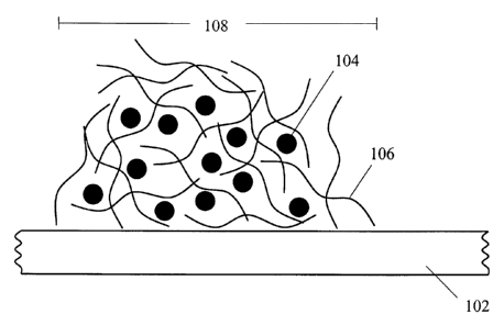
In a preferred embodiment, the microparticles are immobilized on the
substrate by entrapment in a polymeric matrix, as defined herein. An example
of
entrapped microparticles is shown in Figure 1, wherein a polymeric material
106 is
disposed on a substrate 102 and entraps the microparticles 104 within the
polymeric
material 106 and thereby forms a clustered arrangement of microparticles 108.
The polymeric matrix can be composed of a variety of materials that allows
entrapment of the microparticles. Preferred materials for the polymeric matrix
can
be, but are not limited to, synthetic polymers which include polyacrylamide,
polymethacrylamide, polyvinylpyrrolidone, polyacrylic acid, polyethylene
glycol,
polyvinyl alcohol, and poly(HEMA), copolymers thereof, or any combination of
polymers and copolyniars. Natural polymers can aiso be used and include
polysaccharides, for example, polydextrans, glycosaminoglycans, for example,
hyaluronic acid, and polypeptides, for example, soluble proteins such as
albumin
and avidin, and combinations of these natural polymers. Combinations of
natural
and synthetic compounds can also be used. The polymers and copolymers as
described can also be derivitized with a reactive group, for example a
thermally
reactive group or a photoreactive group.
In an alternate embodiment separate crosslinking compounds, for example
photoreactive or thermally activated crosslinkers, can be added to the matrix-
forming material and can be treated to form the matrix. Addition of compounds
such as crosslinkers could serve to make the matrix more durable to use
conditions
and also can create matrices with smaller pore sizes capable of entrapping
smaller
microparticles.
16
CA 02462868 2004-04-02
WO 03/031054 PCT/US02/31707
The polymeric matrix is typically formed by providing an external stimulus
to bind the polymer to the substrate and crosslink the polymer. In one
embodiment,
a slurry including microparticles and polymer is disposed on the substrate and
then
the polymer is treated to crosslink the polymer, for example, by activation of
reactive groups provided by the polymer.
In some embodiments the reactive groups provided on the polymer can be
photoreactive groups and the photoreactive polymer can be crosslinked by
irradiation. The microparticles become entrapped in the polymeric matrix which
is
formed by crosslinking of the polymers. The photoactive groups can also serve
to
bind the polymer to the surface of the substrate upon activation of the
photoreactive
groups. Different concentrations of polymer can be present in the slurry but
generally the concentration shouls be great enough to allow for entrapment of
the
microparticles. The concentration of the polymer can also depend on the size
of the
microparticles used. ror example, the concentration of polymer is at least
0.625
mg/mL for a 1.0 pm microparticle in order to stably entrap the microparticle
in the
polymeric matrix.
According to this embodiment, photoreactive groups can be provided on a
polymer. As used herein, a "photoreactive polymer" can include one or more
"photoreactive groups." A "photoreactive group" includes one or more reactive
moieties that respond to a specific applied external energy source, such as
radiation,
to undergo active species generation, for example, active species such as
nitrenes,
carbenes and excited ketone states, with resultant covalent bonding to an
adjacent
targeted chemical structure. Examples of such photoreactive groups are
described in
U.S. Patent No. 5,002,582 (Guire et al., commonly owned by the assignee of the
present invention), the disclosure of which is incorporated herein in its
entirety.
Photoreactive groups can be chosen to be responsive to various portions of the
electromagnetic spPct~ um, typically ultraviolet, visible or infrared portions
of the
spectrum. "Irradiation" refers to the application of electromagnetic radiation
to a
surface.
The photoreactive groups can be located at any position on the polymer.
Preferably, the number of photoreactive groups present on the polymers is
increased
when smaller microparticles are to be immobilized.
Photoreactive aryl ketones are preferred photoreactive groups on the
photoreactive polymer, and can be, for example, acetophenone, benzophenone,
17
CA 02462868 2004-04-02
WO 03/031054 PCT/US02/31707
anthraquinone, anthrone, and anthrone-like heterocycles (i.e., heterocyclic
analogs
of anthrone such as those having N, O, or S in the 10-position), or their
substituted
(e.g., ring substituted) derivatives. Examples of preferred aryl ketones
include
heterocyclic derivatives of anthrone, including acridone, xanthone and
thioxanthone,
and their ring substituted derivatives. Particularly preferred are
thioxanthone, and its
derivatives, having excitation wavelengths greater than about 360 rtm.
The azides are also a suitable class of photoreactive groups on the
photoreactive polymer and include arylazides (C~RSN3) such as phenyl azide and
particularly 4-fluoro-3-nitrophenyl azide, acyl azides (-CO-N3) such as ethyl
azidoformate, phenyl azidoformate, sulfonyl azides (-SOZ-N3) such as
benzensulfonyl azide, and phosphoryl azides (RO)ZPON3 such as diphenyl
phosphoryl azide and diethyl phosphoryl azide.
Diazo compounds constitute another suitable class of photoreactive groups
on the photoreactive polymers and include diazoalkanes (-CHNz) such as
diazomethane and diphenyldiazomethane, diazoketones (-CO-CHNZ) such as
diazoacetophenone and 1-trifluoromethyl-1-diazo-2-pentanone, diazoacetates (-O-
CO-CHNZ) such as t-butyl diazoacetate and phenyl diazoacetate, and beta-keto-
alpha-diazoacetates (-CO-CNZ-CO-O-) such as 3-trifluoromethyl-3-
phenyldiazirine,
and ketenes (-CH=C=O) such as ketene and diphenylketene.
Exemplary photoreactive groups are shown as follows.
Table 1
Photoreactive Group Bond Formed
aryl azides Amine
acyl azides Amide
Azidoformates Carbamate
sulfonyl azides Sulfonamide
phosphoryl azides phosphoramide
Diazoalkanes new C-C bond
Diazoketones new C-C bond and ketone
Diazoacetates new C-C bond and ester
beta-kPto-alpha-diazoacetatesnew C-C bond and beta-ketoester
aliphatic azo new C-C bond
Diazirines new C-C bond
Ketenes new C-C bond
photoactivated ketonesnew C-C bond and alcohol
The photoreactive polymer can, in some embodiments, comprise a
photoreactive copolymer. The polymer or copolymer can have, for example, a
18
CA 02462868 2004-04-02
WO 03/031054 PCT/US02/31707
polyacrylamide backbone or be a polyethylene oxide-based polymer or copolymer.
One example of a photoreactive polymer comprises a copolymer of
vinylpyrrolidone
and N-[3-(4-Benzoylbenzamido)propyl] methacrylamide (BBA-APMA); another
example is a copolymer of acrylamide and BBA-APMA.
The photoreactive groups of the photoreactive polymer can allow the
formation of a covalent bond between the substrate and the photoreactive
polymer
thereby binding the polymer to the surface of the substrate. The photoreactive
groups of the photoreactive polymer can also serve to crosslink to proximal
polymeric strands together, allowing the formation of a network of covalently
crosslinked polymeric strands that serve as the matrix in which the
microparticles
can be entrapped within. In some embodiments, a non-photoreactive crosslinking
agent can be used to promote the formation of crosslinked polymeric strands.
Use of
a crosslinking reagent, for example, bis-acrylamide, can depend on the
location and
number of photoreactive groups that are present on the polymeric strand.
The polymeric matrix can be composed of a variety of materials that
preferably have pore sizes which allow the entrapment of the microparticle of
the
invention. Preferably the matrix does not allow the microparticles to escape
from
the porous material during steps involving detection of a target in a sample.
For
example, if entrapping microparticle with an average diameter of 2.5 pm, it is
useful
to have a pore size in the range of 50 nm to 2.5 pm; and more preferably in
the range
of 100 nm to 1 Vim.
In another embodiment, immobilization of the microparticles can be
performed by chemical bonding of the microparticle to the polymeric matrix and
the
polymeric matrix to the substrate. A variety of bonds can be formed between
the
microparticles and the matrix material, and the matrix material to the
substrate,
which allows immobilization of the microparticle. These bonds include, for
example, ionic, covalent, cooordinative, hydrogen or Van der Waals bonds. In
this
embodiment, covalent bonds are preferably formed.
According to the invention, arrays are fabricated by providing a substrate,
preparing a slurry containing matrix-forming material and probe-coupled
microparticles, disposing the slurry on the substrate in a manner to form
clustered
arrangements of microparticles, and treating the slurry to form a matrix,
thereby
immobilizing the microparticles in the matrix.
19
CA 02462868 2004-04-02
WO 03/031054 PCT/US02/31707
In one embodiment, slurries including matrix-forming material and probe-
coupled microparticles are printed onto the surface of the substrate to form
an array.
According to this embodiment, printing devices physically spot the slurry onto
the
substrate surface and are available from a variety of sources, including
BioRobotics
Ltd. (MicroGrid arraying robot, Comberton, Cambridge, LTK) and Packard
Instrument Company (BioChip arrayer, Meriden, CT). Alternatively, the probe
can
be applied jet printed to the microparticles through utilization of a
piezoelectric
pump. The BioChip Arrayer (Packard BioChip Technologies) can be used to print
the slurnes onto spots of the substrate.
When printing or spotting techniques are used to apply the probe to the
microparticles, typically a volume of approximately 0.5 to 1.0 nL of solution
containing the probe molecules is applied at each microparticle. Preferably,
the
volumes in the range of 1 pL to 5 pL. When contact printing is used, the
volume of
slurry, containing the microparticles, that is applied to the substrate will
depend
upon such factors as, for example, the size of the microparticles used in the
array,
the desired amount of probe provided on the substrate, the type of printing
pin
utilized, the surface energy of the substrate, the surface tension of the
microparticle-
containing slurry solution, for example, the probe molecules in solvent, and
the like.
When inkjet printing techniques are used, characteristics of the substrate
will not
typically determine the volume of solution containing probe solution applied.
Typically, piezoelectric printing will involve application of approximately 10
- 100
pL of slurry.
The clustered arrangement of microparticles can cover at least a portion of
the surface of the substrate. The clustered arrangements of microparticles can
be
patterned at various locations on the surface of the substrate. Each clustered
arrangement of microparticles typically is coupled to a unique probe,
therefore the
location of probes on the surface of the substrate is determined by the
location or
pattern of the clusters on the substrate. The thickness of the polymeric
matrix of
each cluster can vary and can depend on the size of the microparticles
entrapped in
the polymeric matrix. Preferably, the thickness of the polymeric matrix on the
substrate is greater than the diameter of the largest microparticle being
disposed on
the substrate.
A target can be a molecule, a cell, or a particle suspected to be present in a
sample that can specifically interact with a particular probe, based on
interactions
CA 02462868 2004-04-02
WO 03/031054 PCT/US02/31707
exemplified above. The target can be detected and/or quantitated, according to
the
methods described herein and can be coupled to a "target marker" to accomplish
this. The target can comprise naturally occurnng or man-made molecules, and it
can be used in its unaltered state or as aggregates with other species.
Examples of
targets include antibodies, nucleic acids, receptors, hormones, drugs,
metabolites,
cofactors, peptides, enzymes, viral particles, cells and the like. In one
embodiment,
the target comprises a nucleic acid to be detected in a sample. The probe and
target
are typically members of a specific binding pair, wherein the members of the
pair
are known to bind to each other, while binding little or not at all to other
nonspecific
substances.
In one embodiment, the sample suspected to contain a target is treated to
label the target with a target marker. As used herein, "target marker', refers
to the
detection moiety that is used to visualize the target. As contemplated in this
invention, a target marker comprises a moiety that is detectable using
standard
techniques known in the art. Examples of suitable markers include, but are not
limited to, fluorophores, phosphors, and radioisotopes.
When the target to be detected comprises RNA, the RNA target can be
labeled using molecular biology techniques, such as in vitro run-off
transcription to
generate a labeled RNA sample using RNA polymerises, for example T7, T3, or
SP6 RNA polymerises. Kits for labeling RNA are available from various sources,
for example, Ambion, Inc. (Austin, Texas). This technique can be particularly
useful in generating labeled RNA from, for example, a cDNA library that has
been
cloned into a vector with the appropriate promoters for RNA polymerise
transcription. Techniques can also be used to generate labeled DNA, for
example,
nick translation, PCR amplification, random priming, or primer extension.
These
techniques can be useful for generating labeled DNA from for example, cDNA
libraries or genomic DNA libraries. Modified DNA nucleotides for use as labels
can
also be created from Reverse Transcriptase reactions. For example, an RNA
sample,
such as a polyA-RNA sample can be used as a template in a reaction containing
Reverse Transcriptase, polyT oligonucleotide primer, and modified nucleotide
to
generate labeled-cDNA. Techniques for labeling DNA can be found in various
technical references, for example, Current Protocols in Molecular Biology
(Ausubel
et al., ed., 1990, Greene Pub. Associates and Wiley-Interscience: John Wiley,
New
York).
21
CA 02462868 2004-04-02
WO 03/031054 PCT/US02/31707
RNA and DNA targets can be labeled using modified nucleotides, for
example fluorophore-coupled nucleotides, such as Fluorescein-5[6]-
carboxyamidocaproyl-[5-(3-aminoallyl)uridine 5'triphosphate (Sigma, St. Louis,
MO), biotin-coupled nucleotides, such as (N6-[N-(Biotyinyl-s-aminocaproyl)-6-
aminohexylcarbamoylmethyl]adenosine 5'-triphosphate) or other modified
nucleotides, for example, 5-(3-aminoallyl)uridine 5'-triphosphate (Sigma, St.
Louis,
MO) in order to enable detection of the DNA or RNA. Secondary fluorophore-
coupled reagents, for example, Streptavidin-Cy3 (Caltag, Burlingame, CA) can
be
used to for indirect detection of the modified nucleic acid. Radioactive
nucleotides,
for example 32P-, 33P-, and 35S-labeled ribonucleotides and
deoxyribonucleotides can
be incorporated into the target DNA or RNA present in a sample. These modified
nucleotides can also be used to label sample nucleic acids in other ways, for
example, by 5' or 3' end-labeling with enzymes such as polynucleotide kinase
or
terminal transferase. Optionally, kits and instructions for coupling modified
nucleotides to nucleic acid samples can be obtained commercially from, for
example, CALBIOCHEM (San Diego, CA). Labeled target can optionally be
purified by methods such as gel filtration or purification, spin columns, or
selective
precipitation.
In another embodiment, the sample includes a plurality of protein suspected
of containing a protein target. The protein sample can be obtained from a
tissue
sample, such as a biopsy, or from a fluid sample containing cells, for
example, blood
or bone marrow, or from other body fluids, for example, plasma, sweat, saliva,
or
urine. A protein sample can be recovered from body fluid by a variety of
techniques, for example, by precipitation, filtration, or dialysis. A protein
sample
from cells, for tissue or body fluid, can be prepared by the lysis or
solubilization of
cells in detergents, optionally using methods such as sonication or
homogenization.
Ionic or non-ionic detergents can be used, for example, sodium dodecyl
sulphate
(SDS), Triton X-100, sodium deoxycholate and CHAPS. Cells can also be
disrupted
in the presence of chaotropic reagents, such as urea and guanidine salts.
Other
reagents can be added to the detergent or chaotropic reagent, such as a
buffer, for
example, Tris or HEPES, and salts, for example, KCl or NaCI. Other compounds
can be utilized which stabilize the protein sample, for example, protease
inhibitors,
such as PMSF, pepstatin, or EDTA. However, a variety of methods and buffer
22
CA 02462868 2004-04-02
WO 03/031054 PCT/US02/31707
compositions are available for the lysis or disruption of cells for protein
extraction
and are commonly known in the art. This information can be found in various
references, for example, Current Protocols in Protein Science (Coligan et al.,
eds.,
1996, John Wiley & Sons, New York, NY).
The protein sample is preferably labeled in such a way to enable detection of
the protein target and to retain the ability of the protein target to interact
with the
probe coupled to the microparticle. Reagents are available that allow the
coupling
of a fluorophore to an amino acid residue on a protein target. Amine-reactive
groups, for example, succinimidyl esters, including sulfosuccinimidyl esters,
isothiocyanates and sulfonyl chlorides, or dichlorotriazines, aryl halides and
acyl
azides are.available as fluorophore probes and can be used for protein target
labeling. A variety of these amine-reactive fluorophore probes are
commercially
available, for example, Alexa FluorTM 350 carboxylic acid, succinimidyl ester;
4,4-
difluoro-5-(2-thienyl) -4-bora-3a,4a-diaza-s-indacene- 3-propionic acid,
succinimidyl ester (BODIPYTM 558/568, SE); and 6-carboxy-4',5'-dichloro-2', 7'-
dimethoxyfluorescein, succinimidyl ester (6-JOE, SE) (Molecular Probes,
Eugene,
OR). In other circumstances it can be desirable to label the protein target
with a
fluorescent thiol-reactive derivative, for example, N,N'-didansyl-L-cystine
(Molecular Probes, Eugene, OR). Alternatively, other reagents, for example
fluorescent dyes c~ntuining a hydrazine group, an aromatic diazonium salt, or
an
amine group can be used to label the protein sample.
The protein target can also be coupled to a fluorescent protein, for example
the Green Fluorescent Protein (GFP), using commercially available bifunctional
crosslinking reagents, for example NHS-ASA (Pierce Chemical, Rockford, IL).
Other bifunctional crosslinking agents that are reactive toward amine,
sulfliydryl,
carbohydrate, carboxyl and hydroxyl groups are commercially available (for
example, Pierce Chemical, Rockford, IL) and can be used for coupling a protein
of
interest to the protein target. The protein target can also be coupled to a
primary
reagent for secondary fluorophore detection. In some embodiments, the protein
can
be modified by, for example, sulfo-NHS-biotin, and then coupled to a secondary
fluorophore reagent, for example Streptavidin-Cy3.
Other methods of labeling a protein sample for detection are available. Such
methods include, for :.xample, protein iodination using ~ZSI and protein
phosphorylation using 32P or 33P and a protein kinase can be used.
23
CA 02462868 2004-04-02
WO 03/031054 PCT/US02/31707
In some instances, the target includes a particle, for example, a viral
particle,
a cell, or a portion of a cell. These targets can display molecules on their
surfaces
that allow them to bind a particular probe that can be present in the array.
The
binding of the probe and the molecule present on a viral particle, cell, or
portion of a
S cell can be useful in identifying and quantifying a particular viral
particle, cell, or
portion of a cell which can be present in a sample. For example, the array can
be
useful in determining the presence and quantity of different subtypes of
lymphocytes
present in a sample. The viral particle, cell, or a portion of a cell can be
labeled by a
variety of means, for example, by labeling with an antibody that is coupled to
a
fluorophore. The labeled antibody can be chosen to react specifically with a
subpopulation of the sample or non-specifically with the sample.
Alternatively, the
viral particle, cell, or a portion of a cell can be labeled by incorporation
of a
fluorescent dye into the membrane of these potential targets. Such dyes, for
example, PKH-67 GL (Sigma, St. Louis, MO) or a fluorescent lipophilic probe,
CM-
DiI (Molecular Probes, Eugene, Oregon) are commercially available and can be
used
to label cells.
It is understood by one of skill in the art that there are a wide variety of
techniques available for protein labeling and that the technique chosen can
depend
on the protein or proteins available in the sample targeted for labeling.
In an alternate embodiment, target detection can be performed by employing
a fluorophore:quencher pair on the microparticle. In this embodiment, probe-
coupled microparticles are further coupled to a detectable moiety, such as a
fluorescent molecule, and target is coupled to a compound that quenches the
signal
of the detectable moiety on the microparticle. Binding of the quencher-coupled
target to the probe on the fluorophore-coupled microparticle will result in
the loss of
signal from the clustered arrangement of microparticles. For example, in
clustered
arrangements of probe- and fluorescein-coupled microparticles, a target,
specific for
the probe and coupled to the quenching molecule 6-(N-4'-carboxy-4-
(dimethylamino)azobenzene)-aminohexyl-1-O-(2-cyanoethyl)-(N,N-diisopropyl)-
phosphoramidite (Dabcyl; Glen Research, Sterling, VA) can specifically bind
probe
and effectively quench the fluorescence emission of fluorescein. Quenching of
the
fluorescent signal can be accomplished by positioning the quenching molecule
proximal to the fluorophore.
24
CA 02462868 2004-04-02
WO 03/031054 PCT/US02/31707
Given the description herein, one of skill in the art can select the desired
labeling scheme depending upon the target to be detected. While nucleic acids
and
proteins have been described with particularity, it will be clear that the
teaching
herein can be applied to label samples suspected to contain other types of
targets,
S including, but not limited to, small molecules and the like.
Once formed, the array, which can contain a plurality of clustered
arrangements of microparticles, each clustered arrangement of micropsheres
associated with a unique probe, can be used to detect target suspected to be
contained in a sample. In use, a sample is modified to provide labeled target,
as
described herein. The modified sample is then applied to the array, and the
array
and sample are maintained under conditions suitable for specific binding of
the
target, if present in the sample, to the probe. Such specific binding
conditions can
be determined using techniques known in the art, depending upon the target to
be
detected. For example, when nucleic acid is used as the probe, specific
binding, or
hybridization conditions can be adjusted based on the number of nucleotide
base-
pairs that are formed between the probe nucleic acid and the target nucleic
acid.
After specific binding of the target to the probe, excess sample can be
removed, for example, by washing, and the remaining hybridized targets can be
interrogated. The arrays of the invention can be used to detect any target
suspected
to be present in a sample. Interrogation of a sample can involve one or more
visualization steps depending on the types of markers used for the target
detection
(target marker) and optionally microparticle detection if there is any
microparticle
probe included in the array.
Determination of the presence and amount of one or more targets) in a
sample is typically performed by visualizing the presence or absence of a
detectable
signal associated with one or more clustered arrangements of microparticles on
a
substrate. By knowir_o the probe associated with a particular clustered
arrangement
of microparticles on the substrate, and the signal associated with a
particular
clustered arrangement of microparticles, one can determine the presence and
amount
of particular targets in a sample.
Visualization of the detectable markers can be accomplished using any
suitable visualization technique known in the art. Fluorescence imaging can be
made using a modified epifluorescence microscope or a fluorescence confocal
microscope. Suitable microscopes include, for example, an Olympus BX60 (Tokyo,
CA 02462868 2004-04-02
WO 03/031054 PCT/US02/31707
Japan) or other similar microscopes. Fluorescence images from microscopy
images
can be analyzed for fluorescence intensity using computer software.
Commercially
available microscopy analysis software, for example, Image-Pro Plus (version
4.0)
(Media Cybernetics, L.P., Silver Spring, MD), can be used to define and count
fluorescent signals automatically with optical detection systems.
Alternatively, the
method of fluorescence scanning can be used to visualize particles.
Fluorescence
scanners such as the Scan Array 5000 (GSI Lumonics, Billerica, MA) or Axon
GenePix 4000A (Foster City, CA), which have resolution of approximately 5pm,
can be used for visualization.
EXAMPLES
Example 1
As shown in this example, an array was fabricated by disposing slurries
containing probe-coupled microparticles in a matrix-forming material.
Microparticles were prepared by coupling a probe to the microparticle,
followed by
preparation of a slurry containing a photoreactive polymer and the probe-
coupled
microparticles. These prepared microparticles were then applied to a substrate
to
form an array.
Magnetic, polystyrene-encapsulated microparticles, coupled to a fluorescent
blue dye (excitation/emission maxima of 490/515 nm) and streptavidin were
obtained from Bangs Laboratories (Product # CMO1F; Fishers, IN). The
streptavidin concentration was measured by biotin-conjugate binding and was
determined to be 9.86 ~g biotin-alkaline phosphatase/mg microbeads and 0.77
p,g
biotin-FITC/mg microbeads per manufacturer's measurement. The microparticles
had a diameter of 0.96 pm and a density of 1.7 g/cm3 (1.305e+12
microparticles/g).
The microparticles, were washed twice with deionized water and resuspended in
25
mM phosphate buffered saline, pH 8, at 10 mg/mL.
The streptavidin-coated microparticles were coupled to oligonucleotide
BN30. BN30 is a 30-mer with a 5' biotin modification and a 3' amine
(Integrated
DNA Technologies, Coralville, IA). Coupling was performed using 5 nmole/ml of
BN30 and 1 mg/ml of microparticles (five-fold excess of biotinylated
oligonucleotide, based upon the supplier's stated biotin binding capacity) in
25mM
PBS, pH 8. The coated microparticles and oligonucleotide were incubated for 30
26
CA 02462868 2004-04-02
WO 03/031054 PCT/US02/31707
minutes at room temperature with gentle agitation. After incubation, the
microparticles were washed with deionized water and resuspended at 20 mg/ml in
deionized water.
A slurry of 37 mg/ml photoreactive poly(vinylpyrrolidone) (PVO1;
SurModics, Inc., Eden Prairie, MN) in water was combined with the
microparticle
solution at a ratio of 9:1 (10x dilution of microparticle solution in PVO1).
The slurry
was printed onto an acrylic surface (obtained from Cadillac Plastics,
Minneapolis,
MN) using a Microgrid II arrayer (Biorobotics, Inc. Cambridge, UK) with an
average spot size of 100 pm and a center to center spacing of 250 pm,
providing
approximately 16 spots/mm2. The coated slides were air dried and irradiated
for two
(2) minutes with broad spectrum ultraviolet light (320-390 nm) using a Dymax
LightWelder PC-2 (Dymax Engineering Adhesives, Torrington, CT) having a
typical power output of 2 mW/cm2. The lamp was positioned approximately 10 cm
from the slides which were also placed beneath a cut-off filter (315 nm) to
avoid
potential nucleic acid damage, while gelling the PVO1 solution. After
irradiation,
the substrate was rinsed with 1 X PBS, 0.1 % Tween 20.
The arrays prepared by the above method were stable to washing and
touching, while samples that were not irradiated were not stable. Fluorescence
scanning and light microscopy were used to determine the presence and location
of
the micropaxticles
Sample containing target nucleic acid was applied to the array and incubated.
2.5 ~L of a 33 fM solution of a fluorophore-coupled target nucleic acid in
hybridization buffer (5X SSC, 0.1% SDS, 0.1 mg/ml salmon sperm DNA) per cmz
(array area) placed between a coverslip and the array surface. The slides were
then
be placed in hybridization chambers and heated in a water bath at 45°C
for 2 hours.
The coverslips were then removed with a stream of 4X SSC buffer and the slides
were then washed with 2X SSC/0.1% SDS for five minutes at 45°C,
followed by a
0.2X SSC wash for one minute at room temperature, and finally a wash of 0.1 X
SSC
for one minute at room temperature. The slides were then spun dry and the
target
oligonucleotide detected by methods described below.
Target was detected on the array with a ScanArray 5000 fluorescence
scanner (Packard Bioscience, Billerica, MA).
27
CA 02462868 2004-04-02
WO 03/031054 PCT/US02/31707
Example 2
An array was formed by entrapping microparticles in a polymeric matrix.
Entrapment of the microparticles in the polymeric matrix did not depend on the
formation of covalent or ionic bonds between the substrate and the
microparticles.
S In this example, matrices formed from polyvinyl pyrrolidone)(PVP) were used
to
entrap microparticles. An array was fabricated by preparing a mixture of
photoreactive PVP and underivatized silica microparticles, disposing the
mixture on
a substrate, and then polymerizing the photoreactive PVP to a PVP matrix.
Silanated glass slides ( I x 3 in. x 1 mm) were used in the preparation of
substrates in array fabrication. Glass microscope slides were obtained from
Erie
Scientific, Portsmith, NH (catalog # 2950-W). These soda lime glass microscope
slides were silane treated by dipping in a mixture of 1 % v/v p-
tolyldimethylchlorosilane (T-Silane) and 1% v/v n-decyldimethylchlorosilane (D-
Silane, United Chemical Technologies, Bristol, PA) in acetone for 1 minute.
After
air drying, the slides were cured in an oven at 120°C for one hour. The
slides were
then washed with acetone followed by DI water dipping. The slides were further
dried in an oven for 5-10 minutes. The silanated glass or polypropylene slides
were
then washed in acetone or isopropanol.
A solution of 10 mg/ml photoreactive poly(vinylpyrrolidone) (PVO1;
SurModics, Inc. Eden Prairie, MN) and 2.5 mg/ml of 5 ~m-diameter silica
microparticles (Product #SSOSN, Bangs Laboratories, Fisher, IN) in deionized
water was printed using 25 gauge disposable needles (PrecisionGlide Needles,
Becton Dickinson and Co., Franklin Lakes, NJ) and an x-y programmable stage a
glass microscope slide as prepared above. The slide was then irradiated for
two
minutes with broadband ultraviolet light over the range of 320-390 nm (Dymax
Engineering Adhesives, Dymax LightWelder PC-2, Torrington, CT). Following
this, the slide was rinsed five times with 1X PBS with 0.05% Tween-20, then
incubated for 15 minutes at 45°C in a blocking solution DB02 (50 mM
ethanolamine, O.1M Tris, pH 9; SurModics, Inc., Eden Prairie, MN) supplemented
with 0.1% sodium dodecyl sulphate (SDS). The slide was further rinsed with
deionized water and incubated for one hour at 45°C in 4X SSC (0.6 M
NaCI, 0.06 M
NaCitrate /0.1% SDS. Final rinses included five minutes with 2X SSC at
45°C, then
one minute in 0.2X SSC at room temperature, then one minute in O.IX SSC at
room
temperature followed by drying.
28
CA 02462868 2004-04-02
WO 03/031054 PCT/US02/31707
The micropartscles remained inside the PVP hydrogel array and were
visualized by fluorescence microscopy (Olympus BX60, Tokyo, Japan) and with a
confocal fluorescence scanner (ScanArray 5000, GSI Lumonics, Billerica, MA).
When the microparticles and photoreactive PVP were deposited on the glass
slide without the step of irradiation, the microparticles were not retained in
the gel.
The absence of radiation did not allow crosslinks to form in the polymeric
matrix,
thereby not allowing the polymeric matrix to physically entrap the
microparticles.
In order to define the nature of immobilization of microparticles in the
polymeric matrix, microparticles 5 ~m diameter (Cat no. SSOSN, Bangs
Laboratories, Fisher, IN) were irradiated in a solution of benzophenone dimers
functionalized with quaternary amine groups. A solution of PR03 (ethylene(4-
benzoylbenzyldimethylammonium)dibromide; SurModics, Inc. Eden Prairie, MN) is
described in U.S. Patent No. 5,714,360 (Swan et al., issued 3 Feb., 1998,
commonly
owned by the assignee of the present invention, the disclosure of which is
incorporated herein in its entirety), at a concentration of 5 mg/ml containing
2.5
mg/ml 5 ~m-diameter silica microparticles (Product #SSOSN, Bangs Laboratories,
Fisher, IN) in 10% v/v DMF/water was printed using 25 gauge disposable needles
(PrecisionGlide Needles, Becton Dickinson and Co., Franklin Lakes, NJ) and an
x-y
programmable stage onto a hydrophobic glass slide, as performed in Example 1.
The printed array was irradiated for two minutes with ultraviolet light (Dymax
Engineering Adhesives, Dymax Light Welder PC-2, Torrington, CT) and rinsed
with deionized water, with 1X PBS with 0.05% Tween-20 added, then incubated
for
1 S minutes at 45°C in DB02 (SurModics, Inc., Eden Prairie, MN) with
0.1 % SDS
added. The slide was then rinsed with deionized water and further incubated
for one
hour at 45°C in 4X SSC/0.1% SDS. Final rinses included five minutes
with 2X SSC
at 45°C, then one minute in 0.2X SSC at room temperature, then one
minute in O.1X
SSC at room temperature followed by drying.
When imaged at this point, no microparticles remained in the areas printed
with the PR03 solution. High concentrations of PR03 proved to be insufficient
for
immobilization of the microparticles, as opposed to the PVO1 polymer, which
efficiently trapped the microparticles.
29
CA 02462868 2004-04-02
WO 03/031054 PCT/US02/31707
Example 3
Microparticles were immobilized on the surface of a substrate by entrapment
of the microparticles in a polymeric matrix. This method of immobilization
does not
interfere with the surface functionality of the microparticle since no
chemical bonds
were formed between the microparticle and polymeric matrix. Therefore, the
surface chemistry on the microparticle is not altered. Covalent bonds between
polymer molecules were formed after a slurry containing microparticle and
polymeric material was coated on the device. Covalent bonds were not formed
between the polymer and the microparticle since the microparticles do not
present
carbon-hydrogen bonds of which the activated benzophenone can react with.
Irradiation of the photopolymer crosslinked the polymer with itself encasing
the
microparticles, but did not alter the biomolecules attached to the
microparticles.
To demonstrate this immobilization method, plain silica microparticles of
four different diameters (0.4 Vim, 0.9 pm, 5.0 Vim, and 9.9 Vim) were
immobilized in
photopolymer matrices of differing concentrations. At the lowest concentration
of
photopolymer the polymeric matrix formed was not sufficient to physically
immobilize the microparticles of the sizes tested. As a additional control,
some
photopolymer coatings were not irradiated which resulted in insufficient
crosslinking around the microparticle and leading to microparticle loss from
the
coating. Stringent rinses in detergent and high salt solutions at elevated
temperatures were used to ensure that the photopolymer coatings were robust.
20 p1 of 100 mg/ml aliquots of each 400 nm, 970nm, 5~.m, and 9.9~m silica
microparticles solutions (cat. # SS02N, SS03N, SS05N, and SS06N, respectively;
Bangs Laboratories, Fisher, IN) were pelleted by centrifugation. These pellets
were
individually resuspended in 100 ~1 serial dilutions of photoreactive
poly(vinylpyrrolidone) (PVO1; SurModics, Inc, Eden Prairie, MN). The dilution
series consisted of 10, 5, 2.5, 1.25, 0.6, 0.3, 0.15, 0.08, 0.04 and 0.02
mg/ml
concentration samples of PVO1. The final concentration of microparticles was
20
mg/ml for all sizes. Therefore, the 400 nm microparticle solution contained
significantly more microparticles than did the 9.9 ~m microparticle solution;
however all microparticle solutions contained the same percent solids.
Glass microscope slides, as described in Example 1, that had been coated
with a 1 % v/v solution of n-decyldimethylchlorosilane and
CA 02462868 2004-04-02
WO 03/031054 PCT/US02/31707
tolyldimethylchlorosilane in acetone were used as a substrate for coating. 5
~1 of
each microparticle-PVO1 solution was placed in an area of approximately 10 mm
x 2
mm on a glass slide and allowed to air dry. Once dry, the coatings were
irradiated
with an ultraviolet lamp as detailed in Example 1 (Dymax Lightwelder PC-2,
Dymax Engineering Adhesives,Torrington, CT) for two minutes. A second set of
samples was not irradiated to serve as a control to determine whether
crosslinking
the photopolymer polymeric matrix was necessary to contain the microparticles.
To determine if the microparticles were entrapped well in the polymer
coating, the coated slides were washed in a 1X PBS-0.1% Tween-20 solution with
mild shaking for one hour, followed by two deionized water rinses. The
coatings
were then re-examined by microscope and the microparticle loss evaluated. A
second wash condition of higher salt with higher temperature was then
conducted.
The microparticle coated pieces were incubated and shaken in 4X SSC buffer
with
0.1% SDS for 45 minutes at 45°C, then rinsed twice with deionized water
and
examined microscopically for changes in the microparticle coating. Finally a
longer
high salt wash step was conducted, with the slides incubating in SX SSC buffer
with
0.1% SDS for two hours at 45°C, followed by four rinses of decreasing
concentration of SSC.
The microparticle coatings were examined by qualitative microscopic
examination at SOX magnification (Olympus BX60, Tokyo, Japan) and the changes
were tabulated. Results are shown in Figure 2 and Figure 3.
Figure 2a - 2d shows presence of the microparticles after the final wash.
Figure 2a shows a coating having 400 nm microparticles; Figure 2b shows a
coating
having 970 nm microparticles; Figure 2c shows a coating having Spm nm
microparticles; and Figure 2d shows a coating having 9.9pm microparticles.
A micrograph of microparticles before and after all three washes is show in
Figure 3. Figure 3a shows 9.9 pm microparticles in a 0.3 mg/ml PVO1 matrix
before washing and Figure 3b shows 9.9 pm microparticles in a 0.3 mg/ml PVO1
matrix after washing. The results are shown in Table 2 and Table 3. As
indicated in
Tables 1 & 2: N = No Loss, S = Slight Loss, M = Moderate Loss, H = Heavy Loss,
C = Complete Loss; PBST = 1X PBS-0.1% Tween-20 wash for one hour at room
temperature; 4X SSC = 4X SSC-0.1% SDS wash for 45 minutes at 45°C;
Final =
SX SSC-0.1% SDS wash for two hours at 45°C.
31
CA 02462868 2004-04-02
WO 03/031054 PCT/US02/31707
w.~ ~ ~ x x w.~ ~
~ ~~.
~ _ ~
~~
o ~ >C wn U Urvx x _
U 0
0
w ~ ~ x x ~ ~ a. GG U U U x
E-~ rn E-~
w...~~.-.x x x w~.-~
, w~
a~
~ m
~
o ~ ~C ~ x U x x ~ ~ >C v~ U U U
v~ U vo U
0
0 o. c~ ~ x ~ ~ Q 0.1 U x ~ x
v~ E-' vW
w...~~~ v~vo~ ~ w~.-c
o ~ x v~ v~v~~
vo U ~ ~ >C ~ U U U U
0 ~ U
0 a. as cryv~v~v~ ~. oa x x ~ x
vW v~ H
w~~ ~ v~v~vov~ w.~ ~ ~~~~, r
~.~ ~,~
~:;
~Y~
~~C~~U rr~cnv~v~ ~-' ~rxv~voU U
0 0 ,~~
a. p4 rm~ v~v~ C~, 0.1 U U U x
v~ H v~ E-~
w . ~ v~voz z w ~.-
~ ~ .-. ~
o ~x~~U z z z z o ~x~~U U U U U
H z z z z ~ ~m~H
w... ~ z z z z b w... ~
~.- ~~
~
~x~~U z z z z ~ ,~ o ~x~~U U U U U
H ~' o H
~
~,~,~H z z z z ~,~a~
w..- ~ z z z z z w... ~ ~ ..
~~ ~~ . ~ :
~x~~U z z z z N ~x~~U ~ U U U
H z z z z
w.-.~~~ z z z z w...~~~
~~C~~U z z z z N ~rx~~U U U U U
~.~a~~ z z z z ~.x~~H
w~-~~~ z z z z w._~~.-
~~~~U z z z z ~ ~~~~U U U U U
H z z z z
w.-.~~~ z z z z
~x~~U z z z z o ~~c~~U U U U U
~.~,~~. z z z z ~.a~~H
0
0 0 ~ ~ > .~
~on
32
CA 02462868 2004-04-02
WO 03/031054 PCT/US02/31707
As shown above in Tables 2 and 3, microparticle loss was significantly
increased in samples that had not been irradiated. Also shown above in Tables
2 and
3, microparticle loss was increased when the concentration of polymer was
reduced.
Microparticle loss was increased when a smaller size of microparticle was used
in
the polymeric matrix. This data indicates that irradiation promoted the
formation of
an encasing polymeric network around the microparticles. This data also
indicates
that the microparticles were immobilized by physical constraints of the
polymeric
matrix rather than by chemical bonding.
When printed on a substrate the slurries having smaller microparticles gave
coatings that densely packed with multiple layers of microparticles, whereas
slurries
with larger microparticles gave coatings had loosely arranged microparticles
every
few tenths of a millimeter. Coatings showing the lower density of
microparticles
were as stable to the wash conditions as ones with dense concentrations.
Example 4
Arrays of clustered arrangements of immobilized microparticles were
prepared by creating a slurry of microparticles suspended in a photoreactive
polymer
and contact printing this slurry on a glass slide. The microparticles were
coupled to
nucleic acids thereby creating an array of clustered arrangements of
microparticles
which was used for the detection of the presence of target nucleic acids in a
sample.
To fabricate an array of clustered arrangements of oligonucleotide-coupled
microparticles, 1 pm streptavidin-coated silica microparticles (CSO1N; Bangs
Laboratories, Fisher, IN) were coupled to biotinylated Cy3-labeled 30-mer
oligonucleotides (Integrated DNA Technologies, Coralville, IA) by adding 20 p1
of
a 1.25 p.M solution of the oligonucleotide to 100 p1 of a 4 mg/ml solution of
microparticles in PBS pH 7.5 and allowing the biotin to couple to the
streptavidin
for 30 minutes at room temperature.
The oligonucleotide-coupled microparticles were then pelleted by
centrifugation, the supernatant was removed, fresh deionized water added, and
the
oligonucleotide-coupled microparticle solution was then vortexed to resuspend
the
oligonucleotide-coupled microparticles. This wash procedure was repeated twice
to
remove uncoupled biotinylated oligonucleotide.
33
CA 02462868 2004-04-02
WO 03/031054 PCT/US02/31707
The oligonucleotide-coupled microparticles were introduced into a 2.25
mg/mL solution of PVO1 (Surmodics, Inc. Eden Prairie, MN) in deionized water
to
form a slurry that contained 4.0 mg/mL microparticles. This slurry was contact
printed with a Microgrid II arrayer (Biorobotics, Cambridge, UK) onto a glass
slide
that had been derivatized with a mixture of n-decyldimethylchlorosilane and
tolyldimethylchlorosilane as indicated in Example 1. A pattern of 3 x 3 spots
was
repeated on the surface, with the spots having an average diameter of 100 pm
and a
volume of approximately 0.5 - 1 pL prior to drying. After printing, the slide
was
irradiated for two minutes with an ultraviolet light source as detailed in
Example 1
(Dymax LightWelder PC2, Dymax Engineering Adhesives, Tornngton, CT) through
a 315 nm cutoff filter (Electro-Lite Corporation, Danbury, CT). At this point
the
microarray was imaged on a fluorescence scanner (Packard Biosciences,
ScanArray
5000, Billerica, MN) and representative spots were imaged with a fluorescence
microscope (Olympus BX 60, Tokyo, Japan). Approximately 100-500 S ~m
microparticles were present per 100 pm spot.
Following printing, the array was washed to remove any loosely bound
microparticles. The array was washed three times with 1X PBS (pH 7.4) with
0.1%
v/v Tween-20, rinsed with deionized water, incubated in a solution of DB02
wash
buffer (Surmodics Inc. Eden Prairie, MN) for 1 hour at 50°C, followed
by two rinses
with deionized water. The microarray was then incubated in a solution of 4 X
SSC/
0.1% SDS for two hours at SO°C and rinsed in deionized water.
Hybridization of a target nucleotide to the probe oligonucleotide was
accomplished by placing a solution containing 500 finoles of Cy5-labeled
complementary oligonucleotide, in SX SSC and 0.1% SDS, on the array.
Hybridization was carried out for 2 hours at 45°C. After
hybridization, the
microarray was washed with 2X SSC for 5 minutes at 50°C, 0.2X SSC for
one
minute at room temperature, and finally O.1X SSC for one minute at room
temperature. At this point the microarray was imaged with the fluorescence
scanner,
using Cy 3 and Cy 5 channels and with the fluorescence microscope. The
fluorescence scanner and microscopic images also indicate that the spots on
the
microarray were clearly visible and contain significant numbers of
microparticles, as
well the oligonucleotide on the array hybridizes to the complementary
oligonucleotide. Figure 4 shows the clustered arrangement of microparticles on
the
slide and fluorescence images of the hybridized targets. Figure 4a shows the
34
CA 02462868 2004-04-02
WO 03/031054 PCT/US02/31707
microparticle clusters directly after printing; Figure 4b shows the
fluorescence of the
clusters of the microparticles in the Cy3 channel (60% laser) after
hybridization of
the target; Figure 4c shows fluorescence of the of hybridized Cy5-labeled
target
oligonucleotide in the Cy5 channel (50% laser).
Example 5
In this example, arrays were created using the procedure of Example 3 but
with decreasing concentrations of photoreactive polymer. At lower
concentrations
of photoreactive polymer, the polymeric matrix no longer held the
microparticles in
place following a mild rinse.
Slurries of 5 mg/mL PVO1 (SurModics, Inc. Eden Prairie, MN) were serially
diluted to 5, 2.5, 1.25, 0.625, 0.313 mg/mL in deionized water and 45 ~1 of
each of
the serially diluted PVO1 solutions were added to 5 ~1 of 40 mg/ml
microparticles in
deionized water to create slurnes were the final concentration of
microparticles is 4
mg/ml in each slurry mixture of 50 p1 total.
The slurries were all contact printed on a Microgrid II arrayer (Biorobotics,
Cambridge, UK) and then irradiated for two minutes with ultraviolet light as
detailed
in Example 1 (Dymax LightWelder PC2, Dymax Engineering Adhesives,
Torrington, CT) through a 315 nm cutoff filter (Electro-Lite Corporation,
Danbury,
CT). The arrays were then rinsed three times with 1X PBS with 0.1% Tween-20
and
with deionized water. The resulting spots were imaged with a fluorescence
microscope (Olympus BX 60, Tokyo, Japan) using 100X magnification. As shown
in Figure 5, the PVO1 polymeric matrix immobilized the microparticles until
the
concentration dropped below 0.625 mg/mL (Figure 5d), when the microparticles
were rinsed free from the polymeric matrix. Figure 5a shows microparticles
immobilized in 5 mg/mL PVO1; Figure 5b shows microparticles immobilized in 2.5
mg/mL PVO1, Figure 5c shows microparticles immobilized in 1.25 mg/mL PVO1,
Figure 5d shows microparticles immobilized in 0.625 mg/mL PVO1, Figure 5e
shows microparticles immobilized in 0.313 mg/mL mg/mL PVO1.
Example 6
Different matrix-forming materials were used in another method to
immobilize microparticles on a substrate. Slurries were prepared, each
containing
CA 02462868 2004-04-02
WO 03/031054 PCT/US02/31707
different polymers and microparticles. These slurries were then disposed on a
substrate and treated to form matrices in which microparticles were
immobilized.
Four different photoreactive polymers were used to immobilize 9.9 ~m diameter
silica microparticles on substrates. These photoreactive polymers PA04, PA05,
PVO1, and PVOS are all commercially available from SurModics, Inc. (Eden
Prairie,
MN). PA04 and PA05 are copolymers of acrylamide (AA) and N-[3-(4-
Benzoylbenzamido)propyl] methacrylamide (BBA-APMA) with differing ratios of
BBA-APMA:AA. Similarly PVO1 and PVOS are copolymers of vinylpyrrolidone
(VP) and N-[3-(4-Benzoylbenzamido)propyl] methacrylamide (BBA-APMA) with
differing rations of BBA-APMA:VP. PR03 (as described in Example 4) a non-
polymeric photoreactive material was also used in this Example.
PA04, PA05, PVO1, PVOS, and PR03 were dissolved in deionized water at a
concentration of 2.5 mg/ml. 100 ~l of each solution containing photoreactive
compound was added to a pellet of 2 mg of 9.9 ~m diameter silica
microparticles
(SS06N, Bangs Laboratories, Fisher, IN) that had been washed three times with
deionized water to create a slurry. The final concentration of each slurry was
2.5
mg/ml of photoreactive compound and 20 mg/ml of microparticles. Each slurry
was
printed with 25 gauge disposable needles (PrecisionGlide Needles, Becton
Dickinson and Co., Franklin Lakes, NJ) and an x-y programmable stage (CAMM-3,
Roland Digital Group, Irvine, CA) onto glass microscope slides which had been
functionalized with silanes, as detailed in Example 1 and onto acrylic slides
(Cadillac Plastics, Minneapolis, MN). This printing forms a pattern of
approximately 300- 400 ~m diameter spots on the substrates.
The coated substrates were irradiated for two minutes with ultraviolet light
as
detailed in Example 1. The coated substrates were then imaged with a
fluorescence
microscope (Olympus BX 60, Tokyo, Japan), to ensure that patterning was
successful.
Following this, the coated substrate was washed to remove any loosely
bound microparticles. The coated substrates were washed three times with 1X
PBS
(pH 7.4) with 0.1 % v/v Tween-20, rinsed with deionized water. At this point,
each
was again imaged with the fluorescence microscope to determine microparticle
loss
in the various photoreactive polymer matrices. After imaging, the coated
substrates
were incubated in a solution of DB02 wash buffer (Surmodics Inc. Eden Prairie,
36
CA 02462868 2004-04-02
WO 03/031054 PCT/US02/31707
MN) for 1 hour at 50°C, followed by two rinses with deionized water.
The coated
substrates were then incubated in a solution of 4 X SSC/ 0.1 % SDS for two
hours at
50°C and rinsed in deionized water. Presence of the microparticles
before and after
washing steps are summarized in Table 4.
Table 4
PhotoreactiveSubstratePresence of Presence of Presence of
Compound microparticlesmicroparticlesmicroparticles
before washesafter PBS- after high
Tween wash salt
wash (4X SSC)
PA04 Glass Present No loss No loss
PA04 Acrylic Present No loss No loss
PA05 Glass Present No loss No loss
PA05 Acrylic Present No loss Some loss
PVO1 Glass Present No loss No loss
PVO1 Acrylic Present No loss No loss
PVOS Glass Present No loss No loss
PVOS Acrylic Present No loss No loss
PR03 - non- Glass Present No loss Complete loss
polymer control
PR03 - non- Acrylic Present No loss Complete loss
polymer control
* Sample was touching another slide during the wash.
37