Note: Descriptions are shown in the official language in which they were submitted.
CA 02463789 2004-04-15
WO 03/047712 PCT/US02/33313
1
HYBRID MICROFLUIDIC AND NANOFLUIDIC SYSTEM
This invention was made with government support under grants from
the U.S. Department of Energy (DE FG02 88ER13949 and DE FG02 99ER62797),
the U.S. Defense Advanced Research Projects Agency (F30602-00-2-0567) and
the National Cancer Institute (CA82081 ). The U.S. government has certain
rights
in this invention.
This application claims priority from U.S. Provisional Patent
Application No. 60/330,417 filed October 18, 2001, which is hereby
incorporated
by reference.
Background of the Invention
The present invention relates generally to a microfluidic system, and
more particularly to a microfluidic system having an externally controllable
nanofluidic interconnect.
Microfluidic devices are devices for controlling fluid flow having
dimensions less than about one millimeter. These devices are becoming
increasingly important in chemical and biochemical sensing, molecular
separations, drug delivery and other emerging technologies. New microfluidic
devices and methods for rapidly constructing these devices are being
developed.
However, most prior art devices are two-dimensional. To produce three-
dimensional microfluidic devices, interconnects between two-dimensional
structures often are made. However, creation of these interconnects has proved
challenging. Many prior three-dimensional microfluidic devices use discrete
channels to bridge, rather than connect, independent analysis modules. In
other
words, the channels passively connect the modules and do not have gates or
valves for selectively permitting and preventing flow from one module to the
next.
Although a pressure activated valve has been developed, this interconnect has
limited usefulness because it depends on a variation in pressure of the fluid
for
opening and closing the valve. Thus, there is a need for an externally
controllable
active interconnect to exploit the full three-dimensional capacity of
microfluidic
devices.
CA 02463789 2004-04-15
WO 03/047712 PCT/US02/33313
2
Summary of the Invention
Briefly, the present invention includes a fluid circuit comprising a
membrane having a first side, a second side opposite the first side, and a
pore
extending from the first side to the second side. The fluid circuit also
includes a
first channel containing fluid extending along the first side of the membrane
and a
second channel containing fluid extending along the second side of the
membrane
and crossing the first channel. Further, the circuit comprises an electrical
source in
electrical communication with at least one of the first fluid and second fluid
for
selectively developing an electrical potential between fluid in the first
channel and
fluid in the second channel thereby causing at least one component of fluid to
pass
through the pore in the membrane from one of the channels to the other.
In another aspect, the invention includes a fluid circuit comprising a
membrane having a pore having a width less than about 250 nanometers, a first
channel containing fluid extending along the first side of the membrane, and a
second channel containing fluid extending along the second side of the
membrane.
In yet another aspect, the invention includes a circuit comprising a
membrane, a first channel containing a first fluid having a first Debye length
in fluid
communication with the first side of the membrane, and a second channel
containing a second fluid having a second Debye length at least as long as the
first
Debye length in fluid communication with the second side of the membrane. The
pore in the membrane has a width between about 0.01 and about 1000 times the
first Debye length.
Apparatus of the present invention for constructing a fluid circuit
comprises a membrane, a first channel for containing fluid in fluid
communication
with a first side of the membrane, and a second channel for containing fluid
in fluid
communication with the second side of the membrane. Further, the apparatus
includes an electrical source in electrical communication with at least one of
the
first channel and the second channel for selectively developing an electrical
potential between fluid in the first channel and fluid in the second channel
thereby
causing at least one component of fluid to pass through the pore in the
membrane
from one channel to the other.
CA 02463789 2004-04-15
WO 03/047712 PCT/US02/33313
3
A method of the present invention for isolating a particle having a
selected electrophoretic velocity from a plurality of particles using the
apparatus
described above comprises filling the first channel with a fluid, positioning
the
plurality of particles in the fluid at a first end of the first channel, and
developing an
electrical potential between the first end of the first channel and a second
end of
the first channel opposite the first end so the plurality of particles migrate
along the
first channel from the first end to the second end in an order corresponding
to their
respective electrophoretic velocities. An electrical potential is developed
between
the first channel and the second channel when the particle having the selected
electrophoretic velocity is adjacent the pore in the membrane so the particle
passes through the pore from the first channel to the second channel.
In another method of the present invention, at least one component
of fluid is transferred from a first channel to a second channel. Fluid is
delivered to
the first channel extending along a first side of a membrane and to the second
channel extending along a second side of the membrane. An electrical potential
is
developed between the fluid in the first channel and the fluid in the second
channel
thereby causing at least one component of fluid to pass through the pore in
the
membrane.
In yet another method of the present invention, a selected
component within a fluid comprising a plurality of components is tagged. A
chemical reagent is passed through the pore so the reagent coats a surface of
the
pore. The pore is flushed to remove the reagent from a central portion of the
pore
so at least a portion of the reagent coating remains on the surface of the
pores. At
least one component of the fluid is passed through the pore so the selected
component contacts the reagent.
Another apparatus of the present invention comprises a plurality of
membranes, each having a first side, a second side opposite the first side,
and a
pore extending from the first side to the second side. The apparatus also
includes
a plurality of pairs of channels, each including a first channel adjacent at
least one
of the first sides of the membranes for containing fluid in fluid
communication with
the first side of the respective membrane and a second channel adjacent at
least
one of the second sides of the membranes for containing fluid in fluid
CA 02463789 2004-04-15
WO 03/047712 PCT/US02/33313
4
communication with the second side of the respective membrane. In addition,
the
apparatus includes an electrical source in electrical communication with at
least
one of the channels for selectively developing an electrical potential between
fluid
in at least two of the channels thereby causing at least one component of
fluid to
pass through the pore in at least one of said membranes.
Other features of the present invention will be in part apparent and in
part pointed out hereinafter.
Brief Description of the Drawings
Fig. 1 is a schematic perspective of an apparatus of the present
invention showing bodies of the apparatus in phantom for clarity;
Fig. 2 is detail of a membrane portion of the apparatus of the present
invention;
Fig. 3 is a further detail of a pore in the membrane portion of the
apparatus;
Fig. 4 is a schematic cross section of the apparatus of the present
invention;
Figs. 5a-5c are schematic cross sections of the apparatus illustrating
a steps of a method of the present invention;
Figs. 6a-6d are fluorescence signature graphs for various
experimental transfers;
Figs. 7a-7c are fluorescence signature graphs for various
experimental transfers;
Fig. 8 is a separated perspective of a second apparatus of the
present invention;
Fig. 9a is a perspective showing a fluid circuit formed by the second
apparatus; and
Fig. 9b is a schematic showing an array of fluid circuits formed from
an expansion of the second apparatus.
Corresponding reference characters indicate corresponding parts
throughout the several views of the drawings.
CA 02463789 2004-04-15
WO 03/047712 PCT/US02/33313
Detailed Descriation of the Preferred Embodiment
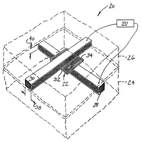
Referring now to the drawings and in particular to Fig. 1, apparatus of
the present invention is designated in its entirety by the reference numeral
20. The
apparatus 20 generally comprises a porous membrane, generally designated by
22, positioned between first and second bodies 24, 26 having first and second
channels 28, 30, respectively, formed therein. The membrane 22 has a first
side
32 facing the first body 24 and a second side 34 opposite the first side
facing the
second body . The first channel 28 is formed in the first body 24 so it
extends
along the membrane 22 adjacent the first side 32 of the membrane. Similarly,
the
second channel 30 is formed in the second body 26 so it extends along the
membrane 22 adjacent the second side 34 of the membrane. As will be explained
in further detail below, the first and second channels 28, 30 each contain
fluid in
communication with the respective side of the membrane 22. In one particularly
useful embodiment of the present invention, the first and second channels 28,
30
cross at an angle. In one embodiment, the first and second channels 28, 30 are
straight and extend perpendicular to each other. Although the first and second
bodies 24, 26 may be made of other materials without departing from the scope
of
the present invention, in one embodiment they are made of polydimethylsiloxane
(PDMS). Although the channels 28, 30 may be made using other techniques
without departing from the scope of the present invention, in one embodiment
the
channels are made using standard rapid prototyping techniques commonly used
for PDMS. Such techniques are described in J.C. McDonald, Electrophoresis 21,
27-40 (2000). Although the resulting channels 28, 30 may have other dimensions
without departing from the scope of the present invention, in one embodiment
each
channel has a width 38 of about 100 micrometer (um) and a depth 40 of about 30
um.
As illustrated in Fig. 2, the nanoporous membrane 22 has at least
one pore (and preferably a plurality of pores) 42 extending from the first
side 32 of
the membrane to the second side 34 of the membrane. As illustrated in Fig. 2,
the
pores 42 in one embodiment each have a width 44 less than about 250
nanometers (nm). In one embodiment, the membrane 22 has a monodisperse
distribution of pore widths 44. In one particularly useful embodiment, each
pore 42
CA 02463789 2004-04-15
WO 03/047712 PCT/US02/33313
6
has a width 44 between about 10 nm and about 230 nm. In still another
embodiment, each pore 42 has a width 44 between about 15 nm and about 220
nm. In most embodiments, the pores 42 are generally cylindrical and the width
44
is a diameter of the cylinder. Although the membrane 22 may have other pore
densities without departing from the scope of the present invention, in one
embodiment the membrane has a pore density of between about 1,000,000 pores
per square centimeter and about 10,000,000,000 pores per square centimeter. In
one particularly useful embodiment, the membrane 22 has a pore density of
between about 100,000,000 pores per square centimeter and about 600,000,000
pores per square centimeter. Although the membrane 22 may have other
thicknesses without departing from the scope of the present invention, in one
embodiment the membrane 22 has a thickness 46 between about 1 um and about
100 um. In one particularly useful embodiment, the membrane 22 has a thickness
46 of about 10 um. Although the membrane 22 may be made of other materials
without departing from the scope of the present invention, in one embodiment
the
membrane is made of nuclear track etched polycarbonate film (PCTE). One such
membrane 22 is available from Osmonics, Inc. of Minnetonka, Minnesota. Such
membranes have been used as active components in bulk solution experiments to
trap and selectively move molecules.
As illustrated in Fig. 1, an electrical source 50 is positioned in
electrical communication with at least one of the channels 28, 30 for
selectively
developing an electrical potential between fluid in the first channel and
fluid in the
second channel. As will be appreciated by those skilled in the art, when the
electrical potential is of the proper polarity and magnitude, it causes one or
more
components (e.g., charged particles or molecules) within the fluid to pass
through
the pore 42 in the membrane 22 from one of the channels 28, 30 to the other by
electrokinetic flow. Although other electrical potentials may be developed by
the
electrical source 50 without departing from the scope of the present
invention, in
one embodiment the potential is between about 10 millivolts and about 200
volts.
As will be appreciated by those skilled in the art, an interior surface
60 defining each pore 42 may be coated with a coating 62 as illustrated in
Fig. 3 so
that individual particles (e.g., molecules) passing through the pore are
likely to
CA 02463789 2004-04-15
WO 03/047712 PCT/US02/33313
7
contact coating. For example, the pores 42 may be coated with a particular
reagent so that desired reactions occur as the particles pass through the
pores.
Further, the coating 62 may be electrically charged if desired. Although the
coatings 62 may have other thicknesses without departing from the scope of the
present invention, in one embodiment the coating has a thickness 64 of about
10
nanometers. In one particular embodiment, the pore 42 is coated with gold by
electroless deposition. Furthermore, the coating may comprise more than one
component. In one embodiment the pore 42 is coated with gold by electroless
deposition and the gold is subsequently derivatized with a linear chemical
agent
terminated with a mercaptan at one end and a selected chemical functional
group
at the other end.
As will be appreciated by those skilled in the art, the separations
capacity factor, which is governed by the surface-to-volume ratio, can be
quite
large. For example, the separations capacity factor increases by about 120
times
when a pore 42 having a width of about 200 nm is coated with a reagent having
a
thickness 64 of about 10 nm compared to a pore having a width of about 20 um
coated with the same coating.
Although in one embodiment the fluid in the first and second
channels 28, 30 have identical chemistries, the fluid in each channel may have
different chemistries without departing from the scope of the present
invention. As
will be appreciated by those skilled in the art, each of the fluids contained
by the
channels 28, 30 has a Debye length which is a measure of the distance at which
the Coulomb field of the charged particles in a plasma cease to interact. The
properties of the flow through the pores 42 is affected by the relationship
between
the width 44 of the pores and the Debye length of the fluid in the channels
28, 30.
In one embodiment, the first channel 28 is filled with a first fluid having a
first
Debye length and the second channel 30 is filled with a second fluid having a
second Debye length at least as long as the first Debye length. Further, the
pore
42 has a width 44 between about 0.01 and about 1000 times the first Debye
length. If the pores have a small width (closer to 0.01 times the first Debye
length),
then flow in the pores is dominated by electroosmosis, whereas if the pores
have a
CA 02463789 2004-04-15
WO 03/047712 PCT/US02/33313
8
large width (greater than 1 first Debye length), then ion migration dominates
the
flow in the pores.
The previously described apparatus 20 can be used to selectively
transfer one or more components of fluid from the first channel 28 to the
second
channel 30 as illustrated in Fig. 4. Fluid is delivered to the first and
second
channels 28, 30, respectively. An electrical potential is developed between
the
fluid in the first channel 28 and the fluid in the second channel 30 thereby
causing
one or more components of fluid (e.g., a particle) to pass through the pore 42
in
the membrane 22.
In addition, the apparatus 20 may be used to tag a selected
component within a fluid. A chemical reagent (e.g., an antibody) is passed
through
the pore 42 so the reagent coats the interior surface 60 of the pore.
Alternatively a
sequence of chemical reagents can be passed through the pore 42 so that a
multilayer structure is built up to coat the interior surface 60 of the pore.
The pore
42 is flushed to remove the reagent from a central portion of the pore so the
reagent coats the surface 60 of the pore. The fluid component to be tagged is
drawn through the pore 42 using a method such as described above so the
selected component contacts the reagent coating 62, and a tagging reaction
results between the selected component and the immobilized chemical reagent.
Although it is envisioned other methods may be used to attract the selected
component to the pore in one embodiment, the electrical potential between the
fluid in channel 28 and the fluid in channel 30 draws the selected component
through the pores. It is further envisioned that the membrane 22 may be
selected
so the pore 42 has a width 44 equal to between about 0.5 and about 100 times
the
Debye length of the fluid plus between about 1 and about 1000 times a width of
the
selected component.
The previously described apparatus 20 also may be used to isolate a
particle having a selected electrophoretic velocity from a plurality of
particles. As
illustrated in Fig. 5a, the first channel 28 is filled with a fluid, and the
plurality of
particles 70 are positioned in the fluid at a first end 72 of the first
channel. An
electrical potential is developed between the first end 72 of the first
channel 28 and
a second end 74 of the first channel opposite the first end so each of the
plurality
CA 02463789 2004-04-15
WO 03/047712 PCT/US02/33313
9
of particles 70 migrate along the first channel from the first end to the
second end
in an order corresponding to their respective electrophoretic velocities as
shown in
Fig. 5b. An electrical potential is developed between the first channel 28 and
the
second channel 30 when the particle having the selected electrophoretic
velocity is
adjacent the pores 42 in the membrane 33 so the particle passes through the
pore
from the first channel to the second channel as illustrated in Fig. 5c.
Although the
electrical potential may be switched nearly instantaneously from the former
condition to the latter condition, in one embodiment the electrical potential
is
adjusted when the particle having the selected electrophoretic velocity is
adjacent
the pores 42 in the membrane 22 so the desired particle stops migrating along
the
first channel 28. Further, in one embodiment the electrical potential between
the
first channel 28 and the second channel 30 is adjusted once the particle
having the
selected electrophoretic velocity has passed through the pores 42 from the
first
channel to the second channel to prevent particles 70 having electrophoretic
velocities other than the selected electrophoretic velocity from passing
through the
pore.
As will be understood by those skilled in the art, fluidic
communication can be established among any number of vertically stacked bodies
and each body can be adapted to perform a specialized fluid handling,
separation
or sensing task. Interconnects as described above can be used to provide
controllable transport of components between bodies. It is further envisioned
that
such systems could be used to perform complex sequences and arrays of fluidic
manipulations as will be explained in further detail below.
Using nanofluidic structures to connect microfluidic channels allows a
variety of flow control concepts to be implemented, leading to hybrid fluidic
architectures of considerable power and versatility. The key characteristic
feature
of nanofluidic channels is that fluid flow occurs in structures of the same
size as
physical parameters that govern the flow. For example, the Debye length which
characterizes the length scale of ionic interactions in solution spans the
range
between about 1 nm and about 50 nm when the ionic strength of the buffer
solution lies in the high-to-low mM range. Because the solution Debye length
is of
the order of the channel dimensions in the nanopores, fluidic transfer may be
CA 02463789 2004-04-15
WO 03/047712 PCT/US02/33313
controlled through applied bias, polarity and density of the immobile nanopore
surface charge, and the impedance of the nanopore relative to the microfluidic
channels. Transfer between microchannels may be operated to produce either
two or three stable transfer rates, illustrating the digital character of the
fluidic
transfer. Furthermore, the separations capacity factor governed by the surface-
to-
volume ratio, can be quite large. For example, the separations capacity factor
is
about 120 times larger for a pore having a width of about 200 nm and a coating
thickness of about 10 nm compared to a pore having a width of about 20 um and
the same coating.
Because gateable transfer of selected solution components between
vertically separated microfluidic channels opens the way to multilevel fluidic
systems, the potential applications of this technology are far reaching. As
one
example, the presence of high salt concentrations degrades electrophoretic
separations. With this technology, one can pre-separate analytes from high-
salt
biological fluids, collect and concentrate particular fractions of the
separation into a
different layer now under optimum conditions for a high resolution second-
dimensional separation. Because the manipulations are displaced vertically one
could readily imagine multi-dimensional separations, not limited by the two in-
plane
spatial directions. One can even envision placing derivatizing chemistry or
immunochemical reagents in a particular channel layer and allow chemical
reactions to take place on a selected analyte band. Given the large variety of
single layer devices already optimized to perform cellular manipulations,
chemical
reactions and complex separations, the ability to combine these individual
architectures into independent layers with external control of the transfer of
individually selectable analytes between layers, will enable many
applications.
As will be appreciated by those skilled in the art, the direction of
particle travel in the apparatus 20 can be controlled by applied potential,
surface
charge density (pH controllable), ionic strength, and even by the impedance of
the
fluidic network in which the interconnect is placed relative to the impedance
of the
membrane 42.
The present invention has been demonstrated through the following
examples:
CA 02463789 2004-04-15
WO 03/047712 PCT/US02/33313
11
Examples
The simple system described above was formed as a proof of
concept. Microfluidic channels were formed in bodies of polydimethylsiloxane
(PDMS) using standard rapid prototyping protocols for PDMS as explained in J.
C.
McDonald, et al., Electrophoresis 21, 27-40 (2000). A 5 um thick nanoporous
membrane was sandwiched between the bodies. Assembly has been
accomplished by centering a 10 mm x 1 mm section of membrane on the lower
body and placing the upper body on the membrane so its channel was
perpendicular to the channel in the lower body.
More sophisticated embodiments of the hybrid microfluidic and
nanofluidic system, such as a seven layer sandwiched structure, may be made
using the following protocol:
(1 ) Etch microchannels and holes in a glass substrate.
(2) Mount a polycarbonate nanopore membrane having desired
pore diameters on a carrier, such as a PDMS slab about 2 mm thick, without
wrinkling or deforming and sufficiently to hold the membrane in place for
subsequent handling, but not so tightly as to permanently bond the membrane to
the carrier.
(3) Apply adhesive type B (as described below) to the substrate
with imprinting, spraying, or screening techniques .
(4) Align the membrane and carrier to the etched glass substrate
and tack them in place.
(5) Release the carrier from the membrane leaving it on the
substrate to form a layered stack.
(6) Repeat step (2) to a solid polycarbonate membrane layer.
(7) Using conventional shadow mask, etch a desired pattern of
channels and holes into the solid membrane using reactive oxygen ion etching,
or
similar etching techniques for polymers.
(8) Apply adhesive type H (as described below) to the solid
membrane, with imprinting, spraying or screening techniques.
CA 02463789 2004-04-15
WO 03/047712 PCT/US02/33313
12
(9) Align the patterned solid membrane with the stack and tack
the membrane in place.
(10) Repeat step (2) to the second nanopore PC membrane
(11 ) With shadow mask, etch desired holes and/or channels into
membrane.
(12) Apply adhesive type H to the substrate.
(13) Repeat steps (4) & (5).
(14) Repeat steps (6) to (9) for a second solid PC membrane.
(15) Repeat steps (10) to (13) for a third nanopore PC membrane.
(16) Apply adhesive type B to a top glass layer having desired
etched holes and channels.
(17) Apply pressure to the entire stack and heat to thermally cure
and activate the adhesives, without degrading the polycarbonate.
A separated view of the resulting apparatus made by this protocol is
shown in Fig. 8. Fig. 9a illustrates the resulting fluid circuit. It is
further envisioned
that such circuits could be assembled to perform complex sequences and arrays
of
fluidic manipulations as illustrated in Fig. 9b.
One of the keys to achieving the desired bond is to use adhesives
that can be dried of solvents after application, and that can be thermally
cured
without evolving sufficient vapors that produce undesired bubbles in the bond.
For the glass/polycarbonate combination, adhesive B is a phenolic-based
adhesive
that is soluble in various non-aqueous solvents, such as ethanol. For the
polycarbonate/polycarbonate combination, adhesive H is a low molecular weight
polycarbonate dissolved in solution. For both adhesives, the adhesives are
diluted
to a low concentration, so that the bond thickness on cure is 1 to 2
micrometers
thick. If too thick of an adhesive layer is applied, the adhesive on curing
can reflow
back into the microfluidic channels and potentially plug the channels and
nanopores. The bonds are then created by applying pressure and heat, typically
over 100 psi and under 150°C. The process steps are still under
development to
determine the optimum bond cycles.
The crossed microfluidic channels spatially define the transport
region and eliminate the need for precise alignment of the nanofluidic
membrane.
CA 02463789 2004-04-15
WO 03/047712 PCT/US02/33313
13
Transport control was monitored with fluorescence spectroscopy and imaging of
fluid streams containing small molecule fluorophores by interrogating the
fluorescence signal on either the originating or the receiving channel side of
the
nanofluidic membrane. Fig. 6a shows the transfer of an aqueous 5 mM phosphate
buffer solution, PBS pH = 8, of the anionic fluorophore, fluorescein, across a
200
nm pore diameter polycarbonate, PCTE, membrane to a receiving channel held
under static, i.e. flow-free, conditions. Successive transfers were affected
by
application of negative bias pulses. Because the receiving channel was held
static,
the fluorophore concentration probed during bias application was a balance
between active transport from the source channel and diffusion along the
receiving
channel. When the bias was removed, diffusion depleted the concentration in
the
region probed, but with successive forward bias applications the concentration
of
probe in the receiving channel increased, thereby diminishing the driving
force for
diffusion after subsequent transfers. Fig. 6b shows a similar experiment in
which
active flow was maintained in the receiving channel. The build-up to steady-
state
at the membrane after bias application results from the balance between active
transport of the analyte across the nanofluidic membrane and its removal by
cross-
flow in the receiving channel, which is clearly more gradual than under static
conditions. An obvious time offset was observed when the detection region was
moved downstream of the interconnect. Figure 6c demonstrates the level of
control and speed of transfer possible with these nanofluidic interconnects.
In this
experiment the off-state voltages were allowed to float, producing a non-zero
level
of transfer intermediate between the forward-bias (-60 V) on-state and the
reverse-
bias (+ 60 V) on-state. Measurements on the changing edges of Fig. 6c indicate
steady state concentration was re-established in the receiver channel within ~
1.2
s of applying the switching voltage. Figure 6d demonstrates the insensitivity
to
charge state by comparing the transfer of the neutral fluorophore, 4,4-
difluoro-5,7-
dimethyl- 4-bora-3a,4a-diaza-s-indacene- 3- succinimidylpropionate (bodipy).
In all of the above experiments the direction of transfer was
controlled by the electroosmotic flow generated by the microfluidic channels.
PDMS exhibits a negative surface charge at pH = 8, so forward bias is expected
when V,~~ - Vsource < 0, as observed. Interestingly, this is directly opposite
to the
CA 02463789 2004-04-15
WO 03/047712 PCT/US02/33313
14
flow direction based on the electroosmotic flow characteristics of the PCTE
membrane alone. The surfaces of the PCTE membrane channels are coated with
polyvinylpyrrolidone (PVP) to render them hydrophilic. The tertiary amine of
the
PVP is susceptible to protonation, making the surface net positive at pH 8,
thus
recruiting a population of negative solution counterions to the interior of
the
nanochannels. Under the low ionic strength conditions used here, the ionic
population in the channel is predominantly H2P04%HP042-, so forward bias is
obtained with Vre~ - Usource > 0, if the nanofluidic channels control the
direction of
transport. Instead, flow in the direction predicated on the (negative) charge
state
of the PDMS surfaces of the microfluidic channels controls transport.
This control can be reversed, as shown in Figs. 7a and 7b, for
transport across a 200 nm pore diameter membrane compared with that across a
15 nm pore diameter membrane. Clearly the polarities of forward- and reverse-
bias have been reversed. This behavior can be understood based on two effects -
the greatly increased resistance to pressure driven flow through the smaller
pores
and the greater voltage drop across the pores in the 15 nm case. Modeling the
impedance network composed of the two microfluidic channels and the membrane
shows that in the network containing the 200 nm pore membrane < 2% of the
potential is dropped across the nanofluidic membrane. However, for 15 nm
pores,
just over 33% of the potential appears across the membrane, so that the PCTE
pore electroosmotic flow dominated overall fluid transport in the device when
15
nm pores were used, but not when larger pores were employed. Thus, by choosing
the pore size, pore and channel surface chemistries, and solution composition,
one
can select either direction of fluid flow for the same externally applied
voltage.
These control concepts have been used to effect preparative
separations on the microscale by incorporating them into a microfabricated
capillary electrophoresis arrangement with a molecular gate membrane placed
between two channel layers just before the detection region. When the gate is
off,
the system acts as a standard electrophoresis system; when the gate is forward
biased, the analyte is collected in the vertically displaced receiving
channel, and
the signal is reduced or eliminated at the detection region. Fig. 7c shows
three
successive injections of a fluorescein-containing plug in the flow-injection
analysis
CA 02463789 2004-04-15
WO 03/047712 PCT/US02/33313
scheme. The inset to Fig. 7c shows a schematic diagram of the preparative
electrophoresis apparatus. The horizontal channel forms the main separation
(electrophoresis) channel with the left-hand vertical channel provided to
provide for
injection of a sample mixture onto the channel for separation. The right-hand
vertical channel is held in a separate vertical plane and is separated from
the main
electrophoresis channel by a molecular gate membrane (denoted by the vertical
rectangle at the crossing point of the vertical and horizontal channels). The
sample bands are all labeled with a fluorescent tag, and are detected in the
horizontal electrophoresis channel just after they pass the molecular gate
membrane. When no sampling gate pulse was applied (left panel), the
fluorescein
is transported past the membrane gate collection region. Application of a
negative
gate pulse to the 200 nm pore diameter polyvinylpyrrolidone free (PVPF)
membrane (middle panel) results in nearly complete removal of the analyte band
from the electrophoresis channel. Another injection made with no gate pulse
reproduces the results of the initial injection. In this experiment a PVPF
membrane
consisting of pores with negative surface charge was used, so the polarity of
transfer was the same as that based on the PDMS microchannels.
Among the advantages of the apparatus 20 of the present invention
is the ability to selectively control flow by controlling the potential
applied across
the pores 42. Flow through the pores 42 can be started and stopped nearly
instantaneously. Systems can be created in which the flow is normally on or
off
until a potential is applied between the fluids in the two channels. Further,
direction of flow through the pores 42 can be instantaneously reversed. Still
further, the apparatus 20 allows certain species to be selectively transported
or
blocked from passage through pores 42 and selected pores within the apparatus
can be controlled using the fluids themselves as the signal path.
Surface charge density is a critical property influencing electrokinetic
flow in these structures, because the enhanced surface-to-volume ratio in
these
nanofluidic channels means that a significant fraction of the total charge is
bound
to the walls and is immobile. Because it determines the magnitude of the
surface
potential and the applicability of the Debye-Huckel approximation, surface
charge
density provides an experimental handle to adjust the microscopic processes
that
CA 02463789 2004-04-15
WO 03/047712 PCT/US02/33313
16
determine transport in the nanopore. Thus, the potential for facile control of
nanofluidic flow by varying the bias, nanochannel wall charge density, charge
polarity, and/or solution ionic strength offers the opportunity to effect
intelligent
transfer of fluid components with extreme ease and versatility.
When introducing elements of the present invention or the preferred
embodiments) thereof, the articles "a", "an", "the" and "said" are intended to
mean
that there are one or more of the elements. The terms "comprising",
"including"
and "having" are intended to be inclusive and mean that there may be
additional
elements other than the listed elements.
As various changes could be made in the above constructions
without departing from the scope of the invention, it is intended that all
matter
contained in the above description or shown in the accompanying drawings shall
be interpreted as illustrative and not in a limiting sense.