Note: Descriptions are shown in the official language in which they were submitted.
CA 02463870 2004-04-15
WO 2004/019787 PCT/US2003/025818
1
TISSUE FASTENERS AND RELATED
DEPLOYMENT SYSTEMS AND METHODS
CROSS-REFERENCE TO RELATED APPLICATION
[001] This application relates to commonly assigned U.S. Application
Serial No. 10/230,682 of Robert Devries et al., filed on the same date as this
application, and entitled "Devices and Methods for Fastening Tissue Layers."
The complete disclosure of that application is incorporated by reference
herein.
DESCRIPTION OF THE INVENTION
Field of the Invention
[002] The present invention relates to surgical tissue fasteners and
related deployment systems and methods for delivering the tissue fasteners.
In particular, the present invention relates to tissue fasteners used in, for
example, a fundoplication procedure for treatment of Gastroesophageal
Reflux Disease (GERD).
Background of the Invention
[003] Gastroesophageal reflux occurs when stomach acid enters the
esophagus. This reflux of acid into the esophagus occurs naturally in healthy
individuals, but also may become a pathological condition in others. Effects
from gastroesophageal reflux range from mild to severe. Mild effects include
heartburn, a burning sensation experienced behind the breastbone. More
severe effects include a variety of complications, such as esophageal erosion,
esophageal ulcers, esophageal stricture, abnormal epithelium (e.g., Barrett's
esophagus), and/or pulmonary aspiration. These various clinical conditions
and changes in tissue structure that result from reflux of stomach acid into
the
esophagus are referred to generally as Gastroesophageal Reflux Disease
(GERD).
[004] Many mechanisms contribute to prevent gastroesophageal
reflux in healthy individuals. One such mechanism is the functioning of the
lower esophageal sphincter (LES). With reference to Fig. 1, the LES 2 is a
ring of smooth muscle and increased annular thickness existing in
approximately the last four centimeters of the esophagus. In its resting
state,
CA 02463870 2004-04-15
WO 2004/019787 PCT/US2003/025818
2
the LES creates a region of high pressure (approximately 15-30 mm Hg
above intragastric pressure) at the opening of the esophagus 3 into the
stomach 7. This pressure essentially closes the esophagus 3 so that contents
of the stomach cannot pass back into the esophagus 3. The LES 2 opens in
response to swallowing and peristaltic motion in the esophagus 3, allowing
food to pass into the stomach. After opening, however, a properly functioning
LES 2 should return to the resting, or closed state. Transient relaxations of
the LES 2 do occur in healthy individuals, typically resulting in occasional
bouts of heartburn.
[005] The physical interaction occurring between the gastric fundus 5
and the esophagus 3 also prevents gastroesophageal reflux. The gastric
fundus 5 is a lobe of the stomach situated at the top of the stomach 7 distal
to
the esophagus 3. In asymptomatic individuals, the fundus 5 presses against
the opening of the esophagus 3 when the stomach 7 is full of food and/or gas.
This effectively closes off the esophageal opening to the stomach 7 and helps
to prevent~acid reflux back into the esophagus 3. More specifically, as the
food bolus is immersed in gastric acid, it releases gas which causes the
fundus 5 of the stomach 7 to expand and thereby exert pressure on the distal
esophagus 3 causing it to collapse. The collapse of the esophagus lumen
reduces the space for the stomach acid to splash past the closed esophagus
lumen and thereby protect the proximal esophagus from its destructive
contact.
[006] In individuals with GERD, the LES 2 functions abnormally, either
due to an increase in transient LES relaxations, decreased muscle tone of the
LES 2 during resting, or an inability of the esophageal tissue to resist
injury or
repair itself after injury. These conditions often are exacerbated by
overeating, intake of caffeine, chocolate or fatty foods, smoking, and/or
hiatal
hernia. Avoiding these exacerbating mechanisms helps curb the negative
side effects associated with GERD, but does not change the underlying
disease mechanism.
[007] A surgical procedure, known generally as fundop(ication, has
been developed to prevent acid reflux in patients whose normal LES
CA 02463870 2004-04-15
WO 2004/019787 PCT/US2003/025818
3
functioning has been impaired, either as a result of GERD or other adverse
effects. This procedure involves bringing the fundus wall 6 into closer
proximity of the esophageal wall 4 to help close off the esophageal opening
into the stomach 7. Traditionally, this procedure has been performed as an
open surgery, but also has been performed laparoscopically.
[008] As with any surgery, the attendant risks are great. Due to
relatively large incisions necessary in the performance of open surgery,
relatively large amount of blood is lost, the risk of infection increases, and
the
potential for post-operative hernias is high. Further, the relatively large
incisions necessary in the performance of open surgery require extended
recovery times for the incision to heal.
[009] A laparoscopic procedure may involve performing laparotomies
for trocar ports (penetrations of the abdominal wall), percutaneous endoscopic
gastronomies (incisions through the skin into the stomach), and the
installation of ports through which, for example, a stapler, an endoscope, and
an esophageal manipulator (invagination device) are inserted. Under view of
the endoscope, the esophageal manipulator is used to pull the interior of the
esophagus 3 into the stomach 7. When the esophagus is in position, with the
fundus 5 of the stomach plicated, the stapler is moved into position around
the
lower end of the esophagus and the plicated fundus is stapled to the
esophagus 3. The process may be repeated at different axial and rotary
positions until the desired fundoplication is achieved. This procedure is
still
relatively invasive requiring incisions through the stomach, which has a risk
of
infection. The location of the incision in the abdominal wall presents a risk
of
other negative effects, such as sepsis, which can be caused by leakage of
septic fluid contained in the stomach.
SUMMARY OF THE INVENTION
[010] Therefore, one embodiment of the present invention provides
less invasive devices and methods for performing the fundoplication
procedure. This is achieved by utilizing tissue fasteners and related
deployment systems which can be endoluminally delivered through the
CA 02463870 2004-04-15
WO 2004/019787 PCT/US2003/025818
4
esophagus 3, thereby eliminating the need for highly invasive, physiologically
insulting surgical procedures.
[011] To attain the advantages and in accordance with the purpose
of the invention, as embedded and broadly described herein, one aspect of
the invention provides a tissue fastener used to join multiple tissue layers.
The tissue fastener includes a proximal member configured to expand from a
delivered state to a deployed state, a distal member configured to expand
from a delivered state to a deployed state, and a connecting member
connecting the proximal member to the distal member. In the deployed state,
the proximal member and the distal member secure the multiple tissue layers
together.
[012] Another aspect of the invention provides a tissue fastener used
to join multiple tissue layers that includes a first member, a second member,
a
connecting member connecting the first member to the second member, and
means for applying a substantially constant force on the tissue layers. In
some embodiments, the applying means may be compressible.
[013] Another aspect of the invention provides a tissue fastener used
to join multiple tissue layers that includes a first member, a second member,
a
connecting member connecting the first member to the second member, and
means for adjusting a length of the connecting member between the first and
second members. In some embodiments, the adjusting means may include
structure associated with the first member for releasably securing the
connecting member to the first member. That structure may be configured to
restrict passage of the connecting member in a direction through the first
member.
[014] Another aspect of the present invention is to provide a delivery
system configured for deployment of an expandable tissue fastener. The
system includes a flexible tube configured to accommodate a tissue fastener
in a contracted state, a pusher for guiding the tissue fastener along a lumen
of
the tube, and a grasper coupled to a distal end of the pusher and having
means to grasp the tissue fastener.
CA 02463870 2004-04-15
WO 2004/019787 PCT/US2003/025818
[015] In yet another aspect of the present invention, a method of
attaching a first layer of tissue to a second layer of tissue includes
providing
an expandable tissue fastener in a contracted state in a device, the tissue
fastener having a proximal member and a distal member, inserting the device
into a body passage leading to the first tissue wall, placing the device
proximate a location on the first tissue layer, passing the device through the
first and second tissue layers, advancing the tissue fastener toward an
opening of the device, such that the distal member is completely protruded
out of the device and expanded against the second tissue layer, and
withdrawing the device and releasing the tissue fastener, such that the
proximal member is expanded against the first tissue layer.
[016] The present invention is depicted in this disclosure and is
particularly suitable in the treatment of GERD, e.g., a fundoplication
procedure. However, the tissue fasteners and related deployment methods
and systems of the present invention can be used to treat any of a number of
different disease conditions, and can be used for fastening any desired body
tissues.
[017] Additional objects and advantages of the invention will be set
forth in part in the description which follows, and in part will be obvious
from
the description, or may be learned by practice of the invention. The objects
and advantages of the invention will be realized and attained by means of the
elements and combinations particularly pointed out in the appended claims.
[018] It is to be understood that both the foregoing general
description and the following detailed description are exemplary and
explanatory only and are not restrictive of the invention, as claimed.
BRIEF DESCRIPTION OF THE DRAWINGS
[019] The accompanying drawings, which are incorporated in and
constitute a part of this specification, illustrate several embodiments of the
invention and together with the description, serve to explain the principles
of
the invention.
[020] In the drawings:
CA 02463870 2004-04-15
WO 2004/019787 PCT/US2003/025818
6
[027] Fig. 1 is a cross-sectional view of the gastrointestinal tract in
the region of the lower esophageal sphincter (LES) and the fundus of the
stomach;
[022] Fig. 2 is a perspective view of a tissue fastener, prior to
deploymenfi into a body, according to an embodiment of the present invention;
[023] Fig. 2A is a perspective view of a proximal fastening member
of the tissue fastener of Fig. 2, showing the proximal fastening member in a
deployed state according to an embodiment of the present invention;
[024] Fig. 2B is a perspective view of a distal fastening member of
the tissue fastener of Fig. 2 in a deployed state, according to an embodiment
of the present invention;
[025] Figs. 3-7C are perspective views of various tissue fasteners
according to various embodiments of the present invention;
[026] Figs. 7D-7F are cross-sectional views of tissue fastener
members according to various embodiments of the present invention;
[027] Figs. 8A-8R are perspective views of various designs of tissue
fastening members according to additional embodiments of the present
invention;
[028] Figs. 9A-9J are perspective views of various connector post
designs according to still other embodiments of the present invention;
[029] Figs. 10A-10SS are perspective views of fasteners according
to various embodiments of the present invention;
[030] Figs. 11A-C are perspective views of an endoluminal
deployment system, illustrating various operational stages of the system for
deployment of a tissue fastener, according to an embodiment of the present
invention;
[031] Fig. 12A is a schematic illustration of the tissue fastener
deployment for a tissue connection procedure, showing the deployment
system of Figs. 11A-C containing the tissue fastener of Fig. 2 and being
inserted into the lower esophagus, according to an embodiment of the present
invention;
CA 02463870 2004-04-15
WO 2004/019787 PCT/US2003/025818
7
[032] Fig. 12B is a schematic illustration of the tissue fastener
deployment, showing the deployment system penetrating the esophageal
wall, according to an embodiment of the present invention;
[033] Fig. 13 is a schematic illustration of the tissue fastener
deployment, showing the deployment system penetrating the esophageal wall
and the fundus wall, according to an embodiment of the present invention;
[034] Fig. 14 is a schematic illustration of the tissue fastener
deployment, showing the expanded distal fastener member of the tissue
fastener holding the fundus wall, according to an embodiment of the present
invention;
[035] Fig. 15 is a perspective view of a proximal fastening member of
the tissue fastener of Fig. 2, viewing from inside the esophagus, after the
deployment is complete, according to an embodiment of the present
invention;
[036] Fig. 16 is a cross-sectional view of the esophageal wall and the
fundus wail, with a tissue fastener in place, after the tissue connection
procedure;
[037] Figs. 17A is a perspective view of a deployment system, with a
straight distal tip, according to another embodiment of the present invention;
[038] Fig. 17B is a perspective view of a deployment system, with a
curved distal tip, according to still another embodiment of the present
invention; and
[039] Figs. 18A and 18B are views of a deployment system
respectively holding and releasing a tissue fastener member.
DESCRIPTION OF THE EMBODIMENTS
[040] Reference will now be made in detail to the exemplary
embodiments of the invention, examples of which are illustrated in the
accompanying drawings. Wherever possible, the same reference numbers
will be used throughout the drawings to refer to the same or like parts.
[041] A newly developed form of fundoplication, referred to as
endoscopic fundoplication, is an endoluminal procedure in which the fundus
wall 6 is folded back onto the esophagus wall 4. The tissue fold formed
CA 02463870 2004-04-15
WO 2004/019787 PCT/US2003/025818
between the esophagus 3 and the fundus 5 then is secured. Endoscopic
fundoplication is intended to be performed as an endoluminal procedure in
which insertion of required medical instruments occurs through the esophagus
3. Such a procedure has the benefits of being less invasive, quicker, and less
expensive as compared to previous techniques.
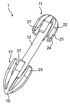
[042] Fig. 2 shows an exemplary embodiment of a tissue fastener 1
for use in a fundoplication procedure. The tissue fastener 1 has a distal
fastening member 13, a proximal fastening member 11, and a connecting
member 12 connecting the distal fastening member 13 and the proximal
fastening member 11 to each other. The distal fastening member 13, the
proximal fastening member 11, and the connecting member 12 are formed as
an integrated unit or two or more components pieced together. The distal
fastening member 13 includes a plurality of anchor hooks or legs 23, and the
proximal fastening member 11 includes a plurality of anchor hooks or legs 21.
The plurality of anchor legs 21, 23 are constructed to expand from a
contracted state to an expanded state. When the legs are in the contracted
state, as shown in Fig. 2, the tissue fastener 1 is low in profile and can be
loaded into a narrow lumen of an endoluminal deployment system for
deployment. Once the deployment system is positioned into a desired
location within a body, the plurality of anchor legs 21, 23 are expanded
outwardly to fasten multiple tissue layers together between the distal and
proximal fastening members 13, 11, thereby enhancing the tissue connection.
[043] In this particular embodiment shown in Fig. 2, each of the distal
and proximal fastening members 11, 13 is provided with a total of six anchor
legs 21, 23, that are equally spaced apart. However, it should be recognized
that the fastening members 11, 13 can be provided with more or less number
of legs 21, 23 with different desired spacing therebetween.
[044] Preferably, the leading edge of the distal fastening member 13
forms a sharp point or edge 15 to assist with penetrating through the tissue
layers. The sharp edge 15 may be a trocar-like cutting edge to perform the
perforation of the layers itself. It should also be recognized that forming
such
a sharp cutting edge 15 may not be necessary if an endoluminal deployment
CA 02463870 2004-04-15
WO 2004/019787 PCT/US2003/025818
9
system includes a cutting edge 40 on its distal tip, as shown in Figs. 17A-B
to
be described herein.
[045] Figs. 2A and 2B show the expanded state of the proximal and
distal fastening members 11, 13, respectively in an installed state. When the
distal fastening member 13 is freed from a restraining means of an
endoluminal deployment system, the anchor legs 23 of the distal fastening
member 13 expand outwardly to form an umbrella-shaped fastening member.
Sharp edge or tip 15 may be removed or made of a dissolvable,
biodegradable material. Similarly, when the proximal fastening member 11 is
freed from a restraining means of an endoluminal deployment system, the
anchor legs 21 of the proximal fastening member 11 expand outwardly to form
a fastening member. Each of the anchor legs 21 of the proximal fastening
member 11 may include one or more bending portions 22 at a location along
the length of the leg 21, such that the distal portion 24 of the leg 21
extends
outwardly with respect to the bending portion 22. The bending portion 22 is a
relatively flexible portion of an otherwise substantially rigid leg 21. The
bending portion 22 may be facilitated by removal of material or change in
geometry along the leg 21. The leg 21 may further comprise a variable
stiffness along the leg 21. For example, the bending portion 22 may consist
of a lower stiffness material to facilitate the bending.
[046] Figs. 3 through 7 are perspective views of various tissue
fasteners in expanded states according to other embodiments of the
invention. In an embodiment shown in Fig. 3, the tissue fastener 101 is
formed of at least two elongate members 150, such as wires or rods,
interconnected in the mid-portion by a connecting member 112. The
interconnection may be made through any suitable method, such as welding,
brazing, molding, a locking mechanism, etc., or members may be
manufactured as an integral piece. In a contracted state, each member 150 is
preferably substantially straight to promote easier insertion through the
esophagus and deployment. In an expanded state, shown in the figure, each
member 150 bends outwardly, substantially perpendicular to the connecting
member 112, to form the distal and proximal fastening members 113, 111.
CA 02463870 2004-04-15
WO 2004/019787 PCT/US2003/025818
The ends of the proximal fastening member 111 could be rounded or
otherwise protected to reduce trauma when in contact with tissue. Similar to
the embodiment shown in Fig. 2, the leading edge of each member 150 may
form a sharp cutting edge 115 to assist with perforating layers of tissues.
[047] Figure 3A illustrates a further embodiment of the fastener of
Figure 3 wherein the distal and proximal ends of the fastener 101 turn inwards
upon deployment through tissue layers. These inward turns, or hooks 114,
would contact tissue and prevent migration of fastener 101.
[048] According to another embodiment shown in Fig. 4A, the tissue
fastener 201 is comprised of a single wire 250 or rod. Similar to the tissue
fastener 101 of Fig. 3, the wire 250 preferably forms a substantially straight
wire in a contracted state. In an expanded state, shown in the figure, the
wire
250 forms spiral-shaped distal and proximal fastening members 213, 211 at
both ends of the wire 250, that are substantially perpendicular to the
connecting member 212. The wire 250a may also form a spring-shaped
fastening member 211 a, 213a at both ends of the wire 250a, as shown in Fig.
4B. In that case, the spring-shaped fastening members 211 a, 213a may be
substantially parallel to the connecting member 212a. The leading edge of
the wire 250 may form a sharp cutting edge 215 to assist with perforating
layers of tissues. As a modification and alternative to fastener 201, a wire
fastener may be delivered in a straight configuration and forms into a coil
that
may be screwed into the tissue layers. As described below, a suitable
material for such fasteners is nitinol.
[049] The tissue fastener 301 shown in Fig. 5A is comprised of
multiple wires integrally attached to a connecting member 312. In a
contracted state, each wire 350 preferably forms a substantially straight wire
and, in an expanded state, shown in the figure, the wires 350 expand
outwardly to form spider-shaped distal and proximal fastening members 313,
311. While the leading edges of the wires 350 may form sharp cutting edges
to assist with perforating the tissue layers, it may be most beneficial to use
an
endoluminal deployment system with a trocar-like cutting edge, such as the
device shown in Figs. 17A-B, or other perforating instrument to form an
CA 02463870 2004-04-15
WO 2004/019787 PCT/US2003/025818
11
opening through the tissue layers prior to the deployment of the tissue
fastener 301. In an alternative embodiment to Fig. 5A, and as shown in Fig.
5B, the fastening members 311,313 may include inwardly bent hooks 352 at
each end portion of the wires 350 to prevent migration of the fastener once
deployed onto the tissue layers.
[050] As shown in Fig. 6, the tissue fastener 401 has a similar
configuration as the tissue fastener 1 shown in Fig. 2, except that the distal
and proximal fastening member 413, 411 have the same configuration, much
like the fastening member 11 shown in Fig. 2. In particular, the distal
fastening member 413 does not include a sharp pointed edge. For this tissue
fastener401, an endoluminal deployment system with a trocar-like cutting
edge (e.g., a device shown in Figs. 17A-B) can be utilized.
[051] Fig. 7A shows a tissue fastener 501, also similar to the tissue
fastener 1 shown in Fig. 2 in certain respects. The tissue fastener 501 has a
distal fastening member 513, a proximal fastening member 511, and an
adjustable connecting member 512. The distal fastening member 513 and the
proximal fastening member 511 function in a similar manner as these
corresponding structures of the tissue fastener 1 shown in Fig. 2. In the
embodiment shown in Fig. 7A, the tissue fastener 501 includes an adjustable
connecting member 512. Once fastener 501 has been placed through the
tissue layers to be connected, tail 520 of fastener 501 is pulled taut as
illustrated in Figure 7B and fastening member 511 is advanced forward. This
action creates a compressive force on the tissues between the fastening
members 511 and 513. Alternatively, the connecting member 512 has an
elastic compressive spring force which exerts pulling force between the distal
and proximal fastening members 513, 511 so that the fastening of the multiple
tissue layers is enhanced.
[052] The elastic compressive force may be further combined with
the adjustable system as described. Figures 7C through 7F illustrate
adjustable fasteners of the present invention. Figure 7C illustrates a
fastener
520 have fastening members 513 and 511 and an adjustable connecting
member 521. Member 521 further includes bumps (or notches) 514 which
CA 02463870 2004-04-15
WO 2004/019787 PCT/US2003/025818
12
allow fastening member 511 to ratchet forward as either member 511 is
advanced or connecting member 521 is pulled taut to connect tissue layers.
Figures 7D through 7F illustrate cross-sectional views of two ratcheting
systems as described. In Figure 7D, the ratcheting action is created by virtue
of flexible tabs 515 located inside fastening member 511. Figures 7E and 7F
illustrate friction based or infinite ratcheting systems wherein the
interference
fit of a single tab 515 of Figure 7E or of multiple tabs 515 of Figure 7F
serve to
keep connecting member 521 from slipping once fastening member 511 is
advanced forward onto tissue or connecting member 521 is pulled taut. If
connecting member 521 is at least partially elastic, the fastener may be self
adjusting once in place or the force within the fastener upon the tissues may
be controlled.
[053] Figs. 8A through 80 show various other exemplary fastening
members according to various embodiments of the present invention. As
shown in the figures, the fastening member can be formed of a flat plate 601
of virtually any geometric shape, a semispherical domed button 602, a flexible
plate 603, a malecot 604, a ratchet 606, a non-orthogonal button 607,
interconnectable multiple pieces 608, a foldable T-shaped bar 609, a twisted
or tied coil, string, or other flexible elongate member 610, a stent-like
configuration 611 wherein the connecting member has a crimped, braided
configuration, a cotterpin 612, a rivot 613, a V-shaped clip 614, 615 with or
without a flattened top (Figs. 8N and 8P), a staple-like configuration (Fig.
80),
and stent-like connectors with dogbone or flared ends (Figs 8Q and 8R).
Each fastening member connects to or comprises a connecting member 650.
[054] These various fastening member embodiments will now be
described in more detail. In an embodiment shown in Fig. 8D, the malecot
604 includes a plurality of legs or rods 604a that assume a cage-like shape
when in its normal, expanded position. In another embodiment shown in Fig.
8F, the fastening member 606 includes at least one skirt-like or frustoconical-
shaped ratchet 606 which permits movement only in one direction. In yet
another embodiment shown in Fig. 8G, a non-orthogonal connection between
the connecting member 650 and the fastening member 607, which may
CA 02463870 2004-04-15
WO 2004/019787 PCT/US2003/025818
13
include any of the described fastening members, is provided. In still another
embodiment shown in Fig. 8H, the fastening member 608 is formed of
multiple pieces that are attached through any suitable means, such as
piercing members or barbs. In still another embodiment shown in Fig. 81, the
fastening member 609 is pivotally coupled to the connecting member 650 to
form a T-shaped configuration. Any conventional connecting devices and
methods may be used to couple the fastening member 609 and the
connecting member 650. In still another embodimenfi shown in Fig. 8J, the
fastening member 610 is formed of a twisted or tied element. Preferably, the
member 610 is made of a single piece and double-looped. In still another
embodiment shown in Fig. 8K, the fastening member 611 is formed of a stent-
like braided configuration. The middle portion of the configuration is crimped
by a plurality of rings or is wrapped with a coil and constitutes the
connecting
member. In an expanded state, rings are removed or a coil is partially
unwrapped at the distal end portion, or the distal end is not otherwise
crimped
like the connecting member portion, to form a fastening member 611 shown in
the figure. Figs. 8Q and 8R illustrate stent type fasteners 660, 670
respectively having a dogbone or flare shape at each end. These fasteners
are deployed in a constricted manner and expanded or allowed to expand into
a fastener comprised of a singular structure. The fasteners of Figs. 8K, SQ,
and 8R may be constructed in any manner similar to fabricating stents as is
known in the art, including but not limited to knitting, weaving, twisting,
laser-
cut tubes, welded wires and molding.
[055] In still another embodiment shown in Fig. 8L, the fastening
member 612 is formed of a bar or a plate, fixedly attached the connecting
member by a cotterpin 612a or a stopper. In still another embodiment shown
in Fig. 8M, the fastening member 613 is formed by attaching a rivot to the
connecting member 651. In this case, the connecting member 651 includes
an engagement member 653 protruding outwardly at its distal end, which
engages the rivot to form the fastening member 613. In still another
embodiment shown in Fig. 8N, the fastening member 614 is a clip having two
or more legs 614b. The clip is expandable in a transverse direction, as
CA 02463870 2004-04-15
WO 2004/019787 PCT/US2003/025818
14
indicated by arrows in the figure, thereby enhancing the tissue connection.
Preferably, one or more bands or O-rings 614a are attached to limit the
expansion to a desired extent and to abut the tissue layers. It should also be
recognized that a plate or other flat surface 614c may be attached to, or
otherwise be integral with, the expanded clip at its distal end, as shown in
Fig.
8P.
[056] In still another embodiment shown in Fig. 80, the fastening
member 616 forms a staple-like configuration. Similar to the embodiments
shown in Figs. 3-5, the wire 616a preferably forms a substantially straight
wire
in a contracted state and, in an expanded state, shown in the figure, the wire
616a expands outwardly or inwardly to form the staple-like configuration.
Alternatively, an anvil can be placed behind tissue layers to bend the wires
616a and form the staple-like configuration. It should be recognized that more
than two wires can be provided. Any other suitable designs providing the
similar function may be utilized. In addition, the fastener may include any
combination of fastening members and connecting posts.
[057] In an embodiment, as shown in Fig. 8E, a tissue fastener 605
also may include a hollow bore 630 passing through the tissue fastener to
inject a therapeutic chemical agent or an adhesion promoting substance. The
tissue fastener is provided with an injection port 670 to permit introduction
of
the therapeutic substances or the adhesion promoting substance. The hollow
bore 630 may be provided with a one-way valve (e.g., check valve) 672 to
prevent a backflow. Fastener 605 may also comprise a weep hole 660 or
other means along post 662 to allow delivery of the chemical agent or
adhesion promoting means between the tissue layers.
[058] Figs. 10A through 1000 show various other exemplary
fastening members according to various embodiments of the present
invention. These various embodiments, in addition to certain others described
throughout this specification, adjust to varying tissue thickness so that
sufficient tension, and in many embodiments substantially constant tension, is
placed on the tissue to hold the tissue layers together. In many of the
disclosed embodiments, such adjustment is performed by adjusting the length
CA 02463870 2004-04-15
WO 2004/019787 PCT/US2003/025818
of the connecting member between the fastening members andlor by
providing a means, in many cases associated with a fastener member, for
providing a substantially constant tension on the tissue layers. Most of the
embodiments shown in Figures 10A-1000 include a pair of opposed fastener
members connected by a connecting member. It is to be understood that
fastener members and connecting members of any embodiment may be used
in combination with fastener members and connecting members of other
embodiments, as desired.
[059] Figure 10A shows a "T"-shaped tissue fastener 720 having a
first fastening member 721, a connecting member 722, and a second
fastening member 723. Member 721 may comprise any suitable
biocompatible elongate member, such as a portion of hypodermic tubing.
Connecting member 722 may comprise suture material, wire, or any other like
biocompatible material that fixedly connects to member 721, preferably in a
fashion that permits member 721 to articulate with respect to connecting
member 722 for ease of delivery and to the extent needed while implanted.
Member 723 may comprise any bead or ball-shaped structure that permits
connection to connecting member 722. Member 723 may include a slot
therein permitting member 722 to slide through so that the length of member
722 between members 721,723 may be adjusted depending on the thickness
of the connected tissue layers. For example, member 723 may be similar to a
fishing weight having a slit therein to hold member 722. As an alternative and
as shown in Figure 10A', member 723 may be a torsion spring 723' having a
pair of arms. When the arms are forced open in the direction of the arrows,
connecting member 722 loosens from within spring 723' and spring 723' can
slide relative to member 722 and be cinched against the tissue layers or an
additional member 721. When the arms are released, spring 723' secures
member 722 in position.
. [060] Figures 10B and 1 OB' show a tissue fastener 730 having first
fastening member 721, connecting member 722, and a second fastening
member 731 having a means for providing a substantially constant force on
the tissue layers. In this case, the means includes a flexible bellows-like
CA 02463870 2004-04-15
WO 2004/019787 PCT/US2003/025818
16
structure. The bellows of member 731 permit adjustment in the length of
connecting member 722 between members 721 and 731. Figure 10B shows
the bellows of member 731 in an expanded, relaxed state. The bellows are
contracted during delivery through the esophagus and to the tissue layers.
Then, as shown in Figure 1 OB', when fastener 730 is applied to tissue layers
735, the bellows will expand to place a sufficient force against the tissue
layers to hold together those layers. In addition, through the use of the
bellow-like structure, a substantially constant force is applied to the tissue
layers, reducing tissue irritation or loosening of the tissue fastener.
[061] Figures 10C, 10D, and 101 show variations on means to
maintain constant force on the tissue layers. In Figure 10C, tissue fastener
740 includes a second fastener member 741 having a spring 742 between
two plate-like structures 743, such as disks or buttons. In Figure 10D, tissue
fastener 743 includes a second fastener member 751 having a balloon 752
between the two structures 743. In Figure 101, tissue fastener 800 includes a
second fastener member 801 having a spring 802. One end of spring 802
connects to structure 743 and the other end of spring 802 is free and places a
substantially constant force against the tissue layers. Any suitable,
biocompatible, compressible structure may take the place of spring 742,
balloon 752, or spring 802.
[062] Figure 1 OE shows a "double-T" shaped tissue fastener 760 that
is essentially of one-piece construction. Fastener 760 has two opposing
fastener members 762, each taking the form of either member 721, member
743, or others described throughout this specification. Members 762 are
connected by an elastic connecting member 761 that permits an adjustable
length of member 761 between members 762. Member 761 may be made of
a elastic, biocompatible material, similar to a rubber-band.
[063] Figure 10H shows a similar arrangement as Figure 10E.
Fastener 790 includes opposing fastener members 762 interconnected by a
bungee cord-like member 792. Member 792 may be pulled tightly (elongated)
between members 762 during delivery to the tissue layers and then released
so that member 792 shortens and grows in diameter. The growth in diameter
CA 02463870 2004-04-15
WO 2004/019787 PCT/US2003/025818
17
may permit an interference fit between member 792 and a hole, notch, slit, or
other opening within one or both of the members 762, so that member 792
secures to members 762.
[064] In addition to certain embodiments already described, other
embodiments include structure associated with one or both fastener members
to permit adjustment of the length of the connecting member between the
fastener members. For example, Figure 10F shows a fastener 770 having
first and second fastener members 771, 772 and a connecting member 773.
Member 773 includes a plurality of beads, balls, or similar enlarged
structures
774 spaced along the length of member 773. As an alternative, the
connecting member may include a plurality of notches spaced along the
length of the member. The structures may be evenly or unevenly spaced as
desired. The structures 774 may be used in combination with a frangible
member 772 having a hole that permits structures 774 to travel through the
hole to shorten the length of member 773 between members 771, 772. To
place more a desired force on the tissue layers, additional structures 774 may
be pulled through the hole in member 772. Member 772 may be constructed
to include a notch therein to accommodate a structure 774. The through hole
in member 772 may be constructed to permit passage of structures 774 in
only one direction, i.e. away from member 771 to shorten the length of
member 773 between members 771, 772. In addition, member 771 may have
a similar construction as member 772 to permit passage of structures 774
therethrough. Excess connecting member 773 may be removed by any
suitable method.
[065] Figure 10G shows a fastener 780 having a fastener member
781 with a ratchet-like mechanism to permit passage of connecting member
782 therethrough in one direction (shown by arrow). Thus, member 781 can
move in the direction opposite the arrow to tighten fastener 780 on the tissue
layers, with the ratchet-like mechanism preventing loosening of member 782
relative to member 781. Similarly, Figure 1 OJ shows a fastener 810 having a
fastener member 811 with teeth 812 to capture connecting member 813 and
CA 02463870 2004-04-15
WO 2004/019787 PCT/US2003/025818
18
prevent loosening. Teeth 812 may be angled in the direction of the applied
force to more strongly grasp member 813.
[066] Figure 10K shows a fastener 820 having a fastener member
821 with a biased locking mechanism 822. The lock 822 includes a ball
bearing 823 or like structure contained within a through hole or passage of
member 821, and a spring 824 biasing bearing 823 into the passage. Bearing
823 will lock connecting member 825 within the passage when member 825 is
not being adjusted in length between the fastener members.
[067] Alternative locking mechanisms are shown in Figures 10M and
10N. Figure 10M shows a locking mechanism 840 that includes a pair of
eccentric wheels 841 that rotate to lock connecting member 842 in position
after member 842 is adjusted in length between fastener members, similar to
eccentric wheel locking mechanisms used on sailboats. Mechanism 840 may
be located within one or both fastener members of a fastener. Figure 10N
shows a similar arrangement, having a locking mechanism 850 with eccentric
wedges used to lock connecting member 852 in position.
[068] Figure 10L shows a fastener 830 having a first fastening
member 831, a connecting member 832, and a second connecting member
833. Member 833 has a two-piece construction that includes a disk 834 that
snaps or otherwise fixedly attaches to a complementary dish-shaped head
835. After connecting member 832 is pulled through a passage in head 835
to adjust the length of member 832 between fastener member 831 and head
835, disk 834 is forced into and attached to head 835, securing member 832
in position.
[069] Figures 100 and 10P show front views of exemplary fastener
members 860 and 870 respectively. Member 860 includes a wedge-shaped
opening 861 therein that accepts a connecting member. Member 871 has a
wedge-shaped notch 871 in its side also for accommodating a connecting
member. After being pulled taut within tissue and between fastener members,
a connecting member may be pulled in the direction of the point of the wedge
opening 861 or notch 871 to lock the connecting member in place.
CA 02463870 2004-04-15
WO 2004/019787 PCT/US2003/025818
19
[070] Figure 10Q shows a fastener 880 having a fastener member
881 having slots around its perimeter that accept connecting member 882.
Member 881 may be twisted, as shown by the arrow, so that member 882
wraps around member 881 to adjust the length of member 881 between
fastener members 881, 883. When the desired length is achieved, member
882 can lock in a notch or like structure on member 881.
[071] Figures 1 OT and 10U show fasteners 910 and 920
respectively. Fastener 910 includes two dome-shaped fastening members
911 that fully invert in the deployed position (bottom of Fig. 10T) to exert
additional force against the tissue layers. Similarly, fastener 920 includes
two
dome-shaped fastening members 921 that invert in their centers when in the
deployed position (bottom of Fig. 10T) to exert additional force against the
tissue layers.
[072] Figure 10V shows a fastener member 930 shaped in a figure
eight and having two openings. A connecting member 931 may be adjusted
in length between fastening members by winding member 931 around and
through the openings, similar to a rappel loop. A small notch in member 930
may hold member 931 once member 930 has been drawn to the desired
tension.
[073] Figure 10W shows a fastener 940 that includes a loop 941 and
a connection member 942. Loop 941 may comprise a cable, wire, or other
suitable biocompatible material. Loop 941 extends through the tissue layers
twice and joins at its ends at connection member 942, which may be located
on either side of the tissue layers, through any suitable means. Loop 941
may be tightened and excess loop portions may be excised by cutting or any
other suitable method. This embodiment results in a relatively small footprint
at the tissue layers.
[074] Figure 10X shows a fastener member 951 that is a spring clip.
Once connecting member 952 is adjusted to the desired tension, spring clip
951 may be inserted over a needle 953 through which connecting member
952 extends (see top portion of Fig. 10X). Spring clip 951 is extended past
CA 02463870 2004-04-15
WO 2004/019787 PCT/US2003/025818
the distal end of needle 953 to grasp connecting member 952 adjacent tissue
layers 954(see bottom portion of Fig. 10X).
[075] The right-hand side of Figure 10Y shows a deployed fastener
960 that includes a collet 961 and a C-spring 962. The left-hand side of
Figure 10Y shows collett 961 and C-spring 962 in undeployed positions, i.e. in
an open mode. Much like open drill bit chuck, collett 961 can fit over any
size
connecting member 963. Once collet 961 is in position over connecting
member 963 and C-spring 962, or an O-ring or other like structure, is slipped
over collet 961, collet 961 closes on member 963. This embodiment permits
fastener adjustments.
[076] Figure 10Z shows a fastener member 971 in both an open
position (left-hand side) and a closed position (right-hand side). Member 971
includes a cap 973 with teeth to grip a connector member 972. Cap 973 is
held to the remainder of member 971 by a hinge, such as a living hinge.
Pulling member 972 opens cap 973 enough to let the connector member 972
adjust. Cap 973 snaps back down to secure member 972. The exit of
connecting member 972 from cap 973 could be at a slot or notch at an edge
of cap 973.
[077] As shown in the variations of Figs. 10DD and 10EE (showing
end views of alternative fasteners 971 with caps 973' and 973" respectively),
the exit of connecting member 972 from cap 973', 973" could be at a through
hole 974 off-center from the fastener hole 975 (Fig. 1 ODD) or a slot 976 in
cap
973 offset from fastener hole 975 (Fig. 10EE). In these alternative
embodiments, pulling member 972 to adjust its length will open the cap 973',
973" slightly.
[078] Figure 10AA shows a fastener 980 in the undeployed (left-hand
side) and deployed (right-hand side) states. Fastener 980 includes fastener
members 981 connected by a connecting member 982. Members 981 have
an inward wedge shape that will displace some force to friction along an axis
of the connecting member 982. The wedge increases surface area contact of
the fastener member 981 with the tissue layers and therefore friction between
the layers and the member 981. The wedge portion may include a fabric
CA 02463870 2004-04-15
WO 2004/019787 PCT/US2003/025818
21
surface to increase friction. The area of the fabric may be made larger than
the surface area of the wedge so that the fabric may bunch up to further
increase surface area and friction. A fabric surface or covering may be placed
over any suitable fastener member described herein.
[079] Figure 10BB shows a fastener 990 having a fastener member
991 and a connecting member 992 attached at its ends to fastening members
993. Member 991 may be a pledget or any other suitable member described
herein or known in the art. Members 993 may be similar to member 921
described above in connection with Figure 10A. In Figure 10BB, member 992
extends through the tissue layers and member 991 in two places. After
deployment of fastener 990, the portion of member 992 extending through
member 991 may be twisted (as shown by the arrow) to place desired tension
against the tissue layers. Connecting member 992 may be similar to member
722 of Figure 10A and may be a polymer suture so that ultrasonic or other
energy may be used to fuse the twisted portion together by melting.
[080] As a variation of the embodiment of Fig. 10BB, Fig. 10CC
shows a fastener 990' having a suture-like member 992 that extends through
member 991 and is formed into a z-like pattern 994 on a surface of member
991 away from the tissue. Member 992 tightens between members 991 and
993 as member 992 is pulled in the direction of the arrows. As a further
variation of the embodiment shown in Fig. 10BB, Fig. 10S shows a fastener
900 similar to fastener 990 except that fastener 900 has a one-piece second
fastener member 902.
[081] Figures 10FF, 10GG, and 10HH show variations of "T"-shaped
fastener embodiments. Fastener 1000 includes fastener members 1002 each
in the shape of an "H" connected by connecting member 1001. Fastener
1010 includes "T" shaped fastener members 1011 connected by a connecting
member that splits into three portions 1012a, 1012b, and 1012c. Fastener
1020 is much like fastener 1010 and includes "T" shaped fastener members
1021 connected by a connecting member that splits into four portions 1022a,
1022b, 1022c, and 1022d. As with all of the embodiments described herein,
CA 02463870 2004-04-15
WO 2004/019787 PCT/US2003/025818
22
any combination of fasteners and connecting members from Figures 10FF-
10HH may be used to form a suitable fastener.
[082] Figure 1011 shows various embodiments of fasteners 1030,
1040, 1050 that include wire frames as fastener members. The wire frames
may be configured in any suitable shape, including triangular, round, and
irregular, as shown. It is preferable that the wire frame be collapsible into
a
flattened shape for ease of delivery and then expand upon deployment.
[083] Figure 10JJ shows embodiments of fasteners 1055, 1056
having fastener members 1057, 1058 in kite-like configurations. The fastener
members 1057, 1058 may include a combination of wire material on the
external perimeter portions and suture material as the spokes 1059 extending
form the connecting members to the wire perimeter. The fastener members
may be configured in any suitable shape, including square, diamond, etc.,
and, as in the Figure 1011 embodiments, collapse into a flattened shape for
delivery and expand upon deployment.
[084] Figure 10KK shows a fastener 1060 made of a combination of
suture material 1061 connecting a pair of hypotubes 1062. the suture
material connects to a connecting member 1063 at an approximate midpoint
of the fastener 1060. The suture material in the embodiments of Figures 10JJ
and 10KK, and any other embodiments disclosed herein, may be elastic,
fuzzy (like dental floss), andlor swell upon deployment.
[085] Figures 10LL-1000 show various embodiments of fasteners
designed to have an increased footprint on the tissue layers. When each of
these embodiments is used in a fundoplication procedure, a button is placed
on the esophageal side of the tissue layers and a mating cup is placed on the
stomach side. These arrangements spread force on the tissue layers beyond
the button and prevent pull-through in the esophagus. For example, Figure
LL shows a button-shaped fastener member 1072 adjacent esophageal tissue
1074 and a mating cup fastener member 1071 adjacent stomach tissue 1075.
A connecting member 1073 extends between button 1072 and cup 1071.
Figures 10MM-1000 show other shaped buttons and mating cups. Figure
10MM shows fastener 1080 with a convex cup 1082 connected by a
CA 02463870 2004-04-15
WO 2004/019787 PCT/US2003/025818
23
connecting member 1083 to a concave mating cup 1081. Figure 10NN shows
fastener 1090 with a wedge-shaped cup 1092 connected by a connecting
member 1093 to a corresponding mating cup 1081. Figure 1000 shows
fastener 1100 with a convex cup 1102 connected by a connecting member
1103 to a concave mating cup 1101 surrounded by a flattened area 1104 that
further increases the footprint on the tissue layers. Convex, wedge, and other
like-shaped buttons increase tissue contact area relative to a flat button
arrangement. Other shaped buttons and mating cups may be used.
[086] Figure 10PP shows a fastener 1110 in a delivered state (top)
and a deployed state (bottom). Fastener 1100 is a tube or rod that includes a
plurality of slits at ends 1111. Upon deployment at the desired site, the
slits at
the ends permit the ends to attain a fastener member arrangement having a
plurality of arms 1112. Such deployment may be attained through a shape
memory material, a deployment mechanism that separates the slits to form
arms 1112, or any other suitable method. Figure 10QQ shows a variation of
the fastener embodiment shown in Figure 10PP. Fastener 1120 includes a
pointed, slotted end 1121 that, when deployed, shapes into arms 1122. The
pointed end may permit puncturing through the tissue layers. The pointed tip
may remain, may be removed through any mechanical, thermal or other
suitable method, or may be made of a absorbable material.
[087] Figure 10RR shows a fastener 1130 that includes a
connecting member 1132 with expandable fastener members 1131 at each
end. The top portion of Figure 10RR shows the delivered state in which
members 1131 remain in a collapsed configuration, and the bottom view
shows members 1131 in a deployed configuration. Members 1131 may be
balloons and may be expanded by fluid actuation or any other suitable
method. Like fastener 1120, fastener 1130 may be modified to include a
pointed tip.
[088] Figure 10SS shows fasteners 1140 and 1150 having fastener
members 1141, 1151 respectively that include non-straight connector
pathways. Such pathways create an interference between the connecting
member 1142, 1152 and the member 1141, 1151 to aid in securing them
CA 02463870 2004-04-15
WO 2004/019787 PCT/US2003/025818
24
together. In the embodiments shown, member 1141 includes a wavy, or
serpentine, pathway for member 1142, and member 1151 includes a pathway
with a bend. Other non-straight pathways may be used.
[089] Figures 10R shows a fastener 890 having a plurality of
connecting members 891 across the tissue layers to spread the force applied
to the tissue surfaces. The connecting members 891 may be unconnected at
their ends, or may be connected at their ends to be a single member wrapped
around the fastener members 892,893.
[090] In further embodiments, friction between the connector
member and the fastener members may be increased by a rough surface on
the connector member (by for example braiding, knots, fibers), an elastic
connecting member, a coated connecting member, or a fabric covered
connecting member. In addition, the through hole of the fastener members)
may include gripping teeth arranged at an angle to prevent reverse movement
of the connecting member. Embodiments as appropriate throughout this
specification also may include a quick-setting, water tolerant adhesive or
glue,
such as cyanoacrylate, may be used to pack the passage or hole within a
fastening member.
[091] Figs. 9A through 9J show various other exemplary connecting
members according to other embodiments of the present invention. As shown
in the figures, a connecting member can be formed of a telescopic bar 701, a
collapsible bellows 702, a cylindrical bar with a ball-socket arrangement 703,
a stent-like braided configuration 704, a spiral-shaped spring 705, a webbed
spiral spring 706, a crimped structure 707, ratchet 708, elastic member 710,
or webbed member 712. In particular, at least the connecting members 701,
702, 705, 706, 710, and 712 shown in Figs. 9A, 9B, 9E, 9F, 91, and 9J are
configured to be expandable/contractible so as to provide compressive forces
to enhance fastening. In an embodiment shown in Fig. 9E, the connecting
member 705 is formed of a spiral-shaped spring. As shown in Fig. 9F, the
spiral-shaped spring may be covered with a conforming material 706a, such
as, for example, polyurethane, silicone, polytetrafluoroethylene, webbed
material or other suitable material. In another embodiment shown in Fig. 9G,
CA 02463870 2004-04-15
WO 2004/019787 PCT/US2003/025818
the connecting member 707 is configured to be coupled to a fastening
member 618. Preferably, a pledget 717 or a ring is utilized to couple between
the connecting member 707 and the fastening member 618 to allow
moveability of the fastening member 618 along the connecting member 707.
In yet another embodiment shown in Fig. 9H, the connecting member 708
includes at least one skirt-like or frustoconical-shaped ratchet 708a which
permits movement only in one direction. Fig. 91 illustrates a post embodiment
wherein the post comprises an elastic material. The embodiment of Fig. 9J
illustrates a post 712 comprising a webbed material, for example, a knit or
woven material. Any other suitable designs providing the similar function may
be utilized.
[092] A connecting member 701-712 is preferably strong enough to
maintain its integrity and utility even under stressful events, such as
running,
swimming, sneezing, coughing, vomiting, or the like. These connecting
members 701-712 can provide compressive forces to enhance the fastening
of the tissues. In addition, these connecting members 701-712 provide
enhanced flexibility for traversing through a tortuous path within a body.
[093] Many of the disclosed tissue fasteners, especially those that
expand from a contracted, delivered state to an expanded, deployed state,
may be made of a shape memory material, such as, for example, nickel
titanium alloy (e.g., Nitinol) or a polymer. By utilizing a shape memory
material, the fastening member is able to transform from its delivered state
to
its deployed, functional state automatically upon deployment. Alternatively,
the reconfiguration can be obtained by utilizing a material that is reactive
to a
predetermined condition, e.g., temperature, pH, electrical current, and/or a
number of other internal and external stimuli or by its material properties
such
as elasticity. The expandability of the distal and proximal fastening members
can also be obtained by utilizing any suitable mechanical means, such as, for
example, a manually actuated expander.
[094] The disclosed tissue fasteners are preferably composed of a
biocompatible and biocompliant material, i.e. similar to the compliance of the
tissue being connected. The fasteners also may be coated with thrombogenic
CA 02463870 2004-04-15
WO 2004/019787 PCT/US2003/025818
26
agents and therapeutics to prevent tissue inflammation. Examples of
materials for a tissue fastener, including its coating or covering, include
silicone-based polymers, nylon-based polymers, hydrid Teflon-textile based
polymers, polypropylene, polyethlylene, or growth factors. Polymer coating
materials further include polycarboxylic acids, cellulosic polymers (e.g.,
cellulose acetate and cellulose nitrate), gelatin, polyvinylpyrrolidone, cross-
linked polyvinylpyrrolidone, polyanhydrides (e.g., malefic anhydride
polymers),
polyamides, polyvinyl alcohols, copolymers of vinyl monomers (e.g., EVA),
polyvinyl ethers, polyvinyl aromatics, polyethylene oxides,
glycosaminoglycans, polysaccharides, polyesters (polyethylene
terephthalate), polyacrylamides, polyethers, polyether sulfone, polycarbonate,
polyalkylenes, halogenated polyalkylenes (e.g., polytetrafluoroethylene),
polyurethanes, polyorthoesters, proteins, glycoproteins, recombinant proteins,
polypeptides, silicones, siloxane polymers, polylactic acid, polyglycolic
acid,
polycaprolactone, polyhydroxybutyrate valerate and blends and copolymers
thereof, coatings from polymer dispersions such as polyurethane dispersions
(e.g., BAYHDROL~), fibrin, collagen and its derivatives, polysaccharides
(e.g.,
celluloses, starches, dextrans, alginates and derivatives), hyaluronic acid,
and
squalene emulsions. Other preferred materials include polyacrylic acid (e.g.,
HYDROPLUS~), described in U.S. Pat. No. 5,091,205, the disclosure of which
is hereby incorporated by reference, and a copolymer of polylactic acid and
polycaprolactone. In a preferred embodiment, the fastener comprises a
bioabsorbable material. A preferred coating would be one which would
minimize tissue inflammation around the fastener.
[095] The disclosed tissue fastener can be made of biodegradable,
bioresorbable, and/or bioabsorbable material. The clamping force created by
the fastener will induce adhesion formation between the tissue layers. This
can occur in a controlled fashion by carefully selecting a material with a
predetermined degradation rate. The bioabsorbable material may be formed
of cross-linked polymer networks that can be manufactured to be responsive
to body temperature, light, pH, and/or a number of other external/internal
stimuli.
CA 02463870 2004-04-15
WO 2004/019787 PCT/US2003/025818
27
[096] Deployment of a tissue fastener is discussed below with
reference to an exemplary endoluminal deployment system, shown in Figs.
11A-C. For illustration purpose, the tissue fastener 1 shown in Fig. 2 is used
to illustrate various deployment stages of a tissue fastener, according to an
embodiment of the present invention. The deployment system, however, may
be used with various other embodiments of tissue fasteners described above.
The deployment system 50 includes a flexible overtube 52 configured to
accommodate a tissue fastener 1 in a delivered state, and a coupling
mechanism 53 for grasping and moving the tissue fastener 1 along the lumen
of the overtube 52. The deployment system 50 may also be configured to
accommodate multiple tissue fasteners that are stacked axially. The coupling
mechanism may be a mechanical grasper 53 that is remotely operable by
suitable actuation means at the proximal end (not shown) of the overtube 52.
The system 50 also includes a pusher 54 for guiding the tissue fastener along
the overtube 52. The pusher 54 may include a hollow lumen through which a
cable for operating the grasper 53 passes. The overtube 52 includes a lumen
56 preferably having a diameter capable of accommodating the tissue
fastener 1. In at least one embodiment, the overtube 52 may be incorporated
into a conventionally known endoluminal fundoplication device, described in
U.S. Pat. No. 6,086,600, the disclosure of which is hereby incorporated by
reference. A device as taught in that patent may be used to fold the
esophageal and fundus walls together, with overtube 52 then used to apply
one or more fasteners. The overtube 52 is preferably made of polymer,
polymer reinforced with metal, or metal hypotube. The overtube is preferably
low in profile and flexible enough to traverse the cricopharyngeal area
transorally.
[097] Figures 18A and 18B show an exemplary embodiment of
deployment system 50, wherein a grasper 53 is fixedly connected to a distal
end of a pusher 54. Grasper 53 includes a pair of arms 53' each terminating
in bent hooks 53". Arms 53' are elastic and biased in the open position
shown in Figure 18B. During delivery of a tissue fastener, arms 53' are
contracted within overtube 52, ands hooks 53" mate with a head 11' of
CA 02463870 2004-04-15
WO 2004/019787 PCT/US2003/025818
28
fastener member 11 to hold the fastener to be deployed. When the fastener
is deployed and pusher 54 is moved distally so that grasper arms 53' move
past the end of overtube 52, arms 53' bias to the open position and release
member 11. Other suitable coupling methods and devices known in the art
for mechanically or otherwise coupling a tissue fastener to a delivery
mechanism may be used, such as for example, graspers, pull wires, hooks,
elastic couplings, springs, cups, and hypotubes.
[098] In another method of detaching a tissue fastener, such as a
fastener made of a single wire or any other suitable fastener described in
this
disclosure, the fastener may be attached to an external mechanism, such as a
wire, that runs outside the body. The external wire then may carry the
electrical current to electrically detach the fastener when installation of
the
tissue fastener is complete. Technology known as a Gugliemi Detachable
Coil (GDC) or GDC coil detachment, such as that described in U.S. Patent
No. 5,423,829, incorporated by reference herein, may be used in connection
with this embodiment.
[099] Referring back to the embodiment shown in Figure 11 B, the
tissue fastener 1 is contained within the flexible overtube 52 and
endoluminally delivered to a desired site in a body to protect the body lumen
from possible damage caused by the sharp edge 15 of the tissue fastener 1.
As shown in Fig. 11 S, once the distal end opening 55 of the overtube 52 is
placed at the desired site, the tissue fastener 1 is advanced toward the
tissue
surface, such that the sharp edge 15 of the tissue fastener 1 makes contact
with the tissues to be perforated. It should be understood that the tissue
fastener may be configured to perforate through the tissue layers, or may be
configured to pass through an opening already made by other perforating
instruments or the overtube 52. For example, the overtube may include a
sheath covering it to puncture through tissue. Once the tissue fastener 1
passes through tissue layers, the distal fastener member 13 is further
advanced out of the overtube 52, and the anchor legs 23 are expanded
outwardly to form an umbrella-shaped fastening member 13, as shown in Fig.
11 C, to hold the tissue layers together.
CA 02463870 2004-04-15
WO 2004/019787 PCT/US2003/025818
29
[0100] Figs. 12A through 16 show implantation of a tissue fastener 1 in
a fundoplication procedure, using the endoluminal deployment system shown
in Figs. 11A-11C. The deployment includes performing the delivery and
placement of the tissue fastener 1 in essentially one step. As shown in Fig.
12A, the deployment system 50 containing the tissue fastener 1 is transorally
inserted into the lower esophagus 3 and positioned adjacent to a desired
location near the distal end of the esophagus 3 for the fundoplication
procedure.
[0101] Preferably, prior to the insertion of the endoluminal deployment
system into the esophagus 3, the fundus wall 6 is lifted and folded toward the
esophageal wall 4 as shown in Fig. 12A. Any suitable mechanism for lifting
the fundus wall 6 toward the esophageal wall 4 may be used for this purpose.
An example of such a suitable device is shown and described in U.S. Patent
No. 6,086,600, the entire disclosure of which is incorporated by reference
herein. It should also be understood that the lifting device may be formed in
combination with the endoluminal deployment system 50.
[0102] After the distal end opening 55 of the overtube 52 positioned
proximate a desired perforation site of the esophageal wall 4, the tissue
fastener 1 is advanced toward the esophageal wall surface, such that the
sharp edge 15 of the tissue fastener makes contact with the surface, as
illustrated in Fig. 12B Tissue fastener 1 preferably extends out of the
overtube 52 only enough so that the edge 15 extends from the opening 55,
yet a portion of legs 23 remain in the overtube 52 so that the distal
fastening
member 13 remains in a contracted state. As discussed above, the tissue
fastener 1 may be configured to cut through the esophageal wall 4 and the
fundus wall 6, or may be configured to penetrate through an opening already
made by other instrument or the overtube 52. As shown in Fig. 13, the
overtube 52 with the sharp edge 15 of the tissue fastener 1 protruded out of
the overtube 52 is advanced through the esophageal wall 4 and the fundus
wall 6.
[0103] Once the tissue fastener 1 passes through the layers of
esophageal and fundus walls 4, 6, the distal fastening member 13 of the
CA 02463870 2004-04-15
WO 2004/019787 PCT/US2003/025818
tissue fastener 1 is further advanced out of the overtube 52 causing the
anchor legs 23 of the distal fastening member 13 to expand outwardly to form
an umbrella-shaped fastening member 13, as shown in Fig. 14. After the
distal fastening member 13 is securely placed against the inside fundus wall
6, the overtube 52 is withdrawn and releases the tissue fastener 1 to
completely expose the proximal fastening member 11 of the tissue fastener 1.
The anchor legs 21 of the proximal fastening member 11 expand outwardly to
form the fastening member 11. Fig. 15 is a perspective view of the proximal
fastening member 11 of the tissue fastener 1, viewing from inside the
esophagus, after the deployment is complete. The distal fastening member
13, i.e., the gastric side, should be robust and resistant to low stomach pH,
and the proximal fastening member 11, i.e., esophageal side, should be low in
profile to prevent possible lumen occlusion.
[0104] Fig. 16 is a cross-section view of the esophageal wall 4 and the
fundus wall 6, with a tissue fastener in place, after the fundoplication
procedure is complete. The expanded distal and proximal fastener members
13, 11 of the tissue fastener 1 hold and maintain the esophageal and fundus
walls 4, 6 together in place to create the fundoplication. The tissue fastener
1
should be strong enough to hold the esophagus and fundus together with a
force of approximately 6 Ibs without damage to the esophageal wall 4 or
fundus wall 6. Furthermore, the tissue fastener 1 should be sufficiently
strong
enough to maintain its integrity and utility even under stressful events, such
as
running, swimming, sneezing, coughing, vomiting, or the like. Depending on
the type of tissue fasteners used and the desired fastening strength, more
than one tissue fastener may be installed. In that instance, a deployment
system capable of loading multiple tissue fasteners can be used.
[0105] The invention is not limited to the exemplary embodiment used
to illustrate the deployment of the tissue fastener 1. Other various tissue
fasteners encompassed in this disclosure can be used with the deployment
systems and methods described above. Moreover, the disclosed tissue
fasteners of the present invention can be used with any other deployment
mechanisms conventionally known in the art. In particular, for the tissue
CA 02463870 2004-04-15
WO 2004/019787 PCT/US2003/025818
31
fasteners 301, 401 that do not encompass sharp cutting edges, an opening
through the esophageal wall 4 and the fundus wall 6 can be made by other
piercing instruments, or the endoluminal deployment system 70 having a
cutting edge 40 on its distal tip, as shown in Figs. 17A and 17B.
[0106] Figs. 17A and 17B illustrate endoluminal deployment systems
70 used for the disclosed tissue fasteners, according to still other
embodiments of the present invention. Similar to the deployment system 50
shown in Figs. 10A-C, the deployment system 70 shown in Figs. 17A and 17B
includes a flexible overtube 71 having either a straight or curved distal
portion
and configured to accommodate a tissue fastener 70 in a contracted state.
Fastener 70 is represented with fastening head 74 and connector 72, but this
illustration is not intended to limit the possible embodiments. In this
embodiment, overtube 71 is represented as a needle. Overtube 71 may be of
any size necessary to deploy fastener 71 while working within the confines of
anatomical space and instrument space needed to fold and retain the fundus
against the esophageal wall. The overtube 71 includes an opening 45 at its
distal tip, through which a tissue fastener passes. The opening 45 is defined
by a slanted edge to form a sharp cutting edge 40. During deployment, the
sharp cutting edge 40 forms an opening through the tissue layers prior to the
deployment of a tissue fastener. The fastener 70 is deployed by releasing a
fastening head on one side of the tissue layers undergoing attachment and
withdrawing overtube 71 from the tissues to release a second fastening head
on the opposite tissue layer side.
[0107] Other devices and methods for delivery and deployment of
tissue fasteners disclosed herein may be used. Suitable, exemplary methods
and devices are disclosed in U.S. Application Serial No. 10/230,682 of Robert
Devries et al., filed on the same day as this disclosure, and entitled
"Devices
and Methods for Fastening Tissue Layers." The complete disclosure of that
application is incorporated by reference herein.
[0108] In addition, although the present invention is depicted in this
disclosure as being used in the treatment of GERD, e.g., a fundoplication
procedure, it is to be understood that the tissue fastener and related
CA 02463870 2004-04-15
WO 2004/019787 PCT/US2003/025818
32
deployment methods and systems of the present invention can be used to
treat any of a number of different disease conditions, and can be used for
fastening any desired body tissues.
[0109] Other embodiments of the invention will be apparent to those
skilled in the art from consideration of the specification and practice of the
invention disclosed herein. It is intended that the specification and examples
be considered as exemplary only, with a true scope and spirit of the invention
being indicated by the following claims.