Note: Descriptions are shown in the official language in which they were submitted.
CA 02464341 2004-04-13
WO 03/049717 PCT/US02/32801
CONDUCTANCE OF IMPROPERLY FOLDED
PROTEINS THROUGH THE SECRETORY PATHWAY AND RELATED
METHODS FOR TREATING DISEASE
ACKNOWLEDGMENT OF FEDERAL SUPPORT
The present invention arose in part from research funded by the following NIH
grants:
GM42136, DK17433, DK53428, DK50230, and HD32573, and the U.S. govermnent
accordingly
has certain rights in this invention.
CROSS-REFERENCE TO RELATED APPLICATIONS
This application claims priority to U.S.S.N. 10/200,607, which is a
continuation-in-part of
U.S.S.N. 09/976,963, filed October 12, 2001, which is a continuation-in-part
ofU.S. Patent No.
6,344,475, both of which are herein incorporated by reference.
FIELD OF THE INVENTION
This invention provides the methodology and agents for treating any disease or
clinical
condition which is at least partly the result of endoplasmic reticulum-
associated retention of
proteins. Thus, the methods and agents of the present invention provide for
the release of
normally retained proteins from the endoplasmic reticulum. The present
invention is particularly
useful for treating any disease or clinical conditiombhich is at least partly
the result of
endoplasmic reticulum-associated retention or degradation of mis-assembled or
mis-folded
proteins.
BACKGROUND
All publications and patent applications herein are incorporated by reference
to the same
extent as if each individual publication or patent application was
specifically and individually
indicated to be incorporated by reference.
A. Introduction
Protein folding and quality control machinery has been implicated in the
molecular
pathogenesis of several human diseases caused by defective intracellular
transport of an
1
CA 02464341 2004-04-13
WO 03/049717 PCT/US02/32801
aberrantly folded protein through the secretory pathway. Exemplary diseases
include pulmonary
emphysema resulting from severe plasma a-antitrypsin deficiency and Cystic
Fibrosis resulting
from mutations in the cystic fibrosis transmembrane conductance regulator
(Amara et al., Trends
Cell. Biol. 2:145-149; Le et al., J. Biol. Chem. 269:7514-7519; Pind et al.,
J. Biol. Chem.
269:12784-12788). This invention is directed to the treatment and cure of such
diseases.
Although the treatment and cure of Cystic Fibrosis and Chronic Obstructive
Pulmonary
Disease have been chosen as representative diseases for the purpose of
describing and explaining
the present invention, the compositions and/or methods of the present
invention are applicable to
the treatment and cure of any disease which involves the defective
intracellular transport of mis-
folded proteins.
B. Cystic Fibrosis - An Overview of the Disease, Protein and Gene
The Disease of Cystic Fibrosis. Cystic Fibrosis (CF) is an inherited mufti-
system
metabolic disorder of the eccrine and exocrine gland function, usually
developing during early
childhood and affecting mainly the pancreas, respiratory system and sweat
glands. Glands which
are affected by CF produce abnormally viscous mucus, usually resulting in
chronic respiratory
infections, impaired pancreatic and digestive function, and abnormally
concentrated sweat. CF is
also called Clarke-Hadfield syndrome, fibrocystic disease of the pancreas and
mucoviscidosis.
CF is the most common fatal autosomal recessive disease in Caucasians
affecting
approximately 1 in 2000 or 2500 live births, with 1 person in 25 being a
heterozygote (Boat et
al., Metabolic Basis of Inherited Disease 2649-2680 (McGraw-Hill, 1989)). It
is a complex
disorder mainly affecting the ability of epithelial cells in the airways,
sweat glands, pancreas and
other organs and tissues to secrete chloride ions (Cl-), leading to a severe
reduction of the
accompanying sodium and water in the mucus. Thus, the primary defect in CF is
thought to be
the relative impermeability of the epithelial cell to chloride ions (Cl-).
This defect results in the
accumulation of excessively thick, dehydrated and tenacious mucus in the
airways, with
subsequent bacterial infections, mucus blockage and inflammation. For a
detailed discussion of
the clinical manifestations, diagnosis, complications and treatment of the
disease, see R.C. Bone,
Cystic Fibrosis, In J.C. Bennett et al., Cecil Textbook of Medicine 419-422
(W.B. Saunders Co.,
1996).
CA 02464341 2004-04-13
WO 03/049717 PCT/US02/32801
Tlie CF Protein and Gene. The gene for CF is located on the long arm of
chromosome
7. For a description of the gene, the expression of the gene as a functional
protein, and
confirmation that mutations of the gene are responsible for CF, see Gregory et
al., Nature
347:382-386 (1990); Rich et al., Nature 347:358-363 (1990); and Watson et al.,
Recombinant
DNA, pp. 525-529 (Scientific American Books, 1992).
The protein encoded by the CF-associated gene is the cystic fibrosis
transmembrane
conductance regulator (CFTR). CFTR is a cyclic AMP-dependent chloride channel
found in the
plasma membrane of certain epithelial cells. CFTR contains approximately 1480
amino acids
and is made up of two repeated elements, each comprising six transmembrane
segments and a
nucleotide binding domain. The two repeats are separated by a large, polar, so-
called R-domain'
containing multiple potential phosphorylation sites. Based on its predicted
domain structure,
CFTR is a member of a class of related proteins which includes the mufti-drug
resistance or P-
glycoprotein, bovine adenyl cyclase, the yeast STE6 protein as well as several
bacterial amino
acid transport proteins (Riordan et al., Science 245:1066-1073 (1989); Hyde et
al., Nature
346:362-365 (1990)). Proteins in this group are characteristically involved in
pumping
molecules into or out of cells.
Gene Mutations Responsible for CF. The metabolic basis for CF results from a
mutational defect in a specific chloride channel. Naturally-occurring, single
amino acid mutations
have been found in the first nucleotide binding fold of CFTR. Although over
800 different
mutations have been identified in the CF associated gene, the most common is a
deletion of three
nucleotides which results in the loss of a phenylalanine residue at position
508 of CFTR (OF508)
(Davis et al., Am J. Respir. Crit. Care Med. 154:1229-1256 (1996); Sheppard
and Welsh,
P_hysiol. Rev. 79:Suppl: 523-545 (1999)).
Additional examples of CFTR mutants include G551D, a mutation in the CFTR gene
resulting in a substitution of aspartic acid for glycine at amino acid 551 of
the CFTR (U.S. Patent
No. 5,602,110), and several naturally-occurring CFTR mutants carrying a defect
in the first
nucleotide binding fold (NFB 1) (LJ.S. Patent No. 5,434,086).
Mutations at position 508 contribute to approximately 90% of all CF cases,
although the
percentage varies by race and geographical location (Kerem et al., Science
245:1073-1080
(1989)). This mutation results in the failure of an epithelial cell chloride
chamzel to respond to
3
CA 02464341 2004-04-13
WO 03/049717 PCT/US02/32801
cAMP (Frizzel et al., Science 233:558-560 (1986); Welsh, Science 232:1648-1650
(1986); Li et
al., Science 244:1353-1356 (1989); Quinton, Clin. Chem. 35:726-730 (1989)).
Although CF-
affected epithelial cells are unable to normally up-regulate apical membrane
Cl- secretion in
response to agents which increase cAMP, they do increase Cl- secretion in
response to increases
in intracellular Caz+.
There are at least three different chloride channels found in epithelial
cells, including
volume sensitive, calcium-dependent and cAMP-dependent. In normal individuals,
chloride
channels are located on the luminal membranes of epithelial cells. When these
channels are
open, chloride ions move into the airway lumen, producing an osmotic gradient
that draws water
into the lumen. In Cystic Fibrosis, the absence or dysfunction of at least one
of these chloride
channels, CFTR, results in the failure to secrete chloride in response to cAMP
stimulation.
Therefore, there is an inadequate amount of water on the luminal side of the
epithelial
membranes as well as excessive sodium reabsorption. In airway cells this
causes abnormal
mucus secretion with inadequate water content, ultimately leading to pulmonary
infection and
epithelial damage. Abnormal electrolytes in the sweat of CF patients probably
results from the
impermeability of the sweat duct epithelium to chloride.
Physiologically, the (~1F508) mutant CFTR is mis-folded and unable to assume
its
appropriate tertiary conformation (Thomas et al., J. Biol. Chem. 267:5727-5730
(1992)), is
retained in the endoplasmic reticulum (ER) as a result of the mutation-induced
mis-folding, and
edentually is targeted for degradation (Cheng et al., Cell 63:827-834 (1990);
Ward et al., Cell 83:
122-127 (1995)). Other examples of processing mutants leading to CFTR chloride
channel
dysfunction, with the frequency of the mutation inparentheses, include: DI507
(0.5), S549I (very
rare), S549R (0.3), A559T (very rare) and N1303K (1.8) (Welsh et al., Cell
73:1251-1254
(1993)). P574H and A455E are additional CF-associated mutants which are also
mis-processed
(Ostedgaard et al., J. Cell. Sci. 112(Ptl3):2091-2098 (1999)). Only 5% to 10%
of the mis-folded
CFTR protein of these two mutants reaches the apical membrane.
Because more than 98% of CF patients die from either respiratory failure or
pulmonary
complications before reaching maximum physiological maturity, the therapeutic
goals have
historically been to prevent and treat the complications of obstruction and
infection in the
airways, enhance mucous clearance, and improve nutrition. The identification
of the ~F508
4
CA 02464341 2004-04-13
WO 03/049717 PCT/US02/32801
defect (and other mutations in CFTR) has facilitated the rapid development of
proposed
treatments for CF, including the therapeutic introduction of the wild-type
CFTR gene via gene
therapy, as well as more traditional drug therapies.
C. Current and Potential Treatments for Cystic Fibrosis
Treatment of Cystic Fibrosis Using Traditional Drugs. Traditional treatments
for CF
include chest physiotherapy (e.g., percussion and postural drainage), various
broncodilators,
nutritional supplements (e.g., pancreatic enzymes and vitamins), exercise and
rehabilitation, and
long-term oxygen therapy for chronic hypoxemia. Aerosolized amiloride has been
administered
to improve the quality of the secretions, thereby improving the air flow in CF
patients (U.S.
Patent Nos. 4,501,729 and 4,866,072). Although these methods have increased
the overall
survival and physical comfort of CF patients, the traditional drugs and
treatment methodologies
do not cure the afflicted individuals and CF-afflicted persons often are not
expected to live
beyond their mid-twenties or early thirties. (R.C. Bone, supra).
DNase Treatment. One identified new drug treatment for CF has been the use of
DNase,
such as human DNase 1, which ameliorates one of the side effects caused by the
defect in CFTR
(New England Journal of Medicine 331:637-642 (1994)). Although the water
content of
bronchial secretions is probably the critical determinant of secretion
viscosity, it is believed that
DNA from lysed cells may add to this index.
Increased Permeability of Epithelial Cells to Cl-. U.S. Patent No. 5,384,128
discloses
a method of treating CF which comprises administration of an epithelial cell
chloride
pernieability enhancing composition which is a nontoxic, nonionic surfactant
having (1) a critical
micelle concentration of less than about 10 mM and a hydrophile-lipophile
balance number of
from about 10 to 20, and (2) a suitable hydrophobic organic group joined by a
linkage to a
suitable hydrophobic polyol. Examples of such compositions include a
saccharide joined with
organic groupings, such as an alkyl, aryl, aralkyl, or fatty acid group;
polyoxyethylenes joined
with an organic grouping; or, alkyl polyoxyethylene sorbitans. The preferred
method of
treatment is by aerosol inhalation.
Treatment of Cystic Fibrosis Using Gene Therapy. Several methods of gene
therapy
have been developed and are being tested for providing the normal CFTR gene
into CF patients.
For example, transfecting the normal CFTR gene into the nasal epithelial cells
of patients has
CA 02464341 2004-04-13
WO 03/049717 PCT/US02/32801
been shown to improve functions of the transmembrane chloride channel. These
results have
raised the hope that delivery of retroviral vectors containing normal CFTR
genes directly to the
lung epithelium by means of aerosol will help alleviate CF. Despite promising
results,
implementation of gene therapy methodologies to "cure" CF by introducing the
normal CFTR
gene into CF patients still remain in the experimental stages. As a result,
efficacious alternatives
including drugs or alternative approaches such as siRNA therapy are needed to
more effectively
treat CF.
D. Chronic Obstructive Pulmonary Disease: An Overview of the Disease, Protein
and Gene.
The Disease. The designation Chronic Obstructive Pulmonary Disease (COPD) is
an
imperfect, although widely used, term because it includes several specific
disorders with different
clinical manifestations, pathologic findings, therapy requirements, and
prognoses. The term
encompasses chronic bronchitis and emphysema. Common to most of these diseases
is chronic
involvement of peripheral (small) airways or, more rarely, localized
obstruction of central (large)
airways. For a comprehensive overview of COPD, see Matthay et al., Chronic
Airways
Diseases, Ifa Cecil Textbook of Medicine (Bennet et al., eds.; W. B. Saunders
Company) 20th
Ed., 52:381-309 (1996)).
Since elastase released by activated neutrophils is rendered inactive by the
inhibitor a-
antitrypsin (AAT), diminished circulating levels of AAT can result in
proteolytic destruction of
lung elastin, a phenomenon implicated in the pathogenesis of COPD (Travis et
al., Annu. Rev.
Biochem. 52:655-709 (1983); Beith, Front. Matrix Biol. 6:1-4 (1978)).
The a-Antitrypsin (AAT) Protein and Gene. Human AAT is a 394-amino acid
protein
glycosylated at three specific asparagin.e residues (Carrell et al., hZ
Proteinase Inhibitors (Barrett
et al., eds.; Elsevier, Amsterdam) 403-420 (1986); Long et al., Biochemistry
23:4828-4837
(1984); Yoshida et al., Arch. Biochem. Biophys. 195:591-595 (1979)). AAT is a
member of the
serine proteinase inhibitor superfamily (Huber et al., Biochemistry 28:8951-
8966 (1989)). It is
folded into a highly ordered tertiary structure containing three [3-sheets,
nine oc helices, and three
internal salt bridges (Loebermaim et al., J. Mol. Bio. 177:531-556 (1984)).
Gene Mutations Responsible for COPD. The human AAT structural gene is highly
polymorphic and several alleles exhibit a distinct mutation predicted to
preclude conformational
CA 02464341 2004-04-13
WO 03/049717 PCT/US02/32801
maturation of the encoded polypeptide following biosynthesis (Brantly et al.,
Arn. .I. Med. 84:13-
31 (1988); Stein et al., Nat. Struct. Biol. 2:96-1'13 (1995)). Genetic
variants of human AAT
unable to fold into the native structural conformation are poorly secreted
from hepatocytes
(Laurell et al., In Protease Inhibitors in Plasma (Putnam, ed.; Academic
Press, New Yorlc) Vol.
1:229-264 (1975); Peters et al., ha Plasma Protein Secretion by the Liver
(Glaumann et al., eds.;
Academic Press, New York) 1-5 (1983); Sifers et al., Semin. Liver Dis. 12:301-
312 (1992);
Sifers et al., Ira The Liver: Biology and Pathology (Arias et al., eds.; Raven
Press Ltd., New
York) 3rd Ed. 1357-1365 (1994)).
Choudhury et al. (J. Biol. Chem. 272(20):13446-13451 (1997)) report on a
secretion-
incompetent variant null of a-antitrypsin designated as Hong Kong.
SUMMARY OF THE INVENTION
E. Overview of the Invention.
The current invention is based on the unexpected discovery that inhibition of
UGGT or
other elements of the ER-chaperon retention machinery allows mis-folded or mis-
assembled
proteins, such as mis-folded mutant (OF508) CFTR protein and mutant a-
antitrypsin (Hong
Kong), to exit the ER instead of being targeted for degradation. By preventing
the normal action
of UGGT and/or other elements of the ER-chaperon retention machinery, the mis-
folded proteins
exit the ER and are targeted to the plasma membrane, where despite the
mutation, they can
function. This invention has practical applications in treating or curing any
disorder or disease
which directly or indirectly results from mis-folded ER proteins including,
but not limited to,
clinical conditions related to the misfolding and/or non-release of the
transmembrane precursors
of the glycosylphosphatidylinositol-linked proteins, low density lipoprotein
receptor, the thyroid
prohormone thyroglobulin (Tg), Class I histocompatibility proteins as occurs
in tumors and in
numerous viral infections, as well as CFTR and a-antitrypsin.
Our approach is the first to attempt to defeat ER retention of mis-folded
proteins by
interfering directly with ER quality control mechanisms. As described in
detail herein, this
invention encompasses various compositions and methods which reduce the
activity of any ER
chaperone including, but not limited to, UGGT and thereby permit exiting of
mis-folded and mis-
assembled proteins from the ER. Such compositions include compounds which
covalently bond
CA 02464341 2004-04-13
WO 03/049717 PCT/US02/32801
to modified UGGT and irreversibly inhibit its catalytic function. Exposure to
oligonucleotides
whose sequences are antisense to the UGGT coding sequence or to siRNA
molecules targeted to
the UGGT transcript will also reduce UGGT expression and activity. Optimal
UGGT activity
requires high concentrations of Ca2+. Our research also demonstrates that
depleting ER Ca2~
stores through various treatments, such as with calcium pump inhibitors,
allows the mis-folded
but functional bF508 CFTR protein to "escape" from the ER and reach the cell
surface, possibly
by interfering with the activity of chaperones such as UGGT. Thus, our
discovery also provides
novel and clinically applicable treatment for reversing or preventing diseases
or clinical
conditions which result from the ER-associated retention or degradation of mis-
assembled or
mis-folded proteins.
The invention provides methods and reagents for treating any disease or
clinical condition
by administering an agent that permits the release of proteins from the ER.
More particularly,
this invention provides such methods wherein the disease or clinical condition
is at least partly
the result of endoplasmic reticulum-associated retention or degradation of mis-
assembled or mis-
folded proteins.
In one embodiment of the invention, methods are provided wherein the agent
permits
release of mis-assembled or mis-folded proteins from the endoplasmic
reticulum. Preferably the
mis-assembled or mis-folded proteins retain sufficient activity to relieve at
least some of the
symptoms of the disease or clinical condition.
In another embodiment of the invention, methods are provided wherein the
proteins being
released are glycoproteins.
The methods of the present invention are useful for treating diseases or
clinical conditions
such as Cystic Fibrosis, Chronic Obstructive Pulmonary Disease, Paroxysmal
Nocturnal
Hemoglobinuria, Familial Hypercholesterolemia, Tay-Sachs Disease, viral
diseases, neoplastic
diseases, Hereditary Myeloperoxidase Deficiency, Congenital Insulin
Resistance, Rhinosinusitis,
Hemochromatosis, Gitelman's Syndrome, Cystinuria, and certain forms of
Nephrogenic
Diabetes Insipidus.
In one embodiment of the invention, the methods involve using agents which act
as
calcium pump inhibitors.
8
CA 02464341 2004-04-13
WO 03/049717 PCT/US02/32801
In another embodiment of the invention, the methods involve using agents which
decrease or inhibit the functional activity of UDP glucose:glycoprotein
glycosyl transferase.
In still another embodiment of the invention, the methods involve using agents
that
decrease or inhibit activity of the endoplasmic reticulum Ca++ ATPase.
In yet another embodiment of the invention, the methods involve using agents
that lower
the concentration of Ca++ in the endoplasmic reticulum.
In another embodiment of the invention, the methods involve using agents that
cause
release of Ca++ from the endoplasmic reticulum.
In yet another embodiment of the invention, the methods involve using agents
that
increase or stimulate IP3 receptor activity.
In yet another embodiment of the invention, the methods involve using agents
that
increase or stimulate ryanodine receptor activity.
In still another embodiment of the invention, the methods involve using agents
that
decrease or inhibit calnexin functional activity.
Examples of agents which are useful in the methods of the present invention
include, but
are not limited to, thapsigargin or a derivative thereof, cyclopiazonic acid
or a derivative thereof,
DBHQ or a derivative thereof, curcumin, curcumin related compounds, or an
analog or derivative
of curcumin, or halothane or a derivative thereof.
Additional examples of agents that are useful in the methods of the present
invention
include, but are not limited to, oligonucleotides which are antisense to UDP
glucose:glycoprotein
glycosyl transferase, calnexin, ER Ca ATPase, or any chaperone involved in the
retention of a
misfolded or misassembled protein in the ER, which retention is associated
with a disease or
clinical condition.
Additional examples of agents that are useful in the methods of the present
invention
include, but are not limited to, siRNAs that are targeted to ER Ca'"~ ATPase,
UDP
glucose: glycoprotein glycosyl transferase, calnexin, or any chaperone
involved in the retention of
a misfolded or misassembled protein within the ER, which retention is
associated with a disease
or clinical condition.
The present invention also provides methods wherein the agents are
administered to the
pulmonary system, such as by using an aerosol.
9
CA 02464341 2004-04-13
WO 03/049717 PCT/US02/32801
The present invention provides methods of releasing a mis-assembled or mis-
folded
glycoprotein from the endoplasmic reticulum of a cell by administering an
agent that decreases or
inhibits the functional activity of UDP glucose:glycoprotein glycosyl
transferase.
The present invention also provides methods of releasing a mis-assembled or
mis-folded
protein from the endoplasmic reticulum of a cell by administering an agent
that decreases or
inhibits activity of the endoplasmic reticulum Cap ATPase.
The present invention also provides methods of releasing a mis-assembled or
mis-folded
protein from the endoplasmic reticulum of a cell by administering an agent
that lowers the
concentration of Ca''-+ in the endoplasmic reticulum.
The present invention also provides methods of releasing a mis-assembled or
mis-folded
protein from the endoplasmic reticulum of a cell by administering an agent
that decreases or
inhibits calnexin functional activity.
The present invention also provides methods of increasing the permeability of
the apical
surfaces of airway epithelial cells to a chloride ion by administering an
agent that decreases or
inhibits the intracellular retention of mis-assembled or mis-folded proteins.
The present invention further provides methods of increasing the permeability
of the
apical surfaces of airway epithelial cells to a chloride ion by administering
an agent that
decreases or inhibits the activity of UDP glucose:glycoprotein glycosyl
transferase.
The present invention also provides methods of increasing the permeability of
the apical
surfaces of airway epithelial cells to a chloride ion by administering an
agent that decreases or
inhibits activity of the endoplasmic reticulum Ca+~ ATPase.
The present invention further provides methods of increasing the permeability
of the
apical surfaces of airway epithelial cells to a chloride ion by administering
an agent that lowers
the concentration of Cap in tile endoplasmic reticulum.
The present invention also provides methods of increasing the permeability of
the apical
surfaces of airway epithelial cells to a chloride ion by administering an
agent that decreases or
inhibits calnexin functional activity.
The present invention further provides methods of treating cystic fibrosis or
alleviating
the symptoms of cystic fibrosis by administering an agent that decreases or
inhibits the activity of
UDP glucose:glycoprotein glycosyl transferase.
l0
CA 02464341 2004-04-13
WO 03/049717 PCT/US02/32801
The present invention also provides methods of treating cystic fibrosis or
alleviating the
symptoms of cystic fibrosis by administering an agent that decreases or
inhibits activity of the
endoplasmic reticulum Cap ATPase.
The present invention further provides methods of treating cystic fibrosis or
alleviating
the symptoms of cystic fibrosis by administering an agent that lowers the
concentration of Ca~ in
the endoplasmic reticulum.
The present invention further provides methods of treating cystic fibrosis or
alleviating
the symptoms of cystic fibrosis by administering an agent that decreases or
inhibits calnexin
functional activity.
The present invention provides methods of screening candidate compounds to
identify an
agent that inhibits endoplasmic reticulum-associated retention or degradation
of a mis-assembled
or mis-folded protein, wherein the method includes the steps of
a) treating a cell exhibiting intracellular retention of a mis-assembled or
mis-
folded protein in the endoplasmic reticulum with the candidate compound; and
b) determining whether the mis-assembled or mis-folded protein is released
from the endoplasmic reticulum, thereby identifying the candidate compound as
an agent that
causes the release of a malformed or mis-folded protein from the endoplasmic
reticulum.
In certain embodiments of the invention the mis-assembled or mis-folded
protein is a
glycoprotein.
The present invention also provides methods of screening candidate compounds
to
identify an agent that inhibits the functional activity of UDP
glucose:glycoprotein glycosyl
transferase, wherein the method includes the steps of:
a) treating a cell exhibiting intracellular retention of a rnis-assembled or
mis-
folded protein in the endoplasmic reticulum with the candidate compound; and
b) determining whether the mis-assembled or mis-folded protein is released
from the endoplasmic reticulum, thereby identifying the candidate compound as
an agent that
causes the release of a mis-assembled or mis-folded protein from the
endoplasmic reticulum.
The invention also provides aerosol formulations of thapsigargin, DBHQ,
cyclopiazonic
acid, or curcumin and also provides aerosol formulations of related compounds,
analogs, or
derivatives of the foregoing compounds.
11
CA 02464341 2004-04-13
WO 03/049717 PCT/US02/32801
In addition, the present invention provides compositions which include two or
more of the
following agents: 1) an agent that decreases or inhibits the activity of TJDP
glucose:glycoprotein
glycosyl transferase, 2) an agent that decreases or inhibits activity of the
endoplasmic reticulum
Ca'"~ ATPase, 3) an agent that increases or stimulates IP3 receptor activity,
4) an agent that
increases or stimulates ryanodine receptor activity, 5) an agent that
decreases or inhibits calnexin
functional activity, and 6) an agent that decreases or inhibits activity of a
chaperone involved in
retention of a misfolded or misassembled protein within the ER, which
retention is associated
with a disease or clinical condition.
DESCRIPTION OF THE DRAWING
Figure 1. CFTR chloride channel activity in excised patches from CF-affected
airway
epithelial cells in control conditions or after treatment with thapsigargin.
Cells were pretreated
with IBMX ( 100p,M) and forskolin (1 OpM) prior to patch excision. Initially
patches were held at
-SOmV, and then stepped through a voltage protocol from +lOmV to +90mV. 1mM
ATP was
present in the bath to prevent channel rundown.
A. Representative single channel current traces from a membrane patch excised
from
untreated IB3-1 cells. No low conductance chloride channel activity was seen.
Arrows indicate
closed state.
B. Representative single channel currents from a membrane patch excised from
an IB3-1
cell after treatment with thapsigargin. Low conductance chloride channel
activity can be seen as
the downward deflections in the current traces. Arrows indicate closed state.
Figure 2. Characteristics of CFTR channels in CF-affected airway epithelial
cells after
thapsigargin treatment.
A. The current versus voltage relationship of the low conductance channels
depicted in
Figure 1B is plotted. The average single channel conductance was 11.8 pS.
B. All points histogram at +80mV. The area under the first peak represents
time spent in
the closed state, while the area under the second peak represents time spent
in the open state. The
calculated open state probability is 0.12.
Figure 3. The effects of elevation of cytosolic cAMP on short circuit current.
Monolayers of CFPAC or T84 cells were exposed to a cAMP-stimulation cocktail
of 10 ~.M
12
CA 02464341 2004-04-13
WO 03/049717 PCT/US02/32801
forskolin and 100~.M IBMX. The bars indicate the % increase in I5~ that is
furosemide sensitive
detected after treatment with the cAMP stimulation cocktail. The asterisks
mark a significant
difference between untreated CFPAC cells (n=12) and either the thapsigargin
treated CFPAC
cells (p= 0.02, *) (n=12) or the T84 cells (p=0.004, **) (n = 12). Error Bars
=SEM.
Figure 4. Confocal immunofluorescent localization of the mutant ~F508 CFTR
protein
in untreated and thapsigargin-treated CF-PAC cells. Untreated CF-PAC cells or
CF-PAC cells
which had been treated with thapsigargin were subjected to confocal
immunofluorescence
labeling using an antibody directed against the CFTR protein.
When viewed era face (A) or in XZ cross-section (C), the untreated cells
revealed a
staining pattern consistent with an exclusively intracellular localization of
the CFTR protein. No
cell surface labeling could be detected. In contrast, thapsigargin-treated
cells viewed eyz. face (B)
or in XZ cross-section (D) reveal bright staining of microvilli at the apical
plasma membrane.
The intracellular signal is markedly diminished in the treated cells. Thus,
thapsigargin treatment
induces the relocalization of the ~1F508 mutant CFTR protein from an
intracellular compartment
to its site of appropriate functional residence at the apical cell surface.
The width of the
monolayer is 11 p,.
Figure 5. Distribution of the 4F508 CFTR protein in CFBE290- CF airway
epithelial
cells exposed to nebulized thapsigargin. CFBE290- airway epithelial cells were
grown to
confluence on permeable filter supports. Cells were exposed to thapsigargin
dissolved in the
media bathing their apical surfaces (A,B), to nebulized thapsigargin (E,F) or
were not
thapsigargin-treated (C,D) and processed for immunofluorescence. Panels A, C
and E depict the
immunofluorescent staining of the ~iF'S08 CFTR protein; panels B, D and F
depict the basolateral
localization of the Na,K-ATPase a-subunit. The OF508 CFTR protein can not be
detected in
untreated cells, but is present to the same extent at the apical surfaces of
cells treated with
nebulized or dissolved thapsigargin. The width of the monolayer is 9 u.
Figure 6. Western blot showing presence of mature CFTR in thapsigargin treated
but not
untreated CFPAC cells.
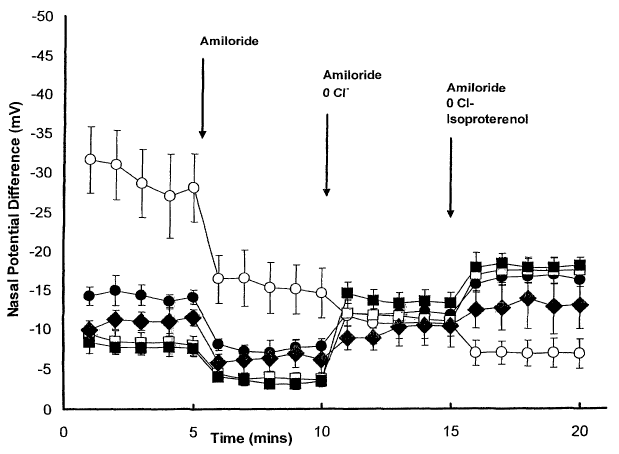
Figure 7. Tracing of transnasal electrical potential (NPD) difference in
normal and CF
mutant mice homozygous for the OF508 mutation. The tracing represents the time
course of the
NPD protocol and the response of NPD readings to perfusion with control Ringer
solution,
13
CA 02464341 2004-04-13
WO 03/049717 PCT/US02/32801
Ringer solution with amiloride, low chloride with amiloride, and the addition
of isoproterenol to
the low chloride solution. For the wild type group CFgroup, n= 4-6 animals.
Legend: open
squares = untreated wild type mice; filled squares = thapsigargin-treated wild
type mice; open
circles = untreated CF mutant mice; filled circles = thapsigargin-treated CF
mutant mice.
Figure 8: Histologic appearance of lung tissue from control and thapsigargin-
treated
wild type mice. Sections of lung tissue from untreated (A) and thapsigargin-
heated (B and C)
mice were stained with hematoxylin and eosin. The scale bar in panel C = 280p.
Figure 9. Immunolocalization of the mutant Delta F508 CFTR protein in
untreated
and curcumin-treated Delta F508 CFTR-expressing CHO cells. CHO cells
expressing Delta
F508 CFTR by transfection were grown to confluence on glass coverslips. Cells
were
exposed to curcumin dissolved in the media (C,D), or were not curcumin-treated
(A,B) and
processed for immunofluorescence using a monoclonal antibody directed against
the CFTR
C-terminus. The images are en face views. The Delta F508 CFTR protein can not
be detected
at the plasma membrane in untreated cells, localizing instead to the ER. The
plasma
membranes of curcumin-treated cells are brightly labeled by the antiCFTR
antibody.
Figure 10. Tracing of transnasal electrical potential (NPD) difference in
normal and
CF mutant mice homozygous for the 4F508 mutation under various treatment
conditions.
The tracing represents the time course of the NPD protocol and the response of
NPD readings
to perfusion with control Ringer solution, Ringer solution with amiloride, low
chloride with
amiloride, and the addition of isoproterenol to the low chloride solution.
Each data point
represents an average of results obtained using groups of animals. Error bars
represent
standard error. Squares represent wild type mice that were either untreated
(open squares) or
thapsigargin treated (closed squares). Circles represent OF508 CFTR mice that
were either
untreated (open circles) or thapsigargin treated (closed circles). Diamonds
represent OF508
CFTR mice treated with nebulized curcumin. N=7 for the wild type untreated and
treated
groups. N=10 for the AF508 CFTR untreated and thapsigargin treated groups. N=6
for the
OF508 CFTR curcumin treated group.
DETAILED DESCRIPTION OF THE INVENTION
14
CA 02464341 2004-04-13
WO 03/049717 PCT/US02/32801
Unless defined otherwise, all technical and scientific terms used herein have
the same
meaning as commonly understood by one of ordinary skill in the art to which
this invention
belongs. Although any methods and materials similar or equivalent to those
described herein can
be used in the practice or testing of the present invention, certain of the
preferred methods and
materials are described.
A. Definitions
Antisense. The term "antisense", as used herein, refers to nucleotide
sequences that are
complementary to a specific DNA or RNA sequence. The term "antisense strand"
is used in
reference to a nucleic acid strand that is complementary to the "sense"
strand. Antisense
molecules may be produced by any method, including synthesis by ligating the
genes) of interest
in a reverse orientation to a viral promoter which permits the synthesis of a
complementary
strand. Once introduced into a cell, this transcribed strand combines with
natural sequences
produced by the cell to form duplexes. These duplexes then block either the
further transcription
or translation. In this manner, mutant phenotypes may be generated. The
designation "negative"
is sometimes used in reference to the antisense strand, and "positive" is
sometimes used in
reference to the sense strand.
Clinical Condition. Any symptom or disorder related to any disease.
Combinatorial Chemistry. "Combinatorial chemistry," as used herein, refers to
the
numerous technologies used to create hundreds or thousands of chemical
compounds, wherein
each of the chemical compounds differ for one or more features, such as their
shape, charge,
and/or hydrophobic characteristics.
Disease. A pathological condition of a cell, body part, an organ, a tissue, or
a system
resulting from various causes, wherein such causes include, but are not
limited to, infections,
genetic defects or environmental stresses.
Mis-assembled. As used herein, "mis-assembled" refers to hetero- or homo-
oligomeric
proteins that have not or can not attain their appropriate or functionally
mature quaternary
structure and/or to hetero- or homo-oligomeric proteins that have a three-
dimensional structure
different to wild type that causes retention in the ER or in an ER-Golgi
compartment.
Mis-folded. As used herein, "mis-folded" refers to proteins that have not or
can not attain
their appropriate or functionally mature tertiary structure and/or to hetero-
or homo-oligomeric
CA 02464341 2004-04-13
WO 03/049717 PCT/US02/32801
proteins that have a three-dimensional structure different to wild type that
causes retention in the
ER or in an ER-Golgi compartment.
Nebulized. As used herein, "nebulized" refers to converting a liquid to a fme
spray. A
medicated spray is one form of the nebulization of a liquid.
Nucleic Acid Sequence. "Nucleic acid sequence," as used herein, refers to an
oligonucleotide, nucleotide, or polynucleotide, and fragments or portions
thereof, and to DNA or
RNA of genomic or synthetic origin which may be single- or double-stranded,
and represents the
sense or antisense strand. Similarly, "amino acid sequence" as used herein
refers to an
oligopeptide, peptide, polypeptide, or protein sequence, and fragments or
portions thereof, and to
naturally occurring or synthetic molecules.
siRNA. A short, intef feYi~ag RNA (siRNA) comprises an RNA duplex that is
approximately 19 basepairs long and optionally further comprises one or two
single-stranded
overhangs or loops. An inventive siRNA may comprise two RNA strands hybridized
together, or may alternatively comprise a single RNA strand that includes a
self hybridizing
portion. When siRNAs utilized in accordance with the present invention include
one or more
free strand ends, it is generally preferred that free 5' ends have phosphate
groups, and free 3'
ends have hydroxyl groups. Inventive siRNAs include a portion that hybridizes
under
stringent conditions with a target transcript. In certain preferred
embodiments of the
invention, one strand of the siRNA (or, the self hybridizing portion of the
siRNA) is precisely
complementary with a region of the target transcript, meaning that the siRNA
hybridizes to
the target transcript without a single mismatch. In most embodiments of the
invention in
which perfect complementarity is not achieved, it is generally preferred that
any mismatches
be located at or near the siRNA termini.
Targeted. An siRNA is considered "targeted" for the purposes described herein
if 1)
the stability of the target gene transcript is reduced in the presence of the
siRNA as compared
with its absence; and/or 2) the siRNA shows at least about 90%, more
preferably at least
about 91%, 92%, 93%, 94%, 95%, 96%, 97%, 98%, 99%, or 100% precise sequence
complementarity with the target transcript for a stretch of at least about 17,
more preferably at
least about 18 or 19 to about 21-23 nucleotides; and/or 3) the siRNA
hybridizes to the target
transcript under stringent conditions appropriately selected for RNA
oligonucleotide
16
CA 02464341 2004-04-13
WO 03/049717 PCT/US02/32801
hybridization to a target sequence.
Treating. As used herein, "treating" includes reversing, alleviating,
inhibiting the
progress of, preventing, or reducing the likelihood of the disease, disorder,
or condition to which
such term applies, or one or more symptoms or manifestations of such disease,
disorder or
condition.
UGGT. As used herein, "UGGT" refers to UDP-Glc:glycoprotein glycosyl
transferase,
also known as UDP glycoprotein glycosyl transferase and as UDP-
glucose:glycoprotein glucosyl
transferase. UGGT is an ER enzyme that attaches glucose to
malformed/improperly folded
glycoproteins, but not to wild type glycoproteins.
B. Elevation of cyclic AMP Levels. As discussed above, CFTR is a CAMP-
dependent
chloride channel. Cyclic AMP is composed of adenosine monophosphate with the
phosphate
group bonded internally to form a cyclic molecule. Cyclic AMP (CAMP) is
generated from
adenosine triphosphate (ATP) by the enzyme adenylcyclase and is active in the
regulation of
gene expression of both prokaryotes and eukaryotes.
Administration of compositions that increase or supplement the cAMP levels of
epithelial
cells has been used in an attempt to activate Cl- conductance to near wild
type levels (U.S. Patent
No. 5,434,086). A preferred compound for increasing cAMP levels is a
phosphodiesterase
inhibitor, such as methylxanthine phosphodiesterase inhibitor.
Phosphodiesterase inhibitors
increase cAMP levels by inhibiting cAMP breakdown. Other examples of
phosphodiesterase
inhibitors include nonspecific inhibitors such as alkylxanthines and cAMP-
specific inhibitors
such as Rolipram (Shearing AG). Preferred alkylxanthines include the
methylxanthines, such as
3-isobutyl-1-methylxanthine (IBMX) and 1,3-dimethylxanthine (theophylline) and
other
xanthines such as papaverine, pentoxifilline and caffeine. For a review of
phosphodiesterase
inhibitors, see Nicholson et al., Trends Pharmacol. Sciences 12:19 (1991) and
Beavo et al.,
Trends Pharmacol. Sciences 11:150 (1990).
Treating OF508-C127 cells and human OF508 airway epithelial cells with a
carboxylic
acid or a carboxylate, such as butyrate (e.g., sodium butyrate), resulted in
the generation of
cAMP-dependent chloride channel activity (LJ.S. Patent No. 5,674,898).
Supplemental CAMP and analogs thereof or beta andrenergic receptor agonists,
such as
isoproterenol and albuterol, can also be used to increase cAMP levels.
17
CA 02464341 2004-04-13
WO 03/049717 PCT/US02/32801
Guanosine monophosphate (GMP) becomes a cyclic molecule by a phosphodiester
bond
between the 3' and 5' atoms. Cyclic GMP (cGMP) acts at the cellular level as a
regulator of
various metabolic processes, possibly as an antagonist to cAMP.
Combination therapy that includes administration of an inhibitor specific for
a cGMP-
inhibited type III cAMP phosphodiesterase, an adenylate cyclase activator, and
a cAMP or a
cAMP analog has also been proposed for treating CF (U.S. Patent No.
5,602,110). Inhibitors
which are specific for a cGMP-inhibited type III cAMP phosphodiesterase
include amrinone,
milrinone, anagrelide, cilostamide and fenoxamine. Adenylate cyclase
activators include
forskolin, cholera toxin and beta-adrenergic receptor agonists.
C. Calcium-ATPase Inhibitors. Correct distribution of Cakz ions within the
cellular
compartments is required for their well-established function as molecular
signals in eukaryotic
cells (Cheek, T. R., Curr. Opin. Cell. Biol. 3:199-205 (1991); Pietrobon et
al., Eur. J. Biochem.
193:599-622 (1990)). ATP-dependent Ca+z uptake from the cytosol to ER lumen is
a prerequisite
for rapid cytostolic signaling through receptor-mediated Ca~z release
(Berndge, M.J., Nature
361:315-325 (1993)).
The ATP-requiring Ca~z transport to the ER lumen is accomplished by a family
of ER
Ca+z ATPases termed SERCA ATPases. Ca'~z-ATPase inhibitors may be
therapeutically useful in
treating CF by improving Cl- secretion in epithelial cells. Proposed Ca+z-
ATPase inhibitors for
use in the present invention, include, but are not limited to, thapsigargin,
cyclopiazonic acid
(CPA), 2,5-di-(tert-butyl)-1,4-hydroquinone (DBHQ) (A.C. Chao et al., J. Clin.
Invest.
96(4):1794-1801 (1995) and U.S. Patent No. 5,384,128), and curcumin.
Thapsigargin is
described in more detail below. CPA is an indole derivative isolated from
liquid culhires of
Pefaicillium cyclopiunz, Aspejgillis flavus and Aspef°gillis versicolor
(Luk et al., Applied and
Environmental Microbiology 211-212 (1977)). DBHQ is a commercially available
non-toxic
synthetic compound chemically unrelated to either thapsigargin or CPA.
Curcumin is described
in further detail below.
Using the CF-derived pancreatic epithelial line CFPAC-l, Chao et al., supra,
found that
DBHQ stimulated'zsI efflux and mobilized intracellular free Ca+z in a dose-
dependent manner.
Pretreatment of monolayers of CFPAC-1 cells with DBHQ for 4-5 minutes
significantly
18
CA 02464341 2004-04-13
WO 03/049717 PCT/US02/32801
increased the Ca+Z-independent or autonomous activity of Ca+Z/calmodulin-
dependent protein
kinase (CaMI~II) assayed in cell homogenates.
D. Opening the ER Ca+2 Channels.
Activators which lower ER Ca+2 by a different mechanism than thapsigargin are
also
encompassed by this invention.
1D-myo-inositol 1,3,4-(or 1,4,5-) triphosphate (IP3), a hydrophilic inositol
phosphate,
induces the intracellular release of Ca+2 stores from the ER through its
specific interactions
with the IP3 receptor (e.g., a calcium channel protein containing an IP3
binding site). Thus,
the present invention also encompasses agents that open ER Ca+Z channels by
acting as IP3
receptor agonists. Adenophostin A is one example of an activator of IP3
receptor activity
(Adkins CE, Wissing F, Potter BV, Taylor CW, Rapid activation and partial
inactivation of
inositol trisphosphate receptors by adenophostin A, Biochem .1., 352 (3): 929-
33, 2000).
A determination of IP3 concentration in cell extracts can be carried out by
means of a
sensitive competitive binding test using an IP3 binding protein, H3 -labeled
IP3 and unlabeled
IP3 (LT.S. Patent No. 5,942,493). An assay kit for this purpose is available
from Amersham
(TRIG 1000) and the determination can be carried out as described in the assay
protocol.
Another calcium channel found in the ER is known as the ryanodine receptor
(RyR).
Mammalian tissues express three different RyR isoforms comprising four 560 kD
(RyR
polypeptide) and four 12 kD (FI~506 binding protein) subunits (reviewed in
Shoshan-Barmatz,
V. and Ashley, R.H., The structure, function, and cellular regulation of
ryanodine-sensitive Ca2+
release channels, IzztRev Cytol, 183: 185-270,1998.) Ryanodine receptors have
been detected in
the lung (Wild, J.S., Giri, S.N., Moore, R., and Pessah, Characterization of
[3H]ryanodine binding
sites in mammalian lung, Arch. Biochezzz Biophys., 379(1):109-18, 2000).
According to the
present invention, treatments that activate or stimulate ryanodine receptors
may be effective in
reducing ER Caz+ concentration in airway epithelial cells. Thus, the present
invention also
encompasses agents that increase or stimulate ryanodine receptors, thereby
increasing Ca2+ exit
from the ER. Such agents include, for example, ryanodine receptor agonists,
compounds that
increase expression of ryanodine receptors, etc. Approaches to modulation of
ryanodine
receptors are discussed in Xu, L., et al., Potential for pharmacology of
ryanodine
receptor/calcium release channels, Azzzz NYAcad Sci, 853: 130-48,1998.
Examples of agents that
19
CA 02464341 2004-04-13
WO 03/049717 PCT/US02/32801
have been shown to increase or stimulate ryanodine receptor activity include,
but are not limited
to, ryanodine (in particular concentrations known in the art) and related
plant allcaloids,
xanthines, 4-Chloro-m-cresol, suramin, and ditalis glycosides. Such agents,
and derivatives
thereof (e.g., pharmaceutically acceptable derivatives), may be used in the
practice of the
invention.
E. Temperature-Dependent Delivery of the Mutant CFTR to the Plasma
Membrane.
Experiments with 3T3 fibroblast cells and C 127 cells grown at lower
temperatures for a
period of time have shown a shift in the glycosylation pattern of OF508 CFTR
towards a more
mature CFTR protein. Normal CFTR protein appears to be unaffected by the lower
temperature.
It has been hypothesized that at reduced temperatures there is an increased
flux of the mutant
protein through the Golgi complex. Thus, it has been suggested that exposing a
patient's lung
epithelia to a temperature below normal body temperature for a period of time
might mobilize
mutant CFTR to the plasma membrane of the lung epithelial cells, where the
mutant CFTR can
mediate chloride transport (LJ.S. Patent No. 5,674,898). One hypothetical
method involves
implanting in the patient's lung a non-toxic, non-immunogenic agent which
lowers the
temperature in the vicinity of the lung so that it is below normal body
temperature.
F. Puriner~ic Receptors and CI- Secretion
Purinergic receptors play an important role in regulating Cl- secretion in
epithelial cells.
moue et al. (Am. J. Physiol. Cell Physiol. 272(6):41-46 (1997)) assayed the
human intestinal
epithelial cell line, Caco-2, for Cl- secretion by measuring the short-circuit
current. The
researchers found that responses to purinergic receptor agonists were
inhibited by pretreatment
with 1,2-bis(2-aminophenoxy)ethane-N,N,N',N'-tetraacetic acid-acetoxymethyl
ester,
thapsigargin or quinine.
G. CF and UDP-Glucose:Glycoprotein Glycosyl Transferase
As discussed above, the primary lesion in cystic fibrosis is associated with
mutations in
the gene encoding the CFTR which prevent it from functioning as a chloride
channel at the apical
surfaces of airway epithelial cells. The most common mutation (OF'S08), which
occurs in 67.2%
of cystic fibrosis patients, results in the synthesis of a CFTR protein which
is unable to fold
CA 02464341 2004-04-13
WO 03/049717 PCT/US02/32801
correctly and assume its appropriate tertiary conformation. Consequently, the
protein is retained
in the ER by the ER's "quality control" machinery. Several other CFTR
mutations also result in
mis-folding and ER retention.
Both nascent a 1-antitrypsin and nascent CFTR form transient associations with
calnexin
(also designated as p88 or IP90), a calcium-binding protein of the ER
membrane. Since calnexin
functions as a molecular chaperone for glycoproteins and interacts with
monoglucosylated
oligosaccharides, reglucosylation may function to initiate assembly between
unfolded
glycoproteins and the molecular chaperone (Hammond et al., Proc. Natl. Acad.
Sci. U.S.A.
91:913-917 (1994)).
The UGGT Protein and Gene. UGGT was found to have an apparent monomeric M~ of
150 kDa following isolation and purification from rat liver microsomes
(Trombetta et al., J.
Biol. Chem. 267:9236-9240 (1992)). The soluble,170 kDa UGGT isolated from
Drosophila has
an amino acid sequence of 1548 amino acids beginning with a signal peptide and
terminating in a
potential ER retrieval signal, HGEL (C.G. Parker et al., EMBO J. 14(7):1294-
1303 (1995)). The
amino acid sequence was found to lack any putative transmembrane domains. The
gene coding
for UGGT, designated as gptl, has also been identified in Schizosaccharornyces
ponabe
(Fernandez et al., EMBO J. 15(4):705-13 (1996)). This gene codes for a
polypeptide having a
signal peptide of 18 amino acids followed by 1429 amino acids with no
transmembrane domain
and a C-terminal tetrapeptide designated PDEL.
Functional Role of UGGT. UGGT adds glucose from UDP-glucose to high mannose
glycoproteins in the presence of Caz+ ions and the resulting glucosylated
oligosaccharide has the
same structure as the processed intermediate, GlclMan9GlcNAcz (Trombetta et
al., Biochemistry
28:8108-8116 (1989)). Unfolded, denatured glycoproteins are substantially
better substrates for
glycosylation by the enzyme than are the corresponding native proteins.
Proteins that fail to fold properly are retained in the ER (or in an ER-Golgi
intermediate
compartment), where they are proteolytically degraded. UGGT is proposed to be
involved in the
quality control of glycoprotein folding in the ER (Parker et al., supra;
Fernandez et al., supra; M.
C. Sousa and A. J. Parodi, The interaction of UDP-Glc:Glycoprotein Glucosyl
transferase with
the acceptor glycoprotein, Cellular and Molecular Biology 42: 609-616 (1996);
Sousa MC and
Parodi AJ., The molecular basis for the recognition of mis-folded
glycoproteins by the UDP-Glc:
21
CA 02464341 2004-04-13
WO 03/049717 PCT/US02/32801
Glycoprotein Glucosyl transferase, EMBO J 14: 4196-4203 (1995)).
UGGTparticipates together
with lectin-like chaperones that recognize monoglucosylated oligosaccharides
in the control
mechanism by which cells only allow passage of properly folded glycoproteins
to the Golgi
apparatus (Labriola et al., J. Cell Biol. 130(4):771-9 (1995)).
Cycles of transient interaction with UGGT, each resulting in reglucosylation
of attached
oligosaccharides, is believed to facilitate interaction between unfolded
glycoproteins and
calnexin and ensure the intracellular retention of improperly folded
glycoproteins in the ER.
Calnexin binds to glucose residues which are exposed on the N-linked sugar
chains of membrane
proteins.
It has been shown that UGGT requires millimolar calcium concentrations for
optimal
activity (Trombetta and Parodi, 1992). In cells expressing wild type al-
antitrypsin, treatment
with thapsigargin retards or prevents the secretion of the protein (Kuznetsov
et al., 1993; Lodish
and Kong, 1990). This is apparently due to stable association of the newly
synthesized a,l-
antitrypsin with UGGT in the endoplasmic reticulum when calcium levels in the
ER are reduced
(Choudhury et al., 1997). It has also been shown that lowering ER calcium
through application
of thapsigargin or calcium ionophores retards the exit of numerous wild type
proteins from the
ER and increases their rate of degradation (Wilkstrom and Lodish, 1993;
Sudbeck et al., 1997;
van Weering et al. 1998; Clark et al., 1994; Wong et al., 1993; Wileman et
al., 1991; Lodish et
al., 1992; Lodish and Kong, 1990). While not wishing to be bound by any
theory, it may be the
case that if the UGGT enzyme is denied calcium, it binds tightly to its
substrates (i.e. newly
synthesized glycoproteins) but is unable to release them, perhaps because
successful completion
of the glucose transfer step is required to effect release. Of course
retention of misfolded proteins
may occur through any of a number of other mechanisms.
It is interesting to speculate why, in the case of al-antitrypsin,
thapsigargin retards
protein exit from the ER, whereas in the case of ~F508 CFTR exit from the ER
is stimulated by
this drug (see Examples 1-8). Without wishing to be bound by any theory, we
propose that in
cells expressing a mutant protein that is incapable of proper folding, mis-
folded protein is present
in the ER in quantities which constitute a large molar excess over the
resident quantity of UGGT.
Under normal circumstances, the mis-folded protein binds to UGGT, undergoes
addition of a
glucose residue and is rapidly released (Hammond and Helenius, 1995). The
glucosylated
22
CA 02464341 2004-04-13
WO 03/049717 PCT/US02/32801
protein is retained in the ER via interactions with calnexin, and a sufficient
pool of UGGT is
available to interact with mis-folded proteins that have lost their glucose
tag. When ER calcium
is depleted, each molecule of UGGT becomes stably complexed with a mis-folded
protein, and
thus unavailable to interact with the remaining mis-folded proteins in the ER.
Since the mis-
folded proteins are present in large molar excess over the UGGT, the excess
mis-folded protein is
free to escape the UGGT-mediated quality control system and to exit the ER. In
contrast, in cells
that do not express a mutant mis-folded protein, we hypothesize that UGGT
exists in large molar
excess over its potential substrates. Thus, when ER calcium is depleted, UGGT
may act as a sink
that can bind up newly synthesized proteins that have not completed their
folding. Consequently,
the bulk of newly synthesized proteins are retained in the ER.
H. Release of Mis-folded t~F508 CFTR Protein From the ER.
We have developed a novel strategy that releases mis-folded 4F508 CFTR protein
from
the ER and allows it to be functionally expressed at the cell surface. While
not wishing to be
bound by any theory, it is believed that retention of mis-folded membrane
proteins in the ER is
dependent upon interactions with ER resident chaperone proteins. Biochemical
characterization
of chaperone activity reveals that optimal functioning of several of these
proteins requires
calcium concentrations in the millimolar range (S.I~. Nigam, A.L. Goldberg, S.
Ho, M.F. Rohde,
K.T. Bush, M.Y. Sherman, J. Biol. ChenZ. 269,1744, 1994; S.E. Trombetta, A.J.
Parodi, J. Biol.
Cher~z. 267, 9236, 1992). Mobilization of sequestered ER Caz+ stores with
agents such as the ER
Ca2+ pump inhibitor thapsigargin dramatically reduces the ER lumenal calcium
concentration (M.
Montero, J. Alvarez, W.J.J. Scheenen, R. Rizzuto, J. Meldolesi, T., Pozzan,
.l. Cell Biol. 139,
601, 1997). While not wishing to be bound by any theory, we postulate that
exposing cells to
thapsigargin might interfere with the capacity of chaperones to mediate the ER
retention of mis-
folded proteins and that depleting ER Caz+ stores with thapsigargin would
allow the mis-folded
~iF508 CFTR protein to "escape" from the ER and potentially reach the cell
surface, where it
would be able to function as a chloride channel and correct the CF defect.
As described in the Examples, we have shown that treatment of CF airway
epithelial
cells with thapsigargin, which reduces the calcium concentration in the ER
lumen, leads to
functional expression of the OF508-CFTR protein at the cell surface as
revealed by
electrophysiologic and immunofluorescence analysis. In addition, we have shown
that
23
CA 02464341 2004-04-13
WO 03/049717 PCT/US02/32801
treatment with thapsigargin can induce reversal of a phenotypic defect in a
mouse model for
cystic fibrosis (CF mice). The dose of thapsigargin employed in these studies
appears to be
tolerable and induces an effect whose magnitude is probably sufficient to
produce clinically
significant improvements in airway epithelial function in cystic fibrosis
patients.
Finally, it must be noted that the mechanism through which calcium pump
inhibitors
effect the release of OF508 CFTR from the ER may not be related directly to
the calcium
requirements of ER chaperone machinery. It is possible, for example, that
depletion of calcium
from the ER lumen is sufficient to facilitate the spontaneous folding of the
~F508 CFTR protein,
permitting it to acquire a stable conformation and bypass chaperone retention.
In either case, it is
clear that calcium pump inhibition is sufficient to release a cohort of ER-
retained OF508 CFTR
to the cell surface, where it can function appropriately (see Examples 1-8).
I. Rhinosinusitis and CFTR Mutations
Rhinosinusitis, or inflammation of the sinus epithelium, is an exhemely common
condition which can be divided into several subtypes including acute,
recurrent acute, subacute,
and chronic based typically on patient history and physical examination. The
persistent form,
chronic rhinosinusitis (CRS), affects approximately 14% of the U.S. population
and is almost
invariably present in patients with CF. A case-control study in which DNA of
CRS patients
(individuals with more than 8 weeks of nasal or sinus symptoms or with a
history of at least 4
episodes of recurrent symptoms of greater than three weeks' duration in the
prior 12 months) and
controls was typed for 16 mutations that account for 85% of CF alleles in the
general population
and also tested for the presence of additional mutations and variants revealed
that the proportion
of CRS patients who were found to have a CF mutation in one of their copies of
the CF gene
(7%) was significantly higher than in the control group (2%) (Wang, X., et al.
"Mutation in the
Gene Responsible for Cystic Fibrosis and Predisposition to Chronic
Rhinosinusitis in the General
Population", JAMA, Vol. 284, No. 14, 2000). Approximately 90% of the patients
with a CF
mutation carried the ~F508 allele. In addition, most of the CF carriers with
CRS had variants in
their other CFTR gene. In particular, the M470V variant was found in 9 of the
10 CRS patients
with a CF mutation, and in 8 of these patients the M470V variant was in the
gene that did not
carry a CF-causing mutation. The variant with valine at amino acid position
470 has reduced
chloride channel activity compared with that having methionine at position 470
although the
24
CA 02464341 2004-04-13
WO 03/049717 PCT/US02/32801
reduction in activity is not generally sufficient to result in CF, the
diagnosis of which is based in
part on clinical criteria. Data from this study indicate that mutations in the
CFTR gene may be
associated with the development of CRS in the general population. The
importance of CFTR in
normal sinus epithelium function is evident from the fact that CRS occurs in
almost all CF
patients. Less severe decreases in CFTR activity, as may occur in individuals
that are
heterozygous for a CF mutation (particularly if they also have a variant CFTR
allele at the other
locus), may lead to CRS in the absence of CF. While not wishing to be bound by
any theory,
reduced CFTR activity may lead to abnormal viscosity and electrolyte
composition of sinus
secretions. Such abnormalities may increase the likelihood that rhinosinusitis
will develop
initially and/or that it will become chronic. These findings suggest that
agents such as those
described herein, which increase the functional activity of mutant CFTR, may
be useful for
prophylaxis and/or treatment of CRS.
It is noted that diagnosis of sinusitis is based at least in part on clinical
criteria, and that
various classification schemes may be applied (See, e.g., International
Rhinosinusitis Advisory
Board. "Infectious rhinosinusitis in adults: classification, etiology and
management." Ear; Nose,
Ths~oat J: 76(12 supply: l-22). Determinations of whether a given patient
suffers from a particular
subtype may vary, and it is likely that certain individuals suffering from
rhinosinusitis who carry
a CF allele and/or CF variant will not be classified as having CRS but rather
as having one of the
other subtypes. Thus the agents described herein may also be useful for
treatment or prophylaxis
in individuals who suffer from rhinosinusitis that has not been classified as
chronic
rhinosinusitis. Such agents would be particularly appropriate for patients
with rhinosinusitis who
are CF carriers, patients who are CF Garners and have a CFTR variant at the
second locus, and
patients who are homozygous for a CFTR variant. As is well known in the art,
patients who are
CF Garners and/or have a CFTR variant may be identified by DNA analysis as
described, for
example, in Wang, X., et al. Thus the present invention provides a method for
treating
rhinosinusitis comprising administering an agent that permits the release of
proteins from the
endoplasmic reticulum. In certain embodiments of the invention the method
further comprises
providing an individual suffering from rhinosinusitis, e.g., from chronic
rhinosinusitis. In certain
embodiments of the invention such individual carnes a CF mutation, e.g.,
~F508. In certain
embodiments of the invention the individual carnes a CF variant, e.g., M470V.
CA 02464341 2004-04-13
WO 03/049717 PCT/US02/32801
In certain embodiments of the invention the method comprises administering an
agent
that permits the release of proteins from the endoplasmic reticulum, an agent
that
decreases or inhibits the activity of UDP glucose:glycoprotein glycosyl
transferase, an agent that
decreases or inhibits activity of the endoplasmic reticulum Cap ATPase, an
agent that lowers the
concentration of Ca++ in the endoplasmic reticulum, an agent that causes
release of Cap from the
ER, an agent that stimulates or increases IP3 receptor activity, an agent that
decreases or inhibits
calnexin functional activity, or an agent that increases or activates
ryanodine receptor activity.
Particular agents that may be used in the practice of the invention include
thapsigargin or a
derivative thereof, cyclopiazonic acid or a derivative thereof, DBHQ or a
derivative thereof, and
halothane or a derivative thereof.
In certain embodiments of the invention the agent is delivered intranasally
according to
methods well known in the art and widely used for treatment of allergies, etc.
Of course the
agent can be delivered by various other means as well.
Applications for release of normally assembled or folded proteins from the ER
As described above, the present invention contemplates enhancing release of
misassembled and/or misfolded proteins from the ER. According to certain
embodiments of
the invention release is enhanced by lowering the Caz+ concentration within
the ER lumen.
While not wishing to be bound by any theory, it is possible that lowering the
ER Caz
concentration may alter or interfere with the activity of chaperone proteins
that would
otherwise bind to a misassembled or misfolded protein and prevent its release
from the ER.
The interaction of normal and mutant proteins with various ER chaperones is a
subject
of ongoing investigation. For example, in the case of CFTR it appears that the
protein
interacts with at least two ER chaperones, heat shock protein 90 (hsp90) and
heat shock
cognate 70 (hsc70) (refs). In a manner that is not yet fully understood and
which depends at
least in part on the primary sequence of the newly synthesized CFTR protein
(e.g., whether it
is wild type or mutant), these interactions ultimately lead to release of the
protein from the
ER, retention of the protein in the ER, and/or ubiquitination of the protein
and ultimately
ubiquitin-dependent degradation by the proteasome (refs). Only approximately
25% of the
wild type CFTR protein attains a stable conformation (stable B) that allows it
to exit the ER,
26
CA 02464341 2004-04-13
WO 03/049717 PCT/US02/32801
while the remainder is ubiquitinated in the ER and thereby targeted for
degradation. In the
case of folding mutants an even smaller fraction of the protein reaches the
stable B form.
Very little if any ~F508 CFTR protein reaches stable B, and thus essentially
all the protein is
ubiquitinated and degraded. While not wishing to be bound by any theory, it is
possible that
association with chaperones is involved both in proper folding of CFTR protein
and in
allowing ubiquitination of both normal and mutant CFTR. Thus it is possible
that an agent
that alters or interferes with chaperone activity may lead to decreased
ubiquitination of wild
type CFTR and thereby allow a greater amount of wild type CFTR to exit the ER.
In the case
of an individual who carries one wild type allele of the CFTR gene and one
allele that
encodes a misfolded CFTR protein, it is possible that treatment with such an
agent would
lead to increased cell surface expression of wild type CFTR, thus compensating
for any
decrease in cell surface expression resulting from the mutation.
It is therefore contemplated that the compositions and methods of the present
invention may be useful not only to increase release of misassembled and/or
misfolded
proteins from the ER but also to increase release of wild type proteins from
the ER,
particularly in cases where a large fraction of the wild type protein is not
released (as is the
case for the normal CFTR protein). The compositions and methods may similarly
be useful
to increase release of mutant proteins from the ER even in cases in which the
mutant proteins
are not necessarily misassembled and/or misfolded.
Thus the compositions and methods of the invention may be used to treat
individuals
suffering from a condition associated with misassembly or misfolding of a
protein, in whom
one copy of a particular gene associated with the condition encodes a
misassembled or
misfolded protein while the other copy encodes a wild type protein or a mutant
protein where
the mutation does not result in misassembly or misfolding but instead results
in a protein that
functions at less than wild type levels for some other reason. As described
above, such
individuals may include individuals with rhinosinusitis, where the individuals
have a
mutation in at least one copy of the CFTR gene, regardless of whether the
mutation results in
synthesis of a misfolded protein. Such individuals also include individuals
suffering from
CF, where the individuals have different mutations in their two copies of the
CFTR gene,
only one of which results in production of a misfolded protein.
27
CA 02464341 2004-04-13
WO 03/049717 PCT/US02/32801
J. Applications For Non-CF Protein Release
In addition to CF, a large and growing list of disease states is associated
with protein
retention in the ER (Amara J, Cheng S and Smith A., Trends in Cell Biol 2:145-
149 (1992);
Bychkova V and Ptitsyn O, Folding intermediates are involved in genetic
diseases?, FEBS Lett
359:6-8 (1995)). Several are listed and briefly discussed below.
al-antitrypsin Deficiency. The al-antitrypsin protein is synthesized in the
liver and
secreted into the circulation. It serves to prevent damage to the lungs
induced by inflammatory
processes. Absence of this protein leads to pulmonary scarring and emphysema.
In the most
common forms of human al-antitrypsin deficiency, a mutation leads to the
synthesis of an al-
antitrypsin molecule which can not fold properly and is consequently not
secreted but rather is
retained in the liver cell ER (Yu M, Lee K and I~im J, The Z type variation of
human alpha 1-
antitrypsin causes a protein folding defect, Nature Structural Biology 2:363-
367 (1995)).
Paroxysmal Nocturnal Hemoglobinuria. In red blood cells, the inventory of
glycosylphosphatidylinositol (GPI) linked proteins includes a pair of
polypeptides, Decay
Accelerating Factor (DAF) and CD59, which help to protect the erythrocytes
from being
accidentally injured by complement-mediated cell lysis. One of the proteins
which participates in
the synthesis of the GPI anchor is a sugar transferase encoded by the PIG-A
gene
(phospatidylinositol glycan-class ~. This gene is located on the X chromosome.
In Paroxysmal
Nocturnal Hemoglobinuria, a spontaneous mutation occurs in the PIG-A gene in
just one of the
many precursor cells which give rise to erythrocytes. All of the erythrocytes
which arise frolll
this particular precursor, therefore, are deficient in GPI-linked protein
synthesis. The
transmembrane precursors of the GPI-linked proteins are retained in the ER and
degraded.
Consequently, these cells lack DAF and CD59 expression and are susceptible to
complement
attack and lysis. Patients with Paroxysmal Nocturnal Hemoglobinuria are likely
to become
anemic and can suffer life threatening disorders of clotting and bone marrow
function. A
treatment which liberated the transmembrane precursors of GPI-linked proteins
from the ER and
allowed them to travel to the cell surface might prevent or ameliorate the
symptoms of this
disease.
Familial Hypercholesterolemia. The disease known as Familial
Hypercholesterolemia
(FHC) is caused by a defect in the gene encoding the low density lipoprotein
(LDL) receptor
28
CA 02464341 2004-04-13
WO 03/049717 PCT/US02/32801
which results in the synthesis of receptors that can not internalize LDL from
the cell surface
(Goldstein et al., Receptor-Mediated Endocytosis: Concepts Emerging from the
LDL Receptor
System, Ann. Rev. Cell Biol. 1, 1-39 (1985)). In the absence of functional LDL
receptors, cells
are unable to import exogenous cholesterol. Even though serum cholesterol
levels rise to
extraordinarily high levels, cells are unaware of its presence since they lack
the machinery that
allows them to endocytose LDL. The excess cholesterol synthesis results in the
build up of
cholesterol-filled lipid droplets in cells throughout the body. Accumulation
of these cholesterol
inclusions in the smooth muscle cells that populate arterial walls produces
atherosclerotic
plaques, which can go on to occupy and occlude the lumens of the blood vessels
themselves. A
subset of the mutations in the gene encoding the LDL receptor which lead to
FHC in humans (the
class II mutations) lead to the synthesis of LDL receptors which can not fold
properly and which
are retained in the ER (Yamamoto et al., Deletion in cysteine-rich region of
LDL receptor
impedes transport to cell surface in WHHL rabbit, Science 232:1230-1237,
1986).
Consequently, they can not participate in the internalization of plasma LDL-
bound cholesterol.
Pharmacologic treatments which liberate these mis-folded LDL receptors from
the ER and
allowed them to proceed to the cell surface might allow them to function
properly in cholesterol
metabolism and prevent the formation of atherosclerotic plaques.
Tay-Saclis Disease. A number of human diseases have been traced to genetic
deficiencies in specific lysosomal hydrolases (Griffiths et al., The Mamzose-6-
Phosphate
Receptor and the Biogenesis of Lysosomes, Cell 52:329-341 (1988)). Children
who suffer from
Tay-Sachs disease, for example, carry a homozygous mutation in the gene
encoding the
lysosomal enzyme hexosaminidase A. Consequently, their lysos'omes are unable
to degrade
substances containing certain specific sugar linkages. Since they can not be
broken down, these
substances accumulate in lysosomes. Over time they come to fill the lysosomes,
which swell and
crowd the cytoplasm. The resulting derangements of cellular function are toxic
to a number of
cell types and ultimately underlie this disease's uniform fatality within the
first few years of life.
At least one mutation which has been shown to induce Tay-Sachs disease leads
to deletion of the
last 22 amino acids of the protein, preventing its proper folding (Lau MMH and
Neufeld EF, A
frameshift mutation in a patient with Tay-Sachs disease causes premature
termination and
defective intracellular transport of the alpha-subunit of beta-hexosaminidase,
J Biol Chem
29
CA 02464341 2004-04-13
WO 03/049717 PCT/US02/32801
264:21376-213 80 (1989)). The mutant protein is retained in the ER and does
not travel to its site
of functional residence in the lysosome. Releasing this protein from the ER
might prevent the
Tay-Sachs pathology in patients who carry this allele.
Immune surveillance of tumors and virally infected cells. In order for the
immune
system to detect and destroy tumor cells and virally infected cells, these
target cells must present
peptide fragments derived from tumor or viral antigens at their cell surfaces
in association with
MHC class I molecules. These peptide fragments are derived from proteasome-
mediated
digestion of the foreign antigens followed by TAP-mediated transport of these
fragments into the
lumen of the ER, where they can assemble with MHC class I and ~i2-
microglobulin to form the
mature MHC complex. Only the mature, peptide-containing MHC complex can depart
the ER
and be transported to the cell surface. In the absence of peptides in the
lumen of the ER, the
incompletely assembled MHC I-(32-microglobulin complex is retained in the ER
through
interactions with calnexin.
Several viruses and tumors avoid immune detection by blocking the surface
expression of
the mature MHC class I complex. The herpes simplex virus induces host cells to
synthesize the
ICP47 protein, which directly inhibits the TAP transporter (Hughes E, Hammond
C and
Cresswell P, Mis-folded maj or histocompatibility complex class I heavy chains
are translocated
into the cytoplasm and degraded by the proteasome, PNAS 94:1896-1901 (1997)).
In a number
of tumors, expression of the genes encoding the two polypeptides which
constitute the TAP
transporter is lost (Pogador et al., Natural killer cell lines kill autologous
[32-microglobulin-deficient melanoma cells: Implications for cancer
immunotherapy, PNAS
94:13140-13145 (1997)). Consequently, the immune system is unable to respond
adequately to
the pathologic condition. To assist the immune system in recognizing and
destroying virally
infected or transformed cells, it might be desirable to release the peptide-
free MHC class
I-(32-microglobulin complex from calnexin-mediated ER retention. This complex
would then
travel to the cell surface, where it could associate with a specific peptide,
administered to the
patient by infusion and chosen to maximize the immunogenicity of the resulting
peptide-MHC-class I-(32-microglobulin complex. Thus, drugs which release mis-
assembled
proteins from the ER might prove efficacious in the treatment of a variety of
viral and neoplastic
diseases.
CA 02464341 2004-04-13
WO 03/049717 PCT/US02/32801
Hereditary Myeloperoxidase Deficiency. Phagocytes, in particular neutrophils,
respond
to stimulation with a burst of oxygen consumption. The oxygen consumed is
converted to
hydrogen peroxide by myeloperoxidase (MPO), which is released from the
neutrophil granules,
and a complex is formed that is capable of oxidizing a large variety of
substances, and that has,
as a result, important anti-microbial properties (Klebanoff, Myeloperoxidase,
Proc. Assoc. Alll.
Physicians, 111(5):383-389, 1999).
In the endoplasmic reticulum, MPO precursors interact transiently with
calrecticulin and
calnexin, presumably as molecular chaperones. MPO deficiency is a relatively
common disorder,
and several missense mutations have been identified where the mutant precursor
is retained in the
endoplasmic reticulum due to prolonged binding to calnexin. The mis-folded
protein is
eventually degraded (Nauseef, Quality CoyZtf°ol in the Endoplasmic
Reticulmn: Lessons f-ona
Hef°editar~ Myeloperoxidase Deficiency, J. Lab. Clin. Med., 134(3):215-
221 (1999)). Here as
well, a treatment that would allow the protein to exit the ER might restore
anti-bacterial
phagocytic function to individuals suffering from MPO deficiency.
Congenital Insulin Resistance. The hormone binding site of the insulin
receptor is
contained in the extracellular region of the protein. In this form of type A
insulin resistance,
substitution mutations of residues located in the beta-sheet and at the
hormone-binding region
completely disrupt intracellular folding and movement of the protein,
resulting in aberrant
retention at an incorrect cellular location.
Misfolded receptors remain bound to calnexin molecules in the endoplasmic
reticulum
until they are degraded. As previously discussed in connection with other
diseases, a treatment
providing release and cellular export of the mutant receptor could have wide-
spread therapeutic
use.
Nephrogenic Diabetes Insipidus. Nephrogenic diabetes insipidus is
characterized by an
inability to concentrate urine in spite of normal or increased plasma
concentrations of the
antidiuretic hormone arginine vasopression (AVP), which normally stimulates
water reabsorbtion
in the distal tubules and/or collecting ducts of the kidney by regulating the
expression of "water
channels" known as aquaporins. In the collecting duct, binding of AVP to the
vasopressin 2
receptor triggers a cascade -- activation of the receptor-linked G protein G5,
activation of
adenylate cyclase, and stimulation of protein kinase A, eventually leading to
exocytic insertion of
31
CA 02464341 2004-04-13
WO 03/049717 PCT/US02/32801
specific water channels, aquaporin 2, into the luminal membrane of collecting
duct cells.
Presence of these chamlels increases permeability of the luminal membrane.
Thus short terns
regulation of AQP2 by AVP entails movement of AQP2 from intracellular vesicles
to the plasma
membrane. Longer term regulation occurs through increased abundance of APQ2,
which is
thought to result from increased transcription of the AQP2gene. AVP also
increases renal water
reabsorption through a variety of additional mechanisms. Nephrogenic diabetes
insipidus is
comprehensively reviewed in Morello, J. and Bichet, D., Nephrogenic diabetes
insipidus, Anrazc.
Rev. Pla~siol., 63:607-30, 2001.
Nephrogenic diabetes insipidus can be inherited or acquired. Polyuria and
polydipsia
are the major symptoms. Approximately 90% of patients with congenital
nephrogenic
diabetes insipidus have an X-linked forni of the disorder caused by mutations
in the arginine
vasopressin receptor 2 gene (AYPR2). In less than 10% of families studied the
disorder has
an autosomal recessive or autosomal dominant pattern of inheritance. Mutations
in the
aquaporin-2 gene (AQP2) have been identified in some of these kindreds. Based
on studies
of glycosylation patterns, it is apparent that most AVPR2 mutations lead to
receptors that are
trapped in a pre-Golgi compartment, presumably the ER, and are thus unable to
reach the cell
surface (See Morello and Bichet, 2001 and papers referenced therein). AQP-2
mutations that
cause autosomal recessive nephrogenic diabetes insipidus are also
characterized by misfolded
mutant proteins that are trapped in the ER (Kamsteeg, E.J., et al., An
impaired routing of
wild-type aquaporin-2 after tetramerization with an aquaporin-2 mutant
explains dominant
nephrogenic diabetes insipidus; reviewed in van Os, C.H. and Deen, P.M.,
Aquaporin-2 water
channel mutations causing nephrogenic diabetes insipidus, Pnoc. Assoc. AJn.
Plzysicianas,
110(5): 395-400, 1998). Thus agents and methods such as those described
herein, that allow
release of misfolded proteins from the ER, are likely to be useful in the
treatment of
congenital nephrogenic diabetes.
Hereditary Hemochromatosis. Hemochromatosis is a common autosomal recessive
disorder characterized by excessive accumulation of iron in many organs and
tissues
including the liver, pancreas, heart, joints, and endocrine organs due to
increased absorption
of iron in the gastrointestinal tract. Clinical consequences includes
cirrhosis of the liver,
hepatocellular carcinoma, diabetes, heart failure, arthritis, and
hypogonadism. A large
32
CA 02464341 2004-04-13
WO 03/049717 PCT/US02/32801
number of studies have indicated that hereditary hemochromatosis (HH) is
caused by
mutations in a gene that encodes a novel member of the major
histocompatibility complex
class I family initially called HLA-H but now designated as HFE (See, e.g.,
Feder, J.N., et al.,
Nature Geiaetics, 13: 339-408, 1996; Beutler, E., et al., Blood Cells Mol.
Dis., 22: 187-194,
1996). Most patients with HH are homozygous for the same missense mutation
(C282Y) in
the gene that encodes HFE. A recent study demonstrated that the C282Y mutant
protein is
retained in the ER and middle Golgi compartment and is subject to accelerated
degradation
(Waheed, A., et al., Hereditary hemochromatosis: Effects of C282Y and H63D
mutations on
association with [32-microglobulin, intracellular processing, and cell surface
expression of the
HFE protein in COS-7 cells, Proc. Natl. Acad. Sei., 94: 12384-12389, 1997).
Much of the
newly synthesized C282Y mutant HFE protein occurs in a high molecular weight
aggregate
as is characteristic of misfolded proteins that are retained in the ER or
Golgi. The C282Y
mutation reduces or prevents association of HFE with [32-microglobulin, which
is necessary
for normal intracellular transport of HFE and delivery to the cell surface.
Thus agents, such as
those described herein, that increase or stimulate the release of misfolded
proteins from the
ER may be useful in the prevention or treatment of HH by allowing mutant HFE
to exit the
ER and reach the cell surface.
Gitelman's Syndrome. Gitelman's syndrome is an autosomal recessive disorder
characterized by salt wasting and hypokalemia and is caused by mutations in
the thiazide
sensitive Na-Cl cotransporter (NCC), which is normally expressed in the
mammalian kidney
at the apical membrane of distal convoluted tubule cells (See, e.g., Simon,
D.B., et al.,
Gitelman's variant of Banter's syndrome, inherited hypokalemic alkalosis, is
caused by
mutations in the thiazide-sensitive Na-Cl cotransporter, Nat. Gefaet., 12: 24-
30, 1996). In a
recent study designed to elucidate the pathogenesis of Gitelman's syndrome,
eight mutations
corresponding to eight disease-causing mutations found in Gitelman's syndrome
patients were
introduced into the mouse NCC and studied by functional expression in Xenopus
oocytes
(Kunchaparty, S., et al., Defective processing and expression of thiazide-
sensitive Na-Cl
cotransporter as a cause of Gitelman's syndrome, Afn .l Playsiol., Oct., 277
(4 Pt 2):F643-9,
1999). Results indicated that a number of the mutations interfere with proper
processing and
insertion into the plasma membrane. The nearly complete absence of
glycosylation argues
33
CA 02464341 2004-04-13
WO 03/049717 PCT/US02/32801
that the mutant proteins do not exit the ER. The results suggest that at least
a subset of
Gitelman's mutations, including the most common mutation (G738R), lead to
production of
proteins that are not glycosylated normally because of misfolding during
synthesis. Thus
agents, such as those described herein, that increase or stimulate the release
of misfolded
proteins from the ER may be useful in the prevention or treatment of
Gitelman's syndrome by
allowing mutant NCC to exit the ER and reach the cell surface.
Cystinuria. Cystinuria is a common inherited disorder characterized by
defective
transport of cystine and dibasic amino acids through the epithelial cells of
the renal tubule
and gastrointestinal tract, commonly resulting in the development of cystine
calculi (stones)
in the kidney. Three types of cystinuria have been described. Mutations in
SLC3A1, a gene
encoding a subunit of the rBAT protein (an amino acid transporter), have been
shown to
cause Type I cystinuria. In a recent study designed to investigate the
pathogenesis of Type I
cystinuria, the most common point mutation, M467T and the related mutation
M467K were
introduced into rBAT and studied by functional expression in Xe~zopus oocytes
(Chillaron, J.,
et al., An Intracellular Trafficking Defect in Type I rBAT Mutants M476T and
M467K, J.
Biol. Ch.em., 272(14), 9543-9549, 1997). The study indicated that the
mutations interfered
with proper intracellular processing and transport to the plasma membrane.
Unlike wild type
rBAT, the mutant proteins were primarily located in an intracellular
compartment, most
likely the ER. Evidence also suggested that, if able to reach the cell
surface, as is the case if
the experimental system is saturated with cDNA encoding the mutant, the mutant
proteins are
functional. As for the other mutant proteins described herein, it is likely
that mutations in
rBAT lead to rnisfolding and retention in the ER. Thus agents, such as those
described
herein, that increase or stimulate the release of misfolded proteins from the
ER may be useful
in the prevention or treatment of Type I cystinuria (and possibly other forms
of cystinuria that
may involve rBAT) by allowing mutant rBAT to exit the ER and reach the cell
surface.
With respect to the disorders and conditions discussed above, in certain
embodiments of
the invention the method for treatment and/or prevention or prophylaxis
comprises administering
an agent that permits the release of proteins from the endoplasmic reticulum,
an agent that
decreases or inhibits the activity of UDP glucose: glycoprotein glycosyl
transferase, an agent that
decreases or inhibits activity of the endoplasmic reticulum Ca+~ ATPase, an
agent that lowers the
34
CA 02464341 2004-04-13
WO 03/049717 PCT/US02/32801
concentration of Ca~+ in the endoplasmic reticulum, an agent that causes
release of Ca++ from
the ER, an agent that decreases or inhibits IP3 receptor activity, an agent
that decreases or inhibits
calnexin functional activity, or an agent that increases or activates
ryanodine receptor activity.
Particular agents that may be used in the practice of the invention include
thapsigargin or a
derivative thereof, cyclopiazonic acid or a derivative thereof, DBHQ or a
derivative thereof, and
halothane or a derivative thereof.
K. Thapsi~ar~in
General Description. Thapsigargin and related sesquiterpene lactones are
naturally-
occurring compounds known to selectively inhibit all of the SERCA ATPases, a
family of Ca+2-
pumping ATPases present in the ER of all mammalian cells, with subnanomolar
potency. These
inhibitors have no effect on the Ca 2-ATPase of the plasma membrane or on
other P-type
ATPases. Members of this class of inhibitors include thapsigargin and
thapsigargicin, both
isolated from Thapsia gaf gaiaica, thapsivillosin A (TvA), isolated from
Tlaapsia villosa, and
trilobolide, extracted from Lasef° t~ilobum (Wictome et al., Biochem.
J. 310:859-868 (1995)).
Functional Role. Thapsigargin appears to induce a conformational state of the
pump in
which several of the partial reactions (e.g., Ca+2 binding, Ca+2-independent
phosphorylation by P;,
nucleotide binding) are blocked (Inesi et al., Arch. Biochem. Biophys. 298:313-
317 (1992)).
Studies utilizing a series of thapsigargin analogues indicated that the
compound fits into a
sterically discriminating cleft involving the hydrophobic transmembrane region
of the ATPases
(Christensen et al., Federation of European Biochemical Societies 335(3):345-
348 (1993)).
Clark et al. (J. Orthop. Res. 12(5):601-611 (1994)) reported that "the calcium-
mobilizing
agents thapsigargin and 2,5-di-(tert-butyl)-1,4-benzohydroquinone were shown
to markedly
elevate the intracellular calcium concentration of chick embryo chondrocytes
in a dose-dependent
manner." The observed effects of the two compounds on secretion of chondrocyte
proteins,
including collagen and proteoglycan, was speculated as being due to the
specific depletion of the
calcium sequestered in the ER.
Addition of 2 mmollliter Ca+2 to thapsigargin-treated CFPAC-1 cells produced a
sustained
increase of Cl- and I~+ currents, which was reversed by Ca'"2 removal
(Galietta et al., Pflugers
Arch. 426(6):534-541 (1994)). The researchers concluded "that CFPAC-1 cells
respond to
CA 02464341 2004-04-13
WO 03/049717 PCT/US02/32801
nucleotide receptor activation with a transient increase in intracellular Ca+z
concentration that
stimulates Ca~"z-dependent Cl- and K+ currents."
It should be noted that it would not be obvious that long term exposure to
thapsigargin
will increase functional expression of CFTR. For example, down-regulation of
CFTR gene
expression was observed by others after exposure of HT-29 human colon
carcinoma cells to: (1)
agents which increase intracellular divalent cation concentrations (e.g.,
agents such as the
divalent cation ionophores A23187 and ionomycin); (2) thapsigargin; and, (3)
growth media
containing increased extracellular concentrations of Ca+z or Mg+z (Bargon et
al., Mol. Cell. Biol.
12(4):1872-1878 (1992)). These researchers stated that thapsigargin was "an
agent that releases
Ca+z from intracellular stores" resulting in a higher intracellular level of
divalent canon
concentration. The authors concluded that "despite the independence of Ca~z-
dependent Cl-
channels and cyclic AMP-dependent CFTR-related Cl- channels in epithelial
cells, increases in
intracellular divalent canon concentrations down-regulate the expression of
the CFTR gene at the
transcriptional level, with consequent decreases in CFTR mRNA and protein."
Exposure of tumor sections from BALB/IJrd mice to ionomycin or thapsigargin
resulted
in a concomitant efflux of lzsh 3601 and $~Rb (Basavappa et al.,
Gastr°oehterology 104(6):1796-
1805 (1993)).
L. Curcumin
General Description. Curcumin (diferuoylmethane or 1,7-bis(4-hydroxy-e-
methoxyphenol)-1,6-heptadiene-3,5-dione) is a naturally occurring compound
found in the
Asian spice turmeric and is responsible for its characteristic yellow color.
Curcumin has been
shown to have anti-cancer activity, perhaps as a consequence of its
antioxidant and/or anti-
angiogenic properties (Kelloff, G., et al., J. Cell. Biochem., 26:1-28, 1996;
Xu, Y., et al, Exp.
Hematol., 25:413-422, 1997). Studies suggest that curcumin and/or certain
curcumin
derivatives can affect a variety of cellular processes including: activation
of apoptosis
(Piwocka, K., et al., Exp. Cell Res., 249: 299-307, 1999), inhibition of
platelet aggregation
(Shah, B., et al., Biochem. Pharmacol., 58: 1167-1172, 1999), and inhibition
of cytokine
production (Abe, Y., et al., Pharmacol. Res., 39: 41-47, 1999). It has been
shown that
curcumin can affect the activity of a number of cellular enzymes including
cyclooxygenase
(Zhang, F., et al., Carcinogenesis, 20: 445-451, 1999), protein kinase C (Liu,
J., et al.,
36
CA 02464341 2004-04-13
WO 03/049717 PCT/US02/32801
Carcinogenesis, 14, 857-861, 1998), protein tyrosine kinases (Chen, H., et a.,
J. Pharmacol.,
124: 1029-1040, 1998), phosphorylase kinase (W00070949), NFxB (US 5,891,924),
and an
endonuclease (Chen, Y., et al., Mol. Carcinog., 17: 224-234). Curcumin is
thought to possess
anti-inflammatory properties, possibly as a result of NFKB and/or
cyclooxygenase inhibition .
Functional Activity. Curcumin and related 1,7-diaryl-1,6-heptadiene-3,5-diones
are
known to inhibit members of the SERCA family of Ca2+-pumping ATPases, which
are
present in the ER of all mammalian cells, with micromolar potency (Bilmen, J.,
et al., Eur. J.
Biochem. 268: 6318-6327, 2001; Logan-Smith, M., et al, J. Biol. Chem. 276(50):
46905-
46911, 2001). These inhibitors appear to have little or no effect on the Ca+Z-
ATPase of the
plasma membrane or on other P-type ATPases.
While not wishing to be bound by any theory, it appears that curcumin induces
a
conformational state of the SERCA pump in which ATP is prevented from binding,
thus
inhibiting overall ATPase activity and Ca(2+) transport by interfering with
phosphoenzyme
formation with ATP or P(i). These effects may be particularly evident at
higher curcumin
concentrations (e.g., 5-30 ~M or greater). While not wishing to be bound by
any theory,
inventors suggest that inhibition of SERCA results in a lower concentration of
Caz+ within the
ER. Reduction in Caz+ concentration may reduce the activity of Caz+-dependent
chaperones,
thereby allowing the release of misfolded or misassembled proteins such as
mutant CFTR
that would otherwise be retained within the ER.
Production of Curcumin and Curcumin Derivatives. Curcumin and certain
homologs (demethoxy curcumin, and bis demethoxy curcumin) can be isolated from
plants
such as Cureuma longa (See, e.g., US 5,861,415). However, such processes are
relatively
time-consuming and typically result in isolation of a mixture of curcumin
related compounds.
For purposes of the present invention the term "curcumin related compound"
includes 1,7-
diaryl-1,6-heptadiene-3,5-diones. According to certain embodiments of the
invention the
term includes homologs and analogs or such compounds. A variety of methods for
synthesis
of curcumin and a wide range of related compounds and derivatives are known in
the art. For
example, U.S.S.N. 5,679,864 discloses a process for the synthesis of curcumin
and curcumin-
related compounds including demethoxycurcumin and ethyl curcumin by reacting
the enol
form of a 2,4-diketone with a monocarbocyclic aldehyde in the presence of an
organic amine
37
CA 02464341 2004-04-13
WO 03/049717 PCT/US02/32801
catalyst. US publication 20020019382 and WO0140188 disclose synthesis and
biological
activity of a group of curcumin analogs. Anto, R, et al., Mutat. Res.,
370(2):127-31, 1998
discloses biological activity of various synthetic curcuminoids. WO0070949
discloses
structures of a wide variety of curcuminoids and curcumin derivatives
including compounds
designated as furfural curcuminoids, salicyl curcuminoids, veratryl
curcuminoids, p-anisyl
curcuminoids, piperonal curcuminoids, tetrahydrocurcuminoids, etc. Each of the
foregoing
constitutes a family of curcumin analogs, wherein the family members may
contain various
different substituents.
Thus the present invention contemplates the use of both the naturally
occurring
curcuminoids, curcumin I (diferuloylmethane), curcumin II (feruloyl-p-
hydroxycinnamoylmethane) and curcurnin III (bis-(p-hydroxycinnamoyl)methane)
and also
curcumin related compounds and derivatives for the treatment of cystic
fibrosis. As
described above, inventors hypothesize that curcumin's ability to inhibit
SERCA may be
important in terms of its ability to cause release of misfolded proteins from
the ER.
Therefore, preferred curcumin related compounds, analogs, or derivatives for
use in certain
embodiments of the invention are compounds that inhibit SERCA. The ability of
any
particular compound to inhibit SERCA can readily be tested using methods known
in the art,
such as those described in Blimen, et al, referenced above. For example, Caz+
ATPase
activity can be measured using purified protein, membranes, membrane vesicles,
microsomes,
etc. Caz+ uptake can be conveniently measured in microsomes. In performing
such
measurements it is important to test a range of concentrations of the
candidate compound
since at low concentrations (e.g., below approximately 0.8 uM) curcumin has
been shown to
cause an increase in SERCA activity in certain experimental settings (Logan-
Smith, et al.,
referenced above). According to certain embodiments of the invention preferred
curcumin
related compounds include those having an -OH group at the 4-position of the
phenyl rings.
The efficacy of any particular curcumin related compound, curcumin analog or
derivative, etc., for treatment of cystic fibrosis may readily be tested using
the assays
described herein, e.g., ability of the compound to cause translocation of
mutant CFTR to the
plasma membrane of cells expressing mutant CFTR, ability of the compound to
restore a
phenotypic characteristic such as the nasal potential difference of a CF
knockout mouse to a
38
CA 02464341 2004-04-13
WO 03/049717 PCT/US02/32801
more normal value, etc. Similar assays may be employed to identify particular
curcumin
related compounds, derivatives, or analogs for use in treatment of other
diseases characterized
by retention of misfohded or misassembled proteins within the ER and/or
failure of such
proteins to reach their normal subcellular location. In performing such assays
it is important
to test a range of concentrations of the candidate compound. In addition to
the use of irz vitr~
studies and studies in animal models, the efficacy of curcumin or any
particular curcumin
related compound, curcumin analog or derivative, etc., can be tested in
individuals suffering
from the condition to be treated. The level or severity of any symptom or
manifestation of
the condition in inviduuals who have been treated can be compared with the
severity of such
symptom or manifestation in untreated individuals or in individuals treated
with a different
agent. In individuals suffering from cystic fibrosis, for example, nasal
potential difference,
mucociliary clearance, sweat chloride levels, or sputum cohesiveness may be
evaluated in
human subjects. (See, e.g., Robinson, M. et al., Pediatric Pulmonology.
30(1):16-24, 2001;
Robinson, M., et al, American Journal of Respiratory & Critical Care Medicine.
153(5):1503-
9; Robinson, M., et ah,Pediatric Puhmonology. 32(2):122-8, 2001; Rubenstein,
R. and Zeitlin,
P, American Journal of Respiratory & Critical Care Medicine. 157(2):484-90,
1998;
American Journal of Respiratory ~z Critical Care Medicine. 157(3 Pt 1):710-4,
1998;
McCarty, N., et al., Pediatr Pulmonoh. 2002 Feb;33(2):90-8 for descriptions of
such studies).
Of course any of a variety of other clinical endpoints may be used as
appropriate to the
particular condition.
M. Recombinant DNA
In accordance with the present invention, as described above or as discussed
in the
Examples below, there may be employed conventional molecular biology,
microbiology and
recombinant DNA techniques. Such techniques are explained fully in the
literature. See for
example, Sambrook et al., Molecular Cloning: A Laboratory Manual (Second Ed.,
Cold Spring
Harbor Press, Cold Spring Harbor NY, 1989); DNA Cloning: A Practical Approach,
vol. 1 and 2
(D.N. Glover ed., 1985); Oligonucleotide Synthesis (M.J. Gait ed., 1984);
Nucleic Acid
Hybridization (B.D. Hames et al.,1985); Transcription and Translation (B.D.
Hames et al., eds,
1984); E. Harlow et al., Antibodies: A Laboratory Manual (Cold Spring Harbor
Press, Cold
Spring Harbor NY, 1988); Roe et al., DNA Isolation and Sequencing: Essential
Techniques
39
CA 02464341 2004-04-13
WO 03/049717 PCT/US02/32801
(John Wiley & Sons, NY, 1996) and Ausubel et. al., Current Protocols in
Molecular Biology
(Greene Publishing Co. NY, 1995) to name a few.
For recombinant procedures related to treating cystic fibrosis see, for
example, U.S.
PatentNos. 5,602,110, 5,674,898 and 5,707,855.
N. Antisense and Short Interfering RNA
Antisense molecules are RNA or single-stranded DNA molecules with nucleotide
sequences complementary to a specified mRNA. When a laboratory-prepared
antisense
molecule is injected into cells containing the normal mRNA transcribed by a
gene under study,
the antisense molecule can base-pair with the mRNA, preventing translation of
the mRNA into
protein. The resulting double-stranded RNA or RNA/DNA is digested by enzymes
that
specifically attach to such molecules. Therefore, a depletion of the mRNA
occurs, blocking the
translation of the gene product so that antisense molecules find uses in
medicine to block the
production of deleterious proteins. Methods of producing and utilizing
antisense RNA are well
known to those of ordinary skill in the art (see, for example, C. Lichtenstein
and W. Nellen
(Editors), Antisense Technology: A Practical Approach, Oxford University Press
(December,
1997); S. Agrawal and S.T. Crooke, Antisense Research and Application
(Handbook of
Experimental Pharmacology, Volume 131), Springer Verlag (April,1998); I.
Gibson, Antisense
and Ribozyme Methodology: Laboratory Companion, Chapman & Hall (June, 1997);
J.N.M.
Mol and A.R. Van Der Krol, Antisense Nucleic Acids and Proteins, Marcel
Dekker; B. Weiss,
Antisense Oligodeoxynucleotides and Antisense RNA: Novel Pharmacological and
Therapeutic
Agents, CRC Press (June, 1997); Stanley et al., Antisense Research and
Applications, CRC Press
(June, 1993); C. A. Stein and A. M. Krieg, Applied Antisense Oligonucleotide
Technology
(April, 1998)).
Antisense molecules and ribozymes of the invention may be prepared by any
method
known in the art for the synthesis of nucleic acid molecules. These include
techniques for
chemically synthesizing oligonucleotides such as solid phase phosphoramidite
chemical
synthesis. Alternatively, RNA molecules may be generated by izz vitz~o and izz
vivo transcription of
DNA sequences encoding UGGT. Such DNA sequences may be incorporated into a
wide variety
of vectors with suitable RNA polymerase promoters such as T7 or SP6.
Alternatively, these
CA 02464341 2004-04-13
WO 03/049717 PCT/US02/32801
cDNA constructs that synthesize antisense RNA constitutively or inducibly can
be introduced
into cell lines, cells, or tissues.
RNA molecules may be modified to increase intracellular stability and half
life. Possible
modifications include, but are not limited to, the addition of flanking
sequences at the 5' and/or 3'
ends of the molecule or the use of phosphorothioate or 2'O-methyl rather than
phosphodiesterase
linkages within the backbone of the molecule. This concept can be extended by
the inclusion of
nontraditional bases such as inosine, queosine, and wybutosine, as well as
acetyl-, methyl-, thio-,
and similarly modified forms of adenine, cytidine, guanine, thymine, and
uridine which are not
as easily recognized by endogenous endonucleases.
RNA interference (RNAi) is a mechanism ofpost-transcriptional gene silencing
mediated
by double-stranded RNA (dsRNA), which is distinct from the antisense and
ribozyme-based
approaches described above. dsRNA molecules are believed to direct sequence-
specific
degradation of mRNA in cells of various types after first undergoing
processing by an RNase III-
like enzyme called DICER (Bernstein et al., NatuYe 409:363, 2001) into smaller
dsRNA
molecules comprised of two 21 nt strands, each of which has a 5' phosphate
group and a 3'
hydroxyl, and includes a 19 nt region precisely complementary with the other
strand, so that there
is a 19 nt duplex region flanked by 2 nt-3' overhangs. RNAi is thus mediated
by short interfering
RNAs (siRNA), which typically comprise a double-stranded region approximately
19 nucleotides
in length with 1-2 nucleotide 3' overhangs on each strand, resulting in a
total length of between
approximately 21 and 23 nucleotides. In mammalian cells, dsRNA longer than
approximately 30
nucleotides typically induces nonspecific mRNA degradation via the interferon
response.
However, the presence of siRNA in mammalian cells, rather than inducing the
interferon
response, results in sequence-specific gene silencing.
siRNA has been shown to downregulate gene expression when transferred into
mammalian cells by such methods as transfection, electroporation, or
microinjection, or when
expressed in cells via any of a variety of plasmid-based approaches. RNA
interference using
siRNA is reviewed in, e.g., Tuschl, T., Nat. BioteclaiZOl., 20: 446-448, May
2002. See also Yu, J.,
et al., Proc. Natl. Acad. Sci., 99(9), 6047-6052 (2002); Sui, G., et al.,
Proc. Natl. Acad. Sci.,
99(8), 5515-5520 (2002); Paddison, P., et al., Gefaes aszd Dev., 16, 948-958
(2002);
Brummelkamp, T., et al., Science, 296, 550-553 (2002); Miyagashi, M. and
Taira, I~., Nat.
41
CA 02464341 2004-04-13
WO 03/049717 PCT/US02/32801
Biotech., 20, 497-500 (2002); Paul, C., et al., Nat. Biotech., 20, 505-508
(2002). As described in
these and other references, the siRNA may consist of two individual nucleic
acid strands or of a
single strand with a self complementary region capable of forming a hairpin
(stem-loop)
structure. A number of variations in structure, length, number of mismatches,
size of loop,
identity of nucleotides in overhangs, etc., are consistent with effective
siRNA-triggered gene
silencing. While not wishing to be bound by any theory, it is thought that
intracellular
processing (e.g., by DICER) of a variety of different precursors results in
production of siRNA
capable of effectively mediating gene silencing. Generally it is preferred to
target exons rather
than introns, and it may also be preferable to select sequences complementary
to regions within
the 3' portion of the target transcript. Generally it is preferred to select
sequences that contain
approximately equimolar ratio of the different nucleotides and to avoid
stretches in which a
single residue is repeated multiple times.
siRNAi may thus comprise RNA molecules having a double-stranded region
approximately 19 nucleotides in length with 1-2 nucleotide 3' overhangs on
each strand, resulting
in a total length of between approximately 21 and 23 nucleotides. As used
herein, siRNA also
includes various RNA structures that may be processed in vivo to generate such
molecules. Such
structures include RNA strands containing two complementary elements that
hybridize to one
another to form a stem, a loop, and optionally an overhang, preferably a 3'
overhang. Preferably,
the stem is approximately 19 by long, the loop is about 1-20, more preferably
about 4 -10, and
most preferably about 6 - 8 nt long and/or the overhang is about 1-20, and
more preferably about
2-15 nt long. In certain embodiments of the invention the stem is minimally 19
nucleotides in
length and may be up to approximately 29 nucleotides in length. Loops of 4
nucleotides or
greater are less likely subject to steric constraints than are shorter loops
and therefore may be
preferred. The overhang may include a 5' phosphate and a 3' hydroxyl. The
overhang may but
need not comprise a plurality of U residues, e.g., between 1 and 5 U residues.
Accordingly, the invention provides siRNA compositions targeted ,to UDP
glucose:glycoprotein glycosyl transferase, calnexin, endoplasmic reticulum Cap
ATPase, or any
chaperone involved in retention of a misfolded or misassembled protein in the
ER, which
retention is associated with a disease or clinical condition. The siRNAs of
the invention may be
prepared by any method known in the art for the synthesis of nucleic acid
molecules. These
42
CA 02464341 2004-04-13
WO 03/049717 PCT/US02/32801
include techniques for chemical synthesis such as solid phase phosphoramidite
chemical
synthesis. Inventive siRNAs may be comprised entirely of natural RNA
nucleotides, or may
instead include one or more nucleotide analogs and/or modifications as
mentioned above for
antisense molecules. The siRNA structure rnay be stabilized, for example by
including nucleotide
analogs at one or more free strand ends in order to reduce digestion, e.g., by
exonucleases. This
may also be accomplished by the inclusion. Alternatively, siRNA molecules may
be generated by
in vitro transcription of DNA sequences encoding the relevant molecule. Such
DNA sequences
may be incorporated into a wide variety of vectors with suitable RNA
polymerase promoters
such as T7, T3, or SP6.
siRNA may be generated by intracellular transcription of small RNA molecules,
which may be followed by intracellular processing events. For example,
intracellular
transcription is achieved by cloning siRNA templates into RNA polymerase III
transcription
units, e.g., under control of a U6 or H1 promoter. In one approach, sense and
antisense
strands are transcribed from individual promoters, which may be on the same
construct. The
promoters may be in opposite orientation so that they drive transcription from
a single
template, or they may direct synthesis from different templates. In a second
approach
siRNAs are expressed as stem-loop structures. The siRNAs of the invention may
be
introduced into cells by any of a variety of methods. For instance, siRNAs or
vectors
encoding them can be introduced into cells via conventional transformation or
transfection
techniques. As used herein, the terms "transformation" and "transfection" are
intended to
refer to a variety of art-recognized techniques for introducing foreign
nucleic acid (e.g., DNA
or RNA) into a host cell, including calcium phosphate or calcium chloride co-
precipitation,
DEAF-dextran-mediated transfection, lipofection, injection, or
electroporation.
Vectors that direct ira vivo synthesis of siRNA constitutively or inducibly
can be
introduced into cell lines, cells, or tissues. In certain preferred
embodiments of the invention,
inventive vectors are gene therapy vectors (e.g., adenoviral vectors, adeno-
associated viral
vectors, retroviral or lentiviral vectors, or various nonviral gene therapy
vectors) appropriate
for the delivery of an siRNA-expressing construct to mammalian cells, most
preferably
human cells. Thus the present invention includes gene therapy approaches to
the treatment of
43
CA 02464341 2004-04-13
WO 03/049717 PCT/US02/32801
diseases or clinical conditions associated with retention of misassembled or
misfolded
proteins in the ER.
The invention includes methods of treating a disease or clinical condition
associated
with retention of misfolded or misassembled proteins in the ER comprising
administering
siRNA compositions comprising siRNA that targets LTDP glucose:glycoprotein
glycosyl
transferase, calnexin, endoplasmic reticulurn Ca++ ATPase, or a chaperone
involved in
retention of a misfolded or misassembled protein in the ER, which retention is
associated
with the disease or clinical condition, to a subject in need of treatment for
the disease or
clinical condition. In certain embodiments of the invention the condition is
CF and the
protein is mutant CFTR. The compositions may be administered parenterally,
orally,
inhalationally, etc. The invention also includes methods of treating a disease
or clinical
condition associated with retention of a misfolded or misassembled protein in
the ER
comprising administering vectors or constructs capable of directing
transcription of siRNA
that targets LTDP glucose:glycoprotein glycosyl transferase, calnexin,
endoplasmic reticulum
Cap ATPase, or a chaperone involved in retention of a misfolded or
misassembled protein in
the ER, which retention is associated with the disease or clinical condition,
to a subject in
need of such treatment. According to certain embodiments of the invention the
condition is
CF, and the protein is mutant CFTR.
Preferred siRNA compositions reduce the level of the target transcript and its
encoded
protein by at least 2-fold, preferably at least 4-fold, more preferably at
least 10-fold or more. The
ability of a candidate siRNA to reduce expression of the target transcript
and/or its encoded
protein may readily be tested using methods well known in the art including,
but not limited to,
Northern blots, RT-PCR, microarray analysis in the case of the transcript, and
various
immunological methods such as Western blot, ELISA, immunofluorescence, etc.,
in the case of
the encoded protein. In addition, the potential of any siRNA composition for
treatment of a
particular condition or disease associated with retention of a misfolded or
misassembled protein
in the ER may be assessed by examining the ability of the siRNA composition to
enhance
transport of the misassembled or misfolded protein to its correct cellular
location. Efficacy may
also be tested in appropriate animal models or in human subjects.
44
CA 02464341 2004-04-13
WO 03/049717 PCT/US02/32801
The foregoing methods are applicable generally to a wide range of diseases or
clinical
conditions associated with retention of misfolded or misassembled proteins in
the ER
including, but not limited to, chronic obstructive pulmonary disease,
paroxysmal nocturnal
hemoglobinuria, familial hypercholesterolemia, Tay-Sachs disease, viral
diseases, neoplastic
diseases, hereditary myeloperoxidase deficiency, congenital insulin
resistance, nephrogenic
diabetes insipidus, rhinosinusitis, hemochromatosis, Gitelman's Syndrome, and
cystinuria.
O. High-Throughput Screening
The power of high throughput screening is utilized in the search for new
compounds (in
addition to thapsigargin, curcumin, etc.) which are capable of mobilizing mis-
folded or
incompletely assembled proteins from the ER, thus enabling their surface
delivery. The
following protocol is designed to permit rapid automated screening of large
numbers of
compounds useful for practicing the claimed invention. The demonstration that
thapsigargin
and/or curcumin produces a positive result when tested in the high-throughput
screening assays
will act as a positive control. For general information on high-throughput
screening, see, for
example, Cost-Effective Strategies for Automated and Accelerated High-
Throughput Screening,
IBCS Biomedical Library Series, IBC United States Conferences (February,
1996); John P.
Devlin (Editor), High Throughput Screening, Marcel Kedder (1998); U.S.
PatentNo. 5,763, 263;
CTL-Mediated Cell Lysis. Cytotoxic T cells recognize their targets through
interactions
with Major Histocompatibility Complex (MHC) class I proteins expressed on the
target cell
surfaces. MHC class I is a complex composed of the MHC class I heavy chain (a
transmembrane
protein) and (32-microglobulin ((32m). MHC class I heavy chains assemble with
~i2m during their
post-synthetic residence in the ER. Each MHC class I heavy chain also binds to
a peptide
produced by cytosolic proteolysis catalyzed by the proteasome and transported
into the lumen of
the ER by the ATP-dependent transporter associated with antigen processing
(TAP). The
complete MHC class I heavy chain-(32m-peptide complex must be fully assembled
before it can
depart the ER and be delivered to the cell surface. In the absence of (32m or
of peptide, MHC
class I is retained in the ER and is unavailable for recognition by T cells.
For general information on the Major Histocompatibility Complex, see, for
example,
Srivastava et al., Immunogenetics of the Major Histocompatibility Complex, Vch
Pub. (March,
1991); B. Pernis and H. J. Vogel, Cell Biology of the Major Histocompatibility
Complex,
CA 02464341 2004-04-13
WO 03/049717 PCT/US02/32801
Academic Press (October, 1995); T. W. Mak and J. Simard, Handbook of Immune
Response
Genes, Plenum Pub. Corp. (February, 1998); R. E. Humphreys and S. K. Pierce,
Anti en
Processing and Presentation, Academic Press (August, 1994); J. Klein and D.
Klein, Molecular
Evolution of the Major Histocompatibility Complex, NATO Asi Series, Series H,
Cell Biology,
Vol: 59, Springer Verlag (January, 1992); L. B. Schook and S. J. Lamont, The
Major
H_istocompatibility Complex Region of Domestic Animal Species, CRC Series in
Comparative
Immunology, CRC Press (September, 1996); U.S. Patent Nos. 5,364,762, 5,639,458
and
5,734,023.
The .174 line of lymphoblastoid cells (hereinafter, 'the .174 cells') carnes a
mutation that
eliminates the function of the TAP transporter (DeMars et al., Mutations that
impair a
posttranscriptional step in expression of HLA-A and-B antigens, PNAS 82:8183-
8187 (1985);
Hughes E, Hammond C and Cresswell P, Mis-folded major histocompatibility
complex class I
heavy chains are translocated into the cytoplasm and degraded by the
proteasome, PNAS
94:1896-1901 (1997)). Consequently, proteasome-processed peptides are not
available for
assembly with MHC class I molecules in these cells. As a result, most MHC
class I molecules
(with the exception of those which can assemble with signal sequence peptides)
are retained in
the ER.
An assay based on cytotoxic T lymphocyte (CTL)-mediated cell lysis is used to
identify
compounds which permit MHC class I molecules to be released from the ER and
expressed at the
surface of .174 cells. A line of .174 cells expressing a specific MHC class I
allele will be
prepared by standard cDNA transfection techniques. CTL's which recognize a
specific antigenic
peptide in association with this class I allele will also be prepared by
standard techniques (Yap K
and Ada G, Cytotoxic T cells specific for influenza virus-infected target
cells, Immunology 32:
151-159 (1977)). The .174 cells will be aliquoted into the wells of a 96 well
cell culture plate.
Each well will receive a quantity of a compound to be tested, after which they
will be incubated
for 90 minutes at 37°C. The 96 well plates will be centrifuged to
pellet the .174 cells, after which
the cells will be resuspended in normal media without any added test compound.
The media will
contain the specific antigenic peptide. After a further two hour incubation at
37°C, CTLs will be
added to each well. Cell lysis will be measured using a standard automated
fluorometric assay
for T cell toxicity (Brenan -M and Parish C. Automated fluorometric assay for
T cell toxicity. J
46
CA 02464341 2004-04-13
WO 03/049717 PCT/US02/32801
Immuno. Methods 112:121-131, 1988). Any well which has received a compound
that permits
the incompletely assembled MHC class I-(32M complex to depart the ER, reach
the cell surface
and bind the antigenic peptide present in the medium will be susceptible to
CTL-mediated lysis.
A duplicate 96 well assay plate will receive the same chemical compounds but
will not receive
CTL cells. Detection of cell lysis on this duplicate plate will identify
compounds which lyse
cells directly, rather than through the MHC-mediated pathway. This assay will
permit rapid and
reliable identification of compounds which permit the release of incompletely
assembled or
mis-folded proteins from the ER. Furtherniore, the assay is designed to be
employed in the high
throughput screening of libraries consisting of natural products or of
combinatorially synthesized
chemicals.
Immunodiagnostics/Immunoassays. This group of techniques is used for the
measurement of specific biochemical substances, commonly at low concentrations
in complex
mixtures such as biological fluids, that depend upon the specificity and high
affinity shown by
suitably prepared and selected antibodies for their complementary antigens. A
substance to be
measured must, of necessity, be antigenic - either an immunogenic
macromolecule or a haptenic
small molecule. To each sample a known, limited amount of specific antibody is
added and the
fraction of the antigen combining with it, often expressed as the bound:free
ratio, is estimated,
using as indicator a form of the antigen labeled with radioisotope
(radioimmunoassay),
fluorescent molecule (fluoroimmunoassay), stable free radical (spin
immunoassay), enzyme
(enzyme immunoassay), or other readily distinguishable label.
Antibodies can be labeled in various ways, including: enzyme-linked
immunosorbent
assay (ELISA); radioimmuno assay (RIA); fluorescent immunoassay (FIA);
chemiluminescent
immunoassay (CLIA); and labeling the antibody with colloidal gold particles
(immunogold).
Common assay formats include the sandwhich assay, competitive or competition
assay,
latex agglutination assay, homogeneous assay, microtitre plate format and the
microparticle-
based assay.
Enzyme-linked immunosorbent assay (ELISA). ELISA is an immunochemical
technique that avoids the hazards of radiochemicals and the expense of
fluorescence detection
systems. Instead, the assay uses enzymes as indicators. ELISA is a form of
quantitative
immunoassay based on the use of antibodies (or antigens) that are linked to an
insoluble Garner
47
CA 02464341 2004-04-13
WO 03/049717 PCT/US02/32801
surface, which is then used to'capture' the relevant antigen (or antibody) in
the test solution. The
antigen-antibody complex is then detected by measuring the activity of an
appropriate enzyme
that had previously been covalently attached to the antigen (or antibody).
For infornzation on ELISA techniques, see, for example, J.R. Crowther, El isa:
Theory and
Practice (Methods in Molecular Biology, Vol. 42), Human Pr. (1995);
Challacombe and
Kemeny, ELISA and Otlz.er Solid Plaase Imfnunoassays: T7Zeor etical and
Practical Aspects, John
Wiley & Son Ltd. (1998); D.M. Kemeny, A Practical Guide to Elisa, Pergamon Pr.
(1991); and
E. Ishikawa, Ultrasefasitive and Rapid Erazyfne Ifnnaunoassay (Laboratory
Techniques in
Biochemistry and Molecular Biology, V. 27), Elsevier Advanced Technology
(1991).
Colorimetric Assays for Enzymes. Colorimetry is any method of quantitative
chemical
analysis in which the concentration or amount of a compound is determined by
comparing the
color produced by the reaction of a reagent with both standard and test
amounts of the compound,
often using a colorimeter. A colorimeter is a device for measuring color
intensity or differences
in color intensity, either visually or photoelectrically.
Standard colorimetric assays of beta-galactosidase enzymatic activity are well
known to
those skilled in the art (see, for example, Norton et al., Molecular &
Cellular Biology 5:281-290
(1985)). A colorimetric assay can be performed on whole cell lysates using O-
nitrophenyl-beta-
D-galactopyranoside (ONPG, Sigma, St. Louis, Mo.) as the substrate in a
standard colorimetric
beta-galactosidase assay (Maniatis et al., Cold Spring Harbor, N.Y., Cold
Spring Harbor Lab.
(1990)). Automated colorimetric assays are also available for the detection
ofbeta-galactosidase
activity, as described in U.S. Patent No. 5,733,720.
Immunofluorescence Assays. Immunofluorescence or immunofluorescence microscopy
is a technique in which an antigen or antibody is made fluorescent by
conjugation to a fluorescent
dye and then allowed to react with the complementary antibody or antigen in a
tissue section or
smear. The location of the antigen or antibody can then be determined by
observing the
fluorescence by microscopy under ultraviolet light.
For general information on inununofluorescent techniques, see, for example,
Knapp et al.,
Immunofluorescence and Related Staining Techniques, Elsevier/North-Holland
Biomedical Press
(1978); V.J. Allan, Protein Localization byFluorescentMicroscopy:
APracticalAppT°oach (The
Practical Approach Series, 218), Oxford Univ. Press (1999); E.H. Beutner,
Defined
48
CA 02464341 2004-04-13
WO 03/049717 PCT/US02/32801
Ifranaurt.ofluorescence and Related Cytocla.ernical Methods, New York Academy
of Sciences
(1983); and E.O. Caul, Inamuraofluorescence Antigen Detection Techniques irt.
Diagnostic
Micf~obiology, Cambridge Univ. Press (1993). For detailed explanations of
immunofluorescent
techniques applicable to the present invention, see, U.S. Patent Nos.
5,912,176; 5,869,264;
5,866,319; and 5,861,259.
P. Combinatorial Chemistry
Combinatorial chemistry can be utilized to generate compounds which are
chemical
variations of compounds useful in the present invention. Such compounds can be
evaluated
using the high-throughput screening methods of the present invention. Basic
combinatorial
chemistry concepts are well known to one of ordinary skill in the chemical
arts and can also be
found in Nicholas K. Terrett, Combinatorial Chemistry (Oxford Chemistry,
Masters), Oxford
Univ. Press (1998); Anthony W. Czarnik and Sheila Hobbs Dewitt (Editors), A
Practical Guide
to Combinatorial Chemistry, Amer. Chemical Society (1997); Stephen R. Wilson
(Editor) and
Anthony W. Czarnik (Contributor), Combinatorial Chemistry: Synthesis and
Application, John
Wiley & Sons (1997); Eric M. Gordon and James F. Kerwin (Editors),
Combinatorial Chemistry
and Molecular Diversity in Drug Discovery, Wiley-Liss (1998); Shmuel Cabilly
(Editor),
Combinatorial Peptide Library Protocols (Methods in Molecular BioloQ;y), Human
Press (1997);
John P. Devlin, High Throughput Screening, Marcel Dekker (1998); Larry Gold
and Joseph
Alper, Keeping pace with genomics through combinatorial chemistry, Nature
Biotechnology 15,
297 (1997); Aris Persidis, Combinatorial chemistry, Nature Biotechnology 16,
691-693 (1998).
Q. Modifying Thapsi~ar~in, Cyclopiazonic Acid , DBHQ, and Curcumin To
Increase Therapeutic Efficacy
Thapsigargin, cyclopiazonic acid, 2,5-di-(teYt-butyl)-1,4-hydroquinone (DBHQ),
and
curcumin inhibit the ER Ca-ATPase, resulting in the transient elevation of
cytosolic calcium
levels and the depletion of ER calcium stores. While this activity underlies
the proposed
therapeutic benefit of these compounds in CF, it is possible that it may also
produce toxic side
effects by activating calcium-dependent processes in a wide variety of cells.
Since the primary
affected organ in CF is the lung, correction of the CF defect in airway
epithelial cells would
dramatically reduce the morbidity associated with this disease. It would be
desirable, therefore,
to construct derivatives of these compounds which could be applied locally to
the airway by
49
CA 02464341 2004-04-13
WO 03/049717 PCT/US02/32801
aerosol inhalation and which would not diffuse out of the airway epithelial
cells to enter the
systemic circulation. Such derivatives would be much less likely to exhibit
systemic toxic side
effects.
A non-specific esterase activity is present in the cytoplasm of most
eukaryotic cell types.
This activity has been exploited in the design of numerous compounds whose
purpose is to enter
the cytoplasm of target cells and subsequently remain trapped there. These
compounds, which
include several indicator dyes used to measure intracellular ionic
concentrations, are synthesized
as acetoxymethylesters (Grynkiewicz G, Poenie M and Tsien RY, A new generation
of Ca
indicators with greatly improved fluorescence properties, J. Biol. Chem.
260:3440-3450 (1985)).
In this form they are membrane permeant and can diffuse across the cell
membrane to enter the
cytoplasm. The action of the cytoplasmic esterase removes methanol groups,
leaving behind
negatively charged carboxylic acid residues on the compound of interest. In
this charged state,
the compound is no longer membrane permeant and it is thus trapped in the
cytosol.
Thapsigargin, cyclopiazonic acid, DBHQ, and curcumin may be modified to
incorporate
acetoxymethylester groups. These modified compounds would then be administered
by aerosol
inhalation. Presumably, they would enter the surface airway epithelial cells
by diffusing across
their apical plasma membranes. Once inside the airway epithelial cells, they
would become
substrates for the action of the cytoplasmic esterase. Esterase action on the
derivatized
compounds would leave these compounds with negatively charged carboxylic acid
residues, thus
preventing their departure from the airway epithelial cells. Consequently, the
compounds would
only gain access to and exert effects upon airway epithelial cells, which are
their intended target.
The potential for systemic side effects would thus be greatly reduced.
This strategy will succeed only if the addition of one or more carboxylic acid
groups to
thapsigargin, cyclopiazonic acid, DBHQ, or curcumin does not markedly reduce
their inhibitory
effects on the ER Ca-ATPase. No modifications may be necessary to reduce the
toxicity of at
least some of these compounds. Animal toxicity has not been associated with
DBHQ (Chao et
al., Calcium- and CaMKII-dependent chloride secretion induced by the
microsomal Ca-ATPase
inhibitor 2,5=di-(tert-butyl)-1,4-hydroquinone in cystic fibrosis pancreatic
epithelial cells, J.
Clin. Invest. 96:1794-1801 (1995)) or with curcumin.
R. Pharmaceutical Preparations
CA 02464341 2004-04-13
WO 03/049717 PCT/US02/32801
General. The therapeutics compositions of this invention can be used in the
form of a
medicinal preparation, for example, in solid, semi-solid or liquid form which
contains the
composition of the present invention, as an active ingredient, in admixture
with an organic or
inorganic carrier or excipient suitable for external, enteral or parenteral
applications. The active
ingredient may be compounded, for example, with the usual non-toxic
pharmaceutically
acceptable carriers for tablets, pellets, capsules, inhalants, suppositories,
solutions, emulsions,
suspensions, and any other form suitable for use. Formulations of the present
invention
encompass those which include carriers such as water, talc, glucose, lactose,
gum acacia, gelatin,
mannitol, starch paste, magnesium trisilicate, corn starch, keratin, colloidal
silica, potato starch,
urea and other carriers suitable for use in manufacturing preparations, in
solid, semisolid or liquid
form and in addition auxiliary, stabilizing, thickening and coloring agents
and perfumes may be
used.
Solid Compositions. For preparing solid compositions such as tablets or
capsules, the
principal active ingredients are mixed with a pharnlaceutical Garner ( e.g.,
conventional tableting
ingredients such as corn starch, lactose, sucrose, sorbitol, talc, stearic
acid, magnesium stearate,
dicalcium phosphate or gums) and other pharmaceutical diluents (e.g., water)
to form a solid
preformulation composition containing a substantially homogeneous mixture of a
composition of
the present invention, or a non-toxic pharmaceutically acceptable salt
thereof. When referring to
the preformulation compositions as substantially homogenous, it is meant that
the active
ingredients are dispersed evenly throughout the composition so that the
composition may be
readily subdivided into equally effective unit dosage forms such as tablets,
pills and capsules.
This solid preformulation composition is then subdivided into unit dosage
forms of appropriate
amounts.
The tablets or pills of the novel composition can be coated or otherwise
compounded to
provide a dosage form affording the advantage of prolonged action. For
example, the tablet or
pill can comprise an inner dosage an outer dosage component, the latter being
in the form of an
envelope over the former. The two components can be separated by an enteric
layer which serves
to resist disintegration in the stomach and permits the inner component to
pass intact into the
duodenum or to be delayed in release. A variety of materials can be used for
such enteric layers
or coatings such materials including a number of polymeric acids and mixtures
of polymeric
51
CA 02464341 2004-04-13
WO 03/049717 PCT/US02/32801
acids with such materials as shellac, cetyl alcohol and cellulose acetate. The
active compounds
may also be formulated in rectal compositions such as suppositories or
retention enemas, e.g.,
containing conventional suppository bases such as cocoa butter or other
glycerides.
Inhalants. For intranasal administration or administration by inhalation, the
active
compounds are conveniently delivered in the form of a solution or suspension
from a pump spray
container that is squeezed or pumped by the patient, or as an aerosol spray
presentation from a
pressurized container or nebulizer, with the use of a suitable propellant
(e.g.,
dichlorodifluoromethane, trichlorofluoromethane, dichlorotetrafluoroethane,
carbon dioxide or
other suitable gas). In the case of a pressurized aerosol, the dosage unit may
be determined by
providing a valve to deliver a metered amount. The pressurized container or
nebulizer may
contain a solution or suspension of the active compound. Capsules and
cartridges (made, for
example, from gelatin) for use in an inhaler or insufflator may be formulated
containing a powder
mix of an active compound and a suitable powder base such as lactose or
starch.
Thapsigargin treatment leads to acute elevations of cytosolic calcium
concentrations in a
wide variety of cell types (Hofer and Machen, Proc. Nat. Acad. Sci. 90:2598-
2602 (1993)).
Since release of calcium from intracellular stores acts as a second messenger
controlling an
enormous list of critical cellular processes, including muscle contraction,
hormone secretion and
neuronal communication (Berridge, Mol. Cell. Endocrin. 98:119-24 (1994)) it is
perhaps
surprising that thapsigargin is so well tolerated when administered in
nebulized form. The
chemical structure of thapsigargin includes 3 ester groups (Christensen et.
al, FEBS Lett.
335:345-348 (1993)). The cytoplasm of most eukaryotic cells is richly endowed
with non-
specific esterase activity, which has been shown to rapidly de-esterify
xenobiotic compounds that
enter the cells by diffusion (Tsien et al., J. Cell Biol. 94:325-334 (1982)).
It is likely, therefore,
that after entering airway epithelial cells by diffusion across their apical
membranes, thapsigargin
is modified by the esterase activity. Loss of the ester groups reduces
thapsigargin's efficacy as a
calcium pump inhibitor by at least 40-fold (Christensen et. al., supra). Thus
thapsigargin may
possess the desirable pharmacologic characteristic of being converted at its
target organ into an
inactive metabolite.
If this is indeed the case, thapsigargin can be applied locally to the airway
by aerosol
inhalation and does not diffuse out of the airway epithelial cells to enter
the systemic circulation
52
CA 02464341 2004-04-13
WO 03/049717 PCT/US02/32801
in a bioactive form. Future derivatives that exploit this feature might be
even less likely to
exhibit systemic toxic side effects. It is also interesting to note that no
toxicity may be associated
with at least some compounds that should mimic the desired thapsigargin
effect. No animal
toxicity has been attributed to DBHQ or curcumin, compounds that share
thapsigargin's ability to
inhibit ER Ca-ATPase activity. (Chao et al., J. Clin. Invest. 96:1794-1801
(1995)).
Finally, other classes of compounds in addition to calcium pump inhibitors are
also likely
to be of potential therapeutic utility in treating clinical conditions
associated with ER retention of
mis-folded proteins. Any compound which directly inhibits the function of the
ER retention
chaperone machinery or which alters the environment of the ER lumen so that
these proteins can
not function properly may possess potential clinical value.
Liquid Forms. The liquid forms, in which the novel composition of the present
invention may be incorporated for administration orally or by injection,
include aqueous solution,
suitably flavored syrups, aqueous or oil suspensions, and flavored emulsions
with edible oils
such as cottonseed oil, sesame oil, coconut oil, or peanut oil as well as
elixirs and similar
pharmaceutical vehicles. Suitable dispersing or suspending agents for aqueous
suspensions
include synthetic natural gums, such as tragacanth, acacia, alginate, dextran,
sodium
carboxymethyl cellulose, methylcellulose, polyvinylpyrrolidone or gelatin.
Liquid preparations for oral administration may take the form of, for example,
solutions,
syrups or suspensions, or they may be presented as a dry product for
reconstitution with water or
other suitable vehicles before use. Such liquid preparations may be prepared
by conventional
means with pharmaceutically acceptable additives such as suspending agents
(e.g., sorbitol syrup,
methyl cellulose or hydrogenated edible fats); emulsifying agents (e.g.,
lecithin or acacia); non-
aqueous vehicles (e.g., almond oil, oily esters or ethyl alcohol);
preservatives (e.g., methyl or
propyl p-hydroxybenzoates or sorbic acid); and artificial or natural colors
and/or sweeteners.
Buccal Administratian. For buccal administration, the composition rnay take
the form
of tablets or lozenges formulated in conventional manners.
The active compounds may be formulated for parenteral administration by
injection,
which includes using conventional catheterization techniques or infusion.
Formulations for
injection may be presented in unit dosage form, e.g., in ampules, or in mufti-
dose containers,
with an added preservative. The compositions may take such forms as
suspensions, solutions or
53
CA 02464341 2004-04-13
WO 03/049717 PCT/US02/32801
emulsions in oily or aqueous vehicles, and may contain formulating agents such
as suspending,
stabilizing, and/or dispersing agents. Alternatively, the active ingredients
may be in powder form
for reconstitution with a suitable vehicle, e.g., sterile pyrogen-free water,
before use.
Formulations. Formulations of the compounds of this invention are prepared for
storage
or administration by mixing the compound having a desired degree of purity
with physiologically
acceptable Garners. excipients, stabilizers etc., and may be provided in
sustained release or timed
release formulations. Acceptable carriers or diluents for therapeutic use are
well known in the
pharmaceutical field, and are described, for example, in Remington's
Pharmaceutical Sciences,
Mack Publishing Co., (A.R. Gennaro edit. 1985). Such materials are nontoxic to
the recipients at
the dosages and concentrations employed, and include buffers such as
phosphate, citrate, acetate
and other organic acid salts, antioxidants such as ascorbic acid, low
molecular weight (less than
about ten residues) peptides such as polyarginine, proteins, such as serum
albumin, gelatin, or
imrnunoglobulins, hydrophilic polymers such as polyvinylpyrrolidinone, amino
acids such as
glycine, glutamic acid, aspartic acid, or arginine, monosaccharides,
disaccharides, and other
carbohydrates including cellulose or its derivatives, glucose, mannose or
dextrins, chelating
agents such as EDTA, sugar alcohols such as mannitol or sorbitol, counterions
such as sodium
and/or nonionic surfactants such as Tween, Pluronics or polyethyleneglycol.
Without further description, it is believed that one of ordinary skill in the
art, using the
preceding description and the following illustrative examples, can make and
utilize the
compounds of the present invention and practice the claimed methods.
EXAMPLES
The following working examples which disclose effects of thapsigargin or
curcumin
treatment in vitro and i~a vivo in cell lines and/or in a mouse model of
cystic fibrosis specifically
point out certain embodiments of the present invention. These examples are not
to be construed
as limiting in any way the scope of the invention. Other examples involving ER
chaperone and
UGGT regulation as well as other proteins that regulate intracellular
targeting of mis-folded
proteins will be apparent to the skilled artisan. Assays analogous to those
described below can be
54
CA 02464341 2004-04-13
WO 03/049717 PCT/US02/32801
utilized in examining other agents that regulate UGGT or other proteins that
regulate mis-folded
proteins.
Tissue culture/ Cell lines
IB3-1 (Zeitlin et al., 1991) and CFBE290- (Kunzelman et al., Am. J. Resp.
Cell. Mol.
Biol. 8:522-529.(1993)) cells are CF-affected airway epithelial cell lines.
Both IB3-1 and
CFBE290- are immortalized, well-characterized human bronchial epithelial cell
lines derived
from CF-patients. The cell lines retain the diagnostic feature of CF-affected
epithelial cells: a
lack of cAMP-stimulated, PKA-activated Cl- channel activity. Genotypically,
IB3-1 is a
compound heterozygote containing the t1F508 mutation and W 1282X, a nonsense
mutation with
a premature termination signal. The W1282X mutation does not result in a
stable mRNA and
yields no protein (Hamosh et al., Hum. Mol. Gen. 1:542-544.(1992)). Therefore,
the only stable
CFTR protein produced in the IB3-1 cells is the OF'F508 product.
The CFBE290- cell line is derived from a patient homozygous for the ~F508
mutation.
Both cell lines were grown at 37° in 5% CO2. The IB3-1 cells were
maintained in LHC-8 media
(Biofluids) supplemented with 5% fetal calf serum, tobramycin (20 ug/ml),
penicillin (100 TJ/ml),
streptomycin (100 ug/ml). The CFBE290- cells were maintained in Dulbucco's
Modified Eagles
medium (DMEM) supplemented with 10% fetal calf serum, tobramycin (20 ug/ml),
penicillin
(100 LT/ml), and streptomycin (100 ug/ml).
The CFPAC-1 cell line is a ductal pancreatic adenocarcinoma cell line derived
by
differential trypsinization of explant cultures from a metastatic lesion in
the liver of a 26 year
old male with CF (Schoumacher et al., Proc. Natl. Acad. Sci. 87:4012-4016
(1990)). The cell
line is homozygous for expression of OF'S08 CFTR and has the ion transport
properties of
CF-affected epithelia. CFPAC-1 cells show epithelial morphology and
polarization with
apical microvilli.
CFPAC cells were grown at 37° in 5% COZ and maintained in Isocove's
modified
Dulbucco's medium supplemented with 10% fetal calf serum. Both for
measurements of
short circuit current and for immunofluorescence experiments, these cells were
grown on
collagen coated permeable supports (Transwell Snapwell filter cups, Corning
Costar,
Cambridge, MA). The well characterized T84 intestinal epithelial cell line was
grown
according to standard methods (Cohn et al., Proc. Nat. Acad. Sci. 89:2340-2344
(1992); Bell
CA 02464341 2004-04-13
WO 03/049717 PCT/US02/32801
and Quinton, Am. J. Physiol. 262:C555-C562.(1992)) and were also plated on
permeable
supports for short circuit current assays.
Experiment 1. Patch clamp analysis.
Materials and Methods. Single channel patch clamp studies were perfornied
using
conventional procedures on the CF-affected bronchial epithelial cell lines,
IB3-1 and
CFBE290- (Egan et al., Am. J. Physiol. 268:C243-C251 (1995)). Cells were grown
in culture
flasks on glass chips coated with collagen (150 ug/ml), fibronectin (10
ug/ml), and bovine
serum albumin (10 ug/ml).
When cells were at 75% confluence they were incubated with luM thapsigargin
(or
vehicle alone) for 1.5 hours at 37°C using the following protocol.
First, the LHC-8 media or
DMEM was removed from the tissue culture dish and the cells were rinsed with
phosphate
buffered saline. Fresh LHC-8 media containing luM thapsigargin was added to
the cell
culture dish. After the 1.5 hour thapsigargin exposure, cells were rinsed with
fresh media and
allowed to incubate for 2 hours at 37°G prior to patch clamping. The
patch clamp bath
solution contained (in mM) 150 NaCI, 2MgC12, 1 EGTA, 5 HEPES, and 0.5 CaClz,
pH=7.3.
The pipette solution contained (in mM) 150 NaCI, 2 MgCl2, 5 HEPES, and 2
CaClz, pH=7.3.
Patch clamp studies were performed at 22-25°C. Data were amplified on
an Axopatch
200A patch clamp amplifier and recorded on videotape for later analysis. Data
were low pass
filtered and digitized at lkHz. Data were analyzed using Pclamp6.
Results. The surface expression of OF508 CFTR was initially examined by patch
clamp analysis performed on two different treated and untreated CF-affected
respiratory
epithelial cell lines, IB3-1 (Zeitlin et al., Am. J. Resp. Cell. Mol. Biol.
4:313-319 (1991)) and
CFBE290-(Kunzelman et al., Am. J. Resp. Cell. Mol. Biol. 8:522-529 (1993)).
In the untreated CF-affected cells, no low conductance chloride channels could
be
activated with a cAMP-stimulation cocktail containing IBMX and forskolin
(Figure lA).
These findings are consistent with the primary CF defect. In contrast,
treatment with
thapsigargin dramatically enhanced the IB3-1 and CFBE290-cells' chloride
conductance.
Cells were incubated in 1 pM thapsigargin for 90 minutes, after which they
were
incubated for 2 hours in the absence of the drug. Patch clamp analysis of the
treated cells
revealed that their plasma membranes now contained abundant low conductance
chloride
56
CA 02464341 2004-04-13
WO 03/049717 PCT/US02/32801
channel activity (Figure 1B and Table 1). The biophysical characteristics of
the channel
activity were consistent with those of the chamiel formed by the ~F'S08 CFTR
protein
(Dalemans et al., Nature 354:526-528 (1991); Egan et al., Am. J. Physiol.
268:C243-C251
(1995); Rubenstein et al., J. Clin. Invest. 100:2457-2465 (1997); Haws et al.,
Am. J. Physiol.
270:C1544-C1555.(1996); Hwang et al., Am J Physiol. 273:C988-998 (1997)).
Thus, the
current versus voltage relationship is linear (Figure 2A), revealing an
average single channel
conductance of 11.8 pS. Furthermore, analysis of an open state histogram
(Figure 2B)
produces a calculated Po of 0.12. Channel activity could be inhibited by
glibenclamide (data
not shown). The levels of functional expression achieved through the
manipulation (Table 1)
are in line with the level of expression that has been suggested to be
required to reverse the
cystic fibrosis defect (Johnson et al., Nature Gen. 2:21-25(1992)).
Patch clamp experiments were also tamed out on thapsigargin-treated cells
after they
were allowed to incubate for 8 hours or 24 hours following a single
thapsigargin exposure to
deternzine how long the effect of this treatment on the expression of the CFTR-
like channel
could persist. After an 8 hour recovery period CFTR-like channel activity was
observed in 7
of 20 excised patches (35%). However after a 24 hour recovery period 0 of 10
patches (0%)
demonstrated any CFTR-like channel activity.
Treatment with calcium pump inhibitors leads to a transient rise in
intracellular
calcium concentrations, which has been shown to acutely stimulate chloride
currents in CF
epithelial cells (Chao et al., J. Clin. Invest. 96:1794-1801(1995)). To
ascertain if the change
in CFTR channel activity was due to this short term effect of thapsigargin,
cells were treated
with a short exposure to thapsigargin (15 minutes) and then allowed to recover
for 2 hours
prior to patch clamping. No CFTR-like channel activity was stimulated in 10
patches
following this protocol (data not shown), suggesting that short-term
elevations of intracellular
calcium concentrations that follow treatment with thapsigargin do not result
in detectable
long term increases in CFTR-like channel activity.
Table 1.
57
CA 02464341 2004-04-13
WO 03/049717 PCT/US02/32801
Cell Type Incubation ConditionPatches with CFTR channel
activity
IB3-1 control, no treatment0/10 (0%)
(in previous studies 0/35)
CFBE290- control, no treatment0/8 (0%)
IB3-1 thapsigargin 25/76 (32.8%)
treated
CFBE290- thapsigargin 8/24 (33.3%)
treated
Combined control, no treatment0/28 (0%)
Combined thapsigargin 33/100 (33%)
treated
1 Note:
Normally
in unaffected
airway
epithelial
cells CFTR
channel
activity
can be detected
via patch
clamp techniques
in 70%
of patches.
Experiment 2. Short circuit current measurements.
Materials and Methods. CFPAC-1 or T84 cells were grown on collagen coated
permeable supports (Transwell Snapwell filter cups, Corning Costar, Cambridge,
MA). Cells
were fed every one to two days from the basolateral surface of the monolayer
while the apical
surface was exposed to the humidified 5% COz environment. Filters were
cultured until a
tight monolayer was achieved.
Prior to electrical studies some of the monolayers were treated with luM
thapsigargin
using the following protocol. Culture media containing luM thapsigargin was
added to the
58
CA 02464341 2004-04-13
WO 03/049717 PCT/US02/32801
apical surface of the monolayer and incubated for 1.5 hours at 37°C.
Cells were then rinsed
with fresh thapsigargin-free media and allowed to incubate for 2 hours at
37°C, after which
they were used for Ussing chamber studies. The Ussing chamber bath solution
was a
nominally bicarbonate-free Ringer's solution that was composed of (in mM) 140
NaCI, 1.2
MgCl2, 5 I~zHP04, 0.5 KHzPOø, 5 HEPES, 1.2 CaCl2, and 5 glucose pH=7.4. Bath
solutions
were warmed to 37°C.
Ag-AgCl wires were embedded in 3M I~Cl agar bridges were used as voltage and
current electrodes on each side of the monolayer contained in an Ussing
chamber system
(World Precision Instruments, WPI). Voltage was clamped using an EC-825
voltage clamp
amplifier (Warn.er Instruments) with a digital current and voltage readout.
The transepithelial
potential difference (V~e) is continuously recorded. At 5-minute intervals the
Vte is clamped
to 0 and the short circuit current (IS°) was determined. Under
IS° conditions a voltage pulse
between 20 and 40 mV was applied and the change in current was used to
calculate the
transepithelial resistance (Rte).
After cells were mounted in the Ussing chamber electrical parameters were
assessed
for 20 to 30 minutes (control period). Following the control period a CAMP-
stimulating
cocktail (10 uM forskolin and 100 uM IBMX) was added to the apical chamber.
Electrical
parameters were monitored for 20-30 minutes following this treatment to assess
for changes
in IS°, Vie, and Rte. Furosemide (10~' M), an inhibitor of chloride
secretion, was then added to
the basolateral bath for 20 minutes to assess its affect on chloride
secretion. In the continued
presence of furosemide, 10-4M amiloride, an inhibitor of sodium absorption,
was added to the
apical bath for 10 minutes. During these maneuvers, electrical parameters were
continuously
monitored.
Results. To determine whether the thapsigargin effect on CFTR channel activity
is of
sufficient magnitude to increase epithelial short circuit current, CFPAC-1
cells (Schoumacher
et al., Proc. Natl. Acad. Sci. 87:4012-4016 (1990)) were grown on collagen-
coated permeable
supports and examined in Ussing chambers. When monolayers of untreated CFPAC-1
cells
were exposed to a cAMP-stimulation there was no increase in the short circuit
current (-
0.38+1.8 %, n=12) (Figure 3). The lack of response to the elevation of
cytosolic cAMP
59
CA 02464341 2004-04-13
WO 03/049717 PCT/US02/32801
concentrations is consistent with the CF phenotype (Grubb et al., Am. J. Resp.
Cell. Mol.
Biol. 8:454-460 (1993)).
In contrast, when thapsigargin treated CFPAC-1 monolayers were exposed to the
cAMP-stimulation cocktail, there was a 14.6+ 6.6 % increase in short circuit
current (n=12,
p=0.02) which was inhibited by furosemide, suggesting it was due to an
increase in net
chloride secretion. The presence of the CAMP-stimulated chloride secretion in
the
thapsigargin-treated CFPAC cells is consistent with a partial correction of
the CF ion
transport defect and it is similar in magnitude to that seen with T84 cell
monolayers (Figure
3). T84 cells are a human colonic epithelial cell line that expresses high
levels of wild-type
CFTR (Cohn et al., Proc. Nat. Acad. Sci. 89:2340-2344.(1992); Bell and
Quinton, Am. J.
P, hysio1.262:C555-C562.(1992)).
Experiment 3. Immunofluorescence analysis.
CFPAC and CFBE290- epithelial cells were grown to confluence on 0.45 micron
Transwell filter inserts (Corning Costar, Cambridge, MA) under the same
conditions
described for the short circuit current measurements. Prior to
immunofluorescence analysis,
filter grown cell monolayers were treated for 90 min with 1 ~M thapsigargin at
37°C, present
in both the apical and basolateral media compartments. The media was then
changed to
standard Iscove's growth medium or DMEM without thapsigargin, and cells were
incubated
for 2 or 4 hours at 37°C. Control cells underwent the same media
changes but were not
subjected to thapsigargin treatment.
Following the second incubation, the filter grown monolayers were washed once
with
phosphate buffered saline supplemented with calcium and magnesium (150 mM
NaCl, 10
mM NaP;, pH 7.4, 1 mM MgCl2, 0.1 mM CaCl2), after which they were fixed for 10
minutes
in -20°C 100% methanol. Immunofluorescence labeling was performed using
the well
characterized 169 and 181 antibodies (gift of W. Guggino, Johns Hopkins
University)
directed against the R domain and the prenucleotide binding fold of the CF'TR
protein,
respectively (Crawford et al., Proc. Nat. Acad. Sci. 88:9262-9266 (1991)) and
a monoclonal
antibody directed against the a,-subunit of the Na,K-ATPase (Gottardi and
Caplan, J. Cell
Biol. 121:283-293 (1993)).
CA 02464341 2004-04-13
WO 03/049717 PCT/US02/32801
Incubations with primary and rhodamine-conjugated secondary antibodies were
performed as previously described (Gottardi and Caplan, Id. . Labeled cells
were examined
using a Zeiss LSM 410 laser scanning confocal microscope. All images are the
product of 8-
fold line averaging. Contrast and brightness settings were chosen so that all
pixels were in
the linear range. XZ cross sections were generated using a 0.2 motor step.
Results. To examine further the effects of thapsigargin on the subcellular
distribution
of the OF508 protein, we performed immunofluorescent localization of the CFTR
protein in
treated and untreated CFPAC cells. In untreated cells, CFTR staining is barely
detectable in a
diffuse cytoplasmic pattern surrounding the nucleus (Figure 4). This pattern
is consistent with
the localization of the OF508-CFTR protein to the ER in the untreated cells.
In treated cells,
viewed both eya face and in XZ cross section, bright labeling of apical
microvilli could be
detected in most of the cells. Cells that were incubated for 2 hours following
the thapsigargin
treatment exhibited only apical staining. No intracellular ER labeling could
be detected in
these cells. Cells that were incubated for 4 hours following the thapsigargin
treatment
exhibiting CFTR staining both at the apical membrane and in the ER (data not
shown). Thus,
treatment with thapsigargin leads to redistribution of the mutant ~F508-CFTR
protein from
the ER to the apical membrane.
As evidenced by the pattern observed in cells incubated for 4 hours after the
removal
of thapsigargin, 4F508-CFTR protein synthesized following the removal of the
drug is
retained in the ER. These observations are consistent with the interpretation
that thapsigargin
treatment permits mis-folded dF508-CFTR protein to be released from the ER and
travel to
its appropriate site of functional residence at the apical plasma membrane.
It is likely that the mechanism through which thapsigargin effects the
redistribution of
the OF'S08 CFTR protein from the ER to the cell surface is related to this
compound's
capacity to reduce the ER's intralumenal Cap concentration. It is also
possible, however, that
thapsigargin might interact directly with the OF508 CFTR protein to alter its
tertiary
structure. CFTR is related to the MDR family of ABC transport proteins.
Members of the
MDR family are capable of interacting with and transporting a wide variety of
chemical
compounds (Higgins, Ann. Rev. Cell Biol. 8:67-113 (1992)). It has been
demonstrated that
MDR proteins that carry mutations resulting in mis-folding and ER retention
can be
61
CA 02464341 2004-04-13
WO 03/049717 PCT/US02/32801
functionally rescued through exposure to compounds that are substrates for the
particular
MDR protein's transport activity (Loo and Clarke, J. Biol. Chem. 272:709-712
(1997); Loo
and Clarke, J. Biol. Chem. 273:14671-14674 (1998)). Presumably, binding
substrate
compounds stabilizes the protein's conformation sufficiently to permit it to
elude the ER's
quality control machinery.
In light of the homology relating CFTR to the MDR proteins, it is possible
that
thapsigargin exerts its effect on OF508-CFTR through a similar mechanism. If
CFTR
manifests an MDR-like activity, thapsigargin could conceivably be a substrate
analogue
whose interaction with a binding site on CFTR could stabilize and modify this
protein's
structure. According to this model, thapsigargin's effect on calcium pumps and
ER lumenal
calcium concentrations would not be relevant to its mode of action in rescuing
4F508-CFTR.
To test this possibility, we exposed CFBE290- cells to the calcium pump
inhibitors
DBHQ and cyclopiazonic acid, which are structurally unrelated to thapsigargin
(ILhan et al.,
Biochem. 34:14385-14393 (1995); Whitcome et al., Biochem. J. 310:859-868
(1995)). As
assayed by immunofluorescence microscopy (data not shown), both compounds were
able to
recapitulate thapsigargin's capacity to induce OF508-CFTR surface delivery.
Since DBHQ
and cyclopiazonic acid are chemically quite distinct from thapsigargin and
from each other, it
is likely that their effects on OF508-CFTR arise from their shared capacity to
release calcium
from the ER lumen rather than from any direct interaction with the CFTR
protein itself.
To ensure that thapsigargin-induced appearance of immunoreactive ~F508-CFTR at
the plasma membrane is due to the release of an ER retained cohort rather than
to stimulation
of new OF'S08-CFTR synthesis, protein synthesis was blocked during
thapsigargin treatment
and post-treatment chase periods through the addition of 10 mm cycloheximide.
Inhibition of
protein synthesis did not abrogate the thapsigargin effect (data not shown),
demonstrating that
thapsigargin releases a pre-synthesized pool of OF508-CFTR to the cell
surface.
While not wishing to be bound by any theory, we speculate that thapsigargin
exerts its
effect by reducing the ER's intralumenal Ca2+ concentration, thus interfering
with the
functioning of calcium-dependent chaperone mechanisms. To establish whether
the
thapsigargin effect is indeed due to a reduction in intraorganellar Ca2+
rather than the
consequent rise in cytosolic Ca'+, we repeated the experiment in cells
preloaded with
62
CA 02464341 2004-04-13
WO 03/049717 PCT/US02/32801
BAPTA, which should chelate Caz+ released into the cytosol by thapsigargin
treatment
(Tsien, R.Y., Bioclaem. 19, 2396 (1980)). The presence of BAPTA did not
inhibit the
thapsigargin-induced delivery of ~F508-CFTR to the cell surface (data not
shown),
demonstrating that this effect is not due to increases in cytoplasmic Ca2~
concentration.
Experiment 4. Nebulized thapsigargin.
A nebulization chamber was constructed using an 8 quart plastic container with
a lid
that creates an air tight seal. A 'T piece nebulizer device' (Hudson RCI T-up
Draft Nebumist
Nebulizer) was inserted into the container via an opening located on the side
of the chamber.
The nebulization device was filled with 5 mls of 1 ~M thapsigargin dissolved
in physiologic
saline solution. The gas source (high pressure air) was attached to the set up
to create a flow
rate of >12 liters per minute. Flow was adjusted to maintain a fme visible
mist throughout
the chamber. Numerous small ventilation holes were placed at the top of the
chamber to
ensure the escape of carbon dioxide. The nebulization chamber was kept in a
fume hood
during the experiments to allow for dispersion of any escaped mist.
Mice or cells were placed into the chamber prior to the onset of nebulization.
Mice
were observed continuously during the nebulization treatments and observations
were
documented every 15-30 minutes. Lungs were prepared for histologic analysis
according to
methods described previously (Courtois-Coutry et al., Cell 90:501-510 (1997)).
Results. Thapsigargin treatment results in the transient elevation of
cytosolic calcium
levels and the depletion of ER calcium stores (Hofer and Machen, Proc. Nat.
Acad. Sci.
90:2598-2602 (1993), Montero et al., J. Cell Biol. 139:601-611 (1997)). While
this activity
underlies the proposed therapeutic benefit of these compounds in CF, it is
possible that it may
also produce toxic side effects by activating calcium-dependent processes in a
wide variety of
cells (Berndge, Mol. Cell. Endocrin. 98:119-24 (1994)). Since the primary
affected organ in
CF is the lung (Davis et al., Am. J. Respir. Crit. Care Med. 154:1229-1256
(1996); Pilewski
and Frizell, Physiol. Rev. 79:Suppl: 5215-5255 (1999); Rosenstein and Zeitlin,
Lancet
351:277-282 (1998); Johnson et al. Nature Gen. 2:21-25 (1992)), correction of
the CF defect
in airway epithelial cells would dramatically reduce the morbidity associated
with this
disease. It is important, therefore, to determine whether therapeutically
efficacious doses of
thapsigargin applied directly to the lung by inhalation are clinically
tolerable.
63
CA 02464341 2004-04-13
WO 03/049717 PCT/US02/32801
To examine this issue, six mice were exposed for 3 to 4 hours per day for 14
days to a
nebulized solution of 1 ~,M thapsigargin in normal saline. The animals
exhibited no obvious
ill effects either during or between treatments. At the end of the 2 week
trial, the animals
were sacrificed and 4 were processed for histopathologic examination of the
lungs. In all
cases, the cellular architecture of the lungs (i. e., alveolar and bronchiolar
architecture) was
completely normal (See Figure 8). One of the specimens exhibited a moderate
peribronchiolar lymphocytic infiltration, while in the other 3 the density of
peribronchiolar
lymphocytes was within normal limits (data not shown).
To ensure that the dose of thapsigargin received by the mice was sufficient to
rescue
t1F508-CFTR in airway epithelial cells, we examined the effect of nebulized
thapsigargin on
CFBE290-cells. These airway epithelial cells were cultured on permeable filter
supports and
grown with an air-liquid interface. Thus, their apical membranes are separated
from the
atmosphere by only a thin film of fluid, as are the apical membranes of airway
epithelial cells
irz situ (Davis et al., Am. J. Respir. Crit. Care Med. 154:1229-1256 (1996);
Pilewski and
Frizell, Physiol. Rev. 79:Suppl: 5215-5255 (1999)). Filter-grown CFBE290-
cells were
exposed to 1 ~.M nebulized thapsigargin for 3 hours and the distribution of
OF'S08-CFTR was
evaluated by immunofluorescence.
As can be seen in Figure 5, treatment of cells with nebulized thapsigargin was
sufficient to produce a dramatic redistribution of ~1F508-CFTR to the apical
plasmalemma.
Since the upper airway epithelial cells in the mice must have experienced a
dose of
thapsigargin similar to that received by the cultured cells, it would appear
that mice tolerate
long-term doses of thapsigargin sufficient to produce a clinical effect
without experiencing
any readily detectable or significant physiologic morbidity.
Experiment 5. Secretion of a1-antitrypsin from secretion incompetent null
variant
affected-hepatocytes after thapsigargin treatment.
Experiment 2 is repeated using a cell line that expresses a retention mutation
for al-
antitrypsin, such as the secretion-incompetent variant, null (Hong Kong),
retained in stably
transfected mouse hepatoma cells (J. Biol. Chem. 269:7514-7519 (1994)).
Changes in the
cell phenotype are assessed by assaying cells for secretion of al-antitrypsin
(detailed
description in J. Biol. Chem. 268:2001-2008 (1993)).
64
CA 02464341 2004-04-13
WO 03/049717 PCT/US02/32801
Briefly, cell monolayers are pulse labeled with [35S] methionine for 30
minutes, after
which the radiolabeled media is removed and replaced with media containing an
excess of
unlabeled methionine. During the chase period, one set of monolayers is
treated with 1 ~M
thapsigargin for 3 hours, while another set is incubated for 3 hours in drug
free media.
Secretion of al- antitrypsin into the media is assessed by immunoprecipitation
followed by
electrophoresis and autoradiography.
Results. Cells expressing the secretion-incompetent variant of al-antitrypsin,
null
(Hong Kong), are pulse labeled for 30 minutes with [35S] methionine, after
which they were
incubated in non-radioactive media for 3 hours in the presence or absence of 1
~M
thapsigargin. After this chase incubation, the media is collected and
subjected to
imrnunoprecipitation with anti -al- antitrypsin antibodies. Immunoprecipitates
are analyzed
by SDS-PAGE followed by autoradiography.
Radiolabeled al-antitrypsin protein is present in the media from thapsigargin
treated
cells and is absent from media collected from untreated cells. These results
demonstrate that
thapsigargin treatment releases the mis-folded al- antitrypsin protein from
the endoplasmic
reticulum and allows it to be secreted from the cell.
Experiment 6. Toxicity Tests for Thapsigargin.
Genetically uniform lab mice were given either normal drinking water (control)
or
drinking water which contained thapsigargin (1 ~M final concentration). The
non-control
group of mice were given the thapsigargin-treated water over a 3 to 7 day time
period. There
were no deaths, illnesses or side effects noted in the mice that were given
the thapsigargin
water (same as control group).
Experiment 7. Western Blot Analysis Establishes Maturation of the OF50~-CFTR
Protein in Thapsigargin-Treated Cells.
Materials and Methods. CFPAC cells were grown to confluence in 10 cm2 plates
(Corning Costar, Cambridge, MA). Following thapsigargin treatment performed as
described
in Example 3, cells were harvested by scraping in PBS, lysed by sonication,
and a crude
membrane pellet was recovered by centrifugation at 50,000 x g for 2 hrs.
Electrophoresis and
Western blotting were performed as described (Gottardi, C.J. and Caplan, M.J.,
J. Cell. Biol.
121, 2~3 (1993)). CFTR protein was detected using an antibody directed against
the CFTR
CA 02464341 2004-04-13
WO 03/049717 PCT/US02/32801
nucleotide binding domain 1 (Catalog number 05585, clone L12B4) from Upstate
Biotechnology (Lake Placid NY).
Results. Figure 6 presents a Western blot comparing the level of mature CFTR
in
thapsigargin treated and untreated CFPAC cells. Lane 3 is a positive control
showing the
170 kDa mature form of the OF508-CFTR protein in T84 cells. In untreated CPFAC
cells
no mature CFTR could be detected in whole lysates, consistent with the
retention and
degradation of the OF508-CFTR protein in the ER (lane 1). Lysates of
thapsigargin-treated
cells contained the 170 kDa mature form of the OF508-CFTR protein, indicating
that the
protein had been released from the ER and allowed to proceed along the
biosynthetic pathway
through the Golgi complex (lane 2).
Experiment 8. Thapsigargin Treatment Can Induce Reversal of a Phenotypic
Defect in
CF Mice.
Materials and Methods. CF mice, which were the kind gift of Mitch Drumm, have
had the ~F508 mutation introduced into their endogenous copies of the CFTR
gene by
homologous recombination and are homozygous for the ~F508 mutation.
Construction of
these mice is described in Zeiher, G.B., et al., A mouse model for the
deltaF508 allele of
cystic fibrosis. J. Clin. Invest. 96: 2051-2064, 1995, and additional studies
of these mice are
described in Steagall WIC and Drumm ML, Stimulation of cystic fibrosis
transmembrane
conductance regulator-dependent short-circuit currents across Delta F508
murine intestines.
GastYOesaterology, 116(6):1379-88, 1999.
Nasal potential difference was measured essentially as described in Grubb,
B.R., Vick, R.
N., and Boucher, R.C., Hyperabsorptior~ of Nay and raised Caz+-mediated Cl-
secretion in nasal
epithelia of CF mice, Af~z. J. Playsiol.,266: C1478-1483, 1994 and
Ramjeesingh, M., et al.,
Assessment of the efficacy of in vivo CFTR protein replacement therapy in CF
mice, Huf~z GeTZe
Ther., 9(4):521-8, 1998.
CF mutant and wild type mice were maintained under standard conditions except
that
Colyte was substituted for drinking water. Substitution of drinking water with
Colyte (an
electrolyte solution containing 6% polyethylene glycol) has been shown to
allow certain CF
mutant mice to consume mouse food, which assists in prolonging their life span
and has certain
advantages over a liquid diet (Grubb, B. R., Ana. J. Plzysiol. 268: 6505-6513,
1995.)
66
CA 02464341 2004-04-13
WO 03/049717 PCT/US02/32801
Wild type and CF mice were exposed to a humidified atmosphere (produced as
described
in Example 4) containing 1 p,M thapsigargin for 3 hours/day for 7-14 days. For
histologic
examination of lung tissue, wild type animals exposed to this treatment for 21
days revealed no
gross pathologic changes (Figure 8). The night before the NPD procedure was
performed, mice
were taken off Colyte and given water or alimentum (liquid formula). Wet food
was also
withheld. These steps were taken to decrease the risk of dehydration and/or
intestinal obstruction
that occur with sedation and dehydration.
The NPD protocol was performed as follows:
A 10 ml syringe was filled with each test solution, making sure that there
were no bubbles
in the microperfusion pump system. The 4 solutions used were: i ) control-
Ringers, ii) Ringers
with amiloride 10-5 M, iii) Ringers with 0mM chloride and amiloride 10-5 M,
iv) Ringers with
OmM chloride, amiloride 10-5 M, and isoproterenol 10-SM. The electrodes were
then attached to a
voltmeter. One electrode was used as a subcutaneous reference electrode (27
gauge butterfly
needle placed either in the belly or the tail) and the other was included in
the system leading to
the nose. Before attaching the electrodes to the system, agar bridges were
placed in control
Ringer solution, and the electrodes were zeroed. The syringe pump was set to
recognize 10 ml
syringes, and the flow rate was set to 0.15 mls/hr.
Mice were anaesthetized with Ketamine 1 OOmg/kg (range 75-100 mg/kg) and
Xilazine 10
mg/kg (range 5-lOmg/kg) (ketamine and xilazine were either prediluted with
saline and then
mixed in a 1 ml syringe or were mixed in a microfuge tube with rnicropipetters
and then diluted
with saline). A total volume of O.Smls was used for intraperitoneal injection
into the right side of
the lower abdomen. If second dosages of anaesthesia were needed
intraperitoneal injection of
SOmg/kg ketamine and Smg/kg xilazine (0.5 ml total volume) was performed.
A heat pad was warmed in a microwave oven for 1 min and then for a further 30
sees to
achieve an appropriate temperature for maintaining mice during the NPD
procedure. Each
mouse was placed on a heat pad and PE10 tubing inserted into the nose. The end
of the tubing
was previously pulled to a very small diameter under the microscope to
minimize trauma to
mouse nasal mucosa. Saline eye drops were applied intermittently to decrease
risk of corneal
abrasions during the procedure.
67
CA 02464341 2004-04-13
WO 03/049717 PCT/US02/32801
After obtaining a stable baseline reading, infusion of Ringers solution was
begun, and
recording was initiated. After 5 minx of stable reading, the solution was
changed successively to:
(i) Ringer solution containing amiloride 10-5 M; (ii) Ringer solution
containing 0 mM chloride
and 10-5 M amiloride; (iii) Ringer solution containing 0 mM chloride, 10'5 M
amiloride, and 10-5
M isoproterenol. NPD was recorded for each solution for 5 minutes of stable
values. Following
the procedure lcc of warm saline was injected IP to aid with rehydration.
After recovery mice
were maintained on a liquid diet overnight.
Each data point in Figure 7 represents an average of results obtained using
groups of 4 - 6
animals. Error bars represent standard error. Statistical analysis was
performed using Jandel's
Sigmastat and Excel.
Results. In human CF patients, both upper and lower airways exhibit reduced or
absent
cAMP-mediated Cl- secretion and hyperabsorption of Na~. It is believed that
the hyperabsorption
of Na~' and osmotically linked water absorption contribute substantially to
the thick, viscous
mucus that characterizes the disease. In humans with CF, measurement of the
electrical potential
across the nasal mucosa in vivo has been used to demonstrate hyperabsorption
of Na+ across the
airway epithelium (Knowles, M., Gatzy, J., and Boucher, J., "Increased
bioelectric potential
difference across respiratory epithelia in cystic fibrosis", N. Engl. J. Med.
305: 1489-1495, 1981).
The same technique has been applied to the mouse. CF patients and various CF
mouse models
in which the murine CFTR gene has been mutated, deleted, or replaced by a
mutant CFTR gene
containing a mutation corresponding to a CF-causing mutation in humans
(referred to herein as
CF mice) exhibit a raised (i.e., more negative) baseline transnasal potential
difference (NPD) as
compared to that in normal subj ects (Grubb, B.R., et al., Inefficient gene
transfer by adenovirus
vector to cystic fibrosis airway epithelia of mice and humans, Nature, 371:
802-806, 1994;
Grubb, B.R., Vick, R. N., and Boucher, R.C., Am. J. Physiol., 266: C1478-
1483,1994; reviewed
in Grubb, B. and Boucher, R.C., Pathophysiology of gene-targeted mouse models
for cystic
fibrosis, Physiological Reviews, 79 (Suppl 1), 1999). Furthermore, various CF
mice display a
significantly greater decrease in NPD in response to amiloride, a drug that
blocks electrogenic
Na''~ absorption, than do control mice. In normal mice and humans perfusion of
the nasal mucosa
with a solution containing a low Cl- concentration leads to a
hyperpolarization of the NPD. In
contrast, in CF individuals either no change or a slight depolarization of the
basal PD is observed
68
CA 02464341 2004-04-13
WO 03/049717 PCT/US02/32801
under such conditions. Thus the alterations in NPD that characterize CF mice
appear to
accurately reflect those seen in human CF subjects. These results suggest that
treatments tending
to restore the behavior of the NPD in CF mice towards that observed in normal
mice will have
similar effects in human CF patients and are likely to be effective treatments
for CF.
To determine the effect of thapsigargin treatment irt vivo, we measured nasal
potential
difference (NPD) in thapsigargin treated or untreated wild type and
genotypically CF mice. The
transnasal potential difference (NPD) reports the electrical potential
difference across the nasal
epithelial cells, and thus permits the assessment of these cells' capacity to
participate in
absorption and secretion of Na'~ and Cl-.
As can be seen in Figure 7, treated (open squares) and untreated (filled
squares) wild type
animals manifest a small lumen negative transepithelial potential that is
further reduced by the
addition of the sodium channel blocker amiloride. Replacement of the fluid in
the lumen with a
solution containing 0 mM Cl- results in increases in the magnitude of the
lumen negative
potential. This effect is further enhanced through the addition of
isoproterenol, which stimulates
CFTR by raising intracellular CAMP levels.
These results are consistent with the interpretation that, in normal mice (and
humans), the
nasal epithelium carries out electrogenic Nay absorption, mediated by an
amiloride-sensitive Na+
channel. The presence of the CFTR chloride channel on the apical surfaces of
these cells allows
CY to follow Na+ and thus reduces the magnitude of the transepithelial
potential. In the presence
of amiloride and in the absence of lumenal Cl-° CFTR permits net Cl-
secretion, which is further
stimulated by activation of CFTR through isoproterenol treatment. Mice
homozygous for a CF-
causing mutation (open circles) exhibit a markedly increased amiloride-
sensitive lumen negative
potential, consistent with the absence of a conductive pathway for Cl-.
Similarly, removal of
lumen Cl- and isoproterenol treatment do not enhance net Cl' secretion in CF
mice. In
thapsigargin-treated CF mice (filled circles), the NPD is markedly reduced
relative to that in
untreated CF mice (p < 0.05), approximating that seen in wild type mice.
Normal levels of net
Cl- secretion are detected in CF mice that have been treated with thapsigargin
when lumen Cl- is
removed in the presence of amiloride and isoproterenol, whereas untreated CF
mice exhibit a
markedly reduced (i.e., less negative) NPD (p < 0.05).
69
CA 02464341 2004-04-13
WO 03/049717 PCT/US02/32801
All of the animals tolerated thapsigargin treatment without exhibiting any
obvious
morbidity. Figure 8 shows the histologic appearance of lung tissue from
control mice and mice
treated with thapsigargin for 21 days. Panel A shows sections of lung tissue
from untreated mice,
and Panels B and C show lung tissue sections from thapsigargin-treated mice.
Alveolar and
bronchiolar architecture was normal in all sections examined. Moderate
accumulation of
peribronchiolar lymphocytes was detected in sections from one of the treated
mice (C), whereas
the density of peribronchiolar lymphocytes in the other treated mice was
within normal limits
(compare A and B) The scale bar in panel C = 280 ~,m.
These results demonstrate that thapsigargin treatment can be clinically
tolerated in doses
sufficient to induce a significant reversal of a phenotypic defect in CF mice.
Experiment 9. Immunofluorescence analysis of OF508 CFTR in cells treated with
curcumin.
Materials and Methods. Delta F508-expressing CHO cells (Prod S. Riordan JR.
Williams DB. Journal of Biological Chemistry, 269(17):12784-8, 1994) were
grown to
confluence on glass coverslips and exposed to media containing curcumin (50
microM) for
90 min, followed by a 90 min incubation in the absence of the drug.
Immunofluorescence
labeling was performed using a monoclonal antibody directed against the CFTR C-
terminus
(MAB25031, R&D Systems). Images were acquired on a Zeiss LSM 410 laser
scanning
confocal microscope. Contrast and brightness settings were chosen so that all
pixel values
were within the linear range. Images are the product of 8-fold line averaging.
Results. To determine whether the effects of curcumin on Delta F508 CFTR
function
are associated with alterations in the subcellular distribution of the Delta
F508 protein,
immunofluorescent localization of the CFTR protein was performed in treated
and untreated
CHO cells that express Delta F508 CFTR by transfection. In untreated cells,
CFTR staining
was detectable in a diffuse cytoplasmic pattern surrounding the nucleus
(Figure 9A and 9B).
This pattern is consistent with the localization of the Delta F508-CFTR
protein to the ER in
the untreated cells. In treated cells, bright labeling of the cell surface was
detected in all of
the cells (Figure 9C and 9D). Images in Figure 9 are en face views. Treated
and untreated
cells were imaged at the same contrast and brightness and settings. Thus the
differences in
brightness between panels depicting untreated cells (A, B) and treated cells
(C, D) is likely to
CA 02464341 2004-04-13
WO 03/049717 PCT/US02/32801
reflect the accumulation of more CFTR protein in the treated cells. The
misfolded protein
trapped in the ER has a very short half life and is rapidly degraded. The
cohort of protein that
gets to the cell surface appears to have a longer half life. Consequently, the
treated cells
possess more CFTR protein, as well as having it localized at the cell surface.
No staining was
seen in samples exposed only to the secondary antibodies (not shown).
Experiment 10. Curcumin Treatment Can Induce Reversal of a Phenotypic Defect
in
CF Mice.
Materials and Methods. CF mice were as described in Experiment 8. CF mutant
and
wild type mice were maintained as described in Experiment 8. Treatment with
curcumin,
thapsigargin, DBHQ, or control (saline) and determination of the nasal
potential difference
(NPD) were performed essentially as described in Experiment 8 except that the
nebulization
device was filled with 5 ml of 1 microM thapsigargin dissolved in physiologic
saline solution
or 50 microM curcumin dissolved in normal saline. Each data point in Figure 10
represents
an average of results obtained using groups of animals. N = 7 for the wild
type treated and
untreated groups. N = 10 for the OF'S08 CFTR untreated and thapsigargin
treated groups. N
= 6 for the 4F508 CFTR curcumin treated group. Error bars represent standard
error.
Statistical analysis was performed using Jandel's Sigmastat and Excel.
Thapsigargin and
curcumin were obtained from Sigma.
Results: Gene targeted mice homozygous for the OF508 mutation were exposed to
a
humidified atmosphere containing 1 microM aerosolized thapsigargin, 12 microM
aerosolized DBHQ or 50 microM aerosolized curcumin for 3 hourslday for 7-14
days. At
the end of this treatment period, the membrane potential difference across
their nasal epithelia
(NPD) was measured. Figure 10 shows plots of nasal potential difference (NPD)
measurements in treated and untreated wild-type and OF'S08 CFTR mice. The
tracing
represents the time course of NPD measurement. The response of NPD readings
when
perfusion in control Ringer's solution was switched to perfusion with Ringer's
solution
containing amiloride, low chloride with arniloride, and isoproterenol in low
chloride with
amiloride is indicated by the arrows. Circles represent ~F508 CFTR mice,
squares represent
wild-type mice. Open circles and squares correspond to untreated animals.
Closed circles and
71
CA 02464341 2004-04-13
WO 03/049717 PCT/US02/32801
squares correspond to thapsigargin-treated animals. Closed diamonds represent
~F508 mice
that have been treated with nebulized curcumin. Curcumin and thapsigargin
treatments
dramatically reduce the basal NPD in Of508 CFTR mice and result in responses
to 0 Cl- and
isoproterenol that are nearly indistinguishable from those of wild-type mice.
CF and wild-
type mice exposed to nebulized normal saline as a control exhibit no changes
in NPD (data
not shown).
In untreated CF-affected animals the nasal epithelium exhibited a large, lumen-
negative potential that was sensitive to amiloride, reflecting clectrogenic
Na+ absorption (see
Figure 10). Removal of luminal Cl- and exposure to isoproterenol did not
substantially alter
the potential in untreated OF508 animals. In marked contrast, curcumin and
thapsigargin-
treated 4F508 animals manifested a significantly smaller baseline NPD,
approaching the
small lumen-negative potentials seen at rest in wild-type animals. Removal of
luminal Cl-
increased the magnitude of this potential in both treated and wild-type
animals. This effect
was further enhanced by isoproterenol, presumably as a result of the CAMP-
mediated
activation of apical Cl- secretion through functional CFTR channels.
All of the animals tolerated curcumin treatment without exhibiting any obvious
morbidity. These results demonstrate that curcumin treatment can be clinically
tolerated in doses
sufficient to induce a significant reversal of a phenotypic defect in CF mice.
The foregoing detailed description has been given for clearness of
understanding only
and no unnecessary limitations should be understood therefrom as modifications
will be
obvious to those skilled in the art.
While the invention has been described in connection with specific embodiments
thereof, it will be understood that it is capable of further modifications and
this application is
intended to cover any variations, uses, or adaptations of the invention
following, in general,
the principles of the invention and including such departures from the present
disclosure as
come within known or customary practice within the art to which the invention
pertains and
as may be applied to the essential features hereinbefore set forth and as
follows in the scope
of the appended claims.
72