Note: Descriptions are shown in the official language in which they were submitted.
CA 02464889 2004-04-27
S P E C I F I C A T I O N
CYTOKINE-INDUCING MATERIAL AND CYTOKINE-INDUCING INSTRUMENT
TECHNICAL FIELD
The present invention relates to cytokine-inducing
material and instrument for use in the cytokine-inducing therapy
or the like, more particularly to cytokine-inducing material
and instrument which enable effective induction of cytokines.
BP.CKGROUND ART
Cytokine is a general term for a diversity of factors of
cell signal transductions. Examples of cytokines include
interferon-a, interferon-I3, interferon- y ( IFN- y ) , interleukin
l, interleukin 18, tumor necrosis factor-a (Tumor Necrosis
Factor-a, TNF-a), tumor necrosis factor-!3 (Tumor Necrosis
Factor-I3), transforming growth factor-a (Transforming Growth
Factor-a), transforming growth factor-f3 (Transforming Growth
Factor-f3, TGF-f3) and various cell growth factors (Special 1995
number of Clinical ImmunityVol . 27, "All about cytokines", Kagaku
Hyoron-sha, Clinical Immunity Vo1.36, 39 - 44, 2001).
Cytokine is known to exhibit various activities in vivo
and be involved in various diseases. A cytokine- inducing
therapy has been conventionally conducted which causes such
activities of cytokine in vivo to thereby treat for diseases.
1
CA 02464889 2004-04-27
In the cytokine-inducing therapy, a cytokine inducer is dosed
to a patient to cause induction of cytokine in vivo. Various
materials are known to serve as material inducing cytokine for
use in such a cytokine-inducing therapy. Examples of known
materials inducing cytokine include microorganism-derived
materials such as OK-432, Bacillus Callete Guerin (BCG) , Bestatin,
Maruyama vaccine and romurtide, and basidiomycetes- derived
materials such as Krestin, lentinan and sizofiran.
For example, OK-432, BCG and the like are known to induce
cytokines, such as interleukin 1 and interferon-y , from blood
(Gifu University Medical Report 43: 166 - 177, 1995, Molecular
Medicine Vo1.36, extra edition, 220 - 229, 1999).
Although possible to induce cytokines in vivo, the
above-described cytokine-inducing therapy is hard to induce
cytokines in a sufficient amount and is accordingly difficult
to provide their strong efficacies, which has been a problem.
A dosage of a cytokine inducer may be increased to effectively
inducecytokines. However, theincreased dosageheightensside
effects to result in the failure to achieve effective therapy.
Japanese Patent Laying-Open No. Sho 60-120821 describes
a leukocyte stimulator for treatment of malignant tumor, which
comprises a stimulating agent covalently bonded to an insoluble
carrier. Also, Japanese Patent Laying-Open No. Sho 61-277628
describes a leukocyte stimulator for treatment of cancer, which
comprises interleukin l, OK-432, recombinant interleukin 2 or
2
CA 02464889 2004-04-27
interferon-y covalently bondedto aninsolublecarrier. These
leukocyte stimulators induce tumor cytotoxic cells. These
prior art references provide no description as to cytokine
induction.
DISCLOSURE OF THE INVENTION
In view of the current state of the above-described prior
art, it is an object of the present invention to provide novel
cytokine-inducing material and cytokine-inducing instrument
which enableestablishmentofa moreeffectivecytokine-inducing
therapy relative to conventional cytokine-inducing therapies.
The cytokine-inducing material of the present invention
contains a cytokine-inducing agent and a water-insoluble
induction enhancer.
The inventors of this application have completed the
present invention as a consequence of their finding that a
cytokine-inducing material, either containing a
cytokine-inducing agent and a water-insoluble induction
enhancer or comprising an insoluble induction enhancer
incorporating a cytokine-inducing agent fixed thereto, has the
capability to induce a markedly large amount of cytokines.
Thecytokine-inducinginstrumentofthe presentinvention
has a container, and a cytokine-inducing material constituted
according to the present invention and accommodated in the
container.
3
CA 02464889 2004-04-27
The present invention is below described in detail.
Although not limiting, the induction enhancer in the
present invention comprises a water-insoluble material, either
metallic, organic or inorganic. The induction enhancer
preferably comprises a water-insoluble organic material, more
preferably a water-insoluble polymeric material.
Examples of metallic induction enhancer include metals
such as Bold, gold alloys, silver, silver alloys, titanium,
titanium alloys and stainless steel.
Examples of inorganic induction enhancers include active
carbon, glass, glass derivatives, silica-based compositions,
alumina and hydroxyapatite.
Examples of organic or polymeric induction enhancers
include celluloses, agaroses, dextrans, polystyrenes, acrylic
esters, polyethylene terephthalates, nylons, polyvinyl
alcohols, polysulfons, polyamides, polyacrylonitriles,
polyethylenes, polyurethanes, polypropylenes and polyesters.
Examplesof polystyrenesinclude divinylbenzene-styrene
copolymers. Examples of acrylic esters include polymethyl
methacrylate and polyhydroxyethyl methacrylate. Particularly
preferred are polymeric materials such as based on polystyrenes,
acrylic esters, nylons, polyesters, celluloses and polyvinyl
alcohols.
The induction enhancer is nonpolar and may be hydrophobic.
In this case, a polystyrene-based polymeric material can be used
4
CA 02464889 2004-04-27
for the induction enhancer. Such an induction enhancer can be
hydrophilicized at its surface as by surface modification or
surface coating.
The shape or form of the induction enhancer is not
particularly specified. The induction enhancer may have a
generally-known form, such as a fiber, non-woven, sponge,
particulate, film or hollow fiber.
The induction enhancer, if particulate, preferably has
a size between SO um and 2 mm and, if fibrous, preferably has
a diameter of up to 10 um, more preferably up to 5 um. If a
fibrous form is selected, the induction enhancer may preferably
comprise a non-woven fabric. In such a case, it is particularly
preferred that a constituent fiber has a diameter of up to 3
um.
It is particularly preferred that the induction enhancer
comprises a leukocyte adsorbent material. Examples of useful
leukocyte adsorbent materials includepolymericmaterials such
as based on polystyrenes, acrylic esters, polyesters, nylons,
polyvinyl alcohols and cellulose based materials, e.g.,
cellulose acetate; and glass-based materials.
Also preferable for use as the induction enhancer are
materials having a surface roughness imparted thereto. Such
materials preferably have a surface irregularity, as specified
by a centerline average roughness Ra value within the range of
0.2 um - 10 um and a mean spacing Sm value of unevenness within
5
CA 02464889 2004-04-27
the range of 5 um - 200 um.
The Ra value refers to a centerline average roughness
according to JIS B 0601-1982. The mean spacing Sm value of
unevenness is defined as follows.
Mean Spacing Sm Value of Unevenness
The latest JIS provides a standard as to an information
of surface roughness in the height direction but provides no
standard as to an information of surface roughness in the planar
direction. However, the unevenness in the present invention
can also be specified by the spacings of irregularity in the
planar direction, as will become apparent from the below-given
Examples. Accordingly, the presentinvention utilizesthe mean
spacing Sm value of unevenness in specifying the degree of
irregularity in the planar direction.
The mean spacing Smvalue of unevenness is given as follows .
First, an upper count level C and a lower count level D
are drawn to locate above and below a center line B of a roughness
curve A shown in Figure 1 at a determined interval. When one
or more intersections of the upper count level C and the roughness
curve A exist between adjacent two points at which the lower
count level D crosses the roughness curve A, "peak" is defined
as a unit peak.
As shown in Figure 2, a centerline length of each peak
which exists in a standard length L is given by Sm~. Then, a
mean spacing Sm value of unevenness is given by the following
6
CA 02464889 2004-04-27
equation ( 1 ) .
n
Sm= 1 F Sm i
n i.l
(n: number of peaks) w (1)
That is, the mean spacing Sm value of unevenness indicates
a mean value of spacing of peaks present in the standard length
L. As such, the conditions of irregularity along the planar
direction can be defined by the mean spacing Sm value of
unevenness.
The surface roughness may be attributed to the porosity
of the induction enhancer. Porous polymeric materials such as
based on polystyrenes, acrylic esters, polyesters, nylons,
celluloses and polyvinyl alcohols are suitable for use as the
induction enhancer.
Alternatively, the surface roughness may be attributed to
the fibrous form of the material used. Fibrous or non-woven
materials are suitable for use as the induction enhancer.
Polymeric materials in the fibrous or non-woven form are
particularly preferred. Examples include fibrous and non-woven
polymeric materials such as composed of polystyrenes, acrylic
esters, polyesters, nylons, celluloses and polyvinyl alcohols.
As stated above, induction of cytokines are remarkably
enhanced by imparting a proper surface roughness, at least 0.2
7
CA 02464889 2004-04-27
um in terms of an Ra value, to the induction enhancer. The size
of a leukocyte is 10 um - 20 pm. When these facts are taken
into consideration, it is preferred that the induction enhancer
has an Ra value of 0.2 um - 10 um. Because the specified Ra
value is much smaller than the size of a leukocyte, it does not
seem that the cytokine induction enhancing effect of such surface
roughness simply results from the increase of a contact area.
Examplesofcytokine-inducing agentsincludebacteriaand
their components such as BCG, BCG-CWS, PPD (Purified protein
derivatives Tuberculin), Nocardia-CWS, OK-432 and
Muramyldipeptide; polysaccharides such as PSK (Klestin),
lentinan and sizofiran; polymers such as poly I :C and poly A:U;
and chemical substances such as Levamisole, DNCB, Azimexon,
Tilorone and Bestatin. Besides the above physiologically
active substances, a variety of substances can also be used as
the cytokine-inducing agent, including bacteria, componentsof
bacteria, peptides, nucleic acids, proteins, sugarsandlipids.
Among the above-listed substances, bacteria and
substances derived from such bacteria are preferred for use as
the cytokine-inducing agent.
Acid-fast bacteria and substances derivedfrom acid-fast
bacteria are also preferred for use as the cytokine-inducing
agents. Among them, tubercule bacilli and substances derived
from tubercule bacilli are particularly preferred for use as
the cytokine-inducing agent. BCG, an attenuated strain of
8
CA 02464889 2004-04-27
mycobacterium bovis, and substances derived from BCG are also
particularly preferred.
Also, hemolytic streptococci and substances derivedfrom
hemolytic streptococci are preferred for use as the
cytokine-inducing agent.
Also, actinomycetes and substances derived from
actinomycetes are preferred for use as the cytokine-inducing
agent.
Some of the above cytokine-inducing agents, if alone, may
be difficult to fully exhibit cytokine-inducing capabilities.
However, they can exhibit their cytokine-inducing activities,
when used in combination with the insoluble induction enhancer.
This accordingly permits the use of various substances, other
than the conventionally-used cytokine-inducing substances, as
the cytokine-inducing agent in the present invention.
Various techniques generally known in the art, such as
physical adsorption, covalent bonding and ionic bonding, can
be utilized to fix the cytokine-inducing agent onto a surface
of the induction enhancer. In the case of covalent bonding or
the like, a spacer having an optional length may be introduced
at a location where the cytokine-inducing agent is to be bonded
to the induction enhancer, if necessary.
The cytokine-inducing agent, if comprised as of bacteria,
may be optionally subjected to various pretreatments such as
wash, fractionatingand grinding operationsofbacteria, before
9
CA 02464889 2004-04-27
it is fixed. The cytokine-inducing agent, if comprised as of
viable bacteria, can be subj ected to various methods, a . g. , heat,
chemical, radiation and gas sterilization treatments, either
before or during or after it is fixed, to kill those bacteria.
The heat treatment can be illustrated by an autoclaving treatment .
Examples of chemical treatments include glutaric aldehyde,
formalin and ethanol treatments. The radiation and gas
sterilization treatments can be illustratedbya y-ray treatment
and an ethylene oxide gas treatment, respectively.
The cytokine-inducing agent comprised of a microorganism
such as BCG can be fixed to the induction enhancer by the bonding
of amino acid or saccharic substances, which are contained in
outer surface walls of bacteria, to a functional group in the
induct ion enhancer such as a carboxyl ic, amino and/or epoxy group .
During this operation, a spacer may be introduced having various
chain lengths and structures, when necessary.
When an outer layer of amicroorganism such as BCG is covered
as with a lipid, this lipid may be optionally removed by wash,
before the fixation is carried out.
The preferredfixation techniqueisaphysicaladsorption
technique. The cytokine-inducing agent can be fixed onto the
induction enhancer by the physical adsorption technique.
Particularly when the induction enhancer has a hydrophobic
surface, physical adsorption can be utilized effectively to fix
BCG or the like cytokine-inducing agent onto such an induction
CA 02464889 2004-04-27
enhancer. The cytokine-inducing agent, if comprised of a
microorganism or its components, may carry a charge at its surface .
In this case, such a cytokine-inducing agent can be fixed to
the induction enhancer having an oppositely charged surface by
physical adsorption.
The usage of the induction enhancer is not particularly
specified. When the induction enhancer is used in a particulate
form, a bulk volume of the induction enhancer is generally in
the approximate range of 0.02 ° - 80 ~, preferably 0.1 0 - 50 0,
relative to a blood volume.
The amount of the cytokine-inducing agent used is not
particularly specified. For example, the cytokine-inducing
agent, if comprising BCG, is preferably added to blood in the
concentration of 0.001 mg - 10 mg/mL, on a dry weight basis,
and, if comprising OK-432, is preferably added to blood in the
concentration of 0.0001 KE - 10 KE/mL.
When in use of the cytokine-inducing instrument in
accordance with the present invention, the cytokine-inducing
material, which contains the induction enhancer and cytokine-
inducing agent, is allowed to contact with blood, a blood
component or the like whereby cytokines are induced effectively
in the blood, blood component or the 1 ike . In this case, a contact
temperature is preferably maintained within the range of 15 -
42 °C. This enables more effective induction of cytokines.
The induction enhancer and cytokine-inducing agent may
11
CA 02464889 2004-04-27
be mixed together before they are allowed to contact with blood.
Alternatively, they may be separately allowed to contact with
the blood, blood component or the like.
In the present invention, a construction of the container
used to accommodate the cytokine-inducing material is not
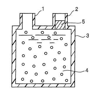
particularly specified. As schematically shown in Figure 3,
the container 3 preferably has an inlet 1 for introducing blood
or the like and an outlet 2 from which blood 4 or the like,
subsequent to contact with the cytokine-inducing material, is
guided to an outside of the container.
It is particularly preferred that the container used to
accommodate the cytokine-introducing material is constructed
in the form of a column, a blood bag or the like.
More preferably, the cytokine-inducing instrument has a
mechanismfor preventing outflow ofcytokine-inducing material,
when blood is allowed to contact with the cytokine-inducing
material and then guided to an outside of the container.
As schematically shown in Figure 3, the mechanism 5 for
preventing outflow of cytokine-inducing materialmay befixedly
mounted within the container so as to prevent outflow of the
cytokine-inducing material. Themechanism may beprovided with
a separation membrane or filter. Also, the cytokine-inducing
material may be separated from blood by centrifuging.
I f a therapeutic need arises, a plasma or serum component
may be separated from blood after it is contacted with the
12
CA 02464889 2004-04-27
cytokine-inducing material.
Specifically, the cytokine-inducing material containing
the induction enhancer in a particulate, fibrous or nonwoven
form or the 1 ike is packed blood bag having an inlet and an outlet .
Blood, a blood component or the like is passed through the inlet
into the bag. If necessary, the blood, blood component or the
like after induction of cytokines may be removed through the
outlet for use. The blood bag preferably has a volume of 50
mL - 1, 000 mL, particularly preferably 100 mL - 400 mL. Such
a blood bag preferably constitutes the cytokine-inducing
instrument by accommodating a particulate, fibrous or nonwoven
cytokine-inducing material.
Examples of particularly important cytokines include
interferon-y(IFN-y), interleukin 2 (IL-2), interleukin 10
(IL-10), interleukinl2 (IL-12), tumornecrosisfactor-a (Tumor
Necrosis Factor-a, TNF-a) and transforming growth factor-f3
(TGF-I3) . For example, IFN-y is a cytokine which plays a very
important role in immune diseases such as rheumatism,
inflammatory diseases, allergic diseases, cancers and other
various diseases and can be expected as having a therapeutic
effect on these diseases.
The above-described cytokine-inducing material and
cytokine-inducinginstrument caninducecytokinesnotonlyfrom
blood, a blood component or the like but also from various
cytokine-producing cells, examples of which include cells
13
CA 02464889 2004-04-27
collected from tissues such as of bone marrow-derived cells,
epidermal cells, fibroblasts, hepatocytes, osteoblasts, blood
stem cells, embryonic stem cells, cultured cells and established
cells.
BRIEF DESCRIPTION OF THE DRAWINGS
Figure 1 is a schematic view which explains surface
roughness and "peaks" of irregularities;
Figure 2 is a schematic view which explains a mean spacing
Sm value of irregularities for surface roughness; and
Figure 3 is a schematic sectional view which shows an
embodimentof a cytokine-inducinginstrumentin accordancewith
the present invention.
BEST MODE FOR CARRYING OUT THE INVENTION
The following examples illustrate the present invention
more specifically but are not intended to be limiting thereof .
In the following examples, IFN-y, TNF-a, IL-2, IL-10 and
IL-12 in human plasma and rat plasma were quantified by using
an R&D Systems ELISA kit, an Endogen ELISA kit and a Genzyme
Techne ELISA kit. TGF-f3 in human plasma was quantitatively
determined by using a Promega ELISA kit.
(EXAMPLE 1)
An induction enhancer -1 (synthetic aromatic adsorbent,
productofMitsubishiChemicalCorp.,productname:DIAION HP-50)
14
CA 02464889 2004-04-27
was washed via decantation with purl f ied water (product of Otsuka
Pharmaceutical Co., Ltd.) and then with methanol (product of
Wako Pure Chemicals Industries, Ltd., for HPLC use). The
inductionenhancer-lwasthereafterwashed via decantation with
physiological saline for injection (product of Otsuka
Pharmaceutical Co., Ltd.). The induction enhancer-1 in the
particle bulk volume of 50 uL was then packed in a sterilized
tube (product of DIATRON Corp., Eppendorf tube for 1.5 ml use) .
Blood was collected from a healthy human to prepare venous
blood containing 15 IU/ml of heparin. BCG (product of Japan
BCG Manufacturing Co. , Ltd. ) was added in the concentration of
1 mg/mL of blood. Here, BCG was prepared with physiological
saline. A ratio by volume of the physiological saline to blood
was brought to 1.25 0.
About 1.45 mL of the BCG-containing blood was introduced
into the tube packed with the induction enhancer-1.
The tube was tumbled to stir the blood and then attached
to a rotary mixer (product of Taitech Corp.) which was
subsequently rotated at 6 rpm in a constant temperature vessel
to effect incubation at 37 °C for 24 hours. After incubation,
the blood was centrifuged at 3,500 rpm (product of Tomy Seiko
Co., Ltd., micro high-speed centrifuge MRX-150) at 4 °C for 15
minutes . Blood plasmas were then collected from the blood for
cryopreservation at -20 'C. The preserved plasmas were then
melted and the amount of IFN-y induced in the plasmas was
CA 02464889 2004-04-27
determined using a Human IFN-y ELISA kit (product of R&D Systems
or ENDOGEN) . The results are shown in the following Table 1.
(COMPARATIVE EXAMPLE 1)
The induction enhancer-1 was excluded and the
BCG-incorporated blood was used in the amount of 1.5 mL.
Otherwise, the procedure of Example 1 was followed to collect
the plasmas and determine the amount of IFN-y induced in the
plasmas. The results are shown in the following Table 1.
(COMPAR.ATIVE EXAMPLE 2)
The procedure of Example 1 was followed, except that BCG
was not added to blood, to determine the amount of IFN- y induced.
The results are shown in the following Table 1.
Table 1
Induction 'I Concentration Amount of
Enhancer 1 ~ of ~~
Bulk Volume L~ BCG ~~ IFN-y Induced
l (mg/mL) /m
Ex. l 50 ' 1 162
Com . Ex. - 1 38
l
Com .Ex.2 50 - < 10
(EXAMPLE 2)
The duration of incubation at 37 °C was altered from 24
hours to 4 hours. Otherwise, the procedure of Example 1 was
followed to obtain cryopreserved plasmas. The cryopreserved
plasmas were then melted and the amount of TNF-a induced in the
plasmas was determined using a Human TNF-a ELISA kit (product
of R&D System) . The results are shown in the following Table
16
CA 02464889 2004-04-27
2.
(COMPARATIVE EXAMPLE 3)
The procedure of Example 2 was followed, except that the
induction enhancer-lwasexcluded andtheBCG-incorporated blood
was used in the amount of 1.5 mL, to determine the amount of
TNF-a induced. The results are shown in the following Table
2.
(COMPARATIVE EXAMPLE 4)
The procedure of Example 2 was followed, except that BCG
was not added to blood, to determine the amount of TNF-a induced.
The results are shown in the following Table 2.
Table 2
Induction Concentration Amount of
Enhancer 1 of ~~ '~
Bulk Volume BCG '~ TNF-a Induced
(uL) (mg/mL) n /mL
Ex.2 ~, 50 ~ 1 4.7
Com . Ex.3 '~~ - 1 1.1
Comp.Ex.4 ~ 50
<0.8
I -
(EXAMPLE 3)
The bulk volume of the induction enhancer-1 was altered
from 50 uL to 5 uL and the blood was added in the amount of 1 .495
mL. Otherwise, the procedure of Example 1 was followed to
determine the amount of IFN-v induced in the plasmas. The
results are shown in the following Table 3.
(EXAMPLE 4)
The bulk volume of the induction enhancer-1 was altered
17
CA 02464889 2004-04-27
from 50 uL to 10 uL and the blood was added in the amount of
1.49 mL. Otherwise, the procedure of Example 1 was followed
to determine the amount of IFN- y induced in the plasmas . The
results are shown in the following Table 3.
(EXAMPLE 5)
The bulk volume of the induction enhancer-1 was altered
from 50 uL to 20 uL and the blood was added in the amount of
1.48 mL. Otherwise, the procedure of Example 1 was followed
to determine the amount of IFN- y induced in the plasmas . The
results are shown in the following Table 3.
( EXAMPLE 6 )
The procedure of Example 1 was followed to determine the
amount of I FN- y induced in the plasmas . The results are shown
in the following Table 3.
(EXAMPLE 7)
The bulk volume of the induction enhancer-1 was altered
from 50 uL to 200 uL and the blood was added in the amount of
1.3 mL. Otherwise, the procedure of Example 1 was followed to
determine the amount of IFN-y induced in the plasmas. The
results are shown in the following Table 3.
(COMPARATIVE EXAMPLE 5)
The induction enhancer-1 was excluded and the
BCG-incorporated blood was used in the amount of 1.5 mL.
Otherwise, the procedure of Example 3 was followed to determine
the amount of IFN-y induced. The results are shown in the
18
CA 02464889 2004-04-27
following Table 3.
(COMPARATIVE EXAMPLE 6)
The procedure of Example 3 was followed, except that BCG
was not added to blood, to determine the amount of IFN- y induced.
The results are shown in the following Table 3.
(COMPARATIVE EXAMPLE 7)
The procedure of Example 4 was followed, except that BCG
was not added to blood, to determine the amount of IFN- y induced.
The results are shown in the following Table 3.
(COMPARATIVE EXAMPLE 8)
The procedure of Example 5 was followed, except that BCG
was not added to blood, to determine the amount of IFN- y induced.
The results are shown in the following Table 3.
(COMPARATIVE EXAMPLE 9)
The procedure of Example 6 was followed, except that BCG
was not added to blood, to determine the amount of IFN- y induced.
The results are shown in the following Table 3.
(COMPARATIVE EXAMPLE 10)
The procedure of Example 7 was followed, except that BCG
was not added to blood, to determine the amount of IFN- y induced.
The results are shown in the following Table 3.
19
CA 02464889 2004-04-27
Table 3
Induction I Concentration Amount of
~ Enhancer 1 of IFN-y Induced
j BCG ~p mL
Bulk Volume m m~ I
L '
Ex.3 5 1 115
Ex.4 j 10 - 1 152
Ex.S 20 ~ 1 I 297
Ex.6 50 1
__ I 573
Ex.7 200 1 453
~Com .Ex.S - 1 104
Com . Ex.6 5 - < 10
Com .Ex.7 10 - < 10
Com .Ex.8 I 20 - <10
Com .Ex.9 50 - < 10
Com .Ex.10 200 - < 10
(EXAMPLE 8)
The procedure of Example 1 was followed, except that the
bulk volume of the induction enhancer-1 was altered to 20 ~L,
to prepare the tube packed with the induction enhancer-1.
Picibanil (product of Chugai Pharmaceutical Co., Ltd.,
OK-432) , in place of BCG, was added to each blood sample in a
concentration of 0.1 KE/mL. This OK-432 was prepared using an
RPMI medium. The ratio of the RPMI medium to blood was 20 0.
The OK-432 incorporated blood was poured into the tube packed
with a 20 uL bulk volume of the induction enhancer-1 to a tube
scale of 1.5 mL. That is, the blood was added in the amount
of about 1.48 mL.
Then, the amount of IFN-y induced was determined in the
same manner as in Example 1. The results are shown in the
following Table 4.
(EXAMPLE 9)
CA 02464889 2004-04-27
The bulk volume of the induction enhancer-1 was altered
to 50 uL and the OK-432 incorporated blood was used in the amount
of 1.45 mL. Otherwise, the procedure of Example 8 was followed
to determine the amount of IFN- y induced. The results are shown
in the following Table 4.
( EXAMPLE 10 )
The bulk volume of the induction enhancer-1 was altered
to 100 uL and the OK-432 incorporated blood was used in the amount
of 1.40 mL. Otherwise, the procedure of Example 8 was followed
to determine the amount of IFN- y induced. The results are shown
in the following Table 4.
(COMPARATIVE EXAMPLE 11)
The induction enhancer-1 was excluded and the blood
incorporatedOK-432 was used in the amount of l.SmL. Otherwise,
the procedure of Example 8 was followed to determine the amount
of IFN-y induced. The results are shown in the following Table
4.
(COMPARATIVE EXAMPLE 12)
The procedure of Example 8 was followed, except that
Picibanil (OK-432) was not added, to determine the amount of
IFN-y induced. The results are shown in the following Table
4.
(COMPARATIVE EXAMPLE 13)
The procedure of Example 9 was followed, except that
Picibanil (OK-432) was not added, to determine the amount of
21
CA 02464889 2004-04-27
IFN-y induced. The results are shown in the following Table
4.
(COMPARATIVE EXAMPLE 14)
The procedure of Example 10 was followed, except that
Picibanil (OK-432) was not added, to determine the amount of
IFN-y induced. The results are shown in the following Table
4.
Table 4
Induction Concentration Amount of I
Enhancer 1 of ' IFN-y Induced
Bulk Volume OK-432 mL
L ~ KE mL ~
Ex.8 20 0.1 ' 360
Ex.9 ' S0 0.1 i 367
Ex.10 I 100 0.1 323
Com .Ex.ll - 0.1 114
~ '
Com .Ex.l2 20
~ i <10
I
Com .Ex.l3 I 50 - < 10
Comp.Ex.14 ~ 100 - < 10
( EXAMPLE 11 )
An induction enhancer-2 (synthetic acrylic ester resin,
product of Organo Corp., product number: AMBERLITE XAD-7), in
place of the induction enhancer-l, was used. Otherwise, the
procedure of Example 1 was followed to determine the amount of
IFN-y induced in the plasmas. The results are shown in the
following Table 5.
( EXAMPLE 12 )
The bulk volume of the induction enhancer-2 was altered
from 50 uL to 200 uL and the BCG-incorporated blood was used
22
CA 02464889 2004-04-27
in the amount of 1.3 mL. Otherwise, the procedure of Example
11 was followed to determine the amount of IFN-y induced. The
results are shown in the following Table 5.
(COMPARATIVE EXAMPLE 15)
The induction enhancer-2 was excluded and the
BCG-incorporated blood was used in the amount of 1.5 mL.
Otherwise, the procedure of Example 11 was followed to determine
the amount of IFN-y induced. The results are shown in the
following Table 5.
(COMPARATIVE EXAMPLE 16)
The procedure of Example 11 was followed, except that BCG
was not added to blood, to determine the amount of IFN- y induced.
The results are shown in the following Table 5.
(COMPARATIVE EXAMPLE I7)
The procedure of Example 12 was followed, except that BCG
was not added to blood, to determine the amount of IFN- y induced.
The results are shown in the following Table 5.
Table 5
Induction ~ Concentration Amount of
Enhancer 2 of IFN-Y Induced
Bulk Volume BCG ~ mL
L m mL
Ex. l l SO 1 299
Ex.l2 ~ 200 1 218
Com . Ex.15- 1 104
Com . Ex.16SO - < 12
Gomp.Ex.17 200 - < 12
The following Examples and Comparative Examples
23
CA 02464889 2004-04-27
illustrate induction of cytokines in rat blood.
(EXAMPLE 13)
The procedure of Example 1 was followed to obtain the
sterilized tube packed with a 50 uL bulk volume of the induction
enhancer-1.
Venous blood containing 15 IU/ml of heparin was collected
from a Wister rat (7 weeks old, male, purchased from Japan SLC,
Inc.). Picibanil (product of Chugai Pharmaceutical Co., Ltd.,
OK-432) was added to the blood in the concentration of 0.1 KE/mL.
This OK-432 was prepared~using an RPMI medium. The ratio of
the RPMI medium to blood was 20 ° . The OK-432 incorporated blood
was added to the tube, packed with the induction enhancer-1,
to a tube scale of 1.5 mL.
Subsequently, the procedure of Example 1 was followed to
collect blood plasmas and determine the amount of IFN-y induced
therein by using a Rat IFN-y quantification kit (product of
Genzyme Techne) . The results are shown in the following Table
6.
(EXAMPLE 14)
The bulk volume of the induction enhancer-1 was altered
to 500 uL and the OK-432 incorporatedblood was used in the amount
of 1.0 mL. Otherwise, the procedure of Example 13 was followed
to determine the amount of IFN- y induced. The results are shown
in the following Table 6.
(COMPARATIVE EXAMPLE 18)
24
CA 02464889 2004-04-27
The induction enhancer-1 was excluded and the OK-432
incorporated blood was used in the amount of 1 . 5 mL. Otherwise,
the procedure of Example 13 was followed to determine the amount
of IFN-y induced. The results are shown in the following Table
6.
(COMPARATIVE EXAMPLE 19)
The procedure of Example 13 was followed, except that
OK-432 was not added to the blood, to determine the amount of
IFN-y induced. The results are shown in the following Table
6.
(COMPARATIVE EXAMPLE 20)
The procedure of Example 14 was followed, except that
OK-432 was not added to the blood, to determine the amount of
IFN-y induced. The results are shown in the following Table
6.
Table 6
Induction Concentration of : Amount
of
Enhancer 1 ~ OK-432 IFN-y Induced
Bulk Volume KE/rnL /mL
L
Ex.13 50 0.1 I 2 51
Ex.14 500 0.1 435
Com . Ex.18- 0.1 72
Com .Ex.l9 T 50 - <40
Com .Ex.20 500 - ~ <40
( EXAMPLE 15 )
The procedure of Example 1 was followed to obtain the
sterilized tube packed with a 50 uL bulk volume of the induction
CA 02464889 2004-04-27
enhancer-1.
Blood was collected from a blister rat (7 weeks old, male,
purchased from Japan SLC, Inc . ) to obtain venous blood containing
15 IU/ml of heparin . BCG was added to this blood in the
concentration of 1 mg/mL. This BCG was prepared using
physiological saline. The ratio in volume of the physiological
saline to the blood was 1 . 25 % . The BCG-incorporated blood was
added to the tube, packed with the induction enhancer-1, to a
tube scale of 1.45 rnL.
Subsequently, the procedure of Example 1 was followed to
collect blood plasmas and determine the amount of IFN- y induced
therein by using a Rat IFN-y quantification kit (product of
Genzyme Techne) . The results are shown in the following Table
7.
(EXAMPLE 16)
The bulk volume of the induction enhancer-1 was altered
from 50 uL to 500 uL and the BCG-incorporated blood was used
in the amount of 1.0 mL. Otherwise, the procedure of Example
15 was followed to determine the amount of IFN-y induced. The
results are shown in the following Table 7.
(COMPARATIVE EXAMPLE 21)
The induction enhancer-1 was excluded and the
BCG-incorporated blood was used in the amount of 1.5 mL.
Otherwise, the procedure of Example 15 was followed to determine
the amount of IFN-y induced. The results are shown in the
26
CA 02464889 2004-04-27
following Table 7.
(COMPARATIVE EXAMPLE 22)
The procedure of Example 15 was followed, except that BCG
was not added to the blood, to determine the amount of LFN-
y induced. The results are shown in the following Table 7.
(COMPARATIVE EXAMPLE 23)
The procedure of Example 16 was followed, except that BCG
was not added to the blood, to determine the amount of IFN-
y induced. The results are shown in the following Table 7.
Table 7
Induction i Concentration
Enhancer 1 ~ of Amount f
Bulk Volume '
L ~' BCG IFN-y Induced
m mL mL
Ex.lS 50 1 t, 132
Ex.l6 i 1 233
500 i
Com . Ex.21- 1 68
'
Com .Ex.22 50 - <40
Com .Ex.23 500 - <40
(EXAMPLE 17)
The duration of incubation at 37 °C was altered from 24
hours to 4 hours. Otherwise, the procedure of Example 15 was
followed to collect blood plasmas and determine the amount of
TNF-a induced therein by using a Rat Tumor Necrosis Factor-a
quantification kit (product of Genzyme Techne). The results
are shown in the following Table 8.
(COMPARATIVE EXAMPLE 24)
The induction enhancer-1 was excluded and the
27
CA 02464889 2004-04-27
BCG-incorporated blood was used in the amount of 1.5 mL.
Otherwise, the procedure of Example 17 was followed to collect
blood plasmas and determine the amount of TNF-a induced therein.
The results are shown in the following Table 8.
(COMPARATIVE EXAMPLE 25)
The procedure of Example 17 was followed, except that BCG
was not added to the blood, to determine the amount of TNF-a
induced. The results are shown in the following Table 8.
Table 8
j Induction Concentration of Amount of
Enhancer 1 BCG TNF-a Induced j
Bulk Volume (uL) , (mg/mL) ~ n /mL 'i
Ex.17 50 1 I 12.7
Com .Ex.24 I - 1 2.1
Comp.Ex.25 ! 50 - < 1.2
(EXAMPLES 18 - 23)
Induction Enhancer-3 : A CA film (product of Artplus Corp . ,
cellulose acetate film, product name: ACETYFILMVR-R) was used.
A plasticizer was extracted from the film by soxhlet extraction
for 24 hours in methyl alcohol. After removal, the film was
air dried for 15 hours and further dried at 80 °C for 5 hours .
Thereafter, the CA film was polished, using a Struers (Denmark)
automatic polisher(productname:PLANOPOL-PEDEMAX)with a220-,
500-, 1200-, 2400- or 4000-mesh sandpaper secured thereto, to
provide a polished CR film having surface roughness on its both
surfaces. This film was washed with methyl alcohol and then
28
CA 02464889 2004-04-27
subdivided to 2 . 5 mm x 2 . 5 mm pieces . These pieces, measuring
a bulk volume of about 200 uL, were washed with physiological
saline for injection and then packed in a sterilized tube for
1.5 mL use.
Blood was collected from a healthy human to prepare venous
blood containing 15 IU/ml of heparin. BCG (product of Japan
BCG Manufacturing Co., Ltd.) was added to the blood in the
concentration of 1 mg/mL. About 1.3 mL of the resulting blood
was added to the tube . Subsequently, the amount of IFN- y induced
was determined in the same manner as in Example 1 . The results
are shown in the following Table 9.
Table 9
Exam 1~
T
18 I 19 '~, 20
21 ~ 22 23~
' Polished
r
Unpol- 220-
4000- I 2400- 1200-
~ 500-
I fished ;
~
I Mesh
Mesh Mesh i Mesh
Mesh
Ra V~ ue 0.05 I 0.05 I~ 0.06 0.58 1.28
I ~'
2.37
Sm Value ~ I
I
~ 250 31 30 44 57
! 125
I
m
Concentra- '
tion of ~ 1 ~ 1 ~ 1 ~ 1 1 1
BCG
m mL , '
' Concentra- '
tion of 38 72 79 ~ 172 193 189
IFN-y ~
~Pg/mL) I I ~
(EXAMPLES 24 - 29)
Induction Enhancer-4 : A PET film (product of Unitika Ltd. ,
polyethylene terephthalate film, product name: EMBLET S-75) was
29
CA 02464889 2004-04-27
used. The film was at its surfaces washed with methyl alcohol.
Thereafter, this PET film was polished, using a Struers (Denmark)
automatic polisher(productname:PLANOPOL-PEDEMAX)with a220-,
500-, 1200-, 2400- or 4000-mesh sandpaper secured thereto, to
provide a polished CA film having surface roughness on its both
surfaces. This film was washed with methyl alcohol and then
subdivided to 2 . 5 mm x 2 . 5 mm pieces . These pieces, measuring
a bulk volume of about 200 uL, were washed with physiological
saline for injection and then packed in a sterilized tube for
1.5 mL use. Blood was collected from a healthy human to obtain
venous blood containing 15 IU/ml of heparin. BCG (product of
Japan BCG Manufacturing Co., Ltd.) was added to the blood in
the concentration of 1 mg/mL. About 1.3 mL of the resulting
blood was added to the tube. The procedure of Example 1 was
then followed to determine the amount of IFN-y induced. The
results are shown in the following Table 10.
25
CA 02464889 2004-04-27
Table 10
i Exam
le
24 25 227
X28
9
~i U Polished
l
npo
-
4000- 2400- 1200- 500-
ished ~ 220-
Mesh Mesh Mesh Mesh
; Mesh
i
Ra V
ue
m 0.07 0.12 0.19 0.61 1.55 2.24
j
Sm Value 475 37 126 31 47 ~ 77
m
Concentra , I
tion of 1 1 1 1 1 1
BCG ~
m mL I
Concentra-
tion of 36 71 ~ 73 141 i 158 169 I
IFN-y
mL
(EXAMPLES 30 - 35)
Induction Enhancer-5: A polystyrene film (product of
Mitsubishi-Monsanto Co., Ltd., product name: SANTOCLEAR) was
used. The film was at its surfaces washed with purified water.
Thereafter, the polystyrene film was polished, using a Struers
(Denmark) automatic polisher (product name: PLANOPOL-PEDEMAX)
with a 220-, 500-, 1200-, 2400- or 4000-mesh sandpaper secured
thereto, to provide a polished polystyrene film having surface
roughness on its both surfaces. This film was washed with
purified water and then subdivided to 2.5 mm x 2.5 mm pieces.
These pieces, measuring a bulk volume of about 200 uL, were washed
with physiological saline for injection and then packed in a
sterilized tube for 1.5 mL use. Blood was collected from a
healthy human to obtain venous blood containing 15 IU/ml of
heparin. BCG (product of Japan BCG Manufacturing Co., Ltd.)
31
CA 02464889 2004-04-27
was added to the blood in the concentration of 1 mg/mL. About
1 . 3 mL of the resulting blood was added to the tube . The procedure
of Example 1 was then followed to determine the amount of IFN-
y induced. The results are shown in the following Table 11.
Table 11
Exam
le
'I 30 31 _ 33 34 35
32 ~
l Polished
U
npo
-
fished 4000- 2400- 1200- 500- 220-
Mesh Mesh Mesh Mesh Mesh
Ra Value 0,06 0.08 0.21 0.72 1.75 2.44
i
m ~
Sm Value 375 52 i 43 ~ 27 ~ 39 71
j ~ ~ ~
i- Il.am)
+
I Concept ! I I I I
a I
tion of j 1 ~ 1 i , 1 ~ 1 ' 1
BCG 1 ~
i (mg/mL) i I ! ,
I Concentra-~
tion of , 48 I 92 ~ 94 239 ' 269 ~ 246
IFN-y i
IPg/mL) ~'~ I I , ~ I
(EXAMPLE 36)
Cellulose acetate pellets (product of Artplus Corp.,
ZO containing 30 ° acetyl triethyl citrate as a plasticizer) were
injection molded to prepare 2.5 mm diameter spherical beads.
The plasticizer was extracted from 50 g of these beads by soxhlet
extraction at 50 °C for 24 hours in 300 mL methanol. Subsequent
to extraction of the plasticizer, those beads were transferred
onto a stainless-steel batting, air dried for 15 hours and then
further dried at 80 °C for 5 hours.
200 mL of such beads and an equal volume of an abrasive
32
CA 02464889 2004-04-27
WHITE ABRRX (WA) #34 (product of Japan Abrasive Company) were
introduced into a pot mill (product of Toyo Engineering Corp.,
product name: 51-ceramic pot mill BP-5). Also, several balls
(product of Toyo Engineering Corp., product name: BB-13) for
a ceramic pot mill were introduced. Polishing was applied for
5 hours by a ball polishing machine (pot mill manufactured by
Nitto Kagaku Co . , Ltd. , product name : AN-3S ) . As a result, beads
were obtained having an Ra value of 1.36 um and an Sm value of
97.2 um (induction enhancer-6).
The obtained beads were washed three times with methanol
and then five times with physiological saline for injection.
These beads, measuring a bulk volume of 400 uL, were packed in
a sterilized tube for 1 . 5 mL use in the same manner as in Example
1. The procedure of Example 1 was then followed, except that
the blood was added in the amount of 1.1 mL, to determine the
amount of IFN- y induced. The results are shown in the following
Table 12.
(EXAMPLE 37)
The procedure of Example 36 was followed, except that
polishing was not applied, to prepare the induction enhancer-6
having an Ra value of 0.186 um and an Sm value of 298.7 um. This
induction enhancer was allowed to contact with the blood in the
same manner as in Example 36. The results are shown in the
following Table 12.
(COMPARATIVE EXAMPLE 26)
33
CA 02464889 2004-04-27
The induction enhancer-6 was excluded and the
BCG-incorporated blood was used in the amount of 1.5 mL.
Otherwise, the procedure of Example 36 was followed to determine
the amount of IFN-y induced. The results are shown in the
following Table 12.
Table 12
Centerline Mean Concentra-tAmount of
Average Spacing of ion of IFN-y Induced
BCG
Roughness Unevenness (mg/mL) (pg/mL)
Ra '
m Sm m
I
Ex.36 1.36 97.2 I 1 198
Ex.37 0.19 298.7 1 101
~Com .Ex.26_ - 1 ~ 38
(EXAMPLES 38 - 42)
The procedure of Example 1 was followed, using the
induction enhancer-1 (synthetic aromatic adsorbent, productof
Mitsubishi Chemical Corp., product name: DIAION HP-50), the
induction enhancer-2 (synthetic acrylic ester resin, product
of Organo Corp . , product number : AMBERLITE XAD-7 ) , an induction
enhancer-7 (synthetic aromatic resin, product of Organo Corp.,
product number: AMBERLITE XAD-2), an induction enhancer-8
(synthetic aromatic resin, product of Organo Corp., product
number: AMBERLITE XAD-4) or an induction enhancer-9 (synthetic
aromatic resin, product of Organo Corp., product number:
AMBERLITE XAD-16HP). The results are shown in the following
Table 13.
34
CA 02464889 2004-04-27
(COMPARATIVE EXAMPLE 27)
The induction enhancer was excluded and the
BCG-incorporated blood was used in the amount of 1.5 mL.
Otherwise, the procedure of Example 38 was followed. The results
are shown in the following Table 13.
Table 13
Induction Enhancer Concentra-Amount of
Bulk Volume Mite e~ tion of IFN-y Induced
50 L ~ ~ BCG mL
m mL
Ex.38 Induction EnhancerHP50 1 299 I
1
Ex.39 Induction EnhancerXAD7 1 136 '.
2
Ex.40 Induction EnhancerXAD2 1 283 I
7 ~
Ex.41 Induction EnhancerXAD4 ' 1 ~I 31 S I
8
Ex 42 Induction Enhancer; XAD 1 ~ 307 !,
9 16HP
r Comp I Absent - ~ 1 ; 61
Ex 27
(EXAMPLE 43)
BCG was suspended in physiological saline, diluted with
a phosphate buffer and centrifuged at 4, 000 rpm for 15 minutes.
After removal of a supernatant liquid, the resultant was again
suspended in a phosphate buffer and centrifuged at 4,000 rpm
(product of Tomy Seiko Co., Ltd., micro high-speed centrifuge
MRX-150 ) for 15 minutes . This operation was repeated three times .
The resultant was suspended in a phosphate buffer containing
50 'o ethanol and then centrifuged at 4, 000 rpm for 15 minutes.
Subsequently, the resultant was suspended in ethanol and
centrifuged at 4,000 rpm for 15 minutes. This operation was
repeated twice. Wash with a phosphate buffer was again carried
CA 02464889 2004-04-27
out three times. Such treated BCG in the amount of 2 mg was
covalently bonded to the carboxyl-introduced induction
enhancer-1 (bulk volume of 1 mL) by a carbodiimide process . The
remaining reactive groups were reacted with ethanolamine. The
resultant was washed with a phosphate buffer and then suspended
in a phosphate buffer. The thus-obtained induction enhancer,
measuring 100 uL, was packed in a sterilized tube.
BCG was not added in its individual form. The blood was
added in the amount of 1.4 mL. Otherwise, the procedure of
Example 1 was followed to determine the amount of IFN-y induced.
The results are shown in Table 14.
(COMPARATIVE EXAMPLE 28)
The procedure of Example 43 was followed, except that the
treated BCG was not covalently bonded, to determine the amount
of IFN-y induced. The results are shown in Table 14.
(COMPARATIVE EXAMPLE 29)
The induction enhancer was not added and the
BCG-incorporated (1 mg/mL) blood was used in the amount of 1.5
mL. Otherwise, the procedure of Example 43 was followed to
determine the amount of IFN- y induced. The results are shown
in Table 14.
36
CA 02464889 2004-04-27
Table 14
Induction Enhancer 1 ConcentrationAmount of
~~ Bulk Volume ~ of BCG IFN-y Induced
I (100 uL) i (mg/mL) ~ (Pg/mL)
Ex.43 Covalent Bondin I - ~ 32
Com .Ex.28 No Covalent Bondin- < 10
Com .Ex.29 - 1 i - 20
(EXAMPLE 44)
The polystyrene-divinylbenzene copolymer induction
enhancer-8 (product of Organo Corp., product number: AMBERLITE
XAD-4 ) , measuring a bulk volume of 1 mL, and BCG ( 10 mg/mL) were
intimately mixed in 1 mL physiological saline at 37 °C for 20
hours, so that BCG was physically adsorbed onto particle surfaces .
After the intimate mixing, these particles were fully washed
with physiological saline and again suspended in physiological
saline. Thethus-obtainedinductionenhancer, measuringabulk
volume of 100 uL, were packed in a sterilized tube.
BCG was not added in its individual form. The blood was
added in the amount of 1.4 mL. Otherwise, the procedure of
Example 1 was followed using blood from two healthy humans to
determine the amount of IFN-y induced. The results are shown
in Table 15.
(COMPAR.ATIVE EXAMPLE 30)
The procedure of Example 44 was followed, except that BCG
was not added, to prepare particles. Using thus-obtained
particles, the amount of IFN-y induced was determined in the
same manner as in Example 44. The results are shown in Table
37
CA 02464889 2004-04-27
15.
(COMPARATIVE EXAMPLE 31)
The induction enhancer was not added and the
BCG-incorporated (1 mg/mL) blood was used in the amount of 1.5
mL. Otherwise, the procedure of Example 44 was followed to
determine the amount of IFN-v induced. The results are shown
in Table 15.
Table 15
- (Amount of IFN-y Induced!
Induction Enhancer 8 Concentra
Bulk Volume tion of BCG ~ Blood '
loo uL~ ~ fmg/mL) ~ A B
Ex.44 ' Ph sical Adso tion - 396 109
Com . Ex.30 No Ph sical Adso tion - < 10 < 10
I Comp.Ex.31 , - ; 1 109 ~ 29
( EXAMPLE 4 5 )
The polystyrene-divinylbenzene copolymer induction
enhancer-8, measuring a bulk volume of 1 mL, and BCG (10 mg/mL)
were intimately mixed in 1 mL physiological saline containing
1 o formalin (neutral buffer formalin, product of Wako Pure
Chemicals Industries, Ltd.) at 4 °C for 20 hours, so that BCG
was physically adsorbed onto particle surfaces. Subsequent to
the intimate mixing, these particles were fully washed with
physiological saline. Thereafter, those particles, measuring
a bulk volume of 100 uL, were packed in a sterilized tube (product
of DIATRON Corp., for 1.5 mL use).
38
CA 02464889 2004-04-27
BCG was not added in its individual form. The blood was
added in the amount of 1.4 mL. Otherwise, the procedure of
Example 1 was followed to determine the amount of IFN- y induced.
The results are shown in Table 16.
(COMPARATIVE EXAMPLE 32)
The procedure of Example 45 was followed, except that BCG
was not added, to determine the amount of IFN- v induced. The
results are shown in Table 16.
(COMPAR.ATIVE EXAMPLE 33)
The induction enhancer-8 was excluded and the
BCG-incorporated (1 mg/mL) blood was used in the amount of 1.5
mL. Otherwise, the procedure of Example 45 was followed to
determine the amount of IFN- y . The results are shown in Table
16.
Table 16
i Induction Enhancer ConcentrationAmount of I
8
Bulk Volume of BCG IFN-y Induced
100 L m /mL mL
Ex.45 Ph sical Adso Lion - I 388
Com .Ex.32 No Ph sical Adso - I < 10 I
tion
Com .Ex.33 - 1 87
(EXAMPLE 46)
The induction enhancer-8 was treated in the same manner
as in Example 45 so that BCG was physical ly adsorbed onto particle
surfaces. The treated induction enhancer, measuring a bulk
volume of 4 mL, was packed in a blood bag (product of Terumo
39
CA 02464889 2004-04-27
Corp. , separation bag for 200 mL use) . Blood was collected from
a healthy human to obtain venous blood containing 15 IU/mL of
heparin. 50 mL of the venous blood was introduced into the blood
bag. The blood bag contents were incubated while gently stirred
at 37 °C for 24 hours. The amount of IFN-y present in the blood
was determined in the same manner as in Example 1. The results
are shown in Table 17.
(COMPARATIVE EXAMPLE 34)
The induction enhancer-8 was used without the physical
adsorption treatment of BCG. Otherwise, the procedure of
Example 46 was followed. The results are shown in Table 17.
(COMPARATIVE EXAMPLE 35)
The induction enhancer-8 was excluded and the blood
containing 1 mg/mL of BCG was used in the amount of 50 mL.
Otherwise, the procedure of Example 46 was followed to determine
the amount of IFN-y. The results are shown in Table 17.
Table 17
Induction Enhancer ConcentrationAmount of
8 ~ IFN-y Induced
Bulk Volume of BCG ~ mL
4 mL m mL
Ex.46 Ph sical Adso tion - 362
Com.Ex.34 No Ph sical Adso - <10
tion
-.
-
r Comp.Ex.35- [ 1 ~ 62
~
(EXAMPLE 47)
The procedure of Example 1 was followed, except that
interleukin 2 and interleukin 12 present in the blood were
CA 02464889 2004-04-27
quantitatively determined. The results are shown in Table 18.
(COMPARATIVE EXAMPLE 36)
The procedure of Example 47 was followed, except that BCG
was not added. The results are shown in Table 18.
(COMPARATIVE EXAMPLE 37)
The induction enhancer-1 was not added and the
BCG-incorporated blood was used in the amount of 1.S mL.
Otherwise, the procedure of Example 47 was followed. The results
are shown in Table 18.
Table 18
i Induction 1
ConcentrationAmount of Amount of
Enhancer
1
of BCG IL-2 InducedIL-12 Induced
Bulk Volume j
!
(mg/mL) ~ (pg/mL) (pg/mL)
i L
Ex.47 ~ .- 50 ~ 1 31 121
Com .Ex.36 50 - <6 i <8
~
Com .Ex.37 1 10 ~ _4~ _
,' -
( EXAMPLE 4 8 )
The procedure of Example 1 was followed, except that the
BCG concentration was altered to 0.1 mg/mL and TGF-!3 present
in the blood was quantitatively determined. The results are
shown in Table I9.
(COMPARATIVE EXAMPLE 38)
The induction enhancer-1 was not added and the
BCG-incorporated blood plasma was used in the amount of 1 .5 mL.
Otherwise, the procedure of Example 48 was followed. The results
41
CA 02464889 2004-04-27
are shown in Table 19.
Table 19
I Induction ~ fount
of TGF-(3
Induced
Enhancer 1 ~ Concentrationn mL
Bulk Volume ~ of BCG Blood
~ L
, (mg/m A B C
(uL) )
~
i 50 0.1 80 101 63
Ex.48
Com .Ex.38 ' - 0.1 21 35 19 I
(EXAMPLE 49)
The procedure of Example 48 was followed, except that
OK-432, in place of BCG, was used in the amount of 0.1 KE/mL.
The results are shown in Table 20.
(COMPARATIVE EXAMPLE 39)
The induction enhancer-1 was not added and the OK-432
incorporated blood was used in the amount of 1 . 5 mL. Otherwise,
the procedure of Example 49 was followed. The results are shown
in Table 20.
Table 20
Induction ~unt
of
TGF-(i
Induced
Enhancer Concentrationn /mL
1 2
Bulk Volume of OK-43 Blood
(uL) (KE/mL) A B C
Ex.49 ~ 50 0.1 71 111 71
i Comp.Ex.39i - ~ 0.1 25 33 26
(EXAMPLE SO)
The procedure of Example 1 was followed, except that OK-432,
in place of BCG, was used in the amount of 0.01 KE/mL and IL-10
42
CA 02464889 2004-04-27
was quantitatively determined. The results are shown in Table
21.
(COMPARATIVE EXAMPLE 40)
The induction enhancer-1 was not added and the OK-432
incorporated blood was used in the amount of 1 . 5 mL. Otherwise,
the procedure of Example 50 was followed. The results are shown
in Table 21.
Table 21
Induction C fount
of
IL-10
Induced
Enhancer centration (pg/mL)
1 ~,
, of OK-432 Blood
Bulk Volume '
!
(1zL) I A ~
(KE/mL) , C
~
Ex.50 ! 50 j 560 285 421
' 0.01 1
Com .Ex.40I - 0.01 74 I 39 112
(EXAMPLES 51 AND 52)
The procedure of Example 1 was followed, except that BCG
was added in the concentration of 0.1 mg/mL or 1 mg/mL. The
results are shown in Table 22.
(COMPARATIVE EXAMPLE 41)
The procedure of Example 51 was followed, except that BCG
was not added. The results are shown in Table 22.
(COMPARATIVE EXAMPLE 42)
The induction enhancer-1 was not added and the
BCG-incorporated (0.1 mg/mL) blood was used in the amount of
1.5 mL. Otherwise, the procedure of Example 51 was followed.
The results are shown in Table 22.
43
CA 02464889 2004-04-27
(COMPAR.ATIVE EXAMPLE 43)
The induction enhancer-1 was not added and the
BCG-incorporated ( 1 mg/mL) blood was used in the amount of 1 . 5
mL. Otherwise, the procedure of Example 52 was followed. The
results are shown in Table 22.
Table 22
Induction Concentra-tfount
Enhancer 1 i of
f BCG IFN-y
Induced
mL
Bulk Volume on o Blood
L
~mg~m A B C
)
Ex.51 50 ! 0.1 73 272 104
I I
Ex.52 50 I 1 376 350 401
!
Com .Ex.41 50 ~ - <2 ' 12 8
I Com .Ex.42- i 0.1 i 9 93 36
I I
L- Comp.Ex.43'i - ~ 1 '~ ', 110 96 -
56 ~
(EXAMPLES 53 AND 54)
The procedure of Example 1 was followed, except that OK-432,
in place of BCG, was added in the concentration of 0.01 KE/mL
or 0.1 KE/mL. The results are shown in Table 23.
(COMPARATIVE EXAMPLE 44)
The procedure of Example 53 was followed, except that
OK-432 was not added. The results are shown in Table 23.
(COMPARATIVE EXAMPLE 45)
The induction enhancer-1 was not added and the OK-432
incorporated (0.01 KE/mL) blood was used in the amount of 1.5
mL. Otherwise, the procedure of Example 53 was followed. The
results are shown in Table 23.
44
CA 02464889 2004-04-27
(COMPARATIVE EXAMPLE 46)
The induction enhancer-1 was not added and the OK-432
incorporated (0.1 KE/mL) blood was used in the amount of 1.5
mL. Otherwise, the procedure of Example 54 was followed. The
results are shown in Table 23.
Table 23
Induction Concentration~unt
Enhancer f OK of
1 432 IF1V-y
Induced
~pg~m~-
Bulk Volume o Blood
-
~KE/
L)
~l~L) m A B C I
Ex.53 50 0.01 79 355 128 '~
Ex.54 50 0.1 267 466 432
Com .Ex.44 50 - <2 12 8
Com .Ex.45 - i 0.01 I 8 I 94 59
Com . Ex.46- 0.1 84 176 i 89
(EXAMPLE 55)
0.04 g (about 300 uL in terms of a bulk volume when packed
in a 1.5 mL tube) of nylon wool (product of Wako Pure Chemicals
Industries, Ltd.) was used as an induction enhancer-10. The
BCG-incorporated blood was added in the amount of 1.2 mL.
Otherwise, the procedure of Example 1 was followed. The results
are shown in Table 24.
( EXAMPLE 5 6 )
0.04 g (1 cm x 3 cm, about 300 uL in terms of a bulk volume)
of a polyester nonwoven fabric (product of Japan Vilene Co.,
Ltd., product designation: EL-5600) was used as an induction
enhancer-11. TheBCG-incorporated blood wasaddedintheamount
CA 02464889 2004-04-27
of 1.2 mL. Otherwise, the procedure of Example 1 was followed.
The results are shown in Table 24.
(EXAMPLE 57)
0. 04 g ( 1 cm x 3 cm, about 220 uL in terms of a bulk volume)
of a polyester nonwoven fabric (product of Japan Vilene Co.,
Ltd., product designation: EW-7180) was used as an induction
enhancer-12. The BCG-incorporated blood wasaddedintheamount
of 1.28 mL. Otherwise, the procedure of Example 1 was followed.
The results are shown in Table 24.
(COMPARATIVE EXAMPLE 47)
The procedure of Example 55 was followed, except that BCG
was excluded. The results are shown in Table 24.
(COMPARATIVE EXAMPLE 48)
The procedure of Example 56 was followed, except that BCG
was excluded. The results are shown in Table 24.
(COMPARATIVE EXAMPLE 49)
The procedure of Example 57 was followed, except that BCG
was excluded. The results are shown in Table 24.
(COMPARATIVE EXAMPLE 50)
The induction enhancer was excluded and the
BCG-incorporated blood was used in the amount of 1.5 mL.
Otherwise, the procedure of Example 55 was followed. The results
are shown in Table 24.
46
CA 02464889 2004-04-27
Table 24
I I Concentration Amount of
Induction IFN-y Induced
Enhancer ~
~~~ of mL
BCG
i i m mL
Ex.55 Induction Enhancer 10 '~ 1 117
Ex.56 Induction Enhancer 11 1 381
Ex.57 Induction Enhancer 12 ' 1 338
Com .Ex.47 Induction Enhancer 10 - < 10
Com . Ex.48Induction Enhancer 11 ~ - < 10
Com .Ex.49 Induction Enhancer 12 - < 10 I
Com .Ex.50 - 1 37
(EXAMPLE 58)
Actinomyces S. nobilis (JCM 4274) obtained from Institute
of Physical and Chemical Research was shake cultured at 30 °C
for 40 hours in 100 mL of a starch-ammonium medium containing
0. 2 ~ yeast extract to obtain bacteria. The procedure of Example
1 was followed, except that such bacteria, in place of BCG, were
added to blood in the dry weight of 1 mg/mL, to determine the
amount of IFN-y. The results are shown in Table 25.
(COMPARATIVE EXAMPLE 51)
The procedure of Example 58 was followed, except that the
bacteria of actinomyces were excluded. The results are shown
in Table 25.
(COMPARATIVE EXAMPLE 52)
The induction enhancer-1 was excluded and the actinomyces
incorporated blood was used in the amount of 1 . 5 mL. Otherwise,
the procedure of Example 58 was followed. The results are shown
in Table 25.
47
CA 02464889 2004-04-27
Table 25
i
Enhancer 8 Concentration of Amount of IFN-y I
Bulk Volume L Bacteria (mg/mL); Induced (pg/mL) ';
Ex.58 ! 50 1 161
Comp.Ex.S~ 50 _ - ; < 10
Comp.Ex.52 - 1 32 ;
( EXAMPLE 5 9 )
As an induction enhancer-13 (gel particles of polyvinyl
alcohol, particle diameter of about 1.1 mm), gel particles of
polyvinyl alcohol-Fe(III) complex were prepared in accordance
with "Complex gels of PVA and PAA" by Hiroshi Yokoi (Polymer
Processing, Vo1.40, No.ll, 1991). The induction enhancer-13
was suspended in physiological saline. Subsequently, gel
particlesofpolyvinylalcohol-Fe(III)complex,measuringabulk
volume of 300 uL, were packed in a sterilized tube for 1.5 mL
use. The procedure of Example 1 was then followed, except that
the blood was added in the amount of 1.2 mL. The results are
shown in Table 26.
(COMPARATIVE EXAMPLE 53)
The procedure of Example 58 was followed, except that BCG
was not added. The results are shown in Table 26.
(COMPARATIVE EXAMPLE 54)
The induction enhancer-13 was excluded and the
BCG-incorporated blood was added in the amount of 1.5 mL.
Otherwise, the procedure of Example 58 was followed. The results
are shown in Table 26.
48
CA 02464889 2004-04-27
Table 26
Induction ConcentrationPrmount of
Enhancer 13 of BCG IFN-y Induced
i Bulk Volume m /mL /mL
L
Ex.59 300 1 129
Com .Ex.53 300 - < 10
Com . Ex.54- 1 42
UTILITY IN INDUSTRY
Cytokines can be induced more effectively by the use of
the cytokine-inducing material in accordance with the present
invention than by conventional cytokine-inducing agents.
Becausethe cytokine-inducing materialofthe presentinvention,
when allowed to contact with blood or a blood component, can
induce cytokines effectively, the cytokine-inducing material
and cytokine-inducinginstrument in accordancewiththepresent
invention can be suitably used to treat various diseases for
which induction of cytokines is therapeutically effective.
49