Note: Descriptions are shown in the official language in which they were submitted.
CA 02465051 2004-04-23
S WITCHED PHOTODYNAMIC APPARATUS
BACKGROUND OF THE INVENTION
O1 In a procedure called Photodynamic Therapy (PDT) a phototoxic drug is
combined with
light to destroy malignant tumours. Activated phototoxic drugs will react with
oxygen, dissolved
in tissue, to create a highly reactive form of oxygen called singlet oxygen.
Singlet oxygen will
oxidize tissue (mainly biomembranes) resulting in cell death and injury.
Typically,
photosensitive drug is injected intravenously into the patient. After a delay
period, needed for
the drug to perfuse the tumour and be cleared from normal tissue, the tumour
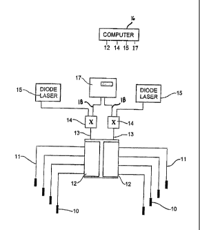
is exposed to light
from a lamp or a laser. The wavelength of the light must be suitable to
activate the drug. The
phototoxic drug Benzo-Porphyrin, for example, has a light activation
wavelength of about
680nm.
02 One limitation to this method is that the optical transparency of human
tissues is limited;
optical penetration depth is typically only a few millimetres. This depth of
penetration is
adequate for the treatment of superficial tumours af, for example, the human
airway and skin but
is too shallow for the treatment of most solid tumours. One method of
overcoming this
shortcoming is to use fibre optic light sources, known as interstitial light
probes or cylindrical
probes in the art, implanted into the tumour. In this method the optical
probes are typically
arranged in a parallel array, each spaced between half and one centimetre from
the adjacent
probe. In a typical geometry the probes are arrayed in an icosahedral pattern.
The cylindrical
source probes are typically an integral part of a light delivery fibre optic
where the distal end (1-
4cm) of the fibre optic is either coated or clad with optical scattering
material. This scattering
material allows light to radially diffuse from the side of the fibre optic;
over a length from one up
to several centimetres, perpendicular to fibreoptic axis. Light from a probe
forms a cylindrical
distribution of emission. When a parallel array of cylindrical probes is used
to illuminate tissue,
variations of light dose occur between the probes. The illumination of tissue
is greatest in the
vicinity of the source and lowest at a point equidistant from adjacent
sources. The illumination
along the length of the cylindrical probes depends upon the design of the
probe but is relatively
uniform.
CA 02465051 2004-04-23
2
03 Light delivery fibre optic cables used for interstitial PDT must be
illuminated with a laser
since lamp sources cannot be focussed onto the small aperture needed for fibre
optic excitation.
In the art, light from a single laser is usually split between several fibre
optic cables using either
beam-splitters or fibre optic sputters. This method of fibre optic
illumination is limited because
control of illumination of an individual fibre optic is not possible.
Biological tissues are
unpredictable and significant variations of the optical properties may exist
with time and across a
tumour. This is the result, for example, of variation in blood profusion and
the existence of
different structural regions within the tumour. Since exposing all points of a
tumour to a lethal
light dose is a necessary part of successful therapy and since over exposure
of tissue may result
in complications associated with damage to normal tissues surrounding the
tumour it is highly
desirable to control the illumination of an individual fibre optic so as to
expose all points of the
tissue to lethal light dose.
04 The problem of achieving a uniformly lethal light dose across a tumour is
further
complicated by dynamic changes in tissue eptical properties over the course of
treatment. These
changes occur as the result of, for example, damage to blood vessels and
consequent changes in
blood perfusion. The therapeutic effect of PDT is known to be principally the
result of the
destruction of blood vessel in and around the tumour. Tumours are well known
to have poorly
developed vascularization and lymphatic drainage as a result of tumour induced
angiogenesis.
As a result phototoxic drugs tend to accumulate in the reticuloendothelial
system within the
tumour. As well, phototoxic drugs tend to accumulate in the walls of blood
vessels of all sizes. In
addition the oxygen concentration is highest at this site such that photo-
toxins associated with
PDT cause blood vessel coagulation and collapse. Loss of blood perfusion to
the tumour results
in ischemia and indirect tumour cell death in addition to direct cell death.
The denaturization of
tissue over the course of treatment may result in changes of optical
properties and a non-uniform
application of light dose. Some drugs will act as significant tissue
chromophores if they are
present in relatively large concentrations. These drugs may also photo bleach
and produce a slow
increase in tissue transmissivity over the course of treatment.
CA 02465051 2004-04-23
OS Complications associated with PDT are associated with damage to vital
tissues in the
vicinity of the treated tissue. For example; treatment of prostate cancer with
PDT carries with it
the risk of damage to the rectum. Prostate tumours tend to occur in the
posterior part of the
prostate, which is adjacent to the rectum. PDT damage to the rectum will be
similar to that
associated with cryotherapy and can result in fistula and which may create a
complex surgical
problem for repair. Currently the only method that will prevent collateral
damage to surrounding
tissues is the control of light dose at the margins of the treatment zone.
When injected
systemically, known phototoxic drugs distribute approximately uniformly across
the patients
body tissues. Some evidence of selective accumulation of phototoxic drugs in
tumours has been
reported but the ratio of drug concentration between the tumour and its
surrounding tissues is
limited and not of significant therapeutic benefit.
06 A method of monitoring interstial light dose during PDT has been described
in the art. In
this device light from a single laser was split, using beam splitters, into
six fibre optics
connected to interstitial cylindrical probes. A mechanical apparatus was used
to obstruct five of
the six sources and place an optical detector at the proximal end of the
obstructed fibres. Light
from the remaining illuminated source was collected by the five obstructed
cylindrical probes
and the photodetector readings from the five obstructed fibres were used to
estimate the
uniformity of light dose throughout the tissue. This apparatus has the
limitation that the light
dose to each cylindrical source may not be controlled. Moreover the
photodetector switching
apparatus is relatively complicated and slow and requires direct current
motors, geaxboxes and
friction clutches to swing the gate like structures in place. This apparatus
provided an indication
of the uniformity of light dose but provided no means of correcting an
inhomogeneous dose
distribution. Because of variations of light dose among the probes, because of
beam splitting
variations and because of detector variations it was necessary to calibrate
this system using a
bath of intralipid. This procedure is not compatible with clinical practice. A
short treatment
period is required for inter-arterial drug delivery and some modern PDT drugs,
which are active
for only minutes following administration, so in these cases a. fast dose
monitoring protocol is
essential.
CA 02465051 2004-04-23
SUMMARY OF THE INVENTION
07 We disclose here several methods and apparatus for the treatment cancer
that overcomes
limitations of conventional PDT. According to an aspect of this invention,
phototoxic drug is
not applied intravenously but is applied to the arterial system of the target,
followed by
illumination of the target tissue by drug activating light. There is also
provided in accordance
with an aspect of the invention, an apparatus for performing photodynamic
therapy of a target
tissue having an arterial supply, the apparatus comprising a source of
phototoxic drug, a drug
injector having a needle for injection of the phototoxic drug; into t:he
arterial supply, the drug
injector being connected to the source of phototoxic drugt; and a photo-
dynamic light source
arranged to provide drug activating light to the target tissue. 'the
phototoxic drug preferably has
a first-pass effect.
08 We further disclose a method of achieving a uniformly lethal light dose to
the target
tissue, while monitoring in real time light and drug dose. There is therefore
providing in
accordance with an aspect of the invention, an apparatus for delivering drug
activating light to
target tissue, the apparatus comprising plural probes; and a drug activating
light delivery system
arranged to cause the plural probes, in operation, to sequentially deliver
drug activating light to
the target tissue. In addition there is a detection device for drug levels and
light dose. The drug
activating light delivery system may comprises a laser having drug activating
light emission, an
optical switch optically coupled between the laser and the plural probes, the
optical switch
having plural operating positions corresponding to connection of the laser to
respective ones of
the plural probes; and a controller far operation of the laser and the optical
switch. Several lasers
may be coupled to the probes. There is also provided a method for delivering
drug activating
light to target tissue, the method comprising the steps of placing plural
probes in sufficient
proximity to the target tissue to direct drug activating Iight towards the
target tissue and activate
drug in the target tissue; and providing drug activating light from at least
one laser to the plural
probes sequentially.
09 Still further, we disclose an apparatus, called an automatic radiance
probe, which may be
used to perform radiance measurements very rapidly and communicate these
measurements to a
CA 02465051 2004-04-23
S
control computer. Therefore, according to an aspect of the invention, there is
provided an
apparatus for delivering light to target tissue, the apparatus comprising a
light delivery filter
terminating in a radiance probe, a chuck for securing the light deliver fiber,
a motor for rotating
the chuck; and a motor control operably connected to the motor. If optical
properties are
determined throughout the tissue using this apparatus, the light dose needed
to achieve a
homogeneous light dose throughout the tissue, may be predicted. This predicted
dose
distribution allows treatment planning prior to therapy.
Still further, we describe an apparatus and method for mapping the optical
characteristics
of a solid tissue body in a time period that is clinically practical. The
apparatus comprises the
radiance probe in combination with an array of probes, and a computer for
receiving and
analyzing light intensity signals obtained from the probes. In the method of
characterizing optical
properties of a target tissue for photo-dynamic therapy, there are carried out
the steps of placing
an array of probes, for example in a human body, placing a directional probe
in the human body
with target tissue between the directional probe and the array of probes,
rotating the directional
probe, detecting intensity of light that has passed between the directional
probe and respective
probes in the array of probes; and computing optical properties of the target
tissue from the
detected light intensity. In a further aspect of the invention, there is
provided the step of, after
computing optical properties of the target tissue at a first axial location,
advancing the directional
probe in the axial direction and computing optical properties of the target
tissue at a second axial
location. The probes are preferably located in therapeutic position within
suitable needles. Each
probe in the array is preferably illuminated sequentially as the directional
probe rotates by
operation of a stepper motor.
11 In a still further aspect of the invention, a method and apparatus are
disclosed for
monitoring radiation dose applied during photodynamic therapy. In this aspect
of the invention,
light from one set of probes is received by another set of probes located with
target tissue
between the sets of probes. As probes of one set of probes transmit, the dose
applied by those
probes is detected by the other set of probes. Which probes act as
transmitters and which probes
act as receivers is switched to measure the dose applied by both sets of
probes.
CA 02465051 2004-04-23
6
12 Further summary of the invention may be found in the detailed disclosure
that follows
and the claims.
BRIEF DESCRIPTION OF THE DRAWINGS
13 There will now be described preferred embodiments of the invention with
reference to
the figures, for purposes of illustrating examples of the invention, in which:
Fig. 1 shows an apparatus for delivery of drug activating light to plural
probes, including
a detector for use in dose monitoring;
Fig. 2 shows application of phototoxic drug to the arterial supply of a
tumour;
Figs. 3A and 3B shows an automatic radiance probe for use with an embodiment
of the
invention;
Fig. 3C shows a prior art radiance probe;
Fig. 4 is a flow chart of a method of treatment;
Fig. 5 is a flow chart of another method of treatment; and
Fig. 6 is a flow chart of a method of characterizing the optical properties of
tissue.
DETAILED DESCRIPTION OF PREFERRED EMBODIMENTS
14 In this patent document, the word comprising is used in its inclusive sense
and does not
exclude other elements being present. The indefinite article "a" before an
element also does not
exclude more than one of the element being present. The term "light" or "drug
activating light"
refers to electromagnetic radiation of a wavelength suitable for drug
activation, for example
phototoxic drug activation. An "optical" element is an element capable of
transmitting and
guiding drug activating light. The term "probe" refers to a device capable of
delivering drug
activating light to target tissue. Probes are typically connected to laser
light sources through
optical fibres. A probe may also be used as a receiver of light when the probe
is connected to a
detector. A "phototoxic drug" is a drug that is activated by application of
light, and includes
typo-phyllic drugs. The phototoxic drug preferably has a first pass effect, in
which most of the
drug is taken up in the targetted tissue on it's first pass through.
s
CA 02465051 2004-04-23
7
15 Apparatus for achieving a uniformly lethal light dose to target tissue is
shown in Fig. 1.
An array of probes 10 are coupled to a drug activating light delivery system.
In this example, the
drug activating light delivery system comprises optical fibers 11 leading from
the probes 10 to
optical switch 12, which in turn is coupled through optical fibers 13 to
detector switch 14 and
from there to laser 15, or other suitable drug activating light source. A
computer 16, with
suitable labelled output ports 12, 14, 15 indicating to which element the
ports are connected, is
used to control operation of the switches and laser. The detector switch 14 is
not required for
uniform dose application, but is used for dose monitoring, as described below.
The optical
switch 12 is for example an 1 xm fiberoptic switch, where m is the number of
probes 10, for
example 4. An identical set of probes 10 and drug activating light delivery
system composed of
elements 11, 12, 13 and 14 are also shown in Fig. 1 and used for dose
monitoring as described
below.
16 The probes 10 are preferably cylindrical probes inserted interstitially
through
conventional associated needles into a tumour or diseased organ as is
currently practiced in the
art. Unlike conventional practice, however, light from one or more lasers 15
is not split between
the cylindrical probes 10 in the array but is switched sequentially between
the probes 10. In a
preferred operation of this method, all of the light from a laser 15 is
coupled to a single probe 10
until the laser 15 is switched to another probe 10. The fibreoptic switch 12
and computer 16
controls the sequence, exposure time and laser power for each probe 10 in the
array.
17 This method provides several benefits over the currently practiced art.
Since the
exposure time and illumination of each probe 10 may be varied, the dose of
light delivered to a
probe 10 and its surrounding tissue may also be varied. The light dose may be
increased in
relatively opaque parts of the tissue body and decreased in relatively
transparent parts in order to
achieve a uniform light dose. This method may consequently concentrate light
dose in
refractory parts of the tissue without over exposing the rest of the tumour
and risking collateral
damage to surrounding tissues.
CA 02465051 2004-04-23
18 This method of fibreoptic switching, moreover, has the added advantage of
potentiating
the therapeutic effect of PDT. It is well known in the art that PDT is
associated with the
depletion of tissue oxygen since the therapy is essentially one of photo-
oxidation of tissue.
Although depletion of some phototoxic drugs may occur as a result of
photochemical
dissociation, the role of the drug is principally to catalyze photo-oxidation
of tissue. F,e-
oxygenation of tissue occurs through the perfusion of blood. In the method of
switched light
delivery, tissue may be exposed for a first period until de-oxygenation causes
saturation of
therapy. Following this, a second period of no light exposure may allow re-
oxygenation before
the therapy is continued. This cycle is repeated until the required total dose
is delivered to all
probes 10 in the array. This cycle of oxygen depletion and repletion is of
course limited in those
tissue where illumination is mainly from one probe 10. For regions in the
tissue midway
between several probes the tissue will have less time to reoxygenate between
light exposures.
19 In a preferred example of operation of apparatus used fox PDT, drug is not
applied
intravenously but is applied to the arterial system of the target tissues
using angiographic
radiological techniques. For example, as shown in Fig. 2, a source 20 of
phototoxic drug is
connected to supply drug to a drug injector 22 for injection of the phototoxic
drug into tile
arterial supply 24 of a tumour 25. A photo-dynamic light source, here shown by
laser 15, switch
12 and probes 10, is arranged to provide drug activating light to the target
tissue. The drug
injector may for example be a pump 22 that pumps drug into the arterial supply
24 using a drl~g
delivery tube 26 that terminates in an angiocath needle (not shown) inserted
through a tracker
small vessel catheter (not shown). Preferably, a tissue imaging system 27,
such as a conventional
electromagnetic (radiographic) imaging system located near the patient, is
used for coordinating
motion of the angiocath needle in conventional fashion. The phototoxic drug
may for example be
Benzo-Porphyrin or Hypocrellin or other Typo-phyllic drug, and the target
tissue may be W a
prostate. Referring now to Fig. 4, the method of use of the above example is
described, denoted
generally by reference character 40. The phototoxic drug is injected into the
arterial supply of
the target tissue in step 41, and is activated by light in step 42. The motion
of the angiocath
needle at the end of the drug delivery tube 26 is tracked using the tissue
imaging system in step
43.
CA 02465051 2004-04-23
9
20 Lypo-phyllic drugs will aggregate in the blood stream, temporarily embolize
and adhere
effectively to the small capillaries in the vicinity of the infra-arterial
injection point. Application
of drug to the arterial supply of the target tissue may consequently be used
to achieve high
concentration of the drug in the target tissue and low concentration in
surrounding tissues that
draw blood supply from other parts of the vascular system. lFor example,
application of drug to
the arterial supply of prostate will result in the concentration of phototoxic
drug within prostate,
which is over one hundred times greater than that in surrounding tissues such
as the rectum and
urethra. This technique should therefore protect vital surrounding tissues
over the course of
PDT and significantly reduce the incidence and risk of complication. This
protection should
persist until the drug leaks from the target tissue and is distributed
systemically to surrounding
organs and tissues. The period of selective drug uptake is adequately long for
PDT treatment
following infra-arterial application of drug.
21 PDT may be made more effective through treatment planning. Switched fibre
optic light
delivery by apparatus illustrated in Fig. 1 may be used to control the light
dose needed to
overcome the optical inhomogeneity of malignant tissue. In order to plan a
treatment prior to
therapy, the distribution of optical properties throughout the tissue body
must be known.
Optical properties may be determined using a method known in the art as the P3
Approximation
in conjunction with the measurement of tissue radiance. An automatic
directional radiance probe
30, with fiber optic 11 terminating in radiance probe 10, chuck 31, motor 32
and handle 33,
shown in Figs. 3A and 3B, may be used to perform radiance measurements very
rapidly arid
communicate these measurements to a control computer. If optical properties
are determined
throughout the tissue using this apparatus, the light dose needed to achieve a
homogeneous light
dose throughout the tissue, may be predicted. This predicted dose distribution
allows treatment
planning prior to therapy. Referring now to Fig. 6, a method of characterizing
the optical
properties 60 is described. In step 61, an array of probes is placed in a
human body. In step 62,
a directional probe is placed in the human body with target tissue between the
directional probe
and the array of probes. The directional probe is rotated in step 63, and in
step 64, light intensity
of light that has passed between the directional probe and respective probes
in the array of probes
CA 02465051 2004-04-23
is detected. The optical properties of the target tissue are then computed
from the detected light
intensity in step 66. The probe is then advanced in step 66, and the optical
properties of the new
location are also computed by returning to step 63.
22 As described in the art, a conventional radiance probe may be used to
characterize the
optical properties between the probe and a small spherical light source. Fig.
3C shows a sketch
of a radiance probe 10, which includes a fiber optic 11, with conventional
coating 34, for
delivering light to and receiving light from the radiance probe 10. At the end
of the radiance
probe 10 a coated right angle prism 35 is attached with optical epoxy 36 and
protected with a
protective glass dome 37. The probe 10 is inserted into an afterloading needle
38. The radiance
probe 10 will detect a maximum of scattered light when orientated in the
direction of the light
source and a minimum of scattered light when orientated 180 degrees away from
the source. The
distribution of detected light between these two extremes is <;alled a
radiance characteristic and
this may be combined with the P3 Approximation to determine tissue optical
properties between
the source and the probe. Radiance probes described in the art are of very
little clinical utility.
Prior art radiance probes must be oriented toward the measurement path. The
probe and the
source must have the same axial position and the orientation of the probe with
respect to the path
must be known. In order to map the optical characteristics of a solid tissue
body many paths
must be characterized and prior art radiance probes are too slow and laborious
to be clinically
practical.
23 A novel apparatus and method for mapping the optical characteristics of a
solid tissue
body in a time period that is clinically practical is now described.
Conventional transparent
needles are implanted into the tissue body under acoustic or fluoroscopic
imaging guidance.
These needles are placed in a parallel array such that when cylindrical probes
10 are introduced
into the needles, the cylindrical probes 10 will be of suitable length and
suitable spacing for
effective therapy. Prior to treatment the automatic radiance probe 30 is used
to map the optical
properties of the target tissue.
CA 02465051 2004-04-23
11
24 The radiance probe 30 is motorized with stepping motor 32 under the control
of computer
16 or another computer. The fibreoptic cable 11, attached to the radiance
probe 30, is inserted
and clamped using chuck 31, in the rotating head of the stepper motor 32, as
illustrated in Fig.
3A. The chuck 31 rotates under control of the motor 32 between positions 180
degrees each side
of a central position. This allows full 360 degree coverage without risking
breakage of the optical
fiber, which needs to twist as the chuck 31 is rotated. The stepper motor 32
and radiance probe
30 are attached to a handle 33 so that an operator may manually insert the
radiance probe 10 into
the conventional transparent needle (not shown). The handle 33 of the probe 30
also contains a
small microprocessor needed to coordinate the rotation of the probe 30 with
the rest of the
apparatus. Date may be acquired for example at every 10 degrees over each 180
degree sweep.
cylindrical probes are placed in the transparent needles adjacent to the
needle containing the
rotating radiance probe 30. Referring now to Fig. 5, the method of use 50 is
shown. The
radiance probes 50 are placed in proximity to the target tissue in step 51. In
step 52, the radiance
probe 30 and the adjacent cylindrical probes 10 are synchronized so that
typically four adjacent
probes are sequentially illuminated as the probe 30 rotates four times and
records the radiance
data for the four paths between the probes 10 and the radiance probe 30. The
radiance probe 30
is advanced axially by movement of the probe 30 through the needle into which
it is inserted, aaad
the radiance (light intensity) measurement is repeated for typically three to
four points along the
length of the transparent needle. The probes 10 or radiance probe 30 may be
used as either
transmitter or receiver.
25 This method, which makes use of a cylindrical light source 10 rather than a
spherical
source, consequently avoids the time consuming step of aligning a source and
the radiance probe
30 at a specific axial position. The computer records the measured radiance
characteristics and
these characteristics are aligned in software with a stored normalized
radiance characteristic.
This avoids the step of mechanical angular orientation needed with
conventional radiance
probes. The computer compares radiance characteristics with the P3
Approximation and the
optical characteristics of the tissue between the cylindrical source and the
radiance probe are
computed and stored. The radiance probe 30 may be introduced into each needle
and th.e
resulting data may be used to map optical parameters in three dimensions.
Tissues found to be
CA 02465051 2004-04-23
12
more inhomogeneous require more measurement points than those relatively free
from
inhomogeneity. The disclosed method and apparatus consequently avoids the
orientation and
positioning steps required by prior art radiance probes and automatically
computes optical
characteristics a solid tissue body. The time required for optical mapping of
for example the
human prostate depends upon the number of sources used and the homogeneity of
the tissue but
is typically in the order of minutes.
26 Following the characterization of the tissue body, described above, a
treatment plan is
computed. The computer 16 uses an optical transport model and the measured
tissue parameters
to predict the light fluence that will result from illumination of the tissue
body by the array of
cylindrical probes 10. The distribution of light- dose delivered by the
cylindrical probe array
needed to produce a uniform and Lethal light- dose at all points is then
calculated. The dose is
controlled by either time or light level fractionation. In the method of time-
fractionation the
laser power is held constant and the time period, for which each cylindrical
source is connected
to the laser, is varied. The longer the connected time, the greater will be
the integrated light-
dose to tissue surrounding the cylindrical probe 10. In the method of light
level fractionation the
connection time to each cylindrical probe 10 is held fixed and the laser power
delivered to the
cylindrical probes 10 is varied. Although time fractionation is technically
easier than light level
fractionation the exposure period for each source must be chosen so that
reoxygenation of tissue
occurs between sequential treatments and this requirement limits the time
fractionation protocol.
27 Although treatment planning is an essential part of any physical treatment
modality, this
planning process has limitations. Cylindrical probes 10 do not provide control
of light emission
along the length of the probe and typical cylindrical probes have significant
variation in emission
along their length. A planning protocol that factors in manufacturing
variation of cylindrical
probes is too slow and complex. For fast acting drugs characterization must be
performed prior
to treatment. Phototoxic drugs when present in tissue become chromophores and,
in high enough
concentration, will change the optical properties of the tissue. Drug
bleaching and PDT induced
tissue changes are known to change tissue light transmissivity over the course
of treatment. It is
consequently highly desirable to have a method of real time tissue light dose
monitoring so that
CA 02465051 2004-04-23
13
the evolution of light dose may be monitored and modified if necessary from
the beginning to the
termination of treatment.
28 A novel dose monitoring apparatus that overcomes limitations of prior art
devices will
now be described. The method uses fibreoptic switches 12 and 14 shown in Fig.
1 to integrate
the functions of dose delivery and dose monitoring. The switches 12 and 14 may
for example be
bidirection fiberoptic switches available from LIGHTech Fiberoptics Inc.
connectorized with
SMA905 connectors. The apparatus may be used to interactively adjust light
dose delivery to
tissue to ensure a uniformly lethal light dose. Fibre optic light switches 12
and 14 a.re
commonly used in the telecommunications art. They are very fast, reliable,
have reproducible
performance and are mass-produced. Manufacturers of fibre optic switching
apparatus routinely
mount customized switches and fibre optic lasers within a single rack mounted
enclosure and
provide a single digital input for remote computer control, such as by
computer 16. Such a fibre
switch enclosures are compatible with clinical practice. The external
connections to such <~n
enclosure are an array of fibre optics 1 l, 13 terminated with cylindrical
probes 10 and a single
digital control line.
29 Fig. 1 shows a typical embodiment of this apparatus. 'rhe external
fibreoptics 11, 13 are
connected to two 1xN switches 12. In the example shown, N=4. The single ends
of these
switches are connected to two 1x2 switches 14. One side of the lx2 switches 14
connects to a
fibreoptically-coupled laser 15 and the other side of the 1 x2 switch 15
connects to a
fibreoptically-coupled photodetector 17, for example a detector with high
sensitivity such as a
152 mm integrating sphere detector available from Melles Ciroot. In order to
coordinate the
physical distribution of illumination with the computer cont~.°ols,
each fibre 1l, 13 is given a
physical number and computer number. Fibreoptics 11 from the two 1 x4 switches
14 ar.°e
interleaved so that they are connected to adjacent cylindrical probes 10.
30 In therapeutic mode both lasers 15 are connected to the cylindrical probes
10. Both lasers
15 switch sequentially between four probes 10 so that two probes 10 are
simultaneously
illuminated. The delivery sequence of the two lasers 15 is chosen to minimizes
the volume of
CA 02465051 2004-04-23
14
tissue illuminated by both lasers I5. The computer 16 controls the light dose
delivered to each
point in the predetermined manner described earlier. Both the power output of
the two lasers 15
and the exposure time of each probe 10 may be varied to achieve the desired
light dose
distribution.
31 In tissue monitoring mode one of the 1 x2 switches 14~ is connected to the
detector I 7 (a
photodiode for example) and the other is connected to a laser 15. The
cylindrical probes 10
connected to the photodiode 17 collect Light when the probes 10 connected to
the laser 15 are
sequentially illuminated. The collected light is transmitted along the fibre
optic 18, monitored by
the photo detector 17 and recorded by the computer 16. In typical animal
tissues, light fluence
falls exponentially with a penetration depth of a few millimetres so only
those cylindrical probes
adjacent to the illuminated probe collect a significant light level. Following
this the 1x2
switches are thrown and the sequence is reversed between laser 15 and detector
probes 10. Tlhe
computer 16 uses these measurements to estimate the optical extinction
coefficient between each
of the cylindrical probes 10. This information is combined with a light
diffusion model and tlhe
light dose applied to each probe 10 during therapy to create a two-dimensional
plot of light dose
over a plane normal to the probes. This of course is an imaginary plane that
represents the
average dose along the length of the probes 10. The computer 16 may then, if
necessary, iterate
the probe dose to correct for regions of low light dose and a new delivery
protocol, which differs
from the planned protocol, may be implemented at the operators discretion.
Because fibre optic
switches may be switched in milliseconds the time needed to perform dose
monitoring and
information is only seconds.
32 An example of a treatment cycle would have one 2xl switch switched to one
laser source
and the desired output fiber of the related 1x9 switch would. be chosen to
deliver light to the
tissue. Meanwhile, the other 2x1 switch would be selected to detection and the
nearby fibers to
the source would be scanned and an optical power measurement would be taken at
each location.
The power at each location is then communicated and recorded by the computer,
thus allowing
for real-time dose monitoring. This continues until each fibre in the array
has been used as a
source and the corresponding light levels in nearby tissue measured. Note that
the switching
CA 02465051 2004-04-23
speed for the optical switch is on the order of 150ms, which is negligible
when compared to the
total treatment time. The cycle repeats until the desired dose level has been
reached.
33 The lasers used may operate at for example 690 nm or 532 nm, and may be
obtained for
example from Optical Fiber Systems Inc., including laser diode, driver
electronics and cooler.
The laser wavelength is largely dictated by the chosen photasensitizer. OS-QLT-
0074 may be
used, which shows strong absorption at 690 nm. This wavelength penetrates more
through xhe
prostate than light at 630 nm. The fiber optics may be terminated with 15 mm
cylindrical
diffusing tips available from Polymicro Technologies. An icosahedral pattern
of the probes may
be used to assist with dose uniformity, in which pattern the probes are
equally space around
target tissue approximately at the corners of an icosahedron.
34 Immaterial modifications may be made to the embodiments described here
without
departing from the invention.