Note: Descriptions are shown in the official language in which they were submitted.
CA 02465220 2004-04-27
WO 03/040413 PCT/US02/35515
SYSTEM FOR DETECTING BIOLOGICAL MATERIALS IN A SAMPLE
[0001] This application claims the benefit of U.S. Provisional Patent
Application Serial No. 60/332,838, filed November 6, 2001, which is hereby
incorporated by reference in its entirety.
FIELD OF THE INVENTION
[0002] The present invention relates to systems and methods for the
detection of target molecules, such as deoxyribonucleic acids (DNA) or
ribonucleic acids (RNA), from fluid samples.
BACKGROUND OF THE INVENTION
[0003] Nucleic acids, such as DNA or RNA, have become of increasing
interest as analytes for clinical or forensic uses. Powerful new molecular
biology
technologies enable one to detect congenital or infectious diseases. These
same
technologies can characterize DNA for use in settling factual issues in legal
proceedings, such as paternity suits and criminal prosecutions.
[0004] For the analysis and testing of nucleic acid molecules,
amplification of a small amount of nucleic acid molecules, isolation of the
amplified nucleic acid fragments, and other procedures are necessary. The
science of amplifying small amounts of DNA have progressed rapidly and several
methods now exist. These include linked linear amplification, ligation-based
amplification, transcription-based amplification, and linear isothermal
amplification. Linked linear amplification is described in detail in U.S.
Patent No.
6,027,923 to Wallace et al. Ligation-based amplification includes the ligation
amplification reaction (LAR) described in detail in Wu et al., Genomics, 4:560
(1989) and the ligase chain reaction described in European Patent No.
032030881
to Backman et al. Transcription-based amplification methods are described in
detail in U.S. Patent No. 5,766,849 to McDonough et al.; U.S. Patent No.
5,654,142 to Kievits et al., Kwoh et al., Proc. Natl. Acad. Sci. U.S.A.,
86:1173
(1989), and PCT Publication No. WO 88/10315 to.Ginergeras et al. The more
CA 02465220 2004-04-27
WO 03/040413 PCT/US02/35515
-2-
recent method of linear isothermal amplification is described in U.S. Patent
No.
6,251,639 to Kurn.
[0005] The most common method of amplifying DNA is by the
polymerase chain reaction ("PCR"), described in detail by Mullis et al., Cold
S~rin~ Harbor Ouant. Biol., 51:263-273 (1986), European Patent No. 201,184 to
Mullis, U.S. Patent No. 4,582,788 to Mullis et al., European Patent Nos.
50,424,
84,796, 258017, and 237362 to Erlich et al., and U.S. Patent No. 4,683,194 to
Saiki et al. The PCR reaction is based on multiple cycles of hybridization and
nucleic acid synthesis and denaturation in which an extremely small number of
nucleic acid molecules or fragments can be multiplied by several orders of
magnitude to provide detectable amounts of material. One of ordinary skill in
the
art knows that the effectiveness and reproducibility of PCR amplification is
dependent, in part, on the purity and amount of the DNA template. Certain
molecules present in biological sources of nucleic acids are known to stop or
inhibit PCR amplification (Belec et al., Muscle and Nerve, 21(8):1064 (1998);
Wiedbrauk et al., Journal of Clinical Microbiology, 33(10):2643-6 (1995);
Deneer
and Knight, Clinical Chemistry, 40(1):171-2 (1994)). For example, in whole
blood, hemoglobin, lactofernn, and immunoglobulin G are known to interfere
with several DNA polymerases used to perform PCR reactions (Al-Soud and
Radstrom, Journal of Clinical Microbiolo~y, 39(2):48593 (2001); Al-Soud et
al., Journal of Clinical Microbiology, 38(1):345-50 (2000)). These inhibitory
effects can be more or less overcome by the addition of certain protein
agents, but
these agents must be added in addition to the multiple components already used
to
perform the PCR. Thus, the removal or inactivation of such inhibitors is an
important factor in amplifying DNA from select samples.
[0006] On the other hand, isolation and detection of particular nucleic acid
molecules in a mixture requires a nucleic acid sequencer and fragment
analyzer, in
which gel electrophoresis and fluorescence detection are combined.
Unfortunately, electrophoresis becomes very labor-intensive as the number of
samples or test items increases.
[0007] For this reason, a simpler method of analysis using DNA
oligonucleotide probes is becoming popular. New technology, called VLSIPSTM,
has enabled the production of chips smaller than a thumbnail where each chip
CA 02465220 2004-04-27
WO 03/040413 PCT/US02/35515
-3-
contains hundreds of thousands or more different molecular probes. These
techniques are described in U.S. Patent No. 5,143,854 to Pirrung et al., PCT
Publication No. WO 92/10092 to Fodor et al., and PCT Publication No. WO
90/15070 to Fodor et al. These biological chips have molecular probes arranged
in arrays where each probe ensemble is assigned a specific location. These
molecular array chips have been produced in which each probe location has a
center to center distance measured on the micron scale. Use of these array
type
chips has the advantage that only a small amount of sample is required, and a
diverse number of probe sequences can be used simultaneously. Array chips have
been useful in a number of different types of scientific applications,
including
measuring gene expression levels, identification of single nucleotide
polymorphisms, and molecular diagnostics and sequencing as described in U.S.
Patent No. 5,143,854 to Pirrung et al.
[0008] Array chips where the probes are nucleic acid molecules have been
1 S increasingly useful for detection for the presence of specific DNA
sequences.
Most technologies related to array chips involve the coupling of a probe of
known
sequence to a substrate that can either be structural or conductive in nature.
Structural types of array chips usually involve providing a platform where
probe
molecules can be constructed base by base or covalently binding a completed
molecule. Typical array chips involve amplification of the target nucleic acid
followed by detection with a fluorescent label to determine whether target
nucleic
acid molecules hybridize with any of the oligonucleotide probes on the chip.
After exposing the array to a sample containing target nucleic acid molecules
under selected test conditions, scanning devices can examine each location in
the
array and quantitate the amount of hybridized material at that location.
[0009] However, this method requires the use of fluorescent or radioactive
labels as additional materials. Such a system is expensive to use and is not
amenable to being made portable for biological sample detection and
identification. Furthermore, the hybridization reactions take up to two hours,
which for many uses, such as detecting biological warfare agents, is simply
too
long. Therefore, a need exists for a system which can rapidly detect
biological
material in samples.
[0010] The present invention is directed to achieving these objectives.
CA 02465220 2004-04-27
WO 03/040413 PCT/US02/35515
-4-
SUMMARY OF THE INVENTION
[0011] The present invention relates to a detection cartridge containing a
housing defining a first chamber and a detection chip within the first chamber
defined by the housing. The detection chip includes two or more electrically
separated conductors fabricated on a substrate. Capture probes are attached to
the
conductors such that a gap exists between the capture probes on the
electrically
separated conductors. A sample, potentially containing a target molecule, can
be
analyzed for the presence of that target molecule by determining whether the
conductors are electrically connected.
[0012] The present invention also relates to a system for detecting a target
molecule in a sample. The system includes a detection cartridge that contains
a
housing defining a first chamber and a detection chip within the first chamber
defined by the housing. The detection chip includes two or more electrically
1 S separated conductors fabricated on a substrate and capture probes attached
to the
conductors such that a gap exists between the capture probes on the
electrically
separated conductors. A sample, potentially containing a target molecule, can
be
analyzed for the presence of the target molecule by determining whether the
conductors are electrically connected. An electrical connector extends through
the
housing and is coupled to the electrically separated conductors so that the
presence of a target molecule connecting the capture probes on the
electrically
separated conductors can be detected. The system also includes a support unit
with respect to which the detection cartridge can be positioned to carry out a
procedure for detecting the target molecule in a sample. The support unit has
an
electrical coupler suitable for electrical communication with the electrical
connector of the detection cartridge. As a result, the presence of the target
molecule in the sample can be detected and communicated to the support unit.
[0013] Another aspect of the present invention relates to a method of
detecting a target molecule. The method involves providing a detection system
that includes a detection cartridge containing a housing defining a first
chamber
and a detection chip within the first chamber defined by the housing. The
detection chip includes two or more electrically separated conductors
fabricated
CA 02465220 2004-04-27
WO 03/040413 PCT/US02/35515
-5-
on a substrate and capture probes attached to the conductors such that a gap
exists
between the capture probes on the electrically separated conductors. A sample,
potentially containing a target molecule, can be analyzed for the presence of
that
target molecule by determining whether the conductors are electrically
connected.
An electrical connector extends through the housing and is coupled to the
electrically separated conductors so that the presence of a target molecule
connecting the capture probes on the electrically separated conductors can be
detected. The system also includes a support unit with respect to which the
detection cartridge can be positioned to carry out a procedure for detecting
the
1.0 target molecule in a sample. The support unit has an electrical coupler
suitable for
electrical communication with the electrical connector of the detection
cartridge.
A sample, potentially containing the target molecule, is injected into the
first
chamber of the housing. Then, the sample is processed within the first chamber
under conditions effective to permit any of the target molecule present in the
15 sample to bind to the capture probes and thereby connect the capture
probes.
Finally, the presence of the target molecule is detected by determining
whether
electricity is conducted between the electrically separated conductors.
[0014] In comparison to other detection systems which require the use of
fluorescent or radioactive labels and a long reaction time, the present
invention
20 discloses a rapid and economical system for detecting target molecules in a
sample. In particular, the disclosed system is amenable to being made portable
for
biological sample detection and identification, and is, thus, highly effective
for
many uses such as detecting biological warfare agents.
25 BRIEF DESCRIPTION OF THE DRAWINGS
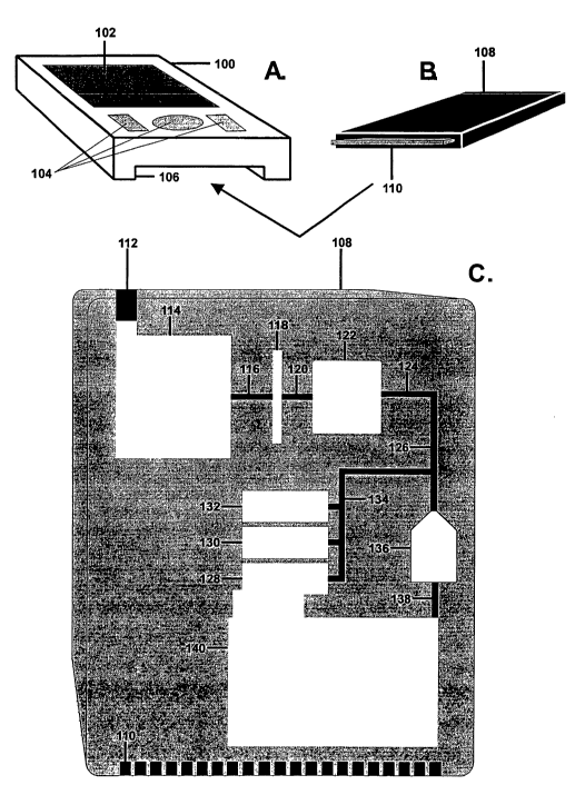
[0015] Figures 1 A-B show a perspective view of a system for detection of
a target nucleic acid molecule from a sample which includes a desk-top
detection
unit and a cartridge which is inserted into the desk-top unit. Figure 1 C
shows a
schematic view of this system.
30 [0016] Figure 2A depicts a single test structure on a detection chip
suitable
to be positioned in first chamber 20 of the system shown in Figures lA-C,
where
oligonucleotide probes are attached to electrical conductors in the form of
spaced
CA 02465220 2004-04-27
WO 03/040413 PCT/US02/35515
-6-
apart conductive fingers. Figure 2B shows how a target nucleic acid molecule
present in a sample is detected by the detection chip.
[0017] Figures 3A-B show a perspective view of a system for detection of
a target nucleic acid molecule which includes a portable detection unit and a
cartridge which is inserted into the portable unit. Figure 3C shows a
schematic
view of this system.
DETAILED DESCRIPTION OF THE INVENTION
[0018] The present invention relates to a detection cartridge containing a
housing defining a first chamber and a detection chip within the first chamber
defined by the housing. The detection chip includes two or more electrically
separated conductors fabricated on a substrate. Capture probes are attached to
the
conductors such that a gap exists between the capture probes on the
electrically
separated conductors. A sample, potentially containing a target molecule, can
be
1 S analyzed for the presence of that target molecule by determining whether
the
conductors are electrically connected.
[0019] The present invention also relates to a system for detecting a target
molecule in a sample. The system includes a detection cartridge that contains
a
housing defining a first chamber and a detection chip within the first chamber
defined by the housing. The detection chip includes two or more electrically
separated conductors fabricated on a substrate and capture probes attached to
the
conductors such that a gap exists between the capture probes on the
electrically
separated conductors. A sample, potentially containing a target molecule, can
be
analyzed for the presence of the target molecule by determining whether the
conductors are electrically connected. A first injection port is provided in
the
housing through which a sample solution can be introduced into the first
chamber.
An electrical connector extends through the housing and is coupled to the
electrically separated conductors so that the presence of a target molecule
connecting the capture probes on the electrically separated conductors can be
detected. The system also includes a support unit into which the detection
cartridge can be positioned to carry out a procedure for detecting the target
molecule in a sample. The support unit has an electrical coupler suitable for
CA 02465220 2004-04-27
WO 03/040413 PCT/US02/35515
electrical communication with the electrical connector of the detection
cartridge.
As a result, the presence of the target molecule in the sample can be detected
and
communicated to the support unit.
[0020] Figures lA-B show a perspective view of a system for detection of
a target nucleic acid molecule from a sample. This system includes a desk-top
detection unit and a detection cartridge which is inserted into the desk-top
unit. In
this embodiment, desk-top detection unit 2 is provided with door 4 for filling
reagents, control buttons 6, and visual display 10. Slot 8 in desk-top
detection
unit 2 is configured to receive detection cartridge 12. Detection cartridge 12
further contains first injection port 14 through wl~ch a sample solution can
be
introduced into a first chamber in cartridge 12 and second injection port 16
through which reagents can be introduced into the first chamber.
[0021] Figure 1 C shows a schematic view of the system utilizing desk-top
detection unit 2 and cartridge 12. In this system, desk-top detection unit 2
contains containers 32A-C suitable for holding reagents and positioned to
discharge the reagents into first chamber 20 of detection cartridge 12 through
second injection port 16 and conduit 21. Containers 32A-C can, for example,
carry a neutralizer, a buffer, a conductive ion solution, and an enhancer. The
contents of these containers can be replenished through door 4. This is
achieved
by making containers 32A-C sealed and disposable or by making them refillable.
[0022] Pump 28 removes reagents from containers 32A-C, through tubes
30A-C, respectively, and discharges them through tube 26 and second injection
port 16 into detection cartridge 12. Instead of using single pump 28 to draw
reagents from containers 32A-C, a separate pump can be provided for each of
containers 32A-C so that their contents can be removed individually.
[0023] Alternatively, the necessary reagents may be held in containers
inside the detection cartridge. The pumps in the detection unit can force a
material, such as air, water or oil, into the detection cartridge to force the
reagents
from the respective containers and into the first chamber. The reagents are
then
changed with each detection cartridge, which eliminates the buildup of salt
precipitates in the detection unit.
[0024] Desk-top detection unit 12 is also provided with controller 38,
which is in electrical communication with the electrical conductors of the
CA 02465220 2004-04-27
WO 03/040413 PCT/US02/35515
-g_
detection cartridge 12 by means of electrical connector 36, to detect the
presence
of the target molecule in the sample. Controller 38 also operates pump 28 by
way
of electrical connector 34. Alternatively, separate controllers can be used
for
operating the pumps and the detection of target molecules. Digital coupling 40
permits controller 38 to communicate data to computer 42 which is external of
desk-top detection unit 12.
[0025] Detection cartridge 12 contains first chamber 20 which, as noted
supra, receives reagents from within desk-top detection unit 2 by way of
second
injection port 16 and conduit 21. A sample to be analyzed is discharged to
first
chamber 20 through first injection port 14 and conduit 18. As described more
fully infra, the presence of a target molecule is detected in first chamber
20.
Detection cartridge 12 is further provided with second chamber 24 for
collecting
material discharged from first chamber 20 by way of connector 22. The
detection
cartridge also contains electrical connector 25 extending through the housing
and
coupled to the electrically separated conductors in first chamber 20 so that
the
presence of a target molecule in a sample can be detected.
[0026] Figure 2A depicts a single test structure on a detection chip suitable
to be positioned in first chamber 20 of the system shown in Figures lA-C.
According to Figure 2A, oligonucleotide probes 46 attached to spaced apart
conductive fingers 44 are physically located at a distance sufficient that
they
cannot come into contact with one another. A sample, containing a mixture of
nucleic acid molecules (i.e. M1-M6), to be tested is contacted with the
fabricated
device on which conductive fingers 44 are fixed, as shown in Figure 2B. If a
target nucleic acid molecule (i.e. M1) that is capable of binding to the two
oligonucleotide probes is present in the sample, the target nucleic acid
molecule
will bind to the two probe molecules. If bound, the nucleic acid molecule can
bridge the gap between the two electrodes and provide an electrical
connection.
Any unhybridized nucleic acid molecules (i.e. M2-M6) not captured by the
probes
is washed away. Here, the electrical conductivity of nucleic acid molecules is
relied upon to transmit the electrical signal. Hans-Werner Fink and Christian
Schoenenberger reported in Nature, 398:407-410 (1999), which is hereby
incorporated by reference in its entirety, that DNA conducts electricity like
a
semiconductor. This flow of current can be sufficient to construct a simple
CA 02465220 2004-04-27
WO 03/040413 PCT/US02/35515
-9-
switch, which will indicate whether or not a target nucleic acid molecule is
present
within a sample. The presence of a target molecule can be detected as an "on"
switch, while a set of probes not connected by a target molecule would be an
"ofl"
switch. The information can be processed by a digital computer which
correlates
the status of the switch with the presence of a particular target. The
information
can be quickly identified to the user as indicating the presence or absence of
the
biological material, organism, mutation, or other target of interest.
Optionally,
after hybridization of the target molecules to sets of biological probes, the
target
molecule can be coated with a conductor, such as a metal. The coated target
molecule can then conduct electricity across the gap between the pair of
probes;
thus producing a detectable signal indicative of the presence of a target
rr~olecule.
[0027] The detection chip, on which conductive fingers 44 are fixed, is
constructed on a support. Examples of useful support materials include, e.g.,
glass, quartz, and silicon as well as polymeric substrates, e.g. plastics. In
the case
1 S of conductive or semi-conductive supports, it will generally be desirable
to
include an insulating layer on the support. However, any solid support which
has
a non-conductive surface may be used to construct the device. The support
surface need not be flat. In fact, the support may be on the walls of a
chamber in a
chip.
[0028] Improved methods of forming large arrays of oligonucleotides,
peptides and other polymer sequences with a minimal number of synthetic steps
are known. See, U.S. Patent No. 5,143,854 to Pirrung et al. (see also, PCT
Publication No. WO 90/15070 to Fodor et al.) and PCT Publication No.
WO 92/10092 to Fodor et al., which are hereby incorporated by reference in
their
entirety, which disclose methods of forming vast arrays of peptides,
oligonucleotides, and other molecules using, for example, light-directed
synthesis
techniques. See also, Fodor et al., Science, 251:767-77 (1991), which is
hereby
incorporated by reference in its entirety. These procedures for synthesis of
polymer arrays are now referred to as VLSIPSTM procedures.
[0029] Methods of synthesizing desired oligonucleotide probes are known
to those of skill in the art. In particular, methods of synthesizing
oligonucleotides
and oligonucleotide analogues can be found in, for example, Oligonucleotide
Synthesis~ A Practical Approach, Gait, ed., IRI Press, Oxford (1984);
Kuijpers,
CA 02465220 2004-04-27
WO 03/040413 PCT/US02/35515
-10-
Nucleic Acids Research 18(17):5197 (1994); Dueholm, J. Org. Chem., 59:5767-
5773 (1994); and Agrawal (ed.), Methods in Molecular Biolo~y, 20, which are
hereby incorporated by reference in their entirety. Shorter oligonucleotide
probes
have lower specificity for a target nucleic acid molecule, that is, there may
exist in
nature more than one target nucleic acid molecule with a sequence of
nucleotides
complementary to the oligonucleotide probe. On the other hand, longer
oligonucleotide probes have decreasingly smaller probabilities of containing
complementary sequences to more than one natural target nucleic acid molecule.
In addition, longer oligonucleotide probes exhibit longer hybridization times
than
shorter oligonucleotide probes. Since analysis time is a factor in a
commercial
device; the shortest possible probe that is sufficiently specific to the
target nucleic
acid molecule is desirable. Both the speed and specificity of binding target
nucleic acid molecules to oligonucleotide probes can be increased if one
electrical
conductor has attached a probe that is complementary to one end of the target
nucleic acid molecule and the other electrical conductor has attached a probe
that
is complementary to the other end of the target nucleic acid. In this case,
even if
short oligonucleotide probes that exhibit rapid hybridization rates are used,
the
specificity of the target nucleic acid molecules to the two probes is high. If
two
different probe molecules are used, it is important that both probes are not
located
on the same electrical conductor, to prevent hybridization of a target nucleic
acid
molecule from one part of an electrical conductor to another part of the same
electrical conductor. If this happens, no signal can be generated from such an
attachment, and the sensitivity of the analysis is lowered.
[0030] The present invention includes chemically modified nucleic acid
molecules or oligonucleotide analogues as oligonucleotide probes. An
"oligonucleotide analogue" refers to a polymer with two or more monomeric
subunits, wherein the subunits have some structural features in common with a
naturally occurring oligonucleotide which allow it to hybridize with a
naturally
occurnng nucleic acid in solution. For instance, structural groups are
optionally
added to the ribose or base of a nucleoside for incorporation into an
oligonucleotide, such as a methyl or allyl group at the 2'-O position on the
ribose,
or a fluoro group which substitutes for the 2'-O group, or a bromo group on
the
ribonucleoside base. The phosphodiester linkage, or "sugar-phosphate backbone"
CA 02465220 2004-04-27
WO 03/040413 PCT/US02/35515
-11-
of the oligonucleotide analogue is substituted or modified, for instance with
methyl phosphonates or O-methyl phosphates. Another example of an
oligonucleotide analogue includes "peptide nucleic acids" in which native or
modified nucleic acid bases are attached to a polyamide backbone.
Oligonucleotide analogues optionally comprise a mixture of naturally occurring
nucleotides and nucleotide analogues. Oligonucleotide analogue arrays composed
of oligonucleotide analogues are resistant to hydrolysis or degradation by
nuclease
enzymes such as RNAase A. This has the advantage of providing the array with
greater longevity by rendering it resistant to enzymatic degradation. For
example,
analogues comprising 2'-O-methyloligoribonucleotides arr. A~asistant to RNAase
A.
[0031] Many modified nucleosides, nucleotides, and various bases suitable
for incorporation into nucleosides are commercially available from a variety
of
manufacturers, including the SIGMA chemical company (Saint Louis, Mo.), R&D
systems (Minneapolis, Minn.), Pharmacia LKB Biotechnology (Piscataway, N.J.),
CLONTECH Laboratories, Inc. (Palo Alto, Calif.), Chem Genes Corp., Aldrich
Chemical Company (Milwaukee, Wis.), Glen Research, Inc., GIBCO BRL Life
Technologies, Inc. (Gaithersberg, Md.), Fluka Chemica-Biochemika Analytika
(Fluka Chemie AG, Buchs, Switzerland), Invitrogen, San Diego, Calif., and
Applied Biosystems (Foster City, Calif.), as well as many other commercial
sources known to one of skill. Methods of attaching bases to sugar moieties to
form nucleosides are known. See, e.g., Lukevics and Zablocka, Nucleoside
Synthesis: Organosilicon Methods Ellis Horwood Limited Chichester, West
Sussex, England (1991), which is hereby incorporated by reference in its
entirety.
Methods of phosphorylating nucleosides to form nucleotides, and of
incorporating
nucleotides into oligonucleotides are also known. See, e.g., Agrawal (ed),
Protocols for Oligonucleotides and Analogues, Synthesis and Properties,
Methods
in Molecular Biolo~y, volume 20, Humana Press, Towota, N.J. (1993), which is
hereby incorporated by reference in its entirety.
[0032] The probes may be targeted to the electrically separated conductors
by using a chemical reaction for attaching the probe or nucleotide to the
conductor
which preferably binds the probe or nucleotide to the conductor rather than
the
support material. Alternatively, the probe or nucleotide may be targeted to
the
conductor by building up a charge on the conductor which electrostatically
attracts
CA 02465220 2004-04-27
WO 03/040413 PCT/US02/35515
-12-
the probe or nucleotide. See U.S. Patent Application Serial No. 10/159,429,
which is hereby incorporated by reference in its entirety.
[0033] Another aspect of the present invention relates to a method of
detecting a target molecule. The method involves providing a detection system
that includes a detection cartridge containing a housing defining a first
chamber
and a detection chip within the first chamber defined by the housing. The
detection chip includes two or more electrically separated conductors
fabricated
on a substrate and capture probes attached to the conductors such that a gap
exists
between the capture probes on the electrically separated conductors. A sample,
potentially containing a target molecule, can be analyzed for the presence of
that
target molecule by determining whether the conductors are electrically
connected.
A first injection port is provided in the housing through which a sample
solution
can be introduced into the first chamber. An electrical connector extends
through
the housing and is coupled to the electrically separated conductors so that
the
presence of a target molecule connecting the capture probes on the
electrically
separated conductors can be detected. The system also includes a support unit
into which the detection cartridge can be positioned to carry out a procedure
for
detecting the target molecule in a sample. The support unit has an electrical
coupler suitable for electrical communication with the electrical connector of
the
detection cartridge. A sample, potentially containing the target molecule, is
injected into the first chamber of the housing. Then, the sample is processed
within the first chamber under conditions effective to permit any of the
target
molecule present in the sample to bind to the capture probes and thereby
connect
the capture probes. Finally, the presence of the target molecule is detected
by
determining whether electricity is conducted between the electrically
separated
conductors. The presence of the target molecule is indicated by the ability to
conduct an electrical signal through the circuit. In the case where the target
molecule is not present, the circuit is not be completed. Thus, the target
molecule
acts as a switch. The presence of a target molecule can be detected as an "on"
switch, while a set of probes not connected by a target molecule would be an
"off'
switch. Due to the direct detection of the target molecule, the method allows
for
extremely sensitive detection of target molecules. The information can be
processed by a digital computer which correlates the status of the switch with
the
CA 02465220 2004-04-27
WO 03/040413 PCT/US02/35515
-13-
presence of a particular target. The computer can also analyze the results
from
several switches specific for the same target, to determine specificity of
binding
and target concentration.
[0034] In one embodiment, the native electrical conductivity of nucleic
acid molecules can be relied upon to transmit the electrical signal. Fink et
al.
"Electrical Conduction through DNA Molecules," Nature. 398:407-410 (1999),
which is hereby incorporated by reference in its entirety, reported that DNA
conducts electricity like a semiconductor. This flow of current can be
sufficient to
construct a simple switch. Thus, another aspect of the present invention
relates to
a method for uetecting a target nucleic acid molecule in a sample. The method
first involves providing an apparatus which includes first and second
electrical
conductors each having detection sites located less than 250 microns apart but
not
in contact with one another. The first electrical conductor is made of a first
type
of conductive material and the second electrical conductor is made of a second
type of conductive material which is different than the first type of
conductive
material. The apparatus also includes a first set of oligonucleotide probes
attached
to the detection sites of the first electrical conductors with an attachment
chemistry which binds the first set of oligonucleotide probes to the first
electrical
conductor but not to the second electrical conductor. Finally, the apparatus
includes a second set of oligonucleotide probes attached to the detection
sites of
the second electrical conductors and spaced apart from the first set of
oligonucleotide probes by a gap. Next, the probes are contacted with a sample
potentially containing a target nucleic acid molecule under conditions
effective to
permit any of the target nucleic acid molecule in the sample to hybridize to
both
of the spaced apart oligonucleotide probes to bridge the gap and electrically
couple the pair of oligonucleotide probes with the hybridized target nucleic
acid
molecule, if any. The electrically coupled pair of oligonucleotide probes and
the
hybridized target nucleic acid molecule are then filled with a filling nucleic
acid
sequence, where the filling nucleic acid sequence is complementary to the
target
nucleic acid molecule and extends between the pair of oligonucleotide probes.
Finally, it is determined if an electrical current can be carried between the
probes,
where the electrical current between the probes indicates the presence of the
target
CA 02465220 2004-04-27
WO 03/040413 PCT/US02/35515
-14-
nucleic acid molecule in the sample which has sequences complementary to the
probes.
(0035] Alternatively, after hybridization of the target nucleic acid
molecule to the oligonucleotide probes, the hybridized target nucleic acid
molecule is coated with a conductive material, such as a metal, as described
in
U.S. Patent Application Serial Nos. 60/095,096 or 60/099,506, which are hereby
incorporated by reference in their entirety. Examples of conductive material
include silver and gold. The coated nucleic acid molecule can then conduct
electricity across the gap between the pair of probes, thus producing a
detectable
1 ~ signal indicative of the presence of a target~nucleic acid. molecule.
Thus, the
present invention relates to a method for detecting a target nucl:;ic acid
molecule
in a sample. The method first involves providing an apparatus which includes
first and second electrical conductors each having detection sites located
less than
2'50 microns apart but not in contact with one another. The first electrical
conductor is made of a first type of conductive material and the second
electrical
conductor is made of a second type of conductive material which is different
than
the first type of conductive material. The apparatus also includes a first set
of
oligonucleotide probes attached to the detection sites of the first electrical
conductors with an attachment chemistry which binds the first set of
oligonucleotide probes to the first electrical conductor but not to the second
electrical conductor. Finally, the apparatus includes a second set of
oligonucleotide probes attached to the detection sites of the second
electrical
conductors and spaced apart from the first set of oligonucleotide probes by a
gap.
Next, the probes are contacted with a sample potentially containing a target
nucleic acid molecule under conditions effective to permit any of the target
nucleic acid molecule in the sample to hybridize to both of the spaced apart
oligonucleotide probes to bridge the gap and electrically couple the pair of
oligonucleotide probes with the hybridized target nucleic acid molecule, if
any. A
conductive material is then applied over the electrically coupled pair of
oligonucleotide probes and the hybridized target nucleic acid molecule.
Finally, it
is determined if an electrical current can be carried between the probes,
where the
electrical current between the probes indicates the presence of the target
nucleic
acid molecule in the sample which has sequences complementary to the probes.
CA 02465220 2004-04-27
WO 03/040413 PCT/US02/35515
-15-
[0036] For instance, the sodium counter ions to DNA phosphate groups
can be replaced with silver ions by flooding the sample area with silver
nitrate
solution. After washing away excess silver nitrate, bathing the area with a
photographic developer such as hydroquinone reduces the silver ions to
metallic
S silver, which is electrically conductive. Braun et al. demonstrated that
silver
could be deposited along a DNA molecule (Braun et al., "DNA-Templated
Assembly and Electrode Attachment of a Conducting Silver Wire," Nature,
391:775-778 (1998), which is hereby incorporated in its entirety). A three-
step
process is used. First, silver is selectively localized to the DNA molecule
through
a Ag+/Na+ ion-exchange (Barton, Bioinor~anic Chemistry eds Be mini; ~: al.,
ch. 8,
University Science Books, Mill Valley, (1994), which is hereby incorporated by
reference in its entirety) and complexes are formed between the silver and the
DNA bases (Spiro, ed., Nucleic Acid-Metal Ion Interactions Wiley Interscience,
New York (1980); Marzeilli, et al., J. Am. Chem. Soc., 99:2797 (1977);
Eichorn,
ed. Inorg_anic Biochemistry, Vol. 2, ch 33-34, Elsevier, Amsterdam, (1973),
which
are hereby incorporated by reference in their entirety). The ion-exchange
process
may be monitored by following the quenching of the fluorescence signal of the
labeled DNA. The silver ion-exchanged DNA is then reduced to form aggregates
with bound to the DNA skeleton. The silver aggregates are further developed
using standard procedures, such as those used in photographic chemistry
(Holgate,
et al., J. Histochem. Cytochem. 31:938 (1983); Birell, et al., J. Histochem.
~ochem. 34:339 (1986), which are hereby incorporated by reference in their
entirety).
[0037] Thus, the detection of a target molecule using a desk-top detection
system, as shown in Figures lA-C, can be carned out as follows. After lysis
and
clarification of the sample, the sample is introduced into detection cartridge
12
through first injection port 14 and conduit 18 and into first chamber 20. Once
the
sample is introduced, detection cartridge 12 is inserted into slot 8 of desk-
top
detection unit 2 so that second injection port 16 is connected to conduit 21
and
electrical connector 36 is coupled to electrical connector 25. The sample is
processed in first chamber 20 containing the capture probes and electrical
conductors for a period of time sufficient for detection of a target nucleic
acid
molecule in the sample. Processing of the sample within first chamber 20 can
CA 02465220 2004-04-27
WO 03/040413 PCT/US02/35515
-16-
involve neutralizing the sample, contacting the neutralized sample with a
buffer,
then treating the sample with conductive ions, and treating the sample with an
enhancer. Molecules that are not captured are expelled from first chamber 20
through second conduit 22 and into second chamber 24. The desk-top detection
system can be programmed by a series of operation buttons 6 on the front of
the
device and the results can be seen on visual display 10.
[0038] Figures 3A-B show a portable detection system. This system is
provided with a portable unit 100 which can be in the form of a portable
personal
digital assistant (e.g., a Palm~ unit, 3Com Corporation, Santa Clara, CA).
Portable unit 100 is provided with visual display 102 and control buttons 104.
Slot 106 is provided to receive detection cartridge 108 having electrical
connector
110.
[0039] Figure 3C shows a schematic diagram of detection cartridge 108
which is used in the portable detection system of the present invention.
Detection
cartridge 108 contains first injection port 112 in the housing through which a
sample solution can be introduced.
[0040] Detection cartridge 108 contains a plurality of containers 128, 130,
and 132 suitable for holding reagents and positioned to discharge the reagents
into
conduit 126 through conduit 134. Containers 128, 130, and 132 can, for
example,
carry a neutralizer, a buffer, and a conductive ion solution.
[0041] Sample pre-treatment chamber 114 is positioned upstream of first
chamber 122, with filter 118 being positioned between pretreatment chamber 114
and first chamber 122. Conduits 116 and 120 provide a path between
pretreatment chamber 114 and first chamber 122. Detection cartridge 108 also
contains conduit 124 that receives material from chamber 122. Conduit 124 has
a
small diameter so that nucleic acid material is sheared as it passes from
first
chamber 122 to detection chamber 136. Detection cartridge 108 also contains a
waste chamber 140 coupled to detection chamber 136 by way of conduit 138 so
that material discharged from the detection chamber 136 is received in waste
chamber 140. Detection cartridge 108 includes a series of electrical
connectors
110 that are coupled to the electrically separated conductors in detection
chamber
136, like those shown in first chamber 20 for the embodiment of Figures lA-C
and 2.
CA 02465220 2004-04-27
WO 03/040413 PCT/US02/35515
-17-
[0042] In operation, the detection of a target molecule using a portable
detection system, as shown in Figures 3A-C, can be carned out as follows.
After
lysis and clarification of the sample, the sample solution is introduced into
detection cartridge 108 through first injection port 112. Within sample
pretreatment chamber 114, cells are lysed to release cellular contents. After
denaturation and deprotination, the sample can be partially purified by
passing it
through filter 118 and depositing the solution into chamber 122. Within first
chamber 138, the neutralized target nucleic acid molecule, if present in the
sample, is permitted to hybridize with the capture probes on the electrically
separated conductors in first chamber 136 in substantially the same way as
described above with reference to Figures 1 A-C and 2. After binding and
washing, the sample is treated with a conductive ion solution from container
128,
such that conductive ions are deposited on the target molecules that have
hybridized to the capture probes on the detection chip. Additionally, after
treatment with a conductive ion solution, the sample can be treated with an
enhancer solution from container 130 to grow a continuous layer of conductive
metal from the deposited conductive ions. Excess buffers and waste buffers
will
exit detection chamber 136 through waste tube 138 and collect in second
chamber
140. The portable detection system can be programmed by operation of a series
of buttons 104 on the front of portable unit 100, and the results are
visualized on
screen 102.
(0043] In carrying out the method of the present invention, a sample
collection phase is initially carried out where bacteria, viruses or other
species are
collected and concentrated. The target nucleic acid molecule whose sequence is
to be determined is usually isolated from a tissue sample. If the target
nucleic acid
molecule is genomic, the sample may be from any tissue (except exclusively red
blood cells). For example, saliva, whole blood, peripheral blood lymphocytes
or
PBMC, skin, hair or semen are convenient sources of clinical samples. These
sources are also suitable if the target is RNA. Blood and other body fluids
are
also a convenient source for isolating viral nucleic acids. If the target
nucleic acid
molecule is mRNA, the sample is obtained from a tissue in which the mRNA is
expressed. If the target nucleic acid molecule in the sample is RNA, it may be
reverse transcribed to DNA, but need not be converted to DNA.
CA 02465220 2004-04-27
WO 03/040413 PCT/US02/35515
-18-
[0044] A plurality of collection methods can be used depending on the
type of sample to be analyzed. Liquid samples can be collected by placing a
constant volume of the liquid into a lysis buffer. Airborne samples can be
collected by passing air over a filter for a constant time. The filter can be
washed
with lysis buffer. Alternatively, the filter can be placed directly into the
lysis
buffer. Waterborne samples can be collected by passing a constant amount of
water over a filter. The filter can then be washed with lysis buffer or soaked
directly in the lysis buffer. Dry samples can be directly deposited into lysis
buffer
for removal of the organism of interest.
10. [004] When whole cells, viruses, or other tissue samples are being
analyzed, it is typically necessary to extract the nucleic acids from the
cells or
viruses, prior to continuing with the various sample preparation operations.
Accordingly, following sample collection, nucleic acids may be liberated from
the
collected cells, viral coat, etc., into a crude extract, followed by
additional
15 treatments to prepare the sample for subsequent operations such as
denaturation of
contaminating (DNA binding) proteins, purification, filtration, desalting, and
the
like.
[0046] Liberation of nucleic acids from the sample cells or viruses, and
denaturation of DNA binding proteins may generally be performed by physical or
20 chemical methods. For example, chemical methods generally employ lysing
agents to disrupt the cells and extract the nucleic acids from the cells,
followed by
treatment of the extract with chaotropic salts such as guanidinium
isothiocyanate
or urea to denature any contaminating and potentially interfering proteins.
Generally, where chemical extraction and/or denaturation methods are used, the
25 appropriate reagents may be incorporated within the extraction chamber, a
separate accessible chamber, or externally introduced.
[0047] Alternatively, physical methods may be used to extract the nucleic
acids and denature DNA binding proteins. U.S. Patent No. 5,304,487 to Wilding
et al., which is hereby incorporated by reference in its entirety, discusses
the use
30 of physical protrusions within microchannels or sharp edged particles
within a
chamber or channel to pierce cell membranes and extract their contents. More
traditional methods of cell extraction may also be used, e.g., employing a
channel
with restricted cross-sectional dimension which causes cell lysis when the
sample
CA 02465220 2004-04-27
WO 03/040413 PCT/US02/35515
-19-
is passed through the channel with sufficient flow pressure. Alternatively,
cell
extraction and denaturing of contaminating proteins may be carried out by .
applying an alternating electrical current to the sample. More specifically,
the
sample of cells is flowed through a microtubular array while an alternating
electric
current is applied across the fluid flow. A variety of other methods may be
utilized within the device of the present invention to effect cell
lysis/extraction,
including, e.g., subjecting cells to ultrasonic agitation, or forcing cells
through
microgeometry apertures, thereby subjecting the cells to high shear stress
resulting
in rupture.
[0048] Following extraction, it is often desirable to separate the n~~clcic
acids from other elements of the crude extract, e.g., denatured proteins, cell
membrane particles, and the like. Removal of particulate matter is generally
accomplished by filtration, flocculation, or the like. Ideally, the sample is
concentrated by filtration, which is more rapid and does not require special
reagents. A variety of filter types may be readily incorporated into the
device.
Samples can be forced through filters that will allow only the cellular
material to
pass through, trapping whole organisms and broken cell debris. Further, where
chemical denaturing methods are used, it may be desirable to desalt the sample
prior to proceeding to the next step. Desalting of the sample, and isolation
of the
nucleic acid may generally be earned out in a single step, e.g., by binding
the
nucleic acids to a solid phase and washing away the contaminating salts or
performing gel filtration chromatography on the sample. Suitable solid
supports
for nucleic acid binding include, e.g., diatomaceous earth, silica, or the
like.
Suitable gel exclusion media is also well known in the art and is commercially
available from, e.g., Pharmacia and Sigma Chemical. This isolation and/or gel
filtration/desalting may be carried out in an additional chamber, or
alternatively,
the particular chromatographic media may be incorporated in a channel or fluid
passage leading to a subsequent reaction chamber.
[0049] Alternatively, the interior surfaces of one or more fluid passages or
chambers may themselves be derivatized to provide functional groups
appropriate
for the desired purification, e.g., charged groups, affinity binding groups
and the
like.
CA 02465220 2004-04-27
WO 03/040413 PCT/US02/35515
-20-
[0050] The oligonucleotide probes of the present invention may be
designed to specifically recognize a variation in the sequence at the end of
the
probe. After the target nucleic acid molecule binds to the probes, the target
nucleic acid molecule is treated with nucleases to remove the ends of the
molecule
which do not bind to the probes. If the confronting ends of the two probes
contain
sequences complementary to the target nucleic acid molecule, treatment with
ligase will join the confronting ends of the two probes. The test chamber can
then
be heated up to denature non-ligated target nucleic acid molecule from the
probes.
Detection of the specific target nucleic acid molecule can then be carried
out.
[0051] In a preferred embodiment of the invention, ligation methods may
be used to specifically identify single base differences in sequences.
Previously,
methods of identifying known target sequences by probe ligation methods have
been reported (U.S. Patent No. 4,883,750 to Whiteley et al.; Wu et al.,
Genomics,
4:560 (1989); Landegren et al., Science, 241:1077 (1988); and Winn-Deen et
al.,
Clin. Chem., 37:1522 (1991), which are hereby incorporated by reference in
their
entirety). In one approach, known as oligonucleotide ligation assay ("OLA"),
two
probes or probe elements which span a target region of interest are hybridized
to
the target region. Where the probe elements basepair with adjacent target
bases,
the confronting ends of the probe elements can be joined by ligation, e.g., by
treatment with ligase. The ligated probe element is then assayed, evidencing
the
presence of the target sequence.
[0052] Hybridization assays on substrate-bound oligonucleotide arrays
involve a hybridization step and a detection step. Homologous nucleotide
sequences can be detected by selectively hybridizing to each other.
Selectively
hybridizing is used herein to mean hybridization of DNA or RNA probes from
one sequence to the "homologous" sequence under stringent or non-stringent
conditions (Ausubel et al., eds., Current Protocols in Molecular Biology, Vol.
I:
2.10.3, Greene Publishing Associates, Inc. and John Wiley & Sons, Inc., New
York (1989), which is hereby incorporated by reference in its entirety).
Hybridization and wash conditions are also exemplified in Sambrook et al.,
Molecular Cloning: A Laboratory Manual, Second Edition, Cold Spring Harbor,
NY (1989), which is hereby incorporated by reference in its entirety.
CA 02465220 2004-04-27
WO 03/040413 PCT/US02/35515
-21 -
[0053] A variety of hybridization buffers are useful for the hybridization
assays of the invention. Addition of small amounts of ionic detergents (such
as N-
lauroyl-sarkosine) are useful. LiCI is preferred to NaCI. Additional examples
of
hybridization conditions are provided in several sources, including: Sambrook
et
al., Molecular Cloning: A Laboratory Manual, 2nd Ed., Cold Spring Harbor, NY
(1989); Berger et al., "Guide to Molecular Cloning Techniques," Methods in
Enzymology, Volume 152, Academic Press, Inc., San Diego, Calif. (1987); and
Young et al., Proc. Natl. Acad. Sci. USA, 80:1194 (1983), which are hereby
incorporated by reference in their entirety. In addition to aqueous buffers,
non-
aqueous buffers may also be used. In particular, non-aqueous buffers which
facilitate hybridization but have low electrical conductivity are preferred.
[0054] The hybridization mixture is placed in contact with the array and
incubated. Contact can take place in any suitable container, for example, a
dish or
a cell specially designed to hold the probe array and to allow introduction of
the
fluid into and removal of it from the cell so as to contact the array.
Generally,
incubation will be at temperatures normally used for hybridization of nucleic
acids, for example, between about 20°C and about 75°C, e.g.,
about 25°C, about
30°C, about 35°C, about 40°C, about 45°C, about
50°C, about 55°C, about 60°C,
or about 65°C. For probes longer than about 14 nucleotides, 37-
45°C is preferred.
For shorter probes, 55-65°C is preferred. More specific hybridization
conditions
can be calculated using formulae for determining the melting point of the
hybridized region. Preferably, hybridization is carned out at a temperature at
or
between ten degrees below the melting temperature and the melting temperature.
More preferred, the hybridization is carned out at a temperature at or between
five
degrees below the melting temperature and the melting temperature. The target
is
incubated with the probe array for a time sufficient to allow the desired
level of
hybridization between the target and any complementary probes in the array.
The
hybridization mixture may contain an isostabilizing agent, a denaturing agent,
or a
renaturation accelerant.
[0055] Including a hybridization optimizing agent in the hybridization
mixture significantly improves signal discrimination between perfectly matched
targets and single-base mismatches. As used herein, the term "hybridization
optimizing agent" refers to a composition that decreases hybridization between
CA 02465220 2004-04-27
WO 03/040413 PCT/US02/35515
-22-
mismatched nucleic acid molecules, i.e., nucleic acid molecules whose
sequences
are not exactly complementary.
[0056] An isostabilizing agent is a composition that reduces the base-pair
composition dependence of DNA thermal melting transitions. More particularly,
the term refers to compounds that, in proper concentration, result in a
differential
melting temperature of no more than about 1 °C. for double stranded DNA
oligonucleotides composed of AT or GC, respectively. Isostabilizing agents
preferably are used at a concentration between 1 M and 10 M, more preferably
between 2 M and 6 M, most preferably between 4 M and 6 M, between 4 M and
1 ~~ i 0 M, and, optimally, at about 5 M. For example, a 5 M agent in 2 x SSPE
(Sodium Chloride/Sodium Phosphate/EDTA solution) is suitable. Betaines and
lower tetraalkyl ammonium salts are examples of suitable isostabilizing
agents.
Betaine (N,N,N,-trimethylglycine) can eliminate the base pair composition
dependence of DNA thermal stability (Rees et al., Biochemistry, 32:137-144
(1993), which is hereby incorporated by reference in its entirety). Unlike
tetramethylammonium chloride ("TMACI"), betaine is zwitterionic at neutral pH
and does not alter the polyelectrolyte behavior of nucleic acids while it does
alter
the composition-dependent stability of nucleic acids. Inclusion of betaine at
about
5 M can lower the average hybridization signal, but increases the
discrimination
between matched and mismatched probes.
[0057] A denaturing agent is a compositions that lowers the melting
temperature of double stranded nucleic acid molecules by interfering with
hydrogen bonding between bases in a double-stranded nucleic acid or the
hydration of nucleic acid molecules. Denaturing agents can be included in
hybridization buffers at concentrations of about 1 M to about 6 M and,
preferably,
about 3 M to about S.5 M. Denaturing agents include formamide, formaldehyde,
dimethylsulfoxide ("DMSO"), tetraethyl acetate, urea, guanidine thiocyanate
("GuSCN"), glycerol and chaotropic salts. As used herein, the term "chaotropic
salt" refers to salts that function to disrupt van der Waal's attractions
between
atoms in nucleic acid molecules. Chaotropic salts include, for example, sodium
trifluoroacetate, sodium tricholoroacetate, sodium perchlorate, and potassium
thiocyanate.
CA 02465220 2004-04-27
WO 03/040413 PCT/US02/35515
- 23 -
[0058] A renaturation accelerant is a compound that increases the speed of
renaturation of nucleic acids by at least 100-fold. They generally have
relatively
unstructured polymeric domains that weakly associate with nucleic acid
molecules. Accelerants include heterogenous nuclear ribonucleoprotein ("hnRP")
A1 and cationic detergents such as, preferably, cetyltrimethylammonium bromide
("CTAB") and dodecyl trimethylammonium bromide ("DTAB"), and, also,
polylysine, spermine, spermidine, single stranded binding protein ("SSB"),
phage
T4 gene 32 protein, and a mixture o~ ammonium acetate and ethanol.
Renaturation accelerants can be included in hybridization mixtures at
concentrations of about 1 mu M to about 10 mM and, preferably, 1 nra ~~= to
about
1 mM. The CTAB buffers work well at concentrations as low as 0.1 mM.
[0059] After incubation with the hybridization mixture, the array usually is
washed with the hybridization buffer, which also can include the hybridization
optimizing agent. These agents can be included in the same range of amounts as
for the hybridization step, or they can be eliminated altogether. Then, the
array
can be examined to identify the probes to which the target has hybridized.
[0060] Nucleases can be used to remove probes which are attached to the
wrong conductor. More particularly, a target nucleic acid molecule may be
added
to the probes. Targets which bind at both ends to probes, one end to each
conductor, will have no free ends and will be resistant to exonuclease
digestion.
However, probes which are positioned so that the target cannot contact both
conductors will be bound at only one end, leaving the molecule subject to
digestion. Thus, improperly located probes can be removed while protecting the
properly located probes. After the protease is removed or inactivated, the
target
nucleic acid molecule can be removed and the device is ready for use.
[0061] The number of probes may be increased in order to determine
concentrations of the target nucleic acid molecule. If a plurality of each
pair of
oligonucleotide probes is provided, the method of the present invention can be
used to identify the number of pairs of identical oligonucleotide probes
between
which electrical current passes to quantify the amount of the target nucleic
acid
molecule present in the sample. For example, several thousand repeated probes
may be produced in the detection apparatus. The circuit would be able to count
CA 02465220 2004-04-27
WO 03/040413 PCT/US02/35515
-24-
the number of occupied sites. Calculations could be done by the unit to
determine
the concentration of the target nucleic acid molecule.
[0062] The method of the present invention can be used for numerous
applications, such as detection of pathogens or viruses. For example, samples
S may be isolated from drinking water or food and rapidly screened for
infectious
organisms, using probes that are complementary to the genetic material of a
pathogenic bacteria. In recent times, there have been several large recalls of
tainted meat products. The method of the present invention can be used for the
in-
process detection of pathogens in foods and the subsequent disposal of the
contaminated materials. This could significantly iii prove food safety,
prevent
food borne illnesses and death, and avoid costly recalls. Detection devices
with
oligonucleotide probes that are complementary to the genetic material of
common
food borne pathogens, such as Salmonella and E. coli., could be designed for
use
within the food industry.
[0063] In yet another embodiment, the method of the present invention
can be used for real time detection of biowarfare agents, by using probes that
are
complementary to the genetic material of a biowarfare agent. With the recent
concerns of the use of biological weapons in a theater of war and in terrorist
attacks, the device could be configured into a personal sensor for the combat
soldier or into a remote sensor for advanced warnings of a biological threat.
The
devices which can be used to specifically identity of the agent, can be
coupled
with a modem to send the information to another location. Mobile devices may
also include a global positioning system to provide both location and pathogen
information.
[0064] In yet another embodiment, the present invention may be used to
identify an individual, by using probes that are complementary to the genetic
material of a human. A series of probes, of sufficient number to distinguish
individuals with a high degree of reliability, are placed within the device.
Various
polymorphism sites are used. Preferentially, the device can determine the
identity
to a specificity of greater than one in 1 million, more preferred is a
specificity of
greater than one in one billion, even more preferred is a'specificity of
greater than
one in ten billion. The present invention may be used to screen for mutations
or
polymorphisms in samples isolated from patients.
CA 02465220 2004-04-27
WO 03/040413 PCT/US02/35515
- 25 -
[0065] This invention may also be used for nucleic acid sequencing using
hybridization techniques. Such methods are described in U.S. Patent No.
5,837,832 to Chee et al., which is hereby incorporated by reference in its
entirety.
EXAMPLES
[0066] The following examples are provided to illustrate embodiments of
the present invention but are by no means intended to limit its scope.
Example 1- Detection of Target Nucleic Acid Molecules in a Sample
Containing Purified DNA
[0067] In a prophetic example, a 10 pl sample containing approximately
100 ng of purified DNA dissolved in hybridization buffer (100 mM NaPhosphate,
pH 7.5, 0.1% SDS) with a defined length of 5.7 kilobases is injected into the
denaturation chamber. The nucleic acid denatures for approximately 1 minute
before the chamber is evacuated and the sample passed along to the
hybridization
chamber. The nucleic acid sample resides in the hybridization chamber over the
test structures for 5 minutes at a temperature of 55 degrees. The sample is
evacuated from the hybridization chamber with a 10 sample volume wash with
hybridization buffer. The nucleic acid sample is washed into the waste
chamber.
A 10 sample volume wash with distilled and deionized water rinses out the
chamber and prepares the sensor for chemical coating. The metallization
chemistry is then mixed on a card having electrically separated conductors and
passed through the hybridization chamber at a fixed flow rate such that the
test
structures are in contact with the solution for a defined time. The test
structures
are rinsed with 10 sample volumes of distilled and deionized water. The test
structures are then electrically probed individually to determine the
resistance of
each test structure. Resistance is obtained by passing a current (200 nA)
through
one of the two electrical test pads on each test structure and measuring the
resistance between the two electrodes. Low resistance indicates the
metallization
process has fused two electrodes and is a positive result.
CA 02465220 2004-04-27
WO 03/040413 PCT/US02/35515
-26-
Example 2 - Detection of Target Nucleic Acid Molecules in a Sample
Containing Bacteria
[0068] In a prophetic example, a known quantity of bacteria are placed
S into lysis solution (Tris-CL, SDS) for 1 minute to break open bacteria. The
cell
debris is removed via filtration and the genomic DNA sheared by passing the
solution through a point-sink shearing cartridge (65 p.m diameter tubing). A
10 pl
sample of the partially purified lysate in hybridization buffer (100 mM
NaPhosphate, pH 7.5, 0.1% SDS) is injected into the denaturation chamber. The
nucleic acid denatures for approximately 1 minute before the chamber is
evacuated and the sample is passed along to the hybridization chamber. The
nucleic acid sample resides in the hybridization chamber over the test strucW
ies
for 5 minutes at a temperature of SS degrees. The sample is evacuated from the
hybridization chamber with a 10 sample volume wash with hybridization buffer.
The nucleic acid sample is washed into the waste chamber. A 10 sample volume
wash with distilled and deionized water rinses out the chamber and prepares
the
sensor for chemical coating. The metallization chemistry is then mixed on a
card
having electrically separated conductors and passed through the hybridization
chamber at a fixed flow rate such that the test structures are in contact with
the
solution for a defined time. The test structures are rinsed with 10 sample
volumes
of distilled and deionized water. The test structures are then electrically
probed
individually to determine the resistance of each test structure. Resistance is
obtained by passing a current (200 nA) through one of the two electrical test
pads
on each test structure and measuring the resistance between the two
electrodes.
Low resistance indicates the metallization process has fused two electrodes
and is
a positive result.
[0069] Although the invention has been described in detail for the purpose
of illustration, it is understood that such detail is solely for that purpose,
and
variations can be made therein by those skilled in the art without departing
from
the spirit and scope of the invention which is defined by the following
claims.