Note: Descriptions are shown in the official language in which they were submitted.
CA 02465490 2004-04-29
WO 03/068306 PCT/US02/28830
1
VASCULAR EXCLUSION CATHETER
BACKGROUND OF THE INVENTION
Field of the Invention
This invention relates generally to apparatus and methods for at
least partially occluding flow within a body conduit.
Discussion of the Relevant Art
Body conduits commonly provide for a flow of fluid from one
location in the body to another location in the body. Typical of these fluid
conduits are arteries and veins of the vascular system which provide a flow of
blood between the heart and the organs of the body. When a particular
procedure requires that the vessel be accessed, the flow of blood can be
expected to exit the conduit through any access hole. This not only results in
a
loss of fluid such as blood, but also invades the general surgical environment
with the fluid. In one such procedure, it is desirable to harvest the
saphenous
vein from the leg and to connect that vein to the ascending aorta in a
Coronary
Artery Bypass Grafting (CABG) procedure.
CA 02465490 2004-04-29
WO 03/068306 PCT/US02/28830
2
In the past, surgeons used an occlusion catheter to stop the flow of
blood through the conduit or vessel. This catheter was provided with a
spherical
balloon which, when inflated, would totally obstruct the blood flow within the
vessel. Particularly in the case of blood vessels, this is undesirable as an
uninterrupted flow of blood is necessary to maintain the tissues of the body.
In order to avoid total occlusion, another procedure has been
developed whereby the blood is totally removed upstream of the operative site
and introduced down-stream of the operative site. In this procedure, commonly
referred to as an "on-pump" (OPCABG) procedure, there is continuous
uninterrupted beating of the heart. Nevertheless, this procedure requires
management of blood flow from the aortotomy in order to create a viable
proximal anastomosis. It is for this reason that many CAPD procedures are
still
performed off-pump.
Presently the surgeon's primary tool to accomplish sensation of
blood flow from the aortotomy is a Partial Occluding Clamp. In these off-pump
procedures, the Partial Occlusion Clamp is often used to engage the conduit or
vessel exteriorly and thereby isolated a small portion of the vessel from the
ongoing fluid flow.
While the partial occluding clamp is relatively simple to use, it is
perceived by many to be very traumatic. Its use has been reported to cause
secondary complications such as the fracturing of plague and resultant
Transient
Ischemic Attack or Cerebral Vascular Accident, with both local and global
consequences. The partial occluding clamp also consumes much of the
CA 02465490 2004-04-29
WO 03/068306 PCT/US02/28830
procedural area not only with its jaws on the aorta, but also with its clamp
handles in the surgical field.
SUMMARY OF THE INVENTION
These deficiencies of the past are overcome with the present
invention which includes a catheter with a dilation assembly capable of
maintaining fluid flow through a conduit while excluding a portion of the
conduit
from this fluid flow. Importantly, this catheter is non-invasive and is
inserted
endoluminally so that it does not require major space in the surgical
environment.
The dilation assembly of the catheter is capable of maintaining fluid flow
within
the conduit while producing an exclusion cavity that isolates a portion of the
conduit from this fluid flow.
In one aspect the invention, a fluid-control device is adapted for
disposition in a body conduit for controlling a flow of body fluids in the
body
conduit. The device includes a wall of separation having a first surface and
an
opposing second surface. The first surface defines a flow passage facilitating
the
flow of body fluids within the body conduit; and the second surface of the
wall
defines an exclusion chamber sealed from the flow passage and the flow of body
fluids through the body conduit.
In another aspect of the invention, a catheter is adapted for
disposition in a body conduit and includes a shaft which extends along an axis
between a proximal end and a distal end. A dilation assembly is disposed at
the
distal end of the shaft and includes a first dilator operable to move between
a
CA 02465490 2004-04-29
WO 03/068306 PCT/US02/28830
4
high-profile state and a low-profile state. A second dilator is included in
the
assembly and is operable to move generally independently of the first dilator
between the high-profile state and the low-profile state. A sleeve is carried
by
the first dilator and the second dilator between the high-profile state and
the low-
profile state.
In another aspect of the invention, a catheter is adapted for
disposition in a body conduit. The catheter includes a shaft and a dilation
assembly disposed at a distal end of the shaft. The dilation assembly has a
low-
profile state facilitating insertion of the assembly into the body conduit and
a
high-profile state facilitating operation of the assembly within the body
conduit.
The shaft includes an inner member which is disposed in a telescoping
relationship with an outer member. A dilator has a first end carried by the
outer
member and a second end carried by the inner member. These first and second
ends have a generally proximate relationship when the dilator is in the high-
profile state and a generally spaced relationship when the dilator is in the
low-
profile state.
In another aspect of the invention, the dilation assembly includes a
balloon that is inflatable to move the balloon to the high-profile state. In
this
state, first portions of the balloon define a fluid flow path to facilitate a
flow of
fluids in the body conduit.
In a further aspect, the invention includes an endovascular method
for restricting blood flow along a predetermined area of a vessel without
occluding blood flow through the vessel. This method includes the step of
CA 02465490 2004-04-29
WO 03/068306 PCT/US02/28830
providing a catheter with a dilation assembly having a wall movable between a
high-profile state and a low-profile state. The assembly is inserted into the
vessel to an operative site in the low-profile state. At the operative site,
the
assembly is dilated to move the wall to the high-profile state where the wall
5 defines with the predetermined area of the vessel an occlusion cavity
isolated
from the blood flow within the vessel.
These and other features and advantages of the invention will be
further discussed with reference to preferred embodiments of the invention and
reference to the associated drawings.
DESCRIPTION OF THE DRAWINGS
FIG. 1 is an axial cross-section of a catheter having a dilation
assembly in accordance with the present invention;
FIG. 2 is an axial cross-section view similar to FIG. 1 and
illustrating the dilation assembly in a low-profile state;
FIG. 3 is an axial cross-section view showing the dilation assembly
in a high-profile state and disposed within a body conduit;
FIG. 4 is an end view of the dilation assembly taken along lines 4-4
of FIG. 3;
FIG. 5 is an axial cross-section view similar to FIG. 3 and
illustrating movement of the dilation assembly between the high-profile state
and
the low-profile state;
CA 02465490 2004-04-29
WO 03/068306 PCT/US02/28830
6
FIG. 6 is an axial cross-section view similar to FIG. 5 and
illustrating a sleeve carried by dilators in a body conduit having a variable
diameter;
FIG. 7 is an axial cross-section view similar to FIG. 6 and
illustrating formation of a flow passage and in an exclusion cavity in
accordance
with the present invention;
FIG. 8 is an axial cross-section view of an additional embodiment
wherein the exclusion cavity has an annular circumferential configuration;
FIG. 9 is a perspective view showing the embodiment of FIG. 8 in a
body conduit;
FIG. 10 is an axial cross-section view showing use of the dilation
assembly to occlude a secondary conduit without occluding a primary conduit;
FIG. 11 is a side-elevation view of a further embodiment of the
invention;
FIG. 12 is a radial cross-section view taken along lines 12-12 of
FIG. 11;
FIG. 13 is a perspective view of the embodiment of FIG. 11
showing an inflatable dilator in a high-profile state;
FIG. 14 is a perspective view of the dilator illustrated in FIG. 13
disposed in a body conduit;
FIG. 15 is a perspective view similar to FIG. 14 and showing
dilation assembly of FIG. 11;
CA 02465490 2004-04-29
WO 03/068306 PCT/US02/28830
7
FIG. 16 illustrates placement of two sheets of material to form the
inflatable dilator;
FIG. 17 illustrates the formation of heat seals to form seams of the
balloon;
FIG. 18A illustrates a step for forming a seal line to define a lateral
recess;
FIG. 18B is a radial cross-section view of the balloon taken along
lines 18B-18B of FIG. 18A;
FIG. 19 is a perspective view of a further embodiment of the
invention including circumferential connection lines;
FIG. 20 is a radial cross-section view taken along lines 20-20 of
FIG. 19;
FIG. 21 is a further embodiment of the invention, including diagonal
connection lines; and
FIG. 22 is a radial cross-section view taken along lines 22-22 of
FIG. 21.
DESCRIPTION OF PREFERRED EMBODIMENTS
AND BEST MODE OF THE INVENTION
An exclusion catheter apparatus is illustrated in Figure 1 and designated
generally by the reference numeral 10. This particular apparatus 10 is adapted
to exclude a segment of a body conduit while facilitating flow through the
CA 02465490 2004-04-29
WO 03/068306 PCT/US02/28830
8
remainder of the conduit. The apparatus 10 comprises a handle assembly 20
with a hand piece 22 and an axially movable thumb slide 24. The thumb slide 24
is coupled to an inner elongate member 26 of a tube assembly 31. In the
preferred embodiment, the inner elongate member 26 comprises a tube with a
hollow core or lumen 27. Alternatively, the inner elongate member 26 may have
a solid core and, thus, comprise a wire, for example.
The handle assembly 20 is coupled to the tube assembly 31, which
in this embodiment comprises a first proximal outer tube 33 coupled to a
distal
portion 35 of the handle assembly 20. The inner elongate member 26 is
disposed within the proximal outer tube 33, and extends distally outwardly
from a
distal tip 37 of the outer tube 33. A second floating outer tube 39 is
disposed
distally of the proximal outer tube 33 and is slidingly carried by the inner
member
26. A third distal outer tube 42 is disposed distally of the floating outer
tube 39
and secured to a distal portion 44 of the inner elongate member 26.
The tube assembly 31 includes a first proximal dilator 46 and a
second distal dilator 48 which are movable between a low profile state, as
illustrated in Figures 1 and 2, and a high profile state as illustrated in
Figure 3.
The dilators 46, 48, are each provided with a permeable configuration in order
to
facilitate fluid flow through the dilators 46,48 in the high-profile state. In
a
preferred embodiment, each dilator 46, 48 comprises a braided tube which may
be composed of a mesh of wires configured in a diamond or crisscross pattern
as
shown in Figure 4. As best illustrated in the detail of Figure 3, the proximal
dilator 46 comprises a first dilator proximal end 51 secured to the proximal
outer
CA 02465490 2004-04-29
WO 03/068306 PCT/US02/28830
9
tube 33, and a first dilator distal end 53 secured to a proximal portion of
the
floating outer tube 39.
The distal dilator 48 comprises a second dilator proximal end 55
secured to a distal portion of the floating outer tube 39, and a second
dilator
distal end 57 secured to the distal outer tube 42. The ends of each dilator
46, 48
are configured to move with respect to each other in order to facilitate
transition
between a spaced-apart relationship, associated with the low profile state,
and a
proximate relationship, associated with the high profile state. It follows
that the
distance between the ends of each of the dilators 46, 48 determines the
profile
state of that dilator.
A sleeve 60 is coupled to the proximal dilator 46 and the distal
dilator 48. The sleeve 60 surrounds the floating tube 39 and adjacent portions
of
the dilators 46, 48. In a preferred embodiment the sleeve 60 is composed of a
thin-walled elastomeric material which is coupled to the dilators 46, 48
through a
heat-sealing process. As fluid passes through the sleeve 60, the resulting
fluid
pressure expands the wall of the sleeve 60. An indented or recessed side
portion 66 of the sleeve 60 is adapted to form an isolated exclusion chamber
or
recess 67 when the sleeve 60 is expanded to the high profile state. In an
alternative embodiment, the sleeve 60 may omit the recess 67 and thus comprise
an axially uniform cylinder.
In order to effect a low-profile state in the embodiment of Figures 1
and 2, the thumb slide 24 can be moved in a distal direction along the
handpiece
22 causing the inner elongate member 26 to extend distally. Accordingly, the
CA 02465490 2004-04-29
WO 03/068306 PCT/US02/28830
distal outer tube 42 is spaced apart from the floating outer tube 39 which is
in
turn spaced apart from the proximal outer tube 33. As these gaps are formed
between the outer tubes 42, 39, 33, spaced-apart relationships are facilitated
between first dilator proximal end 51 and the first dilator distal end 53, as
well as
5 between the second dilator proximal end 55 and the second dilator distal end
57.
The low-profile state of the dilators 46, 48 enables smoother
introduction and removal of the apparatus 10 through body conduits, thereby
minimizing trauma to the patient. Furthermore, the dilators 46, 48, and the
sleeve 60 can be coated with an antithrombin agent and/or a hydrophilic
coating
10 to eliminate any potential thrombogenic response from the body conduit.
To effect a high-profile state of the dilators 46, 48, the thumb slide
24 is moved in a proximal direction along the handpiece 22, causing the inner
elongate member 26 to move proximally. Fixed to the inner elongate member
26, the distal outer tube 42 also moves proximally carrying with it the second
dilator distal end 57. The proximally directed force may also move the
floating
tube 39 in a proximal direction toward the proximal outer tube 33. As a
result,
the first dilator distal end 53 and the first dilator proximal end 51 move
closer
together. Similarly, the second dilator distal end 57 and the second dilator
proximal end 55 also move closer together. Maximum dilation of the dilator 46,
48 may be achieved when the distal outer tube 42 is directed proximally to
abut
the floating tube 39, and when the floating tube is directed proximally to
abut the
proximal outer tube 33. In this configuration, the distal ends 53, 57 of the
dilators
46, 48 are moved closely adjacent to the respective proximal ends 51, 55, as
CA 02465490 2004-04-29
WO 03/068306 PCT/US02/28830
11
shown in Figure 3. An incremental locking mechanism (not shown) may be
provided on the thumb slide 24 to releasably lock each of the dilators 46, 48
to a
preferred expanded diameter.
Figure 5 illustrates two additional features which may be associated
with the present invention. First, it will be noted that the elongate member
26 can
be provided with the axial lumen 27 to facilitate insertion of the catheter
apparatus 10 over a guidewire 61. Second, Figure 5 illustrates that the
catheter
apparatus 10 can be used in a conduit which is smaller than the maximum
diameter which can be achieved by the dilators 46 and 48.
In Figure 5 it will be noted that these dilators, 46, 48 have
expanded to meet the body conduit portion 68 and to carry the sleeve 60 into
contact with this body conduit portion 68. This desirable result is achieved
even
though the dilators 46 and 48 have not been expanded to their maximum
diameter as discussed with reference to in Figure 4.
When the dilator 48 has a diameter less than its maximum
diameter, it will also have an increased width along the axis of the catheter
apparatus 10. This increased width is associated with a greater separation
between the distal end of the floating outer tube 39, and the proximal end of
the
distal outer tube 42. Similarly, when the dilator 46 has a diameter less than
its
maximum diameter, it will have an increased width along the axis of the
catheter
apparatus 10 and greater separation between the distal end of the proximal
outer
tube 33 and the proximal end of the floating outer tube 39.
CA 02465490 2004-04-29
WO 03/068306 PCT/US02/28830
12
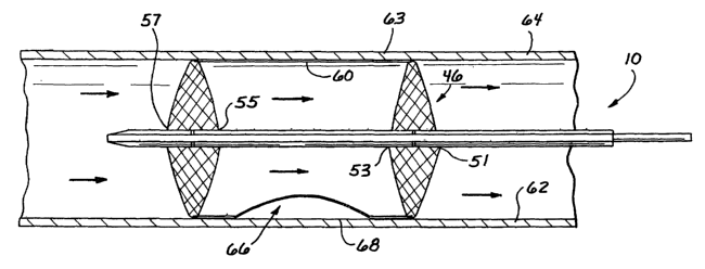
In Figure 6 and 7, the catheter apparatus 10 is illustrated in the
body conduit 64. However in this case the conduit 64, more realistically, has
a
variable rather than a constant diameter. This is illustrated more
specifically by
the diameter D1 in proximity to the dilator 46, and the larger diameter D2 in
proximity to the dilator 48. With the intent of maximizing flow through the
sleeve
60, the catheter apparatus 10 is operable to move the sleeve 60 into contact
with
the inner wall 62 even when the conduit 64 has a variable diameter.
In operation, the elongate member 26 is moved proximally which
initiates the process of expanding the dilators 46 and 48 as previously
discussed.
It is likely that only one of the dilators 46, 48 will expand until it
contacts the inner
wall 62. This will fix the floating outer tube 39 so that further proximal
movement
of the elongate member 26 will expand the diameter of the other dilator. In
Figure 6, the elongate member 26 is moved proximally along with the distal
outer
tube 42 and the floating outer tube 39. This closes the spacing between the
proximal outer tube 33 and the floating outer tube 39, and accordingly
increases
the diameter of the dilator 46. When the dilator 46 reaches the diameter D1 of
the inner wall 62, the movement of the floating outer tube 39 stops. The
continued proximal movement of the elongate member 26 brings the distal outer
tube 42 into closer proximity with the floating outer tube 39 thereby
increasing the
diameter of the dilator 48. The diameter of the dilator 48 will increase until
it
reaches the diameter D2 associated with the inner wall 62 at that location.
CA 02465490 2004-04-29
WO 03/068306 PCT/US02/28830
13
With reference to Figure 7, it will be noted that the sleeve 60 is
brought into contact with the inner wall 62, notwithstanding the variable
diameters of the body conduit 64. Notably, this highly desirable feature is
achieved because the dilators 46 and 48 can be provided with individual
diameters that are independent of each other.
It will also be appreciated that full expansion of both dilators 46, 48
is accomplished when the force exerted against a first adjacent body wall by
the
first dilator 46 is equal to the force exerted against a second adjacent body
wall
by the second dilator 48. Therefore, in any body conduit wherein the diameters
of the conduit portions adjacent to the dilators are not uniform, the self-
adjusting
characteristics of the apparatus 10 enable each dilator 46, 48 to expand to
contact the respective adjacent portions with the same force.
As previously noted, the recessed sleeve portion 66 is radically
spaced from the isolated body conduit portion 68 between the dilators 46, 48.
The permeable dilators 46, 48 enable fluid to pass through the sleeve 60 with
a
resulting fluid pressure which distends the sleeve 60 to contact the inner
wall 62
of the body conduit 64. Thus, the sleeve 60 facilitates flow through the body
conduit 64 while isolating the particular body conduit portion 68. As a
result, an
isolated exclusion chamber 67 is defined by the recessed sleeve portion 66 and
the isolated body conduit portion 68.
This optimizes the flow of fluid passing by the selected surgical site
while the isolated portion 68 of the conduit remains excluded. Thus drugs,
such
as therapeutics, and fluids, such as irritants, may be delivered to or
aspirated
CA 02465490 2004-04-29
WO 03/068306 PCT/US02/28830
14
from the exposed conduit portion without risk of leakage into the isolated
conduit
portion 68. Tissue biopsy could also be obtained via the lateral access recess
66. An anastamosis or repair of the conduit portion 68 could also be performed
while body fluid continues to flow through the remainder of the conduit 64. In
particular, the body conduit portion 68 may be accessed exteriorly via a
puncture,
for example. Blood loss is minimized since only the volume contained in the
isolated chamber 67 would be subject to loss. The sleeve 60 directs the
passing
fluid through the body conduit 62 and thus prevents any fluid communication
between the flow channel of the sleeve 60 and the isolated chamber 67.
An additional embodiment of the invention is illustrated in Figures 8
and 9 where structural elements similar to those previously described are
designated by the same reference numeral followed by the lower case letter
"b."
Thus, in the embodiment of Figures 8 and 9, an alternative sleeve 60b is
provided. The elongate member 26b in this embodiment includes the proximal
outer tube 33b and the distal outer tube 42b which telescopes within the
proximal
outer tube 33b. A skeletal structure 70 is formed by a plurality of bendable
members such as wires 72, each having two ends, one fixed to the outer
proximal tube 33b and the other fixed to the distal outer tube 42b. With this
construction, the distal outer tube 42b can be moved relative to the proximal
outer tube 33b to provide the skeletal structure 70 with both a low-profile
state
and a high-profile state.
CA 02465490 2004-04-29
WO 03/068306 PCT/US02/28830
For example, if the distal outer tube 42b is moved distally of the
proximal outer tube 33b, the ends of the wires 72 are widely separated. This
causes the wires 72 to move into close proximity with the elongate member 26b
5 in the low-profile state. However, when the distal outer tube 42b is moved
proximally relative to the proximal outer tube 33b, the ends of the wires 72
become closely spaced. This causes the wire 72 to move generally radially to a
high profile state as illustrated in Figure 8 and 9.
In order to form the sleeve 60b, a cover 74 is disposed over the
10 skeletal structure 70. This cover 74 is typically formed of a distensible
or
elastomeric material and provided with a tubular configuration so that it at
least
partially covers the skeletal structure 70. At a central portion or waist 83,
the
cover 74 is provided with a collar or belt 85 which maintains the waist 83 at
a
reduced diameter in the high-profile state. As a result, the combination of
the
15 cover 74 and belt 85 provide the sleeve 60b with an hour-glass
configuration. On
either side of the belt 85, the cover 74 is free to expand to a relatively
large
diameter with the wires 72. However, at the central portion of the waist 83,
the
belt 85 limits this expansion to a reduced diameter.
Thus, the belt 85 provides the sleeve 60b with the recess 67b
which in this case is formed circumferentially between the dilators 46b and
48b.
When operatively disposed as illustrated in Figure 9, the sleeve 60b isolates
the
body conduit portion 68b which in this case comprises a full 360 degree or
circular portion of the body conduit 64b. Notwithstanding this isolated
conduit
CA 02465490 2004-04-29
WO 03/068306 PCT/US02/28830
16
portion 67b, the sleeve 60b is capable of continuing fluid flow within the
body
conduit 64b. Thus, a surgeon may exteriorly remove or puncture any part of the
full circular conduit portion 68b without disrupting fluid flow through the
remainder
of the conduit 64b.
In some cases, it may not be necessary to operate on the isolated
body conduit portion 68, but only to isolate the body conduit portion 68 from
the
flow in the main body conduit 64. In these instances, an embodiment such as
that illustrated in Figure 10 may be appropriate. In Figure 10, elements of
structure similar to those previously disposed are designated with the same
reference numeral followed by the lower-case letter "c". Thus, the sleeve 60c
in
this embodiment comprises an axial uniform cylinder which omits any recessed
portion. In this case, the cylindrical sleeve 60c completely isolates a body
conduit portion 91 which includes a branch conduit 93, for example. With the
intent of merely isolating this branch conduit 93 from the flow in the main
conduit
94c, there is no need for a recess such as that designated by the reference
numeral 67b in the embodiment of Figure 9.
From the foregoing description, it will be apparent that the dilators
46 and 48 may comprise a variety of structures. In the embodiment of Figures
11-15, elements of structure similar to those previously discussed or
designated
with the same reference numeral followed by the lower-case letter "d."
In Figures 11-13, the vascular exclusion catheter apparatus 10d
includes a single inflatable dilator or balloon 112, that also serves as a
dilating
sleeve 113. This dilating sleeve 113 is coupled to a catheter shaft 114 that
CA 02465490 2004-04-29
WO 03/068306 PCT/US02/28830
17
extends from the handle assembly 20d. The shaft 114 comprises an outer tube
116 which in this case defines a relatively large through-lumen 118. The
through-lumen 118 is sized and configured to receive a standard guidewire
which
can be used to place the catheter apparatus 10d and to otherwise orient the
dilating sleeve 112 at the operative site.
The handle assembly 20d includes a stopcock 119 which controls
access to the through-lumen 118. An inner tube 121 having an inflation lumen
122 accessible through an inflation port 123, is coupled to a proximal portion
of
the outer tube 116. In a preferred embodiment, the inner tube 121 extends only
partially along the through-lumen 118 terminating within the through-lumen 118
in
proximity to the dilating sleeve 112. Thus, an inflation gas exiting from the
inflation lumen 122 is directed through the through-lumen 118 into the
dilating
sleeve 112.
In this manner, gas from the inflation lumen 123 inflates the balloon
or dilating sleeve 112 to a high profile state. In this state, the sleeve is
circumferentially inflated but defines an axial flow passage shown by the
arrows
124 in Figure 13.
A preferred method of constructing the balloon 112 is illustrated in
Figures 16, 17, 18a and 18b. In accordance with this method, the balloon 112
is
formed of two layers, 125 and 126, of thermoplastic material, each sealed or
otherwise joined together, for example, along seams 127, 128 and 129. The
layers 125 and 126 can also be spot welded at a plurality of layer-joining
connection points 130. With this construction, the balloon 112 is formed
between
CA 02465490 2004-04-29
WO 03/068306 PCT/US02/28830
18
the layers 125 and 126 and bounded by the seams 127-129. The catheter shaft
114 can be inserted between, and sealed to the seams 127 and 128. This gives
the catheter shaft 114, and particularly the inflation lumen 122 access to the
interior of the balloon 112 between the layers 125 and 126. With this
construction, the balloon 112 can be formed into the cylindrical configuration
of
the sleeve 113 by rolling the layers 125 and 126 back on themselves and
attaching the seam 127 to the seam 129 as illustrated in Figure 18b. In this
embodiment, if the recess 66d is desired, it can be formed by removing a
portion
138 as illustrated in Figure 18a and forming a seal of 139 to join the four
edges of
the layers 125 and 126.
As illustrated in Figure 18b, the catheter shaft 114 can be formed
with multiple lumens, namely, the through-lumen 118 and the inflation lumen
122.
With this construction, at least one skive 131 can be cut in the shaft 114 to
access the inflation lumen 122. In operation, the inflation gas will pass from
the
inflation lumen 122 through the skive 131 and into the balloon 112 between the
layers 125 and 126.
With further reference to Figure 14 and 15, it will be noted that the
dilating sleeve 113 facilitates maximum fluid flow while excluding or
isolating a
specific area 144 of a body conduit 146 to form an isolated chamber 148. The
lateral recess 66d facilitates access to a portion of the body conduit for
fluid or
therapeutic administration, tissue biopsy, anastomosis procedure, or for
repairing
damage while body fluid continues to flow through the conduit. As with
previous
CA 02465490 2004-04-29
WO 03/068306 PCT/US02/28830
19
embodiments, the apparatus 10d may be introduced to the surgical site through
either percutaneous or direct access.
In an alternate embodiment shown in Figures 19 and 20, elements
of structure similar to those previously disclosed are designated with the
same
reference numeral followed by the lower-case letter "e." Thus, this embodiment
includes the tube assembly 31 e, the balloon 112e, the outer tube 116e, and
the
inflation lumen 122e. However, in this case additional tubes 151, 153 are
disposed within the tube 116. These additional tubes 151, 153 provide further
lumens 155, 157, respectively, for fluid administration. Also, the dilating
sleeve
113e comprises an inner balloon layer joined to the outer balloon layer via
transverse connection lines 159, instead of the connection points 130 shown in
the embodiment of Figures 16-18.
In a further embodiment, illustrated in Figures 21 and 22, elements
of structure similar to those previously disclosed are designated with the
same
reference numeral followed by the lower-case letter "f." Thus, this embodiment
includes the tube assembly 31f, the balloon 112f and the connection lines
159f.
In this case, however; the dilating sleeve 112f may be formed without any
recess
67 (Figure 5) and thus may comprise an axially uniformed cylinder.
Furthermore, the outer layer 126f of the balloon 112f may be
provided with a lesser thickness than the inner layer 125f thereof. This
difference
in layer thickness facilitates expansion of the balloon 112f toward the
thinner
area upon inflation. Thus, as the outer layer 127f is expanded, the inner
layer
129f is uniformly pulled along with the outer layer.
CA 02465490 2004-04-29
WO 03/068306 PCT/US02/28830
With the specific disclosure of the foregoing embodiments, it will be
apparent that many alterations and modifications can be made without departing
from the spirit and scope of the invention. It is for this reason that the
illustrated
embodiments are set forth only as examples and should not be taken as limiting
the invention.
The words used in this specification to describe the invention and
its various embodiments are to be understood not only in the sense of their
commonly defined meanings, but to include by special definition in this
specification the generic structure, material or acts of which they represent
a
10 single species