Note: Descriptions are shown in the official language in which they were submitted.
CA 02465754 2010-12-17
FLEXIBLE SUBCUTANEOUS IMPLANTABLE CARDIOVERTER-DEFIBRILLATOR
CROSS-REFERENCE TO RELATED APPLICATIONS
The present application is a continuation-in-part of U. S. patent application
entitled
"CANISTER DESIGNS FOR IMPLANTABLE CARDIOVERTER-DEFIBRILLATORS", having
Serial No., 09/940,599 filed August 27, 2001 (now U.S. Patent No. 6,950,705),
which is a
continuation-in-part of U. S. patent application entitled "SUBCUTANEOUS ONLY
IMPLANTABLE CARDIOVERTER-DEFIBRILLATOR AND OPTIONAL PACER", having
Serial No. 09/663, 607, filed September 18, 2000, (now U.S. Patent No.
6,721,597) and U. S. patent
application entitled "UNITARY SUBCUTANEOUS ONLY IMPLANTABLE CARDIOVERTER-
DEFIBRILLATOR AND OPTIONAL PACER ", having Serial No. 09/663,606, filed
September
18, 2000, (now U.S. Patent No. 6,647,292).
BACKGROUND OF THE INVENTION
Defibrillation/cardioversion is a technique employed to counter arrhythmic
heart conditions
including some tachycardias in the atria and/or ventricles. Typically,
electrodes are employed to
stimulate the heart with electrical impulses or shocks, of a magnitude
substantially greater than
pulses used in cardiac pacing. Because current density is a key factor in both
defibrillation and
pacing, implantable devices may improve what is capable with the standard
waveform where the
current and voltage decay over the time of pulse deliver. Consequently, a
waveform that maintains
a constant current over the duration of delivery to the myocardium may improve
defibrillation as
well as pacing.
Defibrillation/cardioversion systems include body implantable electrodes that
are connected
to a hermetically sealed container housing the electronics, battery supply and
capacitors. The entire
system is referred to as implantable cardioverter/defibrillators (ICDs). The
electrodes used in ICDs
can be in the form of patches applied directly to epicardial tissue, or, more
commonly, are on the
distal regions of small cylindrical insulated catheters that typically enter
the subclavian venous
system, pass through the superior vena cava and, into one or more endocardial
areas of the heart.
Such electrode systems are called intravascular or transvenous electrodes. U.
S. Pat. Nos.
4,603,705, 4,693,253, 4,944,300, 5,105,810 disclose intravascular or
transvenous electrodes,
employed either alone, in combination with other intravascular or transvenous
electrodes, or in
combination with an epicardial patch or subcutaneous electrodes. Compliant
epicardial defibrillator
electrodes are disclosed in U. S. Pat. Nos. 4,567, 900 and 5,618, 287. A
sensing epicardial electrode
configuration is disclosed in U. S. Pat No. 5,476, 503.
In addition to epicardial and transvenous electrodes, subcutaneous electrode
systems have
also been developed. For example, U. S. Patent Nos. 5,342, 407 and 5,603, 732,
teach
1
CA 02465754 2010-12-17
the use of a pulse monitor/generator surgically implanted into the abdomen and
subcutaneous
electrodes implanted in the thorax. This system is far more complicated to use
than current ICD
systems using transvenous lead systems together with an active can electrode
and therefore it has
no practical use. It has in fact never been used because of the surgical
difficulty of applying such
a device (3 incisions), the impractical abdominal location of the generator
and the electrically poor
sensing and defibrillation aspects of such a system.
Recent efforts to improve the efficiency of ICDs have led manufacturers to
produce ICDs
which are small enough to be implanted in the pectoral region. In addition,
advances in circuit
design have enabled the housing of the ICD to form a subcutaneous electrode.
Some examples of
ICDs in which the housing of the ICD serves as an optional additional
electrode are described in
U. S. Pat. Nos. 5,133,353, 5,261,400, 5,620,477, and 5,658, 321.
ICDs are now an established therapy for the management of life threatening
cardiac rhythm
disorders, primarily ventricular fibrillation (V-Fib). ICDs are very effective
at treating V-Fib, but
are therapies that still require significant surgery.
As ICD therapy becomes more prophylactic in nature and used in progressively
less ill
individuals, especially children at risk of cardiac arrest, the requirement of
ICD therapy to use
intravenous catheters and transvenous leads is an impedimentto very longterm
management as most
individuals will begin to develop complications related to lead system
malfunction sometime in the
5-10 year time frame, often earlier. In addition, chronic transvenous lead
systems, their
reimplantation and removals, can damage major cardiovascular venous systems
and the tricuspid
valve, as well as result in life threatening perforations of the great vessels
and heart. Consequently,
use of transvenous lead systems, despite their many advantages, are not
without their chronic
patient management limitations in those with life expectancies of >5 years.
The problem of lead
complications is even greater in children where body growth can substantially
alter transvenous
lead function and lead to additional cardiovascular problems and revisions.
Moreover, transvenous
ICD systems also increase cost and require specialized interventional rooms
and equipment as well
as special skill for insertion. These systems are typically implanted by
cardiac electrophysiologists
who have had a great deal of extra training.
In addition to the background related to ICD therapy, the present invention
requires a brief
understanding of a related therapy, the automatic external defibrillator
(AED). AEDs employ the
use of cutaneous patch electrodes, rather than implantable lead systems, to
effect defibrillation under
the direction of a bystander user who treats the patient suffering from V-Fib
with a portable device
containing the necessary electronics and power supply that allows
defibrillation. AEDs can be
nearly as effective as
2
CA 02465754 2012-07-25
an ICD for defibrillation if applied to the victim of ventricular fibrillation
promptly, i.e., within 2 to 3
minutes of the onset of the ventricular fibrillation.
AED therapy has great appeal as a tool for diminishing the risk of death in
public venues such
as in air flight. However, an AED must be used by another individual, not the
person suffering from
the potential fatal rhythm. It is more of a public health tool than a patient-
specific tool like an ICD.
Because >75% of cardiac arrests occur in the home, and over half occur in the
bedroom, patients at
risk of cardiac arrest are often alone or asleep and can not be helped in time
with an AED. Moreover,
its success depends to a reasonable degree on an acceptable level of skill and
calm by the bystander
user.
What is needed therefore, especially for children and for prophylactic long
term use for those
at risk of cardiac arrest, is a combination of the two forms of therapy which
would provide prompt
and near-certain defibrillation, like an ICD, but without the long-term
adverse sequelae of a
transvenous lead system while simultaneously using most of the simpler and
lower cost technology of
an AED. What is also needed is a cardioverter/defibrillator that is of simple
design and can be
comfortably implanted in a patient for many years.
SUMMARY OF THE INVENTION
One embodiment of the present invention provides an implantable cardioverter-
defibrillator
for subcutaneous positioning over a patient's ribcage, the implantable
cardioverter-defibrillator
including a housing, wherein the housing conforms to the patient's ribcage
when subcutaneously
positioned; an electrode disposed upon a portion of the housing; and an
electrical circuit located
within the housing, wherein the electrical circuit is electrically coupled to
the electrode.
In summary, an implantable cardioverter-defibrillator for subcutaneous
positioning over a
patient's ribcage is provided, the implantable cardioverter-defibrillator
comprising: a housing,
wherein the housing conforms to the patient's ribcage when subcutaneously
positioned; an electrode
disposed upon a portion of the housing; and an electrical circuit located
within the housing, wherein
the electrical circuit is electrically coupled to the electrode; wherein a
portion of the housing
comprises a pleated section having a plurality of individual pleats, and
wherein individual pleats may
uniformly expand or contract, allowing the housing to increase or decrease its
overall length, and a
portion of individual pleats may expand or contract, allowing the housing to
bend.
Also provided is an implantable cardioverter-defibrillator for subcutaneous
positioning within
a patient, the implantable cardioverter-defibrillator comprising: a housing
having a first end and a
second end, and wherein a portion of the housing is compliant; a first
electrode disposed upon the first
3
CA 02465754 2012-07-25
end of the housing; a second electrode disposed upon the second end of the
housing; and an electrical
circuit located within the housing, wherein the electrical circuit is
electrically coupled to the first
electrode and the second electrode; wherein the portion of the housing being
compliant comprises a
section of housing forming a plurality of individual pleats; and wherein
individual pleats may
uniformly expand or contract, allowing the housing to increase or decrease its
overall length, and a
portion of individual pleats may expand or contract, allowing the housing to
bend.
BRIEF DESCRIPTION OF THE DRAWINGS
For a better understanding of the invention, reference is now made to the
drawings where like
numerals represent similar objects throughout the figures where:
FIG. I is a schematic view of a Subcutaneous ICD (S-ICD) of the present
invention;
FIG. 2 is a schematic view of an alternate embodiment of a subcutaneous
electrode of the
present invention;
FIG. 3 is a schematic view of an alternate embodiment of a subcutaneous
electrode of the
present invention;
FIG. 4 is a schematic view of the S-ICD and lead of FIG. 1 subcutaneously
implanted in the
thorax of a patient;
FIG. 5 is a schematic view of the S-ICD and lead of FIG. 2 subcutaneously
implanted in an
alternate location within the thorax of a patient;
3a
CA 02465754 2004-05-04
WO 03/039668 PCT/IB02/04516
FIG. 6 is a schematic view of the S-ICD and lead of FIG. 3 subcutaneously
implanted in the
thorax of a patient;
FIG. 7 is a schematic view of the method of making a subcutaneous path from
the preferred
incision and housing implantation point to a termination point for locating a
subcutaneous electrode of the
present invention;
FIG. 8 is a schematic view of an introducer set for performing the method of
lead insertion of any
of the described embodiments;
FIG. 9 is a schematic view of an alternative S-ICD of the present invention
illustrating a lead
subcutaneously and serpiginously implanted in the thorax of a patient for use
particularly in children;
FIG. 10 is a schematic view of an alternate embodiment of an S-ICD of the
present invention;
FIG. 11 is a schematic view of the S-ICD of FIG. 10 subcutaneously implanted
in the thorax of a
patient;
FIG. 12 is a schematic view of yet a further embodiment where the canister of
the S-ICD of the
present invention is shaped to be particularly useful in placing
subcutaneously adjacent and parallel to a
rib of a patient; and
FIG. 13 is a schematic of a different embodiment where the canister of the S-
ICD of the present
invention is shaped to be particularly useful in placing subcutaneously
adjacent and parallel to a rib of a
patient.
FIG. 14 is a schematic view of a Unitary Subcutaneous ICD (US-ICD) of the
present invention;
FIG. 15 is a schematic view of the US-ICD subcutaneously implanted in the
thorax of a patient;
FIG. 16 is a schematic view of the method of making a subcutaneous path from
the preferred
incision for implanting the US-ICD.
FIG. 17 is a schematic view of an introducer for performing the method of US-
ICD implantation;
and
FIG. 18 is an exploded schematic view of an alternate embodiment of the
present invention with a
plug-in portion that contains operational circuitry and means for generating
cardioversion/defibrillation
shock waves.
FIG. 19 is a top perspective view of an alternative S-ICD canister of the
present invention
depicting the top side of the canister housing and a lead electrode coupled to
the S-ICD canister;
FIG. 20 is an exploded bottom perspective view of the S-ICD canister of FIG.
19 showing an
electrode in the shape of a thumbnail positioned on the bottom surface of the
canister housing;
FIG. 21 is a front elevational view of the S-ICD canister of FIG. 19 depicting
the curved canister
housing;
4
CA 02465754 2004-05-04
WO 03/039668 PCT/IB02/04516
FIG. 22 is a partial schematic view of the S-ICD canister of the present
invention implanted
subcutaneously in the thorax of the recipient patient;
FIG. 23A is a top plan view of an alternative S-ICD canister of the present
invention having a
duckbill-shaped end to the canister housing at the proximal end;
FIG. 23B is a top plan view of an alternative S-ICD canister of the present
invention having a
duckbill-shaped canister housing with an alternative proximal head
configuration;
FIG. 24A is a top plan view of an alternative S-ICD canister of the present
invention having a
rectangular-shaped canister housing;
FIG. 24B is a top plan view of an alternative S-ICD canister of the present
invention having a
square-shaped canister housing with a triangular shaped electrode;
FIG. 24C is a top plan view of an alternative S-ICD canister of the present
invention having a
square-shaped canister housing with a square shaped electrode;
FIG. 25A is a top plan view of an alternative S-ICD canister of the present
invention having a
tongue depressor-shaped canister housing;
FIG. 25B is a top plan view of an alternative S-ICD canister of the present
invention having a
modified tongue depressor-shaped canister housing;
FIG. 26A is a top plan view of an alternative S-lCD canister of the present
invention having a
multi-segment canister housing;
FIG. 26B is a front elevational view of the S-ICD canister of FIG. 26A
depicting the curved
proximal segment and the planar distal segment of the multi-segment canister
housing;
FIG. 26C is a front elevational view of the S-ICD canister of FIG. 26A
depicting the curved
proximal segment and the curved distal segment of the multi-segment canister
housing;
FIG. 27 is a perspective view of a US-ICD comprised, in part, of a compliant
material;
FIG. 28A is a front elevational view of an alternative S-ICD canister of the
present invention
having a multi-segment canister housing;
FIG. 28B is an enlarged perspective view of a hinge of the multi-segment
canister housing of
FIG. 28A;
FIG. 29A is a bottom plan view of an alternative S-ICD canister of the present
invention having a
section of the canister housing that is pleated; and
FIG. 29B is a front elevational view of the S-ICD canister of FIG. 29A.
DETAILED DESCRIPTION OF THE INVENTION
Turning now to FIG. 1, the S-ICD of the present invention is illustrated. The
S-ICD consists of
an electrically active canister 11 and a subcutaneous electrode 13 attached to
the canister. The canister
5
CA 02465754 2010-12-17
has an electrically active surface 15 that is electrically insulated from the
electrode connector block
17 and the canister housing 16 via insulating area 14. The canister can be
similar to numerous
electrically active canisters commercially available in that the canister will
contain a battery supply,
capacitor and operational circuitry. Alternatively, the canister can be thin
and elongated to conform
to the intercostal space. The circuitry will be able to monitor cardiac
rhythms for tachycardia and
fibrillation, and if detected, will initiate charging the capacitor and then
delivering
cardioversion/defibrillation energy through the active surface ofthe housing
and to the subcutaneous
electrode. Examples of such circuitry are described in U. S. Patent Nos.
4,693, 253 and 5,105, 810.
The canister circuitry can provide cardioversion/defibrillation energy in
different types of
waveforms. In one embodiment, a 100 uF biphasic waveform is used of
approximately 10-20 ms
total duration and with the initial phase containing approximately 2/3 of the
energy, however, any
type of waveform can be utilized such as monophasic, biphasic, multiphasic or
alternative
waveforms as is known in the art.
In addition to providing cardioversion/defibrillation energy, the circuitry
can also provide
transthoracic cardiac pacing energy. The optional circuitry will be able to
monitor the heart for
bradycardia and/or tachycardia rhythms. Once a bradycardia or tachycardia
rhythm is detected, the
circuitry can then deliver appropriate pacing energy at appropriate intervals
through the active
surface and the subcutaneous electrode. Pacing stimuli can be biphasic in one
embodiment and
similar in pulse amplitude to that used for conventional transthoracic pacing.
This same circuitry can also be used to deliver low amplitude shocks on the T-
wave for
induction of ventricular fibrillation for testing S-ICD performance in
treating V-Fib as is described
in U. S. Patent No. 5,129, 392. Also the circuitry can be provided with rapid
induction of
ventricular fibrillation or ventricular tachycardia using rapid ventricular
pacing. Another optional
way for inducing ventricular fibrillation would be to provide a continuous low
voltage, I. e. , about
3 volts, across the heart during the entire cardiac cycle.
Another optional aspect of the present invention is that the operational
circuitry can detect
the presence of atrial fibrillation as described in Olson, W. et al. "Onset
And Stability For
Ventricular Tachyarrhythmia Detection in an Implantable Cardioverter and
Defibrillator,"Computers in Cardiology (1986) pp. 167-170. Detection can be
provided via R-R
Cycle length instability detection algorithms. Once atrial fibrillation has
been detected, the
operational circuitry will then provide QRS synchronized atrial
defibrillation/cardioversion using
the same shock energy and waveshape characteristics used for ventricular
defibrillation/cardioversion.
The sensing circuitry will utilize the electronic signals generated from the
heart and will
primarily detect QRS waves. In one embodiment, the circuitry will be
programmed to detect only
6
CA 02465754 2010-12-17
ventricular tachycardias or fibrillations. The detection circuitry will
utilize in its most direct form,
a rate detection algorithm that triggers charging of the capacitor once the
ventricular rate exceeds
some predetermined level for a fixed period of time: for example, if the
ventricular rate exceeds 240
bpm on average for more than 4 seconds. Once the capacitor is charged, a
confirmatory rhythm
check would ensure that the rate persists for at least another 1 second before
discharge. Similarly,
termination algorithms could be instituted that ensure that a rhythm less than
240 bpm persisting
for at least 4 seconds before the capacitor charge is drained to an internal
resistor. Detection,
confirmation and termination algorithms as are described above and in the art
can be modulated to
increase sensitivity and specificity by examining QRS beat-to-beat uniformity,
QRS signal
frequency content, R-R interval stability data, and signal amplitude
characteristics all or part of
which can be used to increase or decrease both sensitivity and specificity of
S-ICD arrhythmia
detection function.
In addition to use of the sense circuitry for detection of V-Fib or V-Tach by
examining the
QRS waves, the sense circuitry can check for the presence or the absence of
respiration. The
respiration rate can be detected by monitoring the impedance across the thorax
using subthreshold
currents delivered across the active can and the high voltage subcutaneous
lead electrode and
monitoring the frequency in undulation in the waveform that results from the
undulations of
transthoracic impedance during the respiratory cycle. If there is no
undulation, then the patent is not
respiring and this lack of respiration can be used to confirm the QRS findings
of cardiac arrest. The
same technique can be used to provide information about the respiratory rate
or estimate cardiac
output as described in U. S. Patent Nos. 6,095, 987,5, 423,326, 4,450, 527.
The canister of the present invention can be made out of titanium alloy or
other presently
preferred electrically active canister designs. However, it is contemplated
that a malleable canister
that can conform to the curvature of the patient's chest will be preferred. In
this way the patient can
have a comfortable canister that conforms to the shape of the patient's rib
cage. Examples of
conforming canisters are provided in U. S. Patent No. 5,645, 586. Therefore,
the canister can be
made out of numerous materials such as medical grade plastics, metals, and
alloys. In the preferred
embodiment, the canister is smaller than 60 cc volume having a weight of less
than 100 gms for long
term wearability, especially in children. The canister and the lead of the S-
ICD can also use fractal
or wrinkled surfaces to increase surface area to improve defibrillation
capability. Because of the
primary prevention role ofthe therapy and the likely need to reach energies
over 40 Joules, a feature
of one embodiment is that the charge time for the therapy, is intentionally
left relatively long to
allow capacitor charging within the limitations of device size. Examples of
small ICD housings are
disclosed in U. S. Patents Nos. 5,597, 956 and 5,405, 363.
Different subcutaneous electrodes 13 of the present invention are illustrated
in FIGS. 1-3.
7
CA 02465754 2010-12-17
Turning to FIG. 1, the lead 21 for the subcutaneous electrode is preferably
composed of silicone or
polyurethane insulation. The electrode is connected to the canister at its
proximal end via connection
port 19 which is located on an electrically insulated area 17 of the canister.
The electrode illustrated
is a composite electrode with three different electrodes attached to the lead.
In the embodiment
illustrated, an optional anchor segment 52 is attached at the most distal end
of the subcutaneous
electrode for anchoring the electrode into soft tissue such that the electrode
does not dislodge after
implantation.
The most distal electrode on the composite subcutaneous electrode is a coil
electrode 27 that
is used for delivering the high voltage cardioversion/defibrillation energy
across the heart. The coil
cardioversion/defibrillation electrode is about 5-10 cm in length. Proximal to
the coil electrode are
two sense electrodes, a first sense electrode 25 is located proximally to the
coil electrode and a
second sense electrode 23 is located proximally to the first sense electrode.
The sense electrodes are
spaced far enough apart to be able to have good QRS detection. This spacing
can range from I to
10 cm with 4 cm being presently preferred. The electrodes may or may not be
circumferential with
the preferred embodiment. Having the electrodes non-circumferential and
positioned outward,
toward the skin surface, is a means to minimize muscle artifact and enhance
QRS signal quality. The
sensing electrodes are electrically isolated from the
cardioversion/defibrillation electrode via
insulating areas 29. Similar types of cardioversion/defibrillation electrodes
are currently
commercially available in a transvenous configuration. For example, U. S.
Patent No. 5,534,022
discloses a composite electrode with a coil cardioversion/defibrillation
electrode and sense
electrodes. Modifications to this arrangement is contemplated within the scope
of the invention. One
such modification is illustrated in FIG. 2 where the two sensing electrodes 25
and 23 are non-
circumferential sensing electrodes and one is located at the distal end, the
other is located proximal
thereto with the coil electrode located in between the two sensing electrodes.
In this embodiment
the sense electrodes are spaced about 6 to about 12 cm apart depending on the
length of the coil
electrode used. FIG. 3 illustrates yet a further embodiment where the two
sensing electrodes are
located at the distal end to the composite electrode with the coil electrode
located proximally
thereto. Other possibilities exist and are contemplated within the present
invention. For example,
having only one sensing electrode, either proximal or distal to the coil
cardioversion/defibrillation
electrode with the coil serving as both a sensing electrode and a
cardioversion/defibrillation
electrode.
It is also contemplated within the scope of the invention that the sensing of
QRS waves (and
transthoracic impedance) can be carried out via sense electrodes on the
canister housing or in
combination with the cardioversion/defibrillation coil electrode and/or the
subcutaneous lead
sensing electrode (s). In this way, sensing could be performed via the one
coil electrode located on
the subcutaneous electrode and
8
CA 02465754 2004-05-04
WO 03/039668 PCT/IB02/04516
the active surface on the canister housing. Another possibility would be to
have only one sense electrode
located on the subcutaneous electrode and the sensing would be performed by
that one electrode and
either the coil electrode on the subcutaneous electrode or by the active
surface of the canister. The use of
sensing electrodes on the canister would eliminate the need for sensing
electrodes on the subcutaneous
electrode. It is also contemplated that the subcutaneous electrode would be
provided with at least one
sense electrode, the canister with at least one sense electrode, and if
multiple sense electrodes are used on
either the subcutaneous electrode and/or the canister, that the best QRS wave
detection combination will
be identified when the S-ICD is implanted and this combination can be
selected, activating the best
sensing arrangement from all the existing sensing possibilities. Turning again
to FIG. 2, two sensing
electrodes 26 and 28 are located on the electrically active surface 15 with
electrical insulator rings 30
placed between the sense electrodes and the active surface. These canister
sense electrodes could be
switched off and electrically insulated during and shortly after
defibrillation/ cardioversion shock
delivery. The canister sense electrodes may also be placed on the electrically
inactive surface of the
canister. In the embodiment of FIG. 2, there are actually four sensing
electrodes, two on the
subcutaneous lead and two on the canister. In the preferred embodiment, the
ability to change which
electrodes are used for sensing would be a programmable feature of the S-ICD
to adapt to changes in the
patient physiology and size (in the case of children) over time. The
programming could be done via the
use of physical switches on the canister, or as presently preferred, via the
use of a programming wand or
via a wireless connection to program the circuitry within the canister.
The canister could be employed as either a cathode or an anode of the S-ICD
cardioversion/defibrillation system. If the canister is the cathode, then the
subcutaneous coil electrode
would be the anode. Likewise, if the canister is the anode, then the
subcutaneous electrode would be the
cathode.
The active canister housing will provide energy and voltage intermediate to
that available with
ICDs and most AEDs. The typical maximum voltage necessary for ICDs using most
biphasic waveforms
is approximately 750 Volts with an associated maximum energy of approximately
40 Joules. The typical
maximum voltage necessary for AEDs is approximately 2000-5000 Volts with an
associated maximum
energy of approximately 200-360 Joules depending upon the model and waveform
used. The S-ICD and
the US-ICD of the present invention uses maximum voltages in the range of
about 50 to about 3500 Volts
and is associated with energies of about .5 to about 350 Joules. The
capacitance of the devices can range
from about 25 to about 200 micro farads.
In another embodiment, the S-ICD and US-ICD devices provide energy with a
pulse width of
approximately one millisecond to approximately 40 milliseconds. The devices
can provide pacing current
of approximately one milliamp to approximately 250 milliamps.
9
CA 02465754 2004-05-04
WO 03/039668 PCT/IB02/04516
The sense circuitry contained within the canister is highly sensitive and
specific for the presence
or absence of life threatening ventricular arrhythmias. Features of the
detection algorithm are
programmable and the algorithm is focused on the detection of V-FIB and high
rate V-TACH (>240
bpm). Although the S-ICD of the present invention may rarely be used for an
actual life-threatening
event, the simplicity of design and implementation allows it to be employed in
large populations of
patients at modest risk with modest cost by non-cardiac electrophysiologists.
Consequently, the S-ICD of
the present invention focuses mostly on the detection and therapy of the most
malignant rhythm disorders.
As part of the detection algorithm's applicability to children, the upper rate
range is programmable
upward for use in children, known to have rapid supraventricular tachycardias
and more rapid ventricular
fibrillation. Energy levels also are programmable downward in order to allow
treatment of neonates and
infants.
Turning now to FIG. 4, the optimal subcutaneous placement of the S-ICD of the
present invention
is illustrated. As would be evidence to a person skilled in the art, the
actual location of the S-ICD is in a
subcutaneous space that is developed during the implantation process. The
heart is not exposed during
this process and the heart is schematically illustrated in the figures only
for help in understanding where
the canister and coil electrode are three dimensionally located in the left
mid-clavicular line
approximately at the level of the inframammary crease at approximately the 5th
rib. The lead 21 of the
subcutaneous electrode traverses in a subcutaneous path around the thorax
terminating with its distal
electrode end at the posterior axillary line ideally just lateral to the left
scapula. This way the canister and
subcutaneous cardioversion/defibrillation electrode provide a reasonably good
pathway for current
delivery to the majority of the ventricular myocardium.
FIG. 5 illustrates a different placement of the present invention. The S-ICD
canister with the
active housing is located in the left posterior axillary line approximately
lateral to the tip of the inferior
portion of the scapula. This location is especially useful in children. The
lead 21 of the subcutaneous
electrode traverses in a subcutaneous path around the thorax terminating with
its distal electrode end at
the anterior precordial region, ideally in the inframammary crease. FIG. 6
illustrates the embodiment of
FIG. 1 subcutaneously implanted in the thorax with the proximal sense
electrodes 23 and 25 located at
approximately the left axillary line with the cardioversion/defibrillation
electrode just lateral to the tip of
the inferior portion of the scapula.
FIG. 7 schematically illustrates the method for implanting the S-ICD of the
present invention. An
incision 31 is made in the left anterior axillary line approximately at the
level of the cardiac apex. This
incision location is distinct from that chosen for S-ICD placement and is
selected specifically to allow
both canister location more medially in the left inframammary crease and lead
positioning more
posteriorly via the introducer set (described below) around to the left
posterior axillary line lateral to the
CA 02465754 2004-05-04
WO 03/039668 PCT/IB02/04516
left scapula. That said, the incision can be anywhere on the thorax deemed
reasonably by the implanting
physician although in the preferred embodiment, the S-ICD of the present
invention will be applied in this
region. A subcutaneous pathway 33 is then created medially to the inframmary
crease for the canister and
posteriorly to the left posterior axillary line lateral to the left scapula
for the lead.
The S-ICD canister 11 is then placed subcutaneously at the location of the
incision or medially at
the subcutaneous region at the left infranvnary crease. The subcutaneous
electrode 13 is placed with a
specially designed curved introducer set 40 (see FIG. 8). The introducer set
comprises a curved trocar 42
and a stiff curved peel away sheath 44. The peel away sheath is curved to
allow for placement around the
rib cage of the patient in the subcutaneous space created by the trocar. The
sheath has to be stiff enough
to allow for the placement of the electrodes without the sheath collapsing or
bending. Preferably the
sheath is made out of a biocompatible plastic material and is perforated along
its axial length to allow for
it to split apart into two sections. The trocar has a proximal handle 41 and a
curved shaft 43. The distal
end 45 of the trocar is tapered to allow for dissection of a subcutaneous path
33 in the patient. Preferably,
the trocar is cannulated having a central Lumen 46 and terminating in an
opening 48 at the distal end.
Local anesthetic such as lidocaine can be delivered, if necessary, through the
lumen or through a curved
and elongated needle designed to anesthetize the path to be used for trocar
insertion should general
anesthesia not be employed. The curved peel away sheath 44 has a proximal pull
tab 49 for breaking the
sheath into two halves along its axial shaft 47. The sheath is placed over a
guidewire inserted through the
trocar after the subcutaneous path has been created. The subcutaneous pathway
is then developed until it
terminates subcutaneously at a location that, if a straight line were drawn
from the canister location to the
path termination point the line would intersect a substantial portion of the
left ventricular mass of the
patient. The guidewire is then removed leaving the peel away sheath. The
subcutaneous lead system is
then inserted through the sheath until it is in the proper location. Once the
subcutaneous lead system is in
the proper location, the sheath is split in half using the pull tab 49 and
removed. If more than one
subcutaneous electrode is being used, a new curved peel away sheath can be
used for each subcutaneous
electrode.
The S-ICD will have prophylactic use in adults where chronic
transvenous/epicardial ICD lead
systems pose excessive risk or have already resulted in difficulty, such as
sepsis or lead fractures. It is
also contemplated that a major use of the S-ICD system of the present
invention will be for prophylactic
use in children who are at risk for having fatal arrhythmias, where chronic
transvenous lead systems pose
significant management problems. Additionally, with the use of standard
transvenous ICDs in children,
problems develop during patient growth in that the lead system does not
accommodate the growth. FIG.
9 illustrates the placement of the S-ICD subcutaneous lead system such that he
problem that growth
presents to the lead system is overcome. The distal end of the subcutaneous
electrode is placed in the
11
CA 02465754 2004-05-04
WO 03/039668 PCT/IB02/04516
same location as described above providing a good location for the coil
cardioversion/defibrillation
electrode 27 and the sensing electrodes 23 and 25. The insulated lead 21,
however is no longer placed in
a taught configuration. Instead, the lead is serpiginously placed with a
specially designed introducer
trocar and sheath such that it has numerous waves or bends. As the child
grows, the waves or bends will
straighten out lengthening the lead system while maintaining proper electrode
placement. Although it is
expected that fibrous scarring especially around the defibrillation coil will
help anchor it into position to
maintain its posterior position during growth, a lead system with a distal
tine or screw electrode anchoring
system 52 can also be incorporated into the distal tip of the lead to
facilitate lead stability (see FIG. 1).
Other anchoring systems can also be used such as hooks, sutures, or the like.
FIGS. 10 and 11 illustrate another embodiment of the present S-ICD invention.
In this
embodiment there are two subcutaneous electrodes 13 and 13' of opposite
polarity to the canister. The
additional subcutaneous electrode 13' is essentially identical to the
previously described electrode. In this
embodiment the cardioversion/defibrillation energy is delivered between the
active surface of the canister
and the two coil electrodes 27 and 27'. Additionally, provided in the canister
is means for selecting the
optimum sensing arrangement between the four sense electrodes 23, 23', 25, and
25'. The two electrodes
are subcutaneously placed on the same side of the heart. As illustrated in
FIG. 6, one subcutaneous
electrode 13 is placed inferiorly and the other electrode 13' is placed
superiorly. It is also contemplated
with this dual subcutaneous electrode system that the canister and one
subcutaneous electrode are the
same polarity and the other subcutaneous electrode is the opposite polarity.
Turning now to FIGS. 12 and 13, further embodiments are illustrated where the
canister 11 of the
S-ICD of the present invention is shaped to be particularly useful in placing
subcutaneously adjacent and
parallel to a rib of a patient. The canister is long, thin, and curved to
conform to the shape of the patient's
rib. In the embodiment illustrated in FIG. 12, the canister has a diameter
ranging from about 0.5 cm to
about 2 cm without 1 cm being presently preferred. Alternatively, instead of
having a circular cross
sectional area, the canister could have a rectangular or square cross
sectional area as illustrated in FIG. 13
without falling outside of the scope of the present invention. The length of
the canister can vary
depending on the size of the patient's thorax. In an embodiment, the canister
is about 5 cm to about 40
cm long. The canister is curved to conform to the curvature of the ribs of the
thorax. The radius of the
curvature will vary depending on the size of the patient, with smaller
radiuses for smaller patients and
larger radiuses for larger patients. The radius of the curvature can range
from about 5 cm to about 35 cm
depending on the size of the patient. Additionally, the radius of the
curvature need not be uniform
throughout the canister such that it can be shaped closer to the shape of the
ribs. The canister has an
active surface, 15 that is located on the interior (concave) portion of the
curvature and an inactive surface
16 that is located on the exterior (convex) portion of the curvature. The
leads of these embodiments,
12
CA 02465754 2004-05-04
WO 03/039668 PCT/IB02/04516
which are not illustrated except for the attachment port 19 and the proximal
end of the lead 21, can be any
of the leads previously described above, with the lead illustrated in FIG. 1
being presently preferred.
The circuitry of this canister is similar to the circuitry described above.
Additionally, the canister
can optionally have at least one sense electrode located on either the active
surface of the inactive surface
and the circuitry within the canister can be programmable as described above
to allow for the selection of
the best sense electrodes. It is presently preferred that the canister have
two sense electrodes 26 and 28
located on the inactive surface of the canisters as illustrated, where the
electrodes are spaced from about 1
to about 10 cm apart with a spacing of about 3 cm being presently preferred.
However, the sense
electrodes can be located on the active surface as described above.
It is envisioned that the embodiment of FIG. 12 will be subcutaneously
implanted adjacent and
parallel to the left anterior 5th rib, either between the 4th and 5th ribs or
between the 5th and 6th ribs.
However other locations can be used.
Another component of the S-ICD of the present invention is a cutaneous test
electrode system
designed to simulate the subcutaneous high voltage shock electrode system as
well as the QRS cardiac
rhythm detection system. This test electrode system is comprised of a
cutaneous patch electrode of
similar surface area and impedance to that of the S-ICD canister itself
together with a cutaneous strip
electrode comprising a defibrillation strip as well as two button electrodes
for sensing of the QRS.
Several cutaneous strip electrodes are available to allow for testing various
bipole spacings to optimize
signal detection comparable to the implantable system.
Figures 14 to 18 depict particular US-ICD embodiments of the present
invention. The various
sensing, shocking and pacing circuitry, described in detail above with respect
to the S-ICD embodiments,
may additionally be incorporated into the following US-ICD embodiments.
Furthermore, particular
aspects of any individual S-ICD embodiment discussed above, may be
incorporated, in whole or in part,
into the US-ICD embodiments depicted in the following figures.
Turning now to Fig. 14, the US-ICD of the present invention is illustrated.
The US-ICD consists
of a curved housing 1211 with a first and second end. The first end 1413 is
thicker than the second end
1215. This thicker area houses a battery supply, capacitor and operational
circuitry for the US-ICD. The
circuitry will be able to monitor cardiac rhythms for tachycardia and
fibrillation, and if detected, will
initiate charging the capacitor and then delivering
cardioversion/defibrillation energy through the two
cardioversion/defibrillating electrodes 1417 and 1219 located on the outer
surface of the two ends of the
housing. The circuitry can provide cardioversion/defibrillation energy in
different types of waveforms. In
one embodiment, a 100 uF biphasic waveform is used of approximately 10-20 ms
total duration and with
the initial phase containing approximately 2/3 of the energy, however, any
type of waveform can be
utilized such as monophasic, biphasic, multiphasic or alternative waveforms as
is known in the art.
13
CA 02465754 2010-12-17
The housing of the present invention can be made out of titanium alloy or
other presently
preferred ICD designs. It is contemplated that the housing is also made out of
biocompatible plastic
materials that electronically insulate the electrodes from each other.
However, it is contemplated
that a malleable canister that can conform to the curvature of the patient's
chest will be preferred.
In this way the patient can have a comfortable canister that conforms to the
unique shape of the
patient's rib cage. Examples of conforming ICD housings are provided in U. S.
Patent No. 5,645,
586. In the preferred embodiment, the housing is curved in the shape of a 5th
rib of a person.
Because there are many different sizes of people, the housing will come in
different incremental
sizes to allow a good match between the size of the rib cage and the size of
the US- ICD. The length
of the US-ICD will range from about 15 to about 50 cm. Because of the primary
preventative role
of the therapy and the need to reach energies over 40 Joules, a feature of the
preferred embodiment
is that the charge time for the therapy, intentionally be relatively long to
allow capacitor charging
within the limitations of device size.
The thick end of the housing is currently needed to allow for the placement of
the battery
supply, operational circuitry, and capacitors. It is contemplated that the
thick end will be about 0.5
cm to about 2 cm wide with about 1 cm being presently preferred. As
microtechnology advances,
the thickness of the housing will become smaller.
The two cardioversion/defibrillation electrodes on the housing are used for
delivering the
high voltage cardioversion/defibrillation energy across the heart. In the
preferred embodiment, the
cardioversion/defibrillation electrodes are coil electrodes, however, other
cardioversion/defibrillation electrodes could be used such as having
electrically isolated active
surfaces or platinum alloy electrodes. The coil cardioversion/defibrillation
electrodes are about 5-10
cm in length. Located on the housing between the two
cardioversion/defibrillation electrodes are
two sense electrodes 1425 and 1427. The sense electrodes are spaced far enough
apart to be able
to have good QRS detection. This spacing can range from 1 to 10 cm with 4 cm
being presently
preferred. The electrodes may or may not be circumferential with the preferred
embodiment. Having
the electrodes non-circumferential and positioned outward, toward the skin
surface, is a means to
minimize muscle artifact and enhance QRS signal quality. The sensing
electrodes are electrically
isolated from the cardioversion/defibrillation electrode via insulating areas
1423. Analogous types
of cardioversion/defibrillation electrodes are currently commercially
available in a transvenous
configuration. For example, U. S. Patent No. 5,534, 022, discloses a composite
electrode with a coil
cardioversion/defibrillation electrode and sense electrodes. Modifications to
this arrangement is
contemplated within the scope of the invention. One such modification is to
have the sense
electrodes at the two ends ofhe housing and have the
cardioversion/defibrillation electrodes located
in between the
14
CA 02465754 2004-05-04
WO 03/039668 PCT/IB02/04516
sense electrodes. Another modification is to have three or more sense
electrodes spaced throughout the
housing and allow for the selection of the two best sensing electrodes. If
three or more sensing electrodes
are used, then the ability to change which electrodes are used for sensing
would be a programmable
feature of the US-ICD to adapt to changes in the patient physiology and size
over time. The
programming could be done via the use of physical switches on the canister, or
as presently preferred, via
the use of a programming wand or via a wireless connection to program the
circuitry within the canister.
Turning now to Fig. 15, the optimal subcutaneous placement of the US-ICD of
the present
invention is illustrated. As would be evident to a person skilled in the art,
the actual location of the US-
ICD is in a subcutaneous space that is developed during the implantation
process. The heart is not
exposed during this process and the heart is schematically illustrated in the
figures only for help in
understanding where the device and its various electrodes are three
dimensionally located in the thorax of
the patient. The US-ICD is located between the left mid-clavicular line
approximately at the level of the
inframammary crease at approximately the 5th rib and the posterior axillary
line, ideally just lateral to the
left scapula. This way the US-ICD provides a reasonably good pathway for
current delivery to the
majority of the ventricular myocardium.
Fig. 16 schematically illustrates the method for implanting the US-ICD of the
present invention.
An incision 1631 is made in the left anterior axillary line approximately at
the level of the cardiac apex.
A subcutaneous pathway is then created that extends posteriorly to allow
placement of the US-ICD. The
incision can be anywhere on the thorax deemed reasonable by the implanting
physician although in the
preferred embodiment, the US-ICD of the present invention will be applied in
this region. The
subcutaneous pathway is created medially to the inframammary crease and
extends posteriorly to the left
posterior axillary line. The pathway is developed with a specially designed
curved introducer 1742 (see
Fig. 17). The trocar has a proximal handle 1641 and a curved shaft 1643. The
distal end 1745 of the
trocar is tapered to allow for dissection of a subcutaneous path in the
patient. Preferably, the trocar is
cannulated having a central lumen 1746 and terminating in an opening 1748 at
the distal end. Local
anesthetic such as lidocaine can be delivered, if necessary, through the lumen
or through a curved and
elongated needle designed to anesthetize the path to be used for trocar
insertion should general anesthesia
not be employed. Once the subcutaneous pathway is developed, the US-ICD is
implanted in the
subcutaneous space, the skin incision is closed using standard techniques.
As described previously, the US-ICDs of the present invention vary in length
and curvature. The
US-ICDs are provided in incremental sizes for subcutaneous implantation in
different sized patients.
Turning now to Fig. 18, a different embodiment is schematically illustrated in
exploded view which
provides different sized US-ICDs that are easier to manufacture. The different
sized US-ICDs will all
have the same sized and shaped thick end 1413. The thick end is hollow inside
allowing for the insertion
CA 02465754 2004-05-04
WO 03/039668 PCT/IB02/04516
of a core operational member 1853. The core member comprises a housing 1857
which contains the
battery supply, capacitor and operational circuitry for the US-ICD. The
proximal end of the core member
has a plurality of electronic plug connectors. Plug connectors 1861 and 1863
are electronically connected
to the sense electrodes via pressure fit connectors (not illustrated) inside
the thick end which are standard
in the art. Plug connectors 1865 and 1867 are also electronically connected to
the
cardioverter/defibrillator electrodes via pressure fit connectors inside the
thick end. The distal end of the
core member comprises an end cap 1855, and a ribbed fitting 1859 which creates
a water-tight seal when
the core member is inserted into opening 1851 of the thick end of the US-ICD.
The S-ICD and US-ICD, in alternative embodiments, have the ability to detect
and treat atrial
rhythm disorders, including atrial fibrillation. The S-ICD and US-ICD have two
or more electrodes that
provide a far-field view of cardiac electrical activity that includes the
ability to record the P-wave of the
electrocardiogram as well as the QRS. One can detect the onset and offset of
atrial fibrillation by
referencing to the P-wave recorded during normal sinus rhythm and monitoring
for its change in rate,
morphology, amplitude and frequency content. For example, a well-defined P-
wave that abruptly
disappeared and was replaced by a low-amplitude, variable morphology signal
would be a strong
indication of the absence of sinus rhythm and the onset of atrial
fibrillation. In an alternative embodiment
of a detection algorithm, the ventricular detection rate could be monitored
for stability of the R-R
coupling interval. In the examination of the R-R interval sequence, atria]
fibrillation can be recognized by
providing a near constant irregularly irregular coupling interval on a beat-by-
beat basis. A R-R interval
plot during AF appears "cloudlike" in appearance when several hundred or
thousands of R-R intervals are
plotted over time when compared to sinus rhythm or other supraventricular
arrhythmias. Moreover, a
distinguishing feature compared to other rhythms that are irregularly
irregular, is that the QRS
morphology is similar on a beat-by-beat basis despite the irregularity in the
R-R coupling interval. This is
a distinguishing feature of atrial fibrillation compared to ventricular
fibrillation where the QRS
morphology varies on a beat-by-beat basis. In yet another embodiment, atrial
fibrillation may be detected
by seeking to compare the timing and amplitude relationship of the detected P-
wave of the
electrocardiogram to the detected QRS (R-wave) of the electrocardiogram.
Normal sinus rhythm has a
fixed relationship that can be placed into a template matching algorithm that
can be used as a reference
point should the relationship change.
In other aspects of the atrial fibrillation detection process, one may include
alternative electrodes
that may be brought to bear in the S-ICD or US-ICD systems either by placing
them in the detection
algorithm circuitry through a programming maneuver or by manually adding such
additional electrode
systems to the S-ICD or US-ICD at the time of implant or at the time of follow-
up evaluation. One may
also use electrodes for the detection of atrial fibrillation that may or may
not also be used for the detection
16
CA 02465754 2004-05-04
WO 03/039668 PCT/IB02/04516
of ventricular arrhythmias given the different anatomic locations of the atria
and ventricles with respect to
the S-ICD or US-ICD housing and surgical implant sites.
Once atrial fibrillation is detected, the arrhythmia can be treated by
delivery of a synchronized
shock using energy levels up to the maximum output of the device therapy for
terminating atrial
fibrillation or for other supraventricular arrhythmias. The S-ICD or US-ICD
electrode system can be used
to treat both atrial and ventricular arrhythmias not only with shock therapy
but also with pacing therapy.
In a further embodiment of the treatment of atrial fibrillation or other
atrial arrhythmias, one may be able
to use different electrode systems than what is used to treat ventricular
arrhythmias. Another
embodiment, would be to allow for different types of therapies (amplitude,
waveform, capacitance, etc.)
for atrial arrhythmias compared to ventricular arrhythmias.
The core member of the different sized and shaped US-ICD will all be the same
size and shape.
That way, during an implantation procedures, multiple sized US-ICDs can be
available for implantation,
each one without a core member. Once the implantation procedure is being
performed, then the correct
sized US-ICD can be selected and the core member can be inserted into the US-
ICD and then
programmed as described above. Another advantage of this configuration is when
the battery within the
core member needs replacing it can be done without removing the entire US-ICD.
Figures 19-26 refer generally to alternative S-ICD/US-ICD canister
embodiments. Although the
following canister designs, various material constructions, dimensions and
curvatures, discussed in detail
below, may be incorporated into either S-ICD or US-ICD canister embodimens,
hereinafter, these
attributes will be discussed solely with respect to S-ICDs.
The canisters illustrated in these Figures possess a configuration that may 1)
aid in the initial
canister implantation; 2) restrict canister displacement once properly
positioned; 3) create a consistently
focused array of energy delivered toward the recipient's heart with less
disbursement to other areas of the
thorax; 4) allow for good signal reception from the heart by an S-ICD system;
or 5) provide significant
comfort and long-term wearability in a broad spectrum of patients with
differing thoracic sizes and
shapes. More particularly, Figures 19-26 detail various material
constructions, dimensions and curvatures
that are incorporated within the numerous S-ICD canister designs detailed in
Figures 19-26C.
Referring now to the particular embodiments, Fig. 19 depicts an S-ICD canister
190 of an
embodiment of the present invention. The shell of the S-ICD canister 190
comprises a hermetically
sealed housing 192 that encases the electronics for the S-ICD canister 190. As
with the previously
described S-ICD devices, the electronics of the present embodiment include, at
a minimum, a battery
supply, a capacitor and operational circuitry. Fig. 19 further depicts a lead
electrode 191 coupled to the
shell of the canister through a lead 193. A dorsal fin 197 may be disposed on
the lead electrode 191 to
facilitate the positioning of the lead electrode.
17
CA 02465754 2010-12-17
The S-ICD devices of the present invention provide an energy (electric field
strength
(V/cm), current density (A/cm2), voltage gradient (V/cm) or other measured
unit of energy) to a
patient's heart. S- ICD devices of the present invention will generally use
voltages in the range of
700 V to 3150 V, requiring energies of approximately 40 J to 210 J. These
energy requirements will
vary, however, depending upon the form of treatment, the proximity of the
canister from the
patient's heart, the relative relationship of the S-ICD canister's electrode
to the lead electrode, the
nature of the patient's underlying heart disease, the specific cardiac
disorder being treated, and the
ability to overcome diversion of the S- ICD electrical output into other
thoracic tissues.
Ideally, the emitted energy from the S-ICD device will be directed into the
patient's anterior
mediastinum, through the majority of the heart, and out to the coupled lead
electrode positioned in
the posterior, posterolateral and/or lateral thoracic locations. Furthermore,
it is desirable that the S-
ICD canister 190 be capable of delivering this directed energy, as a general
rule, at an adequate
effective field strength of about 3-5 V/cm to approximately 90 percent of a
patient's ventricular
myocardium using a biphasic waveform. This delivered effective field strength
should be adequate
for defibrillation of the patient's heart-an intended application of an
embodiment of the present
invention.
Increased energy requirements necessitate larger, or alternatively, additional
batteries and
capacitors. The latter of these two options is often more desirable in order
to reduce the overall
depth of the resulting S-ICD canister 190. Increasing the number of batteries
and capacitors,
however, will increase the length and possibly the depth of the S-ICD canister
190. Therefore,
numerous S-ICD devices of varying depth, widths and lengths are manufactured
to accommodate
the particular energy needs of a variety of patient recipients. For example,
an overweight adult male
may require a larger and bulkier S- ICD canister 190 than a young child. In
particular, the young
child is generally smaller, has a relatively lower resistance to current flow,
and contains less current
diverting body mass than the overweight adult male. As a result, the energy
required to deliver an
effective therapy to the young child's heart may be considerably less than for
the overweight adult
male, and therefore, the young child may utilize a smaller and more compact S-
ICD canister 190.
In addition, one may find that individuals, despite equivalent body size, may
have different therapy
requirements because of differences in their underlying heart disease. This
may allow some patients
to receive a smaller canister compared to another patient of equal body size
but with a different type
of heart disease.
The spatial requirements of a resulting S-ICD canister 190 are additionally
dependent upon
the type of operational circuitry used within the device. The S-ICD canister
190 maybe programmed
to monitor cardiac rhythms for tachycardia and fibrillation, and if detected,
will initiate charging
the capacitor (s) to deliver the appropriate cardioversion/defibrillation
energy. Examples of such
circuitry are described in U. S. Patent Nos. 4,693, 253 and 5,105, 810. The S-
18
CA 02465754 2004-05-04
WO 03/039668 PCT/IB02/04516
ICD canister 190 may additionally be provided with operational circuitry for
transthoracic cardiac pacing.
This optional circuitry monitors the heart for bradycardia and/or tachycardia
rhythms. In the event a
bradycardia or tachycardia rhythm is detected, the operational circuitry
delivers the appropriate pacing
energy at the appropriate intervals to treat the disorder.
In additional embodiments, the operational circuitry may be: 1) programmed to
deliver low
amplitude shocks on the T-wave for induction of ventricular fibrillation for
testing the S-ICD canister's
performance; 2) programmed for rapid ventricular pacing to either induce a
tachyarrhythmia or to
terminate one; 3) programmed to detect the presence of atrial fibrillation;
and/or 4) programmed to detect
ventricular fibrillation or ventricular tachycardia by examining QRS waves,
all of which are described in
detail above. Additional operational circuitry, being known in the art for
sensing, shocking and pacing
the heart, are additionally incorporated herein as being within the spirit and
scope of the present
invention.
The primary function of the canister housing 192 is to provide a protective
barrier between the
electrical components held within its confines and the surrounding
environment. The canister housing
192, therefore, must possess sufficient hardness to protect its contents.
Materials possessing this hardness
may include numerous suitable biocompatible materials such as medical grade
plastics, ceramics, metals
and alloys. Although the materials possessing such hardnesses are generally
rigid, in particular
embodiments, it is desirable to utilize materials that are pliable or
compliant. More specifically, it is
desirable that the canister housing 192 be capable of partially yielding in
its overall form without
fracturing.
Compliant canister housings 192 often provide increased comfort when implanted
in patient
recipients. S-ICD canisters 190 formed from such materials permit limited, but
significant, deflection of
the canister housing 192 with certain thoracic motions. Examples of permitted
deflections are ones that
are applied to the canister housing 192 by surrounding muscle tissue. The use
of a compliant canister
housing is particularly beneficial in canister housing 192 embodiments that
extend over a significant
portion of a patient's thorax. For example, a compliant canister housing 192
is particularly beneficial in
US-ICD devices.
FIG. 27 illustrates a US-ICD canister housing embodiment 310 comprising, in
part, of a
compliant material. The US-ICD canister housing 310 includes a first end 312
and a second end 314. As
described in detail above, US-ICD canister housing 310 further comprise a
plurality of electrodes
contained on the canister housing 310. In particular embodiments, and as
depicted in FIG. 27, two
electrodes are used, a first electrode 316 and a second electrode 318 (shown
in phantom). In this
embodiment, the two electrodes 316 and 318 are positioned at each end 312 and
314 of the US-ICD
canister housing 310. Additionally, a connecting member 320 is positioned
between the two ends 312
19
CA 02465754 2004-05-04
WO 03/039668 PCT/IB02/04516
and 314 of the canister housing 310 to join the canister housing's two ends
and encase the US-ICD's
electronic circuitry. In this particular embodiment, a compliant or otherwise
pliable material is used to
form the connecting member 320.
The length of the US-ICD canister housing 310 allows the single unitary device
to correctly
position the electrodes 316 and 318 within the patient to form a
depolarization vector (described in detail
below) with respect to the patient's heart. To accommodate the individual body
shape of the patient
recipient, however, embodiments of the present invention utilize compliant
materials to adjust the
positioning of the US-ICD device to obtain this proper depolarization vector.
The compliant materials
used in these embodiments may comprise a portion of the US-ICD canister
housing 310, or alternatively,
may comprise the canister housing 310 in its entirety.
Compliant materials suitable for the S-ICD canister housings'I92 (and US-ICD
canister housings
310) of the present invention include polyurethanes, polyamides,
polyetheretherketones (PEEK),
polyether block amides (PEBA), polytetrafluoroethylene (PTFE), polyethylene,
silicones, and mixtures
thereof
The use of compliant materials allows the US-ICD depicted in FIG. 27 to bend
and conform
specifically to an individual's ribcage. Thus, the use of compliant materials
permits the proper sizing of
the US-ICD canister housing 310 to numerous patients, without requiring the
manufacturer to generate
numerous canister housings 310 of various specific shapes and sizes. Moreover,
the correct material
selection (or combination thereof) for the US-ICD device is helpful in
eliminating patient awareness of
the device and in improving the long-term wearability of the implanted device.
Materials selected for the canister housing 192 should further be capable of
being sterilized.
Often commercial sterilization processes involve exposure to elevated
temperatures, pressures or
chemical treatments. It is important, therefore, that the materials used in
forming the canister housing be
capable of withstanding such exposures without degrading or otherwise
compromising their overall
integrity.
Polymeric materials suitable for the canister housing 192 of the present
invention include
polyurethanes, polyamides, polyetheretherketones (PEEK), polyether block
amides (PEBA),
polytetrafluoroethylene (PTFE), silicones, and mixtures thereof. Ceramic
materials suitable for the
canister housing 192 of the present invention include zirconium ceramics and
aluminum-based ceramics.
Metallic materials suitable for the canister housing 192 of the present
invention include stainless steel,
and titanium. Alloys suitable for the canister housing 192 of the present
invention include stainless steel
alloys and titanium alloys such as nickel titanium. In certain embodiments of
the present invention,
classes of materials may be combined in forming the canister housing 192. For
example, a nonconductive
polymeric coating, such as parylene, may be selectively applied over a
titanium alloy canister housing
CA 02465754 2004-05-04
WO 03/039668 PCT/IB02/04516
192 surface in order to allow only a specific surface area, such as that at
the undersurface of the duckbill
distal end, to receive signals and/or apply therapy.
In general, it is desirable to maintain the size of the S-ICD canister housing
192 under a total
volume of approximately 50 cubic centimeters. In alternative embodiments of
the present invention, it is
desirable to maintain the size of the S-ICD canister housing 192 under a total
volume of approximately
100 cubic centimeters. In yet alternative embodiments of the present
invention, it is desirable to maintain
the size of the S-ICD canister housing 192 under a total volume of
approximately 120 cubic centimeters.
Moreover, it is additionally desirable to maintain the total weight of the S-
ICD canister 190, as a
whole (including the canister housing, operational circuitry, capacitors and
batteries), under
approximately 50 grams. In alternative embodiments of the present invention,
it is desirable to maintain
the total weight of the S-ICD canister 190 under approximately 100 grams. In
yet alternative
embodiments of the present invention, it is desirable to maintain the total
weight of the S-ICD canister
190 under approximately 150 grams.
Maintaining the weight and size within the above identified parameters is
primarily for patient
comfort depending upon the shape of the device. The implantation of a S-ICD
canister 190 is a long-term
solution to heart dysfunction, and as such, will ideally remain in the patient
until the device's batteries
need replacement or an alternative therapy eventually leads to its removal.
Accordingly, a considerable
amount of engineering is devoted to minimizing discomfort associated with the
installed device.
Weight and size considerations are particularly important to younger patient
recipients. Children
possessing ICDs are more likely to be cognitive of any additional weight or
bulkiness associated with
heavier and/or larger devices. The present invention overcomes these problems
by designing a S-ICD
canister 190 that takes into consideration the concerns of these smaller sized
patient recipients. For
example, lighter materials may be utilized to minimize discomfort associated
with heavier materials.
Furthermore, the S-ICD canister 190 (length, width and depth) in its entirety,
or only a portion thereof,
may be modified in order to accommodate a variety of sized patient recipients.
For example, the shape of
the S-ICD canister housing 192 may also be manufactured in a variety of
anatomical configurations to
better insure comfort and performance in younger children or smaller adults,
throughout the life of their
S-ICD canisters 190. In order to accommodate certain patients, a physician may
place the canister 190
posteriorly with the lead electrode positioned anteriorly with the patient's
body, the reverse of the
canister's 190 usual positioning. This canister 190 placement is particularly
useful when implanted in
very small children. Such canister 190 placement generally optimizes comfort
for these smaller stature
recipients. Moreover, the shape of the canister 190 may be altered
specifically to conform to a female's
thorax, where breast tissue may alter comfort and performance requirements.
21
CA 02465754 2004-05-04
WO 03/039668 PCT/IB02/04516
Referring now to specific portions of the canister housing 192, Fig. 19
depicts a canister housing
192 in accordance with one embodiment of the present invention having a top
surface 194, a bottom
surface 196 and surrounding sides 198 connecting these two surfaces. The S-ICD
canister housing 192
depicted in Fig. 19 further includes a distal end 200 and a proximal end 202.
In particular canister
housing embodiments, the canister housing 192 may lack a proximal end and a
distal end.
The top surface 194 of the canister housing 192 is generally smooth and void
of appendages and
apertures. The smooth top surface 194 enables the S-ICD canister 190 to
advance effortlessly through the
subcutaneous tissues during an implantation procedure. Smoothing the top
surface 194 reduces the
coefficient of friction of the S-ICD canister 190. Such measures reduce
abrasion, and concurrently, also
reduce inflammation associated with the device's insertion and advancement.
The benefits of a reduction
in surface friction also continues on long after implantation through a
significant reduction in
inflammation and soreness, lending to an overall heightened feeling of
wearability and comfort.
In alternative embodiments, the top surface 194 of the canister housing 192
may include one or
more apertures, sensors, electrodes, appendages, or a combination thereof.
Apertures on the top surface
194 of the canister housing 192 are generally in the form of a connection port
203, or multiple connection
ports, for coupling ancillary devices to the canister itself. More
specifically, the connection ports 203
couple the operational circuitry housed within the canister to these ancillary
devices, as well as to a lead
electrode 191. Connection ports 203 may be positioned anywhere along the
canister housing 192,
however, in particular embodiments, the connection ports 203 are located at
the distal end 200 or
proximal end 202 of the canister housing 192. The connection ports 203 may
additionally be positioned
along the canister housing's sides 198 and bottom surface 196.
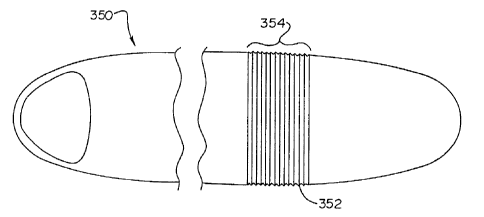
In yet another embodiment, connection ports 203 are located at both the distal
end 200 and the
proximal end 202 of the canister housing 192. Positioning connection ports 203
at both the canister's
distal end 200 and the proximal end 202 may enhance the care provided by the S-
ICD canister 190. In
particular, this canister arrangement allows the operational circuitry in the
S-ICD canister 190 to utilize
multiple electrodes and sensors to best regulate and treat the particular
condition experienced by the
patient recipient. Examples of ancillary devices suitable for attachment
include a lead 193, such as a lead
for sensing, shocking and pacing. Additional ancillary devices suitable for
attachment to the S-ICD
canister 190, being known in the art, (e.g., heart failure monitoring sensors)
are additionally incorporated
as being within the spirit and scope of the present invention.
The top surface 194 of the canister housing 192 may additionally include
particular appendages.
Appendages are especially useful in anchoring the canister housing 192 in a
fixed relative position, or
alternatively, in advancing the canister housing 192 within the patient
recipient. An example of an
appendage that may be incorporated into the top surface 194 of the canister
housing 192 is an extending
22
CA 02465754 2004-05-04
WO 03/039668 PCT/IB02/04516
fin. A fin-like appendage may extend from the canister housing 192 in order to
better direct the S-ICD
canister 190 during the implantation procedure. In this capacity, the extended
fin acts as a rudder
preventing the advancing S-ICD canister 190 from deviating from its desired
path. The extended fin may
additionally aid in preventing the S-ICD canister 190 from displacing from its
original position after
implantation - particularly in the direction perpendicular to the fin's
length. Extending fins suitable for
the present invention may extend the entire length of the canister housing
192, or alternatively, a segment
of the length. Additionally, extending fins may be disposed on the bottom
surface 196 of the canister
housing 192 in order to provide similar functions.
Appendages may also aid physicians in advancing the S-ICD canister 190 to a
desired location
within the patient. Motility-enhancing appendages enable the physician to
push, pull or otherwise direct
the S-ICD canister 190 in a particular fashion throughout the patient's body.
During the procedure, a
physician generally attaches a medical instrument to the motility-enhancing
appendage. This attachment
step may occur either before or after the S-ICD canister 190 has been inserted
within the patient. An
example of one medical instrument capable of attaching to the motility-
enhancing appendage is a
hemostat. Other similar medical instruments, known to those skilled in the
art, may also be utilized in
this attachment step. The physician then advances the hemostat in a desired
direction to properly seat the
S-ICD canister 190 within the patient's body.
The surrounding sides 198 of the canister housing 192 are generally smooth and
substantially
rounded between the top surface 194 and the bottom surface 196 of the canister
housing 192. Smoothing
the side surfaces 198 aids in the insertion of the S-ICD canister 190 during
the implantation procedure.
More specifically, smoother side surfaces 198 permit the S-ICD canister 190,
as a whole, to slide easily
through the surrounding bodily tissue while minimizing abrasion. In addition,
rounded, smooth transition
surfaces allow the surrounding tissues to better conform to the presence of
the device making the device
more comfortable to the patient during chronic implantation.
In contrast, sharp edge formations may have the tendency to ablate, or at a
minimum, irritate the
surrounding tissue during the implantation process. Subsequent tissue
irritation may also occur long after
the implantation process. Minor fluctuations in the positioning of a sharp
edged canister may cause an
inflammatory response in the surrounding tissue. These minor fluctuations are
often the result of simple
day-to-day movements. Movement of the arms, bending at the waist and rotation
of the torso are all daily
activities that may cause surrounding bodily tissue to chafe against the
installed canister. Smoothing
these edges, however, would greatly reduce tissue abrasion, and thereby,
reduce the soreness and
discomfort associated with the implanted S-ICD canister 190.
Referring now to Fig. 20, the bottom surface 196 of the S-ICD canister 190 of
Fig. 19 is shown.
In particular, an electrode 204 possessing an electrically conductive surface
is depicted within the
23
CA 02465754 2004-05-04
WO 03/039668 PCT/IB02/04516
confines of, and hermetically sealed within, the S-ICD canister housing 192.
Although an electrode 204
is specifically illustrated, any sensor capable of receiving physiological
information and/or emitting an
energy may be similarly situated on the canister housing 192. For example, a
sensor may be located on
the canister housing 192 that may monitor a patient's blood glucose level,
respiration, blood oxygen
content, blood pressure and/or cardiac output.
Specifically with reference to Fig. 20, the exposed electrode 204 is
electrically coupled to the
operational circuitry encased within the canister housing 192. The electrode
204, therefore, performs
many of the functions defined by the operational circuitry's programming. More
specifically, the
electrode 204 is the vehicle that actually receives the signals being
monitored, and/or emits the energy
required to pace, shock or otherwise stimulate the heart. Although only a
single electrode 204 is shown
for illustrative purposes, certain S-ICD canister embodiments 190 may be
manufactured with multiple
electrodes. For these embodiments, the multiple electrodes are often task
specific, wherein each electrode
204 performs a single function. In alternate embodiments, a single electrode
204 may perform both
monitoring and shocking functions.
The electrodes 204 are generally positioned at the ends 200 and 202 of the
canister housing 192.
In the S-ICD canister 190 depicted in Fig. 20, the electrode 204 is placed at
the distal end 200 of the
canister housing 192. Although the electrode 204 is positioned in close
proximity to the distal end 200,
the side 198 of the canister housing 192 nearest the distal end 200 should
generally refrain from exposing
any portion of the electrically conductive surface of the electrode 204.
Additionally, although the
electrode is generally planar, in particular embodiments, the electrode may
possess a curved shape.
The size of the electrically conductive surface of an electrode 204, in one
particular embodiment,
is approximately 500 square millimeters in area. In alternate embodiments, it
is desirable to maintain the
size of the electrically conductive surface between approximately 100 square
millimeters and
approximately 2000 square millimeters in area. As with the size of the
canister housing 192, the size of
the electrically conductive surface may vary to accommodate the particular
patient recipient.
Furthermore, the shape and size of an electrode 204 may vary to accommodate
the placement of the
electrode 204 on the canister housing 192. The shape and size of an electrode
may also be varied to adapt
to specified diagnostic and therapeutic functions performed by the canister
190. For example, the
electrode's 204 size and shape may be altered to minimize energy loss to
surrounding bodily tissues, or
for minimizing the diversion of current away from the heart.
One factor in minimizing current diversion is in maintaining an equal current
density distribution
throughout an electrode's 204 conductive surface. A controlling factor in an
electrode's 204 current
density distribution is the electrode's 204 overall shape. Certain electrode
204 shapes draw current to
particular areas on the electrode's 204 conductive surface (e.g., sharp
angles). As a result, these
24
CA 02465754 2004-05-04
WO 03/039668 PCT/IB02/04516
electrodes 204 create an unequal current density distribution. Electrodes 204
possessing sharp corners,
for example, may have higher current densities in the regions defined by the
sharp corner. This unequal
current density distribution results in confined "hot spots". The formation of
hot spots may be desirable
and intentional, such as when attempting to increase current density adjacent
to the sternum. On the other
hand, hot spots may be undesirable as these high current density locations may
scorch or singe
surrounding tissue during the electrode's 204 emission of electrical energy.
Moreover, electrodes 204
possessing numerous hot spots on the electrode's 204 conductive surface
consequently generate areas of
low current density - or "cold spots". This unequal distribution may render
the electrode 204, as a whole,
highly ineffective.
Electrode 204 embodiments of the present invention, in contrast, are
substantially rounded. In
particular, regions of the electrode 204 traditionally possessing sharp comers
are rounded to prevent
extreme hot spots. Nevertheless, the distal most segment of the electrode 200
is slightly angulated in
order to modestly concentrate current at the tip, and therefore, direct
current more through the
mediastinum and into the patient's heart.
Another controlling factor in an electrode's 204 current density distribution
is the electrode's 204
overall size. The relatively small conductive surfaces of electrodes 204 of
the present invention, as
discussed above, minimize the likelihood of forming either hot or cold spots.
Larger electrodes, in
contrast, possess large surface areas that may be more prone to generate more
regions of unequal current
distribution.
As discussed above, electrodes 204 may vary in shape and size to accommodate
an assortment of
canister housing 192 designs. For illustrative purposes, Fig. 20 and Figures
23A-25A show various
electrode shapes disposed upon various canister housings 192. The canister
housings 192 depicted in
these figures, however, are not limited to the electrode shape specifically
illustrated.
The electrode 204 depicted in Fig. 20 is "thumbnail" shaped. The distal end
margin 206 of this
shaped electrode 204 generally follows the outline of the rounded distal end
200 of the canister housing
192. As the electrode 204 moves proximally along the length of the canister
housing 192, the conductive
surface terminates. In the thumbnail embodiment, the electrode's conductive
surface is generally
contained within the rounded portions of the distal end 200 of the canister
housing 192. In alternate
embodiments, the electrode's conductive surface may extend proximally further
within the canister
housing 192. In yet another thumbnail shaped electrode embodiment, the margins
of the electrode's
conductive surface refrain from following the exact rounded contour of the
canister housing 192.
A "spade" shaped electrode 236 is depicted in Fig. 23A. The distal end of the
spade shaped
electrode also generally follows the outline of the rounded distal end 234 of
the canister housing 220. As
the spade shaped electrode 236 moves proximally along the length of the
canister housing 220, the
CA 02465754 2004-05-04
WO 03/039668 PCT/IB02/04516
conductive surface terminates in a rounded proximal end. Similar to the
thumbnail embodiment
described above, the spade shaped electrode's conductive surface is generally
contained within the distal
end 234 of the canister housing 220. In alternate embodiments, the spade shape
electrode's conductive
surface may extend proximally further within the canister housing 220. In yet
another spade shaped
electrode 234 embodiment, the margins of the spade shaped electrode's
conductive surface refrain from
following the exact rounded contour of the canister housing 220, but
substantially form a spade shaped
configuration.
A circular shaped electrode 238 is illustrated in Fig. 23B.
A rectangular shaped electrode 246 is shown in Fig. 24A. Rectangular shaped
electrodes 246
also incorporate electrodes that are substantially rectangular in shape. In
particular to Fig. 24A, the
corners of the rectangular shaped electrode 246 are rounded. Moreover, one
margin of the rectangular
shaped electrode's conductive surface generally follows the rounding of the
distal end 246 of the canister
housing 241.
A triangular shaped electrode 254 is depicted in Fig. 24B. Triangular shaped
electrodes 254 also
incorporate electrodes that are substantially triangular in shape. In
particular to Fig. 24B, the corners of
the triangular shaped electrode 254 are rounded.
A square shaped electrode 257 is depicted in Fig. 24C. Square shaped
electrodes 257 also
incorporate electrodes that are substantially square in shape. In particular
to Fig. 24C, the corners of the
square shaped electrode 257 are rounded.
An ellipsoidal shaped electrode 268 is depicted in Fig. 25A. The distal end of
the ellipsoidal
shaped electrode 268 generally follows the outline of the rounded distal end
264 of the canister housing
260. As the ellipsoidal shaped electrode 268 moves proximally along the length
of the canister housing
260, the conductive surface elongates and then again reduces in length to form
a rounded proximal end.
Similar to the thumbnail and spade shaped embodiments described above, the
ellipsoidal shaped
electrode's conductive surface is generally contained within the distal end
264 of the canister housing
260. In alternate embodiments, the ellipsoidal shape electrode's conductive
surface may extend
proximally further within the canister housing 260. In yet another ellipsoidal
shaped electrode 264
embodiment, the margins of the ellipsoidal shaped electrode's conductive
surface refrain from following
the exact rounded contour of the canister housing 260, but substantially form
an ellipsoidal shaped
configuration.
Energy emissions from any of the above described electrodes 204 generally
follow a path of least
resistance. The intended pathway of the emission, therefore, may not
necessarily be the pathway that the
emission ultimately travels. This is particularly a problem with emissions
made within the human
anatomy where tissue conductivities are highly variable. Obstructing, or low
conductivity tissues like
26
CA 02465754 2004-05-04
WO 03/039668 PCT/IB02/04516
bone material, fat, and aerated lung may all redirect released energy away
from the heart. Alternatively,
surrounding non-cardiac or striated muscle tissue, being generally a high
conductivity tissue, may divert
energy emissions away from the heart. This is a particular concern for the
pectoralis, intercostal, and
latissimus dorsus musculature, as well as other thoracic, non-cardiac
musculature found between the
treating electrodes of the S-ICD. Since the S-ICD canister 190 of the present
invention does not directly
contact the heart muscle itself, such low and high conductivity tissues will
impede and/or shunt a
percentage of the emissions from the present invention's electrode 204 -
permitting the heart to receive a
fraction of the total emitted energy.
The present invention minimizes the effect of impeding and/or obstructing
tissues by designing
an electrode 204 and canister housing 192 capable of focusing the electrode's
array of emitted energy.
Focusing the electrode's array of energy into a highly concentrated beam
enables the resulting beam to be
only minimally impeded or shunted away by any surrounding bodily tissue. This
focused array,
therefore, delivers more of the originally emitted energy directly into the
mediastinum, and subsequently,
into the intended heart muscle than would otherwise occur if the entire
canister, or a majority of the
canister, were electrically active - as is the case with standard transvenous
ICD systems. The present
invention provides an electrode 204 and canister housing 192 design that
creates a consistently focused
array of energy directed toward the chambers of a recipient's heart.
Generally, it is desirable to have the electrode's longest conductive surface
plane positioned
perpendicular to the extending ribs within a recipient's rib cage. Aligning
the electrode 204 in this
manner removes the longest conductive plane from possibly extending directly
over any one particular
rib. If the longest conductive surface were to extend along the length of a
rib, a greater percentage of
emitted energy would be distributed through the rib material, and
consequently, may fail to reach the
heart muscle. When aligned perpendicular to the ribs, only a portion of the
conductive surface is directly
over any particular rib. This alignment permits only a small percentage of the
emitted energy to be
obstructed by the impeding rib material. Therefore, in particular S-ICD
canister 190 embodiments that
extend parallel with a recipient's rib cage, the width 205 of the electrode's
conductive surface is
approximately greater than or equal to the length 207 of the electrode's
conductive surface. This
electrode 204 sizing is best illustrated with reference to Fig. 20. The
conductive surface of the thumbnail-
shaped electrode in Fig. 20 is depicted as both shallow and wide. In contrast,
S-ICD canister 190
embodiments that extend perpendicular with a recipient's rib cage, can have
their conductive surface's
length 207 being greater than their conductive surface's width 205. The
appropriate S-ICD canister 190
alignment, and subsequently the appropriate electrode 204 alignment, is
determined by the style of S-ICD
canister 190 chosen for the patient recipient. Figures 23A-26C illustrate
numerous S-ICD canister
housing embodiments 192 for properly positioning an electrode 204 over a
recipient's heart. The
27
CA 02465754 2004-05-04
WO 03/039668 PCT/IB02/04516
embodiments depicted, however, are for illustrative purposes only, and are not
intended to limit the scope
of the present invention.
Another solution to the problem of thoracic tissues interfering with energy
delivery is by
designing a canister housing 192 that may be strategically positioned in close
proximity to the patient's
heart. One embodiment of the present invention possesses a curved canister
housing 192 that enables the
S-ICD canister 190 to be advanced just over the patient recipient's ribcage.
Moreover, in another
embodiment, the curvature of the S-ICD canister 190 directly mimics the
natural curvature of the ribcage.
Referring now to Fig. 21, the S-ICD canister 190 of Fig. 19 is shown from the
side. Fig. 21
shows the S-ICD canister's top surface 194, the bottom surface 196 and the
side 198 of the canister
housing 192. In the embodiment depicted, both the top surface 194 and the
bottom surface 196 of the
canister housing 192 are curved. In fact, throughout most of the proximal end
202 of the canister housing
192, the curvature is generally similar, and indeed can be identical, between
the top surface 194 and the
bottom surface 196. In alternative embodiments of the present invention, the
top surface 194 may be
generally planar while the bottom surface 196 is curved. In yet another
embodiment of the present
invention, the top surface 194 may be curved and the bottom surface 196 is
generally planar.
Referring back to the embodiment depicted in Fig. 21, the curvatures between
the top surface 194
and the bottom surface 196 are shown differing toward the distal end 200 of
the canister housing 192. At
the S-ICD canister's distal end 200, the canister housing's top surface 194
curvature tapers downwardly
toward the canister's bottom surface 196. This tapering causes the distal end
200 of the canister housing
192 to be narrower (of a decreased depth) than the canister's proximal end
202. In certain embodiments,
this tapering in depth may be gradual throughout the length of the canister's
housing 192, or alternatively,
the tapering may be confined to a particular area.
Tapering the depth of the canister housing 192 may improve the overall
performance of the S-
ICD canister 190. In particular, a tapered distal end 200 may aid in insertion
and advancement of the S-
ICD canister 190 within the patient recipient's body. A tapered distal end 200
enables the S-ICD canister
190 to easily traverse through narrow subcutaneous spaces. In particular, a
physician generally tries to
create a passageway into the patient's body that is appropriately sized for
the canister, especially in regard
to positioning the distal segment of the canister with the end containing the
electrode in close proximity to
the sternum. Tapering the distal end of the canister eliminates unnecessary
trauma to the patient in the
tight spaces adjacent to the sternum. For larger canisters, however, this
tight subcutaneous space is
difficult to traverse. Subsequently, these larger canisters cause the
physician to undertake extensive sharp
and blunt dissection of the patient's tissues in order to place the larger
canister in the desired location.
Regardless of the extent of the dissection, however, larger non-tapered distal
segments may prove
28
CA 02465754 2004-05-04
WO 03/039668 PCT/IB02/04516
extremely uncomfortable if forced into a parasternal position to satisfy the
needs of focusing energy
through the mediastinum, and subsequently, to the patient's heart.
In contrast, embodiments of the present invention having narrow canister
housings 192 may
easily traverse such passageways. Moreover, tapering the S-ICD canister's
distal end 200 further
streamlines the canister housing 192, and therefore, enhances the ease of the
implantation procedure.
Tapering the S-ICD canister's distal end 200 is particularly important when
positioning the distal end of
the canister housing as near the left border of a patient's sternum as
possible. This canister housing 192
placement optimizes energy delivery to the mediastinum, and therefore, to the
patient's heart.
The depth of the canister housing 192 is shown as being very narrow as to the
canister housing's
length 207. The canister's housing depth is less than approximately 15
millimeters. In alternate
embodiments, the depth of the canister's housing depth is approximately 5
millimeters to approximately
10 millimeters. At the tapered distal end 200, the canister housing may have a
depth of approximately 1-
4 millimeters.
In certain embodiments of the present invention, it is desirable to position
the S-ICD canister 190
in close proximity to the patient recipient's heart, without directly
contacting the heart. A favored
location for this S-ICD canister 190 placement is just over the patient's
ribcage. More particularly, in
certain embodiments it is favored to place the S-ICD canister 190 just to the
left of, and adjacent to, the
sternum with a segment at the distal end 200 containing the electrode 204
closest to the sternum. Fig. 22
depicts the placement of the S-ICD canister 190 according to one embodiment of
the present invention
with the lead electrode traversing the subcutaneous tissues laterally toward
the axilla and then posteriorly
to "catch" the current as it is emitted from electrode 204 parasternally and
anteriorly toward the lead
electrode 191 as it receives current exiting the posterior mediastinum and
paraspinal tissues.
During the implantation procedure, a single incision 210 is made in the left
anterior axillary line
approximately at the level of the cardiac apex, or around the fifth to the
sixth intercostal space. The
location of this single incision 210 enables the physician to position both
the S-ICD canister 190 and the
canister's ancillary devices (e.g., pacing leads, shocking leads, etc.) from
this single incision 210. Once
this incision 210 is made, the physician may insert surgical instruments or a
specially designed tool (not
shown) through the incision 210 to shape a passageway for the S-ICD canister
190 to navigate. Although
a tool may be utilized in particular embodiments, a tool is not required -
standard surgical instruments,
together with the general shape of the S-ICD canister 190, are sufficient to
facilitate proper positioning of
the device in the left anterior thorax as adjacent as possible to the sternum.
In particular embodiments, a physician advances both the S-ICD canister 190
and the lead
electrode 191 within the patient to form a depolarization vector with respect
to the patient's heart 218.
The depolarization vector is a vector having an origin, a first end point and
a second end point.
29
CA 02465754 2004-05-04
WO 03/039668 PCT/IB02/04516
In one embodiment, the origin of the depolarization vector originates
approximately within the
chambers of the patient's heart 218. Similarly, the first vector end point
comprises the S-ICD canister
electrode's 204 positioning with respect to the patient's heart 218. Finally,
the second vector end point
comprises the lead electrode's 191 positioning with respect to the patient's
heart 218. In alternate
embodiments, the second vector end point comprises a second canister
electrode.
The lead electrode may be positioned at various positions within the body
because the length of
the lead 193. may be varied. For example, S-ICD devices of the present
invention may have leads with
lengths between 5 centimeters and 55 centimeters. Therefore, the S-ICD
canister 190 and lead electrode
191 of the present invention may create numerous depolarization vectors.
In particular embodiments, a degree of separation of 180 degrees or less
exists between the S-ICD
canister electrode 204 and the lead electrode 191. In alternative embodiments,
the degree of separation
between the S-ICD canister electrode 204 and the lead electrode 191 is
approximately 30 degrees to
approximately 180 degrees.
In order to obtain the desired degree of separation for the depolarization
vector, generally one
device (either the S-ICD canister 190 or the lead electrode 191) must be
advanced anteriorily while the
other device is advanced posteriorily from the initial incision 210.
Accordingly, when the S-ICD canister
190 is advanced subcutaneously and anteriorily from the incision 210, the lead
electrode 191 must be
advanced subcutaneously and posteriorily from the incision 210. With this
particular embodiment, a
physician may advance the S-ICD canister 190 medially toward the patient's
left inframammary crease to
a location proximate the patient's sternum 212.
Alternatively, the physician may advance, and subsequently position the S-ICD
canister 190
within the anterior portion of the patient's ribcage 216. This anterior
placement may further include the
patient's left parasternal region, an anterior placement within the region of
the patient's third and the
patient's twelfth rib 214, or generally any subcutaneous ribcage 216 placement
anterior to the patient's
heart 218. In order to complement the S-ICD canister's 190 placement, and
obtain the correct
depolarization vector, the lead electrode 191 must be advanced posteriorily
toward the paraspinal or
parascapular region of the patient's ribcage 216.
In another embodiment of the present invention, the spatial positioning of the
S-ICD canister 190
and the lead electrode 191, described in detail above, are reversed.
Referring back to Fig. 21, the curvature of particular S-ICD canister
embodiments 190 may be
designed to generally mimic the natural curvature of a patient's ribcage 216.
These S-ICD canister
embodiments 190 restrict canister displacement and heighten comfort for the
patient implanted with the
S-ICD canister 190. The anatomical shape of a patient recipient's ribcage 216
varies. The present
invention includes numerous S-ICD canister housing 192 curvatures to
accommodate these varying
CA 02465754 2004-05-04
WO 03/039668 PCT/IB02/04516
shapes. In particular, the present invention includes S-ICD canisters 190
sized and shaped to properly fit
children, as well as ones to properly fit fully developed adults.
The curvature of the canister housing 192 is generally arc-shaped. The degree
of curvature for
any particular embodiment of the present invention is measured through a
curvature vector theta (0). The
curvature vector 0 is a vector having an origin 199, a first end point and a
second end point.
In one embodiment, the origin 199 of the curvature vector 0 originates
approximately at the
center of the S-ICD canister 190 (lengthwise). The first vector end point in
this embodiment comprises
the distal end 200 of the S-ICD canister 190 and the second vector end point
comprises the proximal end
202 of the S-ICD canister 190. In particular embodiments, the curvature vector
0 possesses a degree of
separation between 30 degrees and 180 degrees. For example, a canister housing
192 having a degree of
separation of 180 degrees is planar. Decreasing the degree of curvature 0
causes the canister housing to
become more arcuate in shape.
In alternative embodiments, the origin 199 of the curvature vector 0 may
originate at a point other
than the center of the S-ICD canister 190. Origins 199 shifted from the center
of the S-ICD canister 190
produce regions of greater curvature, as well as areas of lesser curvature, in
the same S-ICD canister 190.
Similarly, a S-ICD canister 190 may possess multiple curvature vectors 0
having origins 199 throughout
the length of the S-ICD canister 190. Multiple curvature vectors 0 produce
various non-linear or
nonsymmetrical curves that, in certain circumstances, remain generally arc-
shaped. Canister housings
possessing multiple curvature vectors 0 are particularly suitable for S-ICD
canister 190 placement near
the patient's sides (generally in the area under the patient's arms where the
thorax has a more marked
degree of curvature). Canister housings 192 incorporating a nonsymmetrical
curvature are generally
longer S-ICD canisters 190 that span over the front and sides of the patient's
ribcage. In particular, these
S-ICD canisters 190 span areas of the ribcage 216 that are generally planar
(around the patient's sternum
212), as well as areas that are highly curved (generally in the area under the
patient's arms).
Curved canister housings 192 are generally for S-ICD canisters 190 that extend
lengthwise, or
approximately horizontally, along the length of the ribs in the ribcage 216.
For certain embodiments,
however, it is desired to orient the length of the S-ICD canister 190 to be
perpendicular to the length of
the ribs in the ribcage 216. A perpendicularly orientated S-ICD canister 190
generally requires very little,
if any, curvature to conform to the ribcage 216.
Figures 23A-26C depict particular S-ICD canister 190 designs. In each of these
particular S-ICD
canister designs, the various material constructions, dimensions and
curvatures, discussed in detail above,
may be incorporated within each individual S-ICD canister design. Furthermore,
particular aspects of any
31
CA 02465754 2004-05-04
WO 03/039668 PCT/IB02/04516
individual S-ICD canister design may be incorporated, in whole or in part,
into another depicted S-ICD
canister design.
Turning now to Fig. 23A, a S-ICD canister 220 having a duckbill-shaped
canister housing 222 is
shown. The duckbill-shaped canister housing 222 has a proximal end 226 and a
distal end 234. The
proximal end 226 of the duckbill-shaped canister housing 222 further includes
a main housing member
228 and a distal housing member 230. The distal housing member 230 is an
elongated segment extending
distally from the distal end of the main housing member 228. Although the two
segments differ in their
size and shape, the distal housing member 230 and main housing member 228 are
generally contiguously
and fluidly attached to one another and may be formed from a single mold. In
alternative embodiments,
however, the distal housing member 230 may be hinged to the main housing
member 228. The distal
housing member 230 also generally comprises a material that is similar in
composition to that forming the
main housing member 228. In alternate embodiments, however, the distal housing
member 230 may
include a material that possess enhanced electrically insulated
characteristics.
The main housing member 228 generally encases the operational circuitry,
batteries and
capacitors of the duckbill-shaped S-ICD canister 220. The width and length of
the main housing member
228 enable the main housing member 228 to accommodate batteries and capacitors
for delivering a
shocking energy of approximately 50 J of energy, 75 J of energy, 100 J of
energy, 125 J of energy, 150 J
of energy and 200 J of energy.
Although a specific number of batteries and capacitors are required for
delivering these charges,
their positioning within the canister housing 222 is highly modifiable. More
specifically, the width of the
main housing member 228 may be altered to accommodate a longer or shorter
canister. For example, the
width of the main housing member 228 may be increased in order to obtain a
main canister housing 228
of decreased length. Modification of the sizing and orientation of the main
housing member 228 allow
manufacturers to create a variety of differing sized duckbill-shaped S-ICD
canisters 220. Increased
specificity in the canister housing's shape and size enhance the comfort and
wearability for the patient
recipient.
In general, the width of the main housing member 228 is approximately 10 cm
wide or less.
Likewise, the length of the main housing member 228 is approximately 20 cm
long or less. In particular
embodiments the width of the main housing member 228 is 4 cm. In an
alternative embodiment, the
width of the main housing member 228 is 8 cm.
The distal housing member 230 is an elongated segment of canister housing that
possesses a
width that differs from that of the main housing member 228. The distal
housing member's width
decreases as the distal housing member 230 extends distally. This tapering in
width results in the
formation of a shoulder region 232. In particular embodiments, the rate with
which the width decreases
32
CA 02465754 2004-05-04
WO 03/039668 PCT/IB02/04516
as the proximal housing member 230 extends distally is constant. In alternate
embodiments, the rate is
variable. A variable rate shoulder region 232 taper proceeds at a rate of
tapering where a unit of tapering
width is not directly related to a unit of length in the distal direction. In
either of the embodiments,
however, bilateral symmetry is maintained throughout the length of the distal
housing member 230.
The shoulder region 232 is a generally rounded and smooth region of the
canister housing 222.
As discussed in detail above, rounding the edges along the canister's surface
enhances insertion of the S-
ICD canister 220. The rounded edges also reduce abrasion and inflammation
associated with short-term
and long-term wearability.
Extending distally beyond the shoulder region 232 is the distal head 234 of
the distal housing
member 230. The distal head 234 is the distal termination point of the
duckbill-shaped S-ICD canister
220. The distal head 234 includes a generally rounded end. In one embodiment,
illustrated in Fig. 23B,
the distal head 234 has a width greater than the width at a location within
the shoulder region 232 of the
distal housing member 230. In alternative embodiments, the distal head's width
is equal to or less than
the width at any point in the shoulder region 232 of the distal housing member
230, as illustrated in 23A.
The length of the duckbill-shaped S-lCD canister 220 may depend highly upon
the shape and size
of the distal housing member 230. In particular embodiments, the duckbill-
shaped S-lCD canister 220 is
approximately 30 centimeters long or less. In alternative embodiments, the
duckbill-shaped S-ICD
canister 220 is approximately 10 centimeter or less. In particular
embodiments, the length of the duckbill-
shaped S-ICD canister 220 may be curved, or alternatively, or a portion of the
length (i.e., the shoulder
region 232 and distal head 234) are curved.
The electrode 236 for the duckbill-shaped S-ICD canister 220 is generally
seated within a portion
of the distal housing member 230. Fig. 23A diagrams in phantom the approximate
location of an
electrode 236 on the duckbill-shaped canister housing 222. Although the
electrode 236 is depicted as
generally circular in shape (in Fig. 23B), the electrode may also be "spade
shaped" (depicted in Fig. 23A),
thumbnail shaped, square, rectangular, triangular or ellipsoidal. The
electrode 236 is electrically coupled
to the operational circuitry within the main housing member 228 of the S-ICD
canister 220.
In certain embodiments of the present invention, an associated feature of the
electrode 236 at the
distal end is the presence of a margin of insulated material 237 around the
active electrode 236. The
margin of insulated material 237 may aid in directing emitted energy from the
electrode 236 inwardly
toward the patient's heart instead of dispersing energy outward toward the
patient's chest wall. This
margin of insulated material 237 typically ranges from 1-5 mm in width and may
extend to the margin of
the housing. Moreover, in certain embodiments, the margin of insulated
material 237 comprises a
ceramic material or other material designed to facilitate focusing of current
inward toward the heart.
33
CA 02465754 2004-05-04
WO 03/039668 PCT/IB02/04516
In certain embodiments of the present invention, the electronic components
(e.g., circuitry,
batteries and capacitors) of the S-ICD canister 220, are generally absent from
the distal housing member
230. As such, the depth of the distal housing member 230 may be greatly
reduced. In these
embodiments, a depth of approximately 1 millimeter may be obtained at the
distal head 234 of the
duckbill-shaped S-ICD canister 220.
The duckbill-shaped distal housing member 230 enhances navigation during
canister
implantation. The distal head 234 of the distal housing member 230 is blunt at
its end to reduce trauma
suffered to surrounding tissue during the S-ICD canister's advancement or
during chronic implantation.
Similarly, the narrower distal head 234 (width-wise and depth-wise) is easier
to control during the
advancement procedure. The smaller distal head 234 also enables a physician to
navigate the smaller and
more compact tissues adjacent to the sternum, which a larger head might
otherwise find unobtainable.
Moreover, the narrower distal head 234 may be advanced to a location in close
proximity to the patient
recipient's heart 218 without concern of distorting or stressing the skin in
the left parasternal region.
The closer the electrode 236 is to the patient's heart 218, the less energy is
required to achieve an
adequate electric field or current density to defibrillate the heart, A
desirable anatomical position for
reducing this energy requirement is just lateral to the sternum 212 of the
patient. The area surrounding
the patient's sternum 212 generally lacks a considerable accumulation of
bodily tissue. Thus,
subcutaneous S-ICD canister 190 positioning over the sternum 212, or some
other location just over the
rib cage 216, provides a significant lessening of the required energy - due to
proximity to the heart 218
and a reduction in impeding surrounding tissue. Positioning an ICD canister of
normal contour in this
area has proven difficult, however, and is additionally aesthetically
displeasing. The reduced profile of
the duckbill-shaped S-ICD canister 220, however, provides such optimal
electrode 236 placement in a
more aesthetically and less physically obtrusive manner.
Structurally, a reduction in the energy requirement frees space within the
canister housing 222.
This space was previously occupied by batteries and capacitors needed for the
higher energy
requirements. This space, however, is no longer required. The duckbill-shaped
S-ICD canister 220,
therefore, can be smaller in length, width and depth. Eliminating batteries
and capacitors also reduces the
weight of the present invention. As described in detail above, reducing the
weight of the S-ICD canister
enhances patient recipient comfort.
Fig. 24A illustrates another embodiment of a S-ICD canister having a generally
rectangular-
shaped canister housing 240. The rectangular-shaped canister housing 240
includes a top surface 241, a
bottom surface (not shown) and surrounding sides 248 connecting these two
surfaces. The rectangular-
shaped canister housing 240 further includes a distal end 242 and a proximal
end 244. The electrode 246,
shown in phantom, is generally positioned at either the distal end 242 or the
proximal end 244 of the
34
CA 02465754 2004-05-04
WO 03/039668 PCT/IB02/04516
canister housing 240. In alternative embodiments, the rectangular-shaped
canister housing 240 may
include two or more electrodes 246. When two electrodes are utilized, one
electrode is positioned at the
distal end 242 of the canister housing 240 while the second electrode is
positioned at the proximal end
244 of the canister housing 240.
The length of the rectangular-shaped canister housing 240 is approximately 30
centimeters long.
In alternative embodiments, the rectangular-shaped canister housing 240 is
approximately 10 centimeter
long or less. The width of the rectangular-shaped canister housing 240 is
approximately 3 centimeters to
approximately 10 centimeter wide.
Figures 24B and 24C depict additional embodiments of a S-ICD canister having a
generally
square-shaped canister housing 250. The square-shaped canister housing 250
includes a top surface 251,
a bottom surface (not shown) and surrounding sides 252 connecting these two
surfaces. The sides 252 of
the square-shaped canister housing are generally of the same length. The
electrode 254, shown in
phantom, is generally positioned in the center and to one side of the square-
shaped canister housing 250.
A triangular shaped electrode 254 is specifically illustrated at the corner of
the square-shaped canister
housing 250 in Fig. 24B. In alternate embodiments, however, the electrode 254
may be positioned
toward the center of one of the sides 252 of the square-shaped canister
housing 250, or at the center of the
square-shaped canister housing 250, or rotated more. A square shaped electrode
257 is specifically
illustrated at the side of the canister housing 250 in Fig. 24C.
The length and width of the square-shaped canister housing 250 is
approximately 6 centimeters to
approximately 8 centimeter long and wide.
Fig. 25A depicts yet another embodiment of a S-ICD canister having a "tongue
depressor-
shaped" canister housing 260. The tongue depressor-shaped canister housing 260
includes a top surface
261, a bottom surface (not shown) and surrounding sides 262 connecting these
two surfaces. The tongue
depressor-shaped canister housing 260 further includes a distal end 264 and a
proximal end 266. The
distal end 264 and the proximal end 266 of the tongue depressor-shaped
canister housing 260, however,
are rounded. In one embodiment, the rounded ends extend outwardly away from
the canister housing 260
in either the corresponding distal or proximal direction. The rounded ends
generally are circular arc-
shaped curves, however, the rounded ends may also be elliptical or
nonsymmetrical arc-shaped curves.
The electrode 268, shown in phantom, is generally positioned at either the
distal end 264 or the
proximal end 266 of the canister housing 260. In alternative embodiments, the
tongue depressor-shaped
canister housing 260 may include two or more electrodes 268. When two
electrodes are utilized, one
electrode is positioned at the distal end 264 of the canister housing 260
while the second electrode is
positioned at the proximal end 266 of the canister housing 260.
CA 02465754 2004-05-04
WO 03/039668 PCT/IB02/04516
The length of the tongue depressor-shaped canister housing 260 is
approximately 30 centimeters
long or less. In alternative embodiments, the tongue depressor-shaped canister
housing 260 is
approximately 15 centimeter long or less. The width of the tongue depressor-
shaped canister housing 260
is approximately 3 centimeters to approximately 10 centimeters wide.
Referring now to Fig. 25B, where a modified tongue depressor-shaped canister
housing 270 is
shown. The modified tongue depressor-shaped canister housing 270 is similar to
the tongue depressor-
shaped S-ICD canister 260 depicted in Fig. 25A, however, the modified tongue
depressor-shaped canister
housing 270 comprises only has a single rounded distal end 272. The proximal
end 274 of the modified
tongue depressor-shaped canister housing 270 is generally square.
Figures 26A-26C illustrate another embodiment of a S-ICD canister having a
multi-segment
canister housing 280. The multi-segment canister housing 280 includes at least
two canister housing
segments that are coupled together. The S-ICD canister depicted in Fig. 26A,
26B and 26C specifically
have a distal segment 282 and a proximal segment 284 hinged, or otherwise
coupled, together.
The distal segment 282 includes a top surface 292, a bottom surface (not
shown) and surrounding
sides 286 connecting these two surfaces. The distal most end 288 of the distal
segment 282 comprises a
rounded region. An electrode 290 is disposed within this rounded region of the
distal segment 282
(shown in phantom). The electrode 290 generally follows the outline of the
rounded region of the distal
most end 288 of the canister housing, however, the electrode 290 may comprise
of other shapes and sizes.
In an embodiment of the multi-segment canister housing 280, both the electrode
290 and the
electronics are disposed within the distal segment 282. In alternative
embodiments, the electrode 290 is
disposed within the distal segment 282 and the electronics are located within
the proximal segment 284 of
the multi-segment canister housing 280.
Fig. 26B shows the distal segment 282 of the multi-segment canister housing
280 being curved to
mimic the anatomical shape of a patient recipient's ribcage 216. In the
embodiment depicted, both the
top surface 292 and the bottom surface 294 of the proximal segment 282 are
curved. The curvature,
however, differs at the distal most end 288 of the distal segment 282. At the
distal segment's distal most
end 288, the distal segment's top surface 292 tapers downwardly toward the
distal segment's bottom
surface 294. This tapering causes the distal most end 288 of the distal
segment 282 to be narrower than
the distal segment's distal end 296. In certain embodiments, this tapering in
depth may be gradual
throughout the length of the distal segment 282, or alternatively, the
tapering may be confined to a
particular area.
The proximal segment 284 also includes a top surface 298, a bottom surface 300
and surrounding
sides 302 connecting these two surfaces. The proximal segment 284 depicted in
Fig. 26B, however, is
36
CA 02465754 2004-05-04
WO 03/039668 PCT/IB02/04516
generally planar. In alternative embodiments, depicted in Fig. 26C, the
proximal segment 284 may also
be curved and may also be of a different curvature to that of the distal
segment.
The length of the multi-segment canister housing 280 is approximately 30
centimeters long or
less. In alternative embodiments, the multi-segment canister housing 280 is
approximately 20 centimeters
or less. In yet another embodiment, the multi-segment canister housing 280 is
approximately 12
centimeters or less. The width of multi-segment canister housing 280 is
approximately 3 centimeters to
approximately 10 centimeters wide.
Figures 28A illustrate another embodiment of an S-ICD canister having a multi-
segment canister
housing 330. The multi-segment canister housing 330 includes at least two
canister housing segments
that are coupled together. The S-ICD canister depicted in Fig. 28A
specifically include a distal segment
332 and a proximal segment 334 coupled together. The coupling of adjacent
segments occurs through a
hinging mechanism. As illustrated in Figure 28A, a first hinging mechanism 336
and a second hinging
mechanism 338 are positioned between the proximal segment 334 and the distal
segment 332 of the
multi-segment canister housing 330. Although two hinging mechanisms 336 and
338 are specifically
depicted, any number of hinging mechanisms may be utilized in coupling
adjacent segments. Moreover,
the hinging mechanisms may be positioned at any suitable location on the
canister housing 330 that
enables adjacent segments to pivot about the hinging mechanism.
FIG. 28B generally depicts one embodiment of the present invention for hinging
the proximal end
334 of the multi-segment canister 330 to the distal segment 332 of the multi-
segment canister 330. More
specifically, FIG. 28B illustrates the first hinging mechanism 336 of FIG.
28A.
As shown, the proximal segment 334 includes a threaded hole 340 that traverses
through the
entire depth of the proximal segment's canister housing 334. The distal
segment 332, in contrast,
includes a threaded ball 342 (a ball having a single orifice that is threaded)
retained by a plurality of leaf
springs 344. The leaf springs 344 in the distal segment 332 prevent the
displacement of the threaded ball
342 from the distal segment 332, while permitting rotational movement of the
threaded ball 342. In
certain embodiments of the present invention, four leaf springs 344 are
utilized to apply a positive
pressure against the threaded ball 342.
The coupling of the proximal segment 334 with the distal segment 332 occurs by
advancing a
screw 346 through the threaded hole 340 of the proximal segment 334 and into
the threaded ball 342 held
within the distal segment 332. The length of the screw 346 enables a portion
of the screw 346 to be
retained within both the proximal segment 334 and the distal segment 332 of
the multi-segment canister
housing 330. As such, the two segments permit some pivoting action between the
segments while
remaining positively coupled together. Moreover, the screw 346 may further act
as a conduit for the
37
CA 02465754 2004-05-04
WO 03/039668 PCT/IB02/04516
transfer of electrical energy between segments. Electrical energy may be
transferred from an electrical
circuit in one segment, through the screw 346, and into an adjacent segment.
The use of a threaded ball 342 enables a degree of rotational freedom between
the two segments.
The segments, therefore, may be translated in numerous directions with respect
to one another. This
added freedom permits the multi-segment canister housing 340 to conform to an
array of shapes, and
subsequently, to an array of patient recipient body types.
FIG. 29A is a bottom plan view of an alternative S-ICD canister 350 of the
present invention
having a canister housing region that is pliable. The pliable region of the
canister housing 350 comprises
a plurality of pleats 352 in the material forming the canister housing 350. In
particular embodiments,
materials forming the pleated sections 354 of the canister housing 350 include
polymeric materials such
as polyurethanes, polyamides, polyetheretherketones (PEEK), polyether block
amides (PEBA),
polytetrafluoroethylene (PTFE), polyethylenes, silicones, and mixtures
thereof. Metallic materials
suitable for the pleated sections 354 of the canister housing 350 include
stainless steel, and titanium.
Alloys suitable for the pleated sections 354 of the canister housing 350
include stainless steel alloys and
- titanium alloys such as nickel titanium.
FIG. 29B is a side view of the S-ICD canister embodiment of Fig. 29A. As
depicted, the
individual pleats 352 in the pleated section 354 may uniformly expand or
contract; enabling the canister
housing 350 to either increase or decrease its overall length. Moreover,
individual pleats 352 may expand
in one region and contract in another to allow the canister housing 350 to
bend either inwardly or
outwardly. For example, the pleats 352 may expand along the top surface 356 of
the canister housing 350
while contract along the bottom surface 358 of the canister housing 350 to
enable the canister housing
350 to reduce the degree of separation in the canister housing's curvature
vector 0 (described in detail
above).
The use of a pleated section 354 in a canister housing 350 allows the S-ICD/US-
ICD to bend and
conform specifically to an individual's ribcage. Thus, the use of a pleated
section 354 permits the proper
sizing of a single canister housing 350 in numerous patients, without
requiring the manufacturer to
generate numerous canister housings of various specific shapes and sizes.
Moreover, the use of pleated
canister housings 350 is helpful in eliminating patient awareness of the
implanted device and in
improving the long-term wearability of the implanted device.
Numerous characteristics and advantages of the invention covered by this
document have been
set forth in the foregoing description. It will be understood, however, that
this disclosure is, in many
aspects, only illustrative. Changes may be made in details, particularly in
matters of shape, size and
arrangement of parts without exceeding the scope of the invention. The
invention's scope is defined, of
course, in the language in which the appended claims are expressed.
38