Note: Descriptions are shown in the official language in which they were submitted.
CA 02466333 2004-05-07
WO 03/040304 PCT/IB02/04930
METHOD OF PROLIFERATION IN
NEUROGENIC REGIONS
FIELD OF THE INVENTION
This application is directed to compounds that disrupt EphA7 and ephrin-
AS interaction or EphA7 and ephrin-A2 interaction. Further, this application
is directed
to methods for the use of these compounds and to the use of the compounds for
the
alleviation of one or more symptoms of a neurological disease or disorder.
BACKGROUND OF THE INVENTION
Receptor tyrosine kinases (RTKs) are important mediators of effects from
signalling proteins in both the developing and the adult organism. The Eph
receptors
constitute the largest family of RTKs. Ephrins are membrane-bound ligands for
the Eph
protein tyrosine kinase receptor family. This class of molecules is further
subdivided into
A-class and B-class ephrins that couple to A- and B-type receptors,
respectively. One
exception to this rule is EphA4, which elicits binding to both A and B
ligands. Both
classes of ligands are anchored to the membrane even though the A ligands only
are
attached to the outer leaflet of the membrane in contrast to the B ligands
that span the
entire membrane (Frisen, J. et al., 1999. EMBO J. 18: 5159-5165; Wilkinson,
D.G.,
2001. Nat Rev Neurosci 2(3): 155-64). It has been shown that in order to
activate the
receptor, the ligand has to be clustered into oligomers (Davis, S. et al.,
1994. Science
266: 816-819). Upon binding to the ligand complex the receptor itself
dimerizes,
enabling cross-phosphorylation of the tyrosine kinase domains, thus triggering
a signal
transduction cascade. One feature of Eph-ephrin signalling is the bi-
directional signalling
made possible by the membrane-attached ligands. The bi-directional signalling
allows the
ligand to act as a receptor and vice versa. This type of reverse signalling is
well
established with regard to the ephrin-Bs Henkemeyer, M. et al., 1996. Nature
383: 722-
725) and recent evidence suggests that the same is true for the ephrin-As
(Davy, A. et al.,
1999. Genes Dev. 13: 3125-3135; Huai, et al., 2001. J Biol Chem 276(9): 6689-
94).
Eph receptors and ephrins show widespread expression in the developing nervous
system
as well as in the adult central nervous system (CNS) ( Frisen, J. et al.,
1999. EMBO J.
18: 5159-5165). First shown to act as repellent guidance cues for growing
axons, recent
1
CA 02466333 2004-05-07
WO 03/040304 PCT/IB02/04930
research has revealed an astounding functional versatility of ephrins and Eph
receptors
(Wilkinson, D.G., 2001. Nat Rev Neurosci 2(3): 155-64).
Sites of neurogenesis are retained in the adult brain. Among these, two
locations exhibit high levels of Eph receptor and ephrin expression: the
dentate gyros of
the hippocampus and the lateral ventricular wall. The exact identity of the
stem cells
residing in the SVZ remains to be proven. Evidence for an ependymal as well as
a
subependymal origin for the stem cells exists ( Johansson, C. et al., 1999.
Cell 96: 25-
34; Doetsch, F. et al., 1999. Cell 97: 703-716). Nevertheless it is possible
to dissect the
lateral wall, dissociate the tissue and cultivate the stem cells as buoyant
spheres,
denominated neurospheres). The neurospheres have self renewal capacity and the
developmental potential to differentiate into neurons, oligodendrocytes and
astrocytes
(Johansson, C. et al., 1999. Cell 96: 25-34). l~ vivo the stem cells give rise
to neural
progenitors that migrate along the lateral wall and feed into the
rostromigratory stream,
eventually ending up in the olfactory bulb (Doetsch, F. et al., 1996. Science
271: 978-
981). To keep the cells in a low proliferative, undifferentiated mode one
could postulate a
non-autonomous mechanism where an extracellular protein could, when activated
through
binding to a ligand/receptor, act as a repressor on proliferation and/or
differentiation. The
lack of such an activation would results in increased proliferation or
differentiation. The
high expression of ephrin-A2, and EphA7 in the above mentioned neurogenic
regions
could be an indication of such a model.
2
CA 02466333 2004-05-07
WO 03/040304 PCT/IB02/04930
BRIEF SUMMARY OF THE INVENTION
The Eph tyrosine kinase receptors and their ephrin ligands confer short
range communication between cells in the developing organism regulating
diverse
processes such as axon guidance, cell migration and neural tube formation (
Wilkinson,
D.G., 2001. Nat Rev Neurosci 2(3): 155-64). Even though both receptors and
ligands are
widely expressed in the adult nervous system, the knowledge concerning their
roles in the
adult is limited. Neurogenic areas in the adult brain, including the lateral
wall of the
lateral ventricle and the dentate gyrus of the hippocampus, express EphA7 and
the ligands
ephrin-A2. Mice lacking the receptor EphA7 exhibit increased cellular
proliferation in
the tissue on the lateral side of the lateral ventricle. We show that in the
wild type
organism the ephrin or Eph are negative regulators of proliferation, keeping
it at a basal
level. This effect involves reversed signalling through the ligand upon
binding to the
EphA7 receptor. Upon injection of the freely soluible form of ephrin-AS-Fc,
ephrin-A2
or EphA7 either as monomers or as oligomers into the lateral ventricle, the
number of
proliferating cells as measured by BrdU-labelling was significantly higher
than in sham
injected mice. The ephrin-AS-Fc, ephrin-A2 or EphA7 proteins presumably
disrupt the
binding between the endogenous ligands and receptors, thus blocking signalling
through
the ligands and allowing a higher rate of proliferation.
Mice lacking EphA7 have minimal and compressed lateral ventricles due
to increased amount of tissue in the lateral side of the ventricle. In the
EphA7 null
mutants BrdU injections show that the rate of proliferation in the ventricular
wall is
significantly higher than in the wild type. We have also performed i~ vitro
studies that
show a dramatic decrease in proliferation and/or differention capacity of
neurospheres that
are grown on a surface coated with EphA7 proteins in a conformation that can
activate the
ephrin ligands (clustered) whereas the opposite is true when EphA7 is
presented in a form
that will only block the ligands and not activate them (unclustered). The
latter case
mimics the mouse mutants with the coated EphA7 blocking the endogenous binding
of
EphA7 to ephrin-A2 in the neurospheres thus silencing the repressing activity
of the
ephrin-A ligand. Furthermore, when cultivated, stem cells from the lateral
ventricular
wall of an EphA7 null mutant mouse give rise to significantly higher numbers
of spheres
3
CA 02466333 2004-05-07
WO 03/040304 PCT/IB02/04930
than corresponding tissue from a wild type mouse. We delivered ephrin-AS or
ephrin-A2
ligands through intracranial infusion into rodent lateral ventricle and
measured
proliferation in the lateral wall through BrdU labeling of dividing cells. We
reasoned that
the endogenous binding between EphA7 and the ephrin ligands would be
interrupted and
allow a higher rate of proliferation. This turned out to be the case as the
number of
proliferative cells was significantly increased in comparison with sham-
injected animals.
The interpretation that we believe best fits our data is one in which the
ephrin-A2 are
activated upon binding the EphA7 receptors. The activated ligand suppresses
proliferation in the stem cell population, whereas if this activation is
blocked, the
proliferation is increased. When expressed within the same cell population as
the full-
length EphA7 receptor, a truncated splice form lacking the intracellular
tyrosine kinase
could act as a dominant negative EphA7 receptor, silencing the repellent
activity of the
ligand-bound full-length EphA7 ( Holmberg, J. et al., 2000. Nature 408: 203-
206).
Furthermore, after intracranial infusion of ephrin-A5, we observed more BrdU
positive
cells in the olfactory bulb indicating the presence of functional neurogenesis
by the
increasing the number of stem cells in the neurogenic regions.
One embodiment of the invention is directed to a method of alleviating a
symptom of a disease or disorder of the nervous system. In the method, a
modulator that
can modulate an activity of a neural stem cell or a neural progenitor cell is
administered in
vivo to a patient suffering from the disease or disorder of the nervous
system. The term
"modulator" is defined as a compound that can disrupt an interaction between
EphA7 and
ephrin-AS or an interaction between EphA7 and ephrin-A2.
All the methods of the invention may use the following dosage range for
administration of the modulator. The modulator may be administered in the
dosage range
of 0.1 ng/kg/day to 10 mg/kg/day; preferably about 1 ng/kg/day to 10
mg/lcg/day; more
preferably about 1' ng/kg/day to 5 mg/kg/day; and in particular about 0.1
~g/kg/day to 5
mg/kg/day. In another method of dosage, the modulator may be administered so
that a
target tissue achieve a modulator concentration of O.lnM to 50 nM. The target
tissue (for
any of the methods of this invention that refer to target tissue for
administration) may be
selected from the group consisting of tissue adjacent to the lateral
ventricular wall,
hippocampus, alveus, striatum, substantia nigra, retina, nucleus basalis of
Meynert, spinal
4
CA 02466333 2004-05-07
WO 03/040304 PCT/IB02/04930
cord and cortex. In particular, the targeted tissue may be a region of the
brain damaged by
a disorder, stroke, or ischemia. One method of accomplishing this is to
administer the
modulator to a patient, determine the concentration of the modulator in the
target tissue,
and then depending on the outcome of the concentration measurement, decide on
whether
to continue to administer the modulator. Further, as the concentration is
decreased over
time, additional administration and measurements may be made.
The neural stem cell or neural progenitor cell referred to in this application
may be a cell that is isolated from adult bone marrow, spinal cord, epithelial
skin,
epithelial intestinal, pancreas, hemapoetic system, blood, umbilical cord and
muscle. In
this embodiment, neural stem cell or neural progenitor cell is not limited to
cells only
found in an adult nervous system. For example, a puripotent stem cell may be
isolated
from the tissues listed and contact with the modulator may cause, directly or
indirectly,
the stem cell to become a neural stem cell or neural progenitor cell. As a non
limiting
illustration of this concept, an embryonic stem cell is the ultimate
puripotent stem cell and
yet it is not found in adult neuro tissue. Further examples would include the
reported
isolation of puripotent stem cells of the immune system that have been found
in body fat.
Thus, a neural stem cell or neural progenitor cell that can be derived from a
pluripotent
stem cell contacted to the modulator is also considered to be a neural stem
cell or neural
progenitor cell of this patent. Naturally, neural stem cell or neural
progenitor cell is
derived from tissue enclosed by dura mater, peripheral nerves or ganglia are
of particular
interest and is contemplate in the definition of all references to "neural
stem cell or neural
progenitor cell" in this application.
All the methods of this disclosure that involve modulator administration
may use the following methods. The modulators may be administered orally or by
injection. The term injection, throughout this application, encompasses all
forms of
injection known in the art and at least the more commonly described injection
methods
such as subcutaneous, intraperitoneal, intramuscular, intracerebroventricular,
intraparenchymal, intrathecal and intracranial injection.
The modulator may be, for example, a EphA7 protein or a soluble
fragment or an extra-cellular fragment of EphA7. Similarly, the modulator may
be
CA 02466333 2004-05-07
WO 03/040304 PCT/IB02/04930
ephrin-A2 or ephrin-AS or a soluble fragment or an extra-cellular fragment of
these two
proteins.
Where administration is by means other than injection, all known means
are contemplated including administration by through the buccal, nasal or
rectal mucosa.
Commonly known delivery systems include administeration by peptide fusion to
enhance
uptake or by via micelle delivery system.
Any of the methods of the invention may be used to alleviate a symptom of
a diseases such as neurodegenerative disorders, neural stem cell disorders,
neural
progenitor disorders, ischemic disorders, neurological traumas, affective
disorders,
neuropsychiatric disorders and learning and memory disorders. Disease or
disorder of the
nervous system may be Parkinson's disease and Parkinsonian disorders,
Huntington's
disease, Alzheimer's disease, amyotrophic lateral sclerosis, spinal ischemia,
stroke
(including ischemic stroke), spinal cord injury and brain/spinal cord injury
(especially
cancer related brain/spinal cord injury). Disease or disorder of the nervous
system may be
schizophrenia, psychoses, depression, bipolar depression/disorder, anxiety
syndromes/disorders, phobias, stress and related syndromes, cognitive function
disorders,
aggression, drug and alcohol abuse, obsessive compulsive behaviour syndromes,
seasonal
mood disorder, borderline personality disorder, cerebral palsy, mufti-infarct
dementia,
Lewy body dementia, age related/geriatric dementia, epilepsy and injury
related to
epilepsy, spinal cord injury, brain injury, trauma related brain/spinal cord
injury, anti-
cancer treatment related brain/spinal cord tissue injury, infection and
inflammation related
brain/spinal cord injury, environmental toxin related brain/spinal cord
injury, multiple
sclerosis, autism, attention deficit disorders, narcolepsy, retinal
degenerative disorders,
injury or trauma to the retina and sleep disorders. The complete and permenant
treatment
of the above diseases are also contemplated.
The term "neural stem cell or neural progenitor cell activity" includes
activities such as proliferation, differentiation, migration or survival.
Another embodiment of the invention is directed to a method of
modulating ephrin receptor or an ephrin ligand on the surface of a neural stem
cell or
neural progenitor cell. In the method, such cells expressing the receptor, or
ligand are
6
CA 02466333 2004-05-07
WO 03/040304 PCT/IB02/04930
contacted to exogenous reagent, antibody, or affibody, wherein the exposure
induces the
neural stem cell or neural progenitor cell to proliferation, differentiation,
migration or
survival. The antibody may be a monoclonal (including a mixture of different
monoclonals) or a polyclonal antibody. As described above, the neural stem
cell or neural
progenitor cell may be derived from fetal brain, adult brain, neural cell
culture or a
neurosphere.
Another embodiment of the invention is directed to a method of
determining an isolated candidate ephrin receptor modulator or an isolated
candidate
ephrin ligand modulator for its ability to modulate neural stem cell or neural
progenitor
cell activity. The steps of the method included (a) administering said
isolated candidate
compound to a non-human mammal and (b) determining if the candidate compound
has
an effect on modulating the neural stem cell or neural progenitor cell
activity in the non-
human mammal. The neural stem cell or neural progenitor cell is a cell that
can be
isolated from adult bone marrow, spinal cord, epithelial skin, epithelial
intestinal,
pancreas, hemapoetic system, blood, umbilical cord and muscle. Further the
neural stem
cell or neural progenitor cell may be derived from a pluripotent stem cell
contacted to said
modulator (details concerning the neural cells are described in previous
paragraphs). The
determining step may be comparing the neurological effects of said non-human
mammal
with a referenced non-human mammal not administered the candidate compound.
The
compound may be any compound that has the described effect. For example, the
compound may be a peptide, a small molecule, a soluble receptor a receptor
agonist and a
receptor antagonist. In a preferred embodiment, the compound is (1) EphA7; (2)
ephrin-
A2; (3) ephrin-A5; (4) a soluble fragment of (1) (2) or (3); or an extra-
cellular fragment of
(1), (2) or (3).
Another embodiment of the invention is directed to a method for reducing
a symptom of a disease or disorder of the central nervous system in a mammal
in need of
such treatment. In the method, an ephrin receptor or ephrin ligand modulator
(i.e., the
"modulator" as defined previously) is administered to the mammal, wherein the
modulator disrupts an interaction between EphA7 and ephrin-AS or an
interaction
between EphA7 and ephrin-A2. It should be noted that while the patent refer to
an ephrin
receptor modulator or ephrin ligand modulator, it is also contemplated that in
some cases
7
CA 02466333 2004-05-07
WO 03/040304 PCT/IB02/04930
a compound may be both a ephrin receptor modulator and a ephrin ligand
modulator. The
useful dosages, including dosage to achieve a tissue concentration, and
physical methods
(injection etc.) of dosage administration are as previously described for all
methods
involving modulator administration. The targeted tissue includes tissue
adjacent to the
lateral ventricular wall, hippocampus, alveus, striatum, substantia nigra,
retina, nucleus
basalis of Meynert, spinal cord and cortex, and a region of the brain damaged
by a
disorder, stroke, or ischemia (as described in detail in the beginning of this
section). The
modulator may be selected from the group consisting of an antibody, an
affibody, a small
molecule and a receptor. Any of the method previously described may also be
used in this
embodiment for administration. For example, administration may be local or
systemic.
In addition, administration of the modulator, in any of the methods of this
disclosure, may include the details described in this paragraph. The modulator
administration may be accompanied by administration of a ventricle wall
permeability
enhancer that is delivered before, during or after administration of ephrin
receptor
modulator or ephrin ligand modulator. As necessary or desired, the modulator
may be
admixed with a pharmaceutically acceptable carrier. Other reagents that may be
administered before, during or after modulator administration include stem
cell mitogens,
survival factors, glial-lineage preventing agents, anti-apoptotic agents, anti-
stress
medications, neuroprotectants, anti-pyrogenics and a combination thereof.
Another embodiment of the invention is directed to a method for inducing
the iu situ proliferation differentiation, survival or migration of a neural
stem cell or
neural progenitor cell located in the neural tissue of a mammal. The method
comprises
administering a therapeutically effective amount of a modulator to the neural
tissue,
wherein the modulator disrupts an interaction between EphA7 and ephrin-AS or
an
interaction between EphA7 and ephrin-A2. The administration of the modulator
may be
systemic or local. The administration may be used to alleviates a symptom of a
diseases
or disorders of the nervous system which include any disease or disorder
listed above for
other methods of the invention. .
Another embodiment of the invention is directed to a method for
accelerating , the growth of neural stem cells or neural progenitor cells in a
desired target
8
CA 02466333 2004-05-07
WO 03/040304 PCT/IB02/04930
tissue in a subject, comprising administering intramuscularly to the subject
an expression
vector containing an ephrin gene in a therapeutically effective amount. The
expression
vector may be a non-viral expression vector encapsulated in a liposome.
Another embodiment of the invention is directed to a method of enhancing
neurogenesis in a patient suffering from a disease or disorder of the central
nervous
system, by intraventricular infusion of a modulator which disrupts an
interaction between
EphA7 and ephrin-AS or an interaction between EphA7 and ephrin- A2. The
disease or
disorder may be neurodegenerative disorders, neural stem cell disorders,
neural progenitor
disorders, ischemic disorders, neurological traumas, affective disorders,
neuropsychiatric
disorders and learning and memory disorders.
Another embodiment of the invention is directed to a method for
producing a population of cells enriched for human neural stem cells or human
neural
progenitor cells which can initiate neurospheres. The method comprises the
steps of (a)
contacting a population containing neural stem cells or neural progenitor
cells with a
reagent that recognizes a determinant on ephrin receptor; and (b) selecting
for cells in
which there is contact between the reagent and the determinant on the surface
of the cells
of step (a), to produce a population highly enriched for central nervous
system stem cells.
The reagent may be a soluble receptor, a small molecule, a peptide, an
antibody and an
affibody. The antibody may be a monoclonal or a polyclonal antibody. The
population
containing neural stem cells or neural progenitor cells may be obtained from
any
population of cells which gives rise to neural tissue. The neurotissue may be
from a fetal
brain or an adult brain.
Another embodiment of the invention is directed to a method for treating a
disease or disorder of the central nervous system. In the method, a population
of cells as
described in the previous paragraph is administered to a mammal in need of the
treatment.
This include mammals (such as humans) with the disease or disorder. Another
embodiment of the invention is directed to a non-human mammal engrafted with
the
enriched human neural stem cells or neural progenitor cells as described in
the previous
paragraph.
9
CA 02466333 2004-05-07
WO 03/040304 PCT/IB02/04930
Examples of nonhuman mammals referred to in this disclosure include
rats, mice, rabbits, horses, sheep, pigs and guinea pigs. The disease or
disorders described
are not limited to nonhumans and would include humans. Thus, naturally,
references to
patients include humans and other non human animals.
Another embodiment of the invention is directed to a method of activating
an ephrin receptor on a neural stem cell or neural progenitor cell, the method
comprising
exposing a neural stem cell or neural progenitor cell expressing a receptor to
exogenous
reagent, antibody, or affibody, wherein the exposure induces the neural stem
cell or neural
progenitor cell to proliferate or differentiate. The antibody may be a
monoclonal or a
polyclonal antibody. The neural stem cell or neural progenitor cell may be
derived from
fetal brain, adult brain, neural cell culture or a neurosphere.
Another embodiment of the invention is directed to a method of reducing a
symptom of a disease or disorder of the central nervous system in a subject
comprising
the steps of administering into the spinal cord of the subject a composition
comprising a
population of isolated primary neurons obtained from a fetus; and an ephrin
receptor
modulator such that the symptom is reduced.
Another embodiment of the invention is directed to a method of gene
delivery and expression in a target cell of a mammal. The steps of the method
include
introducing a viral vector into the target cell, wherein the viral vector has
at least one
insertion site containing a nucleic acid encoding for EphA7, ephrin-A5, ephrin-
A2, a
soluble fragment thereof, or an extra-cellular fragment thereof; the nucleic
acid gene
operably linked to a promoter capable of expression in the host. The viral
viral vector
may be a non-lytic viral vector.
Another embodiment of the invention is directed to a method of gene
delivery and expression in a target cell of a mammal. The steps of the method
include (a)
providing an isolated nucleic acid fragment encoding EphA7, ephrin-A5, or
ephrin-A2 a
soluble fragment thereof, or an extra-cellular fragment thereof; (b) selecting
a viral vector
with at least one insertion site for insertion of the isolated nucleic acid
fragment operably
linked to a promoter capable of expression in the target cells; (c) inserting
the isolated
nucleic acid fragment into the insertion site, and (d) introducing the vector
into the target
CA 02466333 2004-05-07
WO 03/040304 PCT/IB02/04930
cell wherein the gene is expressed at detectable levels. The virus may be a
retrovirus,
adenovirus, or pox virus. One preferred pox virus is vaccinia. Other viruses
include
retrovirus, adenovirus, iridoviruses, coronaviruses, togaviruses,
caliciviruses
picornaviruses, adeno-associated viruses and lentiviruses. All the viruses may
be from a
strain that has been genetically modified or selected to be non-virulent in a
host.
Another embodiment of the invention is directed to a method for
alleviating a symptom of a disease or disorder of the central nervous system
in a patient.
The method involves the steps of (a) providing a population of neural stem
cells or neural
progenitor cells; (b) suspending the neural stem cells or neural progenitor
cells in a
solution comprising a mixture comprising an ephrin receptor modulator to
generate a cell
suspension; and (c) delivering the cell suspension to an injection site in the
central
nervous system of the patient to alleviate the symptom. An optional addition
step may
include the step of injecting the injection site with the growth factor for a
period of time
before, after, or during (coinjection) the step of delivering the cell
suspension.
11
CA 02466333 2004-05-07
WO 03/040304 PCT/IB02/04930
BRIEF DESCRIPTION OF DRAWINGS
Figure 1 depicts mRNA expression and immuno staining of (a) Ephrin-A2-Fc
staining of the elateral ventricular wall; (b) In situ hybridization showning
mRNA for the EphA7-gene; (d) EphA7-Fc staining of the elateral
ventricular wall; and (e) EphA7-Fc staining of the lateral ventricular wall.
Figure 2 depicts RT-PCR results from cultured human stem cells.
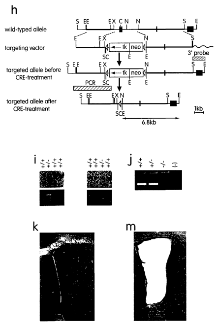
Figure 3 (h) depicts the strategy for the targeted disruption of the EphA7
gene; (i)
genotype analysis of EphA7 homozygous (+/+) and heterozygous (+/-) ES
cells before (upper left panel) and after (upper right panel) the transfection
with the Cre recombinase expression plasmid. Genomic DNA was
isolated, digested with EcoRI and subjected to Southern Blot analysis
using 3' external probe shown in A. Alleles bearing the ephA7 mutation
show a 6.8 kb band whereas a 9.7 kb band is observed in the wild type
alleles. For PCR analysis, primer pairs amplifying a 3.6 kb (lower left
panel, see also A) or a 0.5 kb (lower right panel) band in the case of
successful recombination were used; (j) RT-PCR analysis of total RNA
isolated from brain of adult animals of the indicated genotypes. Primers
were chosen to amplify part of exon I of EphA7 (314 bp), (-) denoted no
template control; (k) ventricular tissue architecture of an EphA7-/- mouse;
(m) ventricular tissue architecture of a wild type mouse. In all figures,
lateral is to the left and dorsal is up.
Figure 4 depicts in vitro proliferation of neurospheres.
Figure 5 depicts that EphA7 knockout mice have increased cell proliferation.
Figure 6 depicts the quantification of an increased in the number of BrdU
positive
cells (proliferation) in ephrin-A2-Fc infused animals.
Figure 7 depicts Ephrin-AS-Fc treatment indicates an increased proliferation
and
neurogenesis in the olfactory bulb in comparison to negative control
(vehicle treated animals).
Figure 8 depicts that EphA7 knockout mice have increased number of cells in
the
cortex.
12
CA 02466333 2004-05-07
WO 03/040304 PCT/IB02/04930
DETAILED DESCRIPTION OF INVENTION
It has been discovered that certain reagents are capable of modulating the
differentiation, migration, proliferation and survival of neural
stem/progenitor cells both
in vitro and in vivo. As used herein, the term "modulate" refers to having an
affect in
such a way as to alter the differentiation, migration, proliferation and
survival of neural
stem cell (NSC) or neural progenitor cell (NPC) activity. Since
undifferentiated,
pluripotent stem cells can proliferate in culture for a year or more, the
invention described
in this disclosure provides an almost limitless supply of neural precursors.
As used herein, the term "neural stem cells" (NSCs) can be identified by
their ability to undergo continuous cellular proliferation, to regenerate
exact copies of
themselves (self renew), to generate a large number of regional cellular
progeny, and to
elaborate new cells in response to injury or disease. The terms "neural
progenitor cells"
or "neural precursor cells" (NPCs) mean cells that can generate progeny that
are either
neuronal cells (such as neuronal precursors or mature neurons) or glial cells
(such as glial
precursors, mature astrocytes, or mature oligodendrocytes). Typically, the
cells express-
some of the phenotypic markers that are characteristic of the neural lineage.
Typically,
they do not produce progeny of other embryonic germ layers when cultured by
themselves
i~ vitro unless dedifferentiated or reprogrammed in some fashion.
As used herein, the term "reagent" refers to any substance that is
chemically and biologically capable of activating a receptor, including
peptides, small
molecules, antibodies (or fragments thereof), affibodies and any molecule that
dimerizes
or multimerizes the receptors or replaces the need for activation of the
extracellular
domains. In one embodiment, the reagent is a small molecule.
As used herein, the term "antibody" as used in this disclosure refers to both
polyclonal and monoclonal antibody. The ambit of the term deliberately
encompasses not
only intact immunoglobulin molecules, but also such fragments and derivatives
of
immunoglobulin molecules (such as single chain Fv constructs, diabodies and
fusion
constructs) as may be prepared by techniques known in the art, and retaining a
desired
antibody binding specificity. The term "affibody" (U.S. Patent No. 5,831,012)
refers to
highly specific affinity proteins that can be designed to bind to any desired
target
13
CA 02466333 2004-05-07
WO 03/040304 PCT/IB02/04930
molecule. These antibody mimics can be manufactured to have the desired
properties
(specificity and affinity), while also being highly robust to withstand a
broad range of
analytical conditions, including pH and elevated temperature. The specific
binding
properties that can be engineered into each capture protein allow it to have
very high
specificity and the desired affinity for a corresponding target protein. A
specific target
protein will thus bind only to its corresponding capture protein. The small
size (only 58
amino acids), high solubility, ease of further engineering into
multifunctional constructs,
excellent folding and absence of cysteines, as well as a stable scaffold that
can be
produced in large quantities using low cost bacterial expression systems, make
affibodies
superior capture molecules to antibodies or antibody fragments, such as Fab or
single
chain Fv (scFv) fragments, in a variety of Life Science applications.
Preferred reagents of the invention include EphA7, ephrin-AS or ephrin-
A2 and any molecule that can interfere with EphA7 and ephrin-AS interaction or
EphA7
and ephrin-A2 interaction. The invention provides a method for in vivo
disruption of
EphA7/ephrin-AS interaction or EphA7/ephrin-A2 activity and for therapeutic
administration of EphA7, ephrin-AS or ephrin-A2 and drug screening. . In a
preferred
embodiment, the neural tissue is fetal or adult brain. In yet another
embodiment, the
population containing neural or neural-derived cells is obtained from a neural
cell culture
or neurosphere.
Production of Reagents
Reagents for treatment of patients are recombinantly produced, purified
and formulated according to well known methods.
Reagents of the invention, and individual moieties or analogs and
derivatives thereof, can be chemically synthesized. A variety of protein
synthesis
methods are common in the art, including synthesis using a peptide
synthesizer. See, e.g.,
Peptide ~'hemishy, A Practieal Textbook, Bodasnsky, Ed. Springer-Verlag, 1988;
Merrifield, Science 232: 241-247 (1986); Barany, et al, Intl. J. Peptide
Protein Res. 30:
705-739 (1987); Dent, Ann. Rev. Biochem. 57:957-989 (1988), and Kaiser, et al,
Science 243: 187-198 (1989). The peptides are purified so that they are
substantially free
of chemical precursors or other chemicals using standard peptide purification
techniques.
14
CA 02466333 2004-05-07
WO 03/040304 PCT/IB02/04930
The language "substantially free of chemical precursors or other chemicals"
includes
preparations of peptide in which the peptide is separated from chemical
precursors or
other chemicals that axe involved in the synthesis of the peptide. In one
embodiment, the
language "substantially free of chemical precursors or other chemicals"
includes
preparations of peptide having less than about 30% (by dry weight) of chemical
precursors or non-peptide chemicals, more preferably less than about 20%
chemical
precursors or non-peptide chemicals, still more preferably less than about 10%
chemical
precursors or non-peptide chemicals, and most preferably less than about 5%
chemical
precursors or non-peptide chemicals.
Chemical synthesis of peptides facilitates the incorporation of modified or
unnatural amino acids, including D-amino acids and other small organic
molecules.
Replacement of one or more L-amino acids in a peptide with the corresponding D-
amino
acid isoforms can be used to increase the resistance of peptides to enzymatic
hydrolysis,
and to enhance one or more properties of biologically active peptides, i.e.,
receptor
binding, functional potency or duration of action. See, e.g., Doherty, et al.,
1993. J.
Med. Chem. 36: 2585-2594; I~irby, et al., 1993, J. Med. Chem. 36:3802-3808;
Morita,
et al., 1994, FEBS Lett. 353: 84-88; Wang, et al., 1993 Int. J. Pept. Protein
Res. 42:
392-399; Fauchere and Thiunieau, 1992. Adv. Drug Res. 23: 127-159.
Introduction of covalent cross-links into a peptide sequence can
conformationally and topographically constrain the peptide backbone. This
strategy can
be used to develop peptide analogs of reagents with increased potency,
selectivity and
stability. A number of other methods have been used successfully to introduce
conformational constraints into peptide sequences in order to improve their
potency,
receptor selectivity and biological half life. These include the use of (i) Ca
methylamino
acids (see, e.g., Rose, et al., Adv. Protein Chem. 37: 1-109 (1985); Prasad
and Balaram,
CRC C~it. Rev. Biochem., 16: 307-348 (1984)); (ii) Na methylamino acids (see,
e.g.,
Aubry, et al., Int. J. Pept. Protein Res., 18: 195-202 (1981); Manavalan and
Momany,
Biopolymers, 19: 1943-1973 (1980)); and (iii) a,~3-unsaturated amino acids
(see, e.g.,
Bach and Gierasch, Biopolymers, 25: 5175-5192 (1986); Singh, et al.,
Biopolymers, 26:
819-829 (1987)). These and many other amino acid analogs are conunercially
available,
CA 02466333 2004-05-07
WO 03/040304 PCT/IB02/04930
or can be easily prepared. Additionally, replacement of the C- terminal acid
with an
amide can be used to enhance the solubility and clearance of a peptide.
Alternatively, a reagent may be obtained by methods well-known in the art
for recombinant peptide expression and purification. A DNA molecule encoding
the
protein reagent can be generated. The DNA sequence is known or can be deduced
from
the protein sequence based on known codon usage. See, e.g., Old and Primrose,
Principles of Gene Manipulation 3'a ed., Blackwell Scientific Publications,
1985; Wada et
al., Nucleic Acids Res. 20: 2111-2118(1992). Preferably, the DNA molecule
includes
additional sequence, e.g., recognition sites for restriction enzymes which
facilitate its
cloning into a suitable cloning vector, such as a plasmid. Nucleic acids may
be DNA,
RNA, or a combination thereof. Nucleic acids encoding the reagent may be
obtained by
any method known within the art (e.g., by PCR amplification using synthetic
primers
hybridizable to the 3'- and 5'-termini of the sequence and/or by cloning from
a cDNA or
genomic library using an oligonucleotide sequence specific for the given gene
sequence,
or the like). Nucleic acids can also be generated by chemical synthesis.
Any of the methodologies known within the relevant art regarding the
insertion of nucleic acid fragments into a vector may be used to construct
expression
vectors that contain a chimeric gene comprised of the appropriate
transcriptional/translational control signals and reagent-coding sequences.
Promoter/enhancer sequences within expression vectors may use plant, animal,
insect, or
fungus regulatory sequences, as provided in the invention.
A host cell can be any prokaryotic or eukaryotic cell. For example, the
peptide can be expressed in bacterial cells such as E. coli, yeast, insect
cells, fungi or
mammalian cells (such as Chinese hamster ovary cells (CHO) or COS cells).
Other
suitable host cells are known to those skilled in the art. In one embodiment,
a nucleic
acid encoding a reagent is expressed in mammalian cells using a mammalian
expression
vector. Examples of mammalian expression vectors include pCDMB (Seed (1987)
Nature
329:840) and pMT2PC (Kaufinan et al. (1987) EMBO J 6: 187-195).
The host cells, can be used to produce (i.e., overexpress) peptide in culture.
Accordingly, the invention further provides methods for producing the peptide
using the
16
CA 02466333 2004-05-07
WO 03/040304 PCT/IB02/04930
host cells of the invention. In one embodiment, the method comprises culturing
the host
cell of invention (into which a recombinant expression vector encoding the
peptide has
been introduced) in a suitable medium such that peptide is produced. The
method further
involves isolating peptide from the medium or the host cell. Ausubel et al.,
(Eds). In:
Current Protocols in Molecular Biology. J. Wiley and Sons, New York, NY. 1998.
An "isolated" or "purified" recombinant peptide or biologically active
portion thereof is substantially free of cellular material or other
contaminating proteins
from the cell or tissue source from which the peptide of interest is derived.
The language
"substantially free of cellular material" includes preparations in which the
peptide is
separated from cellular components of the cells from which it is isolated or
recombinantly
produced. In one embodiment, the language "substantially free of cellular
material"
includes preparations of peptide having less than about 30% (by dry weight) of
peptide
other than the desired peptide (also referred to herein as a "contaminating
protein"), more
preferably less than about 20% of contaminating protein, still more preferably
less than
about 10% of contaminating protein, and most preferably less than about 5%
contaminating protein. When the peptide or biologically active portion thereof
is
recombinantly produced, it is also preferably substantially free of culture
medium, i.e.,
culture medium represents less than about 20%, more preferably less than about
10%, and
most preferably less than about 5% of the volume of the peptide preparation.
The invention also pertains to variants of a reagent that function as either
agonists (mimetics) or as antagonists. Variants of a reagent can be generated
by
mutagenesis, e.g., discrete point mutations. An agonist of a reagent can
retain
substantially the same, or a subset of, the biological activities of the
naturally occurring
form of the reagent. An antagonist of the reagent can inhibit one or more of
the activities
of the naturally occurring form of the reagent by, for example, competitively
binding to
the receptor. Thus, specific biological effects can be elicited by treatment
with a variant
with a limited function. In one embodiment, treatment of a subject with a
variant having
a subset of the biological activities of the naturally occurring form of the
reagent has
fewer side effects in a subject relative to treatment with the naturally
occurring form of
the reagent.
17
CA 02466333 2004-05-07
WO 03/040304 PCT/IB02/04930
Preferably, the analog, variant, or derivative reagent is functionally active.
As utilized herein, the term "functionally active" refers to species
displaying one or more
known functional attributes of a full-length reagent. "Variant" refers to a
reagent
differing from naturally occurring reagent, but retaining essential properties
thereof.
Generally, variants are overall closely similar, and in many regions,
identical to the
naturally occurring reagent.
Variants of the reagent that function as either agonists (mimetics) or as
antagonists can be identified by screening combinatorial libraries of mutants
of the
reagent for peptide agonist or antagonist activity. In one embodiment, a
variegated library
of variants is generated by combinatorial mutagenesis at the nucleic acid
level and is
encoded by a variegated gene library. A variegated library of variants can be
produced by,
for example, enzymatically ligating a mixture of synthetic oligonucleotides
into gene
sequences such that a degenerate set of potential sequences is expressible as
individual
peptides, or alternatively, as a set of larger fusion proteins (e.g., for
phage display)
containing the set of sequences therein. There are a variety of methods which
can be used
to produce libraries of potential variants from a degenerate oligonucleotide
sequence.
Chemical synthesis of a degenerate gene sequence can be performed in an
automatic DNA
synthesizer, and the synthetic gene then ligated into an appropriate
expression vector. LTse
of a degenerate set of genes allows for the provision, in one mixture, of all
of the
sequences encoding the desired set of potential sequences. Methods for
synthesizing
degenerate oligonucleotides are known in the art (see, e.g., Narang (1983)
Tetrahedron
39:3; Itakura et al. (1984) Annu Rev Biochem 53:323; Itakura et al. (1984)
Science
198:1056; Ike et al. (1983) Nucl. Acids Res. 11:477.
Derivatives and analogs of the reagent or individual moieties can be
produced by various methods known within the art. For example, the polypeptide
sequences may be modified by any number of methods known within the art. See
e.g.,
Sambrook, et al., 1990. Molecular Clohi~g: A Laboratory Manual, 2hcl ed.,
(Cold Spring
Harbor Laboratory Press; Cold Spring Harbor, NY). Modifications include:
glycosylation, acetylation, phosphorylation, amidation, derivatization by
known
protecting/blocking groups, linkage to an antibody molecule or other cellular
reagent, and
the like. Any of the numerous chemical modification methodologies known within
the art
18
CA 02466333 2004-05-07
WO 03/040304 PCT/IB02/04930
may be utilized including, but not limited to, specific chemical cleavage by
cyanogen
bromide, trypsin, chymotrypsin, papain, V8 protease, NaBH4, acetylation,
formylation,
oxidation, reduction, metabolic synthesis in the presence of tunicamycin, etc.
Derivatives and analogs may be full length or other than full length, if said
derivative or analog contains a modified nucleic acid or amino acid, as
described infra.
Derivatives or analogs of the reagent include, but are not limited to,
molecules comprising
regions that are substantially homologous in various embodiments, of at least
30%, 40%,
50%, 60%, 70%, 80%, 90% or preferably 95% amino acid identity when: (i)
compared to
an amino acid sequence of identical size; (ii) compared to an aligned sequence
in that the
alignment is done by a computer homology program known within the art (e.g.,
Wisconsin GCG software) or (iii) the encoding nucleic acid is capable of
hybridizing to a
sequence encoding the aforementioned peptides under stringent (preferred),
moderately
stringent, or non-stringent conditions. See, e.g., Ausubel, et al., Current
Protocols in
Molecular Biology, John Wiley and Sons, New York, NY, 1993.
Derivatives of the reagent may be produced by alteration of their sequences
by substitutions, additions or deletions that result in functionally-
equivalent molecules.
One or more amino acid residues within the reagent may be substituted by
another amino
acid of a similar polarity and net charge, thus resulting in a silent
alteration. Conservative
substitutes for an amino acid within the sequence may be selected from other
members of
the class to which the amino acid belongs. For example, nonpolar (hydrophobic)
amino
acids include alanine, leucine, isoleucine, valine, proline, phenylalanine,
tryptophan and
methionine. Polar neutral amino acids include glycine, serine, threonine,
cysteine,
tyrosine, asparagine, and glutamine. Positively charged (basic) amino acids
include
arginine, lysine and histidine. Negatively charged (acidic) amino acids
include aspartic
acid and glutamic acid.
The reagent can be administered locally to any loci implicated in the CNS
disorder pathology, i. e. any loci deficient in neural cells as a cause of the
disease. For
example, the reagent can be administered locally to the ventricle of the
brain, substantia
nigra, striatum, locus ceruleous, nucleus basalis Meynert, pedunculopontine
nucleus,
cerebral cortex, and spinal cord.
19
CA 02466333 2004-05-07
WO 03/040304 PCT/IB02/04930
Neural stem cells and their progeny can be induced to proliferate and
differentiate in vivo by administering to the host a reagent, alone or in
combination with
other agents, or by administering a pharmaceutical composition containing the
reagent
that will induce proliferation and differentiation of the cells.
Pharmaceutical
compositions include any substance that blocks the inhibitory influence and/or
stimulates
neural stem cells and stem cell progeny to proliferate and ultimately
differentiate. Such in
vivo manipulation and modification of these cells allows cells lost, due to
injury or
disease, to be endogenously replaced, thus obviating the need for
transplanting foreign
cells into a patient.
Antibodies
Included in the invention are antibodies to be used as reagents. The term
"antibody" as used herein refers to immunoglobulin molecules and
immunologically
active portions of immunoglobulin (Ig) molecules, i.e., molecules that contain
an antigen
binding site that specifically binds (immunoreacts with) an antigen. Such
antibodies
include, but are not limited to, polyclonal, monoclonal, chimeric, single
chain, Fab, Fab'
and F~ab')2 fragments, and an Fab expression library. In general, antibody
molecules
obtained from humans relates to any of the classes IgG, IgM, IgA, IgE and IgD,
which
differ from one another by the nature of the heavy chain present in the
molecule. Certain
classes have subclasses as well, such as IgGI, IgG2, and others. Furthermore,
in humans,
the light chain may be a kappa chain or a lambda chain. Reference herein to
antibodies
includes a reference to all such classes, subclasses and types of human
antibody species.
An isolated protein of the invention intended to serve as an antigen, or a
portion or fragment thereof, can be used as an immunogen to generate
antibodies that
immunospecifically bind the antigen, using standard techniques for polyclonal
and
monoclonal antibody preparation. The full-length protein can be used or,
alternatively,
the invention provides antigenic peptide fragments of the antigen for use as
immunogens.
An antigenic peptide fragment comprises at least 6 amino acid residues of the
amino acid
sequence of the full length protein and encompasses an epitope thereof such
that an
antibody raised against the peptide forms a specific immune complex with the
full length
protein or with any fragment that contains the epitope. Preferably, the
antigenic peptide
CA 02466333 2004-05-07
WO 03/040304 PCT/IB02/04930
comprises at least 10 amino acid residues, or at least 15 amino acid residues,
or at least 20
amino acid residues, or at least 30 amino acid residues. Preferred epitopes
encompassed
by the antigenic peptide are regions of the protein that are located on its
surface;
commonly these are hydrophilic regions.
In certain embodiments of the invention, at least one epitope encompassed
by the antigenic peptide is a region of EphA7, ephrin-AS or ephrin-A2 that is
located on
the surface of the protein, e.g~., a hydrophilic region. A hydrophobicity
analysis of the
human those protein sequences will indicate which regions of the polypeptide
are
particularly hydrophilic and, therefore, are likely to encode surface residues
useful for
targeting antibody production. As a means for targeting antibody production,
hydropathy
plots showing regions of hydrophilicity and hydrophobicity may be generated by
any
method well known in the art, including, for example, the I~yte Doolittle or~
the Hopp
Woods methods, either with or without Fourier transformation. See, e.g., Hopp
and
Woods, 1981, Pr~oc. Nat. Acad. Sci. USA 78: 3824-3828; I~yte and Doolittle
1982, J.
Mol. Biol. 157: 105-142, each incorporated herein by reference in their
entirety.
Antibodies that are specific for one or more domains within an antigenic
protein, or
derivatives, fragments, analogs or homologs thereof, are also provided herein.
The term "epitope" includes any protein determinant capable of specific
binding to an immunoglobulin or T-cell receptor. Epitopic determinants usually
consist
of chemically active surface groupings of molecules such as amino acids or
sugar side
chains and usually have specific three dimensional structural characteristics,
as well as
specific charge characteristics. A EphA7, ephrin-AS or ephrin-A2, or a
fragment thereof
comprises at least one antigenic epitope. An anti- EphA7, ephrin-AS or ephrin-
A2
antibody of the present invention is said to specifically bind to .the antigen
when the
equilibrium binding constant (KD) is <_1 ~,M, preferably <_ 100 nM, more
preferably <_ 10
nM, and most preferably 5 100 pM to about 1 pM, as measured by assays such as
radioligand binding assays or similar assays known to those skilled in the
art.
Various procedures known within the art may be used for the production of
polyclonal or monoclonal antibodies directed against a protein of the
invention, or against
derivatives, fragments, analogs homologs or orthologs thereof (see, for
example,
21
CA 02466333 2004-05-07
WO 03/040304 PCT/IB02/04930
Antibodies: A Laboratory Manual, Harlow E, and Lane D, 1988, Cold Spring
Harbor
Laboratory Press, Cold Spring Harbor, NY, incorporated herein by reference).
Some of
these antibodies are discussed below.
Polyclonal Antibodies
For the production of polyclonal antibodies, various suitable host animals
(e.g., rabbit, goat, mouse or other mammal) may be immunized by one or more
injections
with the native protein, a synthetic variant thereof, or a derivative of the
foregoing. An
appropriate immunogenic preparation can contain, for example, the naturally
occurring
immunogenic protein, a chemically synthesized polypeptide representing the
immunogenic protein, or a recombinantly expressed immunogenic protein.
Furthermore,
the protein may be conjugated to a second protein known to be immunogenic in
the
mammal being immunized. Examples of such immunogenic proteins include but are
not
limited to keyhole limpet hemocyanin, serum albumin, bovine thyroglobulin, and
soybean
trypsin inhibitor. The preparation can further include an adjuvant. Various
adjuvants
used to increase the immunological response include, but are not limited to,
Freund's
(complete and incomplete), mineral gels (e.g., aluminum hydroxide), surface
active
substances (e.g., lysolecithin, pluronic polyols, polyanions, peptides, oil
emulsions,
dinitrophenol, etc.), adjuvants usable in humans such as Bacille Calmette-
Guerin and
Corynebacterium parvum, or similar immunostimulatory agents. Additional
examples of
adjuvants which can be employed include MPL-TDM adjuvant (monophosphoryl Lipid
A, synthetic trehalose dicorynomycolate).
The polyclonal antibody molecules directed against the immunogenic
protein can be isolated from the mammal (e.g., from the blood) and further
purified by
well known techniques, such as affinity chromatography using protein A or
protein G,
which provide primarily the IgG fraction of immune serum. Subsequently, or
alternatively, the specific antigen which is the target of the immunoglobulin
sought, or an
epitope thereof, may be immobilized on a column to purify the immune specific
antibody
by immunoaffinity chromatography. Purification of immunoglobulins is
discussed, for
22
CA 02466333 2004-05-07
WO 03/040304 PCT/IB02/04930
example, by D. Wilkinson (The Scientist, published by The Scientist, Inc.,
Philadelphia
PA, Vol. 14, No. 8 (April 17, 2000), pp. 25-28).
Monoclonal Antibodies
The term "monoclonal antibody" (MAb) or "monoclonal antibody
composition", as used herein, refers to a population of antibody molecules
that contain
only one molecular species of antibody molecule consisting of a unique light
chain gene
product and a unique heavy chain gene product. In particular, the
complementarity
determining regions (CDRs) of the monoclonal antibody are identical in all the
molecules
of the population. MAbs thus contain an antigen binding site capable of
immunoreacting
with a particular epitope of the antigen characterized by a unique binding
affinity for it.
Monoclonal antibodies can be prepared using hybridoma methods, such as
those described by I~ohler and Milstein, Nature, 256:495 (1975). In a
hybridoma method,
a mouse, hamster, or other appropriate host animal, is typically immunized
with an
immunizing agent to elicit lymphocytes that produce or are capable of
producing
antibodies that will specifically bind to the immunizing agent. Alternatively,
the
lymphocytes can be immunized in vitro.
The immunizing agent will typically include the protein antigen, a
fragment thereof or a fusion protein thereof. Generally, either peripheral
blood
lymphocytes are used if cells of human origin are desired, or spleen cells or
lymph node
cells are used if non-human mammalian sources are desired. The lymphocytes are
then
fused with an immortalized cell line using a suitable fusing agent, such as
polyethylene
glycol, to form a hybridoma cell (Goding, Monoclonal Antibodies: Principles
and
Practice, Academic Press, (1986) pp. 59-103). Immortalized cell lines are
usually
transformed mammalian cells, particularly myeloma cells of rodent, bovine and
human
origin. Usually, rat or mouse myeloma cell lines are employed. The hybridoma
cells can
be cultured in a suitable culture medium that preferably contains one or more
substances
that inhibit the growth or survival of the unfused, immortalized cells. For
example, if the
parental cells lack the enzyme hypoxanthine guanine phosphoribosyl transferase
(HGPRT
23
CA 02466333 2004-05-07
WO 03/040304 PCT/IB02/04930
or HPRT), the culture medium for the hybridomas typically will include
hypoxanthine,
aminopterin, and thymidine ("HAT medium"), which substances prevent the growth
of
HGPRT-deficient cells.
Preferred immortalized cell lines are those that fuse efficiently, support
stable high level expression of antibody by the selected antibody-producing
cells, and are
sensitive to a medium such as HAT medium. More preferred immortalized cell
lines are
marine myeloma lines, which can be obtained, for instance, from the Salk
Institute Cell
Distribution Center, San Diego, California and the American Type Culture
Collection,
Manassas, Virginia. Human myeloma and mouse-human heteromyeloma cell lines
also
have been described for the production of human monoclonal antibodies
(I~ozbor, J.
Immunol., 133:3001 (1984); Brodeur et al., Monoclonal Antibody Production
Techniques
and Applications, Marvel Dekker, Inc., New York, (1987) pp. 51-63).
The culture medium in which the hybridoma cells are cultured can then be
assayed for the presence of monoclonal antibodies directed against the
antigen.
Preferably, the binding specificity of monoclonal antibodies produced by the
hybridoma
cells is determined by immunoprecipitation or by an in vitro binding assay,
such as
radioimmunoassay (RIA) or enzyme-linked immunoabsorbent assay (ELISA). Such
techniques and assays are known in the art. The binding affinity of the
monoclonal
antibody can, for example, be determined by the Scatchard analysis of Munson
and
Pollard, Anal. Biochem., 107:220 (1980). It is an objective, especially
important in
therapeutic applications of monoclonal antibodies, to identify antibodies
having a high
degree of specificity and a high binding affinity for the target antigen.
After the desired hybridoma cells are identified, the clones can be
subcloned by limiting dilution procedures and grown by standard methods
(Goding,
Monoclonal antibodies: principles and practice, Academic press, (1986) pp. 59-
103).
Suitable culture media for this purpose include, for example, Dulbecco's
Modified Eagle's
Medium and RPMI-1640 medium. Alternatively, the hybridoma cells can be grown
in
vivo as ascites in a mammal.
The monoclonal antibodies secreted by the subclones can be isolated or
purified from the culture medium or ascites fluid by conventional
immunoglobulin
24
CA 02466333 2004-05-07
WO 03/040304 PCT/IB02/04930
purification procedures such as, for example, protein A-Sephaxose,
hydroxylapatite
chromatography, gel electrophoresis, dialysis, or affinity chromatography.
The monoclonal antibodies can also be made by recombinant DNA
methods, such as those described in U.S. Patent No. 4,816,567. DNA encoding
the
monoclonal antibodies of the invention can be readily isolated and sequenced
using
conventional procedures (e.g., by using oligonucleotide probes that are
capable of binding
specifically to genes encoding the heavy and light chains of marine
antibodies). The
hybridoma cells of the invention serve as a preferred source of such DNA. Once
isolated,
the DNA can be placed into expression vectors, which are then transfected into
host cells
such as simian COS cells, Chinese hamster ovary (CHO) cells, or myeloma cells
that do
not otherwise produce immunoglobulin protein, to obtain the synthesis of
monoclonal
antibodies in the recombinant host cells. The DNA also can be modified, for
example, by
substituting the coding sequence for human heavy and light chain constant
domains in
place of the homologous marine sequences (LT.S. Patent No. 4,816,567;
Morrison,
Nature 368, 812-13 (1994)) or by covalently joining to the immunoglobulin
coding
sequence all or part of the coding sequence for a non-immunoglobulin
polypeptide. Such
a non-immunoglobulin polypeptide can be substituted for the constant domains
of an
antibody of the invention, or can be substituted for the variable domains of
one antigen-
combining site of an antibody of the invention to create a chimeric bivalent
antibody.
Hutilanized Antibodies
The antibodies directed against the protein antigens of the invention can
further comprise humanized antibodies or human antibodies. These antibodies
are
suitable for administration to humans without engendering an immune response
by the
human against the administered immunoglobulin. Humanized forms of antibodies
are
chimeric immunoglobulins, immunoglobulin chains or fragments thereof (such as
Fv,
Fab, Fab', F(ab')a or other antigen-binding subsequences of antibodies) that
are principally
comprised of the sequence of a human immunoglobulin and contain minimal
sequence
derived from a non-human immunoglobulin. Humanization can be performed
following
the method of Winter and co-workers (Jones et al., Nature, 321:522-525 (1986);
CA 02466333 2004-05-07
WO 03/040304 PCT/IB02/04930
Riechmann et al., Nature, 332:323-327 (1988); Verhoeyen et al., Science,
239:1534-1536
(1988)), by substituting rodent CDRs or CDR sequences for the corresponding
sequences
of a human antibody. (See also U.S. Patent No. 5,225,539.) In some instances,
Fv
framework residues of the human immunoglobulin are replaced by corresponding
non-
human residues. Humanized antibodies can also comprise residues which are
found
neither in the recipient antibody nor in the imported CDR or framework
sequences. In
general, the humanized antibody will comprise substantially all of at least
one, and
typically two, vaxiable domains, in which all or substantially all of the CDR
regions
correspond to those of a non-human immunoglobulin and all or substantially all
of the
framework regions are those of a human immunoglobulin consensus sequence. The
humanized antibody optimally also will comprise at least a portion of an
immunoglobulin
constant region (Fc), typically that of a human immunoglobulin (Jones et al.,
1986;
Riechmann et al., 1988; and Presta, Curr. Op. Struct. Biol., 2:593-596
(1992)).
Human Antibodies
Fully human antibodies essentially relate to antibody molecules in which
the entire sequence of both the light chain and the heavy chain, including the
CDRs, arise
from human genes. Such antibodies are termed "human antibodies", or "fully
human
antibodies" herein. Human monoclonal antibodies can be prepared by the trioma
technique; the human B-cell hybridoma technique (see I~ozbor, et al., 1983
Immunol
Today 4: 72) and the EBV hybridoma technique to produce human monoclonal
antibodies
(see Cole, et al., 1985 In: MONOCLONAL ANTIBODIES AND CANCER THERAPY, Alan R.
Liss, Inc., pp. 77-96). Human monoclonal antibodies may be utilized in the
practice of
the present invention and may be produced by using human hybridomas (see Cote,
et al.,
1983. Proc Natl Acad Sci USA 80: 2026-2030) or by transforming human B-cells
with
Epstein Barr Virus in vitro (see Cole, et al., 1985 In: MONOCLONAL ANTIBODIES
AND
CANCER THERAPY, Alan R. Liss, Inc., pp. 77-96).
In addition, human antibodies can also be produced using additional
techniques, including phage display libraries (Hoogenboom and Winter, J. Mol.
Biol.,
227:381 (1991); Marks et al., J. Mol. Biol., 222:581 (1991)). Similarly, human
26
CA 02466333 2004-05-07
WO 03/040304 PCT/IB02/04930
antibodies can be made by introducing human immunoglobulin loci into
transgenic
animals, e.g., mice in which the endogenous immunoglobulin genes have been
partially or
completely inactivated. Upon challenge, human antibody production is observed,
which
closely resembles that seen in humans in all respects, including gene
rearrangement,
assembly, and antibody repertoire. This approach is described, for example, in
U.S.
Patent Nos. 5,545,807; 5,545,806; 5,569,825; 5,625,126; 5,633,425; 5,661,016,
and in
Marks et al. (Bio/Technology 10, 779-783 (1992)); Lonberg et al. (Nature 368
856-859
(1994)); Morrison ( Nature 368, 812-13 (1994)); Fishwild et al,( Nature
Biotechnology
14, 845-51 (1996)); Neuberger (Nature Biotechnology 14, 826 (1996)); and
Lonberg and
Huszar (Intern. Rev. Immunol. 13 65-93 (1995)).
Human antibodies may additionally be produced using transgenic
nonhuman animals which are modified so as to produce fully human antibodies
rather
than the animal's endogenous antibodies in response to challenge by an
antigen. (See
PCT publication W094/02602). The endogenous genes encoding the heavy and light
immunoglobulin chains in the nonhuman host have been incapacitated, and active
loci
encoding human heavy and light chain immunoglobulins are inserted into the
host's
genome. The human genes are incorporated, for example, using yeast artificial
chromosomes containing the requisite human DNA segments. An animal which
provides
all the desired modifications is then obtained as progeny by crossbreeding
intermediate
transgenic animals containing fewer than the full complement of the
modifications. The
preferred embodiment of such a nonhuman animal is a mouse, and is termed the
XenomouseTM as disclosed in PCT publications WO 96/33735 and WO 96/34096. This
animal produces B cells which secrete fully human immunoglobulins. The
antibodies can
be obtained directly from the animal after immunization with an immunogen of
interest,
as, for example, a preparation of a polyclonal antibody, or alternatively from
immortalized
B cells derived from the animal, such as hybridomas producing monoclonal
antibodies.
Additionally, the genes encoding the immunoglobulins with human variable
regions can
be recovered and expressed to obtain the antibodies directly, or can be
further modified to
obtain analogs of antibodies such as, for example, single chain Fv molecules.
An example of a method of producing a nonhuman host, exemplified as a
mouse, lacking expression of an endogenous immunoglobulin heavy chain is
disclosed in
27
CA 02466333 2004-05-07
WO 03/040304 PCT/IB02/04930
U.S. Patent No. 5,939,598. It can be obtained by a method including deleting
the J
segment genes from at least one endogenous heavy chain locus in an embryonic
stem cell
to prevent rearrangement of the locus and to prevent formation of a transcript
of a
rearranged immunoglobulin heavy chain locus, the deletion being effected by a
targeting
vector containing a gene encoding a selectable marker; and producing from the
embryonic
stem cell a transgenic mouse whose somatic and germ cells contain the gene
encoding the
selectable marker.
A method for producing an antibody of interest, such as a human antibody,
is disclosed in U.S. Patent No. 5,916,771. It includes introducing an
expression vector
that contains a nucleotide sequence encoding a heavy chain into one mammalian
host cell
in culture, introducing an expression vector containing a nucleotide sequence
encoding a
light chain into another mammalian host cell, and fusing the two cells to form
a hybrid
cell. The hybrid cell expresses an antibody containing the heavy chain and the
light chain.
In a further improvement on this procedure, a method for identifying a
clinically relevant epitope on an immunogen, and a correlative method for
selecting an
antibody that binds immunospecifically to the relevant epitope with high
affinity, are
disclosed in PCT publication WO 99/53049.
Fab Fragments and Single Chain Antibodies
According to the invention, techniques can be adapted for the production
of single-chain antibodies specific to an antigenic protein of the invention
(see e.g., U.S.
Patent No. 4,946,778). In addition, methods can be adapted for the
construction of Fab
expression libraries (see e.g., Huse, et al., 1989 Science 246: 1275-1281) to
allow rapid
and effective identification of monoclonal Fab fragments with the desired
specificity for a
protein or derivatives, fragments, analogs or homologs thereof. Antibody
fragments that
contain the idiotypes to a protein antigen may be produced by techniques known
in the art
including, but not limited to: (i) an F~ab~~2 fragment produced by pepsin
digestion of an
antibody molecule; (ii) an Fab fragment generated by reducing the disulfide
bridges of an
28
CA 02466333 2004-05-07
WO 03/040304 PCT/IB02/04930
F(ab')2 fragment; (iii) an Fab fragment generated by the treatment of the
antibody molecule
with papain and a reducing agent and (iv) F,, fragments.
Bispecific Antibodies
Bispecific antibodies are monoclonal, preferably human or humanized,
antibodies that have binding specificities for at least two different
antigens. In the present
case, one of the binding specificities is for an antigenic protein of the
invention. The
second binding target is any other antigen, and advantageously is a cell-
surface protein or
receptor or receptor subunit.
Methods for making bispecific antibodies are known in the art.
Traditionally, the recombinant production of bispecific antibodies is based on
the co-
expression of two immunoglobulin heavy-chain/light-chain pairs, where the two
heavy
chains have different specificities (Milstein and Cuello, Nature, 305:537-539
(1983)).
Because of the random assortment of immunoglobulin heavy and light chains,
these
hybridomas (quadromas) produce a potential mixture of ten different antibody
molecules,
of which only one has the correct bispecific structure. The purification of
the correct
molecule is usually accomplished by affinity chromatography steps. Similar
procedures
are disclosed in WO 93/08829, published 13 May 1993, and in Traunecker et al.,
EMBO
J., 10:3655-3659 (1991).
Antibody variable domains with the desired binding specificities
(antibody-antigen combining sites) can be fused to immunoglobulin constant
domain
sequences. The fusion preferably is with an immunoglobulin heavy-chain
constant
domain, comprising at least part of the hinge, CH2, and CH3 regions. It is
preferred to
have the first heavy-chain constant region (CH1) containing the site necessary
for~light-
chain binding present in at least one of the fusions. DNAs encoding the
immunoglobulin
heavy-chain fusions and, if desired, the immunoglobulin light chain, are
inserted into
separate expression vectors, and are co-transfected into a suitable host
organism. For
further details of generating bispecific antibodies see, for example, Suresh
et al., Methods
in Enzymology, 121:210 (1986).
29
CA 02466333 2004-05-07
WO 03/040304 PCT/IB02/04930
According to another approach described in WO 96/27011, the interface
between a pair of antibody molecules can be engineered to maximize the
percentage of
heterodimers which are recovered from recombinant cell culture. The preferred
interface
comprises at least a part of the CH3 region of an antibody constant domain. In
this
method, one or more small amino acid side chains from the interface of the
first antibody
molecule are replaced with larger side chains (e.g. tyrosine or tryptophan).
Compensatory "cavities" of identical or similar size to the large side chains)
are created
on the interface of the second antibody molecule by replacing large amino acid
side chains
with smaller ones (e.g. alanine or threonine). This provides a mechanism for
increasing
the yield of the heterodimer over other unwanted end-products such as
homodimers.
Bispecific antibodies can be prepared as full length antibodies or antibody
fragments (e.g. F(ab')2 bispecific antibodies). Techniques for generating
bispecific
antibodies from antibody fragments have been described in the literature. For
example,
bispecific antibodies can be prepared using chemical linkage. Brennan et al.,
Science
229:81 (1985) describe a procedure wherein intact antibodies are
proteolytically cleaved
to generate F(ab')a fragments. These fragments are reduced in the presence of
the dithiol
complexing agent sodium arsenite to stabilize vicinal dithiols and prevent
intermolecular
disulfide formation. The Fab' fragments generated are then converted to
thionitrobenzoate (TNB) derivatives. One of the Fab'-TNB derivatives is then
reconverted to the Fab'-thiol by reduction with mercaptoethylamine and is
mixed with an
equimolax amount of the. other Fab'-TNB derivative to form the bispecific
antibody. The
bispecific antibodies produced can be used as agents for the selective
immobilization of
enzymes.
Additionally, Fab' fragments can be directly recovered from E. coli and
chemically coupled to form bispecific antibodies. Shalaby et al., J. Exp. Med.
175:217-225 (1992) describe the production of a fully humanized bispecific
antibody
F(ab')2 molecule. Each Fab' fragment was separately secreted from E. coli and
subjected
to directed chemical coupling in vitro to form the bispecific antibody. The
bispecific
antibody thus formed was able to bind to cells overexpressing the ErbB2
receptor and
normal human T cells, as well as trigger the lytic activity of human cytotoxic
lymphocytes
against human breast tumor taxgets.
CA 02466333 2004-05-07
WO 03/040304 PCT/IB02/04930
Various techniques for making and isolating bispecific antibody fragments
directly from recombinant cell culture have also been described. For example,
bispecific
antibodies have been produced using leucine zippers. Kostelny et al., J.
Immunol.
148(5):1547-1553 (1992). The leucine zipper peptides from the Fos and Jun
proteins
were linked to the Fab' portions of two different antibodies by gene fusion.
The antibody
homodimers were reduced at the hinge region to form monomers and then re-
oxidized to
form the antibody heterodimers. This method can also be utilized for the
production of
antibody homodimers. The "diabody" technology described by Hollinger et al.,
Proc.
Natl. Acad. Sci. USA 90:6444-6448 (1993) has provided an alternative mechanism
for
making bispecific antibody fragments. The fragments comprise a heavy-chain
variable
domain (VH) connected to a light-chain variable domain (VL) by a linker which
is too
short to allow pairing between the two domains on the same chain. Accordingly,
the VH
and VL domains of one fragment are forced to pair with the complementary VL
and VH
domains of another fragment, thereby forming two antigen-binding sites.
Another
strategy for making bispecific antibody fragments by the use of single-chain
Fv (sFv)
dimers has also been reported. See, Gruber et al., J. Immunol. 152:5368
(1994).
Antibodies with more than two valencies are contemplated. For example,
trispecific
antibodies can be prepared. Tutt et al., J. Immunol. 147:60 (1991).
Exemplary bispecific antibodies can bind to two different epitopes, at least
one of which originates in the protein antigen of the invention.
Alternatively, an
anti-antigenic arm of an immunoglobulin molecule can be combined with an arm
which
binds to a triggering molecule on a leukocyte such as a T-cell receptor
molecule (e.g.
CD2, CD3, CD28, or B7), or Fc receptors for IgG (FcyR), such as FcyRI (CD64),
FcyRII
(CD32) and Fc~yRIII (CD16) so as to focus cellular defense mechanisms to the
cell
expressing the particular antigen. Bispecific antibodies can also be used to
direct
cytotoxic agents to cells which express a particular antigen. These antibodies
possess an
antigen-binding arm and an arm which binds a cytotoxic agent or a radionuclide
chelator,
such as EOTUBE, DPTA, DOTA or TETA. Another bispecific antibody of interest
binds
the protein antigen described herein and further binds tissue factor (TF).
31
CA 02466333 2004-05-07
WO 03/040304 PCT/IB02/04930
Immunoliposomes
The antibodies disclosed herein can also be formulated as
immunoliposomes. Liposomes containing the antibody are prepared by methods
known
in the art, such as described in Epstein et al., Proc. Natl. Acad. Sci. USA,
82: 3688
(1985); Hwang et al., Proc. Natl Acad. Sci. USA, 77: 4030 (1980); and U.S.
Pat. Nos.
4,485,045 and 4,544,545. Liposomes with enhanced circulation time are
disclosed in
U.S. Patent No. 5,013,556.
Particularly useful liposomes can be generated by the reverse-phase
evaporation method with a lipid composition comprising phosphatidylcholine,
cholesterol, and PEG-derivatized phosphatidylethanolamine (PEG-PE). Liposomes
are
extruded through filters of defined pore size to yield liposomes with the
desired diameter.
Fab' fragments of the antibody of the present invention can be conjugated to
the liposomes
as described in Martin et al., J. Biol. Chem., 257: 286-288 (1982) via a
disulfide-
interchange reaction.
Antibody Therapeutics
Antibodies of the invention, including polyclonal, monoclonal, humanized
and fully human antibodies, may used as therapeutic agents such as one of this
invention.
Such agents will generally be employed to treat or prevent a disease or
pathology,
specifically neurological disease, in a subject. An antibody preparation,
preferably one
having high specificity and high affinity for its target antigen, is
administered to the
subj ect and will generally have an effect due to its binding with the target.
Such an effect
may be one of two kinds, depending on the specific nature of the interaction
between the
given antibody molecule and the target antigen in question. In the first
instance,
administration of the antibody may abrogate or inhibit the binding of the
target with an
endogenous EphA7, ephrin-AS or ephrin-A2 ligand to which it naturally binds.
In this
case, the antibody binds to the target and masks a binding site of the
naturally occurring
ligand, wherein the ligand serves as an effector molecule. Thus, the receptor
mediates a
signal transduction pathway for which ligand is responsible.
32
CA 02466333 2004-05-07
WO 03/040304 PCT/IB02/04930
Alternatively, the effect may be one in which the antibody elicits a
physiological result by virtue of binding to an effector binding site on the
target molecule.
In this case the target, a EphA7, ephrin-AS or ephrin-A2 cell surface receptor
having an
endogenous ligand which needs to be modulated, binds the antibody as a
surrogate
effector ligand, initiating a receptor-based signal transduction event by the
receptor.
A therapeutically effective amount of an antibody of the invention relates
generally to the amount needed to achieve a therapeutic objective. As noted
above, this
may be a binding interaction between the antibody and its target antigen that,
in certain
cases, interferes with the functioning of the target, and in other cases,
promotes a
physiological response. The amount required to be administered will
furthermore depend
on the binding affinity of the antibody for its specific antigen and the rate
at which an
administered antibody is depleted from the free volume of the subject to which
it is
administered.
Diseases and Disorders
Diseases and disorders that are characterized by altered (relative to a
subject not suffering from the disease or disorder) levels or biological
activity may be
treated with therapeutics that antagonize (i.e., reduce or inhibit) EphA7,
ephrin-AS or
ephrin-A2 activity. Therapeutics that antagonize activity may be administered
in a
therapeutic or prophylactic manner. Therapeutics that may be utilized include,
but are not
limited to: (i) an aforementioned peptide, analog, derivatives, fragments or
homologs
thereof; (ii) antibodies to an aforementioned peptide; (iii) nucleic acids
encoding an
aforementioned peptide; (iv) administration of antisense nucleic acid and
nucleic acids
that are "dysfunctional" (i. e., due to a heterologous insertion within the
coding sequences
of coding sequences to an aforementioned peptide) that are utilized to
"knockout"
endogenous function of an aforementioned peptide by homologous recombination
(see,
e.g., Capecchi, 1989. Science 244: 1288-1292); or (v) modulators ( i.e.,
inhibitors,
agonists and antagonists, including additional peptide mimetic of the
invention or
antibodies specific to a peptide of the invention) that alter the interaction
between an
aforementioned peptide and its binding partner.
33
CA 02466333 2004-05-07
WO 03/040304 PCT/IB02/04930
Diseases and disorders that are characterized by altered (relative to a
subject not suffering from the disease or disorder) levels or biological
activity may be
treated with therapeutics that increase (i.e., are agonists to) activity. ,
Therapeutics that
upregulate activity may be administered in a therapeutic or prophylactic
manner.
Therapeutics that may be utilized include, but are not limited to, an
aforementioned
peptide, analog, derivatives, fragments or homologs thereof; or an agonist
that increases
bioavailability.
Increased or decreased levels can be detected by quantifying peptide and/or
RNA, by obtaining a patient tissue sample (e.g., from biopsy tissue) and
assaying it ih
vitro for RNA or peptide levels, structure and/or activity of the expressed
peptides (or
mRNAs of an aforementioned peptide). Methods that are well-known within the
art
include, but are not limited to, immunoassays (e.g., by Western blot analysis,
immunoprecipitation followed by sodium dodecyl sulfate (SDS) polyacrylamide
gel
electrophoresis, immunocytochemistry, etc.) and/or hybridization assays to
detect
expression of mRNAs (e.g., Northern assays, dot blots, in situ hybridization,
and the like).
Therapeutic Methods
Another aspect of the invention pertains to methods of modulating EphA7,
ephrin-AS or ephrin-A2 expression or activity for therapeutic purposes. The
modulatory
method of the invention involves contacting a cell with an agent that
modulates one or
more of the activities of EphA7, ephrin-AS or ephrin-A2 protein activity
associated with
the cell. An agent that modulates this protein activity can be an agent as
described herein,
such as a nucleic acid or a protein, a naturally-occurring cognate ligand of a
EphA7,
ephrin-AS or ephrin-A2 receptor, a peptide, a EphA7, ephrin-AS or ephrin-A2
peptidomimetic, or other small molecule. In one embodiment, the agent
stimulates the
activity of the EphA7, ephrin-AS or ephrin-A2 signaling pathway. Examples of
such
stimulatory agents include active EphA7, ephrin-AS or ephrin-A2 protein and a
nucleic
acid molecule encoding EphA7, ephrin-AS or ephrin-A2 that has been introduced
into the
cell. In another embodiment, the agent inhibits EphA7, ephrin-AS or ephrin-A2
signaling. Examples of such inhibitory agents include antisense nucleic acid
molecules
34
CA 02466333 2004-05-07
WO 03/040304 PCT/IB02/04930
and antibodies. These modulatory methods can be performed ih vitro (e.g., by
culturing
the cell with the agent) or, alternatively, i~ vivo (e.g., by administering
the agent to a
subject). As such, the invention provides methods of treating an individual
afflicted with
a disease or disorder, specifically a neurological disorder. In one
embodiment, the
method involves administering an reagent (e.g., an reagent identified by a
screening assay
described herein), or combination of reagents that modulate (e.g., up-
regulates or down-
regulates) EphA7, ephrin-AS or ephrin-A2 expression or activity. In another
embodiment, the method involves administering a EphA7, ephrin-AS or ephrin-A2
protein or nucleic acid molecule as therapy to modulate proliferation,
differentiation or
survival of NSCs/NPCs.
Determination of the Biological Effect of the Therapeutic
In various embodiments of the invention, suitable in vitro or ih vivo assays
are performed to determine the effect of a specific therapeutic and whether
its
administration is indicated for treatment of the affected tissue.
In various specific embodiments, i~ vitro assays may be performed with
representative stem cells or newly differentiated cells involved in the
patient's disorder, to
determine if a given therapeutic exerts the desired effect upon the cell
type(s).
Compounds for use in therapy may be tested in suitable animal model systems
including,
but not limited to rats, mice, chicken, cows, monkeys, rabbits, and the like,
prior to testing
in human subjects. Similarly, for in vivo testing, any of the animal model
system known
in the art may be used prior to administration to human subjects.
Pharmaceutical Compositions
The invention provides methods of influencing central nervous system
cells to produce progeny that can replace damaged or missing neurons in the
central
nervous system by exposing a patient, suffering from a neurological disease or
disorder, to
a reagent (e.g. EphA7, ephrin-AS or ephrin-A2) in a suitable formulation
through a
CA 02466333 2004-05-07
WO 03/040304 PCT/IB02/04930
suitable route of administration, that modulates NSC or NPC activity in vivo.
A
"neurological disease or disorder" is a disease or disorder which results in
the disturbance
in the structure or function of the central nervous system resulting from
developmental
abnormality, disease, injury or toxin. Examples of neurological diseases or
disorders
include neurodegenerative disorders (e.g. associated with Parkinson's disease,
Alzheimer's disease, Huntington's disease, Shy-Drager Syndrome, Progressive
Supranuclear Palsy, Lewy Body Disease or Amyotrophic Lateral Sclerosis);
ischemic
disorders (e.g. cerebral or spinal cord infarction and ischemia, stroke);
traumas (e.g.
caused by physical injury or surgery, and compression injuries; affective
disorders (e.g.
stress, depression and post-traumatic depression); neuropsychiatric disorders
(e.g.
schizophrenia, multiple sclerosis or epilepsy); and learning and memory
disorders.
This invention provides a method of treating a neurological disease or
disorder comprising administering a reagent that modulates neural stem cell or
neural
progenitor cell activity in vivo to a mammal. The term "mammal" refers to any
mammal
classified as a mammal, including humans, cows, horses, dogs, sheep and cats.
In one
embodiment, the mammal is a human.
The invention provides a regenerative cure for neurodegenerative diseases
by stimulating ependymal cells and subventricular zone cells to proliferate,
migrate and
differentiate into the desired neural phenotype targeting loci where cells are
damaged or
missing. I~ vivo stimulation of ependymal stem cells is accomplished by
locally
administering a reagent to the cells in an appropriate formulation. By
increasing
neurogenesis, damaged or missing neurons can be replaced in order to enhance
brain
function in diseased states.
A pharmaceutical composition useful as a therapeutic agent for the
treatment of central nervous system disorders is provided. For example, the
composition
includes a reagent of the invention, which can be administered alone or in
combination
with the systemic or local co-administration of one or more additional agents.
Such
agents include preservatives, ventricle wall permeability increasing factors,
stem cell
mitogens, survival factors, glial lineage preventing agents, anti-apoptotic
agents, anti-
stress medications, neuroprotectants, and anti-pyrogenics. The pharmaceutical
36
CA 02466333 2004-05-07
WO 03/040304 PCT/IB02/04930
composition preferentially treats CNS diseases by stimulating cells (e.g.,
ependymal cells
and subventricular zone cells) to proliferate, migrate and differentiate into
the desired
neural phenotype, targeting loci where cells are damaged or missing.
A method for treating a subject suffering from a CNS disease or disorder is
also provided. This method comprises administering to the subject an effective
amount of
a pharmaceutical composition containing a reagent (1) alone in a dosage range
of 0.5
ng/kg/day to 500 ng/kg/day, (2) in a combination with a ventricle wall
permeability
increasing factor, or (3) in combination with a locally or systemically co-
administered
agent.
A pharmaceutical composition of the invention is formulated to be
compatible with its intended route of administration. Examples of routes of
administration include parenteral, e.g., intravenous, intradermal,
subcutaneous, oral (e.g.,
inhalation), transdermal (topical), transmucosal, and rectal administration.
Solutions or
suspensions used for parenteral, intradermal, or subcutaneous application can
include the
following components: a sterile diluent such as water for injection, saline
solution, fixed
oils, polyethylene glycols, glycerine, propylene glycol or other synthetic
solvents;
antibacterial agents such as benzyl alcohol or methyl parabens; antioxidants
such as
ascorbic acid or sodium bisulfate; chelating agents such as
ethylenediaminetetraacetic
acid; buffers such as acetates, citrates or phosphates, and agents for the
adjustment of
tonicity such as sodium chloride or dextrose. The pH can be adjusted with
acids or bases,
such as hydrochloric acid or sodium hydroxide. The parenteral preparation can
be
enclosed in ampoules, disposable syringes or multiple dose vials made of glass
or plastic.
Pharmaceutical compositions suitable for injectable use include sterile
aqueous solutions (where water soluble) or dispersions and sterile powders for
the
extemporaneous preparation of sterile injectable solutions or dispersion. For
intravenous
administration, suitable carriers include physiological saline, bacteriostatic
water,
Cremophor ELTM (BASF, Parsippany, N.J.) or phosphate buffered saline (PBS). In
all
cases, the composition must be sterile and should be fluid to the extent that
easy
syringability exists. It must be stable under the conditions of manufacture
and storage and
must be preserved against the contaminating action of microorganisms such as
bacteria
37
CA 02466333 2004-05-07
WO 03/040304 PCT/IB02/04930
and fungi. The carrier can be a solvent or dispersion medium containing, for
example,
water, ethanol, polyol (for example, glycerol, propylene glycol, and liquid
polyethylene
glycol, and the like), and suitable mixtures thereof. The proper fluidity can
be
maintained, for example, by the use of a coating such as lecithin, by the
maintenance of
the required particle size in the case of dispersion and by the use of
surfactants.
Prevention of the action of microorganisms can be achieved by various
antibacterial and
antifungal agents, for example, parabens, chlorobutanol, phenol, ascorbic
acid,
thimerosal, and the like. In many cases, it will be preferable to include
isotonic agents,
for example, sugars, polyalcohols such as manitol, sorbitol, sodium chloride
in the
composition. Prolonged absorption of the injectable compositions can be
brought about
by including in the composition an agent which delays absorption, for example,
aluminum
monostearate and gelatin.
Sterile injectable solutions can be prepared by incorporating the active
compound (e.g., chimeric peptide) in the required amount in an appropriate
solvent with
one or a combination of ingredients enumerated above, as required, followed by
filtered
sterilization. Generally, dispersions are prepared by incorporating the active
compound
into a sterile vehicle that contains a basic dispersion medium and the
required other
ingredients from those enumerated above. In the case of sterile powders for
the
preparation of sterile injectable solutions, methods of preparation are vacuum
drying and
freeze-drying that yields a powder of the active ingredient plus any
additional desired
ingredient from a previously sterile-filtered solution thereof.
Oral compositions generally include an inert diluent or an edible carrier.
They can be enclosed in gelatin capsules or compressed into tablets. For the
purpose of
oral therapeutic administration, the active compound can be incorporated with
excipients
and used in the form of tablets, troches, or capsules. Oral compositions can
also be
prepared using a fluid carrier for use as a mouthwash, wherein the compound in
the fluid
carrier is applied orally and swished and expectorated or swallowed.
Pharmaceutically
compatible binding agents, and/or adjuvant materials can be included as part
of the
composition. The tablets, pills, capsules, troches and the like can contain
any of the
following ingredients, or compounds of a similar nature: a binder such as
microcrystalline
cellulose, gum tragacanth or gelatin; an excipients such as starch or lactose,
a
38
CA 02466333 2004-05-07
WO 03/040304 PCT/IB02/04930
disintegrating agent such as alginic acid, Primogel, or corn starch; a
lubricant such as
magnesium stearate or Sterotes; a glidant such as colloidal silicon dioxide; a
sweetening
agent such as sucrose or saccharin; or a flavoring agent such as peppermint,
methyl
salicylate, or orange flavoring.
For administration by inhalation, the compounds are delivered in the form
of an aerosol spray from pressured container or dispenser which contains a
suitable
propellant, e.g., a gas such as carbon dioxide, or a nebulizer.
Systemic administration can also be by transmucosal or transdermal
means. For transmucosal or transdermal administration, penetrants appropriate
to the
barrier to be permeated are used in the formulation. Such penetrants are
generally known
in the art, and include, for example, for transmucosal administration,
detergents, bile salts,
and fusidic acid derivatives. Transmucosal administration can be accomplished
through
the use of nasal sprays or suppositories. For transdermal administration, the
active
compounds are formulated into ointments, salves, gels, or creams as generally
known in
the art.
The compounds can also be prepared in the form of suppositories (e.g.,
with conventional suppository bases such as cocoa butter and other glycerides)
or
retention enemas for rectal delivery.
In one embodiment, the active compounds are prepared with carriers that
will protect the compound against rapid elimination from the body, such as a
controlled
release formulation, including implants and microencapsulated delivery
systems.
Biodegradable, biocompatible polymers can be used, such as ethylene vinyl
acetate,
polyanhydrides, polyglycolic acid, collagen, polyorthoesters, and polylactic
acid.
Methods for preparation of such formulations will be apparent to those skilled
in the art.
The materials can also be obtained commercially from Alza Corporation and Nova
Pharmaceuticals, Inc. Liposomal suspensions (including liposomes targeted to
infected
cells with monoclonal antibodies to viral antigens) can also be used as
pharmaceutically
acceptable carriers. These can be prepared according to methods known to those
skilled
in the art, for example, as described in U.S. Pat. No. 4,522,811.
39
CA 02466333 2004-05-07
WO 03/040304 PCT/IB02/04930
It is especially advantageous to formulate oral or parenteral compositions
in dosage unit form for ease of administration and uniformity of dosage.
Dosage unit
form as used herein refers to physically discrete units suited as unitary
dosages for the
subject to be treated; each unit containing a predetermined quantity of active
compound
calculated to produce the desired therapeutic effect in association with the
required
pharmaceutical carrier. The specification for the dosage unit forms of the
invention are
dictated by and directly dependent on the unique characteristics of the active
compound
and the particular therapeutic effect to be achieved, and the limitations
inherent in the art
of compounding such an active compound for the treatment of individuals.
Nucleic acid molecules encoding a proteinaceous agent can be inserted
into vectors and used as gene therapy vectors. Gene therapy vectors can be
delivered to a
subject by, for example, intravenous injection, local administration (see U.S.
Pat. No.
5,328,470) or by stereotactic injection (see e.g., Chen et al. (1994) PNAS
91:3054-3057).
The pharmaceutical preparation of the gene therapy vector can include the gene
therapy
vector in an acceptable diluent, or can comprise a slow release matrix in
which the gene
delivery vehicle is imbedded. Alternatively, where the complete gene delivery
vector can
be produced intact from recombinant cells, e.g., retroviral vectors, the
pharmaceutical
preparation can include one or more cells that produce the gene delivery
system.
The pharmaceutical compositions can be included in a container, pack, or
dispenser together with instructions for administration.
In another embodiments, the reagent is administered in a composition
comprising at least 90°1o pure reagent. The reagent can be, for example
EphA7, ephrin-AS
or ephrin-A2 or a EphA7, ephrin-AS or ephrin-A2 receptor, or any combination
thereof.
Preferably the reagent is formulated in a medium providing maximum
stability and the least formulation-related side-effects. In addition to the
reagent, the
composition of the invention will typically include one or more protein
carrier, buffer,
isotonic salt and stabilizer.
In some instances, the reagent can be administered by a surgical procedure
implanting a catheter coupled to a pump device. The pump device can also be
implanted
CA 02466333 2004-05-07
WO 03/040304 PCT/IB02/04930
or be extracorporally positioned. Administration of the reagent can be in
intermittent
pulses or as a continuous infusion. Devices for injection to discrete areas of
the brain axe
known in the art (see, e.g., U.S. Patent Nos. 6,042,579; 5,832,932; and
4,692,147).
Reagents containing compositions can be administered in any conventional
form for administration of a protein. A reagent can be administered in any
manner known
in the art in which it may either pass through or by-pass the blood-brain
barrier. Methods
for allowing factors to pass through the blood-brain barrier include
minimizing the size of
the factor, providing hydrophobic factors which may pass through more easily,
conjugating the protein reagent or other agent to a carrier molecule that has
a substantial
permeability coefficient across the blood brain barrier (see, e.g., U.S.
Patent 5,670,477).
Reagents, derivatives, and co-administered agents can be incorporated into
pharmaceutical compositions suitable for administration. Such compositions
typically
comprise the agent and a pharmaceutically acceptable carrier. As used herein,
"pharmaceutically acceptable carrier" is intended to include any and all
solvents,
dispersion media, coatings, antibacterial and antifungal agents, isotonic and
absorption
delaying agents, and the like, compatible with pharmaceutical administration.
The use of
such media and agents for pharmaceutically active substances is well known in
the art.
Except insofar as any conventional media or agent is incompatible with the
active
compound, use thereof in the compositions is contemplated. Supplementary
active
compounds can also be incorporated into the compositions. Modifications can be
made to
the agents to affect solubility or clearance of the peptide. Peptidic
molecules may also be
synthesized with D-amino acids to increase resistance to enzymatic
degradation. In some
cases, the composition can be co-administered with one or more solubilizing
agents,
preservatives, and permeation enhancing agents.
For example, the composition can include a preservative or a carrier such
as proteins, carbohydrates, and compounds to increase the density of the
pharmaceutical
composition. The composition can also include isotonic salts and redox-control
agents.
In some embodiments, the composition administered includes the reagent
and one or more agents that increase the permeability of the ventricle wall,
i. e. "ventricle
wall permeability enhancers." Such a composition can help an injected
composition
41
CA 02466333 2004-05-07
WO 03/040304 PCT/IB02/04930
penetrate deeper than the ventricle wall. Examples of suitable ventricle wall
permeability
enhancers include, for example, liposomes, VEGF (vascular endothelial growth
factor),
IL-s, TNFa, polyoxyethylene, polyoxyethylene ethers of fatty acids, sorbitan
monooleate,
sorbitan monolaurate, polyoxyethylene monolaurate, polyoxyethylene sorbitan
monolaurate, fusidic acid and derivatives thereof, EDTA, disodium EDTA, cholic
acid
and derivatives, deoxycholic acid, glycocholic acid, glycodeoxycholic acid,
taurocholic
acid, taurodeoxycholic acid, sodium cholate, sodium glycocholate,
glycocholate, sodium
deoxycholate, sodium taurocholate, sodium glycodeoxycholate, sodium
taurodeoxycholate, chenodeoxycholic acid, urosdeoxycholic acid, saponins,
glycyrrhizic
acid, ammonium glycyrrhizide, decamethonium, decamethonium bromide,
dodecyltrimethylammonium bromide, and dimethyl-[3-cyclodextrin or other
cyclodextrins.
Drug Screening
The invention also provide a method of using the receptors or
receptor/reagent complexes for analyzing or purifying certain stem or
progenitor cell
populations, using e.g. antibodies, against the receptors or receptor/reagent
complexes.
In another aspect, the invention provides a method for screening for
reagents that influence stem and progenitor cells. In some applications,
neural cells
(undifferentiated or differentiated) are used to screen factors that promote
maturation into
neural cells, or promote proliferation and maintenance of such cells in long-
term culture.
For example, candidate reagents are tested by adding them to cells in culture
at varying
dosages, and then determining any changes that result, according to desirable
criteria for
further culture and use of the cells. Physical characteristics of the cells
can be analyzed
by observing cell and neurite growth with microscopy. The induction of
expression of
increased levels of proliferation, differentiation and migration can be
analyzed with any
technique known in the art which can identify proliferation and
differentiation. Such
techniques include RT-PCR, in situ hybridisation, and ELISA.
In one aspect, novel receptor/reagents in undifferentiated neurospheres was
examined using RT-PCR techniques. In particular, genes that are up-regulated
in these
42
CA 02466333 2004-05-07
WO 03/040304 PCT/IB02/04930
undifferentiated neurospheres were identified. As used herein, the term "up-
regulation"
refers to a process that increases reagent/receptor interactions due to an
increase in the
number of available receptors. The presence of these genes suggests a
potential role in
the mediation of signal transduction pathways in the regulation of NSC/NPC
function.
Furthermore, by knowing the levels of expression of the receptors or their
various
reagents, it is possible to diagnose disease or determine the role of stem and
progenitor
cells in the disease. By analyzing the genetic or amino-acid sequence
variations in these
genes or gene products, it is possible to diagnose or predict the development
of certain
diseases. Such analysis will provide the necessary information to determine
the
usefulness of using stem or progenitor cell based treatments for disease.
In another aspect, in situ hybridization is performed on adult mouse brain
sections to determine which cells in the adult brain express these signalling
pathways.
This data is helpful in determining treatment options for vaxious neurological
diseases.
In yet another aspect, quantitative PCR is performed on RNA prepared
from undifferentiated and differentiated neurospheres. In some embodiments,
certain
receptor-reagent combinations reveal much higher expression in the
undifferentiated
neurospheres as compared to neurospheres that have been induced to
differentiate, while
in other embodiments, other receptor-reagent combinations reveal the opposite.
Undifferentiated neurospheres (which are rapidly proliferating cells with the
capacity to
differentiate into neurons and glial cells, which express higher levels of
these receptor-
reagent combinations) are involved in the pathways of proliferation and
differentiation of
NSC/NPC. For certain signalling pathways, the data indicating that they are
expressed
more in differentiated neurospheres suggests a role for this receptor-reagent
combination
in cells embarking or proceeding on a differentiation pathway.
To determine the effect of a potential reagent on neural cells, a culture of
NSCs/NPCs derived from multipotent stem cells can be obtained from normal
neural
tissue or, alternatively, from a host afflicted with a CNS disease or
disorder. The choice
of culture will depend upon the particular agent being tested and the effects
one wishes to
43
CA 02466333 2004-05-07
WO 03/040304 PCT/IB02/04930
achieve. Once the cells are obtained from the desired donor tissue, they are
proliferated i~
vitro in the presence of a proliferation-inducing reagent.
The ability of various biological agents to increase, decrease or modify in
some other way the number and nature of the stem cell progeny proliferated in
the
presence of the proliferative factor can be screened on cells proliferated by
the methods
previously discussed. For example, it is possible to screen for reagents that
increase or
decrease the proliferative ability of NSCs/NPCs which would be useful for
generating
large numbers of cells for transplantable purposes. In these studies precursor
cells are
plated in the presence of the reagent in question and assayed for the degree
of
proliferation and survival or progenitor cells and their progeny can be
determined. It is
possible to screen neural cells which have already been induced to
differentiate prior to
the screening. It is also possible to determine the effects of the reagent on
the
differentiation process by applying them to precursors cells prior to
differentiation.
Generally, the reagent will be solubilized and added to the culture medium at
varying
concentrations to determine the effect of the agent at each dose. The culture
medium may
be replenished with the reagent every couple of days in amounts so as to keep
the
concentration of the reagent somewhat constant.
Changes in proliferation are observed by an increase or decrease in the
number of neurospheres that form and/or an increase or decrease in the size of
the
neurospheres, which is a reflection of the rate of proliferation and is
determined by the
numbers of precursor cells per neurosphere.
Using these screening methods, it is possible to screen for potential drug
side-effects on prenatal and postnatal CNS cells by testing for the effects of
the biological
agents on stem cell and progenitor cell proliferation and on progenitor cell
differentiation
or the survival and function of differentiated CNS cells.
Other screening applications of this invention relate to the testing of
pharmaceutical compounds for their effect on neural tissue. Screening may be
done either
because the compound is designed to have a pharmacological effect on neural
cells, or
because a compound designed to have effects elsewhere may have unintended side
effects
44
CA 02466333 2004-05-07
WO 03/040304 PCT/IB02/04930
on the nervous system. The screening can be conducted using any of the neural
precursor
cells or terminally differentiated cells of the invention.
Effect of cell function can be assessed using any standard assay to observe
phenotype or activity of neural cells, such as receptor binding,
proliferation,
differentiation, survival-either in cell culture or in an appropriate model.
Therapeutic Uses
The fact that neural stem cells are located in the tissues lining ventricles
of
mature brains offers several advantages for the modification and manipulation
of these
cells in vivo and the ultimate treatment of various neurological diseases,
disorders, and
injury that affect different regions of the CNS. Therapy for these diseases
can be tailored
accordingly so that stem cells surrounding ventricles near the affected region
would be
manipulated or modified ih vivo using the methods described herein. The
ventricular
system is found in nearly all brain regions and thus allows easier access to
the affected
areas. In order to modify the stem cells ih vivo by exposing them to a
composition
comprising a reagent, it is relatively easy to implant a device that
administers the
composition to the ventricle and thus, to the neural stem cells. For example,
a cannula
attached to an osmotic pump may be used to deliver the composition.
Alternatively, the
composition may be injected directly into the ventricles. The neural stem cell
progeny
can migrate into regions that have been damaged as a result of injury or
disease.
Furthermore, the close proximity of the ventricles to many brain regions would
allow for
the diffusion of a secreted neurological agent by the stem cells or their
progeny.
In an additional embodiment, a reagent of the invention is administered
locally, as described above, in combination with an agent administered locally
or
systemically. Such agents include, for example, one or more stem cell
mitogens, survival
factors, glial-lineage preventing agents, anti-apoptotic agents, anti-stress
medications,
neuroprotectants, and anti-pyrogenics, or any combination thereof.
CA 02466333 2004-05-07
WO 03/040304 PCT/IB02/04930
The agent is administered systemically before, during, or after
administration of the reagent of the invention. The locally administered agent
can be
administered before, during, or after the reagent administration.
For treatment of Huntington's Disease, Alzheimer's Disease, Parkinson's
Disease, and other neurological disorders affecting primarily the forebrain, a
reagent alone
or with an additional agent or agents is delivered to the ventricles of the
forebrain to affect
in vivo modification or manipulation of the stem cells. For example,
Parkinson's Disease
is the result of low levels of dopamine in the brain, particularly the
striatum. It is
therefore advantageous to induce a patient's own quiescent stem cells to begin
to divide ih
vivo and to induce the progeny of these cells to differentiate into
dopaminergic cells in the
affected region of the striatum, thus locally raising the levels of dopamine.
Normally the cell bodies of dopaminergic neurons are located in the
substantia nigra and adjacent regions of the mesencephalon, with the axons
projecting to
the striatum. The methods and compositions of the invention provide an
alternative to the
use of drugs and the controversial use of large quantities of embryonic tissue
for treatment
of Parkinson's disease. Dopamine cells can be generated in the striatum by the
administration of a composition comprising a reagent of the invention to the
lateral
ventricle.
For the treatment of MS and other demyelinating or hypomyelinating
disorders, and for the treatment of Amyotrophic Lateral Sclerosis or other
motor neuron
diseases, a reagent of the invention, alone or with an additional agent or
agents is
delivered to the central canal.
In addition to treating CNS tissue immediately surrounding a ventricle, a
reagent of the invention, alone or with an additional agent or agents can be
administered
to the lumbar cistern for circulation throughout the CNS.
In other aspects, neuroprotectants can also be co-administered systemically
or locally before, during andlor after infusion of a regent of the invention.
Neuroprotectants include antioxidants (agents with reducing activity, e.g.,
selenium,
vitamin E, vitamin C, glutathione, cysteine, flavinoids, quinolines, enzymes
with reducing
46
CA 02466333 2004-05-07
WO 03/040304 PCT/IB02/04930
activity, etc), Ca-channel modulators, Na-channel modulators, glutamate
receptor
modulators, serotonin receptor agonists, phospholipids, unsaturated- and
polyunsaturated
fatty acids, estrogens and selective estrogen receptor modulators (SERMS),
progestins,
thyroid hormone and thyroid hormone-mimicking compounds, cyclosporin A and
derivatives, thalidomide and derivatives, methylxanthines, MAO inhibitors;
serotonin-,
noradrenaline and dopamine uptake blockers; dopamine agonists, L-DOPA,
nicotine and
derivatives, and NO synthase modulators.
Certain reagents of the invention may be pyrogenic following IV injection
(in rats; Am. J. Physiol. Regul. Integr. Comp. Physiol. 2000 278:81275-81).
Thus, in
some aspects of the invention, antipyrogenic agents like cox2 inhibitors,
indomethacin,
salicylic acid derivatives and other general anti-inflammatory/anti-pyrogenic
compounds
can be systemically or locally administered before, during.and/or after
administration of
the reagent of the invention.
In another aspect of the invention, anti-apoptotic agents including caspase
inhibitors and agents useful for antisense-modulation of apoptotic enzymes and
factors
can be administered before, during, or after administration of the reagent of
the invention.
Stress syndromes lower neurogenesis, therefore in some aspects, it may be
desirable to treat a subject with anti-stress medications such as, e.g., anti-
glucocorticoids
(e.g., RU486) and beta-blockers, administered systemically or locally before,
during
and/or after infusion of the reagent of the invention.
Methods for preparing the reagent dosage forms are known, or will be
apparent, to those skilled in this art.
The amount of reagent to be administered will depend upon the exact size
and condition of the patient, but will be from 0.1 ng/kg/day to 10 mg
ng/kg/day in a
volume of 0.001 to 10 ml.
The duration of treatment and time period of administration of reagent will
also vary according to the size and condition of the patient, the severity of
the illness and
the specific composition and method being used.
47
CA 02466333 2004-05-07
WO 03/040304 PCT/IB02/04930
The effectiveness of each of the foregoing methods for treating a patient
with a CNS disease or disorder is assessed by any known standardized test for
evaluating
the disease.
Other features of the invention will become apparent in the course of the
following description of exemplary embodiments which are given for
illustration of the
invention and are not intended to be limiting thereof. All references, patents
and patent
applications cited are hereby incorporated by reference in their entirety.
48
CA 02466333 2004-05-07
WO 03/040304 PCT/IB02/04930
EXAMPLES
I EXAMPLE 1 EPHA7-FL EPHA~-T1 AND EPHA7-T2 EXPRESSION IN NORMAL AND
MUTANT ANIMALS
EphA7-FL, EphA7-T1 and EphA7-T2 are expressed in the neurogenic
lateral wall of the lateral ventricle (See Figure 1). EphA7-~- mutants have
small and
narrow lateral ventricles due to increased amount of parenchymal tissue,
indicating
increased proliferation. The EphA7-~- single mutant displays severely altered
tissue
architecture in the lateral ventricles. The tissue in the lateral side of the
ventricle has
expanded into the ventricular space, which efficiently narrows the lateral
ventricle. When
injected with BrdU to label dividing cells, adult EphA7-~- mutant mice show
significantly
increased labelling in the neurogenic SVZ compared to wild type mice. Ephrin-
A2,
ephrin-AS and ephA7 are expressed in neurospheres obtained from the lateral
wall of the
lateral ventricle.
When cultivated, the total yield of neurospheres obtained from ephA7-~-
mice is higher than the yield from wild type mice. Primary cultures obtained
from the
lateral ventricle's lateral wall of adult EphA7~~- mutant mice contain a
higher number of
neurospheres than cultures obtained from wild type mice. The amount of
neurospheres
from EphA7-~- being 1.33 times higher than spheres obtained from wild type
animals
(n=3).
Intracranial infusion of ephrin-AS-Fc increases the number of brdu positive
cells in the anterior part of the lateral wall of the lateral ventricle.
Osmotic pumps filled
with ephrin-AS-Fc were allowed to deliver the proteins through intracanial
infusion into
the lateral ventricles of adult mice for 3.5 days. Intraperitoneal injections
of BrdU were
performed prior to collection of samples for in vitro study. Wholemount
preparations
labeled with an antibody towards BrdU clearly show increased proliferation in
the ephrin-
AS-Fc infused animals. The BrdU-positive cells in the infused animals have a
clustered
hyperplasia-like appearance. The increased proliferation could be a result of
the infused
ephrin-AS interfering with endogenous Eph-Ephrin signaling. Unclustered EphA7-
Fc
proteins appear to be able to induce the same effect.
49
CA 02466333 2004-05-07
WO 03/040304 PCT/IB02/04930
If plated on a surface coated with EphA7-Fc, the neurosphere response will
depend on whether the EphA7-Fc protein is preclustered or not, indicating a
ligand
signaling pathway. Poly-0-lysine coated surfaces were coated with EphA7-Fc in
preclustered or unclustered conformation. Neurospheres allowed to attach and
differentiate exhibited diametrically different behavior depending on whether
the EphA7-
Fc proteins were preclustered or not. Cells in neurospheres seeded on
unclustered
EphA7-Fc displayed fast and increased migration and differentiation along with
increased
size of the attached sphere indicating an increase in proliferation. Cells in
neurospheres
seeded on clustered EphA7-Fc showed none or minimal migration, differentiation
or
proliferation.
These spheres remained small and undifferentiated with a rounded
morphology. The difference in neurosphere response to clustered vs.
unclustered EphA7
indicates a signaling pathway that goes in the reverse direction, that is
through the ephrin-
AS and/or ephrin-A2 ligand. Taken together with the examples mentioned above
we
believe that these results can best be explained with a model where the Ephrin-
A ligand
upon receptor binding negatively regulates proliferation in the neurogenic
region of the
ventricular wall.
EXAMPLE 2 PREPARATION OF SAMPLES
IN SITU HYBRIDIZATION -- For EphA7-FLITIIT~, ephrih AS and eph~~ih A2
mRNA expression adult mice were perfused with 4% paraformaldehyde, the brains
were
put into 10 % sucrose overnight. After the overnight incubation, the brain was
cryosectioned into slices of 12~,m thick. Digoxygenin-labeled riboprobes
complementary
to the targeted~genes were used according to well know in situ hybridization
methods such
as those described in Henrique et al., (1995).
BRDU-LABELING AND IMMUNOHISTOCHEMISTRY -- Adult mice received
three intraperitoneal injections of BrdU with two hour intervals and were then
sacrificed
and perfused with 4% paraformaldehyde. After dissection, the brains were post-
fixed for
between one and two hours and put into 10% sucrose overnight. The brains were
either
cryosectioned l2p,m thick or processed for wholemount labeling using common
CA 02466333 2004-05-07
WO 03/040304 PCT/IB02/04930
techniques such as those described in Conover et al., (2000). For
immunohistochemistry
on the cyrosections, antisera against Bromodeoxyuridine (BrdU) (BD Biosciences
Pharmingen, San Diego, CA) was used at a dilution of 1:200 and visualized with
an anti-
mouse alexa-488 secondary antibody at a dilution of 1:500. Wholemount BrdU-
labeling
was performed using common techniques such as those described in ( Conover,
J.C. et
al., 2000. Nat Neurosci 3: 1091-1097).
NEUROSPHERE CULTURES -- Neurosphere cultures from adult mice were
prepared using techniques described in Johansson et al., 1999. Cell 96: 25-34.
INTRAVENTRICULAR INFUSION -- Osmotic pumps filled with either ephrin-
AS-Fc (200~,g/ml) or EphA7-Fc (200~,g/ml) fusion proteins were fitted on wild
type adult
mice as previously described (Conover, J.C. et al., 2000. Nat Neurosci 3: 1091-
1097).
The investigation of the role of relevant ligands/receptors in vivo using
healthy and/or models for disease/trauma/disorders will be conducted according
to the
following protocol (intravenous administration), here described for rats, but
available also
for mice:
NEUROGENESIS -- In vivo testing of compounds. The animals used were
commercially purchased male rats and mice.
ANIMAL HUSBANDRY -- Animals were housed in a regiment of 12 hours
light/ 12 hours darkness and were fed standard pellets with food and water
provided ad
libitum. Rats were housed in the standard capacity of 5 animals per standard
cage;
COMPOUND ADMINISTRATION -- Brain infusion was performed by osmotic
mini-pumps. Typical duration of administration is one to 14 days with BrdU or
3H-
thymidine or other relavent compounds such as marker of proliferation. The
animals were
studied for 0 - 4 weeks post infusion. Animal handling and surgery were
performed as
described as in Pencea V et al., J. Neurosci Sept 1 (2001), 21(17):6706-17.
REMOVAL OF PUMPS - Pumps were removed after 1 to 14 days under
proper anesthesia of the animals.
51
CA 02466333 2004-05-07
WO 03/040304 PCT/IB02/04930
SAMPLE COLLECTION - Animals were transcaxdial perfused with NaCl until
cessation of vital signs. This was followed by perfusion with a 4%
paraformaldehyde
solution. Brains were removed and stored in 4% paraformaldehyde overnight and
transferred to 30% sucrose solution at 4°C. The bulbus olfactorius (OB)
was separated
from the rest of the brain. Freezing was performed by submersion in -
80°C (in liquid
methylbutane) and long term storage was performed in the -80°C freezer.
SECTIONING -- Sagittal sectioning of ipsilateral OB and coronal sectioning
of rest of brain on cryotome.
EXAMPLE 3 BIOPOLYMER SEQUENCES
The DNA and protein sequences referenced in this patent are as listed
below. These sequence Genbank accession numbers are also listed.
Mouse EphA7
BC026153 Mus musculus, clone MGC:14056 IMAGE:3991628, mRNA, complete cds
X79082 M.musculus mRNA for kinase 1
X81466 M.musculus mRNA for Ebk receptor tyrosine kinase
X79083 M.musculus mRNA for kinase l, truncated variant 1
X79084 M.musculus mRNA for kinase 1, truncated variant 2
Human EphA7
L36642 Homo Sapiens receptor protein-tyrosine kinase (HEI~l 1) mRNA, complete
cds
NM 004440 Homo sapiens EphA7 (EPHA7), mRNA
Mouse ephrin-A2, Efna2
U14941 Mus musculus ELF-1 precursor mRNA, complete cds
U14752 Mus musculus Cek7 ligand mRNA, complete cds
NM 007909 Mus musculus ephrin A2 (Efna2), mRNA
Human ephrin-A2, EFNA2
AJ007292 Homo Sapiens mRNA for ephrin-A2
NM 001405 Homo sapiens ephrin-A2 (EFNA2), mRNA
52
CA 02466333 2004-05-07
WO 03/040304 PCT/IB02/04930
Mouse ephrin-A5, efna5
U90664 Mus musculus ligand AL-1/RAGS mRNA, complete cds
NM 010109 Mus musculus ephrin AS (EfnaS), mRNA
U90665 Mus musculus ligand AL-1 s/RAGS mRNA, complete cds
Human ephrin-A5, EFNAS
U26403 Human receptor tyrosine kinase ligand LERK-7 precursor (EPLG7) mRNA,
complete cds
NM 001962 Homo sapiens ephrin-AS (EFNAS), mRNA
EXAMLPLE 4 HUMAN STEM CELL (HSC) CULTURES
A biopsy from the anterior lateral wall of the lateral ventricle was taken
from an adult human patient and enzymatically dissociated in PDD (Papain
2.SU/ml;
Dispase lU/ml; Dnase I 250 U/ml) in DMEM containing 4.5 mg/ml glucose and
37°C for
20 min. The cells were gently triturated and mixed with three volumes of Human
Stem
Cell Plating Medium (HSCPM) (DMEM/F12; 10% FBS). The cells were pelleted at
250
x g for 5 min. The supernatant was subsequently removed and the cells
resuspended in
HSCPM, plated out on fibronectin coated culture dishes and incubated at
37°C in 5%
C02. The following day the expansion of the culture was initiated by change of
media to
HSC culture media (DMEM/F12; BIT 9500; EGF 20ng/ml; FGF2 20ng/ml). The HSC
were split using trypsin and EDTA under standard conditions. FBS was
subsequently
added to inhibit the reaction and the cells collected by centrifugation at 250
x g for 5 min.
The HSC were replated in HSC culture media.
EXAMPLE S IN VIVO TESTING OF EPHRIN-AS-FC
Male rats (12 hours light /dark regime; feeding and drinking ad libitum; 5
animals in standard cage) were infused (Alzet minipumps) in the left lateral
ventricle with
human recombinant ephrin-AS-FC (R&D sytems Inc, USA, MN) for 14 days at a
daily
dose of 2,4 ~,g/day (8 animals/group). Bromodeoxyuridine (BrdU) was also
included in
the infusion vehicle (artificial cerebrospinal fluid) to enable measurement of
proliferation
by quantification of BrdU incorporation in the DNA. Animals were sacrificed at
28 days
53
CA 02466333 2004-05-07
WO 03/040304 PCT/IB02/04930
after start of treatment and brains were dissected and prepared for sectioning
and
immunohistochemistry (Pencea V et al., J. Neurosci Sept 1 (2001), 21(17):6706-
17).
Proliferation was measured by BrdU incorporation and diaminobenzidine
(DAB) staining of HRP conjugated secondary antibodies (FIG. 7). Cells were
counted in
a phase contrast microscope. Neural phenotype is estimated to at least 84% of
the
newborn cells in olfactory bulb (Petreanu L and Alvarez-Buylla A, J Neurosci.
Jul 15
(2002), 22(14):6106-13).
EXAMPLE 6 RT-PCR
RT-PCR -- Total-RNA was isolated froxil neurospheres and dissected
lateral ventricular wall tissue with the RNeasyTM kit (Qiagen). Reverse
transcription was
performed with Superscript-II [Invitrogen] and the cDNA was amplified with
primers
specific for the Ephrin-A & Bs and the EphA& Bs.
The following primer pairs were designed to specifically identify the
presence of EphA7 (Gene bank Acc no L36642), ephrin-AS (Gene bank Acc no
U26403 ,
and ephrin-A2 (Gene bank Acc no AJ007292 gene expression in human stem cell
cultures. Estimated band sizes for each primer pair are given below:
Band size
(base airs)
EphA7 5'-TGGACAGCACCCGAAGCCAT-3' (SEQ ID NO:1) 517
5'-GATGACCAACCAGTGTGATCCCT-3' (SEQ ID NO:2)
EphA7 5'-AAAAAGCTAAACGTGGAGCAGCC-3' (SEQ ID N0:3) 347
5'-CCATTGGGTGGAGAGGAAATCC-3' (SEQ ID N0:4)
ephrin-AS 5'-GATTCCTTTTTCCTCCTGAACCC-3' (SEQ ID NO:S) 375
5'-TTCCAGTAGACAGCGTAGCGGT-3' SEQ ID NO:6)
ephrin-AS 5'-GATTCCTTTTTCCTCCTGAACCC-3' (SEQ ID N0:7) 509
5'-CCATGTAGAGGACATAGCGCTCA-3' (SEQ ID N0:8)
ephrin-A2 5'-CGCTGCTGCTCCTGCTGTTA-3' (SEQ ID N0:9) 363
5'-GGAACTTCTCCGAGAACTTGAGC-3' (SEQ ID NO:10)
ephrin-A2 5' -CGCTGCTGCTCCTGCTGTTA-3' (SEQ ID NO:11) 509
5'-CTCGTACAGGGTCTCGTTGGTC-3' SEQ ID N0:12)
54
CA 02466333 2004-05-07
WO 03/040304 PCT/IB02/04930
Human stem cells (HSC) were prepared and cultured as stated above.
Total RNA isolated using Qiagen's RNeasy Mini Kit according to the
manufacturer's
instructions and DNase treated using Ambion DNase I and according to protocol.
Life
Technology's One-Step RT-PCR Kit was used to detect the presence of EphA7,
ephrin-
AS and ephrin-AZ mRNA. Briefly, SOng of total RNA was used in each reaction,
with an
annealing temperature of 55°C. To further ensure that genomic
contamination of the total
RNA did not give rise to false positive results, an identical reaction in
which the RT-taq
polymerase mix was replaced by taq polymerase alone and was run in parallel
with the
experimental RT-PCR. The reactions were electrophoresed on a 1.5% agarose gel
containing ethidium bromide and the bands visualised under UV light. Bands
corresponding to the estimated length of PCR products of the desired genes
were cloned
into the cloning vector pCR II TOPO (Invitrogen) and sequenced to verify their
identity.
The results of this experiment may be seen in Figure 2. In Figure 6,
EphA7, ephrin A2 and ephrin AS genes are expressed in cultured Human Neural
Stem
Cells. RT-PCR was performed on total RNA prepared from cultured HSC using
primer
pairs specific for the above genes. The bands indicated with an arrow
correspond to the
bands of the desired size (EphA7 [lane2 517bp; lane3 347bp], eplZrin A2 [lane4
no
product; lanes 509bp], ephrin AS [lane6 363bp; lane? 509bp]), verifying that
they
represent correct product. A parallel control experiment without using any
reverse
transcriptase, only taq polymerase, ruled out false positive bands through
genomic
contamination.
EXAMPLE 7 IMMUNOHISTOCHEMISTRY
Analysis and quantification will be done for proliferative brain regions,
migratory streams and areas of clinical relevance (some, but not all, of these
areas are
exemplified below).
DAB (diamine benzidine) or fluorescence visualization using one or
several of the following antibodies: as neuronal markers NeuN, Tuj 1, anti-
tyrosine
hydroxylase, anti-MAP-2 etc.; as glial markers anti-GFAP, anti-S 100 etc.; as
oligodendrocyte markers anti-GaIC, anti-PLP etc. For BrdU visualization: anti-
BrdU.
CA 02466333 2004-05-07
WO 03/040304 PCT/IB02/04930
Quantification:
I) DAB-BrdU-Imniunohistochemistry
Stereological quantification in ipsilateral brain regions
II) 4-weeks-survival-group: ipsilateral hemisphere
a) Quantification of BrdU + cells via DAB-Immunohistochemistry
(stereology)
dorsal hippocampus dentate gyros
dorsal hippocampus CAl/alveus
olfactory bulb (OB)
subventricular zone (SVZ)
striatum
b) Quantification of double-staining with confocal microscope for every
(OB, DG, CA1/alveus, SVZ, wall-to-striatum) structure: checking of BrdU+ for
double-
staining with the lineage markers. For further experimental details, see
Pencea V et al., J.
Neurosci Sept 1 (2001), 21(17):6706-17.
CLUSTERING OF EPHA7 AND COATING -- EphA7-Fc fusion protein (R~zD
systems) were clustered with anti-human IgG (Jackson) as previously described
(Davis et
al., 1994). Plastic petri dishes were coated with poly-0-ornthinine O/N at
37°C and then
coated with clustered or unclustered EphA7-Fc (lOq.g/ml) O/N at 37°C.
Neurospheres
were then seeded onto the plates and subsequently analyzed with light-
microscopy.
In vivo experiments to define the therapeutic potential of ephrin-A2 or -AS
and their receptor EphA7
Ultimately the identification of proliferating and differentiating factors is
likely to provide insights into the stimulation of endogenous neurogenesis, in
the adults,
56
CA 02466333 2004-05-07
WO 03/040304 PCT/IB02/04930
for the treatment of neurological diseases and disorders. A role for tyrosine
kinases
(RTKs) in neurogenesis and neuronal differentiation has begun to emerge. In
particular,
Eph receptor and ephrin ligand signaling has recently been explored to have
role in these
processes (Miao, H., B.R. Wei, et al. (2001) Nat Cell Biol 3(5): 527-30,
Conover, J.C.,
F. Doetsch, et al. (2000) Nat Neurosci 3(11): 1091-7).
To gain evidence as to whether receptors Ephs or their ligands ephrins
(e.g. EphA7 ephrin-AS or ephrin-A2) can stimulate neurogenesis, mediated
through
interacting and/or interrupting binding through receptor and/or ligand, in
vivo studies is
conducted. A number of studies have been carried out testing the potency of
growth
factors to influence neurogenesis by the method of intraventricular infusion.
Infusion of
both EGF and basic FGF have been shown to proliferate the ventricle wall cell
population, and in the case of EGF, extensive migration of progenitors into
the
neighboring striatal parenchyma (Craig, C. G., V. Tropepe, et al. (1996) J
Neurosci
16(8): 2649-58; Kuhn, H. G., J. Winkler, et al. (1997) J Neurosci 17(15): 5820-
9.)
Differentiation of the progenitors was predominantly into a glial lineage
while reducing
the generation of neurons (Kahn, H. G., J. Winkler, et al. (1997) J Neurosci
17(15):
5820-9.). A recent study found that intraventricular infusion of BDNF in adult
rats
promotes increases in the number of newly generated neurons in the olfactory
bulb and
rostral migratory stream, and in parenchymal structures, including the
striatum, septum,
thalamus and hypothalamus (Pencea, V., K. D. Bingaman, et al. (2001). J
Neurosci
21 (17): 6706-17.).
A new study (Neuronova, unpublished results) utilizing intraventricular
infusion of the receptor EphA7 and the ligand ephrinA2-Fc fusion proteins
confirms the
increase in proliferation that was observed after the first round of
experiments. In
addition to the clustered and unclustered EphA7 and ephrin-AS proteins,
ephrinB 1 was
included as an extra control. The control used in the previous experiment, the
anti-Fc
antibody, was also included. In the study performed by Conover, J.C., F.
Doetsch, et al.
(2000), ephrin-B 1 elicited no proliferative effect and this is confirmed by
the results in
our recent round of experiments. If the results of both experiments are taken
into account
an increase in BrdLT-incorporation in the lateral wall was observed.
57
CA 02466333 2004-05-07
WO 03/040304 PCT/IB02/04930
However, the initial idea that clustered EphA7 would be able to activate
ephrin-A2 and thus suppress proliferation can clearly be challenged. This can
be due to
failure of activation of ephrin-A2 by the clustered EphA7 complex in solution
or if it
some other signaling properties of the proliferating cells. It appears as if
both clustered
and un-clustered EphA7 and ephrin-A2 are able to disrupt endogenous signaling
and
increase proliferation.
Interestingly there appears to be a slight (1.25X) increase of tunel-positive
(TdT-mediated dUTP digoxigenin nick end labeling) cells (apoptosis) in the
lateral wall
of mice infused with clustered ephrin-A2. This indicates that at least the
clustered ephrin-
A2 is able to exert other effects than blocking endogenous ephrin signaling.
The in vitro
results show increased proliferation of neurospheres of material from EphA7
and ephrin-
A2 knockout animals. Both the number of sphere-forming cells and the
proliferative
capacity of those cells are increased. This is in line with the increased
number of BrdU+
cells in the lateral wall of EphA7 and ephrin-A2 knockouts. In addition to
this increase
double staining with the mitotic marker (PCNA) and BrdU reveals a shortened
cell cycle
in these knockouts. The mechanisms underlying these changes in proliferation
capacity
remain unsolved but the results indicate that disruption of endogenous
EphA7/ephrin-A2
signaling is a crucial component. This notion is supported both by data from
the
knockouts, the Eph/ephrin infusions and the in vitro data.
To determine the effects on neurogenesis, unclustered and/or clustered
EphA7, ephrin-A5, ephrin-A2 or alternative binding proteins, derivatives,
orthologs,
paralogs, mimetics, small molecular weight compounds, antibodies or affibodies
will be
intraventricularly infused at a range of concentrations into mice and/or rats.
The basic
experimental set up for infusion of unclustered and/or clustered EphA7, ephrin-
A5,
ephrin-A2 or alternative binding molecules (see above) into the rodent lateral
ventricle
and the detection of new neurons and glia is described below.
Further evidence for the importance of ephrins and their receptors can be
gained by the use of knockout mice for these proteins (see examples below) is
shown.
Indeed, taxgeted deletion of EphA7 in mice indicates an increased
proliferation in lateral
ventricular wall. Furthermore, more evidence can be gained by using EphA7,
ephrin-AS
58
CA 02466333 2004-05-07
WO 03/040304 PCT/IB02/04930
and Ephrin-A2 knockout mice singularly or in combination, together with animal
disease
models (see list below), that can improve or worsen the state of the disease
model.
In addition to determine the effects of ephrin-AS or ephrin-A2 that
function through the EphA7 receptor family, in healthy animals, it is
ultimately the
treatment of diseases and disorders through stimulation of neurogenesis that
is the goal.
The list of diseases that may benefit from increased neurogenesis is
extensive, including
Parkinson's, Alzheimer's, all forms of depression, schizophrenia,
Huntington's, and
disorders such as spinal cord injury. To this aim, the above selection of Eph-
A7, ephrin-
AS and ephrin-A2 or related Eph receptors and binding compounds (see above)
may be
applied by intraventricular infusion in rodent and non-human primate disease
models as
potential treatments. Models for Parkinson's in rodents include MPTP or 60HDA
treatment.
The intraventricular infusion of unclustered and/or clustered Eph-A7,
ephrin-A5, ephrin-A2 or alternative binding proteins, derivatives, orthologs,
paralogs,
mimetics, small molecular weight compounds, antibodies or affibodies,
essentially
bypasses systemic side effects of the applied compound. Successful results
from the
above experiments will be carried out to assess this application approach.
Furthermore, the crystal structure of Eph-A7, ephrin-A5, ephrin-A2
singularly or in complex can be used for structure based drug design or
structure based in
silico screening. Recent publications have revealed the crystal structure of
the receptor
Eph-B2 in complex with the ligand ephrin-B2 (Himanen J.P., K.R. Rajashankar et
al
2001. Nature 414(6866): 933-8; Himanen JP and D.B. Nikolov. 2002. Acta
Crystallogr
D Biol Crystallogr 58(Pt 3): 533-5). This information will facilitate the
development of
homology structures of EphA7, ephrin-A5, ephrin-A2, that can be used in the
development of derivatives, mimetics, small molecular weight compounds,
antibodies or
affibodies as well as dissecting the biological functionalities of the ligand-
receptor pairs.
In sununary, the results of the work presented here suggest that the EphA7
and ephrinAS could be used for therapeutic applications. in the treatment of
CNS
conditions.
59
CA 02466333 2004-05-07
WO 03/040304 PCT/IB02/04930
We have shown that the EphA7/ephrinAS-A2 system is expressed in
neurogenic areas of the adult mouse brain, and also by neurospheres derived
from the
lateral ventricular wall. Intraventricular infusion of unclustered
(=inactivating) ephrin-AS
increases the number of newborn cells in the lateral ventricular wall of these
mice.
Infusion of unclustered EphA7 proteins has the same effect. This indicates
that
interfering with the normal Eph-ephrin signaling (both on the receptor and the
ligand side)
releases the proliferation block, resulting in increased proliferation. We
therefore propose
that proteins, peptides, small molecules, antibodies or affibodies that
interact with
ephrinA2, AS or EphA7 and block the normal signaling can be used to enhance
neurogenesis in the adult brain. Conditions such as neurodegenerative disease,
depression, stroke, traumatic injury to the CNS are candidate indications for
treatments
based on stimulated neurogenesis.
Neural stem cell cultures express EphA7 and ephrinAS/A2. We have
shown that the rate of proliferation, migration and differentiation of these
neurospheres in
vitro is dependent on and can be manipulated through the Eph/ephrin system.
Possible
applications for these findings may be in the propagation and/or
differentiation of neural
stem cells for use in transplantation as well as for developing in vitro model
systems for
pharmacological testing.
EXAMPLE 8 ANIMAL MODELS
The following animal models of CNS disease/disorders/trauma are to be
used to demonstrate recovery. The following examples are not meant to be
limiting;
additional/modified models will also be used:
Models of epilepsia, such as: Electroshock-induced seizures (Billington A
et al., Neuroreport 2000 Nov 27;11(17):3817-22), pentylene tetrazol (Gamaniel
K et al.,
Prostaglandins Leukot Essent Fatty Acids 1989 Feb;35(2):63-8) or kainic acid
(Riban V et
al, Neuroscience 2002;112(1):101-11) induced seizures
Models of psychosis/schizophrenia, such as: amphetamine-induced
stereotypes /locomotion (Borison RL & Diamond BI, Biol Psychiatry 1978
Apr;l3(2):217-25), MK-801 induced stereotypies (Tiedtke et al., J Neural
Transm Gen
CA 02466333 2004-05-07
WO 03/040304 PCT/IB02/04930
Sect 1990;81(3):173-82), MAM (methyl azoxy methanol- induced (Fiore M et al.,
Neuropharmacology 1999 Jun;38(6):857-69; Talamini LM et al., Brain Res 1999
Nov
13;847(1):105-20) or reeler model (Ballmaier M et al., Eur J Neurosci 2002
Apr;15(17):1197-205)
Models of Parkinson's disease, such as: MPTP (Schmidt &Ferger, J Neural
Transm 2001;108(11):1263-82), 6-OH dopamine (O'Dell & Marshall, Neuroreport
1996
Nov 4;7(15-17):2457-61) induced degeneration
Models of Alzheimer's disease, such as: fimbria fornix lesion model
(Frugel et al., Int J Dev Neurosci 2001 Jun;l9(3):263-77), basal forebrain
lesion model
(Moyse E et al., Brain Res 1993 Apr 2;607(1-2):154-60)
Models of stroke, such as: Focal ischemia (Schwartz DA et al., Brain Res
Mol Brain Res 2002 May 30;101(1-2):12-22); global ischemia (2- or 4-vessel
occlusion)
(Roof RL et al., Stroke 2001 Nov;32(11):2648-57; Yagita Y et al., Stroke 2001
Aug;32(8):1890-6)
Models of multiple sclerosis, such as: myelin oligodendrocyte glycoprotein
-induced experimental autoimmune encephalomyelitis (Slavin A et al.,
Autoimmunity
1998;28(2):109-20)
Models of amyotrophic lateral sclerosis, such as: pmn mouse model
(Fennel P et al., J Neurol Sci 2000 Nov 1;180(1-2):55-61)
Models of anxiety, such as: elevated plus-maze test (Holmes A et al.,
Behav Neurosci 2001 Oct;115(5):1129-44), marble burying test (Broekkamp et
al., Eur J
Pharmacol 1986 Jul 31;126(3):223-9), open field test (Pelleymounter et al., J
Pharmacol
Exp Ther 2002 Ju1;302(1):145-52)
Models of depression, such as: learned helplessness test, forced swim test
(Shirayama Y et al., J Neurosci 2002 Apr 15;22(8):3251-61), bulbectomy
(O'Connor et
al., Prog Neuropsychopharmacol Biol Psychiatry 1988;12(1):41-51)
61
CA 02466333 2004-05-07
WO 03/040304 PCT/IB02/04930
Models for learning/memory, such as: Morris water maze test (Schenk F &
Morris RG, Exp Brain Res 1985;58(1):11-28)
Models for Huntington's disease, such as: quinolinic acid injection (Marco
S et al., J Neurobiol 2002 Mar;50(4):323-32), transgenics/knock-ins (reviewed
in
Menalled LB and Chesselet MF, Trends Pharmacol Sci. 2002 Jan;23(1):32-9).
EXAMPLE 9 PRODUCTION OF MUTANT MICE
For homologous recombination, 5'EcoRI XhoI 3-kb sequence and 3' NotI-
SaII 5.3-kb sequence flanking exon I (1 - 330 by of the EphA7 cDNA) including
part of
the upstream sequence (-601 to -1) were subcloned into pBluescript vector
(Stratagene,
CA). A loxP-flanked selection cassette containing a neomycin-resistance gene
and the
thymidine kinase gene (tk/neo), both with the phosphoglycerate promoter and
polyadenylation signal, was inserted by cloning between these genomic
sequences. The
Rl embryonic stem cell line was electroporated with the lineaxized targeting
construct and
selected with 6418 for 10 days. A total of 360 clones were expanded, and
homologous
recombinants were identified by Southern blot analysis of genomic DNA from
single
clones digested with EcoRI. See Figure 3H.
The 5' end of the targeted allele was checked for integrity using 5'-
CTTGACAGCTAAATATCTGGATAAAGAGATC-3' (SEQ ID N0:13) sense and 5'-
CATTACACTTCCAGACCTGGGAC-3' (SEQ ID N0:14) reverse primer generating a
3.6-kb band in case of correct homologous recombination. From the 12 resulting
positive
clones, three were transfected with the expression plasmid pIC-Cre coding for
Cre
recombinase in order to remove the loxP-flanked tk/neo selection cassette.
Clones were
counter-selected with the thymidine kinase substrate gancyclovir (2 M).
Surviving clones
were expanded and tested with the genomic probe as described above.
To analyze the removal of the loxP-flanked tk/neo selection cassette,
genomic DNA was tested in a PCR reaction using 5'-CTAAGGTCCTATTTTGCCTG-3'
(SEQ ID NO:15) sense primer and the reverse primer described above, leading to
the
amplification of a 0.5-kb band from the taxgeted allele. Primers used in RT-
PCR for
demonstrating the absence of the signal peptide of EphA7 in transgenic animals
were 5'-
62
CA 02466333 2004-05-07
WO 03/040304 PCT/IB02/04930
GTCTGCAGTCGGAGACTTGCAG-3' (SEQ ID N0:16) and 5'-
CTTCGCAGCCTGCGCCTC-3' (SEQ ID NO:17), amplifying a 314-by band from the
5'-region of the EphA7 mRNA. EphA7 null mice displaying neural tube defects
die
immediately after birth and were not included in the analysis. EphA7 mutant
mice were
genotyped by PCR. The strain had a mixed 129/Sv and C57/b16 genetic background
and
wild type littermates were used as controls in all experiments.
EXAMPLE 1 O IN SITU HYBRIDIZATION, IMMUNOHISTOCHEMISTRY AND EPHRIN/EPH-FC
LABELING
Digoxygenin-labeled riboprobes complementary to ephrin A2, EphA7
(bases 2601-2925) [Ciossek T. et al., Oncogene. 1995 Nov 16;11(10):2085-95)
were
used for in situ hybridization as described (Schaeren-Wiemers, N., and Gerfin-
Moser, A.
(1993) A single protocol to detect transcripts of various types and expression
levels in
neural tissue and cultured cells: in situ hybridization using digoxigenin-
labeled cRNA
probes. Histochemistry 100, 431-440.). Ephrin-A2 and EphA7-Fc binding was
performed after the principle of Cheng and Flanagan [Cheng H.J. and Flanagan
J.G.,
Cell. 7;79(1):157-68).
Tissue dissociation and culture conditions were essentially as described in
Johansson, C. et al. 1999. Cell 96: 25-34). Neural stem cells were passaged by
dissociating neurospheres by using trypsin see Johansson C. et al. 1999. Cell
96: 25-34.
Differentiation of the neural stem cells was induced by plating on poly-o-
ornithine-coated
slides.
CELL PROLIFERATION -- BrdU (65mg/kg in 0,9%NaCI, Sigma-Aldrich) was
delivered by a single intraperitoneal injection, and was detected with
mouseocBrdU in
14~,m cryostat sections.
EPHRIN-A2-FC DELIVERY -- Adult male C57 Bl/6 mice (B&K Universal)
were anesthetized with 2.5 % (v/v) of 2,2,2-tribromethanol (Sigma-Aldrich) and
2-
methyl-2-butanol (Fluka), 1:1, in distilled water (10 mfkg 1, i.p.). Ephrin-A2-
Fc (200
~,g/ml in Y, R&D systems, USA, MN ) or vehicle was delivered with an osmotic
pump
(Alzet 1007D, delivering 0,5 ~l/h) connected to a canula stereotaxically
inserted 0.5 mm
posterior and 0.7 mm lateral to Bregma, 2 mm below the dura mater in the right
lateral
ventricle.
63
CA 02466333 2004-05-07
WO 03/040304 PCT/IB02/04930
EXAMPLE 11 CONFIRMING THE RESULTS IN MICE
Analysis of the expression of all A type ephrins and their EphA receptors
in the mouse brain revealed prominent expression of ephrin-A2 and EphA7 mRNA
and
protein in cells of the lateral ventricle wall (Fig 1). In addition, low
levels of EphA4
mRNA were detected by RT-PCR and in situ hybridization and very low levels of
protein
were seen in the lateral ventricle wall by immunohistochemistry with an
antibody against
EphA4. Neural stem cells reside in proximity to the lumen of the ventricular
system both
during embryogenesis and in the adult brain ( McI~ay R., 1997. Science.
276(5309):66-
71; Doetsch, F. et al., 1999. Cell 97: 703-716; Johansson, C. et al., 1999.
Cell 96: 25-
34; Gage F.H., 2000. Science. 287(5457):1433-8; Rietze R.L. et al., 2001.
Nature
412(6848):736-9; Capela A and Temple S. 2002 Neuron. 35(5):865-75). EphA7 is
expressed in the ventricular zone already at embryonic day 12.5, but
expression of A
ephrins in this region cannot be detected until late in embryonic development
( Rogers
J.H. et al., 1999. Brain Res Mol Brain Res. 74(1-2):225-30; Zhang, J. H. et
al., 1996.
J. Neurosci. 16, 7182-7192 ). In the adult mouse brain, ephrin-A2 expression
is
restricted to the subventricular zone, whereas EphA7 is expressed by cells
both in the
ependymal layer and in the subventricular zone (Fig 1 a-b & d-e). This
expression pattern
appear to be evolutionarily conserved, with ephrin-A2 expression in the
ventricular zone
starting late in embryogenesis in macaque monkeys (Donoghue M.J. and Rakic P.,
1999.
J Neurosci. 19(14):5967-79) and expression of ephrin-A2 and EphA7 mRNA in the
adult
. human lateral ventricle wall (data not shown).
The expression of ephrin-A2 and EphA7 in a neural stem cell niche
prompted us to generate mice carrying a null mutation in the EphA7 gene by
homologous
recombination in embryonic stem cells (Fig 3i) to elucidate the role of these
genes in
neurogenesis. EphA7 null mice are born at a slightly lower frequency (24%)
than
expected from Mendelian inheritance due to prenatal death caused by neural
tube defects
analogous to that found in subpopulation of ephrin-AS -/- mice (Holmberg J. et
al.,
2000., Nature 408, 203-206). However, the majority of EphA7 null mice does not
display
neural tube defects or any other overt phenotype but reaches adulthood and are
fertile.
64
CA 02466333 2004-05-07
WO 03/040304 PCT/IB02/04930
Ephrins, and potentially unknown Eph receptor binding proteins, can be
detected with chimeric proteins consisting of the ectodomain of the Eph
receptor fused to
the Fc part of an immunoglobulin (Eph-Fc) (Cheng, H.-J., and Flanagan, J. G.
1994.
Cell 79, 157-168; Gale, N. W. et al., 1996. Neuron 17, 9-19). Detection of
EphA7
binding proteins in the lateral ventricle wall with EphA7-Fc revealed a
pattern mimicking
that of ephrin-A2 expression. Similarly, ephrin binding proteins can be
visualized by
chimeric ephrin-Fc proteins and ephrin-A2-Fc labeling resulted in a pattern
resembling
that of EphA7 expression, which was abolished in EphA7 -/- mice, arguing that
EphA7 is
the predominant ephrin-A2 receptor expressed in this part of the brain.
However, low
levels of EphA4 expression may partially compensate for the loss of EphA7.
We asked whether ephrin-A2 and EphA7 regulate cell proliferation in the
neural stem cell niche. Bromo-deoxyuridine (BrdU) labeling of dividing cells
in EphA7 -
/- mice revealed a 59,112,4% (mean~SEM, P<0,03., n=4) increase (Fig Sa),
respectively,
in the number of labeled cells compared to wild type littermates, suggesting
that ephrin-
A2 and EphA7 are negative regulators of cell proliferation. However, an
alternative
explanation to the increased number of BrdU labeled cells in the lateral
ventricle wall
could be that ephrin-A2 and EphA7 inhibit apoptosis, resulting in reduced
elimination of
newborn cells. We found no significant changes in (TdT-mediated dUTP
digoxigenin
nick end labeling) TUNEL-positive cells in the knockout (data not shown).
Ephrins and Eph receptors regulate cell migration in several contexts
Wilkinson, D.G., 2001. Nat Rev Neurosci 2(3): 155-64; Holmberg, J., and
Frisen, J.,
2002. Trends Neurosci. 25, 239-243; Kullander, K., and Klein, R., 2002. Nat.
Rev.
Mol. Cell Biol. 3, 475-486) and the increase in BrdU labeling in the lateral
ventricle
wall could potentially be a result of newborn cells failing to leave the
subventricular zone.
Since the increased number of BrdU labeled cells was not accompanied by an
increase in
cell death, one would expect an expansion of the subventricular zone, which
was not seen.
Nevertheless, to directly test if the increase in BrdU labeling in the mutant
mice was due
to increased cell proliferation we assessed the cell cycle length of dividing
cells in the
lateral ventricle wall. We quantified the proportion of PCNA expressing cells
in mutant
and wild type mice that were labeled by a total of three pulses of BrdU, with
two hour
intervals, prior to analysis. Since PCNA is expressed throughout the cell
cycle of mitotic
CA 02466333 2004-05-07
WO 03/040304 PCT/IB02/04930
cells, whereas BrdU only will be incorporated in nuclei of cells in S phase, a
shortening of
the cell cycle will result in an increase in the proportion of PCNA expressing
cells that are
labeled by a pulse of BrdU. We found that 45.64.9% (mean~SEM) of PCNA-
immunoreactive cells had incorporated BrdU 8 hours after the injection in wild
type mice.
The BrdU/PCNA labeling index was 67.71.7% (mean~SEM, P<0.01.) in EphA7 -l-
mice, demonstrating a 48-68,8% reduction in cell cycle length. The increased
cell
proliferation in the absence of EphA7 establishes this protein as negative
regulators of cell
proliferation in the brain.
The lateral ventricle wall harbors several distinct cell types including all
maturational stages from neural stem cells to neuroblasts (Doetsch, P. et al.,
1997. J.
Neurosci. 17, 5046-5061). To test if ephrin-A2 and EphA7 regulate the number
of neural
stem cells, rather than controlling the proliferation exclusively of some
other cell
population in the ventricular wall, we established primary cell cultures from
EphA7 -l-
mice and wild type littermates and quantified the number of neural stem cell
clones
(neurospheres). There was no decrease in the number of neurospheres that were
able to
give rise to all three neural lineages, i.e. neurons, astrocytes and
oligodendrocytes from
the mutant mice compared to wild type mice, confirming that counted clones
indeed
derived from neural stem cells. We found a significantly higher number (34-
40%) of
neurospheres in cultures established from the mutant mice compared to wild
type
littermates (Fig 4d), demonstrating an increased number of neural stem cells
in the adult
brain in the absence of EphA7.
Both ephrin-A2 and EphA7 are expressed in neurospheres, which allowed
us to further characterize their role in the regulation of neural stem cell
proliferation. We
measured cell proliferation in neurospheres, revealing a significant increase
in [3H]-
thymidine incorporation and cell number in cultures from EphA7 -/- mice
compared to
wild type littermates. However, after a neurosphere is formed from a single
neural stem
cell, an increasing heterogeneity will ensue as some cells within the clone
will gain
commitment to certain fates, not making it possible to conclude that the
increase in
proliferation is in the neural stem cell population rather than in more
restricted progenitor
cells. To directly assay the number of neural stem cells that were generated
in vitro, we
quantified the number of cells that could form new neurospheres. We found that
the
66
CA 02466333 2004-05-07
WO 03/040304 PCT/IB02/04930
absolute number of secondary neurospheres was higher in EphA7 null compared to
wild
type cultures. This expansion was greater than anticipated from the increase
in cell
number (Figure 4e), suggesting that ephrin-A2 and EphA7 do not only repress
neural stem
cell proliferation, but also promote the generation of differentiated progeny,
at least in
vitro. J3-catenin, tcf, Pten and Emx2 are examples of modulators of neural
stem cell
proliferation which may act as intracellular effectors (Chenn, A., and Walsh,
C. A., 2002.
Science 297, 365-369; Megason, S. G., and McMahon, A. P., 2002. Development
129,
2087-2098; Groszer, M et al., 2001. Science 294, 2186-2189; Galli, R. et al.,
2002.
Development 129, 1633-1644) although none of these proteins have been reported
to
interact with or be regulated by Eph receptors.
We next analyzed the consequence of increased stem cell proliferation on
the number of cells in the brain. The size of the lateral ventricles is
drastically reduced in
the EphA7 -/- mice, leaving only a minimal lumen (Fig. 3, I~-M). In spite of
the
increased cell proliferation in the lateral ventricle wall this does not
appear to be a result
of a thickening of the ependymal layer or the subventricular zone compressing
the
ventricle. Detailed histological analysis did not reveal an obvious expansion
of any
individual brain region, but there rather appears to be a uniform increase in
the volume of
brain regions resulting in the reduced volume of the lateral ventricle (Fig 3,
I~-M). We
quantified the number of cells in the brain cortex of wild type, and EphA7
mutant mice
(Fig 8). The 14~,m Cryosections were stained with DAPI to visualize cell
nuclei. The
nuclei in one 20x microscopic field of a defined area of the cortex were
counted. We
found that EphA7 -/- mice have significantly more cells (mean~SEM, P<0,05) in
their
brain cortex compared to wild type littermates. An increase in brain volume
during
development, for example due to hydrocephalus or null mutations in genes
regulating
apoptosis or cell intrinsic determinants of proliferation, results in an
enlargement of the
brain and altered shape of the cranium. If the increase in intracranial volume
instead
starts late in development, the head shape will not be altered, but an
increase in brain
volume can only expand into the ventricular system. Ephrin-A2 expression
commences
late during embryogenesis, and EphA7 null mice do not have an abnormal head
shape,
although the reduction of the lumen of the lateral ventricle is manifest
already at postnatal
day 3. Neural stem cell proliferation and neurogenesis drops sharply
perinataly, and we
67
CA 02466333 2004-05-07
WO 03/040304 PCT/IB02/04930
conclude that negative regulation by ephrin-A2 and EphA7 contribute to this
development.
Cell transplantation is a well-established therapy for several hematopoietic
disorders and is a promising approach for the treatment of type 1 diabetes and
Parkinson's
disease (Bjorklund, A., and Lindvall, O., 2000. Nature Neuroscience 3, 537-
544; Shapiro,
A. M. et al., 2000. N. Engl. J. Med. 343, 230-238). Stem cells represent an
attractive
source for transplantable cells, not least for neuronal replacement (Gage, F.
H., 1998.
Nature 392 suppl., 18-24; I~im, J. H. et al , 2002. Nature 418, 50-56). An
alternative to
neuronal replacement by cell transplantation is to stimulate neurogenesis from
endogenous stem cells. Several studies have shown that infusion of mitogens
can
increase cell proliferation in the lateral ventricle wall of the adult brain
and in some
situations even result in an increase in neurogenesis (Craig, C. G. et al.,
1996. J.
Neurosci. 16, 2649-2658; I~uhn, H. G. et al., 1997. J. Neurosci. 17, 5820-
5829;
Nakatomi, H. et al., 2002. Cell ll D, 429-441). The identification of ephrin-
A2 and
EphA7 as negative regulators of neural stem cell proliferation raised the
question whether
it may be possible to stimulate neurogenesis in the adult brain by blocking
the binding of
ephrin-A2 to EphA7. Ephrins need to be clustered in the cell membrane to
activate Eph
receptors, which can be mimicked by clustering recombinant soluble ephrins
with
antibodies (Davis, S. et al., 1994. Science 266, 816-819). Unclustered soluble
ephrins
function as antagonists of Eph signaling (Davis, S. et al., 1994. Science 266,
816-819).
We delivered unclustered ephrin-A2-Fc directly into the lateral ventricle of
adult wild
type mice over a three day period via osmotic pumps to test whether we could
block the
repression of neural stem cell proliferation mediated by the interaction of
ephrin-A2 with
EphA7. This resulted in a 33.57.2% (mean~SEM, p<0,01, n=7) increase in cell
proliferation in the lateral ventricle wall compared to vehicle infused
animals (Fig.6e),
approaching the level seen in EphA7 null mice. The increase in cell number in
the adult
brain achieved by blocking the binding of ephrin-A2 to EphA7 with ephrin-A2-Fc
and
disrupting the suppression on proliferation establishes inhibition of a
negative regulator as
a potential therapeutic strategy to expand a stem cell derived population in
vivo.
The identification of an extracellular pathway that negatively regulates
stem cell proliferation demonstrates a novel control mechanism in a stem cell
niche.
68
CA 02466333 2004-05-07
WO 03/040304 PCT/IB02/04930
Ephrins and Eph receptors have recently been identified in screens for genes
expression is
common to several stem cell populations (Ivanova, N. B. et al., 2002. Science
29~, 601-
604; Ramalho-Santos, M. et al., 2002. Science 29~, 597-600). Interestingly,
increased
Eph receptor signaling in hematopoietic stem cells by over expression of
EphB4, a
receptor which is normally expressed in these cells, reduced the number of
stem cells in
an in vitro assay (Wang, Z. et al., 2002. Blood 99, 2740-2747). Repression of
stem cell
proliferation by ephrins and Eph receptors may be a general mechanism to
control cell
number in organs. a
69
CA 02466333 2004-05-07
WO 03/040304 PCT/IB02/04930
SEQUENCE LISTING
SEQUENCE LISTING
<110> NeuroNova AB
Holmberg, ~ohan
Frisen, 7onas
<120> ACTIVATION OF EPHRIN-A2/A5 SUPPRESSES PROLIFERATION IN NEUROGENIC
REGIONS OF THE ADULT MURINE BRAIN
<130> 21882-505
<140> To be determined
<141> 2002-11-07
<150> 60/345,206
<151> 2001-11-09
<150> 60/393,272
<151> 2002-07-02
<160> 17 I
<170> Patentln version 3.1
<210> 1
<211> 20
<212> DNA
<213> Artificial
<220>
<223> PCR primer
<400> 1
tggacagcac ccgaagccat 20
<210> 2
<211> 23
<212> DNA
<213> Artificial
<220>
<223> PCR Primer
<400> 2
gatgaccaac cagtgtgatc cct 23
<210> 3
<211> 23
<212> DNA
<213> Artificial
<220>
<223> PCR Primer
<400> 3
aaaaagctaa acgtggagca gcc 23
<210> 4
<211> 22
<212> DNA
<213> Artificial
1
CA 02466333 2004-05-07
WO 03/040304 PCT/IB02/04930
<220>
<223> PCR Primer
<400> 4
ccattgggtg gagaggaaat cc 22
<210> S
<211> 23
<212> DNA
<213> Artificial
<220>
<223> PCR Primer
<400> 5
gattcctttt tcctcctgaa ccc 23
<210> 6
<211> 22
<212> DNA
<213> Artificial
<220>
<223> PCR Primer
<400> 6
ttccagtaga cagcgtagcg gt 22
<210> 7
<211> 23
<212> DNA
<213> Artificial
<220>
<223> PCR Primer
<400> 7
gattcctttt tcctcctgaa ccc 23
<210>
<211> 23
<212> DNA
<213> Artificial
<220>
<223> PCR Primer
<400>
ccatgtagag gacatagcgc tca 23
<210> 9
<211> 20
<212> DNA
<213> Artificial
<220>
<223> PCR Primer
<400> 9
cgctgctgct cctgctgtta 20
<210> 10
2
CA 02466333 2004-05-07
WO 03/040304 PCT/IB02/04930
<211> 23
<212> DNA
<213> Artificial
<220>
<223> PCR Primer
<400> 10
ggaacttctc cgagaacttg agc 23
<210> 11
<211> 20
<212> DNA
<213> Artificial
<220>
<223> PCR Primer
<400> 11
cgctgctgct cctgctgtta 20
<210> 12
<211> 22
<212> DNA
<213> Artificial
<220>
<223> PCR Primer
<400> 12
ctcgtacagg gtctcgttgg tc 22
<Z10> 13
<211> 31
<212> DNA
<213> Artificial
<220>
<223> PCR Primer
<400> 13
cttgacagct aaatatctgg ataaagagat c 31
<210> 14
<211> 23
<212> DNA
<213> Artificial
<220>
<223> PCR Primer
<400> 14
cattacactt ccagacctgg gac 23
<210> 15
<211> 20
<212> DNA
<213> Artificial
<220>
<223> PCR Primer
<400> 15
3
CA 02466333 2004-05-07
WO 03/040304 PCT/IB02/04930
ctaaggtcct attttgcctg 20
<210> 16
<211> 22
<212> DNA
<213> Artificial
<220>
<223> PCR Primer
<400> 16
gtctgcagtc ggagacttgc ag 22
<210> 17
<211> 18
<212> DNA
<213> Artificial
<220>
<223> PCR Primer
<400> 17
cttcgcagcc tgcgcctc 1g
4