Note: Descriptions are shown in the official language in which they were submitted.
CA 02467871 2004-06-09
4 g
1
METHOD FOR ASSAY OF ANTIBODIES AND ANTIBODY ASSAY DEVICE
This is a divisional application of Canadian Patent
Application Serial No. 2,293,677 filed on April 9, 1999.
TECHNICAL FIELD
The present invention relates to a method of detecting
or quantitating antibodies in samples and more particularly
to a method by which antibodies against sources of infection
such as bacteria and viruses as occurring in clinical body
fluid samples, particularly urine samples, can be detected
or assayed with high accuracy, expediently, and with good
specificity.
The present invention in a further aspect relates to a
device for detecting or quantitating an antibody in a sample
and more particularly to a device with which the antibody
against a source of infection as occurring in clinical body
fluid samples, particularly urine samples, can be detected
or assayed with high accuracy, expediently, and with good
specificity.
The invention further relates to an antibody assay
reagent kit which is useful for the above antibody assay
method and the assay method using said antibody assay device.
It should be understood that the expression ,the
invention,, and the like encompasses the subject matter of
both the parent and the divisional application.
BACKGROUND ART
Detection of antibodies specific to various sources of
infection (pathogens) such as bacteria and viruses, which
may occur in body fluids, is a useful indirect means for the
CA 02467871 2004-06-09
t ,.
2
diagnosis of an infection. Therefore, immunological assay
techniques and devices designed to detect an antibody by
utilizing a pathogen or a component of the pathogen as an assay
antigen have heretofore been used in a broad field of diagnosis.
Motoyashiki et al. describes in JP 05 180 837 use of
an antigen produced by a recombinant DNA technique using an
E. coli host.
Such an immunoassay method using a pathogen or a
component thereof as an assay antigen is advantageous in that
the necessary assay system can be easily established but is
not fully satisfactory in sensitivity and specificity, thus
leaving room for improvement.
As an immunoassay device for use in such immunological
assays, there can be mentioned a strip of porous material on
which a binding assay (antigen-antibody reaction) is carried
out. An assay device of this type takes advantage of the
capillary property of a porous substrate, that is to say a body
fluid applied to one end of a porous strip migrates toward the
other end. Thus, when a test sampl.e (liquid) containing a
substance to be assayed is applied to one end of the strip
carrying various reagents disposed successively in
strategical positions, the sample migrates by capillary action
along the strip and encounters those reagents in said positions
in succession to undergo reactions. The existence of the
substance to be assayed can be confirmed and its amount be
determined by detecting a signal from the detectable label
CA 02467871 2004-06-09
, { . .
3
included in the ligand-receptor coupling system.
The immunoassay technique utilizing the above principle
is often called immunocapillary assay or imunochromatographic
assay, and has been described in WO No.87/02774, EP No. 0306772
and other publications. As to modifications of the technique,
the inventions described in Japanese Unexamined Patent
Publication NO.63865/1989, Japanese Unexamined Patent
Publication NO.299464/1989 and Japanese Unexamined Patent
Publication NO.167497/1994 can be mentioned.
The above-mentioned devi.ce =is advantageous in that no
specific instrument is required for determination and the
assay can be completed easily and within a short time but have
room for improvement in sensitivity and specificity.
In addition, because the device performs one test only,
a negative or positive control sample cannot be concurrently
determined, with the consequent disadvantage that it is
impossible to judge whether the result is a reliable data
generated by the proper determination.
Generally speaking, urine and saliva, among body fluids,
are favored as clinical test samples because its collection
requires no invasive procedure and is easy and safe as compared
with blood.
However, it is usual that the concentrations of
antibodies present in such samples are extremely low, for
CA 02467871 2004-06-09
4
example of the order of one-thousandth to one-ten thousandth
of the concentrations in blood. In addition, urine samples
collected from subjects who have taken large quantities of
water are extremely thin, with the result that a large variation
is inevitable in antibody titer among samples.
In such cases, with the conventional assay device
described above, the test will be negative when the sample is
too thin to detect an antibody, so that the problem arises that
the case of "true negative" cannot be differentiated from the
case of "negative (false negative)" occasioned by the low
concentration of the sample.
Furthermore, when samples lean in antibodies are to be
tested, a highly sensitive assay system is required but in that
case there is the problem that byproducts formed by nonspecific
reactions due to contaminants in the samples are liable to be
simultaneously detected to give false positive results.
Therefore, an antibody assay system insuring
sufficiently high detection sensitivity even when such body
fluids as urine and saliva are used as samples, that is to say
a reliable assay system contributory to reduced chances for
false negative and false positive tests because of high
specificity, is required.
The first object of the present invention is to provide
an antibody assay technology (antibody assay method and
CA 02467871 2004-06-09
antibody assay device) which is capable of detecting
antibodies against sources of infection occurring in test
samples such as body fluids with high sensitivity and high
specificity.
5 The second object of the present invention is to
provide an antibody assay method which enables
determinations with high accuracy through suppression of
"false positive" reactions arising from contaminants in
samples even when the samples are those of urine or other
body fluid which are comparatively lean in the target
antibody.
The third object of the present invention is to
provide an antibody assay method as an improvement in
immunocapillary assay or immunochromatographic assay, by
which the existence and amount of the target antibody as
the object of detection in a sample can be accurately
determined with a clear demarcation between a "false
negative reaction" arising from the nature of the sample
and.a "true negative" reaction.
According to one aspect of the present invention,
there is provided an antibody assay device comprising a
solid phase support having at least (a) a first region to
which a sample is applied and (b) a second region in which
the antibody in the test sample is reacted as arranged in
such a sequence that the sample is transported from the
CA 02467871 2004-06-09
5a
first region to the second region by capillary action, and
a labeling means for detecting the result of reaction in
the second region, the (b) second region having (i) a test
site in which a ligand for capturing the target antibody
to be detected has been immobilized and which has been
constructed to carry out a coupling reaction of the target
antibody with the ligand in the present of an E. coli
component, and (ii) a control site where a ligand for
capturing an arbitrary antibody in the sample has been
immobilized.
BRIEF DESCRIPTION OF THE DRAWINGS
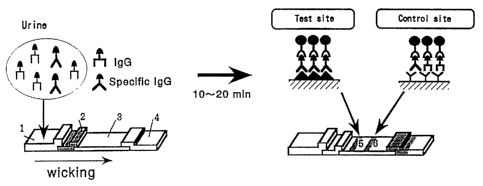
Fig. 1 is a diagram showing a solid phase support in
the form of a strip as a constituent element of the
antibody assay device of the invention. In Fig. 1, the
code 1 represents a first region, 2 a tracer region, 3 a
second region, 4 a third region, 5 a test zone and 6 a
control zone.
Fig. 2 is a diagram illustrating the.principle of
assay
CA 02467871 2004-06-09
6
of the target aritibody in a sample with the antibody assay
device of the invention. The respective codes used have the
same meanings as in Fig. 1.
Fig. 3 is schematic diagrams showing a strip of solid
phase support (A) and a housing (B) accommodating said solid
phase support included in the antibody assay device of the
invention. In Fig. 3, the codes 1-6 have the same meanings
as in Fig. 1, and the code 7 represents an upper section of
the housing, 8 a lower section of the same, 9 a sample inlet
port, and 10 a detection window.
Fig. 4 is a diagrammatic representation of the results
of determination of anti-H. pylori antibody in urine in Example
1 (5) (i) . In Fig. 4, the.open circles represent data on urine
samples from subjects with H. p_ylori infection who gave a
positive 13C-UBT test and the closed circles represent data on
urine samples from subjects who gave a negative 13C-UBT test.
Fig. 5 is a diagrammatic representation of the data on
anti-H. pvlri antibody in urine as determined in Example 1
(5) (ii) . In Fig. 5, the ordinate represents absorbance (O.D.
450 nm) and the abscissa represents the 11. pvlori-positive and
H. pylori-negative groups established according to the 13C-
UBT test.
Fig. 6 is a diagrammatic representation of the data on
CA 02467871 2004-06-09
7
anti-R. gylori antibody in urine as determined in Example 1
(6). In Fig. 6, open circles represent data on urine samples
from subjects who gave a positive 13C-UBT test and closed
circles represent data on urine samples from subjects who gave
a negative 13C-UBT test.
Fig. 7 is a diagrammatic representation of data on
anti-HBc antibody in urine as generated in Example 2 (2). In
Fig. 7, closed circles represent data on urine samples from
subjects who gave a positive test for blood anti-HBc antibody
and open circles represent data on urine samples from subjects
who gave a negative test for blood anti-HBc antibody.
Fig. 8 is a diagram showing gel permeation chromatograms
of urine samples giving false positive reactions in the
determination of anti-HIV antibody in urine and the antibody
reactivity of each fraction (Example 3 (1)). In Fig. 8, the
ordinate represents absorbance (O.D.) and the abscissa
represents the gel permeation chromatographic fraction
(fraction No. ). The solid line represents the absorbance of
the protein at 280 nm, the black dot-line represents data
generated with anti-human (IgG+igM) antibody, open
triangle-line represents data generated with anti-human IgG
(Fc-specific) antibody; and the closed triangle-line
represents data generated with anti-human IgG (Fab-specific)
antibody.
CA 02467871 2004-06-09
8
Fig. 9 is a diagramniatic representation of data on anti-Ii.
py1 ori antibody in urine as determined in Example 5. In Fig.
9, the ordinate represents absorbance (O.D. 450-650 nm) and
the abscissa represents the FI. pylori-positive group (+: n=56)
and -negative group (-: n=44) as classified by the 13C-UBT test.
Fig. 10 is a diagrammatic representation of data on
anti-rubella antibody in urine as generated in Example 6. In
Fig. 10, the ordinate represents absorbance (O.D. 450-650 nm)
and the abscissa represents the anti-rubella antibody-
positive group (+: n=76) and -negative group (-: n=23) as
classified according to the serum level measured with a
commercial kit.
Fig. 11 is a diagram showing the test site and control
site in the second region of the antibody assay device of the
invention [Example 7 (3)].
Fig. 12 is a histogram showing assay data on anti-H.
pylori antibody in the urine, whole blood and plasma as
generated with the antibody assay device of the invention in
comparison with the corresponding data generated with control
devices (A-E). In Fig. 12, "Specificity" represents the
percentage of negative tests (negative rate) relative to the
total number of tests when samples from subjects verified by
the 13C-UBT test to be negative were determined for each test
CA 02467871 2004-06-09
9
item with each assay device and "Sensitivity"! represents the
percentage of positive tests (positive rate) relative to the
total number of tests when samples from subjects verified by
the 13C-UBT test to be positive were determined for each test
item with each assay device. The control devices A-H mean the
following devices.
A: Helitest (manufactured by Cortecs Diagnostics)
B: H. pylori-Check-i
(manufactured by Bio-Medical Products)
C: First Check H. pylori
(manufactured by Worldwide Medical Corp)
D: Biocard Helicobacter pylori IgG
(manufactured by Anti Biotech Oy)
E: Insta Test H. Pylori
(manufactured by Cortez Diagnostics Inc.)
F: One Step H. pylori Test
(manufactured by Teco Diagnostics)
G: H. pylori SPOT
(manufactured by International Immuno-Diagnostics)
H: Quick Stripe H. pylori
(manufactured by Diatech Diagnostics Inc.)
DISCLOSURE OF INVENTION
The inventors of the present invention did much research
for establishing an assay system which would enable high-
CA 02467871 2004-06-09
precision determination of target antibodies even when samples
are lean in the antibodies, for example urine samples, and found
that an antibody component which nonspecifically binds the
antigen in an antigen-antibody reaction (hereinafter referred
5 to as the nonspecific binding antibody component) exists in
the assay system to give rise to nonspecific reactions, thus
causing a false positive result and hence lowering the accuracy
of detection.
Based on the above findings the inventors did further
10 research and found that said nonspecific reactions can be
suppressed by conducting the antigen-antibody reaction
between the target antibody to be assayed and the antigen
specific to the particular antibody in the presence of an
Escherichia coli (Fa. coli) component, whereby the false
positive rate can be reduced to achieve a significant
improvement in the accuracy of detection.
The inventors_further discovered that said nonspecific
binding antibody component comprises IgG fragments and/or
their denaturation products which retain the antigenicity of
the light (L) chain or F (ab) region of the IgG and that this
antibody component cross-reacts with the ordinary antibody
assay reagents (e.g. secondary antibodies) used in serum
antibody assay systems, thus leading to false positive tests.
CA 02467871 2004-06-09
11
Based on the above findings, the inventors of the present
invention further confirmed that the nonspecific reactions in
an antibody assay system can be inhibited by using a reagent
having a specific affinity for the Fc region of the assay target
antibody IgG as an antibody assay reagent, whereby the false
positive rate can be reduced to improve the accuracy of
detection in a significant degree.
Meanwhile, the inventors endeavored to improve the
antibody assay hardware (the imrnunocapillary assay device and
iinmunochromatographic assay device) and found that "true
negative" reactions can be accurately detected excluding
"false negative" reactions by establishing a "control site"
for detecting an arbitrary antibody in samples in addition to
the site ( tes t si te ) for detecting the target antibody in the
reaction zone (evaluation zone) of the strip as a part of the
assay device. Thus, in such an assay system, when the sample
is an inappropriate sample which cannot be assayed for reasons
such as too low a concentration of the antibody (that is to
say the total amount of the antibody is too small) , the "control
site" gives a negative signal indicating that the sample is
not assayable. On the other hand, when the sample has an
appropriate antibody concentration, the "control site" gives
a positive signal indicating that the sample is appropriate
for the intended assay of the target antibody. Then, according
CA 02467871 2004-06-09
12
to the result in this "test site", one may know for certain
the presence or absence of the target antibody in the sample,
that is to say whether the sample is "positive" or "true
negative ".
In this connection, Japanese Unexamined Patent
Publication N0.299464/1989 and Japanese Unexamined Patent
Publication NO.167497/1994, both disclosing improvements in
the antibody assay hardware (immunocapillary assay device and
immunochromatographic assay device), describe the devices
including a control site in addition to a test site. However,
the control site in these devices is designed to ascertain
whether or not a label disposed in an upstream region of the
strip has traversed through the test site by capillary action
and, therefore, is quite different from the control site in
accordance with the invention.
The present inventors further confirmed that when the
coupling reaction between the target antibody and the
corresponding antigen by means of the above improved antibody
assay device is conducted in the presence of an E. coli
component, the nonspecific reaction in this antigen-antibody
reaction system is inhibited and that when a reagent having
a specific affinity for the Fc region of the IgG is used as
the antibody assay reagent, the nonspecific reaction with the
antigen-antibody complex is inhibited, thus leading to a
CA 02467871 2004-06-09
13
significant decrease in the incidence of a false-positive test.
The present invention has been developed on the basis
of the above several findings.
In a first aspect thereof, the present invention provides
a high-precision method for assaying an antibody with a reduced
incidence of false positive reaction.
(1-1) As one mode thereof, the above antibody assay method for
detecting a target'antibody in a sample by utilizing an
antigen-antibody reaction is characterized in that said
reaction is carried out between said antibody and an assay
antigen in the presence of an F,. coli component.
This method for assaying an antibody includes the
following specific methods.
(a) The antibody assay method in which said E. coli component
is at least one member selected from the group consisting of
the soluble fraction and lipopolysaccharide fraction of
Escherichia coli.
(b) The antibody assay method wherein the F,. coli component
is used in a proportion of about 0.1-100 ,cl g, preferably about
0.5-50 gg, per gg of the assay anti gen .
(1-2) As another mode, the antibody assay method comprises
detecting a target antibody in a sample by the sandwich
technique, characterized in that a reagent comprising a
CA 02467871 2004-06-09
14
secondary antibody having a specific affinity for the Fc region
of the target antibody IgG is used as an antibody assay reagent.
This method for assaying an antibody includes the
following specific methods. -
(a) The antibody assay method in which the secondary antibody
is an Fc-specific anti-IgG antibody.
(b) The antibody assay method comp'rising an antigen-
antibody reaction step in which the target antibody in the
sample is coupled to an immobilized antigen specific to said
antibody as immobilized on a support and a reaction step in
which the target antibody captured by said immobilized antigen
is reacted with a secondary antibody having a specific affinity
for the Fc region of the antibody IgG.
(C) The above antibody assay method in which the
antigen-antibody reaction is carried out in the presence of
an E. coZi component.
In a second aspect, the present invention relates to an
antibody assay device. This device includes the following
embodiments.
(2-1) An antibody assay device comprising a solid phase support
having at least (a) a first region to which a sample is applied
and (b) a second region in which the antibody in the test sample
is reacted as arranged in such a sequence that the sample is
CA 02467871 2004-06-09
transported from the first region to the second region by
capillary action, and a labeling means for detecting the result
of reaction in the second region, said (b) second region having
(i) a test site where a ligand for capturing the target antibody
5 to be detected has been immobilized and (ii) a control site
where a ligand for capturing an arbitrary antibody in the sample
has been immobilized.
(2-2) The antibody assay device wherein the ligand immobilized
in the test site is an antigen to the target antibody occurring
10 in the sample.
(2-3) The antibody assay device wherein the ligand immobilized
in the control site is an ariti-human immunoglobulin antibody
capable of capturing an arbitrary antibody in the sample.
(2-4) The antibody assay device comprising a labeled ligand
15 to be bound by both the target antibody and arbitrary antibody
as said labeling means.
(2-5) The antibody assay device wherein the labeling means is
a labeled ligand to be bound by both the target antibody and
arbitrary antibody as removably supported upstreams of the
second region of the solid phase support in such a manner that,
upon contact with a sample, it reacts with the target antibody
and arbitrary antibody to form a target antibody/labeled
ligand complex and an arbitrary antibody/labeled ligand
complex, respectively, which are then transported by capillary
CA 02467871 2004-06-09
n 4
16
action to.the second region where they are fixed in the test
site and control site, respectively.
(2-6) The antibody assay device wherein the labeled ligand is
supported in a region (tracer region) intermediate between the
first region and second region of the solid phase support.
(2-7) The antibody assay device wherein the labeled ligand to
be bound by both the target antibody and arbitrary antibody
is a labeled anti-human immonoglobulin antibody.
(2-8) The antibody assay device wherein the anti-human
immunoglobulin antibody is an anti-IgG antibody having a
specific affinity for the Fc region of irnmunoglobulin G.
(2-9) The antibody assay device wherein the solid phase support
is further provided with an absorption region downstreams of
the first and second regions so that the sample transported
from the first region to the second region is further
transported by capillary action to the absorption region.
(2-10) The antibody assay device wherein the coupling reaction
of the target antibody at the test site in the second region
takes place in the presence of an K. coli component.
In a third aspect, the present invention relates to a
method for solid phase assay of a target antibody in a sample.
This method includes the following embodiments.
(3-1) A method for solid phase assay of a target antibody which
comprises applying the sample to the first region of the
CA 02467871 2004-06-09
17
antibody assay device and detecting the development of a color
at the test site in the second region under the condition of
the control site in the second region developing a color.
(3-3) The method for solid phase assay of a target antibody
wherein the coupling reaction of the target antibody at the
test site in the second region of the antibody assay device
takes place in the presence of an E. coli component.
In a fourth aspect, the present invention relates to an
antibody assay reagent kit for use in association with said
antibody assay device. The antibody assay reagent kit may
include the following embodiments.
(4-1) An antibody assay reagent kit characterized by its
comprising an coli component.
(4-2) The antibody assay reagent kit further comprising an
antigen or antibody assay reagent which is optionally-
immobilized.
(4-3) The antibody assay reagent kit characterized by its
containing an Fc-specific anti-IgG antibody as the antibody
assay reagent.
(4-4) The antibody assay reagent kit containing the antibody
assay device according to the invention.
(1) Antibody assay method
In the first place, the antibody assay method as the first
aspect of the present invention is now described in detail.
CA 02467871 2004-06-09
18
The antibody assay method of the invention represents
an improvement in the antibody immunoassay method and is
characterized in that the incidence of false positive reaction
can be decreased through inhibition of non-specific reaction.
(1-1) As an embodiment of the above antibody assay method, there
can be mentioned a method in which the antigen-antibody
reaction between the target antibody in a sample and an antigen
specific to said antibody is carried out in the presence of
an E. coli component. In accordance with this method, the
nonspecific reaction in the antigen-antibody reaction is
significantly inhibited, with the result that the incidence
of a false positive test can be decreased.
The E. coli component is not particularly restricted
provided that it is a component of Escherichia coli, thus
including but not limited to the protein component,
carbohydrate component or lipid component thereof or a mixture
of such components. As a preferred example, a soluble fraction
or lipopolysaccharide (LPS) fraction of E. coli can be
mentioned.
There is no particular limitation on the method for-
preparing such an E. coli component but a variety of methods
can be selectively used. A usual procedure may comprise
CA 02467871 2004-06-09
19
growing an arbitrary F,. coli strain in a medium suited for its
proliferation, harvesting the grown cells, and either
disintegrating the cells physically by means of a sonicator
or solubilizing them with a surfactant or the like to provide
a soluble fraction (extract) . The LPS mentioned above can be
prepared by an extractive procedure using an organic solvent,
e.g. phenol, chloroform or ether, or a mixture of two or three
different organic solvents. It can also be prepared
artificially using a genetic engineering technique. Moreover,
commercial products can be expediently utilized (e.g.
Lipopolysaccharide F. coli. which is available from Difco or
Sigma)
The preferred sample to which the invention can be
applied is a body fluid sample. The body fluid is not
restricted provided that it is a body fluid derived from a human
or other animal in which the target antigen is supposedly
contained. Thus, the term "body fluid" covers a broad variety
of biological fluids which are used as samples in routine
laboratory tests. More particularly, the body fluid includes
blood,*inclusive of serum and plasma, urine, cerebrospinal
fluid, amniotic fluid, saliva, sweat, and so forth.
Particularly the present invention solves the problem of poor
detection accuracy associated with noninvasive samples which
are favored as samples for antibody detection, such as urine,
_ .. _.. . . , _..---- ... .._ ___ __..._.. _. . ..._..._.-_._._,.... .
CA 02467871 2004-06-09
saliva and sweat, particularly urine, and, therefore, those
biological materials can be mentioned as preferred examples
of the body fluid.
The "target antibody", the object of determination, is
5 not particularly restricted provided that it is an antibody
the detection of which is desired, thus including antibodies
against various sources of infection which are foreign bodies
to the host.
The sources of infection are not particularly restricted
10 but include many different pathogens which infect man and other
animals and give rise to antibodies in the hosts. More
particularly, said pathogen includes a variety of viruses such.
as HIV (human immunodeficiency virus), type A, B, C and other
hepatitis viruses, rubella virus, influenza virus, measles
15 virus, cytomegalovirus, herpes simplex virus, varicella-
zoster herpes virus, adenovirus, enterovirus, etc.; bacteria
such as Helicobacter põylori (hereinafter referred to briefly
as Ii. py].ori) , Cldia spp.,Mycobacterium tuberculosis,
spirochetes, gonococci, Tr=onema pallidum, Mycoplasma spp.,
20 etc. (excluding Escherichia coli); and protozoae such as
Toxoplasma gondii, Entamoeba histolytica, Rickettsia
tsutsugamushi, and so forth. Preferred are viruses such as
HIV, hepatitis viruses, rubella virus, influenza virus,
measles virus, herpes virus, etc. and bacteria represented by
CA 02467871 2007-03-13
21
Fielicobacter, twlori etc., with bacteria such as H. nvlori being
particularly preferred.
The antigen for use in the antibody assay method of the
invention is not particularly restricted provided that i;t is
$ an antigen capable of undergoing antigen-antibody reaction
with the target antibody to be detected. Thus, for example,
any of the antigens used in the conventional serum antibody
assay system can be successfully used. Those antigens may not
only be the very pathogens such as said viruses and bacteria
-10 but also be antigens having the antigenic determinant groups
intrinsic to the respective pathogens. Thus, for example,
'inactivated pathogens available upon heat treatment or
irradiation of pathogens, antigens prepared by extracting
pathogens with a surfactant or the like, and antigens
15 artificially prepared by chemical synthesis or recombinant DNA
technology.
Incidentally, whether a candidate antigen may be
successfully used or not in the assay method of the invention
can be easily ascertained typically by testing its reactivity
20 with the target antibody in the conventional manner.
In the assay method of the invention, said antigen may
be optionally used as immobilized on an arbitrary solid phase
beforehand. The solid phase mentioned just above may be any
of the various solid phases in routine use in this field of
CA 02467871 2004-06-09
22
art, thus including but not limited to sticks, beads, plates
(inclusive of microtiter plates) and test tubes made of various
TM
materials, for example glass, cellulose powder, Sephadex,
Sepharose,polystyrene,filter paper,carboxymethylcellulose,
ion exchange resins, dextran, plastic film, plastic.tubing,
nylon, glass beads, silk, polyamine-methyl vinyl ether-maleic
acid copolymer, amino acid copolymer, ethylene-maleic acid
copolymer, etc.
The method for immobilization is not particularly
restricted, either, but may be whichever of physical bonding
andchemical bonding. For example, chemical bonding methods
such as covalent bonding methods, e.g. diazo method, peptide
method (acid amide derivative method, carboxyl chloride resin
method, carbodiimide resin method, maleic anhydride
derivative method, isocyanate derivative method, bromocyan
activated polysaccharide method, cellulose carbonate
derivative method, condensing reagent method,'etc.),
alkylation method; crosslinking agent coupling method (the
method for coupling to a support using gl.utaraldehyde,
hexamethyiene isocyanate or the like as the crosslinking
agent), Ugi reaction coupling method, etc.; ionic binding
methods using ion exchange resins and the like supports; and
physical adsorption methods using glass beads or other porous
glass supports.
CA 02467871 2004-06-09
23
The amourit of the antigen to be used in the assay system
is not particularly restricted but may be freely selected
according to the amount of the antigen which is in routine use
for the particular assay system. For example, when the
sandwich method is used, generally the antigen is used in excess
over the target antibody. Taking. the case in which the
reaction is conducted in a 100 ,Cl 1 reaction system as an example,
the antigen may be used in a proportion of generally about
0.1-100 ,tl g/ml , preferably about 1-10 gg/mi.
The conditions of the antigen-antibody reaction between
said antigen and target antibody are not particularly
restricted but may be the same as those in routine use for
conventional immunoassays except that the reaction should be
conducted in the presence of an F,. c-~oli component. A typical
procedure may comprise incubating or allowing to stand said
antigen, antibody and E. cgli component together at a
temperature of generally not higher than 45 C, preferably about
4-400C, more preferably about 25-40 C, for about 0.5-40 hours,
preferably about 1-20 hours. The solvent for use in the
reaction and its pH are not particularly restricted, either,
as far as the reaction is not interfered with. Thus, the
conventional buffers showing a buffer action in the pH range
of about 5-9, such as citrate buffer, phosphate buffer, tris
buffer, acetate buffer, etc. can be used generally in the
___-_ .. ...., ._..... . . _ ._,_._ ._.. ..._._-...._.._
CA 02467871 2004-06-09
24
routine manner.
The proportion of the E.. coli component in this reaction
system is not particularly restricted but may for example be
generally about 0. 1-100 u g, preferably about 0.5-50 gg, per
S /.tg of the antigen in the reaction system.
The procedure for practicing the antibody assay method
of the invention is not particularly restricted except for the
basic requirement that it comprises an antigen-antibody
reaction step in which the target antibody is reacted with the
corresponding antigen, which may be an immobilized antigen,
in the presence of said E. coli component. Preferably, however,
the method further comprises a step of detecting the target
antibody captured by said antigen (antigen-antibody complex) ,
that is to say a step of reacting the antigen-antibody complex
with an antibody assay reagent.
The method of detecting and quantitating the
antigen-antibody complex obtained by said antigen-antibody
reaction and the conditions thereof are not particularly
restricted but may be those in routine use for immunoassays
in general.
Preferably the present invention can be carried into
practice by the saridwich method. In. the solid phase sandwich
method, for instance, the target antibody in a sample can be
assayed typically by the following procedure.
CA 02467871 2004-06-09
First, an E. coli component and a sample supposedly
containing the target antigen (a body fluid such as urine) are
added to a solid phase antigen which is an immobilized antigen
capable of undergoing a specific antigen-antibody reaction
5 with the target antibody to thereby carry out an antigen-
antibody reaction. After the unbound substances not coupled
to the solid phase antigen are removed by washing, for instance,
an antibody assay reagent is added for reaction with the target
antibody coupled to the solid phase antigen (antigen-antibody
10 complex) and the antigen-antibody complex is detected or
quantitated by a detection means corresponding to the
particular assay reagent.
The selection and modification of various means for such
assays are well known to those.skilled in the art and any of
15 such techniques can be utilized in the practice of the present
invention [e.g. "Rinsho Kensa-hou Teiyo (Outline of Clinical
Test)", Kanehara Publishing Co., 1995].
The antibody assay reagent for use here is riot
particularly restricted but includes a variety of reagents in
20 routine use in the art. For example, secondary antibodies such
as an anti-human immunoglobulin antibody capable of binding
the objective antibody (immunoglobulin) can be mentioned. The
anti-human immunoglobulin antibody mentioned above includes
the antisera, purified products thereof (polyclonal
.... . __
_
CA 02467871 2004 06-_09
26
antibodies) and monoclonal antibodies available from
arbitrary animals immunized using an immunoglobulin in the
class corresponding to the target immunoglobulin as an
immunogen.
Further, as the antibody assay reagent, an anti-IgG
antibody having a specific affinity for the Fc region of the
target antibody (IgG) can also be used. As such an anti-IgG
antibody, an Fc-specific anti-IgG antibody which is not
reactive to the light chain of IgG or the F(ab) region of IgG
or protein A, protein G or the like which is specifically
reactive to the Fc region of IgG can also be used. These can
be used with particular advantage when the target antibody is
an IgG.
Those antibody assay reagents can be prepared in the
conventional manner or purchased from commercial sources.
For detection, the antibody assay reagentinay be directly
modified with a conventional labeling agent or indirectly
modified by an additional detection means.
The labeling agent is not particularly restricted but
any of the agents hitherto-known or expected to come into use
in future can be employed. To mention specific examples,
radioisotopes such as "2SI, 'H, 14C, etc.; enzymes such as
alkaline phosphatase (ALP), peroxidase (e.g. HRP), etc.;
fluorescent substances such as fluorescein isothiocyanate
CA 02467871 2007-03-13
27
(FITC),.tetramethylrhodamine isothiocyanate (RITC), etc.;
1N-(2,2,6,6-tetramethyl-l-oxyl-4-piperidyl)-SN-
(aspartate) =-2, 4-dinitrobenzene (TOPA) , etc. can be used. The
immunoassay methods using the above-mentioned labeling agents
are called radioimmunoassay, enzyme immunoassay,
fluoroimmunoassay, and spin immunoassay, respectively. The
immunochromatoassay method using an antibody assay reagent
prepared by labeling colloidal gold-stained latex particles
can also be.employed.
Labeling with those labeling agents, modifications by
indirect labeling, and their detection can be made by the per
Z-a known methods [Tatsuo Iwasaki et al.: Monoclonal Antibody,
Kodansha Scientific., 1984; and Eiji Ishikawa et al.: Enzyme
Immunoassay, 2nd Edition, Igaku Shoin, 1982, among others).
In the assay method of the invention, it is essential
'that an F,. aolz component be included in the =reaction system
comprising the target antibody to be assayed and the
corresponding antigen and as far as this requirement is
satisfied, the rest of the basal procedure is not particularly
restricted but may be the same as that used in conventional
immunoassays or routinely in the art. The conditions of the
reaction between said antigen-antibody complex and said antibody
assay reagent may for example be the same as mentioned under
(1-1) .
CA 02467871 2007-03-13
28
The presence or absence of the target antibody in a sample
or its content.is evaluated-by measuring the label activity,
which depends on the kind of labeling agent used in the labeling
of the antibody assay reagent (or the indirect label) , in the
routine manner or in terms of the antibody titer calculated
from=the measured value.
(1-2) As an alternative mode of the antibody assay method of
the present invention, there can be mentioned an immunological
method for assay of the target antibody in a sample which
comprises using a reagent having a specific affinity for the
Fc region of the target antibody IgG as the antibody assay
reagent.
In accordance with this method, the reaction between the
nonspecific binding antibody component and the antibody assay
reagent can be significantly suppressed so that the frequency
of false positive tests can be decreased. Thus, this antibody
CA 02467871 2004-06-09
29
assay method may be regarded as an improvement in the sandwich
method for immunoassay of antibodies.
The antibody assay reagent of the invention is reactive
to the target antibody (IgG) and, therefore, can be used for
detection of the antibody and is characterized in that it is
not reactive to the light chain of the target antibody IgG or
the F(ab) region of the target IgG, that is to say it has a
specific affinity for the Fc region of the target IgG.
More particularly, said antibody assay reagent may for
example be an Fc-specific anti-IgG antibody which can be
prepared by using the Fc region of the target antibody IgG as
the immunogen, and includes antisera, purified products
thereof (polyclonal antibodies) and monoclonal antibodies
which can be obtained from arbitrary animals immunized with
'said immunogen. This reagent is not limited to such antibody
preparations but may be protein A, protein G or the like which
is specifically reactive to the Fc region of the antibody IgG.
Those antibody assay reagents can be prepared in the
routine manner or purchased from commercial sources (e.g.
Sigma, Cappel or Jackson Immuno Research Laboratories,Inc.).
For detection, the antibody assay reagent may be directly
modified with a conventional labeling agent or indirectly
CA 02467871 2004-06-09
b ro.
modified by an additional detection means.
The kind of labeling agent, method for labeling, and
method of detecting the label can be the same as those mentioned
under (1-1).
5 The antibody assay method of the invention comprises the
use of the above-mentioned antibody assay reagent as an
essential feature thereof in the detection of the target
antibody, i. e. the antigen-antibody complex, and as far as this
requirement is met, the rest of the basal procedure may be
10 liberally the same as that used in conventional imrnunoassays
by the sandwich technique.
Basically, the antibody assay method of the present
invention is carried into practice by reacting an antigen
capable of reacting with the target antibody in a sample and
15 detecting the target antibody bound to the antigen
(antigen-antibody complex) with said antibody assay reagent.
The assay sample and the antigen may respectively the
same as mentioned under (1-1), and as to the target antibody,
20. too, the same antibodies as those mentioned under (1-1) can
be used, provided that the antibody is an antibody IgG against
the infection source. In the method of the present invention,
the infection source may include Escherichia coli.
Where necessary, the antigen or antibody assay reagent
CA 02467871 2007-03-13
31
in the present invention can be used as immobilized on an
arbitrary solid phase. The solid phase for use and the method
of immobilization may for exarnple be the same as mentioned under
(1-1) .
=; ,
The antigen-antibody reaction between the antigen and the
target antibody may for example be the same as mentioned under
(1-1).
Although it is not mandatory, an'E. coli. component may
be caused to be present in theantigen -antibody reaction as
mentioned under (1-1).
The resulting antigen-antibody complex is then washed
and submitted to a step in which it is reacted with said
specified antibody assay reagent. This reaction can be
carried out.under the same conditions as are generally used
in, or substantially in the same manner as, the conventional
imrnunoassays (sandwich assays). The solvent, for instance,
is not particularly restricted provided that it does=not
interfere with the reaction, thus including but not limited
to buffers at pH about 5-9, such as citrate buffer, phosphate
__..__..__
----
CA 02467871 2004-06-09
32
buffer, tris buffer and acetate 'buffer, to mention just a few
examples. The reaction time and reaction temperature are not
particularly restricted, either, but may for example be the
same as those mentioned for said antigen-antibody reaction.
The presence or absence of the target antibody in a sample
or its content is evaluated by measuring the label activity,
which depends on the kind of labeling agent used in the labeling
of the antibody assay reagent (or the indirect label) , in the
routine manner or in terms of the antibody titer calculated
from the measured value, just as mentioned under (1-1).
(2) Antibody assay device
The antibody assay device according to the second aspect
of the invention is now described in detail.
The antibody assay device of the invention is an improved
method by which the presence and/or quantity of the target
antibody to be detected in a sample can be determined with good
accuracy by a solid phase assay procedure.
More particularly, the antibody assay device of the
invention is an assay device comprising a solid phase support
having at least (a) a first region for contact with a sample
and (b) a second region for reaction of the antibody in the
sample as arranged in such a sequence that the sample is
transported by capillary action from said first region to said
CA 02467871 2004-06-09
33
second region, and a label means for detecting the result of
reaction in said second region, characterized in that said (b)
second region has (i) a test site where a ligand for the target
antibody to be assayed is immobilized and (ii) a control site
where a ligand for capturing an arbitrary antibody in the sample
is immobilized.
The outstanding feature of the antibody assay device of
the invention is that a control site independent of a test site
is provided in the second region, which control site is such
that, when a proper sample is applied and tested in a proper
manner, it forms an indication representing a positive test
in the presence of a label regardless of whether the target
antibody is present or not in the sample while, when an improper
sample is applied or a sample is tested in an improper manner,
it forms an indication representing a negative test in the
presence of a label regardless of whether the target antibody
is present or not in the sample.
Thus, the control site in the second region of this device
is a site giving an indication of whether the result
(particularly the negative result) in the test.site is a valid
assay result regardless of the presence or absence of the target
antibody in the sample. With the antibody assay device of the
invention, thanks to the above construction, it is possible
to determine, qualitatively and quantitatively, the antibody
CA 02467871 2007-03-13
34
.
in the sample with high accuracy (high reliability) with a clear,
distinction between false negative and true negative results.
Furthermore, the antibody assay device of the invention
may be- so designed that the reaction of the target antibody
in the test site is carried out in the presence of an E. coli
component or so designed that a reagent having a specific
affinity for the Fc region of the target antibody IgG is used
as the antibody assay reagent for detecting the result of
reaction in the test site. With the antibody assay device of
the invention, thanks to the above-described construction,
nonspecific reactions are inhibited so that the incidence of
a false positive test is significantly decreased, thus making
it possible to determine, qualitatively and quantitatively,
the target antibody in the sample with good accuracy and high
sens=i tivi ty .
As the assay sample and target, antibody for this antibody
assay device of the invention, those mentioned under(1-1) may
for example be employed.
The present invention first provides a sol'id phase
support comprising at least a first region and a second region.
The first region is a zone where the sample applied comes
into contact with the devi,ce and the second region is a zone
CA 02467871 2004-06-09
where the antibody (the total antibody which may contain the
target antibody) in the sample undergoes reaction and coupling
in the ligand-receptor mode or in the antigen-antibody mode
and the result of reaction is displayed in the presence of a
5 label (reaction zone and evaluation zone). Those regions are
arranged on a solid phase support in.such a manner that the
sample.applied and coming into the first region is transported
by capillary action from said first region to the second region.
Preferably, said regions are so arranged that all or at least
10 a portion of the sample coming into the first region travels,
by capillary action, through a substantially planar layer of
the solid phase support to the second region.
Optionally the solid phase support may have a third
region downstreams of the second region, as a region which
15 absorbs the sample (liquid) migrating, by capillary action,
from the first region to the second region and further
downstreams.
The preferred solid phase support is formed in the shape
of a strip and said first and second regions are arranged on
20 one and the same plane of the strip in such a manner that the
sample applied travels, by capillary action, from a first band
(first region) to a second band (second region) and optionally
further to a third band (third region). While the preferred
form of said solid phase support is a strip as mentioned above,
CA 02467871 2004-06-09
36
any other shape or geometry can be employed as far as the
functions expected of the solid phase support in the present
invention can be implemented.
The solid phase support is capable of absorbing the
sample (liquid) and, when wetted with the sample, allowing at
least the antibody in the sample to travel, by capillary action,
from the first region to the second region of the solid phase
support and optionally further to the third region. Moreover,
the solid phase support is preferably one that is capable of
supporting and immobilizing a ligand which reacts with the
antibody (inclusive of the target antibody) contained in the
sample to capture the latter.
The proper solid phase support includes a variety of
porous materials, e.g. polyethylene, glass fiber, cellulose,
rayon, nylon, crosslinked dextran, various types of
chromatograph paper, nitrocellulose, and filter paper.
The first region, second region and third region, for
instance, of the solid phase support may respectively be
constituted by the same or different members selected from
among the above-mentioned materials, with the choice of
materials depending on the roles and functions of the
respective regions.
The first region of the solid phase support is preferably
constituted by a porous material adapted to absorb the sample
CA 02467871 2004-06-09
37
applied onto its surface and let it travel, by capillary action,
to the second region. The porous material suited for the first
region is not particularly restricted but generally
TM
polyethylene (for example POREX: Porex Technologies, Fairburn,
Georgia), glass fiber, rayon, nylon, and cellulosic materials
inclusive of paper can be used. The preferred material is a
porous polyethylene or a cellulosic material such as filter
paper.
The second region of the solid phase support is
preferably constituted by a porous material which is capable
of allowing the sample (liquid) to be wicked by capillary action
from the first region to the second region and supporting a
ligand for the.antibody (inclusive of the target antibody)
occurring in the sample in a condition not dislodged by
capillary action. The porous material having such properties
includes filter paper, chromatograph paper, glass fiber,
crosslinked dextran, nylon, nitrocellulose, etc. The
preferred material is nitrocellulose because the ligand can
be easily immobilized thereon.
The second region is provided with a test site where a
ligand adapted to specifically recognize the target antibody
in the sample and capture it has been immobilized and a control
site where a ligand adapted to recognize an arbitrary antibody
in the sample and capture it has been immobi].ized. The control
CA 02467871 2004-06-09
38
site is disposed away from the test site with a given interval
therebetween, preferably downstreams in the direction of
capillary flow, and it is preferably so arranged that both sites
are contacted by the sample liquid front under the identical
conditions.
The ligand for the test site is not particularly
restricted provided that it will be specifically coupled to
the target antibody to be assayed but is preferably an antigen
which is specifically recognized and bound by the target
antibody (antigen-antibody reaction). As the antigen
mentioned just above, those antigens which are in routine use
in the conventional serum antibody assay system can be
liberally used. Those antigens may be the very pathogens such
as viruses and bacteria but may also be substances containing
antigenic determinant groups intrinsic to the respective
pathogens. Thus, for example, the pathogens inactivated by
heating or irradiation, the antigens obtained by extracting
the pathogens with a surfactant or the like, and those antigens
which are artificially prepared by chemical synthesis or
recombinant DNA technology can be mentioned.
The ligand for the control site is not particularly
restricted provided that it couples an arbitrary antibody in
the sample but is preferably an antibody (anti-immunoglobulin
antibody) which specifically recognizes and binds an arbitrary
CA 02467871 2004-06-09
39
antibody in urine.
Those ligands are immobilized on the porous material in
the test site and control site, respectively, so that they will
not be dislodged from the respective sites by the capillary
flow of the liquid sample. Thus, each ligand is bound to the
corresponding site on the porous support so that it will not
be caused to diffuse when the second region is wetted by the
sample containing the target antibody but be retained
stationary in the site without being transported to the third
region of the solid phase support.
Immobilization of the ligands on said porous support can
be achieved by the techniques well known to those skilled in
the art, i.e. by physical bonding or chemical bonding.
Thus, for example, chemical bonding methods such as
covalent bonding methods, e.g. diazo method, peptide method
(acid amide derivative method, carboxyl chloride resin method,
carbodiimide resin method, maleic anhydride derivative method,
isocyanate derivative method, bromocyan activated
polysaccharide method., cellulose carbonate derivative method,
condensing reagent method, etc.), alkylation method,
crosslinking agent coupling method (the method for coupling
to a support using glutaraldehyde, hexamethylene isocyanate
or the like as the crosslinking agent) , Ugi reaction coupling
method, etc.; ionic binding methods; and physical adsorption
..._.___.. _. ... . _. _....,__._____.____. .
CA 02467871 2004-06-09
methods can be mentioned. When nitrocellulose is used as the
porous support for the second region, said ligand can be
conveniently iminobilized by non-covalent bonding.
The amount of the ligand (antigen) for use in the test
5 site of the second region is preferably in excess so that
essentially all of the target antibody presumably'present in
the sample will be bound to the test site.
The amount of the ligand (anti-immunoglobulin antibody)
for use in the control site of the second region is also
10 preferably in excess so that essentially all of the arbitrary
antibody (which may contain the target antibody) in the sample
will be bound to the control site.
The coupling reaction between the target antibody and
the_ligand (particularly an antigen) in the test site is
15 preferably carried out in the presence of an E. coli component.
The K. coli component may be supplied to the reaction system
by incorporating it in a dilution of the assay sample and
applying the mixture to the first region or may be removably
immobilized in the test site of the solid phase support
20 upstreams of the test site (e.g. said first region or the tracer
region to be described hereinafter). The E. coli component
is not particularly restricted provided that it is derived from
Escherichia coli as mentioned above. Thus, it may be the
protein fraction, carbohydrate fraction or lipid fraction of
CA 02467871 2004-06-09
41
the cells or a mixture of such fractions. The soluble fraction
obtained by extraction of the cells or the lipopolysaccharide
(LPS) fraction can be mentioned as a preferred example.
As the sample is applied to the first region, the sample
migrates, by capillary action, to the second region where the
target antibody in the sample is bound and fixed to the test
site within the second region and, then, the remaining
arbitrary antibody (inclusive of the residue of the target
antibody which has not been bound to the test site) is bound
and fixed to the control site downstreams of the test site.
The labeling means in the present invention is used to
detect whether the target antibody and arbitrary antibody in
the sample have been coupled and fixed to said test site and
control site, respectively.
The labeling means may consist of a ligand for the
antibody and a detectable label coupled to said ligand.
The ligand-for the antibody is not particularly
restricted as far as it is a molecule which recognizes and binds
the antibody present in the sample but, in the present invention,
is preferably one which binds not only the target antibody to
be assayed but also the arbitrary antibody present in the sample.
As an example of such ligand, there can be mentioned the same
ligand as that mentioned for the control site, particularly
CA 02467871 2004-06-09
42
the same anti-immunoglobulin antibody as that adopted as the
ligand in the control site.
The anti-imrnunoglobulin antibody mentioned above
includes antisera available from arbitrary animals immunized
with a relevant immunoglobulin, e.g. a human immunoglobulin,
as the immunogen, purification products thereof (polyclonal
antibodies) and monoclonal antibodies.
The anti -immunoglobulin antibody as the ligand in the
control site may optionally be an antibody directed to all
classes of antibodies so that it may capture and detect the
total antibody in the sample or be an antibody directed to a
desired class of antibody such as immunoglobulin G (IgG).
Preferably, the ligand is an anti-immunoglobulin antibody
directed to antibodies in the same class as the class to which
the target antibody belongs and this arrangement is preferred
in that as antibodies of the same class as the target antibody
are thus detected in the control site, the optimum indication
is obtained there for judging whether the assay has *been done
properly not only when a urine sample is used but when other
body fluids such as sera are used as samples.
When the target antibody is IgG, the ligand as said
labeling means is more preferably a ligand characterized by
having a specific affinity for the Fc region of the antibody
IgG and, as preferred examples of such ligand, an Fc-specific
CA 02467871 2004-06-09
43
anti-IgG antibody which is not reactive to the light chain of
the antibody IgG or the F(ab) region thereof or Protein A,
Protein G or the like having a specific reactivity to the Fc
region of the antibody IgG can be mentioned. Such anti-
immunoglobulin antibodies or ligands can be prepared in the
routine manner or purchased from commercial sources.
The detectable labeling component is not particularly
restricted as far as it is a detectable label which is known
to be useful for specific binding assays, particularly
immunoassays, or which will be possibly used in the future
[Tatsuo Iwasaki et al: Monoclonal Antibody, Kodansha
Scientific, 1984; Eiji Ishikawa et al: Enzyme Immunoassay, 2nd
Edition, Igaku Shoin, 1982, etc.].
The preferred label is one which undergoes change in
color in the test site or control site of the second region.
Though not restricted, a label undergoing a change of color
which can be visually recognized without the aid of any
instrument is particularly preferred. For example, various
chromogens such as fluorescent substances and absorbing dyes
can be mentioned. The still more preferred is a label in the
form of a powder containing a visually detectable marker.
The suitable particul'ate label includes polymer
particles (e.g. latex or polystyrene beads) , sacks, liposomes,
metallic gels (e.g. colloidal silver, colloidal gold, etc.),
CA 02467871 2004-06-09
j n
44
and polystyrene dye particles. Among them, metal gels such
as colloidal silver and colloidal gold are preferred.
While such a label can be coupled to a ligand, either
chemically or physically, in the conventional manner to
provide a labeled ligand, commercial products can also be
utilized.
The labeling means which can be used in the present
invention may be any label that, when applied to the second
region (inclusive of the test site and control site) of the
solid phase support, indicates the result of the reaction with
the antibody occurring in the sample which has taken place in
the second region and, as far as this function can be achieved,
there is no particular limitation on the mode of its presence.
For example, when the assay device of the present invention
is provided in the form of a flow through device, the labeling
means may be included in the antibody assay reagent kit
independently of the solid phase support.
Preferably, the labeling means is removably imiiiobilized
on a solid phase support, and more preferably it is removably
immobilized upstreams of the second region of the solid phase
support. The still more preferred mode is such that the
labeling means is supported in a region (hereinafter referred
to as tracer region) intermediate between the first and second
regions of the solid phase support. In this mode of use, the
CA 02467871 2004-06-09
sample applied to the first region is wicked by capillary action
to the tracer region where it comes into contact with the
labeled ligand to form the target antibody/labeled ligand
complex and the arbitrary antibody/labeled ligand complex.
5 After passage through the tracer region, the sample containing
those complexes migrates, by capillary action, further to the
second region. The ligand (antigen) disposed in the test site
within the second region is specific to the target antibody
and the ligand (anti-immunoglobulin antibody) disposed in the
10 control site is specific to the arbitrary antibody. Therefore,
the target antibody/labeled ligand complex which has migrated
by capillary action is first captured in the test site and the
remaining arbitrary antibody/labeled ligand-complex
(inclusive of the target antibody/labeled ligand complex) in
15 the sample is then captured in the control site.
The tracer region of the solid phase support is not
particularly restricted as far as it is a support capable of
transporting the test sample containing the target antibody
and arbitrary antibody from the first region to the second
20 region and supporting said labeled ligand in such a manner that
the latter, may be released by the capillary flow. Generally,
it is a porous member made of polyethylene, glass fiber, rayon,
nylon or a cellulosic material inclusive of paper. The
preferred is a material which hardly allows nonspecific
CA 02467871 2004-06-09
46
adsorption, for example glass fiber optionally treated with
polyvinyl alcohol (PVA).
Among preferred embodiments of the invention is an
embodiment wherein said tracer region and said test site in
the second region are disposed with a given interval
therebetween on a solid phase support. As such an interval
region is provided between the tracer region and the test site,
the antibody in the sample coming into contact with the labeled
ligand in the tracer region is blended with the labeled ligand
there before the sample reaches the test site in the second
region, whereby the coupling reaction between the antibody and
the labeled ligand is more positively assured. Thus, this
region provides an incubation environment (time and space) for
the coupling of the target antibody and arbitrary antibody with
the labeled ligands prior to contact of the target antibody
and arbitrary antibody in the sample with the test site and
control site, respectively..
Furthermore, the solid phase support of the present
invention may have a third region downstreams of the second
region.
The sample continues migrating from the first region to
the optional tracer region to the second region and further
to the third region by capillary action. Thus, the third
region functions as a region receiving the liquid coming from
CA 02467871 2004-06-09
47
the second region by capillary action.
Generally the third region need only discharge the
function of receiving the liquid component not bound in the
second region but may be further provided with a site
informative of the completion of an assay upon advance of the
capillary flow of the sample (that is the liquid front) to a
predetermined end-point zone on the solid phase support.
For the above purpose, the solid phase support may be
provided with a visible indicator zone containing a water-
soluble dye such as erythrosine B, saffranine.0, phenol red
or the like downstreamsof the second region. In this case,
as the liquid front of the sample traverses the second region
into the third region, it flows through said indicator zone
and the dye disposed in this zone is carried downstreams by
the capillary flow of the sample (liquid) , thereby informing
that the sample has already passed serially through' the first
- region, tracer region and second region (test and control
sites) and accordingly that the assay has just been completed.
The material for the third region is not particularly
restricted as far as it is capable of absorbing the sample
(liquid), thus including porous films or sheets of
polyethylene, rayon, nylon and cellulosic materials inclusive
of paper. The preferred material is a cellulosic material such
CA 02467871 2004-06-09
48
as paper.
The present invention is now described in detail,
reference being had to the accompanying drawings showing the
antibody assay device and its constituent members. It should,
however, be understood that the illustrated device is a mere
embodiment of the invention and by no means definitive of the
invention.
Fig. 1 is a diagram showing a solid phase support in the
form of a strip (60 x 5 mm) as a member of the device of the
invention. In Fig. 1, the code 1 represents a first region
(16 x 5 mm, 0.92 mm thick) where an assay sample is applied
and brought into contact with the strip; the code 2 represents
a tracer region (5 x 5 mm, 0. 79 mm thick) where a labeled ligand
(e.g. colloidal gold-labeled human IgG antibody) is
immobilized; the code 3 represents a second region (18 mm x
5 mm, 0.1 mm thick) where the reaction with the antibody in
the sample takes place and the result is indicated; and the
code 4 represents a third region (22 x 5 mm, 1.46 mm thick)
where the sample which has migrated from the first region and
second region is absorbed.
The thickness of the first region is generally 0.2-2 mm
and preferably 0.8-1.2 mm, and the thickness of the tracer
region is generally 0.2-1.5 mm and preferably 0.5-1 mm. The
thickness of the second region is generally 0.03~0.2 mm and
CA 02467871 2004-06-09
49
preferably 0.08-0.12 mm, and the thickness of the third region
is generally 0.5-3 mm and preferably 1-2 mm. However, these
ranges are not critical.
Disposed within said second region (3) is a test site
(test line, about 1 x 5 mm) (5) where a ligand having a specific
affinity for the target antibody is supported in position and
a control site (control line, about 1 x 5 mm) (6) where a ligand
having an affinity for the arbitrary antibody (anti-human
immunoglobulin antibody) is supported in position. The test
site (5) is disposed at a given distance (about 6 mm) from the
tracer region and the control site (6) is disposed at a given
distance (about 6 mm) from the test site (5). The test site
has the function of reacting specifically with the target
antibody in the sample and indicating in the presence of a label
whether the target antibody exists or not in the sample and
the control site has the function of indicating in the presence
of a label whether the sample applied is proper or not.
The materials (supports) for the first region, tracer
region, second region and third region constituting the solid
phase support strip may have been simply connected in series
in the longitudinal direction of the strip along which the
sample migrates and there is no restriction to the mode of
connection. Preferably, however, the connection between the
longitudinally front end of the first region and the rear end
CA 02467871 2004-06-09
of the tracer region, the connection between the front end of
the tracer region and the rear end of the second region, and
the connection between the front end of the second region and
the rear end of the third region are respectively in
5 superimposed relation. More preferably, as illustrated in Fig.
1, the longitudinally front end part of the first region is
superimposed on the rear end part of the tracer region and the
longitudinally.front end part of the tracer region is
superimposed on the rear end part of the second region. The
10 rear end part of the third region may be superimposed on the
front end part of the second region. In such a mode of
connection, the sample applied to the first region is allowed
to migrate smoothly in the longitudinal direction of the strip.
The width of the strip in its longitudinal direction is not
15 particularly restricted but may for example be 0.5-10 mm,
preferably 1-2 mm, more preferably 0.8-1.2 mm. It should be
understood that the top/bottom relationships of the respective
regions in the superimposed strip structure are not limited
to those mentioned above but may be reversed.
20 For convenience in use, the above solid phase support
is preferably packaged in the form of an assay unit.
The principle of assay with the assay device of the
invention is now explained with reference to Fig. 2.
(3) Solid phase assay method
CA 02467871 2004-06-09
51
The third aspect of the invention is directed to a solid
phase assay method using the above-described device, which
specifically is a solid phase method for assay of the target
antibody in a sample which comprises bringing the sample into
contact with the first region of the antibody assay device and
detecting the development of a color in the test site of the
second region under the condition in which the control site
of the second region is indicating a color.
In this assay, a sample suspected to contain the target
antibody is first applied to the first region (1) of the solid
phase support.
For application of the sample, a body fluid such as urine
may be used as it is or after dilution with a suitable diluent.
The diluent is not particularly restricted but includes
various buffers having a buffer action within the range of pH
about 5-9, preferably about 6.5-8.5, (e.g. citrate buffer,
phosphate buffer, tris buffer, acetate buffer, borate buffer,
etc.) , surfactants, etc.
Since nonspecific reactions in the antigen-antibody
reaction can be suppressed to reduce the incidence of a false
positive test by conducting the antigen-antibody reaction in
the presence of an E. coli component, it is preferable to use
a diluent containing such an E. coli component. The amount
of the ~. coli component (LPS) to be incorporated in the
__ ..____.~...__. _..
CA 02467871 2004-06-09
52
diluent is not particularly restricted but is preferably such
that about 0.1N10 gg, preferably about 0.5-5 gg, of said
component will be available per gg of the ligand (antigen)
in the reaction system, i.e. the test site.
The amount of the E. coli component (LPS) to be
incorporated in the sample may for example be generally not
less than 5/.l g/ml, preferably 5N100 gg/ml, more preferably
10-50 gg/ml. Although the use of an amount over 100 gg/mi
is not prohibitive, the effect of the invention can be
accomplished at the addition level of up to 100 gg/ml.
As the sample is applied to the first region (1), the
first region 1 is wetted. The sample applied flows through
the first region (1) into the tracer region (2) by capillary
action and comes into contact and reacts with the labeled ligand
(colloidal gold-labeled anti-human IgG.antibody) removably
supported in the tracer region.
Wh,en a suitable sample is used and the target antibody
is contained in the sample, both the target antibody and
arbitrary antibody contained in the sample are coupled to said
labeled ligand in the tracer region (2) to form a target
antibody/labeled ligand complex and an arbitrary
antibody/labeled ligand complex, respectively. After
passage of the same through the tracer region (2), the
respective complexes or the labeled ligand not forming such
CA 02467871 2004-06-09
53
complexes are transported together with the sample downstreams
of the tracer region (2). In a preferred mode, the labeled
ligand not forming a complex yet is given a sufficient time
(space) for forming complexes as it travels with the
antibody-containing sample from the tracer region (2) to the
test site (5) of the second region (3) by capillary action.
As the sample reaches the test site (5) in the second region,
the target antibody/labeled ligand complex in the sample is
coupled to the ligand supported in the test site (5) and
immobilized in situ. The sample further migrates downstreams
by capillary action to reach the control site (6) where the
arbitrary antibody/labeled ligand complex is coupled to the
ligand (anti-human immunoglobulin antibody) in that site and
immobilized there. Then, by detecting the complexes
immobilized in the test site (5) and control site (6) in the
second region according to the label component of the labeled
ligand, the assay result can be indicated as a positive test.
In contrast,-when a sample not containing the target antibody
is applied,- said target antibody/labeled ligand complex to be
immobilized in the test site (5) is not formed so that the label
is not detected in this test site (5) (a negative test).
In this connection, the negative test indication in the
test site (5) includes both a negative (false negative) test
due to a low concentration of the sample (i.e. a'small total
CA 02467871 2004-06-09
n x
54
amount of antibody in the sample) and a negative (true negative)
test due to the absence of the target antibody in the sample.
These negative tests cannot be differentiated from each other
according to the result in the test site (5) alone. However,
in the case of a false negative test, said arbitrary
antibody/labeled ligand complex to be immobilized in the
control site (6) is, not formed so that the control site gives
a negative indication, while in the case of a true negative
test,said arbitrary antibody/labeledligand complex is formed
so that the control site gives a positive indication.
Therefore, according to whether the indication at the control
site is negative or positive, it is possible to tell whether
the negative result in the test site (5) is a false negative
test or a true negative test. Furthermore, when a labeled
ligand which has been deactivated is used or otherwise a proper
assay is not performed in a proper system, the control site
(6) gives a negative indication so that the finding of a false
negative test owing to such causes can be prevented.
The sample liquid containing all the unbound antibodies,
labeled ligands, etc. continues to migrate further downstreams
of the second region (3) to the third region (4). Optionally
an indicator zone may be provided and, in this case, the liquid
front coveys the dye from the indicator zone to the end-point
zone, thus indicating the passage of the liquid and dye through
CA 02467871 2004-06-09
the third region and completion of the assay.
Fig. 3 shows an example of the antibody assay device of
the invention for use in horizontal position. The solid phase
support (A) comprising the first region (1) , tracer region (2),
5 second region (3) (including the test site (5) and control site
(6) ) and third region (4) is accommodated in a housing (B) made
of a suitable material. The housing material is preferably
a moldable plastic material such as polystyrene, although
other materials such as glass, metal and paper can also be used.
10 The housing consists of an upper section (7) having several
apertures and a lower section (8) , and the solid phase support
is disposed on the lower section of the housing and covered
with the upper section on top thereof. The apertures (9) and
(10) in the upper section of the housing are disposed in
15 alignment with the series of regions of the solid phase support
and in the positions corresponding to the first region (1) and
second region (3), respectively, of the solid phase support.
The sample can be applied to the first region (1) of the
20 support from the aperture (9) (sample feeding port) . The
aperture (9) is preferably provided with a projecting
peripheral wall around it so that the wall may assist in the
dripping of the liquid sample onto the first region of the
support. The method for contacting the sample with the first
CA 02467871 2007-03-13
56
region of the solid phase support is not particularly
restricted but the sample is preferably dripped from said
sample feeding port perpendicularly to the plane of the solid
phase support.
The aperture (10) is disposed in a position permitting
a visual access to the test and control sites in the second
region of the solid phase support, whereby the fixation of the
labeled ligand/target antibody complex in the test site and
that of the labeled ligand/arbitrary antibody complex in the
control site can be visually confirmed (detection port,
evaluation port). The aperture (10) need not necessarily be
a single port permitting a.visual access to both the test site
and control site but may.comprise two independent ports for
the test 'site and the control site, respectively.
With the antibody assay device of the present.invention,
the presence or absence of the target antibody in a sample as
well as the amount of the antibody can be determined with high
accuracy by leaving the assay system standing for a few minutes
to30 minutes, preferably 5-20 minutes, after application of
the sample, generally at a temperature of not over 45r,,
preferably 4-4 0'C,"more preferably about 15-309C .
(4) Antibody assay reagent kit
The antibody assay method described under (1) or the
solid-phase assay method using the antibody assay device
CA 02467871 2004-06-09
57
described under (2) can be more expediently carried out when
an antibody assay reagent kit containing a complete set of
various reagents and equipment necessary for determination of
the antibody is utilized.
The present invention, thus, further provides an
antibody assay reagent kit for reducing to practice said
antibody assay method and said solid phase assay method.
The antibody assay reagent kit according to the present
invention is intended for use for the purpose of detecting and
quantitating an antibody in a sample through an antigen-
antibody reaction and, as one preferred mode, includes a kit
containing said E. coli component as a kit component. This
reagent kit further contains an optionally immobilized antigen
adapted to undergo antigen-antibody reaction with a target
antibody to be assayed, an antibody assay reagent, and so forth.
Furthermore, for convenience in assaying, this reagent kit
may further include a suitable reaction medium, diluent, wash
buffer, reaction stopper and/or label activity test reagent.
Moreover, as another mode, the antibody assay reagent
kit of the invention may include a reagent having a specific
affinity for the Fc region of the target antibody IgG,
preferably an Fc-specific anti-IgG antibody.
As a further alternative mode, the antibody assay reagent
-------- --
CA 02467871 2004-06-09
58
kit of the invention may contain said antibody assay device.
The reagent kit may further contain said E. co1i component
or said substance having a specific affinity for the Fc region
of the target antibody IgG or both as reagents. Moreover, the
kit may further contain such accessories as a pipette and an
ampoule (tube) for use in dilution of the sample in addition
to said reaction medium, diluent, wash buffer, reaction
stopper, and label activity test reagent, among other reagents.
BEST MODE FOR CARRYING OUT THE INVENTION
The.following examples are intended to illustrate the
present invention in further detail and should by no means be
construed as defining the technical scope of the invention.
It should be understood that many changes and modifications
may be made by those skilled in the art easily on the basis
of the foregoing disclosure of the invention without departing
from the technical scope of the invention.
Example 1 Assay of H. pylori
(1) Preparation of H. p,ylori antigen
H. pylori (a clinical isolate) was cultured on Brucella
agar medium (Becton) for 48 hours (10% C021 5% 02, 370C) and
the grown cells were harvested into cold PBS. The cells were
centrifugally washed with cold PBS for a total of 5 times, and
cold PBS was added so as to make a cell concentration of 100
CA 02467871 2004-06-09
59
mg/ml. Under stirring, one equivalent of cold 0.2% Triton
TM
X-100/PBS was added. The mixture was stirred for 5 minutes
and centrifuged and the supernatant was recovered as H. pvlori
antigen solution and stored at -80 C.
(2) Preparation of an H. pylori antigen plate
The above H. pylori antigen solution (2.5 9 g protein/ml )
was added to a 96-well plate, 100 91/well, and incubated at
4 C overnight. After the wells were washed with PBS once, a
blocking solution (Dulbecco-PBS [D-PBS), 1t- BSA, 5% sorbitol,
0. 053% NaN3 [pH 7.41) was added, 300 ,tc 1/well , and the plate was
incubated at 4 C overnight. After the blocking solution was
discarded, the plate was dried at 25 C overnight, sealed,
together with*a desiccant, in an aluminum bag and stored at
4 aC until used.
(3) Preparation of an F,. coli component
Escherichia coli (pvc18/JNi109, Takara Shuzo) was
cultured in ampicillin-containing liquid LB medium (Luria-
Bertani medium, Nippon Seiyaku) at 37 C for 18 hours. The
culture was centrifuged to harvest the cells, which were washed
with 2 portions of PBS. To the washed cells was added cold
PBS to make 100 mg/ml, and the mixture was disrupted and
extracted using a sonicator (10 seconds x 3) . The supernatant
was recovered for use as an E. coli component and stored at
-800C (hereinafter referred to as F,. coli extract).
CA 02467871 2004-06-09
(4) Determination of anti-Ii. p.ylori antibody in urine
Using urine as the sample, the anti-H. pylori antibody
in the sample was determined.
To each well of the ~i. py~ antigen plate prepared under
5 (2) ,. 25 g l of a first buffer solution (200 mM Tris-HC1 buffer,
TM
0.14 M NaCl, 2% casein, 0.5% BSA, 0.05% Tween 20, 0.1% NaN3
[pH 7.31) containing 20 gg protein/ml of the E. coli extract
and 100 gl of the urine sample were added. The mixture was
stirred for 10 seconds and allowed to stand at 37 C for 1 hour.
10 The wells were washed with 6 portions of PBST (0.05% Tween
20 and-0.05% NaN3 in PBS) and 100 /11 of a 11,000-fold dilution
of an enzyme (HRP)-labeled anti-human IgG antibody
(peroxidase-conjugated Affini Pure Goat Anti-human IgG (Fc) ,
Jackson Immuno Research) in a second buffer (50 mM Tris-HC1
15 buffer , 0.14 M NaCl, 0.5% BSA, 5% goat serum, 0.05% Tween '20,
0.1% XL-II [pH 7.3] ) was added. The plate was allowed to stand
at 37"C for l hour and, then, washed with 6 portions of PBST.
Then, 100 gl of a color developer solution (50 mM
20 ci.trate-Na2HPOõ 50% TMB solution, 0.0075% H202) was added and
reacted at room temperature for 20 minutes, at the end of which
time 100 91 of a reaction stopper (50% TMB stop solution, 50%
1N-H2SO4) was added and the absorbance was measured.
(5) Results
CA 02467871 2004-06-09
61
(i) In 5 cases of E. pYlOri-positive cases and the same number
of H. pylori-negative cases as diagnosed by the 13C-UBT test
[J. Gastroenterol., 33, pp.6-13, (1998) ] which is regarded as
the most accurate of all the diagnostic methods currently
available for H. py1ori infection, urine was sampled and the anti-H. pylori
antibody in the urine was assayed by the
procedure described under (4).
As a control experiment, the same procedure was applied
to the same urine samples except that the addition of the H.
coli extract was omitted and, based on the results, the effect
of addition of an H. coli extract in accordance with the
invention was evaluated.
The data are presented in Fig. 4.
In Fig. 4, the ordinate represents absorbance (O.D.
450-650 nm) and the abscissa represents the addition (+) or
omission (-) of the K. coli extract. Furthermore, the open
circles represent the results on uri;ne samples from H.
pylori-positive patients by the 13C-UBT test and the closed
circles represent the results on urine samples from H.
pvlori-negative patients by the 13C-UBT test.
It will be apparent from Fig. 4 that by carrying out
assays in the presence of an H. coli extract in accordance with
this invention, H. pylori-positive and -negative cases could
be clearly discriminated in agreement with the results of the
CA 02467871 2004-06-09
62
13C-UBT test, endorsing the high accuracy of the method of the
invention.
(ii) Using urine samples from 99 healthy volunteers having no
history of an eradication treatment against H. pylori and 20
patients with stomach disease (7 cases of gastric ulcer and
13 cases of gastritis), the urinary anti-H. pylori antibody
was assayed in the presence of the E. coli extract in accordance
with the procedure described under (4). The results are
presented in Fig. 5. In Fig. 5, the ordinate represents
absorbance (O.D. 450 nm) and the abscissa represents the groups
according to the ''3C-tJBt test (positive and negative cases).
The results indicated that, in the assay system containing
an H. coli extract, all urine samples from negative patients
gave definitely negative results without a false positive
result.
(iii) The amounts of anti-~.i. pvlori antibody in sera from the
same subjects as enrolled in the experiment (ii) were
determined with commercial ELISA kits and the results were
compared with the results of assays in urine by the method of
the invention in the same subjects in regard of sensitivity,
specificity and accuracy. The serum and urine samples from
each subject were simultaneously collected and prepared for
assays.
The commercial ELISA kits were as follows: HNi-CAPT" kit,
CA 02467871 2004-06-09
63
Enteric Products (HM-CAP) ; Helico Gs" kit, Shield Diagnostic
(Helico G) ; and HEL-p TestT" kit, Amrad Biotech (HEL-p) . Each
asay was performed in conformity with the protocol attached
to each kit. The resutis are shown in =Table 1.
Table 1
Method of Sample Sensitivity Specificity Accuracy
assay
HM-CAP Serum 80* (56/70) 96% (47/49) 87% (103/119)
Helico G Serum 99% (69/70) 88% (43/49) 94% (112/119)
HEL-p Serum 97% (68/70) 90% (44/49) 94% (112/119)
Method of
invention Urine 99~ (69/70) 100%(49/49) 99~c (118/119)
Referring to Table 1, "Sensitivity" means the positive
rate generated with each kit in H. pylori-positive subjects
(infection-positive according to,13C-UBT test: n=70),
"Specificity" means the negative rate generated with each kit.
in H. pvlori-negative subjects (infection-negative according
to 13C-UBT test: n=49), and "Accuracy" means the percentage of
accurate results with each kit in the total population
(70+49=119 patients).
It is clear that despite the use of urine samples
containing only trace amounts of the antibody, the
determination of anti-H. p,ylori antibody by the method of the
invention gave higher detection sensitivity and specificity
as well as significantly higher accuracy as compared with the
conventional blood antibody assay kits.
(6) Assay of anti H. p,ylori antibody in urine
CA 02467871 2004-06-09
64
Using a first buffer containing E. eml jLPS (Difco) (LPS
concentration: 5gg/well) in lieu of the E. agli extract, the
procedure of (4) was otherwise repeated to determine the anti-H.
py ori antibody in urine samples and the results were evaluated
in the same manner as under (5) (i) . The results are presented
in Fig. 6. It is clear from the diagram that similar results
can be obtained by using E. coli LPS in lieu of said E. coli
extract.
Example 2 Assay of anti-hepatitis B virus (HBc) antibody
(1) Assay of anti-hepatitis B virus (HBc) antibody
An antigen plate was prepared using HBc antigen (Chemicon
International) in accordance with the procedure of Example 1
(2) and the assay of anti-hepatitis B (core) (HBc) antibody
in urine samples was carried out in accordance with Example
1 (4).
Thus, 25 gl of first buffer containing the K. coli
extract in a varying concentration and 100 91 of sample urine
were added to each well of the HBc antigen plate and after 10
seconds' stirring, the plate was allowed to sit at 37 C for
1 hour. After the plate was washed with 6 portions of PBST,
100 ;tl 1 of a 11,000-fold dilution of enzyme (HRP) -labeled
anti-human IgG antibody in second buffer was added and the plate
was allowed to sit at 37 C for 1 hour and then washed (6 times
with PBST).
CA 02467871 2004-06-09
Then, 100 91 of a color developer solution was added
and the reaction was carried out at room temperature for 20
minutes, after which 100 91 of a reaction stop solution was
added and the absorbance was measured.
5 (2) Results
In 5 positive and 5 negative blood anti-HBc antibody
cases as classified by assays using a commercial anti-HBc
antibody assay kit (Dinabott), the anti-HBc antibody in urine
was determined by the procedure described under (1).
10 The concentrations of the Z. coli extract in reaction
mixtures were set at 0, 0.78, 1.56, 3.13, 6.25, and 12.5 ~.l
g/ml and the effect of addition of the extract was evaluated.
The results are presented in Fig. 7.
In Fig. 7, the ordinate represents absorbance (O.D.
15 450-650 nm) and the abscissa represents the level of addition
of the E. coli extract. Further, the closed circles represent
data on the urinary antibody in patients with positive blood
anti-HBc antibody and the open circles represent data on the
urinary antibody in patients with negative blood anti-HBc
20 antibody. It is apparent from the diagram that even when urine
samples are used, the difference in the detection level of
anti-HBc antibody between the group of patients with positive
blood anti-HBc antibody and the group of those with negative
blood anti-HBc antibody becomes more prominent in -relation to
CA 02467871 2004-06-09
66
the level of addition of the Z. s4l i extract.
Examnle 3
Using urine samples which gave false positive tests in
the determination of anti-HIV antibody in urine by a known assay
method [Calypte724 HIV-1 Urine EIA: Arch. Pathol. Lab. Med.,119,
139-141 (1995) ; Clinical Infectious Diseases, 12, 1100-1104
(1994) ], an exploratory experiment was carried out to identify
the component supposedly responsible for a nonspecific
reaction in the same assay system.
(1) Each of the above urine samples was adjusted to pH 7.4 with
1 M phosphate buffer (pH 7.7) and filtered through 5.0, 0.8
and 0.2 /lm-cut filters. A 20 ml portion of the filtrate was
concentrated by ultrafiltration (a 10 kDa-cut membrane) to 2
ml. The concentrated urine was subjected to gel permeation
TM
chromatography (Sephacryl S-300, Pharmacia) and each fraction
was tested for its reactivity to HIV antigen.
The reactivity to HIV antigen was confirmed by causing
each fraction to react with an HIV antigen-immobilized plate
prepared by immobilizing HIV antigen (gp160) and detecting the
conjugate (nonspecific binding component) with ALP-labeled
goat anti-human (IgG+IgM) antibody, ALP-labeled goat anti-
human IgG (Fc-specific) antibody or HRP-labeled goat anti-
human IgG (Fab-specific) antibody (all available from Jackson
ImmunoResearch Labs). The results are presented-in Fig. B.
CA 02467871 2004-06-09
67
In Fig. 8, the ordinate represents absorbance (O.D. ) and
the abscissa represents gel permeation chromatographic
fractions (fraction Nos.). The solid line represents the
absorbance of the protein at 280 nm and the closed circle-
line represents the result of detection with said anti-human
(IgG+IgM) antibody, the open triangle-line represents the
result of detection with said anti-human IgG (Fc-specific)
antibody, and the closed triangle-line represents the result
of detection with said anti-human IgG (Fab-specific) antibody.
It is apparent from Fig. 8 that in the detection with
anti-human IgG (Fab-specific) antibody, the mode of reaction
(reactivity with-the nonspecific binding component) is the
same as in the detection with anti-human (IgG+IgM) antibody,
while no reactivity is found with anti-human IgG (Fc-specific)
antibody. This finding suggested that the nonspecific binding
component is a fragment or denaturation product of human IgG
which retains the reactivity with anti-human IgG (Fab-
specific) antibody without Fc region.
Therefore, it was clear that when an anti-human IgG
(Fc-specific) antibody not reactive to such nonspecific
binding component is used as the assay reagent, the nonspecific
reaction in the antibody detection system and, hence, the
CA 02467871 2004-06-09
68
incidence of a false positive test due to such nonspecific
reaction can be inhibited, with the result that a highly
specific and very accurate antibody assay method can be
provided.
Example 4 Assay of anti-HIV antibody in urine
To each well of an HIV antigen plate (CalypteTm HIV-I Urine
EIA, Calypte Biomedical Corp.), 25 91 of first buffer (the
sample buffer of Calypte'r' HIV-I Urine EIA) and 200 91 of sample
urine were added, and after 10 seconds' stirring, the plate
was allowed to sit at 37 C for 1 hour-. After this plate was
washed 6 times (wash buffer: D-PBS, 0.05% Tween 20), 100
1 of a 11,000-fold dilution of HRP-labeled goat anti-human IgG
(Fc-specific) antibody (Peroxidase-conjugated Affini Pure
Goat anti-Human IgG, Fc Fragment Specific, Jackson
ImmunoResearch Labs.) in second buffer (50 mM Tris-HC1 buffer,
0.14M NaCl, 0.5% BSA, 5% Goat Serum, 0.05% Tween 20, 0. 1% XL-II
(pH 7.3) ) was added and the plate was allowed to sit at 37 C
for 1 hour and washed (6 times) in the same manner as above.
Then, 10091 of a color developer (50% TMB solution, 50
mM Citrate-Na2HP0õ 0.0075% H,,OZ) was added and reacted at room
temperature for 10 minutes, at the end of which time 100 ~.t
1 of a reaction stop solution (50% TMB stopper, 50% 1N-H2SO4)
was added and the absorbance (O.D. 450 nm) was measured.
CA 02467871 2004-06-09
69
As a control experiment, the sample was assayed with the
known assay method [CalypteT" HIV-1 Urine EIA: Arch.Pathol.
Lab. Med.,11.9, 139-141 (1995) ; Clinical Infectious Diseases,
1,.9, 1100-1104 (1994)] (control method) using ALP-labeled goat
anti-human immunoglobulin antibody- as a second antibody.
Furthermore, the negative control and positive control of the
above assay kit were measured by the above assay method of the
invention. Since the absorbance values thus found were
comparable to those found with the above kit, the cut-off point
for the method of the invention was set at the value found by
adding 0.180 to the mean absorbance of the above negative
control in accordance with the cut-off value calculation
method of the same kit.
The results of assays in 100 samples (urine) from
subjects with positive serum anti-HIV antibody (2 cases) and
subjects with negative serum anti-HIV antibody (98 cases) are
presented in Table 2.
Table 2
Method of invention
Positive Negative Total.
Control Positive 4* 26 30
method
Negative 0 70 70
Total 4 96 100
*: Of these 4 subjects, 2 are subjects with positive serum anti-HIV
antibody.
CA 02467871 2004-06-09
It can be seen from Table 2 that although both the
sensitivity of the method of the invention and that of the
control method were 100% (2/2), the specificity was 71.4%
(70/98) for the control method vs. 98% (96/98) for the method
5 of the invention.
The above results indicate that as compared with the
control method, the antibody assay method of the invention is
remarkably low in the incidence of a false positive test and
very high in specificity.
10 Exampie 5 Assay of anti-H. pylori antibody in urine
(1) Preparation of an E. ri antigen plate
To a 100 mg/mi suspension of j3elicobacter pylori (a
clinical isolate) in cold Dulbecco-PBS as prepared in the
routine manner [J. Clin. Microbiol., 29: 2587-2589 (1991)),
15 an equal volume of cold 0.2% Triton-X solution was added under
constant stirring with a stirrer and the mixture was further
stirred for 5 minutes and centrifuged (3,000 rpm, 20 min.).
The super.natant was transferred to a new tube for use as the
extract (1-1.5 mg/m1 as protein).
20 This extract was diluted with D-PBS (2.5 gg/ml) and the
dilution was distributed into a 96-well plate, 100 g1 per well,
and incubated at 25 C overnight. After each well was washed,
300 9 1 of a blocking solution (D-PBS, 0. 5% casein, 5% sorbitol,
0.05% NaN3 (pH 7.4) ) was added, followed by incubation at 250C
CA 02467871 2004-06-09
71
overnight. The blocking solution was then discarded and the
plate was dried at 251C overnight, sealed together with a
desiccant in an aluminum bag and stored at 4 C until used.
(2) Assays
Using the immobilized antigen (plate) prepared as above,
anti-,b-i. pvlri antibody in urine samples was assayed as in
Example 4.
Thus, 25 41 of first buffer (200 mM Tris-HC1 buffer,
0.14M NaCI, 2% casein, 0.5% BSA, 0.05% Tween 20, 0.1% NaN31
. 20 9g/ml B. coli extract (pH 7.3)) and 100 91 of sample urine
wa"s added to each well and after 10 seconds' stirring, the plate.
was allowed to sit at 37 C for 1 hour and then washed 6 times.
Just as in Example 4, 100 91 of said dilution of HRP-labeled
goat anti-human IgG (Fc-specific) antibody in second buffer
was added and the plate was allowed to sit at 37'C for 1 hour
to detect the antibody.
The results obtained are presented in Fig. 9 and Table
3.
Table 3
Sample Cut-off Sensitivity Specificity Accuracy
Point 13C-UBT (+) 13C-U$T (-)
Method of Urine 14+3SD(0.104) 56/56(100%) 42/44(95%) 98%_
invention
Urine M+5SD(0.147) 56/56(100%) 43/44(98%) 99%
Control Serum M+3SD(1.27) 53/56(95%) 43/44(98%) 96%
method
Sertrm M+5SD (1.82) 47/56(84%) 43/44(98%) 90%
--------------
CA 02467871 2004-06-09
72
In Fig. 9, the ordinate represents absorbance (O.D.
450-650 nm) and the abscissa represents the positive H. pylori
infection group (+; n=56) and negative Ji. pvlori infection
group (-; n=44) according to the 13C-UBT test [J. Gastroenterol.,
33: pp.6-13 (1998)].
In Table 3, "Sensitivity" denotes the percentage of cases
detected as positive among the positive cases according to
13C-UBT test; "Specificity" denotes the percentage of cases
detected as negative among the negative cases according to
13C-UBT test; and "Accuracy'* denotes the percentage of cases
detected as positive and negative, respectively, among the
positive and negative cases according to 13C-UBT test, the
respective figures corresponding to the cut-off point of mean
M+3SD or M+5SD. As a control experiment, the same urine
samples were assayed with a serum anti-H. pylori antibody assay
kit [HM-CAP; EPI/Kyowa Medics (K.K. )] and the results are also
tabulated (Control method).
The above results indicate that the method of the
invention is superior to the control method in sensitivity and
accuracy in particular.
Examole 6 Assay of anti-rubella virus antibody in urine
(1) Preparation of a rubella antigen plate
Using a commercial rubella antigen [available from
BIO-DESIGN] in a concentration of 1jlg/ml and a blocking agent
CA 02467871 2004-06-09
a s,
73
composed Qf D-PBS, 1$ BSA, 5$ sorbitol and 0. 05% NaN3 (pH 7. 4),
the procedure of Example 5 (1) was otherwise repeated to provide
a rubella antigen plate.
(2) Assays
Using the above antigen plate, the rubella antibody in
urine samples was assayed in the same manner as in Example 5
(2). The results are presented in Fig. 10 and Table 4.
Table 4
Control method
(servzn ELISA)
Positive Negative Total
Method of invent-i.on Positive 76 0 76
(urine ELISA)
Negative 0 23 23
Total 76 23 99
In Fig. 10, the ordinate represents absorbance (O.D.
450-650 nm) and the abscissa represents the serum anti-rubella
antibody-positive group (n=76) and -negative group (n=23)
_ according to the results of determination with a commercial
kit (Rubella IgG (II) -EIA, SEIKEN; available from Denka
Seiken). The data given in Table 4 indicates a complete
agreement between the result obtained in urine by the method
of the invention and the result obtained in sera by the control
method.
According to the above data, the degree of agreement
between the method of the invention and the control method is
CA 02467871 2004-06-09
74
as high as 100% (99/99), thus indicating that the invention
enables detection of the antibody with high sensitivity and
high specificity even in urine which is safe and convenient
and is, therefore, of great use in the laboratory examination.
Example 7 Construction of an antibody assay device
(1) Preparation of an H. pylorit antigen
An H. pylori antigen solution was prepared by the same
procedure as in Example 1 (1) and stored at -80 C.
(2) Preparation of a labeled anti-human IgG antibody-
containing dry glass fiber
To a glass fiber sheet (5.0 mm x 260 mm x 0.8 mm thick;
Whatman) was added 1 ml of a 40 nm (dia.) colloidal gold-labeled
anti-human IgG (Fc-specific) antibody solution and the sheet
was dried overnight. This sheet was stored together with a
desiccant at room temperature until used.
(3) Preparation of a membrane
The H. ip-v-lori antigen solution (3 mg/ml) prepared under
(1) above and an anti-human IgG antibody solution (0.3 mg/ml)
were respectively applied onto a nitrocellulose membrane (26.5
mm x 260 mm x 0.1 mm thick; Advance MicroDevice) by spraying
(1 .5 9 1/cm) in lines at a predetermined spacing as illustrated
in Fig. 11 and dried at 37'C for 120 minutes. After drying,
the membrane was dipped and washed in a skim milk-containing
CA 02467871 2004-06-09
Borax Buffer (pH 8.2) for 30 minutes. The washed membrane was
dried at 37 C for 1 hour and stored in the presence of a
desiccant at room temperature.
(4) Assembly (a solid-phase support)
5 As illustrated in Fig. 3 (A), the above membrane (3),
an absorbent filter paper pad (4) (22 x 260 mm x 1.5 n= thick;
Whatman), said labeled.antibody-containing glass fiber sheet
(2) and a sample pad (1) (15 x 260 mm x 1.0 mm thick; filter
paper, Whatman) were glued together with an adhesive and cut
10 to 5 mm in width.
The solid-phase support thus prepared was set in position
on a bottom section (8) of a plastic housing and a top section
(7) provided with a sample inlet port (9) and a detection window
(10) in series was placed over the solid-phase support and set
15 securely on the, bottom section (8).
Example 8 Assay of anti-,)i_. p.,ylori antibody in urine
Using the device constructed in Example 7, the assay of
anti-H. p-ylori antibody was carried out in 3 kinds of urine
samples, namely samples of urine from subjects with H. p-ylori
20 infection, samples of urine from subjects without H. p-ylori
infection, and extremely lean samples of urine from subjects
with H. p-ylori infection.
First, 500 ul of sample urine was added to 500 gl of
a sample diluent [200 mM Tris-HC1 buffer, 0.14MNaC1-; 2% casein,
CA 02467871 2004-06-09
76
0.5% BSA, 0. 05% Tween 20, 0.1% NaN3 (pH 7.3) ,F,. coli LPS (Difco)
509 1/ml], followed by mixing. Six drops (about 150 J.11) of
the resulting dilution was dripped from the sample inlet port
(9) of the device constructed in Example 7 for adsorption on
the support which was then allowed to sit for 20 minutes. As
a result, when a qualified urine sample was used, a pink - red
color band appeared in the control region of the detection
window (10). On the other hand, when an extremely lean
unqualified urine sample was used, neither the test region nor
the control region of the detection window (10) showed a color
development, indicating that the sample was not evaluable.
When the sample was a qualified urine sample from a subject
without H. Pvlori infection, a pink - red color band appeared
only in the control region of the detection window (10) showing
a negative (true negative) test for H. pylori infection, while
in the presence of H. pylori infection, a pink - red color band
appeared in both the test region and control region of the
detection window (10) , showing a positive test for H. pvlori.
Example 9 Assay of anti-E. pvlori antibody in urine
(1) Preparation of an H. coli component
Escherichia coli (pvc18/JM109; Takara Shuzo) was
cultured in ampicillin-containing liquid LB medium (Luria-
Bertani medium; Nihon Seiyaku) at 379C for 18 hours and the
CA 02467871 2004-06-09
77
grown cells were harvested by centrifugation and washed with
2 portions of PBS. Then, cold PBS was added at a final cell
concentration of 100 mg/ml and the cells were disrupted and
extracted using a sonicator (10 seconds x 3 times). The
resultant supernatant was used as E. coli extract protein.
(2) Using the E. coli extract protein prepared under (1) above
in lieu of the F,. coli LPS added to the sample diluent in Example
8, the procedure of Example 8 was otherwise repeated to
determine anti-H. pylori antibodies in urine samples. As a
result, the same results as described in Example 8 were obtained.
Example 10
Using the whole blood, plasma and urine from 21 H. pylori
infection-positive subjects and the same number of Ii. pylori
infection-negative subjects (a total of 42 cases) according
to the'3C-UBT test jJ. Gastroenterol., 33: 6-13 (1998) ], anti-H.
- p,ylori antibodies in samples were assayed by the proc,edure
described in Example B.
As a control experiment, samples from the same subjects
were respectively assayed using commercial U. pylori antibody
assay kits directed to whole blood or plasma and the
effectiveness of the device of the invention was evaluated from
the data. The results are presented in Fig. 12.
In Fig. 12, control kits A through H were as follows.
CA 02467871 2004-06-09
78
A: Helitest (manufactured by Cortecs Diagnostics)
B: H. pylor.i-Check-1
(manufactured by Bio-Medical Products)
C: First Check H. pystori
(manufactured by Worldwide Medical Corp)
D: Biocard Helicobacter p,vlri IgG
(manufactured by Anti Biotech Oy)
E: Insta Test-H. pvlori
(manufactured by Cortez Diagnostics Inc.)
F: One Step H. p.vlori Test
(manufactured by Teco Diagnostics)
G: H. pylori SPOT
(manufactured by International Immuno-Diagnostics)
H: Quick Stripe H. pylori
(manufactured by Diatech Diagnostics Inc.)
In Fig. 12, "Specificity" denotes the percentage of
negative tests (negative rate) as found by assaying 13C-UBT
test-negative samples with the corresponding kit, and
"Sensitivity" denotes the percentage of positive tests
(positive rate) as found by assaying 13C-UBT-positive samples
with the corresponding kit.
It is apparent from the data in Fig. 12 that the antibody
assay device and solid-phase assay method of the-present
CA 02467871 2004-06-09
79
invention provide excellent assay systems with high detection
specificity and accuracy even when applied to urine samples,
not to speak of blood (whole blood, plasma) samples.
It is also clear from the above results that even when
the sample is a urine sample which is safe and convenient, the
present invention enables high-sensitivity, high-specificity
detection of antibodies, thus being of great use in the
laboratory examination.
Example 11 Effect of an E. coli component on the assay of
antibodies in urine
(1) The effect of an E. coli component on the assay of antibodies
in urine was evaluated using the assay device constructed
in Example 8. Thus, using the urine from H. ipylori
infection-positive and -negative subjects selected by the
''3C-UBT test, anti-H. pvlori antibodies in urine samples
were assayed in a system using a sample diluent not
containing coli LPS (Diluent 1) and systems using the
same diluent supplemented with coli LPS at the various
concentrations shown in Table 5. The line color
development in the test region and control region was
evaluated from the line intensity measured with a
densitometer (manufactured by ATTO) . The results are shown in
Table 5.
CA 02467871 2004-06-09
e w =
Table 5
Level of addition of E. nQLi. LPS
( .Ug/ml)
Diluent 1
100 33.3 11.1 3.7 1.2
Positive Control site 50 51 43 36 41 45
urine
Test site 68 66 62 61 61 67
Negative Control site 26 26 22 18 23 23
urine
Test site 12 0 0 0 13 12
It was found that when F. c!21i LPS was added to the sample
5 at 11.1 gg/ml and higher levels, the nonspecific reaction
observed with Diluent 1 disappeared so that a false positive
test (detection error) could be precluded.
INDUSTRIAL APPLICABILITY
10 The present invention provides an antibody assay
technology by which target antibodies specific to sources of
infection can be detected with high sensitivity and high
specificity even when urine samples which are comparatively
lean in the antibodies are used as test samples. According
15 to the antibody assay method of the invention, the "false
positive" reactions due to contaminants in samples can be
significantly inhibited so that highly accurate and dependable
assay results can be obtained. Moreover, the present
invention provides improvements in immunocapillary or
CA 02467871 2004-06-09
81
irnmunochromatographic assays, whereby the existence of target
antibodies and their amounts in samples can be detected
accurately with a clear distinction between "false negative"
and "true negative".