Note: Descriptions are shown in the official language in which they were submitted.
CA 02467918 2004-05-19
WO 03/053253 PCT/US02/39301
APPARATUS AND METHOD FOR TREATING
GASTROESOPHAGEAL REFLUX DISEASE
BACKGROUND OF THE INVENTION
This application claims priority from provisional application serial no.
60/342,540
filed December 20. 2001, the entire contents of which are incorporated herein
by reference.
1. Field of the Invention
I O The subject invention is directed to a minimally invasive surgical
procedure, and
more particularly, to an endoscopic surgical procedure for treating
gastroesophageal reflux
disease, and apparatus for performing the procedure.
2. Background of the Related Art
Gastroesophageal reflux disease (GERD) is one of the most common upper-
I S gastrointestinal disorders in the western world, with a prevalence of
approximately 360
cases per 100,000 population per year. Approximately 25% of individuals with
GERD
will eventually have recurrent, progressive disease and are candidates to
undergo anti-
reflux surgical procedures for effective long term therapy.
GERD is a condition in which acids surge upward from the stomach into the
20 esophagus. Backflow of acid into the esophagus makes it raw, red and
inflamed,
producing the condition known as esophagitis; it also causes the painful,
burning sensation
behind the breastbone known as heartburn. Backflow or reflux of acid can occur
when the
sphincter or band muscle at the lower end of the esophagus fails to stay
closed. This
sphincter is called the lower esophageal sphincter (LES). The LES acts as a
valve to the
25 stomach, remaining closed until the action of swallowing forces the valve
open to allov~
food to pass from the esophagus to the stomach. Normally the valve closes
immediately
CA 02467918 2004-05-19
WO 03/053253 PCT/US02/39301
after swallowing to prevent stomach contents from surging upward. When the LES
fails to
provide that closure, stomach acids reflux back into the esophagus, causing
heartburn.
The general approach for corrective surgery involves creating a new valve or
tightening the existing valve. This procedure is known as "fundoplication" and
is used to
prevent the back flow of stomach acids into the esophagus. Various
fundoplication
procedures have been developed to correct GERD and are known as Nissen
fundoplication,
Belsey Mark IV repair, Hill repair and Dor repair. Each surgical procedure has
its own
unique attributes; however, each requires an invasive surgical procedure,
whereby the
individual must endure trauma to the thoracic cavity. The individual remains
hospitalized
after the procedure for about six to ten days.
The Nissen fundoplication technique involves enveloping the lower esophagus
with
the gastric fundus by suturing the anterior and posterior fundal folds about
the esophagus.
Modifications of this procedure include narrowing of the esophageal hiatus
posterior to the
esophagus, anchoring of the fundoplication to the preaortic fascia and
surgical division of
the vegus nerve. The degree of the fundal wrap can be modified to incompletely
encircle
the esophageal tube to avoid gas float syndrome and has also been modified to
include a
loose wrap. Similarly, the Belsey Mark IV repair, Hill repair and Dor repair
are directed to
modifications for encirclement of the esophageal tube by fascia.
Complications of these fundoplication procedures include the inability to
belch or
vomit, dysphagia, gastric ulcer, impaired gastric emptying and slippage of the
repair that
may foil the best surgical results. Therefore, the fundoplication procedures
have
been modified to adjust the length and tension of the wrap, include or exclude
esophageal
muscle in the sutures and leaving the vagus nerves in or out of. the
encirclement.
2
CA 02467918 2004-05-19
WO 03/053253 PCT/US02/39301
A relatively new fundoplication technique is known as Nissen fundoplication
laparoscopy. In contrast to the traditional Nissen fundoplication procedure,
which requires
a 6 to 10 inch incision and a 6 to 10 day hospital stay with up to 8 weeks of
recovery
at home, the laparoscopy technique is performed through small openings about
the
~ abdominal cavity and most patients tend to leave the liospital~ in two days
and cam return to
work and other activities within a week or two. Despite the benefits of less
invasive
laparoscopic fundoplication procedures, there is still a need for a minimally
invasive
corrective treatment for GERD that can be performed on an out-patient basis.
SUMMARY OF THE INVENTION
The subject invention is directed to a new and useful minimally invasive
surgical
procedure for treating Gastroesophageal reflux disease by reducing the
diameter of the
esophagus proximate to the lower esophageal sphincter, and to an endoscopic
surgical
apparatus for performing the procedure. The method includes the steps of
forming a fold
of esophageal tissue proximate to the lower esophageal sphincter, and
extending at least
one needle through the fold of esophageal tissue. Each of the needles has an
interior lumen
containing a tissue fastener. The method further includes the steps of
ejecting a distal
portion of the tissue fastener from the interior lumen of each needle such
that the distal
portion of each tissue fastener is disposed against a distal surface of the
fold of esophageal
tissue, and retracting each needle from the fold of esophageal tissue such
that a proximal
portion of each tissue fastener is deployed from the interior lumen of each
needle and is
disposed against a proximal surface of the fold of esophageal tissue.
The method further comprises the step of providing an endoscopic device having
a
an interior lumen for supporting the needles in a manner that permits the
reciprocal
movement thereof, and a tissue reception cavity for receiving the fold of
esophageal tissue.
CA 02467918 2004-05-19
WO 03/053253 PCT/US02/39301
The method includes guiding the endoscopic device through the esophagus to a
location
wherein the tissue reception cavity is disposed proximate to the lower
esophageal
sphincter. Thus, the step of forming the fold of esophageal tissue includes
the step of
drawing esophageal tissue into the tissue reception cavity of the endoscopic
device. This
may be accomplished using suction or with a tissue grasping device.
Preferably, a tissue fastener of shape memory alloy or a similar bio-
compatible .
material having memory characteristics is provided within the interior liunen
of each
needle in a generally elongate orientation. The step of ejecting a tissue
fastener from the
interior lumen of a needle includes permitting the distal portion of the
tissue fastener to
move to a normally unstressed condition (at body temperature) wherein the
distal portion
of the tissue fastener is in a curved or coiled orientation. The step of
retracing the needle
from the fold of esophageal tissue includes permitting the proximal portion of
the tissue
fastener to move to a normally unstressed condition (at body temperature)
wherein the
proximal portion of the tissue fastener is in a curved or coiled orientation.
It is envisioned
that the needles may be extended through the fold of esophageal tissue
simultaneously or
in seriatim. Similarly, the tissue fasteners may be ejected from the needles
simultaneously
or in seriatim. After the fasteners have been ejected from the needles, the
fold of
esophageal tissue is released from the tissue reception cavity, and the
endoscopic device is
withdrawn from the esophagus.
The subject invention is further directed to an endoscopic surgical apparatus
for
performing the method summarized above. The apparatus includes an elongated
tubular
body having opposed proximal and distal end portions and an interior lumen
extending
therethrough. An endoscope may be housed within the interior lumen of the
tubular body.
Preferably, one or more needles are disposed within the elongated tubular body
and are
mounted for reciprocal movement therein between a retracted position and a
protracted
4
CA 02467918 2004-05-19
WO 03/053253 PCT/US02/39301
position. Depending upon the configuration and orientation of the needles
within the
tubular body, it is envisioned that the reciprocal movement thereof may be
either
longitudinal, rotational or helical. Each of the needles has an interior lumen
extending
therethrough. A tissue fastener is disposed within the interior lumen of each
needle. The
fasteners are configured for movement between an initially straight position
within the
interior lumen of a needle and a subsequently coiled or curved position
ejected from the
interior lumen of a needle.
A mechanism is provided for effectuating reciprocal movement of the needle
within the interior bore of the elongated tubular body, and a mechanism if
provided for
ejecting the tissue fasteners from the interior lumen of the needles.
Preferably, a tissue
receiving window is formed within the distal end portion of the elongated
tubular body for
receiving a fold of esophageal tissue. Thus, the retracted position of the
needle is proximal
to or, in some instances lateral to the tissue receiving window and the
protracted position
of the needle is distal of the tissue receiving window.
These and other aspects of the subject invention and the method of using the
same
will become more readily apparent to those having ordinary skill in the art
from the
following detailed description of the invention taken in conjunction with the
drawings
described hereinbelow.
BRIEF DESCRIPTION OF THE DRAWINGS
So that those having ordinary skill in the art to which the subject invention
appertains will more readily understand how to make and use the surgical
apparatus
disclosed herein, preferred embodiments thereof will be described in detail
hereinbelow
with reference to the drawings, wherein:
CA 02467918 2004-05-19
WO 03/053253 PCT/US02/39301
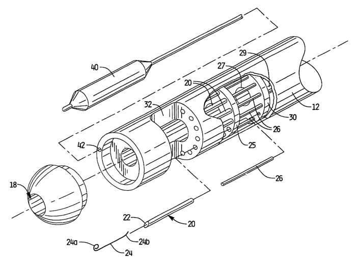
Fig. 1 is a perspective view of a surgical apparatus constructed in accordance
with a
preferred embodiment of the subject invention;
Fig. la is an enlarged localized perspective view, in partial cross-section,
of the
distal portion of the surgical apparatus of Fig. l, with parts separated for
ease of
illustration, wherein the apparatus includes a plurality of elongated needles
mounted for
reciprocal longitudinal movement relative to the longitudinal axis of the
apparatus;
Fig. 2 is a perspective view of another surgical apparatus constructed in
accordance
with a preferred embodiment of the subject invention;
Fig. 2a is an enlarged localized perspective view of the distal portion of the
surgical
apparatus of Fig. 2, with parts separated for ease of illustration, wherein
the apparatus
includes a plurality of curved needles mounted for reciprocal rotational
movement relative
to the longitudinal axis of the apparatus;
Fig. 3 is a perspective view of another surgical apparatus constructed in
accordance
with a preferred embodiment of the subject invention;
Fig. 3a is an enlarged localized perspective view of the distal portion of the
surgical
apparatus of Fig. 3, with parts separated for ease of illustration, wherein
the apparatus
includes a plurality of partially helical needles mounted for reciprocal
helical movement
relative to the longitudinal axis of the apparatus;
Fig. 4 is a side elevational view of the distal portion of the surgical
apparatus of
Fig. 1 illustrating the formation of a fold of esophageal tissue proximate to
the lower
esophageal sphincter during a treatment procedure;
Fig. 5 is a side elevational view the distal portion of the surgical apparatus
of Fig. 1
illustrating the extension of a needle through the fold of esophageal tissue,
wherein the
interior lumen of the needle contains a tissue fastener;
6
CA 02467918 2004-05-19
WO 03/053253 PCT/US02/39301
Fig. 6 is a side elevational view the distal portion of the surgical apparatus
of Fig. 1
illustrating the ejection of a distal portion of the tissue fastener from the
interior lumen of
the needle such that the distal portion of the tissue fastener is disposed
against a distal
surface of the fold of esophageal tissue;
5-- - Fig: 6a is an enlarged localized view of the needle shown iri Fig. 6
illustrating-the
ejection of the fastener from the interior lumen of the needle by the needle
pusher;
Fig. 7 is a side elevational view of the distal portion of the surgical
apparatus of
Fig. 1 illustrating the retraction of the needle from the fold of esophageal
tissue such that a
proximal portion of tissue fastener is deployed from the interior lumen of the
needle and is
disposed against a proximal surface of the fold of esophageal tissue; and
Fig. 7a is an enlarged localized view of the needle shown in Fig. 7
illustrating the
retraction of the needle from the fold of tissue.
DETAILED DESCRIPTION OF PREFERRED EMBODIMENTS
Referring now to the drawings wherein like reference numerals identify similar
structural features of the apparatus disclosed herein, there is illustrated in
Fig. l an
endoscopic surgical apparatus constructed in accordance with a preferred
embodiment of
the subject invention and designated generally by reference numeral 10.
Referring to Fig. 1 in conjunction with Fig. la, endoscopic surgical apparatus
10
includes an elongated flexible tubular body 12 having opposed proximal and
distal end
portions 14, 16 and an interior lumen 18 extending therethrough. Elongated
flexible
needles 20 with tapered leading edges are disposed within the elongated
tubular body 12
and are mounted for reciprocal longitudinal movement therein between a
retracted position
and a protracted position. More particularly, the elongated needles 20 are
supported in
circumferentially spaced relationship within tubular body 12 by a needle block
25. Needle
7
CA 02467918 2004-05-19
WO 03/053253 PCT/US02/39301
block 25 is mounted at the distal end of a tubular drive shaft 27 which is
adapted for
reciprocal axial movement within tubular body 12.
Each elongated needle 20 has an interior lumen 22 extending therethrough. A
tissue fastener 24 formed of a shape memory metal alloy, such as a nickel-
titanium alloy, is
- disposed within the interior lumen of each rieedle~20. The tissue fastener
24 is configured
for movement between an initially straight position within the interior lumen
of the
elongated needle and a subsequently coiled position ejected from the interior
lumen of the
elongated needle. In the straight position, and in the coiled position,
opposed end portions
24a, 24b of the fastener 24 have a generally curved configuration. In Fig. 2a,
the end
portion 24a of fastener 24 is shown in the coiled position, while the opposed
end portion
24b is shov~m in a transitional state between the initially straight position
and the
subsequently coiled or curved position.
An elongated push rod 26 extends through the interior lumen -22 of each
elongated
needle 20 for ejecting at least a portion of the tissue fastener 24 from the
interior lumen 22
of the elongated needle 20. Each push rod 26 is supported in circumferentially
spaced
relationship by a push rod block 30. Push rod block 30 is mounted at the
distal end of a
tubular drive shaft 29 which is mounted coaxial with drive shaft 27. Drive
shaft 29 is
adapted and configured for reciprocal axial motion within tubular body 12.
As best seen in Fig. 1, surgical apparatus 10 further includes an actuation
mechanism 35 operatively associated with a proximal portion 14 of the elongate
body 12.
Actuation mechanism 35 is adapted and configured to effectuate reciprocal
longitudinal
movement of the drive shaft 27 associated with needle block 25 and the drive
shaft 29
associated with push rod block 30. It is envisioned that actuation mechanism
35 can take
the form of a mechanical actuator, a pneumatic actuator, a hydraulic actuator
or an
electrical actuator which transmits force to the drive shafts 27, 29 through
conventional
8
CA 02467918 2004-05-19
WO 03/053253 PCT/US02/39301
mechanisms, such as cooperative linkages, gear trains or combinations thereof.
It is also
envisioned that the fasteners can be fired in a proximal direction.
Surgical apparatus 10 further includes a generally U-shaped or concave tissue
receiving window 32 formed within the distal end portion of the elongated
tubular body
12. In the retracted position, the elongated needles 20 are proximal of the
tissue receiving
window 32 and in the protracted position, the elongated needles 22 travel to a
position that
is distal to the tissue receiving window 32.
As illustrated in Fig. la, as an option, the surgical apparatus 10 of the
subject
invention could be provided with an angioplasty balloon 40 that would be
accommodated
within an elongated lateral lumen 42. It is envisioned that angioplasty
balloon 40 could be
extended from the distal end of tubular body 12 and used as a dilator to
increase the
esophageal diameter prior to placement of the fasteners 24.
Referring to Figs. 2 and 2a, there is illustrated another surgical apparatus
110
constructed in accordance with a preferred embodiment of the subject invention
that
includes an elongated body 112 having opposed proximal and distal end portions
114 and
116, and an interior lumen 118 extending therethrough. The distal end portion
116 has a
tissue receiving window 132 formed therein and the proximal portion 114 has an
actuator
handle 135 operatively associated therewith.
As best seen in Fig. 2a, surgical apparatus 110 includes a plurality of curved
needles 120 each supporting a surgical fasteners 124 in the interior lumen 122
thereof. The
curved needles 120 are supported in axially spaced relationship on a needle
block 125 that
is mounted for reciprocal rotational movement within body portion 112. A
plurality of
curved push rods 126 are supported on a push rod block 130 adjacent needle
block 125.
Each push rod 126 is configured to eject at least a portion of a tissue
fastener 124 from the
interior Iumen 122 of a needle 120 upon actuation of handle 135. Those skilled
in the art
9
CA 02467918 2004-05-19
WO 03/053253 PCT/US02/39301
will readily appreciate that conventional mechanisms such as drive screws or
drive shafts
may be employed to transmit rotational motion from actuation handle 135 to
needle block
125 and push rod block 130.
Referring to Figs. 3 and 3a, there is illustrated another surgical apparatus
210
constructed in accordance with a preferred embodiment of the subjeot invention
that
includes an elongated body 212 having opposed proximal and distal end portions
214 and
216, and an interior lumen 21 ~. A tissue receiving window 232 is formed in
the distal end
portion 216 and an actuator handle 235 is operatively associated with the
proximal potion
214. As best seen in Fig. 3a, surgical apparatus 210 differs from surgical
apparatus 110 in
that it includes a plurality of partially helical needles 220 that are mounted
for reciprocal
helical movement within body portion 212 relative to the longitudinal axis of
body portion
212.
While not shown in Fig. 3a, a surgical fastener fornled from shape memory
alloy is
supported with the interior lumen 222 of each needle 220 and is configured for
deployment
in the manner described above with respect to apparatus 110. Those skilled in
the art will
readily appreciate that conventional mechanisms such as drive screws or drive
shafts may
be employed to transmit helical motion from actuation handle 235 to the needle
block and
push rod block operatively associated with curved needles 220.
The subject invention is also directed to a method of treating
gastroesophageal
reflux disease using a surgical apparatus constructed in accordance a
preferred
embodiment of the subject invention, such as, for example, surgical apparatus
10. Initially,
during a surgical procedure, the elongated body 12 of surgical apparatus 10 is
extended
through the esophagus such that tissue receiving window 32 is positioned in a
location that
is proximate to the esophageal sphincter. Next, as shown in Fig. 4, a fold of
esophageal
tissue is drawn into the tissue receiving window 32. This is preferably done
under visual
CA 02467918 2004-05-19
WO 03/053253 PCT/US02/39301
observation using the flexible endoscope 50 extended through the interior
lumen 18 of
body 12, and is preferably accomplished by suction or using a tissue grasping
device such
as tissue grasper 45.
Thereafter, one or more needles 20 are extended through the fold of esophageal
tissue, as shown imFig. 5: -At such a time, the distal portion-24a of the
tissue fastener 24 in - -
each needle 20 is ejected from the interior lumen 22 of each needle 20 by push
rod 26 such
that the distal portion 24a of each tissue fastener 24 is disposed against a
distal surface of
the fold of esophageal tissue in a curved condition, as shown in Figs. 6 and
6a. Then, as
shown in Figs. 7 and 7a, needles 20 are retracted from the fold of esophageal
tissue such
that the proximal portion 24b of each tissue fastener 24 is deployed from the
interior lumen
22 of needle 20 and is disposed against a proximal surface of the fold of
esophageal tissue.
In instances wherein more than one needle is employed, the needles may be
extended through the fold of esophageal tissue either simultaneously or in
seriatim by
staging the needles at different positions relative to one another. Similarly,
the tissue
fasteners may be ejected from the needles simultaneously or in seriatim by
staging the
push rods at different positions relative to one another. After the needles
have been
retracted, the fold of esophageal tissue is released from the tissue reception
cavity.
Once the fasteners 24 have been deployed, the fold of tissue with which they
are
associated will undergo repetitive movement during peristalsis. Since the ends
of the
fasteners are curved and flexible, they will advantageously comply with the
fold of tissue
as it moves. This flexibility also accommodates belching and vomiting.
Furthermore, the
flexible configuration of the fasteners facilitates the easy removal thereof
from the fold of
tissue should it become necessary to reverse the procedure. This may be done
with a
grasping device, such as that which is illustrated in Fig. 4.
11
CA 02467918 2004-05-19
WO 03/053253 PCT/US02/39301
Preferably, the steps of the subject invention are performed under vision
using an
endoscope which may be provided integral with surgical device 10.
Alternatively, the
treatment method of the subject invention may be performed using either
ultrasound,
fluoroscopy or magnetic resonance imaging.
It is also envisioned and well within the scope of the subject invention that
the
surgical apparatus 10 and the method of using the same can be employed to
reduce the
volume of a patients stomach. In such a procedure, gastric tissue would be
fastened using
the apparatus of the subject invention. Since the ends of the fasteners
utilized in this
procedure are curved and flexible, they will comply, or unfurl with the fold
of tissue as the
stomach expands with the intake of food.
Although the apparatus and method of the subject invention have been described
with respect to preferred embodiments, those skilled in the art will readily
appreciate that
changes and modifications may be made thereto without departing from the
spirit and
scope of the present invention as defined by the appended claims.
12