Note: Descriptions are shown in the official language in which they were submitted.
CA 02468105 2004-05-21
WO 03/045462 PCT/US02/28202
-1-
MEDICAL DEVICES WITH MAGNETIC
RESONANCE VISIBILITY ENHANCING MATERIAL
BACKGROUND OF THE INVENTION
The present invention relates generally to
intralumenal devices for use in magnetic resonance
imaging. More particularly, the present invention
relates to intralumenal devices that incorporate a
magnetic resonance visibility enhancing material, the
devices being adapted for use in magnetic resonance
~ Q. imaging. _
Magnetic resonance imaging (MRI) is a non-
invasive medical procedure that utilizes magnets and
radio waves to produce a picture of the inside of a
body. An MRI scanner is capable of producing
pictures of the inside of a body without exposing the
body to ionizing radiation (X-rays). In addition,
MRI scans can see through bone and provide detailed
pictures of soft body tissues.
A typical MRI scanner includes a magnet
that is utilized to create a strong homogeneous
magnetic field. A patient is placed into or
proximate the magnet. The strong magnetic field
causes atoms within the patient's body to align. A
radio wave is directed at the patient's body,
triggering atoms within the patient's body cavity
tissues to emit radio waves of their own. These
return radio waves create signals (resonance signals)
that are detected by the scanner at numerous angles
around the patient's body. The signals are sent to a
CA 02468105 2004-05-21
WO 03/045462 PCT/US02/28202
computer that processes the information and compiles
an image. or images. Typically, although not
necessarily, the images are in the form of 2-
dimensional "slice" images.
Some MRI applications utilize a contrast
medium, also known as a contrast agent. Typically, a
contrast medium contains paramagnetic material and is
injected into the bloodstream of a patient. The
contrast medium alters the inherent response to
magnetic fields of atoms contained within proximately
located blood and body tissues. In this way,
contrast mediums may enable blood flow to be tracked
and/or a greater sensitivity for MRI detection and
characterization of different body tissues.
Gadolinium, a periodic table element, is an
example of a material that has been utilized within
the context of contrast mediums. Gadolinium has
eight unpaired electrons in its outer shell, which
causes it to be paramagnetic in nature. Gadolinium,
when bound to a chelator retains paramagnetic
properties and is relatively safe for exposure to the
body.
In some MRI applications, a gadolinium-
based contrast medium is introduced into a body
through intravenous injection. When injected in the
bloodstream of a patient, the gadolinium alters the
inherent response to magnetic fields of atoms
contained within proximately located blood and body
tissues. In particular, the gadolinium shortens the
CA 02468105 2004-05-21
WO 03/045462 PCT/US02/28202
-3-
relaxation time of atoms contained in the blood and
tissue that are in regions proximate to the
gadolinium molecules. During the MRI process, this
shortening of relaxation time caused by the
gadolinium-based contrast medium translates into
images that are highlighted or brightened in the
areas of atoms demonstrating the shortened
relaxation.
Within some MRI applications, catheters and
other intralumenal devices may be inserted into a
body during the MRI process. An ability tolocate,
trace and position such devices in their intralumenal
environments is desirable. A material similar to a
contrast medium (i.e., a paramagnetic material) may
be directly disposed on at least a portion of an
intralumenal device to enhance MRI visibility. Under
the typical environmental conditions associated with
the intralumenal manipulation of a medical device,
exposure of the intralumenal device to stationary
body tissue and fluid is limited. As a result,
interaction between fluid/tissue and the material
disposed on the intralumenal device is also limited.
The present invention addresses at least
one of these and other problems and offers advantages
over the prior art.
SUMMARY OF THE INVENTION
The present invention generally pertains to
intralumenal devices adapted to be advanced through a
CA 02468105 2004-05-21
WO 03/045462 PCT/US02/28202
-4-
patient during a magnetic resonance imaging
procedure. In particular, the present invention
provides one or more constructions of such
intralumenal devices that incorporate a magnetic
resonance visibility enhancing material. These and
various other features, as well as advantages that
characterize the present invention, will be apparent
upon a reading of the following detailed description
and review of the associated drawings.
BRIEF DESCRIPTION Of 'T'HE DRAWTNGS
FIG. 1 is a partial block diagram of an
illustrative magnetic resonance. imaging system in which
illustrative embodiments of the present invention ca.n
be employed.
FIG. 2 is a side view of a magnetic
resonance catheter in accordance with an illustrative
embodiment of the present invention.
FIG. 3 PRIOR ART is an enlarged cross-
sectional view of a catheter.
FIG. 4 is an enlarged cross-sectional view
of the catheter shown in FIG. 2, in accordance with an
illustrative embodiment of the present invention.
DETAILED DESCRIPTION OF ILLUSTRATIVE EMBODIMENTS
FIG. 1 is a partial block diagram of an
illustrative magnetic resonance imaging system in
which embodiments of the present invention could be
employed. In FIG. 1, subject 100 on support table
CA 02468105 2004-05-21
WO 03/045462 PCT/US02/28202
-5-
110 is placed in a homogeneous magnetic field
generated by magnetic field generator 120. Magnetic
field generator 120 typically comprises a cylindrical
magnet adapted to receive subject 100. Magnetic
field gradient generator 130 creates magnetic field
gradients of predetermined strength in three mutually
orthogonal directions at predetermined times.
Magnetic field gradient generator 130 is
illustratively comprised of a set of cylindrical
coils concentrically positioned within magnetic field
generavor 120 . A region of subj ect 100 into which a
device 150, shown as a catheter, is inserted, is
located in the approximate center of the bore of
magnetic 120. Illustratively, device 150 could be a
guidewire or some other intralumenal device.
RF source 140 radiates pulsed radio
frequency energy into subject 100 and device 150 at
predetermined times and with sufficient power at a
predetermined frequency to influence nuclear magnetic
spins in a fashion well known to those skilled in the
art. The influence on the spins causes them to
resonate at the Larmor frequency. The Larmor
frequency for each spin is directly proportional to
the strength of the magnetic field experienced by the
spin. This field strength is the sum of the static
magnetic field generated by magnetic field generator
120 and the local field generated by magnetic field
gradient generator 130. In an illustrative
embodiment, RF source 140 is a cylindrical external
CA 02468105 2004-05-21
WO 03/045462 PCT/US02/28202
-6-
coil that surrounds the region of interest of subject
100. Such an external coil can have a diameter
sufficient to encompass the entire subject 100.
Other geometries, such as smaller cylinders
specifically designed for imaging the head or an
extremity can be used instead. Non-cylindrical
external coils such as surface coils may
alternatively be used.
Device 150 is inserted into subject 100 by
an operator. Illustratively, device 150 may
alternatively be a guidewire, a catheter, an abat ion
device or a similar recanalization device, or some
other intralumenal device.
In accordance with one embodiment, but not
by limitation, device 150 illustratively includes an
RF antenna that detects magnetic resonance (MR)
signals generated in both the subject and the device
150 itself in response to the radio frequency field
created by RF source 140. Since the internal device
antenna is small, the region of sensitivity is also
small. Consequently, the detected signals have
Zarmor frequencies, which arise only from the
strength of the magnetic field in the proximate
vicinity of the antenna. The signals detected by the
device antenna are sent to imaging and tracking
controller unit 170 via conductor 180. It should be
emphasized that device 150 need not incorporate a
device antenna to be within the scope of the present
invention.
CA 02468105 2004-05-21
WO 03/045462 PCT/US02/28202
-
In accordance with one embodiment, medical
devices (such as but not limited to catheters) with
the below-described embodiments of integrated
magnetic resonance visibility enhancing material can
be utilized in combination with a device antenna to
assist in tracking and locating the device antenna.
This combination of features illustratively provides
both passive and active image enhancement.
External RF receiver 160 illustratively
detects RF signals emitted by the subject in response
to the radio frequency field created by RF source
140. In an illustrative embodiment, external RF
receiver 160 is a cylindrical external coil that
surrounds the region of interest of subject 100.
Such an external coil can have a diameter sufficient
to have a compass the entire subject 100. Other
geometries, such as smaller cylinders specifically
designed for imaging the head or an extremity can be
used instead. Non-cylindrical external coils, such
as surface coils, may alternatively be used.
External RF receiver 160 can share some or all of its
structure with RF source 140 or can have a structure
entirely independent of RF source 140. The region of
sensitivity of RF receiver 160 is larger than that of
the device antenna and can encompass the entire
subject 100 or a specific region of subject 100.
However, the resolution which can be obtained from
external RF receiver 160 is less than that which can.
be achieved with the device antenna. The RF signals
CA 02468105 2004-05-21
WO 03/045462 PCT/US02/28202
_g_
detected by external RF receiver 160 are sent to
imaging and tracking controller unit 170 where they
are analyzed together with any RF signals detected by
the device antenna.
In accordance with an embodiment of the
present invention, external RF receiver 160 detects
RF signals emitted by device 150 in response to the
radio frequency field created by RF source 140.
Illustratively, these signals are sent to imaging and
tracking controller unit 170 where they are
translated into images of device 150. In accordance
with one embodiment, the position of device 150 is
determined in imaging and tracking controller unit
170 and is displayed on display means 190. In one
illustrative embodiment, the position of device 150
is displayed on display means 190 by superposition of
a graphic symbol on a conventional MR image obtained
by external RF receiver 160. Alternatively, images
may be acquired by external RF receiver 160 prior to
initiating tracking and a symbol representing the
location of the tracked device be superimposed on the
previously acquired image. Alternative embodiments of
the invention display the position of the device
numerically or as a graphic symbol without reference
to a diagnostic image.
FIG. 2 is side view of one illustrative
embodiment of a device that could be utilized similar
to device 150 described above in relation to FIG. 1.
More particularly, FIG. 2 is a side view of a
CA 02468105 2004-05-21
WO 03/045462 PCT/US02/28202
-9-
magnetic resonance catheter 200 (MR catheter 200), in
accordance with an illustrative embodiment of the
present invention. MR catheter 200 includes an
.elongated body 210 having a proximal end 220 and a
distal end 230. Illustratively, an antenna 240 is
optionally disposed proximate distal end 230 and
operates as described above in relation to FIG. 1.
FIG. 3 PRIOR ART is an enlarged cross
sectional view of a typical catheter identified as
catheter 300. Catheter 300 includes a circumference
310 and an axis 320. Catheter 300 also includes a
lumen 330. Zumen 330 is illustratively formed and
defined by a coaxial, tubular catheter body 335 (body
335). Body 335 is typically constructed of a
flexible polymeric material or some other flexible
material. Body 335 includes an optional coaxial
layer 340 of undercoat material. Optional layer 340
is typically constructed of a layer of material such
as urethane, PVC, polyamide, silicon, PTFE,
polyurethane or some other similar material. Body
335 includes an optional coaxial outer protective
layer 345. Any of the body 335, optional layer 340
and optional layer 345 may be formed with additional
layers. For example, a reinforcement layer may be
included to improve certain mechanical
characteristics. FIG. 3 PRIOR ART is provided for
comparative purposes to better illustrate
illustrative embodiments of the present invention.
CA 02468105 2004-05-21
WO 03/045462 PCT/US02/28202
-10-
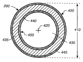
FIG. 4, in accordance with an embodiment of
the present invention, is an enlarged cross-sectional
view of MR catheter 200 taken along line 4--4 in FIG.
2. As is illustrated in FIG. 4, MR catheter 200
includes a circumference 410 and an axis 420, which
each illustratively extend at least from proximal end
220 to distal end 230 (FIG. 2). The MR catheter 200
also includes a lumen 430 that also illustratively
extends between ends 220 and 230. It should be noted
that catheters having additional lumens (mufti-lumen
cathet~er~s) should be considered within the scope of
the present invention.
With further reference to FIG. 4, lumen 430
is illustratively formed and defined by a coaxially
formed tubular catheter body 435 (body 435). In
accordance with one embodiment, body 435 is
constructed of a flexible polymeric material. Body
435, however, may be constructed of other materials
' without departing from the scope of the present
invention.
Body 435 includes an optional coaxial layer
440 of undercoat material. Illustratively, optional
layer 440 could be constructed of a layer of material
such as urethane, PVC, polyamide, silicon, PTFE,
polyurethane or some other material. Body 435 also
includes an optional coaxial outer protective layer
445. Optional layer 445 could illustratively be some
form of a lubricious coating. It should be noted
that, without departing from the scope of the present
CA 02468105 2004-05-21
WO 03/045462 PCT/US02/28202
-11-
invention, any of the body 435, optional layer 440
and optional layer 445 could illustratively be formed
with additional layers. For example, a reinforcement
layer may be included to improve certain mechanical
characteristics. In accordance with one embodiment,
a reinforcement layer is included and is configured
to operate as an internal RF antenna or a device
antenna and provides active MRI image enhancement.
Still referring to FIG. 4, the MR catheter
200, in accordance with an embodiment of the present
invention, further includes magnetic resonance
visibility enhancing material 450 (MR material 450)
disposed on the inside of body 435 (proximate lumen
430) and on the outside of body 435. It should be
noted that, in accordance with embodiments of the
present invention, magnetic resonance material 450
could be disposed either on the inside of body 435 or
on 'the outside of body 435. In addition, MR material
450 need not necessarily be coaxially continuous as
illustrated. Also, MR material 450 could
illustratively be in a general layer that is thinner
or thicker than illustrated without departing from
the scope of the present invention. The precise
configuration details of material 450 are application
dependent and will vary depending on a particular
desired functional outcome.
The MR material 450 is illustratively
disposed on a surface or surfaces of catheter 200.
In accordance with an embodiment of the present
CA 02468105 2004-05-21
WO 03/045462 PCT/US02/28202
-12-
invention, MR material 450 comprises a hydrophilic
polymer. In accordance with one embodiment, MR
material 450 comprises a hydrophilic polymer having a
magnetic resonance material incorporated therein.
The magnetic resonance material may illustratively be
incorporated into the hydrophilic polymer by
traditional means, such as compounding or blending.
In accordance with additional embodiments, the
incorporated magnetic resonance materials may be or
include paramagnetic metal salt, paramagnetic
partioles (i.e., super-magnetic iron oxide,
dysprosium, etc.), paramagnetic metal chelate,
material, gadolinium, Gd-DTPA (Gadolinium
diethylenetriaminepentaacetic acid), or some other
paramagnetic material. In accordance with yet
another embodiment, a soluble gadolinium salt is
incorporated or cross-linked into the hydrophilic
polymer matrix. Illustratively, the soluble
gadolinium salt becomes part of the hydrophilic
polymer.
In accordance with an embodiment of the
present invention, upon contact with body fluid when
catheter 200 is in use, the hydrophilic material in
MR material 450 gets hydrated in a controlled
fashion. In accordance with one embodiment, MR
material 450 is pre-soaked or pre-hydrated
(illustratively but not necessarily with water or
saline and illustratively but not necessarily for
five minutes) before catheter 200 is inserted into
CA 02468105 2004-05-21
WO 03/045462 PCT/US02/28202
-13-
the patient. The hydrophilic polymer in material 450
influences the relaxation time of the atoms captured
within the hydrophilic polymer (i.e., the relaxation
time is shortened) and thereby enhances the MRI
visibility of catheter 200. I17_ustratively, the
hydrophilic polymer modulates 'the relaxation time of
the captured atoms (i.e., shortens t1 and/or t2,
which are relaxation factors known in the art) to
enable creation of an MR image of the catheter. In
accordance with one embodiment, as the result of the
described influenced relaxation time, cat~ae~ter 200
will essentially "light up" under MRI.
In accordance with one illustrative
embodiment, paramagnetic material is incorporated
into the hydrophilic polymer to enhance MRI
visibility. Illustratively, the paramagnetic
material in material 450 influences the relaxation
time of the hydrated polymer (i.e., the relaxation
time is shortened) and thereby enhances the MRI
visibility of catheter 200. In accordance with one
embodiment, as the result of a shortened relaxation
time, catheter 200 will essentially "light up" under
MRI. The paramagnetic material illustratively might
be, but is not limited to, paramagnetic ionic
material.
The MR material 450 can illustratively be
applied to a surface of catheter 200 (or some other
medical device) in a variety of ways. A variety of
hydrophilic polymers having a variety of different
CA 02468105 2004-05-21
WO 03/045462 PCT/US02/28202
-14-
attributes and physical characteristics could be
utilized in the context of the present invention.
Assuming a given selected hydrophilic polymer has
appropriate physical characteristics, the polymer can
illustrati_vr~ly be coated or d_ip coated on a surface
of catheter 200. In accordance with one embodiment,
magnetically resonant components (paramagnetic
material) are incorporated into the hydrophilic
polymer, and both the hydrophilic polymer and the
incorporated materials are coated or dip coated on a
sur:Face of catheter 20U.
Other hydrophilic polymers may demonstrate
different physical characteristics that enable
different modes of integration or attachment with a
medical device. For example, some hydrophilic
polymers could illustratively be integrated or
attached to catheter 200 utilizing an extrusion
process. Some extr_udable hydrophilic polymers may
inherently demonstrate particularly desirable
mechanical characteristics (desirable tensile
strength, du.r_abi7_ity, etc.) following an application
to catheter 200 utilizing an extrusion process.
Other hydrophilic polymers may be less desirable in
terms of~ inheren~l~ mechanical characteristics.
In accordance with an embodiment of the
present invention, a hydrophilic polymer is applied
to catheter 200 through co-extrusion with a
structural polymer. The structural polymer provides
desirable mechanical properties while the hydrophilic
CA 02468105 2004-05-21
WO 03/045462 PCT/US02/28202
-15-
polymer provides magnetic resonance visibility. In
accordance with one embodiment, this co-extruded
hydrophilic material. can be cross-linked to enhance
its durability. Radiation, or other ~ohemical means
can illustratively be utilized to achieve the cross-
linking. In accordance with another embodiment, a
hydrophilic polymer is compounded or blended with a
structural polymer. The compounded or blended
polymers are applied to catheter 200 and provide a
material having structurally beneficial properties.
In accordance with an embodiment of the -
present invention, a hydrophilic polymer, along with
incorporated paramagnetic components (i.e.,
paramagnetic metal salt, paramagnetic metal chelate,
paramagnetic metal complex, other paramagnetic ionic
material, paramagnetic particles, etc), is applied to
catheter 200 through co-extrusion with a structural
polymer. The structural polymer provides desirable
mechanical properties while the hydrophilic polymer,
and its incorporated components, provide magnetic
resonance visibility. In accordance with one
embodiment, this co-extruded hydrophilic material can
be cross-linked to enhance its durability.
Radiation, or other chemical means can illustratively
be utilized to achieve the cross-linking. In
accordance with another embodiment, a hydrophilic
polymer, along with incorporated paramagnetic
components, is compounded or blended with a
structural polymer. The compounded or blended
CA 02468105 2004-05-21
WO 03/045462 PCT/US02/28202
-16-
polymers are applied to catheter 200 and provide a
material having structurally beneficial properties.
In accordance with an embodiment of the present
invention, catheter 20fl is generally~manufactured or
constructed utilizing a structural polymer having a
hydrophilic polymer compounded therein. In other
words, the structural polymer is what generally gives
shape to catheter 200, and it has a hydrophilic
polymer compounded therein. In essence, catheter 20U
is manufactured or constructed to inherently include
material 450. This method of integration/attachment
stands in contrast to the incorporation of a
hydrophilic polymer with a structural polymer that is
itself attached or integrated with catheter 200.
In accordance with an embodiment of the
present invention, catheter 200 is generally
manufactured or constructed utilizing a structural
polymer having a hydrophilic polymer, along with
incorporated paramagnetic components (i.e.,
paramagnetic metal salt, paramagnetic metal chelate,
paramagnetic metal complex, other paramagnetic ionic
material, paramagnetic particles, etc), compounded
therein. In other words, the structural polymer is
what generally gives shape to catheter 200, and it
has a hydrophilic polymer and associated paramagnetic
CA 02468105 2004-05-21
WO 03/045462 PCT/US02/28202
-17-
polymers compounded therein. In essence, catheter
20.0 is manufactured or constructed to inherently
include material 450. This method of
integration/attachment stands in contrast to an
incorporation of components with a structural polymer
that is itself attached or integrated with catheter
200.
The above-described extrusion, co-extrusion
and general compounding applications of material 450
are alternatives beyond coating to provide device 200
with the described magr~eLic resonance
characteristics. In many instances, compared to
coating, extrusion, co-extrusion or general
compounding can be quicker and cheaper than coating
or dip coating.
In accordance with embodiments of the
present invention, the above described co-extrusion
processes could be accomplished such that the co-
extruded components are incorporated into a variety
of potential patterns. Such patterns include a
multiple layer pattern with one component applied
directly on top of the other (one or both layers
illustratively might or might not be totally
continuous). Another pattern is with the components
co-extruded in a striped pattern. For example, but
not by limitation, each co-extrusion component might
alternate every other stripe. Another pattern is
with the components co-extruded in a spiraled
CA 02468105 2004-05-21
WO 03/045462 PCT/US02/28202
-18-
pattern. Other co-extrusion patterns should be
considered within the scope of the present invention.
Referring to FIG. 4, as was previously
mentioned, an MR material 450 may be disposed on the
inside of body .435 (proximate lumen 430) and/or on
the outside of body 435. Illustratively, extrusion
or co-extrusion provides a relatively simple
application means for attaching an MR material 450 to
the inside of body 435 (the tubular inside of
catheter 200). Placement of MR material 450 within
or on the inside of body 435 has certain illustrative
advantages. For example, during use of device 200,
there generally may be less fluid exchange in the
inner lumen of body 435 than on the external or
outside surface of body 435. In the context of
embodiments wherein paramagnetic ions are
incorporated- with a hydrophilic polymer, losses of
paramagnetic material from the hydrophilic polymer.
could be decreased in the case of placement of MR
material within or on the inside of body 435. Such
placement might enable a better longevity of the
magnetic resonance visibility effects.
Examples of hydrophilic polymers suitable
for extrusion or co-extrusion are: polyethylene oxide
(PEO), polypropylene oxide (PPO), polyvinyl
pyrrolidone (PVP), hydrophilic ~ polyurethanes,
polypropylene, starches, polycarboxylic acids,
cellulosic polymers, gelatin, malefic anhydride
polymers, polyamides, polyvinyl aleohols, polyacrylic
CA 02468105 2004-05-21
WO 03/045462 PCT/US02/28202
-19-
acid, and polyethylene oxides. Other hydrophili c
polymers, however, should be considered within the
scope of the present invention. Examples of
structural polymers suitable for co-extrusion are:
Nylon, PEBAX, polyurethane, polyethylene, PEEK,
polyimide, polyester-amide copolymer and polyether-
amide copolymer. Other structural polymers, however,
should be considered within the scope of the present
invention.
Although the present description has been
described in the Context oi: catheter X00, the present
invention could just as easily be applied in the
context of other medical devices, and in particular,
in the context of other intralumenal medical devices.
For example, the above-described material
configurations and attachment/integration methods
could just as easily be applied to produce implant
devices, guide wires, catheters of many types
(including vascular and non-vascular and esophogeal
catheters), ablation devices or any other medical
device having an enhanced MRI visibility. In
accordance with one embodiment, the above-described
material configurations and attachment/integration
methods are applied to produce balloons (i.e.,
angioplasty balloons) having an enhanced MRI
visibility. In the context of tubular devices, the
above-described MR visibility enhancement material
could illustratively be coated, extruded or co-
extruded on an outer surface, inner surface or both
CA 02468105 2004-05-21
WO 03/045462 PCT/US02/28202
-20-
surfaces. Similarly, for non-tubular devices, the
material could be coated, extruded or co-extruded on
one or both sides of a surface.
In accordance with embodiments of the
present invention, optional coatings, sucY~, as but not
limited to coatings similar to optional coatings 440
and 445, disposed on an exposed surface of an MR
material 450. For example, a lubricious coating can
be disposed or placed on an exposed MR material 450.
surface. Alternatively, a coating containing a
therapeutic agent (:i.a., an anti-biotic) could 'rye v_,
disposed or placed on a MR material 450 surface.
Illustratively, such coatings generally must not
completely block access of body fluid to MR material
450 or the hydrophilic polymer will not become
hydrated and the paramagnetic ions incorporated into
the hydrophilic polymer will not be allowed to act
upon captured body fluid.
In conclusion, the present invention
relates to a method of creating and applying a
magnetic resonance visibility enhancing material to a
medical device through, for example, a coating,
compounding (i.e., compoundi_ng elements into
structural polymer that forms a given medical
device), extrusion or co-extrusion process. The
material enables the device to be visible under MRI.
The material generally includes a hydrophilic polymer
but may or may not include an incorporated
paramagnetic material. The devices may be catheters,
CA 02468105 2004-05-21
WO 03/045462 PCT/US02/28202
-21-
such as neuro-interventional micro-catheters, or any
other appropriate MRI medical device. The devices
may illustratively enable physicians to perform
procedures under an open MRI system, instead of under
X-ray. The devices illustratively reduce radiation
exposure to both physicians and patients. The
described MRI materials illustratively help the
tracking and positioning of devices.. The devices may
illustratively be implant devices, so physicians can
check/track the implants under MRI with 3D images.
laltrough the present ' in~remti~n hay ~~i:~eezi
described with reference to illustrative embodiments,
workers skilled in the art will recognize that
changes may be made in form and detail without
departing from the spirit and scope of the invention.