Note: Descriptions are shown in the official language in which they were submitted.
CA 02468259 2004-05-25
WO 03/048337 PCT/US02/38849
ANTIBODY TO LATENT MEMBRANE PROTEINS AND USES THEREOF
FIELD OF THE INVENTION
[001] The present invention relates to antibodies to latent membrane
proteins 1
(LMP1) and 2, (LMP2A and LMP2B) and antigenic fragments thereof, and more
particularly, the extracellular transmembrane loops of the LMP1 protein. The
invention further relates to use of these antibodies in prevention and
treatment of
diseases caused by Epstein-Barr virus (EBV).
BACKGROUND OF THE INVENTION
[002] Latent membrane protein 1 (LMP1) oncogene belongs to a group of
antigens expressed on the surface of cells infected with the Epstein-Barr
virus (EBV)
during the latency period and is considered to be one of the most important pv-
transforming proteins [Meij et al., J. Infectious Diseases, 179:1108-15,
1999].
LMP1 belongs to a group of EBV antigens. Currently known members of this group
of antigens include two EBV encoded noncoding RNAs (EBER1, 2), six Epstein-
Barr nuclear antigens (EBNA1, 2, 3a, 3b, LP), and three latent membrane
proteins
(LMP1, 2A, 2B) [Id.].
[003] LMP1 protein has a short cytoplasmic amino terminus which is involved
in transcriptional activation. The carboxy terminus contains a C-terminal
activation
region 1 (CTAR1) region adjacent to the plasma membrane, including a PXQTX
(SEQ ID NO. 1) core TRAF protein-binding motif and an outermost CTAR2 region
for tumor necrosis factor associated death domain protein (TRADD) and receptor
interacting protein (RIP) binding. In addition to these functional domains,
LMP1
also contains three plasma membrane spanning domains, which expose short loops
to the extracellular space [Knecht et al. Oncology 60:289-302, 2001]. While
these
short loops are present on the surface of the infected cell, the LMP1 oncogene
is
B0S530051.1
CA 02468259 2004-05-25
WO 03/048337 PCT/US02/38849
- 2 -
known to have a very low immunogenicity [Meij et al. J. Infectious Diseases,
179:1108-15, 1999]. This has hampered development of antibodies against it.
Moreover, the extracellular loops of the LMP1 are part of a transmembrane
domain.
Membrane association of LMP1 makes production of antibodies against a
recombinant antigen difficult because such a recombinant antigens do not
present
themselves in a native membrane-bound conformation.
[004] Epstein Barr Virus (EBV) belongs to y-herpesviruses and it is
associated
with various malignant and benign lymphoproliferative disorders [Liebowitz, N.
Eng. J. Med. 338:1413-21 (1998)]. It is the etiologic agent of infectious
mononucleosis. It is also strongly associated with malignancies like Burkitt's
Lymphoma (BL), nasopharyngeal carcinoma, and immunoblastic B cell lymphomas
(non-Hodgkin's lymphoma, NHL) in immunocompromised individuals. EBV has
also been detected in substantial percentage of Hodgkin's disease (HD), in
certain
types of T and NK (natural killer) cell NHL (T-NHL and B-NHL), and gastric
carcinoma patients. In addition to its association of human malignancies, EBV
is
also associated with a spectrum of diseases, collectively called chronic EBV
syndrome, and with oral hairy cell leukoplakia predominantly seen in patients
with
AIDS.
[005] Based upon viral latent gene expression and the expression of surface
antigens, three main types of EBV latency have been characterized. In type I
latency, the EBV infected cells express EBNA-1 and EBER1 and 2, and the
disease
is usually a phenotypically representative BL. Type II latency is seen in
nasopharyngeal carcinoma, Hodgkin's disease, T-cell non-Hodgkin's lymphoma,
and B-cell non-Hodgkin's lymphoma, and in immunocompetent patients and is
characterized by expression of EBNA1, EBER1 and EBER2, and LMP1, LMP2A,
and LMP2B. Type II latency, in which all gene products are expressed, is seen
in B-
NHL in immunocompromised patients.
[006] In many cases, EBV infection results in a lymphoproliferative disease
that may be only temporarily debilitating. However, in immunosuppressed
individuals, the result can be full-blown malignancy. This occurs in
individuals who
are immunosuppressed intentionally, particularly children receiving organ
CA 02468259 2004-05-25
WO 03/048337 PCT/US02/38849
- 3 -
transplants who are treated with cyclosporine A, or opportunistically, as in
the case
with individuals infected with EBV, or genetically, as in the case of affected
males
carrying the XLP (X-linked lymphoproliferative syndrome) gene. In these cases
the
resulting malignancies derive from the polyclonal proliferation of EBV-
infected B
cells. In addition, in such patients uncontrolled epithelial replication of
the virus is
detectable in lesions of oral hairy leukoplakia. Thus, the immune response
plays a
central role in the control of EBV infection.
[007] Vaccination against EBV might be useful for several groups of people
who are seronegative for EBV. These include patients undergoing bone marrow or
organ transplantation, persons with X-linked lymphoproliferative disease,
people in
areas of the world with high incidence of Burkitt's lymphoma (equatorial
Africa) or
nasopharyngeal carcinoma (southern China), and adolescents and adults at risk
for
infectious mononucleosis.
[008] Current treatment of BL and other EBV associated malignancies include
chemotherapy, for example cyclophosphamide, and/or radiation therapy.
Radiation
therapy is frequently used, especially in patients with AIDS, because
chemotherapy
alone is rarely successful. However, these treatments are focused on
unspecific
general destruction of rapidly dividing cells and are associated with a number
of
undesirable side effects.
[009] Some anti-B-cell antibodies, such as monoclonal antibodies to CD21
(EBV receptor), CD24 (pan-B-cell antibody), and CD20 (Rituxan , rituximab,
from
Genetech oncobiology, Inc.) have recently been introduced [Cohen J.I., New
England J. Med. 343:481-492, 2000; Benkerrou et al., Blood 92:3137-47 (1998)].
However, while being B-cell specific, they do not discriminate between EBV
infected and uninfected B-cells and their use results in destruction of all B-
cells with
such surface antigens. Therefore it would be desirable to develop antibodies
that are
specific for EBV infected cells and that elicit sufficient immune responses to
specifically target infected with EBV infected cells. It would also be
desirable to
have new means for determining cells infected by EBV and means which can be
used to differentiate between different disease states.
SUMMARY OF THE INVENTION
CA 02468259 2004-05-25
WO 03/048337 PCT/US02/38849
- 4 -
[0010] We have now discovered antibodies and antibody fragments directed
against extracellular domains of the EBV LMP proteins, including LMP1, LMP2A
and LMP2B. Generating antibodies against LMP1 is preferred. However, the same
methodology can be used for all three proteins. We have also discovered
methods of
treating EBV-associated malignancies using these LMP specific antibodies. For
example, these antibodies can further be expressed intracellularly to bind to
LMP1
protein within the cell and inhibit function of the protein. We have also
discovered
methods of generating immune cells, including cytotoxic T cells, with
specificity for
LMP. We have also discovered methods of treating EBV-associated malignancies
using these LMP-specific immune cells. We have further discovered a method of
using the antigenic fragments of, e.g., LMP1 in eliciting immune response, for
example, in a vaccine formulation.
[0011] One embodiment of the invention provides an antibody or antibody
fragment that binds to Epstein Barr Virus (EBV) latent membrane protein (LMP).
Preferably, the antibody binds to LMP1, LMP2A, or LMP2B. Even more
preferably, LMP 1. In a preferred embodiment, the antibody binds to an
antigenic
fragment which is an extracellular domain of the LMP 1. In a more preferred
embodiment, the antigenic fragment is amino acids 1 ¨207 of LMP1. Preferably,
the antibody is a single chain antibody, a single chain Fv domain (scFv, also
sometimes called sFv), Fab, Fab', F(ab)2, humanized antibody, human antibody,
or
chimeric antibody.
[0012] Another preferred embodiment of the invention provides an
immunogenic proteoliposome containing LMP. Preferably, the proteoliposome
contains at least one antigenic epitope of the extracellular domain of LMP 1.
In a
preferred embodiment, the proteoliposome contains the extracellular domain of
LMP1, which can readily be prepared for instance using LMP1 amino acids 1-207.
Preferably, the proteoliposome contains the entire extracellular domain of the
particular LMP being used.
[0013] Another preferred embodiment of the invention provides a method of
obtaining an LMP-specific antibody, by screening a library of antibodies with
a
proteoliposome containing LMP, and selecting antibodies that bind to the
CA 02468259 2004-05-25
WO 03/048337 PCT/US02/38849
-5..
proteoliposome. Preferably, the library of antibodies is a phage display
library.
Even more preferably, a human single chain antibody phage display library.
[0014] In another embodiment, the invention relates to a pharmaceutical
composition comprising a complex comprising the antibody or antibody fragment
against an antigenic fragment of LMP, e.g., LMP1 and a second moiety, such as
a
cytotoxic molecule attached to the antibody in a pharmaceutically acceptable
carrier.
[0015] In a further embodiment, the invention relates to a method of
treating an
EBV-associated disease. Preferred EBV-associated diseases include an EBV-
associated malignancy, Hodgkin's disease, chronic EBV syndrome, and oral hairy
cell leukoplakia. Preferably, an EBV-associated malignancy including Burkitt's
lymphoma, lymphoproliferative disease, B-lymphoproliferative disease, non-
Hodgkin's lymphoma (NHL), T-NHL, NK-NHL, lymphonasopharyngeal carcinoma,
and gastric carcinoma.
[0016] One preferred embodiment comprises administering the antibody or
antibody fragment against an antigenic fragment of LMP, for example, LMP1 in a
pharmaceutically acceptable carrier to an individual infected with EBV. In one
embodiment, the individual has EBV-infected cells, more preferably an EBV-
associated malignancy. One preferred compound further contains a second moiety
such as a cytotoxic molecule attached to the antibody.
[0017] In yet another embodiment, the invention relates to a method of
vaccinating an individual against EBV infection using an immune response
eliciting
amount of at least one LMP protein, e.g., LMP1, or an antigenic fragment
thereof in
a pharmaceutically acceptable carrier.
[0018] Another preferred embodiment of the present invention provides a
chimeric gene comprising one gene segment encoding at least an antibody heavy
chain binding region which specifically binds LMP, (e.g., a dAb, a scFv) and a
second gene segment encoding partially or entirely the transmembrane and
cytoplasmic domains of an immune cell-triggering molecule. More preferably,
the
first gene segment encodes both the light and heavy chain binding region,
e.g., a
single chain antibody. Such a chimeric gene combines the antibody recognition
sites
CA 02468259 2004-05-25
WO 03/048337 PCT/US02/38849
- 6 -
and the lymphocyte signaling moiety into one continuous chain, and endows a
lymphocyte transformed with this chimeric gene with antibody-type specificity.
Preferably, there is both a transmembrane and a cytoplasmic domain, although
it is
possible to delete the cytoplasmic domain.
[0019] In one preferred embodiment, the antibody binding region is an
scFv.
[0020] In one preferred embodiment, the immune cell-triggering molecule
is a
lymphocyte receptor chain, a polypeptide of the TCR/CD3 complex, a subunit of
the
Fe receptor, or a subunit of the IL-2 receptor. Even more preferably, a chain
of the T
cell receptor. Even more preferably, a cytotoxic T cell receptor.
[0021] Another preferred embodiment provides an expression vector
containing
the chimeric gene combining an LMP-specific antibody heavy chain binding
region
with an immune cell-triggering molecule.
[0022] Another preferred embodiment of the present invention provides
LMP-
specific immune cells, transformed with the chimeric gene combining an LMP-
specific antibody binding region with an immune cell-triggering molecule.
Preferrably, the immune cell is a natural killer cell, a lymphokine activated
cell, a
cytotoxic T cell, a helper T cell, or a subtype thereof. Even more preferably,
a
cytotoxic T cell.
[0023] Another preferred embodiment of the present invention provides
methods
of treating an EBV-associated disease by administering an immune cell
expressing a
chimeric gene combining an LMP-specific scFv with an immune cell-triggering
molecule. Preferably, an EBV-associated malignancy. Preferably, the immune
cell
expressing a chimeric gene is a cytotoxic T cell. Preferably, the binding of
the
immune cells to the LMP-expres sing target cells results in cell death of the
target
cells.
[0024] In another preferred embodiment of the present invention, the
chimeric
gene combining an LMP-specific antibody heavy chain binding region with an
immune cell-triggering molecule is transformed into the lymphocytes of an
individual infected with EBV, and the transformed and thus activated cells are
administered to the individual.
CA 02468259 2004-05-25
WO 03/048337 , PCT/US02/38849
- 7..
BRIEF DESCRIPTION OF THE FIGURES
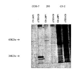
[0025] Figure 1 shows expression of C9-LMP1 and ATES-C9 in COS-7, 293
and
CF2 cells. Cell lysates derived from approximately 107 COS-7 or 293 or CF2
cells
transfected with pC9-LMP1 or pATES-C9 were subjected to immunoprecipitation
with anti-C9 Sepharose beads. Bound proteins were recovered by incubation in
2%
SDS buffer at 55 C. Eluted proteins were separated on a 12% or 15% SDS-PAGE
mini-gel.
[0026] Figure 2 shows an SDS-PAGE gel analysis of different cell/bead
ratios.
Radiolabeled cleared lysates derived from approximately 107 COS-7 cells
incubated
respectively with 106, 3 x 10⁶, 107, 3 x 10⁷ 1D4-streptavidin-coated beads.
Beads
were washed several times and the proteins eluted by incubation at 55 C for
lh.
[0027] Figure 3 shows an SDS-PAGE gel analysis of protein composition of
ATES-C9 proteoliposomes. [35S]methionine and [35S]cysteine labeled cells were
used to obtain proteoliposomes. The proteoliposomes were incubated in SDS-
sample
buffer and the eluted proteins analysed in a SDS-PAGE gel. The position of
ATES-
C9 (24 I(Da) is indicated.
[0028] Figures 4A and B show the results of an ELISA assay after panning
of a
phage library with ATES-C9 proteoliposomes. The ATES¨C9 proteoliposomes were
used to select recombinant antibodies from a human single-chain antibody phage
display library of approximately 1.5 x 10¹⁰ members. Each round of selection
was
performed using approximately 5x10⁷ proteoliposomes; in the first round 2x10¹³
t.u. of phages were used and the output was 105 t.u./ml. After the fourth
round the
output was 5x10⁷ t.u./ml. After the fourth round of panning, 48 of the
selected
individual phages were tested for ability to bind BJAB-WT4 cells relative to
binding
of BJAB cells using a cell-based ELISA. The titer of the captured phages
increased
from 105 to 5x10⁷ from the second to the fourth round of panning. All resulted
positive in the cell-based ELISA assay as shown in the table on Figure 4A.
Figure
4B illustrates the ELISA assay using pools A (including clones 1-5, 7-22, 24-
26, 28-
30, 32-34, 36, 38-46, and 48), B (including clones 6, 23, 31, and 35), and C
(including clones 37 and 47).
CA 02468259 2004-05-25
WO 03/048337 PCT/US02/38849
- 8 -
DETAILED DESCRIPTION OF THE INVENTION
[0029] The present invention is directed to antibodies and antibody
fragments
directed against extracellular domains of the EBV LMP proteins, including
LMP1,
LMP2A and LMP2B. Generating antibodies against LMP1 is preferred. However,
the same methodology can be used for all three proteins. We have also
discovered
methods of treating EBV-associated malignancies using these LMP specific
antibodies. For example, these antibodies can further be expressed
intracellularly to
bind to LMP1 protein within the cell and inhibit function of the protein. We
have
also discovered methods of generating immune cells, including cytotoxic T
cells,
with specificity for LMP. We have also discovered methods of treating EBV-
associated malignancies using these LMP-specific immune cells. We have further
discovered a method of using the antigenic fragments of, e.g., LMP1 in
eliciting
immune response, for example, in a vaccine formulation.
[0030] The antibodies and antibody fragments of the present invention
recognize
the extracellular epitope of an LMP. Preferably, the LMP is LMP1, LMP2A, or
LMP2B. Even more preferably, LMP 1.
[0031] Antibodies and antibody fragments of the present invention include
single chain antibodies, single chain Fv domains (scFv, also sometimes called
sFv),
Fab, Fab', F(ab)2, heavy chain single domain (dAb), humanized antibodies,
human
antibodies, and chimeric antibodies. One preferred antibody is an scFv
antibody.
Even more preferably, a human scFv.
[0032] Any antigen or antigenic determinant which can generate LMP-specific
antibodies may be used to generate such antibodies. Preferrend antigens
include
proteoliposomes containing LMP, as described below.
[0033] Preferably, the antigenic determinant has a conformation
approximating
the wild type (i.e., native) LMP such as LMP1. The invention is further
directed to
antibodies or fragments thereof capable of specifically recognizing an epitope
of an
LMP, such as the LMP1 protein or derivatives thereof and capable of binding
thereto.
CA 02468259 2011-05-19
=
tµPlµ C)
WO 03/048337 PCT/US02/38849
- 9 -
[0034] In one preferred embodiment, the antigenic determinant is part of
an
extracellular loop of LMPl. For example, the antigenic determinant can be made
from about amino acids 1-207 of LMP1 . This construct contains the cytoplasmic
N-
terminus, the six tansmembrane domains and 20 amino acids of the C-terminal
tail.
Obviously, other variations can be made. For example, the C-terminal tail can
be
modified as can the other positions. Preferably, modifications are designed to
emphasize the native antigenic epitopes. More preferably, a conserved epitope
among EBV strains.
[0035] The LMP antigens useful according to the present invention
include
latent membrane protein 1 (LMP1), LMP2A, and LMP2B and antigenic fragments
thereof. In the preferred embodiment, the antigen is LMPI . Preferably, the
antigen
comprises at least one of the extracellular loops of LMP 1. One preferred
method of
generating an antibody is to prepare proteoliposomes which contain an
antigenic
determinant of an LMP protein such as LMPl. The epitope is preferably in a
conformation that approximates the wild type conformation. This can be done by
blown means based upon this disclosure. The basic methodology described below
can be used with all the LMPs.
[0036] One source of antigenic material to generate antibodies is
proteoliposomes containing LMP. One method for expressing proteins including
transmembrane proteins such as LMPs is the use of proteoliposomes, as
described in
PCT/US01/50820 published as WO 02/056831, PCT/US00/35295 published as WO
01/049265, and U.S.S.N. 09/749,240 which corresponds to U.S. Patent No.
6,761,902.
[0037] To prepare proteoliposomes containing LMP, a vector expressing
the
desired portion of LMP is expressed into a host cell, as described below. The
LMP-
expressing cell is then lysed in a buffer with the appropriate detergent and
protease
inhibitors so the LMP can be separated from other cellular debris by
conventional
means without harming the protein, preferably without disrupting the protein's
natural conformation.
[0038] In general, due to their amphipathic properties, transmembrane
proteins
can be solubilized only by agents that disrupt hydrophobic associations and
destroy
the membrane's lipid bilayer. The agents typically used are small amphipathic
CA 02468259 2004-05-25
WO 03/048337 PCT/US02/38849
- 10 -
molecules which tend to form micelles in water. Preferably, the agent is a
detergent.
When mixed with membranes, the hydrophobic regions of the detergent bind to
the
transmembrane domain of proteins, displacing the lipid molecules. The polar
ends
of detergents can either be charged (ionic) or uncharged (non-ionic). Although
integral membrane proteins can be maintained in a native conformation in a
detergent solution, over time many such solubilized proteins undergo
denaturation
and aggregation.
[0039] When a detergent is removed from a transmembrane protein-detergent
complex in the absence of phospholipid, the membrane protein molecules usually
denature, aggregate and precipitate out of solution. If, however, the purified
protein
is mixed with phospholipid before the detergent is removed, the active protein
can
insert into the lipid bilayer formed by the phospholipids. In this manner,
functionally active membrane proteins can be reconstituted from purified
components. An integral membrane protein properly reconstituted into its
native
lipid environment is stable for extended periods of time.
[0040] Additionally, a critical factor for maintaining a functional native
conformation of the LMP transmembrane protein during its purification is the
choice
of detergent used to solubilize the protein. The detergent best suited for a
given
membrane protein is typically determined empirically. If the protein has been
investigated previously, the literature will indicate successful detergents.
Moreover,
one can rely upon the results obtained with related proteins to determine
detergents
that will be successful with other proteins. Thus, research on a related
protein
indicates the type of detergent most likely to extract the protein in an
active form.
[0041] Detergents can be generally classed, depending upon the nature of
their
polar end, into three groups: non-ionic, zwitterionic, and ionic. Strong ionic
detergents (such as SDS) can solubilize most membrane proteins, but tend to
unfold
the protein in the process, making them less useful for reconstituting active
conformations. In general, milder non-ionic detergents are preferred.
[0042] Detergents recommended for gentle solubilization of membrane
proteins
include alkyl glucopyranosides (such as C8-GP and C9-GP), alkyl thio-
glucopyranosides (such as C8-tGP, C10-M, C12-M, Cymal-5, Cymal-6, and Cymal-
CA 02468259 2004-05-25
WO 03/048337 , PCT/US02/38849
- 11 -
7), alkyl sucroses (such as HECAMEG), CHAPSO, digitonin,
hydroxyethylglucamides (such as HEGA-10), oligoethyleneglycol derivatives
(such
as C8E5, C8En, and C12E8), dodecylmaltopyranoside, and phenyl polyoxyethylenes
(such as Triton X-100).
[0043] Preferred detergents include alkyl thioglucopyranosides,
dodecylmaltopypanoside and phenyl polyoxyethydenes. More preferably, Cymal-5,
Cymal-6, Cymal-7, HEGA-10, digitonin, CHAPS , dodecylmaltopyranoside, and
Triton X-100. Still more preferably Cymal-5, Cymal-6, Cymal-7, and
dodecylmaltopyranoside.
[0044] Commercial kits are also available to assist in choosing a
detergent
appropriate for a given membrane protein. For example, both Anatrace and
Calbiochem offer a variety of kits containing mixtures of different
detergents.
[0045] There are many known instances of detergents which have been
successfully used to purify functionally active membrane proteins. For
example,
decylmaltoside was used to purify the K+ channel (Ksc K+) from Streptomyces
lividans, allowing its structure to be determined by X-ray crystallography
(Doyle et
al., Science (1998) 280: 69-77). Cymal-5, Cymal-6, Cymal-7, and
dodecylmaltopypanoside are preferred detergents for GCPRs, more preferably for
chemokine receptors (Mirzabekov, T. et al. (1999), J. Biol. Chem. 274: 28745-
50).
[0046] The cleared cell lysate containing all solubilized LMP membrane
proteins
and other water-soluble cellular proteins can be separated from the other
cellular
debris by conventional means. For example using high speed centrifugation,
such as
150,000 x g. Antibodies directed against the epitope tag on the protein of
interest are
used to capture this protein from the cell lysate onto the solid support
(e.g., beads).
After binding of the solubilized integral membrane protein to the antibodies
immobilized on the solid support, the solid support is washed. Thereafter the
purified
detergent-protein mixture is formed into a proteoliposome as described below.
[0047] The proteoliposome comprises a spherical or elliptoid shape such
as a
bead or other pellet. Preferably, the bead or pellet is at least about 15% the
size of a
eukaryotic cell; still more preferably it is at least about 20% the size of
such a cell;
CA 02468259 2004-05-25
WO 03/048337 PCT/US02/38849
- 12 -
and even more preferably it is at least about 25% the size of such a cell. The
shape
is three-dimensional so that it can be coated on all sides. However, there can
be
substantial variability in the exact shape used. The exact shape chosen will
depend
upon the way the proteoliposome is being used. Thus, in some embodiments
flakes
are preferable to beads, e.g., as an immunogen, in others, a thicker ellipsoid
can be
preferable.
[0048] The spherical or elliptoid shape, e.g. bead, is preferably also
coated with
a substance that will help attract and anchor a lipid layer. For example, one
can use
a compound such as streptavidin or avidin to coat the spherical or elliptoid
shape
such as a bead and add a small amount of biotinylated lipid to the lipid
mixture. For
example, one can use a head group-modified synthetic lipid, such as
dipalmitoylphosphoethanolamine-N-Biotinyl (Biotinyl-DPPE) or
dioleoylphosphoethanolamine-lissamine Rhodamine B (Rho-DOPE) in solution with
lipids. Such a mixture will form a strong uniform coating with, for example, a
streptavidin coated-bead.
[0049] The spherical or elliptoid shape (such as a bead) will also have an
anchor
ligand such as an antibody bound to it that will specifically bind either the
antigenic
tag or a known specific portion of the integral membrane protein that is to be
bound
to the bead, thereby orienting the protein. The lipid solution containing
biotinylated
lipid is added to the beads with the captured protein of interest. Thereafter,
the
detergent is slowly removed by known means. For example, by dialysis, for
e.g., at
least 24 hours. The resulting integral membrane protein-containing proteolipo
some
is stable for an extended period of time. As used herein, an extended period
of time
means at least 12 hours; still more preferably at least one day; even more
preferably
at least one week; still more preferably at least one month; and even more
preferably
at least two months. Not only will the protein retain its conformation in
these
proteoliposomes for long periods of time, but it will do so under a wide range
of
conditions, such as pH and temperature.
[0050] Preferably the spherical or elliptoid surface that is used is a
magnetic
bead. Magnetic beads are well known in the art and can be obtained
commercially.
For example tosylactivated Dynabeads M-(Bikker, J.A., Trumpp-Kallmeyer, S.,
and Humblet, C. (1998) J. Med. Chem. 41, 2911-2927)0 (Dynal, Inc., Lake
Success,
CA 02468259 2004-05-25
WO 03/048337 , PCT/US02/38849
- 13 -
New York). These are particularly useful in assisting in the purification of
the
protein. One can use such proteoliposomes as intermediates and transfer the
stabilized proteoliposome to another surface. For example, a flake. When using
the
proteoliposome for injection into an individual, it is preferable that the
surface is
made of a biodegradable material.
[0051] While the proteoliposome will typically contain only the integral
membrane protein of interest, there are instances where one may want to use
more
than one protein. For example, one can prepare a mixture comprising different
epitopes of LMP1, LMP2A, or LMP2B, or, alternatively, use LMP2A and LMP2B
antigenic fragments together with LMP1 antigenic fragment, or, for example,
other
EBV viral proteins. This can readily be done by tagging the proteins with the
same
epitope tag at the C-terminus and preparing beads with the appropriate tag-
reactive
antibody. Alternatively, the proteins can be tagged with different tags and
one can
prepare beads having mixtures of different antibodies. This would allow one to
vary
the ratios of the proteins in the proteoliposome.
[0052] These stabilized proteoliposomes can be used in a variety of
different
methods. One can obtain high concentrations of the protein on the bead. In
this
manner one can use the proteoliposome as an immunogen to obtain antibodies to
the
native conformation of the protein. One can use the proteoliposomes to obtain
antibodies to different epitopes exposed during different conformations of a
protein.
For example, one protein may assemble into several different multimeric
complexes,
depending for example on the availability of different binding partners.
Proteoliposomes carrying different complexes can be used as immunogens, thus
generating antibodies to different epitopes on a single protein which are
differentially exposed depending on its binding to other proteins.
[0053] . The immunogenic LMP proteoliposomes can be used to generate and also
to identify a range of antibodies. For example, LMP1 antibodies.
[0054] The immunogenic LMP proteoliposomes can be used to generate an
immune reaction in a host by standard means. For example one can administer
the
LMP1 proteoliposome in adjuvant. Alternatively, the LMP proteoliposome can be
CA 02468259 2004-05-25
WO 03/048337 õ PCT/US02/38849
- 14 -
used to select antibodies from an antibody library. For example, one can pan a
human single chain antibody phage display library.
[0055] The LMP proteoliposome is preferably administered with an
adjuvant.
Adjuvants are well known in the art and include aluminum hydroxide, Ribi
adjuvant,
etc. Preferably the proteoliposome is comprised of biodegradable material.
[0056] One can administer the proteoliposomes to individuals by a
variety of
means. For immunization purposes, intradermal, subcutaneous, intramuscular and
mucosal administration can be used.
[0057] The proteoliposomes when used for administration are prepared
under
aseptic conditions with a pharmaceutically acceptable carrier or diluent.
[0058] For preparation of LMP antibodies, any technique that provides
for the
production of antibody molecules may be used. As stated, preferably the
antigen is
present as part of a proteoliposome. However, the present invention is not so
limited. The term "antibodies" is meant to include monoclonal antibodies,
polyclonal antibodies and antibodies prepared by recombinant nucleic acid
techniques that are selectively reactive with a desired antigen, such as LMP1
protein
or an antigenic epitope thereof.
[0059] As used herein, the term "monoclonal antibody" refers to an
antibody
composition having a homogeneous antibody population. The term is not limited
regarding the species or source of the antibody, nor is it intended to be
limited by the
manner in which it is made. The term encompasses whole immunoglobulins as well
as fragments such as Fab, F(ab')2, Fv, single domain heavy chain and others
which
retain the antigen binding function of the antibody. Monoclonal antibodies of
any
mammalian species can be used in this invention. In practice, however, the
antibodies will typically be humanized or of rat or murine origin because of
the
availability of rat or murine cell lines for use in making the required hybrid
cell lines
or hybridomas to produce monoclonal antibodies.
[0060] As used herein, the term "humanized antibodies" means that at
least a
portion of the framework regions of an immunoglobulin are derived from human
immunoglobulin sequences.
CA 02468259 2004-05-25
WO 03/048337 PCT/US02/38849
- 15 -
[0061] As used herein, the term "single chain antibodies" refer to
antibodies
prepared by determining the binding domains (both heavy and light chains) of a
binding antibody, and supplying a linking moiety which permits preservation of
the
binding function. This forms, in essence, a radically abbreviated antibody,
having
only that part of the variable domain necessary for binding to the antigen.
Determination and construction of single chain antibodies are described in
U.S. Pat.
No. 4,946,778 to Ladner et al.
[0062] The term "selectively reactive" refers to those antibodies that
react with
one or more antigenic determinants of the desired antigen, e.g., EBV LMP1
protein,
and do not react appreciably with other polypeptides. For example, in a
competitive
binding assay, preferably less than 5% of the antibody would bind another
protein,
more preferably less than 3%, still more preferably less than 2% and most
preferably
less than 1%. Antigenic determinants usually consist of chemically active
surface
groupings of molecules such as amino acids or sugar side chains and have
specific
three dimensional structural characteristics as well as specific charge
characteristics.
Antibodies can be used for diagnostic applications or for research purposes.
[0063] One method for preparing antibodies is by using hybridoma mRNA or
splenic mRNA as a template for PCT amplification of such genes [Huse, et al.,
Science 246:1276 (1989)]. For example, intrabodies can be derived from murine
monoclonal hybridomas [Richardson, J. H., et al., Biochem and Biophys Res
Comm.
197: 422-427 (1993); Mhashilkar, A. M., et al., EMBO J. 14:1542-1551 (1995)].
These hybridomas provide a reliable source of well-characterized reagents for
the
construction of antibodies and are particularly useful when their epitope
reactivity
and affinity has been previously characterized. Another source for such
construction
includes the use of human monoclonal antibody producing cell lines [Marasco,
W.
A., et al., Proc. NatL Acad. Sci. USA 90:7889-7893 (1993); Chen, S. Y., et
al., Proc.
Natl. Acad. Sci. USA 91:5932-5936 (1994)].
[0064] One preferred method includes the use of an antibody library such as
an
antibody phage display technology to construct new antibodies against
different
epitopes on a target molecule [Burton, D. R., et al., Proc. NatL Acad Sci. USA
88:10134-1-137 (1991); Hoogenboom, H. R., et al., ImmunoL Rev. 130:41-68
(1992); Winter, G., et al., Ann. Rec. Immunol 12:433-355 (1994); Marks, J. D.,
et
CA 02468259 2004-05-25
WO 03/048337 , PCT/US02/38849
=
- 16 -
al., J Biol. Chem. 267:16007-16010 (1992); Nissim, A., etal., EMBO J. 13:692-
698
(1994); Vaughan, T. J., et al., Nature Bio. 14:309-314 (1996); Marks, C., et
al., New
Eng. J. Med. 335: 730-733 (1996)]. For example, very large nave human scFv
libraries have been and can be created to offer a large source of rearranged
antibody
genes against a plethora of target molecules. Smaller libraries can be
constructed
from individuals with autoimmune disorders [Portolano, S,. et al., J. ImmunoL
151:2839-2851 (1993); Barbas, S. M., et al., Proc. NatL Acad Sci. USA 92:2529-
2533 (1995)] or infectious diseases [Barbas, C. F., et al., Proc. Natl. Acad.
Sci. USA
89:9339-9343 (1992); Zebedee, S. L., et al., Proc. Natl. Acad Sci. USA 89:3175-
3179 (1992)] in order to isolate disease specific antibodies. One can then
screen
such libraries to select the appropriate antibodies.
[0065] Other sources include transgenic mice that contain a human
immunoglobulin locus instead of the corresponding mouse locus as well as
stable
hybridomas that secrete human antigen-specific antibodies [Lonberg, N., et
al.,
Nature 368:856-859 (1994); Green, L.L., et al., Nat. Genet. 7:13-21 (1994)].
Such
transgenic animals provide another source of human antibody genes through
either
conventional hybridoma technology or in combination with phage display
technology. In vitro procedures to manipulate the affinity and find
specificity of the
antigen binding site have been reported including repertoire cloning
[Clackson, T., et
al., Nature 352: 624-628); marks, J. D., et al., J MoL Biol. 222: 581-597
(1991);
Griffiths, A.D., et al., EMBO J 12: 725-734 (1993)], in vitro affinity
maturation
[Marks, J.D., et al., Biotech 10: 779-783 (1992); Gram, H., et al., Proc.
Natl. Acad
Sci. USA 89: 3576-3580 (1992)], semi-synthetic libraries [Hoogenboom, H. R.,
supra; Barbas, C.F., supra; Akamatsu, Y., et al., J. ImmunoL 151: 4631-4659
(1993)] and guided selection [Jespers, L.S. et al., Bio Tech 12: 899-902
(1994)].
Starting materials for these recombinant DNA based strategies include RNA from
mouse spleens [Clackson, t., supra] and human peripheral blood lymphocytes
[Portolano, S., et al., supra; Barbas, C.F., et al., supra; Marks, J.D., et
al., supra;
Barbas, C.F., et al., Proc. Natl. Acad. Sci. USA 88: 7978-7982 (1991)].
[0066] For preparation of monoclonal antibodies directed toward an
antigen,
such as the immunogenic proteoliposomes, any technique that provides for the
production of antibody molecules by continuous cell lines may be used. For
CA 02468259 2011-05-19
k
C
WO 03/048337
PCT/US02/38849
- 17 -
example, the hybridoma technique originally developed by Kohler and Milstein
(Nature, 256: 495-7,1973), as well as the trioma technique, the human B-cell
hybridoma technique (Kozbor et al., Immunology Today 4:72), and the EBV-
.
hybridoma technique to produce human monoclonal antibodies, and the like, are
within the scope of the present invention. See, generally Larrick et al., U.S.
Patent
5,001,065 and references cited therein. Further, single-chain antibody (SCA)
methods are also available to produce antibodies against polypeptides encoded
by a
eulcaryotic nucleotide sequence of the invention (Ladner et al., U.S. patents
4,704,694 and 4,976,778).
[0067] The monoclonal antibodies may be human monoclonal
antibodies or
chimeric human-mouse (or other species) monoclonal antibodies. The present
invention provides for antibody molecules as well as fragments of such
antibody
molecules.
[0068] Those of ordinary skill in the art will recognize
that a large variety of
possible moieties can be coupled to the resultant antibodies or preferably to
the
stabilized trimers or to other molecules of the invention. See, for example,
"Conjugate Vaccines", Contributions to Microbiology and Immunology, J. M.
Cruse
and R. E. Lewis, Jr (eds.), Carger Press, New York, 1989.
[0069] Coupling may be accomplished by any chemical
reaction that will bind
the two molecules so long as the antibody and the other moiety retain their
respective activities. This linkage can include many chemical mechanisms, for
instance covalent binding, affinity binding, intercalation, coordinate binding
and
complexation. The preferred binding is, however, covalent binding. Covalent
binding can be achieved either by direct condensation of existing side chains
or by
the incorporation of external bridging molecules. Many bivalent or polyvalent
linking agents are useful in coupling protein molecules, such as the
antibodies of the
present invention, to other molecules. For example, representative coupling
agents
can include organic compounds such as thioesters, carbodiimides, succinimide
esters, disocyanates, glutaraldehydes, diazobenzenes and hexamethylene
diamines.
This listing is not intended to be exhaustive of the various classes of
coupling agents
known in the art but, rather, is exemplary of the more common coupling agents
(see
CA 02468259 2004-05-25
WO 03/048337 PCT/US02/38849
- 18 -
Killen and Lindstrom, J Immunol. 133:1335-2549, 1984; Jansen, F. K., et al.,
Imm.
Rev. 62:185-216, 1982; and Vitetta et al., supra).
[0070] Preferred linkers are described in the literature. See, for example,
Ramakrishnan, S., et al., Cancer Res. 44: 201-208 (1984), describing the use
of
MBS (M-maleimidobenzoyl-N-hydroxysuccinimide ester). See also Umemoto et
al., U.S. Patent 5,030,719, describing the use of a halogenated acetyl
hydrazide
derivative coupled to an antibody by way of an oligopeptide linker.
Particularly
preferred linkers include: (i) EDC (1-ethy1-3-(3-dimethylamino-propyl)
carbodiimide hydrochloride; (ii) SMPT (4-succinimidyloxycarbonyl-alpha-methyl-
alpha-(2-pyridyl-dithio)-toluene (Pierce Chem. Co., Cat. (21558G); (iii) SPDP
(succinimidy1-6 [3-(2-pyridyldithio) propionamido] hexanoate (Pierce Chem.
Co.,
Cat #21651G); (iv) Sulfo-LC-SPDP (sulfosuccinimidyl 6 [3-(2-pyridyldithio)-
propianamide] hexanoate (Pierce Chem. Co. Cat. #2165-G); and (v) sulfo-NHS (N-
hydroxysulfo-succinimide: Pierce Chem. Co., Cat. #24510) conjugated to EDC.
[0071] The linkers described above contain components that have different
attributes, thus leading to conjugates with differing physio-chemical
properties. For
example, sulfo-NHS esters of alkyl carboxylates are more stable than sulfo-NHS
esters of aromatic carboxylates. NHS-ester containing linkers are less soluble
than
sulfo-NHS esters. Further, the linker SMPT contains a sterically hindered
disulfide
bond, and can form conjugates with increased stability. Disulfide linkages,
are in
general, less stable than other linkages because the disulfide linkage is
cleaved in
vitro, resulting in less conjugate available. Sulfo-NHS, in particular, can
enhance
the stability of carbodimide couplings. Carbodimide couplings (such as EDC)
when
used in conjunction with sulfo-NHS, forms esters that are more resistant to
hydrolysis than the carbodimide coupling reaction alone.
[0072] Complexes that form with molecules of the present invention can be
detected by appropriate assays, such as the direct binding assay discussed
earlier
and by other conventional types of immunoassays. In this manner one can screen
cells to determine if they are expressing an LMP. Finally elevated levels of,
for
example, LMP1 can be used diagnostically to confirm a malignancy and
prognostically to determine a particular disease stage.
CA 02468259 2004-05-25
WO 03/048337 PCT/US02/38849
- 19 -
[0073] In a preferred embodiment, one could screen a phage display library
looking to find antibodies to a given protein or find ligands that will bind
to the
protein.
[0074] One can also use the antibody tag to reverse-orient the
proteoliposome.
As used herein a reverse-oriented protein will have the portion of the protein
that is
normally present intracellularly present on the surface of the proteoliposome.
Then
one can screen for compounds or proteins that affect intracellular
interactions. For
example, one can look at the binding of intracellular as well as extracellular
ligands,
as well as compounds or proteins that will affect intracellular as well as
extracellular
binding.
[0075] One can also use this method to identify small antagonists in an
assay that
looks at compounds that affect binding to LMP.
[0076] Accordingly, the LMP proteoliposomes provide an easily manipulable
spherical lipid bilayer containing a relatively large amount of pure, oriented
and
stable LMP membrane protein.
[0077] The proteoliposomes are stable for extended periods of time. The
integrity of the conformational dependent epitope on the proteins, such as the
LMP
proteins, is maintained for extended periods of time permitting the uses
described
above.
[0078] Antibodies can be obtained by screening an antibody library, such as
a
human antibody phage display library. Numerous antibodies can be detected
comprising at least 6 different groups. Tables 1-3 show the structure of three
exemplary unique scFvs antibodies of the present invention (SEQ ID NOS: 2 ¨ 7,
respectfully).
CA 02468259 2004-05-25
WO 03/048337 PCT/US02/38849
-20 -
Table 1:
<- FR1 -->
CAG GTG CAG CTG GTG CAA TCT GGG TCT GAG TTG AAG AAG CCT GGG TCC TCG GTG
4 V 4 L V 4 S G S E L K K P G S S V
E= FR1 4 E- CDR1
AAG GTC TCC TGC AAG GCT TCT GGA GGC ACC TTC AGC AGC TAT GCT ATC AGC TGG
K V S C K A S G G T F S S Y A I S W
E- FR2 4 <-
GTG CGA CAG GCC CCT GGA CAA GGG CTT GAG TGG ATG GGA GGG ATC ATC CCT ATC
/ R 4 A P G 4 G L E W M G G I I P I
CRD2 -) E-=
TTT GGT ACA GCA AAC TAC GCA CAG AAG TTC CAG GGC AGA GTC ACG ATT ACC GCG
F G T A N Y A 4 K F 4 G R V T I T A
E- FR3 -)
GAC AAA TCC ACG AGC ACA GCC TAC ATG GAG CTG AGC AGC CTG AGA TCT GAG GAC
D K S T S T A Y M E L S S L R S E D
- . CDR3 4 E-
ACG GCC GTG TAT TAC TGT GCG AGA GGG AGG GAC GGT ATG GAC GTC TGG GGT CAA
T A V Y Y C A R G R D G M D V W G 4
FR4 4 E- Interchain linker
GGC ACC CTG GTC ACC GTC TCC TCA GGT GGC GGC GGT TCC GGA GGT GGT GGT TCT
G T L V T V S S G G G G S G G G G S
- E- VL FR1
GGC GGT GGT GGC AGC CAG CCT GGG CTG ACT CAG CCA CCC TCA GTG TCC GTG TCC
G G G G S 4 P G L T 4 P P S V S V S
-> =E= CDR1
CCA GGA CAG ACA GCC AGC ATC ACC TGC TCT GGA GAT GAA TTG GGG AAT AGA TAT
P G 4 T A S I T C S G D E L G N R Y
4 <- FR2 - 4.---
GCT TAC TGG TAT CAG CAG AAG CCA GGC CAG TCC CCT GTT CTG GTC ATC TAT CAA
A Y W Y 4 4 K P G 4 S P V L V I Y 4
CDR2 - E- FR3
GAT AGG AAG CGG CCC TCA GGG ATC CCT GAG CGA TTC TCT GGC TCC AAC TCT GGG
D R K R P S G I P E R F S G S N S G
4
AAC ACA GCC ACT CTG ACC ATC AGC GGG ACC ACG GCT ATG GAT GAG GCT GAC TAT
N T A T L T I S G T 4 A M D E A D Y
- E- CDR3 --> E-- FR4
TAC TGT CAG GCG TGG GCC AGC GGC ACT GGA GTC TTC GGA ACT GGG ACC AAG GTC
Y C 4 A W A S G T G V F G T G T K L
-)
ACC GTC CTT
T V L
FG-1
VH Gernaline DP-88/hv1051K
VH Family VH1
CDR3 length 7
VL Gerrnline 3r.9C5/DPL23
VL Family VL3
CDR3 length 9
?
CA 02468259 2004-05-25
WO 03/048337 PCT/US02/38849
- 21 -
Table 2:
E- FR1 4
TAG GTG CAG CTG GTG CAG TCT GGG GCT GAG GTG AAG AAG CCT GGG TCC TCG GTG
V 4 L V 4 S G A E V K K P G S S V
E. FR1 4 <- CDR1 4 E=
AAG GTC TCC TGC AAG GCT TCT GGC GTC ACC TTC AGC AGC TAT GGT ATC AAT TGG
K V S C K A S G V T F S S Y G I N W
E FR2 4 <-
GTC CGA CAG GCC CCT GGA CAA GGA CTT GAA TGG ATG GGA GGA ATC ATT CCT ATC
/ R 0 A P G 4 G L E W M G G I I P I
CRD2 4 E-
TTC GGC ACA GGA AAC TAC GCA CAG AAG TTC CAG GGC CGA CTC ACA ATA AGC GCG
F G T G N Y A 4 K F 4 G R L T I S A
E FR3 -
GAC GAA TCC ACG AGC ACA GCC TAC ATG GAA CTG AAC AGT CTG AGA TCT GAG GAC
D E S T S T A Y M E L N S L R S E D
-) E CDR3 -) E-
ACG GCC GTG TAT TAC TGT GCG AGA GGC AAC CCG TTC GGG CAA ACT TGG GGC CAG
T A V Y Y C A R G N P F G 4 T W G 4
FR4 --) E- Interchain linker
GGA ACC CTG GTC ACC GTC TCC TCA GGT GGC GGC GGT TCC GGA GGT GGT GGT TCT
G T L V T V S S G G G G S G G G G S
4 E- VL FR1
GGC GGT GGT GGC AGC CAG CCT GGG CTG ACT CAG CCA CCC TCA GTG TCC CCA GGA
G G G G S 4 P G L T 4 P P S
V S P G
-) E CDR1 -
CAG ACA GCC AGC ATC ACC TGT TCT GGA GAT AAA TTG GGG GAT AAA TAT GCT TCC
4 T A S I T C S G D K L G D K Y A S
E FR2 -)
E---CDR2----
TGG TAT CAG CTG AAG CCA GGC CAG TCC CCT CTA CTG GTC ATC TAT CAA GAT GTC
W Y 4 L K P G 4 s P L L V I Y 4 D V
4 E- FR3
AAG CGG CCC TCA GGG ATC CCT GAG CGA TTC TCT GGC TCC AAC TCT GGG AAC ACA
K R P S G I P E R F S G S N S G N T
4
GCC ACT CTG ACC ATC AGC GGG ACC CAG GCT ATG GAT GAG GCT GAC TAT TAC TGT
A T L T I S G T 4 A M D E A D Y Y C
E CDR3 -) E FR4
CAG GCG TGG GAC AGC GGC ACT GCG GTT TTC GGC GGG GGG ACC AAG CTG ACC GTC
4 A W D S G T A V F G G G T K L T V
CTG
L
FG-23
VH GER1VILINE DP-10/hv1051
VH Family VH1
CDR3 length 7
VL Germline 3r.9C5/DPL23
VL Family VL3
CDR3 length 9
CA 02468259 2004-05-25
WO 03/048337 PCT/US02/38849
-22 -
Table 3:
E= FR1 4
CAG GTG CAG CTG GTG CAG TCT GGG GCT GAG GTG AAG AAG CCT GGG TCC TCG GTG
4 v 4 L V Q S G A E V K K P G S S V
E- FR1 4 E CDR1 -> (-
AAG GTC TCC TGC AAG GCT TCT GGA GGC ACC TTC AGC AGC TAT GCT ATC AGC TGG
K V S C K A S G G T F s s Y A 1 S w
<- FR2 4 <-
GTG CGA CAG GCC CCT GGA CAA GGG CTT GAG TGG ATG GGA GGG ATC ATC CCT ATC
/ R 4 A P G 4 G L E w m G G I I P I
CRD2 4 <-
TTT GGT ACA GCA AAC TAC GCA CAG AAG TTC CAG GGC AGA GTC ACG ATT ACC GCG
F G T A N Y A 4 K F 4 G R V T I T A
FR3 4
GAC GAA TCC ACG AGC ACA GCC TAC ATG GAG CTG AGC AGC GTG AGA TCT GAG GAC
D E S T S T A Y m E L s s L R s E D
4 <- CDR3 4 (- FR4
ACG GCC GTG TAT TAC TGT GCG AGG CCC TAT TTG GGC TGG GGC CAA GGG ACA ATG
T A V Y Y C A R P Y L G w G Q G T m
-> <- Interchain linker
GTC ACC GTC TCT TCA GGT GGC GGC GGT TCC GGA GGT GGT GGT TCT GGC GGT GGT
/ T V S S G G G G S G G G G s G G G
4 <-----VL FR1
GGC AGC AAT TTT ATG CTG ACT CAG CCC CAC TCT GTG TCG GAG TCT CCG GGG AAG
G S N F M L T 4 P H s v s E s P G K
4 <- CDR1
ACG GTA AAC ATC TCC TGC ACC CGC AGC AGT GGC AGC ATT GCC AGC CAC TAC GTG
T V N 1 s C T R S S G s I A s H Y V
--> (- FR2 4 k-
CAG TGG TTC CAG CAG CGC CCG GGC AGT GCC CCC GCC ACT GTG ATC TAT GAG GAT
4 w F 4 0 R P G s A P A T V I Y E D
--CDR2 4 <-
AAA CAA AGA CCC TCT GGG GTC CCT GAT CGG TTC TCT GGC TCC ATC GAC AGC TCC
K 4 R P S G V P D R F S G' S 1 D s S
(-- FR3
TCC AAC TCT GCC TCC CTC ACC ATC TCT GGA CTG AGG ACT GAA GAC GAG GCT GAC
S N s A S L T I s G L R T E D E A D
-3, E- CDR3 4 E.- FR4
TAC TAC TGC CAG TCT TAT GAT ACC GGC ACT TGG GTG TTC GGC GGA GGG ACC AAG
Y Y c 4 s Y D T G T W V F G G G T K
4
CTG ACT GTC CTG
L T V L
FG-47
VH Gem.line DP-10/hv1051
VH Family VH1
CDR3 length 4
VL Gerrnline 6a.366F5N1-22
VL Family VL6
CDR3 length 9
[0079] Another method of generating such an antibody is by using
hybridoma
mRNA or splenic mRNA as a template for PCR amplification of such genes [Huse,
et al., Science 246:1276 (1989)]. For example, antibodies can be derived from
murine monoclonal hybridomas [Richardson J.H., et al., Proc Nall Acad Sci USA
Vol. 92:3137-3141 (1995); Biocca S., et al., Biochem and Biophys Res Comm,
197:422-427 (1993) Mhashilkar, A.M., et al., EMBO J. 14:1542-1551 (1995)].
These hybridomas provide a reliable source of well-characterized reagents for
the
CA 02468259 2004-05-25
WO 03/048337 PCT/US02/38849
- 23 -
construction of antibodies and are particularly useful when their epitope
reactivity
and affinity has been previously characterized. Another source for such
construction
includes the use of human monoclonal antibody producing cell lines. [Marasco,
W.A., et al., Proc Natl Acad Sci USA, 90:7889-7893 (1993); Chen, S.Y., et al.,
Proc
Natl Acad Sci USA 91:5932-5936 (1994)].
[0080] One can readily screen an antibody to insure that it has a
sufficient
binding affinity for the antigen of interest. The binding affinity (Kd) should
be at
least about 10-7 l/mol, more preferably at least about 10-81/mol.
[0081] For example, cDNA clone encoding LMP1 or a fragment thereof may
be expressed in a host using standard techniques such that 5-20% of the total
protein
that can be recovered from the host is the desired protein. Recovered proteins
can be
electrophoresed using PAGE and the appropriate protein band can be cut out of
the
gel. The desired protein sample can then be eluted from the gel slice and
prepared
for immunization. Alternatively, a protein of interest can be purified by
using
conventional methods such as, for example, ion exchange hydrophobic, size
exclusion, or affinity chromatography.
[0082] Once the protein immunogen is prepared, mice can be immunized
twice intraperitoneally with approximately 50 micrograms of LMP1 protein or a
fragment thereof immunogen per mouse. Sera from such immunized mice can be
tested for antibody activity by immunohistology or immunocytology on any host
system expressing such polypeptide and by ELISA with the expressed
polypeptide.
For immunohistology, active antibodies of the present invention can be
identified
using a biotin-conjugated anti-mouse immunoglobulin followed by avidin-
peroxidase and a chromogenic peroxidase substrate. Preparations of such
reagents
are commercially available; for example, from Zymad Corp., San Francisco,
California. Mice whose sera contain detectable active LMP1 antibodies
according to
the invention can be sacrificed three days later and their spleens removed for
fusion
and hybridoma production. Positive supernatants of such hybridomas can be
identified using the assays described above and by, for example, Western blot
analysis.
CA 02468259 2004-05-25
WO 03/048337 PCT/US02/38849
- 24 -
[0083] To further improve the likelihood of producing a LMP1 specific
antibody, the amino acid sequence of the polypeptide encoded by a eukaryotic
nucleotide sequence of LMP1 protein may be analyzed in order to identify
portions
of amino acid sequence which may be associated with increased immunogenicity.
For example, polypeptide sequences may be subjected to computer analysis to
identify potentially immunogenic surface epitopes. Such computer analysis can
include generating plots of antigenic index, hydrophilicity, structural
features such as
amphophilic helices or amphophilic sheets and the like.
[0084] Another method for preparing anti- LMP antibodies is by in
vitro
immunization techniques, such as using spleen cells, e.g., a culture of murine
spleen
cells, injecting an antigen, and then screening for an antibody produced to
said
antigen. With this method, as little as 0.1 micrograms of LMP antigen (such as
the
LMP proteoliposome) can be used, although about 1 microgram/milliliter is
preferred. For in vitro immunization, spleen cells are harvested, for example,
mice
spleen cells, and incubated at the desired amount, for example, 1 x 10⁷
cells/milliliter, in medium plus with the desired antigen at a concentration
typically
around 1 microgram/milliliter. Thereafter, one of several adjuvants depending
upon
the results of the filter immunoplaque assay are added to the cell culture.
These
adjuvants include N-acetylmuramyl-L-alanyl-D-isoglutamine [Boss, Methods in
Enz,-ymology 121:27-33 (1986)], Salmonella typhimurium mitogen [Technical
Bulletin, Ribi ImmunoChem. Res. Inc., Hamilton, Montana] or T-cell condition
which can be produced by conventional techniques [See, Borrebaeck, MoL
IrnmunoL 21:841-845 (1984); Borrebaeck, ImmunoL 136:3710-3715
(1986)] or obtained commercially, for example, from Hannah Biologics, Inc. or
Ribi
ImmunoChem. Research Inc. The spleen cells are incubated with the antigen for
four days and then harvested.
[0085] Single cell suspensions of the in vitro immunized mouse spleen
cells
are then incubated, for example on antigen-nitrocellulose membranes in
microfilter
plates, such as those available from Millipore Corp. The antibodies produced
are
detected by using a label for the antibodies such as horseradish peroxidase-
labeled
second antibody, such as rabbit anti-mouse IgA, IgG, and IgM. In determining
the
isotype of the secreted antibodies, biotinylated rabbit anti-mouse heavy chain
CA 02468259 2004-05-25
WO 03/048337 PCT/US02/38849
- 25 -
specific antibodies, such as from Zymed Lab., Inc. can be used followed by a
horseradish peroxidase-avidin reagent, such as that available from Vector Lab.
[0086] The insoluble products of the enzymatic reaction are visualized as
blue
plaques on the membrane. These plaques are counted, for example, by using 25
times magnification. Nitrocellulose membrane of the microfilter plaques
readily
absorb a variety of antigens and the filtration unit used for the washing step
is
preferred because it facilitates the plaque assay.
[0087] One then screens the antibodies by standard techniques to find anti-
LMP1 antibodies of interest. Cultures containing the anti-LMP1 antibodies of
interest are grown and induced and the supernatants passed through a filter,
for
example, a 0.45 micromiter filter and then through a column., for example, an
antigen
affinity column or an anti-tag peptide column. The binding affinity is tested
using a
mini gel filtration technique. See, for example, Niedel, J., Biol. Chem.
256:9295
(1981). One can also use a second assay such as a radioimmunoassay using
magnetic beads coupled with, for example, anti-rabbit IgG to separate free
125I-
labeled antigen from 125I-labeled antigen bound by rabbit anti-tag peptide
antibody.
In a preferred alternative one can measure "on" rates and "off' rates using,
for
example, a biosensor-based analytical system such as "BIAcore" from Pharmacia
Biosensor AB [See, Nature 361:186-187 (1993)].
[0088] This latter technique requires less antigen than the in vivo
immunization because the in vivo method typically requires about 50 micrograms
of
antigen per mouse per injection and there are usually two boosts following
primary
immunization for the in vivo method.
[0089] Using any of these antibodies, one can construct VH and VL genes.
For instance, one can create VH and VL libraries from murine spleen cells that
have
been immunized either by the above-described in vitro immunization technique
or by
conventional in vivo immunization and from hybridoma cell lines that have
already
been produced or are commercially available. One can also use commercially
available VH and VL libraries. One method involves using the spleen cells to
obtain
mRNA which is used to synthesize cDNA. Double stranded cDNA can be made by
using PCR to amplify the variable region with a degenative N terminal V region
CA 02468259 2004-05-25
WO 03/048337 PCT/US02/38849
- 26 -
primer and a J region primer or with VH family specific primers, e.g., mouse-
12,
human-7.
[0090] For example, the genes of the VH and VL domains of the desired
antibody such as one to LMP1 can be clone and sequenced. The first strand cDNA
can be synthesized from, for example, total RNA by using oligo dT priming and
the
Moloney murine leukemia virus reverse transcriptase according to known
procedures. This first strand cDNA is then used to perform PCR reactions. One
would use typical PCR conditions, for example, 25 to 30 cycles using e.g. Vent
polymerase to amplify the cDNA of the immunoglobulin genes. DNA sequence
analysis is then performed. [Sanger, et al., Proc. NatL Acad. ScL USA 79:5463-
5467
(1977)].
[0091] Both heavy chain primer pairs and light chain primer pairs can be
produced by this methodology. One preferably inserts convenient restriction
sites
into the primers to make cloning easier.
[0092] Thereafter, the variable region is chosen. This is then added to the
"humanized" framework motif by standard techniques.
[0093] Anti-LMP antibodies of the present invention can be detected by
appropriate assays, e.g., conventional types of immunoassays. For example, a
sandwich assay can be performed in which LMP protein or a fragment thereof is
affixed to a solid phase. Incubation is maintained for a sufficient period of
time to
allow the antibody in the sample to bind to the immobilized polypeptide on the
solid
phase. After this first incubation, the solid phase is separated from the
sample. The
solid phase is washed to remove unbound materials and interfering substances
such
as non-specific proteins which may also be present in the sample. The solid
phase
containing the antibody of interest bound to the immobilized polypeptide is
subsequently incubated with labeled antibody or antibody bound to a coupling
agent
such as biotin or avidin. Labels for antibodies are well-known in the art and
include
radionuclides, enzymes (e.g. maleate dehydrogenase, horseradish peroxidase,
glucose oxidase, catalase), fluorescent molecules (fluorescein isothiocyanate,
rho damine, phycocyanin, fluorescamine), biotin, and the like. The labeled
antibodies are incubated with the solid and the label bound to the solid phase
is
CA 02468259 2004-05-25
WO 03/048337 PCT/US02/38849
- 27 -
measured, the amount of the label detected serving as a measure of the amount
of
anti-urea transporter antibody present in the sample. These and other
immunoassays
can be easily performed by those of ordinary skill in the art. The desired
antibodies
and genes and primers encoding such antibodies can be packaged in kits. The
other
components in the kit will depend upon the use to which the kit is designed.
These
other components can include coupling moieties, vectors, polymerase, etc.
[0094] Another embodiment of the present invention provides chimeric
receptor genes suitable for endowing lymphocytes with antibody-type
specificity to
LMP. Chimeric receptor genes allow one to combine the advantage of the
antibody's
specificity with the homing, tissue penetration, cytokine production and
target-cell
destruction of T lymphocytes and to extend, by ex vivo genetic manipulations,
the
spectrum of anti-tumor specificity of T cells. Such chimeric genes are
sometimes
referred to as chimeric receptor genes or chimeric genes or T-bodies. Such
chimeric
genes are described for example in U.S. Patent Application No. 08/547,263,
filed
October 24, 1995.
[0095] The chimeric receptor genes comprise a first segment encoding at
least
the heavy chain binding region of an antibody (e.g., a dAb, a scFv) and a
second
receptor or co-receptor chain, such as a segment encoding an immune cell-
triggering
molecule. Preferably, the first segment encodes both the heavy and the chain
binding regions.
[0096] The first segment of the chimeric gene encoding at least the heavy
chain binding region of an antibody functions as the antigen recognition unit
of
chimeric molecules. Any heavy chain binding region of an antibody which
confers
LMP-specific binding can be used. In one preferred embodiment, the heavy chain
binding region are scFvs.
[0097] The second segment of the chimeric gene, encoding an immune cell-
triggering molecule, is preferably composed of the transmembrane and
cytoplasmic
domains of receptor molecules of immune cells, such as T cells and natural
killer
(NK) cells. Such receptors can be single or multi-chain in nature and not
necessarily
belong to the Ig gene superfamily. Candidate molecules for immune cell-
triggering
molecule are receptor molecules which take part in signal transduction as an
CA 02468259 2004-05-25
WO 03/048337 PCT/US02/38849
- 28 -
essential component of a receptor complex, such as receptors which trigger T
cells
and NK activation and/or proliferation. In one embodiment the cytoplasmic
domain
can be deleted. However, it is preferable to have both the transmembrane and
cytoplasmic domain present. Examples of triggers of T cells are subunits of
the
TCR, such as the a, p, y, and 8 chains of the TCR, or any of the polypeptides
constituting the CD3 complex which are involved in the signal transduction,
e.g., the
.gamma., .delta., .epsilon., .zeta. and .eta. CD3 chains. Among the
polypeptides of
the TCR/CD3 (the principal triggering receptor complex of T cells), especially
promising are the zeta and its eta isoform chain, which appear as either homo-
or
hetero-S--S-linked dimers, and are responsible for mediating at least a
fraction of the
cellular activation programs triggered by the TCR recognition of ligand
[Weissman
et al., EMBO J. 8:3651-6 (1989); Bauer et al., Proc. Natl. Acad. Sci. USA
88:3842-6
(1991)]. These polypeptides have very short extracellular domains which can
serve
for the attachment of a binding domain such as the scFv.
[0098] Additional examples of immune cell trigger molecules are any
one of
the IL-2 receptor (IL-2R) p55 (.alpha.) or p75 (.beta.) or .gamma. chains,
especially
the p75 and .gamma. subunits which are responsible for signaling T cell and NK
proliferation.
[0099] Further candidate receptor molecules for creation of chimera
receptor
genes in accordance with the present invention include the subunit chains of
Fe
receptors.
[00100] In the group of NK-stimulatory receptors the most attractive
candidates are the .gamma.- and CD16.alpha.-subunits of the low affinity
receptor
for IgG, Fc.gamma.RIII. Occupancy or cross-linking of Fc.gamma.RIII (either by
anti-CD16 or through immune complexes) activates NK cells for cytokine
production, expression of surface molecules and cytolytic activity [Unkeless,
J. C. et
al. Annu. Rev. Immunol. 6:251-281 (1988); Ravetch, J. V. and Kinet, J. -P.
Annu.
Rev. Immunol. 9:457-492 (1991)]. In NK cells, macrophages, and B and T cells,
the
Fc.gamma.RIII appears as a heterooligomeric complex consisting of a ligand-
binding .alpha. chain associated with a disulfide-linked .gamma. or zeta
chain. The
Fc.gamma.RIIIA signalling gamma chain [Wirthmuller, U. et al. J. Exp. Med.
CA 02468259 2004-05-25
WO 03/048337 PCT/US02/38849
- 29 -
175:1381-1390 (1992); Lanier, L. G. et al. J. Immunol. 146:1571-1576 (1991);
Vivier, E. et al. J. Immunol. 147:4263-4270 (1991)] serves also as part of the
Fc.epsilon.RI complex, where it appears as a homodimer, is very similar to the
CD3
zeta chain, and in fact can form heterodimers with it in some cytolytic T
lymphocytes (CTL) and NK cells [Orloff, D. G. et al. Nature (London) 347:189-
191
(1990). Chimeras between these polypeptides and the CD4 [Romeo, C. and Seed,
B.
Cell 64:1037-1046 (1991)], the CD8 [Irving, B. A. and Weiss, A. Cell 64:891-
901
(1991)], IL-2 receptor chain [Letourneur, F. and Klausner, R. D. Proc. Natl.
Acad.
Sci. USA 88:8905-8909 (1991)] or CD16 extracellular domains, can be active in
signalling T cell stimulation even in the absence of other TCR/CD3 components.
[00101] Other lymphocyte accessory and adhesion molecules can be used
such
as CD2 and CD28, which transduce a co-stimulatory signal for T-cell
activation.
[00102] Preferably, desirable immune cell trigger molecules have the
ability to
be expressed autonomously (i.e., as a single chain), the ability to be fused
to an
extracellular domain such that the resultant chimera is expressed on the
surface of an
immune cell into which the corresponding gene was genetically introduced, and
the
ability to take part in signal transduction programs secondary to encounter
with a
target ligand.
[00103] The binding region segment is joined to the immune cell
triggering
segment so the antibody portion will be extracellular when the chimera is
expressed.
This is accomplished by known means, such as joining the antibody segment
either
to the very end of the transmembrane portion opposite the cytoplasmic domain
of the
trigger molecule or by using a spacer which is either part of the endogenous
extracellular portion of the triggering molecule or from other sources. The
chimeric
molecules of the present invention have the ability to confer on the immune
cells on
which they are expressed MHC nonrestricted antibody-type specificity. Thus, a
continuous polypeptide of antigen binding and signal transducing properties
can be
produced and utilized as a targeting receptor on immune cells.
[00104] These cells can be prepared ex vivo, amplified and then
reintroduced
into a subject by known methods such as I.V. administration, subcutaneous
administration, in the form of capsules, liposomes, etc. In vivo, cells
expressing
CA 02468259 2004-05-25
WO 03/048337 PCT/US02/38849
- 30 -
these genetically engineered chimeric receptors will home to their target,
stimulated
by it to attract other effector cells, or, by itself, will mediate specific
destruction of
the target cells.
[00105] In a preferred embodiment, the target cells are EBV infected
cells
expressing LMP and the antibody binding domain is derived from an antibody
specific to an epitope expressed on the tumor cells. It is expected that such
cytolysis
can also be independent of exogenous supply of IL-2, thus providing a specific
and
safer means for adoptive immunotherapy.
[00106] In preferred embodiments, the immune cells are T-cells or NK-
cells.
The antibody design of the present invention will thus involve retargeting
lymphocytes in vivo in an MHC-non-restricted manner. Thus, the T-cells can be
re-
targeted in vivo to EBV infected cells.
[00107] Current methods of administering such transformed cells include
adoptive immunotherapy or cell-transfer therapy. These methods allow the
return of
the transformed immune system cells to the blood stream. Rosenberg, S. A.,
Scientific American 62 (May 1990); Rosenberg et al., The New England Journal
of
Medicine 323(9):570 (1990).
[00108] The invention also provides expression vectors comprising said
chimeric genes and to lymphocytes transformed with said expression vectors.
Various types of lymphocyte cells are suitable, for example, natural killer
cells,
cytotoxic T cells, helper T cells, suppressor T cells, lymphokine activated
cells,
subtypes thereof and any other cell type which can express chimeric receptor
chain.
The transformed cells of the present invention may be administered in the form
of a
pharmaceutical composition with suitable pharmaceutically acceptable
excipients.
Such compositions may be administered to any animal which may experience the
beneficial effects of the transformed cell of the present invention, including
humans.
[00109] The chimeric receptor genes can confer on the lymphocytes the
following functions: antibody-type specificity toward the predefined LMP
antigen;
specific "homing" to their targets; specific recognition, activation, and
execution of
effector function as a result of encountering the target; and specific and
controlled
CA 02468259 2004-05-25
WO 03/048337 PCT/US02/38849
- 31 -
proliferation at the target site. Using an antibody can permit controlled and
selective
blocking of the aforementioned functions using soluble haptens or Fab' of anti-
,
idiotypic antibodies.
[00110] Candidate immune cells to be endowed with antibody specificity
using
this approach are: NK cells, lymphokine-activated killer cells (LAK),
cytotoxic T
cells, helper T cells, and the various subtypes of the above. These cells can
execute
their authentic natural function and also act as carriers of foreign genes
designated
for gene therapy, and the chimeric receptor shall serve in this case to direct
the cells
to their target. This approach can be applied also to anti-idiotypc
vaccination by
using helper T cells expressing chimeric receptors made of Fv of antiidiotypic
antibodies. Such "designer lymphocytes" will interact and stimulate idiotype-
bearing
B cells to produce antigen-specific antibodies, thus bypassing the need for
active
immunization with toxic antigens.
[00111] The antibodies of the present invention can be expressed by a
vector
=
containing a DNA segment encoding the single chain antibody described above.
[00112] These can include vectors, liposomes, naked DNA, adjuvant-assisted
DNA, gene gun, catheters, etc. as discussed above.
[00113] Pox viral vectors introduce the gene into the cells cytoplasm.
Avipox
virus vectors result in only a short term expression of the nucleic acid.
Adenovirus
vectors, adeno-associated virus vectors and herpes simplex virus (HSV) vectors
are
preferred for introducing the nucleic acid into neural cells. The adenovirus
vector
results in a shorter term expression (about 2 months) than adeno-associated
virus
(about 4 months), which in turn is shorter than HSV vectors. The particular
vector
chosen will depend upon the target cell and the condition being treated. The
introduction can be by standard techniques, e.g. infection, transfection,
transduction
or transformation. Examples of modes of gene transfer include e.g., naked DNA,
CaPO4 precipitation, DEAE dextran, electroporation, protoplast fusion,
lipofecton,
cell microinjection, and viral vectors.
[00114] The vector can be
employed to target essentially any desired target
cell, such as a glioma. For example, stereotaxic injection can be used to
direct the
CA 02468259 2004-05-25
WO 03/048337
PCT/US02/38849
- 32 -
vectors (e.g. adenovirus, HSV) to a desired location. Additionally, the
particles can
be delivered by intracerebroventricular (icy) infusion using a minipump
infusion
system, such as a SynchroMed Infusion System. A method based on bulk flow,
termed convection, has also proven effective at delivering large molecules to
extended areas of the brain and may be useful in delivering the vector to the
target
cell (Bobo et al., Proc. Natl. Acad. Sci. USA 91:2076-2080 (1994); Morrison et
al.,
Am. J. Physiol. 266: 292-305 (1994)). Other methods that can be used include
catheters, intravenous, parenteral, intraperitoneal and subcutaneous
injection, and
oral or other known routes of administration.
[00115] These
vectors can be used to express large quantities of antibodies
that can be used in a variety of ways. For example, to detect the presence of
LMP1
protein in a sample.
[00116] In one preferred embodiment, the antibody can also be used to
bind to
and disrupt an LMP, such as an LMP1 interaction. It would be administered as
described earlier.
[00117] The antibody cassette is delivered to the cell by any of the
known
means. For example, a cassette containing these antibody genes, such as the
scFy
gene, can be targeted to a particular cell by a number of techniques as
described
above.
[00118] Preferably the vectors of the present invention use internal
ribosome
entry site (IRES) sequences to force expression of the desired gene, for
example, an
scFv. In another embodiment, one can use an IRES to force a stoichiometric
expression of light chain and heavy chain. This forced expression avoids the
problem of "silencing" where cells expressing the desired protein are
phenotypically
not seen, which may occur with a wide range of gene products. Another
embodiment comprises using the IRES sequences the single chain antibodies to
the
target of interest can be linked with a selectable marker. Selectable markers
are well
known in the art, e.g., genes that express protein that change the sensitivity
of a cell
to stimuli such as a nutrient, an antibiotic, etc. Examples of these genes
include neo
puro, tk, multiple drug resistance (MDR), etc.
CA 02468259 2004-05-25
WO 03/048337 PCT/US02/38849
- 33 -
[00119] The resultant products of that IRES linkage are not fusion
proteins,
and they exhibit their normal biological function. Accordingly, the use of
these
vectors permits the forced expression of a desired protein. Intracellular
immunization strategies that are aimed at inhibiting target gene expression
can be
RNA (antisense, ribozymes, RNA decoys) or protein (antibodies expressed
intracellularly, antibodies delivered to a cell which then enters the cell,
dominant-
negative mutants) based and each group of inhibitors has advantages and
limitations.
[00120] One preferred method of treating EBV infected cells is to
deliver
antibodies to EBV infected cells. This can be done by delivering the already
expressed antibody to the cell or by having the antibody expressed within the
cell.
The antibody will also have an intracellular localization sequence, preferably
one
directed to the Golgi apparatus or the endoplasmic reticulum such as KDEL.
These
antibodies will then target LMP proteins as they are being processed in the
EBV
infected cells and inhibit malignant transformation by silencing an LMP
function
such as LMP 1 resulting in phenotypic and functional knockout. See U.S. Patent
Nos. 5,965,371, 6,004,940, 6,072,036.
[00121] Using any suitable technique known in the art, such as
Northern
blotting, quantitative PCR, etc. the level of the LMP protein or mRNA in
cells,
particularly in potentially malignant cells such as prostate cells, can be
measured.
An increase in the level of expression of LMP1 is associated with malignancy
or
susceptibility for malignancy.
[00122] Alternatively, the antibodies of the invention can be used in
standard
techniques such as Western blotting or immunohistochemistry to detect the
presence
of cells expressing LMP, to quantify the level of expression. Preferably, one
uses
FACS analyses. In this way, the antibodies can be used diagnostically and
prognostically.
[00123] In another embodiment, the invention can be used in passive
immunotherapy. Preferably, as a whole IgG molecule. Alternatively, one can use
a
complex comprising an antibody, preferably a scFv, or antibody fragment
directed
against an immunogenic fragment of a LMP protein linked to a cytotoxic
molecule.
Various immunoconjugates in which antibodies were used to target
CA 02468259 2004-05-25
WO 03/048337 PCT/US02/38849
- 34 -
chemotherapeutic drugs (P. N. Kularni, A. H. Blair, T. I. Ghose, Cancer Res.
41,
2700 (1981); R. Anion, R. and M. Sela, Immunol. Rev. 62, 5 (1982); H. M. Yang
and R. A. Resifeld, Proc. Natl. Acad. Sci. U.S.A., 85, 1189 (1988); R. 0.
Dilman, D.
E. Johnson, D. L. Shawler, J. A. Koziol, Cancer Res. 48, 6097 (1988); L. B.
Shih, R.
M. Sharkey, F. J. Primus, D. M. Goldenberg, Int. J. Cancer 41, 832 (1988); P.
A.
Trail, et al., Cancer Res. 52, 5693 (1992)), or plant and bacterial toxins (I.
Pastan, M.
C. Willingham, D. J. Fitzgerald, Cell 47, 641 (1986); D. D. Blakey, E. J.
Wawrzynczak, P. M. Wallace, P. E. Thorpe, in Monoclonal Antibody Therapy Prog.
Allergy, H. Waldmann, Ed. (Karger, Basel, 1988), pp. 50-90) have been
evaluated in
preclinical models and found to be active in vitro and in vivo. US Patent No.
5,869,045 describes in detail how to make such antibody conjugates and is
hereby
incorporated as reference in its entirety.
[00124] Examples of therapeutic agents that can be conjugated with
anti-LMP
antibodies include, but are not limited to, antimetabolites, alkylating
agents,
anthracyclines, and antimitotic agents. Antimetabolites include methotrexate,
6-
mercaptopurine, 6-thioguanine, cytarabine, 5-fluorouracil decarbazine.
Alkylating
agents include mechlorethamine, thioepa chlorambucil, melphalan, carmustine
(BSNU) and lomustine (CCNU), cyclothosphamide, busulfan, dibromomannitol,
streptozotocin, mitomycin C, and cis-dichlorodiamine platinum (II) (DDP)
cisplatin.
Anthracyclines include daunorubicin (formerly daunomycin) and doxorubicin
(also
referred to herein as adriamycin). Additional examples include mitozantrone
and
bisantrene. Antimitotic agents include vincristine and vinblastine (which are
commonly referred to as vinca alkaloids). Other cytotoxic agents include
procarbazine, hydroxyurea, asparaginase, corticosteroids, mytotane (0,P'-
(DDD)),
and interferons. Further examples of cytotoxic agents include, but are not
limited
to, ricin, doxorubicin, taxol, cytochalasin B, gramicidin D, ethidium bromide,
etoposide, tenoposide, colchicin, dihydroxy anthracin dione, 1-
dehydrotestosterone,
and glucocorticoid. Clearly analogs and homologs of such therapeutic and
cytotoxic
agents are encompassed by the present invention. For example, the
chemotherapuetic
agent aminopterin has a correlative improved analog namely methotrexate.
Further,
the improved analog of doxorubicin is an Fe-chelate. Also, the improved analog
for
1-methylnitrosourea is lomustine. Further, the improved analog of vinblastine
is
vincristine. Also, the improved analog of mechlorethamine is cyclophosphamide.
CA 02468259 2004-05-25
WO 03/048337 PCT/US02/38849
- 35 -
[00125] The invention is further directed to methods for detection of
the
expression of LMP or a derivative thereof in a biological sample using
antibodies to
an LMP, such as LMPl. Such antibodies can be used for diagnosis and/or
prognostics as well as for treatment of individuals affected with EBV. The LMP
antigens of the invention can be used to elicit immune responses, for example
in a
vaccine, to prevent EBV infection in individuals.
[00126] The nucleic acid encoding an LMP antigen or fragments thereof
can be
expressed by any known means. These include creating an expression cassette
(nucleic acid construct), where the nucleic acid is operably linked to a
promoter.
Other enhancing elements are known and may also be used. The codons used to
synthesize the protein of interest may be optimized, converting them to codons
that
are preferentially used in mammalian cells. Optimal codons for expression of
proteins in non-mammalian cells are also known, and can be used when the host
cell
is a non-mammalian cell (for example, insect cells, yeast cells, bacteria).
[00127] The nucleic acid construct encoding LMP or fragments thereof
created
as described above can be introduced into a cell for the expression by known
means.
These include, for example, vectors, liposomes, naked DNA, adjuvant-assisted
DNA, gene gun, catheters, etc. Vectors include chemical conjugates, plasmids,
phage, etc. The vectors can be chromosomal, non-chromosomal or synthetic.
Commercial expression vectors are well known in the art, for example pcDNA
3.1,
pcDNA4 HisMax, pACH, pMT4, PND, etc. Promoters that can be used to express
the gene are well known in the art. The promoters chosen are selected based
upon
the host cell which the protein is expressed in. These include cytomegalovirus
(CMV) intermediate early promoter, a viral LTR such as the Rous sarcoma virus
LTR, HIV-LTR, HTLV-1 LTR, the simian virus 40 (SV40) early promoter, E. coli
lac V5 promoter and the herpes simplex TK virus promoter.
[00128] Preferred vectors include viral vectors, fusion proteins and
chemical
conjugates. Retroviral vectors include Moloney murine leukemia viruses. Other
vectors include pox vectors such as orthopox or avipox vectors, herpesvirus
vectors
such as a herpes simplex I virus (HSV) vector (Geller, A.I. et al., (1995),
.I.
Neurochem, 64: 487; Lim, F., et al., (1995) in DNA Cloning: Mammalian Systems,
D. Glover, Ed., Oxford Univ. Press, Oxford England; Geller, A.I. et al.
(1993), Proc
CA 02468259 2004-05-25
WO 03/048337 PCT/US02/38849
- 36 -
Natl. Acad. Sci.: U.S.A. 90:7603; Geller, A.I., et al., (1990) Proc Natl.
Acad. Sci
USA 87:1149), adenovirus vectors (LeGal LaSalle etal. (1993), Science,
259:988;
Davidson, et al. (1993) Nat. Genet 3: 219; Yang, etal., (1995)J. Virol. 69:
2004)
and adeno-associated virus vectors (Kaplitt, M.G., etal. (1994) Nat. Genet. 8:
148).
The particular vector chosen will depend upon the host cell used.
[00129] The introduction of the LMP nucleic acid construct into the host
cell
can be by standard techniques, e.g. infection, transfection, transduction or
transformation. Examples of modes of gene transfer include e.g., naked DNA,
CaPO4 precipitation, DEAE dextran, electroporation, protoplast fusion,
lipofection,
cell microinjection, and viral vectors.
[00130] An antigenic tag sequence may be inserted in the LMP protein to
assist
in its purification and in orienting the protein on the solid surface.
Preferably, the
tag sequence is present at either the N-terminal end or the C-terminal end of
the
protein. The tag is preferably 6 to 15 amino acids in length, still more
preferably
about 6 to 9 amino acids. The tag is selected and its coding sequence inserted
into
the gene encoding the protein in a manner not to affect the overall
conformation or
function of the protein. Tags can include HA, polyoma, C9, FLAG, etc.
[00131] Doses of the pharmaceutical compositions will vary depending upon
the subject and upon the particular route of administration used. Dosages can
range
from 0.1 to 100,000g/kg a day, more preferably 1 to 10,000 g/kg.
[00132] Routes of administration include oral, parenteral, rectal,
intravaginal,
topical, nasal, direct injection, etc.
[00133] An exemplary pharmaceutical composition is a therapeutically
effective amount of an oligomer, antibody etc., that recognizes the EBV
infected
cells, or that can induce an immune reaction against EBV infected cells,
thereby
acting as a prophylactic immunogen, optionally included in a pharmaceutically-
acceptable and compatible carrier. The term "pharmaceutically-acceptable and
compatible carrier" as used herein, and described more fully below, includes
one or
more compatible solid or liquid filler diluents or encapsulating substances
that are
suitable for administration to a human or other animal. In the present
invention, the
CA 02468259 2004-05-25
WO 03/048337 PCT/US02/38849
- 37 -
term "carrier" thus denotes an organic or inorganic ingredient, natural or
synthetic,
with which the molecules of the invention are combined to facilitate
application.
The term "therapeutically-effective amount" is that amount of the present
pharmaceutical composition which produces a desired result or exerts a desired
influence on the particular condition being treated. For example, the amount
necessary to raise an immune reaction to provide prophylactic protection.
Typically
when the composition is being used as a prophylactic immunogen at least one
"boost" will be administered at a periodic interval after the initial
administration.
Various concentrations may be used in preparing compositions incorporating the
same ingredient to provide for variations in the age of the patient to be
treated, the
severity of the condition, the duration of the treatment and the mode of
administration.
[00134] In one preferred method of immunization one would prime with a
proteoliposome containing LMP1 antigen, comprising at least one of the
extracellular loops of LMP1, and then boost one or more times with an LMP1
proteoliposome comprising all three of the extracellular loops encoded by
amino
acids 1-207.
[00135] One can prepare kits containing the proteoliposome containing
an
LMP, such as LMP1 or LMP2A or LMP2B. The kits would contain the LMP
proteoliposome in sterile and pyrogen free containers. Doses of the
pharmaceutical
compositions of the invention (e.g. the LMP proteoliposome or an antibody to
LMP)
will vary depending on the subject and upon the particular route of
administration
used. Dosages can range from 0.1 to 100,000 lig/kg per day, more preferably 1
to
10,000 ilg/kg. By way of an example only, an overall dose range of from about,
for
example, 1 microgram to about 300 micrograms might be used for human use. This
dose can be delivered at periodic intervals based upon the composition. For
example
on at least two separate occasions, preferably spaced apart by about 4 weeks.
In the
embodiment where the prime is the proteoliposome containing the extracellular
loop
of an LMP1, with the boost of proteoliposomes containing LMP1 and native LMP2A
or B, it is presently preferred to have a series of at least 2 boosts,
preferably 3 to 5
boosts spread out over a year. Other compounds might be administered daily.
Pharmaceutical compositions of the present invention can also be administered
to a
CA 02468259 2004-05-25
WO 03/048337 - 38 - PCT/US02/38849
subject according to a variety of other, well-characterized protocols. For
example,
certain currently accepted immunization regimens can include the following:
(i)
administration times are a first dose at elected date; a second dose at 1
month after
first dose; and a third dose at a subsequent date, e.g., 5 months after second
dose.
See Product Information, Physician's Desk Reference, Merck Sharp & Dohme
(1990), at 1442-43. (e.g., Hepatitis B Vaccine-type protocol); (ii) for
example with
other vaccines the recommended administration for children is first dose at
elected
date (at age 6 weeks old or older); a second dose at 4-8 weeks after first
dose; a third
dose at 4-8 weeks after second dose; a fourth dose at 6-12 months after third
dose; a
fifth dose at age 4-6 years old; and additional boosters every 10 years after
last dose.
See Product Information, Physician's Desk Reference, Merck Sharp & Dohme
(1990), at 879 (e.g., Diphtheria, Tetanus and Pertussis-type vaccine
protocols).
Desired time intervals for delivery of multiple doses of a particular
composition can
be determined by one of ordinary skill in the art employing no more than
routine
experimentation.
CA 02468259 2004-05-25
WO 03/048337 PCT/US02/38849
- 39 -
EXAMPLE
Formation of ATES-C9 proteoliposomes.
[00136] Paramagnetic proteoliposomes containing a truncated form of
LMP1
(ATES, aa 1-207) were obtained. The mutant contains the cytoplasmic N-
terminus,
the six transmembrane domains and 20 aa of the C-terminal tail of LMPl; it
also
contains a C-terminal nonapeptide (C9) tag, recognized by the 1D4 monoclonal
antibody. The protein was expressed in COS-7 cells. COS-7 cells were
transfected
as described. 108 cells were then lysed in 10 ml of solubilization buffer for
30 min at
4 C. Cell debris was removed and the cleared lysate incubated with 2x10⁸ 1D4-
streptavidin-coated beads for 4 h at 4 C on a rocking platform.
[00137] Paramagnetic beads were conjugated with 1D4 and streptavidin
and
used to capture the mutant ATES-C9 from the cell lysate. The beads were than
mixed
with solubilised lipids containing biotynil-DOPE, that self-assemble around
the
beads producing the lipid bilayer.
Protein composition of ATES-C9 proteoliposomes.
[00138] The protein composition of ATES proteoliposomes were analyzed.
COS-7 cells expressing ATES were labeled with [35S]methionine and
[35S]cysteine
and used to produce proteoliposomes. For this purpose, proteoliposomes were
obtained from [35S]methionine and [35S]cysteine labeled cells, incubated for 1
h at
55 C in 2% SDS buffer and the eluted protein was run on an 11% SDS gel. The
gel
was treated in Enhancer, dried and autoradiographed. The labeled protein
incorporated in the proteoliposomes were then analyzed on polyacrylamide gel.
The
band corresponding of ATES (24 KDa) was observed, while other cellular
proteins
were not present.
Panning of a phage library with ATES-C9 proteoliposomes.
[00139] The ATES¨C9 proteoliposomes were used to select recombinant
antibodies from a human single-chain antibody phage display library of
approximately 1.5 x 10¹⁰ members. The selection was performed essentially as
described in "Screening of phage antibody libraries", Meth in Enzimology, Vol
267,
CA 02468259 2004-05-25
WO 03/048337 PCT/US02/38849
- 40 -
83-109. Approximately 5x10⁷ proteoliposomes were blocked for lh at room
temperature, with 4% milk and 1/500Megablock in PBS. 2x10¹³ t.u. of phages was
added and the mixture of phages and proteoliposomes was gently rotated for lh
at
room temperature. Proteoliposomes and captured phages were washed 12 times,
and
the phages were eluted by 100 mM Gly-HC1 pH 2.2 and neutralized.
[00140] Half of the eluted phages was used to infect 10 ml log phase
E. coli
TG1 which were plated on GTY-AMP-Glu plates.
[00141] Each round of selection was performed using approximately
5x10⁷
proteoliposomes; in the first round 2x10¹³ t.u. of phages were used and the
output
was 105 t.u./ml. After the fourth round the output was 5x10⁷ t.u./ml.
[00142] After the fourth round of panning, 48 of the selected
individual phages
were tested for ability to bind BJAB-WT4 cells relative to binding of BJAB
cells
using a cell-based ELISA.
[00143] The titer of the captured phages increased from 105 to 5x10⁷
from the
second to the fourth round of panning. All resulted positive in the cell-based
ELISA
assay.
Fingerprinting and sequencing.
[00144] The 48 positive clones were analyzed by fingerprinting. PCR
analysis
showed the presence of the 800 bp insert, corresponding to the scFv, in all
the
clones. Amplification was carried out for 25 cycles at 94 for 30 sec, 58 for
30 sec,
72 for 1.5 min. 800 bp PCR products were digested using BstNI restriction
enzyme
and visualized on a 2% agarose gel. Based on restriction analysis the 48
clones can
be classified into 6 groups. Sequence analysis showed the presence of 3 unique
scFvs.
Cell lines and Plasmids.
[00145] COS-7 and 293 are human embryonal kidney cell lines. CF2 is a
canine cell line. BJAB is an EBV negative Burkitt's lymphoma cell line. BJAB-
WT4 is an LCL transformed by a recombinant EBV carrying a N-terminal Flag-
CA 02468259 2004-05-25
WO 03/048337 PCT/US02/38849
- 41 -
tagged LMPl. pC9-LMP1 is a pSG5-based plasmid, expressing a N-terminal C9-
Tagged LMPl. pATES-C9 is a pSG5-based plasmid, expressing a truncated form
(aa 1-207) of LMP1, with a C-terminal C9 tag.
Transfection and radiolabeling.
[00146] 293, cos7 and CF2 cells were cultured in Dulbecco's modified
Eagle's
medium supplemented with 10% fetal calf serum. For the transient transfection
cells
were plated on 150mm dishes and the following day were transfected with
plasmids
expressing C9-LMP1 or ATES-C9, by using Gene Porter Reagent (Gene Therapy
System, San Diego, CA).
[00147] After 24h the medium was replaced with 8 ml/dish of
methionine/cysteine free DMEM containing 400 p,Ci each of [35S]methionine and
[35S]cysteine. Labeled cells were harvested with 5mM EDTA in PBS, pelleted and
frozen.
Immunoprecipitation.
[00148] Approximately 107 cells were lysed in lml of solubilization
buffer
(100 mM (NH4)2504, 20 mM Tris-HC1 pH 7.5, 10% glycerol, 1% wN Cymal-5 and
Protease Inhibitor Mixture) for 30 min at 4 C. Cell debris was removed by
centrifugation, 15 min at 14,000 X g and cleared cell lysates were incubated
with
anti-C9 Sepharose beads for 2h at 4 C on a rocking platform. Beads were than
washed 8 times in solubilization buffer and bound proteins were recovered by
incubation in 2% SDS buffer for lh at 55 C. Eluted proteins were separated on
a
12% or 15% SDS-PAGE mini-gel. The gel was incubated for lh with Enhancer
solution, dried and autoradiographed.
Coating of Dynabeads M-280 by antibodies and streptavidin
[00149] The protocol was already described (Mirzabekov et al 2000),
briefely:
6x10⁸ tosyl-activated Dynabeads M-280 (Dynal, Inc) were conjugated with 1 mg
of
1D4 antibodies (National Cell Colture Centre, Minneapolis) and 40p,g of
streptavidin
by a 20 hours incubation at 37 C . The non-covalented bound proteins were
removed
by incubation in 1% Cymal-5, 20mM Tris-HC1 pH 7.5, 100 mM (NH4)2SO4, 1mM
CA 02468259 2011-05-19
WO 03/048337 - 42 - PCT/US02/38849
NaCl. The efficiency of antibody conjugation was checked by FACS using anti-
mouse R-phycoerythrin-conjugated IgG (Boheringer Mannheim).
Optimization of magnetic bead/cell number ratio
[00150] To optimize the cell/bead ratio for the formation of
proteoliposomes,
cos7 cells were transfected with ATES-C9, radiolabeled and lysed as describe
above.
The cleared lysates derived from approximately 107 cells were incubated with
106, 3
x 106, 107, 3 x 10⁷, 5 x 10⁷, 7 x 10⁷, 1 x 10⁸, 3 x 10⁸ 1D4-streptavidin-
coated beads
for 3h at 4 C on a rocking platform. Beads were than washed several times and
incubated at 55 C for lh. The eluted proteins were run on a SDS-PAGE gel and
visliali7ed by autoradiography.
Analysis of the protein composition in ATES-C9 proteoliposomes
[00151] The protein composition of ATES-C9 proteoliposomes was tested
by
autoradiography.
CA 02468259 2004-08-31
SEQUENCE LISTING
<110> DANA-FARBER CANCER INSTITUTE, INC.
<120> ANTIBODY TO LATENT MEMBRANE PROTEINS AND USES THEREOF
<130> 08900589CA
<140> 2,468,259
<141> 2002-12-04
<150> PCT/US02/38849
<151> 2002-12-04
<150> 60/337,294
<151> 2001-12-04
<160> 8
<170> PatentIn Ver. 2.1
<210> 1
<211> 5
<212> PRT
<213> Artificial Sequence
<220>
<221> MOD RES
<222> (2)
<223> Variable amino acid
<220>
<221> MOD RES
<222> (5)
<223> Variable amino acid
<220>
<223> Description of Artificial Sequence: Synthetic
protein-binding motif
<400> 1
Pro Xaa Gln Thr Xaa
1 5
<210> 2
<211> 711
<212> DNA
<213> Epstein-Barr virus
<220>
<221> CDS
<222> (1)..(711)
<400> 2
cag gtg cag ctg gtg caa tct ggg tct gag ttg aag aag cct ggg tcc 48
Gln Val Gln Leu Val Gln Ser Gly Ser Glu Leu Lys Lys Pro Gly Ser
1 5 10 15
tcg gtg aag gtc tcc tgc aag gct tct gga ggc acc ttc agc agc tat 96
Ser Val Lys Val Ser Cys Lys Ala Ser Gly Gly Thr Phe Ser Ser Tyr
1
CA 02468259 2004-08-31
20 25 30
got atc ago tgg gtg cga cag gcc cot gga caa ggg ctt gag tgg atg 144
Ala Ile Ser Trp Val Arg Gln Ala Pro Gly Gin Gly Leu Glu Trp Met
35 40 45
gga ggg atc atc cot atc ttt ggt aca gca aac tac gca cag aag ttc 192
Gly Gly Ile Ile Pro Ile Phe Gly Thr Ala Asn Tyr Ala Gin Lys Phe
50 55 60
cag ggc aga gtc acg att acc gcg gac aaa too acg ago aca gcc tac 240
Gin Gly Arg Val Thr Ile Thr Ala Asp Lys Ser Thr Ser Thr Ala Tyr
65 70 75 80
atg gag ctg ago ago ctg aga tot gag gac acg gcc gtg tat tac tgt 288
Met Glu Leu Ser Ser Leu Arg Ser Glu Asp Thr Ala Val Tyr Tyr Cys
85 90 95
gcg aga ggg agg gac ggt atg gac gtc tgg ggt caa ggc acc ctg gtc 336
Ala Arg Gly Arg Asp Gly Met Asp Val Trp Gly Gin Gly Thr Leu Val
100 105 110
acc gtc too tca ggt ggc ggc ggt too gga ggt ggt ggt tot ggc ggt 384
Thr Val Ser Ser Gly Gly Gly Gly Ser Gly Gly Gly Gly Ser Gly Gly
115 120 125
ggt ggc ago cag cot ggg ctg act cag cca coo tca gtg too gtg too 432
Gly Gly Ser Gin Pro Gly Leu Thr Gin Pro Pro Ser Val Ser Val Ser
130 135 140
cca gga cag aca gcc ago atc acc tgc tot gga gat gaa ttg ggg aat 480
Pro Gly Gin Thr Ala Ser Ile Thr Cys Ser Gly Asp Glu Leu Gly Asn
145 150 155 160
aga tat got tac tgg tat cag cag aag cca ggc cag too cot gtt ctg 528
Arg Tyr Ala Tyr Trp Tyr Gin Gin Lys Pro Gly Gin Ser Pro Val Leu
165 170 175
gtc atc tat caa gat agg aag cgg coo tca ggg atc cot gag cga ttc 576
Val Ile Tyr Gin Asp Arg Lys Arg Pro Ser Gly Ile Pro Glu Arg Phe
180 185 190
tot ggc too aac tot ggg aac aca gcc act ctg acc atc ago ggg acc 624
Ser Gly Ser Asn Ser Gly Asn Thr Ala Thr Leu Thr Ile Ser Gly Thr
195 200 205
cag got atg gat gag got gac tat tac tgt cag gcg tgg gcc ago ggc 672
Gin Ala Met Asp Glu Ala Asp Tyr Tyr Cys Gin Ala Trp Ala Ser Gly
210 215 220
act gga gtc ttc gga act ggg acc aag gtc acc gtc ctt 711
Thr Gly Val Phe Gly Thr Gly Thr Lys Val Thr Val Leu
225 230 235
<210> 3
<211> 237
<212> PRT
<213> Epstein-Barr virus
2
CA 02468259 2004-08-31
<400> 3
Gin Val Gin Leu Val Gin Ser Gly Ser Glu Leu Lys Lys Pro Gly Ser
1 5 10 15
Ser Val Lys Val Ser Cys Lys Ala Ser Gly Gly Thr Phe Ser Ser Tyr
20 25 30
Ala Ile Ser Trp Val Arg Gin Ala Pro Gly Gin Gly Leu Glu Trp Met
35 40 45
Gly Gly Ile Ile Pro Ile Phe Gly Thr Ala Asn Tyr Ala Gin Lys Phe
50 55 60
Gin Gly Arg Val Thr Ile Thr Ala Asp Lys Ser Thr Ser Thr Ala Tyr
65 70 75 80
Met Glu Leu Ser Ser Leu Arg Ser Glu Asp Thr Ala Val Tyr Tyr Cys
85 90 95
Ala Arg Gly Arg Asp Gly Met Asp Val Trp Gly Gin Gly Thr Leu Val
100 105 110
Thr Val Ser Ser Gly Gly Gly Gly Ser Gly Gly Gly Gly Ser Gly Gly
115 120 125
Gly Gly Ser Gin Pro Gly Leu Thr Gin Pro Pro Ser Val Ser Val Ser
130 135 140
Pro Gly Gin Thr Ala Ser Ile Thr Cys Ser Gly Asp Glu Leu Gly Asn
145 150 155 160
Arg Tyr Ala Tyr Trp Tyr Gin Gin Lys Pro Gly Gin Ser Pro Val Leu
165 170 175
Val Ile Tyr Gin Asp Arg Lys Arg Pro Ser Gly Ile Pro Glu Arg Phe
180 185 190
Ser Gly Ser Asn Ser Gly Asn Thr Ala Thr Leu Thr Ile Ser Gly Thr
195 200 205
Gin Ala Met Asp Glu Ala Asp Tyr Tyr Cys Gin Ala Trp Ala Ser Gly
210 215 220
Thr Gly Val Phe Gly Thr Gly Thr Lys Val Thr Val Leu
225 230 235
<210> 4
<211> 705
<212> DNA
<213> Epstein-Barr virus
<220>
<221> CDS
<222> (4)..(705)
<400> 4
tag gtg cag ctg gtg cag tct ggg gct gag gtg aag aag cct ggg tcc 48
Val Gin Leu Val Gin Ser Gly Ala Glu Val Lys Lys Pro Gly Ser
1 5 10 15
3
CA 02468259 2004-08-31
tog gtg aag gtc tcc tgc aag gct tct ggc gtc acc ttc agc agc tat 96
Ser Val Lys Val Ser Cys Lys Ala Ser Gly Val Thr Phe Ser Ser Tyr
20 25 30
ggt atc aat tgg gtc cga cag gcc cot gga caa gga ctt gaa tgg atg 144
Gly Ile Asn Trp Val Arg Gln Ala Pro Gly Gln Gly Leu Glu Trp Met
35 40 45
gga gga atc att cot atc ttc ggc aca gga aac tac gca cag aag ttc 192
Gly Gly Ile Ile Pro Ile Phe Gly Thr Gly Asn Tyr Ala Gln Lys Phe
50 55 60
cag ggc cga ctc aca ata agc gcg gac gaa too acg agc aca gcc tac 240
Gln Gly Arg Leu Thr Ile Ser Ala Asp Glu Ser Thr Ser Thr Ala Tyr
65 70 75
atg gaa ctg aac agt ctg aga tot gag gac acg goo gtg tat tac tgt 288
Met Glu Leu Asn Ser Leu Arg Ser Glu Asp Thr Ala Val Tyr Tyr Cys
80 85 90 95
gcg aga ggc aac ccg ttc ggg caa act tgg ggc cag gga acc ctg gtc 336
Ala Arg Gly Asn Pro Phe Gly Gln Thr Trp Gly Gln Gly Thr Leu Val
100 105 110
acc gtc too tca ggt ggc ggc ggt too gga ggt ggt ggt tot ggc ggt 384
Thr Val Ser Ser Gly Gly Gly Gly Ser Gly Gly Gly Gly Ser Gly Gly
115 120 125
ggt ggc agc cag cct ggg ctg act cag cca ccc tca gtg too cca gga 432
Gly Gly Ser Gln Pro Gly Leu Thr Gln Pro Pro Ser Val Ser Pro Gly
130 135 140
cag aca gcc agc atc acc tgt tot gga gat aaa ttg ggg gat aaa tat 480
Gln Thr Ala Ser Ile Thr Cys Ser Gly Asp Lys Leu Gly Asp Lys Tyr
145 150 155
got too tgg tat cag ctg aag cca ggc cag too cct cta ctg gtc atc 528
Ala Ser Trp Tyr Gln Leu Lys Pro Gly Gln Ser Pro Leu Leu Val Ile
160 165 170 175
tat caa gat gtc aag cgg ccc tca ggg atc cot gag cga ttc tot ggc 576
Tyr Gln Asp Val Lys Arg Pro Ser Gly Ile Pro Glu Arg Phe Ser Gly
180 185 190
too aac tot ggg aac aca goo act ctg acc atc agc ggg acc cag got 624
Ser Asn Ser Gly Asn Thr Ala Thr Leu Thr Ile Ser Gly Thr Gln Ala
195 200 205
atg gat gag got gac tat tac tgt cag gcg tgg gac agc ggc act gcg 672
Met Asp Glu Ala Asp Tyr Tyr Cys Gln Ala Trp Asp Ser Gly Thr Ala
210 215 220
gtt ttc ggc ggg ggg acc aag ctg acc gtc ctg 705
Val Phe Gly Gly Gly Thr Lys Leu Thr Val Leu
225 230
<210> 5
<211> 234
<212> PRT
<213> Epstein-Barr virus
4
CA 02468259 2004-08-31
<400> 5
Val Gln Leu Val Gln Ser Gly Ala Glu Val Lys Lys Pro Gly Ser Ser
1 5 10 15
Val Lys Val Ser Cys Lys Ala Ser Gly Val Thr Phe Ser Ser Tyr Gly
20 25 30
Ile Asn Trp Val Arg Gln Ala Pro Gly Gln Gly Leu Glu Trp Met Gly
35 40 45
Gly Ile Ile Pro Ile Phe Gly Thr Gly Asn Tyr Ala Gln Lys Phe Gln
50 55 60
Gly Arg Leu Thr Ile Ser Ala Asp Glu Ser Thr Ser Thr Ala Tyr Met
65 70 75 80
Glu Leu Asn Ser Leu Arg Ser Glu Asp Thr Ala Val Tyr Tyr Cys Ala
85 90 95
Arg Gly Asn Pro Phe Gly Gln Thr Trp Gly Gln Gly Thr Leu Val Thr
100 105 110
Val Ser Ser Gly Gly Gly Gly Ser Gly Gly Gly Gly Ser Gly Gly Gly
115 120 125
Gly Ser Gln Pro Gly Leu Thr Gln Pro Pro Ser Val Ser Pro Gly Gln
130 135 140
Thr Ala Ser Ile Thr Cys Ser Gly Asp Lys Leu Gly Asp Lys Tyr Ala
145 150 155 160
Ser Trp Tyr Gln Leu Lys Pro Gly Gln Ser Pro Leu Leu Val Ile Tyr
165 170 175
Gln Asp Val Lys Arg Pro Ser Gly Ile Pro Glu Arg Phe Ser Gly Ser
180 185 190
Asn Ser Gly Asn Thr Ala Thr Leu Thr Ile Ser Gly Thr Gln Ala Met
195 200 205
Asp Glu Ala Asp Tyr Tyr Cys Gln Ala Trp Asp Ser Gly Thr Ala Val
210 215 220
Phe Gly Gly Gly Thr Lys Leu Thr Val Leu
225 230
<210> 6
<211> 714
<212> DNA
<213> Epstein-Barr virus
<220>
<221> CDS
<222> (1)..(714)
<400> 6
cag gtg cag ctg gtg cag tct ggg gct gag gtg aag aag cct ggg tcc 48
Gln Val Gln Leu Val Gln Ser Gly Ala Glu Val Lys Lys Pro Gly Ser
1 5 10 15
CA 02468259 2004-08-31
tcg gtg aag gtc tcc tgc aag gct tct gga ggc acc ttc agc agc tat 96
Ser Val Lys Val Ser Cys Lys Ala Ser Gly Gly Thr Phe Ser Ser Tyr
20 25 30
gct atc agc tgg gtg cga cag gcc cct gga caa ggg ctt gag tgg atg 144
Ala Ile Ser Trp Val Arg Gln Ala Pro Gly Gln Gly Leu Glu Trp Met
35 40 45
gga ggg atc atc cct atc ttt ggt aca gca aac tac gca cag aag ttc 192
Gly Gly Ile Ile Pro Ile Phe Gly Thr Ala Asn Tyr Ala Gln Lys Phe
50 55 60
cag ggc aga gtc acg att acc gcg gac gaa tcc acg agc aca gcc tac 240
Gln Gly Arg Val Thr Ile Thr Ala Asp Glu Ser Thr Ser Thr Ala Tyr
65 70 75 80
atg gag ctg agc agc ctg aga tct gag gac acg gcc gtg tat tac tgt 288
Met Glu Leu Ser Ser Leu Arg Ser Glu Asp Thr Ala Val Tyr Tyr Cys
85 90 95
gcg agg ccc tat ttg ggc tgg ggc caa ggg aca atg gtc acc gtc tct 336
Ala Arg Pro Tyr Leu Gly Trp Gly Gln Gly Thr Met Val Thr Val Ser
100 105 110
tca ggt ggc ggc ggt tcc gga ggt ggt ggt tct ggc ggt ggt ggc agc 384
Ser Gly Gly Gly Gly Ser Gly Gly Gly Gly Ser Gly Gly Gly Gly Ser
115 120 125
aat ttt atg ctg act cag ccc cac tct gtg tcg gag tct ccg ggg aag 432
Asn Phe Met Leu Thr Gln Pro His Ser Val Ser Glu Ser Pro Gly Lys
130 135 140
acg gta aac atc tcc tgc acc cgc agc agt ggc agc att gcc agc cac 480
Thr Val Asn Ile Ser Cys Thr Arg Ser Ser Gly Ser Ile Ala Ser His
145 150 155 160
tac gtg cag tgg ttc cag cag cgc ccg ggc agt gcc ccc gcc act gtg 528
Tyr Val Gln Trp Phe Gln Gln Arg Pro Gly Ser Ala Pro Ala Thr Val
165 170 175
atc tat gag gat aaa caa aga ccc tct ggg gtc cct gat cgg ttc tct 576
Ile Tyr Glu Asp Lys Gln Arg Pro Ser Gly Val Pro Asp Arg Phe Ser
180 185 190
ggc tcc atc gac agc tcc tcc aac tct gcc tcc ctc acc atc tct gga 624
Gly Ser Ile Asp Ser Ser Ser Asn Ser Ala Ser Leu Thr Ile Ser Gly
195 200 205
ctg agg act gaa gac gag gct gac tac tac tgc cag tct tat gat acc 672
Leu Arg Thr Glu Asp Glu Ala Asp Tyr Tyr Cys Gln Ser Tyr Asp Thr
210 215 220
ggc act tgg gtg ttc ggc gga ggg acc aag ctg act gtc ctg 714
Gly Thr Trp Val Phe Gly Gly Gly Thr Lys Leu Thr Val Leu
225 230 235
<210> 7
<211> 238
<212> PRT
6
CA 02468259 2004-08-31
<213> Epstein-Barr virus
<400> 7
Gln Val Gln Leu Val Gln Ser Gly Ala Glu Val Lys Lys Pro Gly Ser
1 5 10 15
Ser Val Lys Val Ser Cys Lys Ala Ser Gly Gly Thr Phe Ser Ser Tyr
20 25 30
Ala Ile Ser Trp Val Arg Gln Ala Pro Gly Gln Gly Leu Glu Trp Met
35 40 45
Gly Gly Ile Ile Pro Ile Phe Gly Thr Ala Asn Tyr Ala Gln Lys Phe
50 55 60
Gln Gly Arg Val Thr Ile Thr Ala Asp Glu Ser Thr Ser Thr Ala Tyr
65 70 75 80
Met Glu Leu Ser Ser Leu Arg Ser Glu Asp Thr Ala Val Tyr Tyr Cys
85 90 95
Ala Arg Pro Tyr Leu Gly Trp Gly Gln Gly Thr Met Val Thr Val Ser
100 105 110
Ser Gly Gly Gly Gly Ser Gly Gly Gly Gly Ser Gly Gly Gly Gly Ser
115 120 125
Asn Phe Met Leu Thr Gln Pro His Ser Val Ser Glu Ser Pro Gly Lys
130 135 140
Thr Val Asn Ile Ser Cys Thr Arg Ser Ser Gly Ser Ile Ala Ser His
145 150 155 160
Tyr Val Gln Trp Phe Gln Gln Arg Pro Gly Ser Ala Pro Ala Thr Val
165 170 175
Ile Tyr Glu Asp Lys Gln Arg Pro Ser Gly Val Pro Asp Arg Phe Ser
180 185 190
Gly Ser Ile Asp Ser Ser Ser Asn Ser Ala Ser Leu Thr Ile Ser Gly
195 200 205
Leu Arg Thr Glu Asp Glu Ala Asp Tyr Tyr Cys Gln Ser Tyr Asp Thr
210 215 220
Gly Thr Trp Val Phe Gly Gly Gly Thr Lys Leu Thr Val Leu
225 230 235
<210> 8
<211> 4
<212> PRT
<213> Artificial Sequence
<220>
<223> Description of Artificial Sequence: Illustrative motif
<400> 8
Lys Asp Glu Leu
1
7