Note: Descriptions are shown in the official language in which they were submitted.
CA 02468513 2004-05-25
WO 03/056327 PCT/SE02/02412
1
ANALYSIS METHOD AND SYSTEM THEREFOR
Field of Invention
The present invention concerns an analysis method
and a system for performing this analysis. Specifically
the invention concerns a method for determination of he-
moglobin in unaltered whole blood and a system which can
be used in this determination.
Background Art
A disposable cuvette for sampling a fluid, mixing
the sample with a reagent and directly making optical
analyses of the sample mixed with the reagent is previ-
ously known from U.S. Pat. No. 4,088,448. This known cu-
vette has several advantages as it i.a. simplifies the
sampling procedure, reduces the number of utensils and
considerably improves the accuracy of analysis by making
the analysing procedure independent of the operating
technique of the operator making the analysis. A cuvette
construction based on the same principle and with im-
proved flow characteristics is disclosed in the US patent
5 674 457.
A disposable cuvette developed according to these
patents is currently widely used for hemoglobin measure-
ment (Hb determination) of undiluted whole blood. To this
end the cuvette cavity has been pre-treated with a rea-
gent, such that when a blood sample is drawn into the cu-
vette, the walls of the red blood cells are disintegrated
and a chemical reaction is initiated. The result of the
reaction allows Hb determination by absorption measure-
ment directly through the transparent walls of the cu-
vette which, in the measuring zone, also called the opti-
cal window, has a predetermined and accurately defined
distance between the inner surfaces of the opposing pla-
nar walls. The measurement method is based on a modified
CA 02468513 2004-05-25
WO 03/056327 PCT/SE02/02412
2
azidmethemoglobin method according to Vanzetti, G., Am.J.
Lab.& Clin. Med. 67, 116 (1966).
The spectrophotometric measurements are made at 570
and 880 nm. This quantitative measurement method based on
dry chemistry has met with considerable success as can be
seen in e.g. the article by von Schenck et al in Clinical
Chemistry, vol 32, No 3, 1986 as the method gives equal
or even superior results in comparison with the results
obtained with standardised wet methods for the determina-
tion of Hb. The reagent used is comprised of sodium de-
oxycholate which hemolyses the red blood cells, sodium
azide and sodium nitrite, which converts hemoglobin to
azidmethemoglobin.
Due to the hygroscopic properties of the reagents
used, the shelf life is limited and the storage of the
cuvettes in sealed packages including a drying agent is
required. Even more troublesome is the fact that, in cli-
mates with high humidity, the cuvette has to be used
within a few minutes after the removal from the package,
as otherwise the reagents will be destroyed and the meas-
urement will be inaccurate and thus useless.
The problems originating from the hygroscopic prop-
erties of the reagents used may however be eliminated as
it has been found that these reagents must not be used as
disclosed in the co-pending patent application PCT
SE01/01442 according to which the first absorption meas-
urement is performed at a wavelength range 490-520 nm di-
rectly on the sample in the microcuvette. According to
the invention disclosed in this patent application it is
however necessary that the blood is hemolysed before the
measurement is performed. The cuvette cavity must thus
include a hemolysing agent for disintegrating the red
blood cells and releasing the hemoglobin contained in
these cells. The necessity of using a hemolysing agent
when performing photometric absorbance measurements of
hemoglobin in a blood sample is also disclosed in e.g.
the US patent 5 064 282 (Artel).
CA 02468513 2004-05-25
WO 03/056327 PCT/SE02/02412
3
Quantitative methods for optical determination of
hemoglobin in whole blood without using hemolysing agent
are known but these methods have in common that they are
all comparatively complicated. This depends above all on
the inhomogeneity of the blood due to the high concentra-
tion of red blood cells, a consequence of which is that
light is scattered upon interaction with these particles
of inhomogeneous blood samples. Accordingly the light is
not transmitted directly through the sample but deflected
over a range of scattering angles. Another factor that
causes problems is the fact that blood may contain as
many as five different species of hemoglobin. Patent pub-
lications addressing these problems are i.a. the US pat-
ent 6 262 798 (Shepherd) and WO 01/53806 (Radiometer).
According to the invention disclosed in the US pat-
ent 6 262 798 a plurality of wavelengths are needed in
order to achieve a correct measurement. The fact that
many wavelengths are needed makes the spectrophotometer
comparatively complicated. The wavelengths are selected
by their ability to distinguish the hemoglobin species at
minimum scatter and maximum absorbance. The patent also
discloses the use of a large detector which reduces the
problem of scattering beyond the detection range.
WO 01/53806 discloses an apparatus which is espe-
cially applicable for optical measurements on whole
blood. This apparatus comprises an absorption filter or
an interference filter, which provides correction for
variations in the detector sensitivity and in the effec-
tive optical path length as observed upon varying level
of scattering. The apparatus uses a large detector for
detecting scattered light transmitted through the absorp-
tion filter or the interference filter.
The finding according to the present invention that
an accurate determination of the total amount of hemoglo-
bin in whole blood can be made not only without using a
hemolysing agent but also without using a plurality of
wavelengths as disclosed in the US patent 6 262 798 or a
CA 02468513 2010-07-26
28569-52
4
special absorption or interference filter which provides correction for
variations in
the detector sensitivity and in the effective optical path length as observed
upon
varying level of scattering as disclosed in WO 01/53806 was therefore most
unexpected.
Summary of the Invention
The present invention provides a rapid, quantitative method for the
determination of hemoglobin in unaltered whole blood.
The present invention provides a method for the determination of
hemoglobin in unaltered whole blood, which may be performed in a disposable
microcuvette.
The present invention provides a cuvette with capillary inlet and
without active reagents and hemolysing agent for the determination of
hemoglobin
in unaltered whole blood.
The present invention provides a method of processing results of
absorption measurements for determination of hemoglobin in unaltered whole
blood.
The present invention provides a system for implementing the
methods for the determination of hemoglobin in unaltered whole blood.
In accordance with an aspect of the present invention a method for
providing such a hemoglobin determination comprises the steps of
providing a disposable, capillary cuvette, which has an optical path
length of less than 1 mm;
filling said cuvette with a sample of unaltered whole blood;
performing a first absorption measurement at a wavelength in the
range 490-520 nm directly on the sample in the cuvette,
CA 02468513 2004-05-25
WO 03/056327 PCT/SE02/02412
further conducting a second absorption measurement,
and
processing results of the first and second absorp-
tion measurements to determine the concentration of hemo-
5 globin in the sample, wherein the step of processing com-
prises compensating for scattering in the sample, said
compensating being dependent on the result of the second
absorption measurement.
According to another aspect of the present invention
a method is provided for determining a concentration of
hemoglobin in a sample of undiluted, unhemolyzed whole
blood from a result of a first absorption measurement on
the sample performed at a wavelength in the range 490 -
520 nm and a result of a second absorption measurement on
the sample. The method comprises: processing the results
of the first and second absorption measurements to deter-
mine the concentration of hemoglobin in the sample,
wherein the step of processing comprises compensating for
scattering in the sample, said compensating being depend-
ent on the result of the second absorption measurement.
According to a further aspect of the present inven-
tion a system providing such a hemoglobin determination
comprises:
means for emitting light at a first wavelength in a
first range of 490 - 520 nm and at a second wavelength in
a second range,
a cuvette holder arranged to receive a capillary cu-
vette, which has an optical path length of less than 1 mm
and holds a sample of unaltered whole blood,
a detector for detecting light transmitted through
the sample in a first absorption measurement for light in
said first range and in a second absorption measurement
for light in said second range, and
a processing unit for processing results of the
first and second absorption measurements to determine the
concentration of hemoglobin in the sample, wherein the
processing comprises compensating for scattering in the
CA 02468513 2004-05-25
WO 03/056327 PCT/SE02/02412
6
sample, said compensating being dependent on the result
of the second absorption measurement.
It has thus unexpectedly been found that quantita-
tive determinations of hemoglobin can easily be performed
without not only the chemical reagents sodium azide and
sodium nitrite but also without a hemolysing agent di-
rectly on the unaltered, i.e. undiluted and unhemolysed,
whole blood. Since the unaltered whole blood contains
blood cells, there is substantial scattering of the light
in the sample. Thus, it has heretofore been expected that
a quantitative hemoglobin determination in undiluted, un-
hemolyzed whole blood would require detecting and analys-
ing the scattered light. According to the invention, he-
moglobin determination may be performed by two absorption
measurements without the need for quantitatively knowing
the scattering coefficients of the contents of the blood
or physically reducing the measured effects of scattered
light..It has unexpectedly been found that by compensat-
ing for the level of absorption of the sample in the sec-
and absorption measurement, the effect of scattering may
easily be accounted for. Thus, according to the inven-
tion, hemoglobin determination is simple, requiring only
two absorption measurements.
In accordance with the present invention it has thus
been found that the hygroscopic reagents can be elimi-
nated. Furthermore, it has been found that the time for
obtaining the analytical determination may be reduced. As
the analyses are performed in large amounts in e.g. hos-
pitals and blood banks, the time aspect is important.
In the context of this application, the term
"absorption measurement" should be construed as a meas-
urement related to the absorption in a sample. In an ab-
sorption measurement, the intensity of light detected af-
ter interacting with a sample is compared with the inten-
sity of light irradiated on the sample. The detected
light corresponds to the transmittance through the sam-
ple. The light that does not reach the detector is con-
CA 02468513 2010-07-26
28569-52
7
sidered to be absorbed. Thus, in the results of the measurements the
transmittance may be used instead of the absorption. As the transmittance is
the
inverse of the absorption, detecting transmittance would still be an
absorption
measurement. However, the measured absorption does not only correspond to
light that has been truly absorbed in the sample, since some of the light has
been
scattered in the sample so that it does not reach the detector.
Further, the term "determination" should be construed as the
measurement not necessarily obtaining an absolutely exact value of the
concentration of hemoglobin in the sample. Thus, the concentration of
hemoglobin is "determined" within reasonable margins of error such that the
result
not merely gives an order of magnitude of the concentration, while not
necessarily
giving an absolute value.
Other aspects will be apparent from the following description and the
accompanying claims.
Brief Description of the Drawings
The invention will now by way of example be described in more
detail with reference to the accompanying drawings, on which;
Fig. I is a flow chart of a method according to the invention,
Fig. 2 is a schematic diagram of the absorbance of hemoglobin,
Fig. 3 is a schematic view of a system according to the invention,
Fig. 4A is a diagram illustrating a preliminary evaluation of the
inventive method in comparison with currently used HemoCue microcuvettes.
Fig. 4B is a diagram illustrating a preliminary evaluation of the
inventive method in comparison with an international reference method.
CA 02468513 2010-07-26
28569-52
8
Detailed Description of the Invention
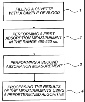
Referring now to Fig. 1, a method for hemoglobin de-
termination according to the invention will now be de-
scribed. First, a disposable, capillary cuvette is filled
with a sample of unaltered whole blood, step 1. Thus, a
sample which is to be analysed is obtained. Then, a first
absorption measurement on the sample is performed at a
wavelength in the range 490 - 520 nm, step 2. Further, a
second absorption measurement is performed on the sample,
step 3. The second absorption measurement is performed at
a wavelength in the range 650 - 1200 nm. This second ab-
sorption measurement is used to compensate for light
scattering in the sample, as will be described in further
detail below. Finally, the results of the measurements
are processed, step 4, using a predetermined algorithm
for determining the concentration of hemoglobin in the
sample.
The disposable microcuvette used according to the
present invention may be of the type disclosed in the US
patent 4 088 448 or preferably in the US patent 5 674 457.
The cuvette
may be defined as a unitary body member including at
least one cavity with an optical window (measuring zone)
wherein two, plane or curved, surfaces facing the cavity
are placed at a predetermined distance from one another
and thus define a predetermined optical path length. This
distance between the surfaces defining the measuring zone
is a critical parameter in providing the proper optical
path length for the hemoglobin measurement. The optical
path length should be less than 1 mm in order to ensure
that the intensity of light transmitted through a sample
in the cuvette is sufficient to enable determination of
hemoglobin in the sample. In a preferred embodiment, this
distance is less than 0.2 mm, and more preferably between
0.05 and 0.2 mm. The distance between the inner surfaces
of the rest of the cavity is preferably in the order of
0.1-2 mm which is effective to permit the sample to enter
CA 02468513 2004-05-25
WO 03/056327 PCT/SE02/02412
9
the cavity by capillary force through the cavity inlet,
which is communicating with the exterior of the body mem-
ber. Furthermore, the cavity has a predetermined fixed
volume of less than about 25 Al. No active additives,
such as reagents or hemolysing agents, are necessary for
the determination according to the inventive method.
The cuvettes according to the present invention may
be formed by any suitable material, which allows the for-
mation of the necessary tight tolerance levels. Prefera-
bly the cuvette is manufactured by injection moulding of
a transparent polymeric material.
In order to overcome problems related to the capil-
lary filling of the cuvette it may be necessary to pre-
treat the inner surfaces of the cuvette in order to im-
part a hydrophilic character to these surfaces. This may
be achieved by coating the surfaces with a suitable de-
tergent, such as Brij 35. Another possibility is to se-
lect a hydrophilic material for the manufacturing of the
cuvette. A critical feature of the inventive method is
that the absorption determination should be carried out
at a wavelength in a range of 490 - 520 nm, more prefera-
bly in the range 500-510 nm, and most preferably at 506
nm. The secondary compensatory absorption measurement is
preferably performed at a wavelength in the range 650 -
1200 nm, more preferably in the range 850 - 910 nm, and
most preferably in the range 860 - 900 nm.
The absorption measurements are performed directly
on the whole blood in the sample, i.e. the blood is unal-
tered (undiluted and unhemolyzed).
In the wavelength range of 490 - 520 nm, the absorp-
tions of the five different forms of hemoglobin, namely
oxy-, deoxy-, carboxy-, met- and sulfhemoglobin, are
similar and significant. Thus, the absorption in this
wavelength range will depend only slightly on the distri-
bution between the different forms of hemoglobin in the
blood. Especially, at 506 nm, the difference between the
absorbances of oxy- and deoxyhemoglobin is close to zero.
CA 02468513 2004-05-25
WO 03/056327 PCT/SE02/02412
Since these forms of hemoglobin are predominant in normal
blood, the absorption of oxy- and deoxyhemoglobin could
advantageously be used for determining an absorption co-
efficient for relating a measured absorption to the con-
5 centration of hemoglobin at 506 nm. Accordingly, some as-
sumptions are made regarding the contents of different
forms of hemoglobin in the blood sample. Thus, the hemo-
globin determination will not be as accurate or the proc-
essing of the measurement results will have to be modi-
10 fied, if a measurement is made on a blood sample having a
very differing distribution of the forms of hemoglobin.
Further, the measurements will only determine the total
concentration of hemoglobin and not the concentrations of
the specific forms of hemoglobin.
A second absorption measurement is performed at a
wavelength, where the absorption of light in blood is
substantially smaller. Such an absorption measurement
could suitably be performed at a wavelength in the range
650 - 1200 nm. The differences between the absorption
measurements is then considered to be due to absorption
of hemoglobin.
However, the scattering of light varies with the
concentration of hemoglobin in the sample, but the scat-
tering of light is not only dependent on the concentra-
tion of hemoglobin. The scattering of light is due to
light interaction with particles in the blood, such as
red blood cells, white blood cells, platelets, lipids and
other macro molecules. According to the invention, it has
unexpectedly been found that the effect of scattering may
be related to the measured result in the second
absorption measurement, as will be explained with
reference to the schematic diagram in Fig. 2. In Fig. 2,
the solid line schematically illustrates measured
absorption in a first sample having a high concentration
of hemoglobin. The absorption includes both true
absorption and light scattered so that it does not reach
a detector. The dashed line in Fig. 2 schematically
CA 02468513 2004-05-25
WO 03/056327 PCT/SE02/02412
11
illustrates measured absorption in a second sample having
a lower concentration of hemoglobin. It should be noted
that the schematic diagram in Fig. 2 only emphasizes the
main features of absorption of samples of whole blood,
and does not illustrate absorption of real samples. As
can be seen in Fig. 2, the difference in absorption for
the first sample between a first wavelength at 506 nm and
a second wavelength at 880 nm is substantially equal to
the corresponding difference in absorption for the second
sample. Therefore, if the concentration of hemoglobin is
determined directly from the differences in the measured
absorptions, an erroneous result would be returned, at
least for one of the samples. Thus, a compensation for
the light scattering will be needed, and according to the
invention it has been found that a compensation for the
level of absorption will account for the scattering and
enables simple hemoglobin determination.
It has empirically been determined that when using a
compensation that is proportional to the level of absorp-
tion, a correct value of the concentration of hemoglobin
may be obtained.
According to the above, the results of the absorp-
tion=measurements should be processed for determining the
concentration of hemoglobin in the sample. This process-
ing may be performed by a predetermined algorithm. This
algorithm calculates the concentration of hemoglobin ac-
cording to the above-described scheme.
The compensation for light scattering is preferably
dependent on the result of the second absorption measure-
ment. A compensation function could be determined by per-
forming absorption measurements on a set of blood samples
having known concentrations of hemoglobin. These absorp-
tion measurements are performed in a measurement arrange-
ment which is to be used. Then, the needed compensation
for light scattering in order to obtain correct results
are compared with the values of the second absorption
measurement. In this way, a function of the second ab-
CA 02468513 2004-05-25
WO 03/056327 PCT/SE02/02412
12
sorption measurement may be found that would give a com-
pensation so that the determined concentrations of hemo-
globin would fall within an acceptable margin of error.
In a simplified model, the compensation is linearly
dependent on the result of the second absorption measure-
ment at least in a range of the result of the second ab-
sorption measurement. This range of the result of the
second absorption measurement may span typical values of
the second absorption measurement that are obtained with
the specific measurement arrangement.
The processing may determine the concentration of
hemoglobin in the sample by computing the following for-
mula:
[Tot Hb] _ (Abs, - Abs2) = k + F(Abs2 )
wherein [Tot Hb] is the total concentration of hemoglobin
in the sample, Abs1 is the measured absorbance of the
first absorption measurement, Abs2 is the measured ab-
sorbance of the second absorption measurement, k is a
calibration coefficient, which depends on the measurement
arrangement, and F(Abs2) is a function that depends on
the measured absorbance of the second absorption measure-
ment. The calibration coefficient k may be specific for
each instrument used for hemoglobin determination. The
compensating function F(Abs2) may have a constant part,
which also is a calibration for each instrument, and a
variable part, which depends on the result of the second
absorption measurement and is obtained as described
above. In this case, the variable part may be zero for a
result of the second absorption measurement that is in
the centre of the range of the results of the second ab-
sorption measurement.
Referring now to Fig. 3, a system implementing the
above-described method will be described. The system com-
prises means 10 for emitting light at a first wavelength
in a first range of 490 - 520 nm and at a second wave-
CA 02468513 2004-05-25
WO 03/056327 PCT/SE02/02412
13
length in a second range of 650 - 1200 nm. This means 10
for emitting light may be implemented by a combination of
a light source emitting at several wavelengths or in
broad wavelength ranges together with filters. Thus, the
light source is arranged to emit light both at the first
wavelength and at the second wavelength. Using the filter
the wavelength emitted could selectively be controlled to
be within one of these ranges. Alternatively, a first and
a second light source may be used for emitting the first
and the second wavelengths, respectively. Light emitting
diodes may be used as light sources. Then, by switching
the two light sources on and off, the means 10 for emit-
ting light may be selectively controlled to emit light in
the first or in the second wavelength.
Preferably, the first wavelength emitted by the
means 10 for emitting light is in the range 500 - 510 nm,
more preferably at 506 nm. Further, the second wavelength
emitted by the means 10 for emitting light is preferably
in the range 850 - 910 nm, and more preferably in the
range 860 - 900 nm.
The system further comprises a cuvette holder 12 ar-
ranged to receive a capillary cuvette, which has an opti-
cal path length of less than 1 mm and holds a sample of
unaltered whole blood. When a cuvette is placed in the
holder 12, the optical window will be correctly posi-
tioned so that it will be irradiated with the light from
the light source. Preferably, the cuvette holder is ar-
ranged to receive a cuvette, which has an optical path
length of less than 0.2 mm, and more preferably in the
range 0.05 - 0.2 mm.
The light transmitted through the sample will be de-
tected by a detector 14 so that a first absorption meas-
urement may be obtained for light in the first range and
a second absorption measurement may be obtained for light
in the second range.
The system further comprises a processing unit 16
for processing results of the first and second absorption
CA 02468513 2004-05-25
WO 03/056327 PCT/SE02/02412
14
measurements to determine the concentration of hemoglobin
in the sample according to the algorithm described above.
The system may suitably be implemented in a photome-
ter comprising the means 10 for emitting light, the cu-
vette holder 12, and the detector 14. Photometers suit-
able for performing these measurements may be obtained by
using photometers modified with suitable wave length fil-
ters and light emitting diodes. According to a preferred
embodiment of the invention a photometer measures the ab-
sorbance at the two wavelengths and a built-in micro
processor calculates, according to a programmed algo-
rithm, the total concentration of hemoglobin in blood.
Thus, no special absorption or interference filter which
provide correction for variations in the detector sensi-
tivity and in the effective optical path length as dis-
closed in WO 01/53806 are necessary.
In the above case, the processing unit 16 is embed-
ded in the photometer. However, the processing unit 16
may also be connected to the photometer, and thus be im-
plemented outside the photometer. For example, a computer
connected to the photometer may be used.
The detector 14 may be arranged to detect essen-
tially only directly transmitted light, since the scat-
tered light need not be detected. This implies that the
detector 14 detects light which is essentially within the
diameter of the light beam irradiated on the sample and
directly transmitted through the sample. Of course, some
light may be scattered, while still being within this di-
ameter. According to a preferred embodiment, the diameter
of a detecting area of the detector 14 may typically be
approximately 2 mm. The detector 14 is preferably ar-
ranged closer than 10 mm to the sample holder. This im-
plies that light which has been scattered to small angles
is detected.
The following non limiting example illustrates the
inventive method.
CA 02468513 2010-07-26
28569-52
It was found that the time period for analysing the
blood was about 30 seconds shorter for the inventive
method in a comparison with the method for determination
of hemoglobin in the known, currently used HemoCue micro-
5 cuvettes. This permits a clear reduction of the total
time of the hemoglobin determination which may be advan-
tageous in busy hospitals and in other situations where
may determinations are made. Another advantage is that
there is no need for a cuvette containing active reagents
10 or hemolysing agents. Thus, storage of the cuvettes is
insensitive to temperature and humidity in the storage
environment, which makes handling of the cuvettes before
their use much simpler.
A preliminary evaluation of the new method in com-
15 parison with the HemoCue method is disclosed in figure
4A. The evaluation was made under laboratory conditions.
As can be seen the agreement between the methods is very
good.
The spectrophotometric absorption measurements were
made at about 570 nm for the known method and about 505
nm for the new method. For both methods compensatory
measurements were made at about 880 nm.
Further, a second evaluation of the new method in
comparison with the standard ICSH method is disclosed in
figure 4B. As can be seen the agreement between these
methods is also very good.