Note: Descriptions are shown in the official language in which they were submitted.
CA 02468635 2004-07-08
WO 03/077779 PCT/US02/38092
APPARATUS FOR CONVERTING A CLAMP INTO
AN ELECTROPHYSIOLOGY DEVICE
BACKGROUND OF THE INVENTIONS
1. Field of Inventions
The present inventions relate generally to structures for positioning
diagnostic and therapeutic elements within the body and, more particularly, to
devices which are particularly well suited for the treatment of cardiac
conditions.
2. Description of the Related Art
There are many instances where diagnostic and therapeutic elements
must be inserted into the body. One instance involves the treatment of cardiac
conditions such as atrial fibrillation and atrial flutter which lead to an
unpleasant,
irregular heart beat, called .arrhythmia.
Normal sinus rhythm of the heart begins with the sinoatrial node (or "SA
node") generating an electrical impulse. The impulse usually propagates
uniformly across the right and left atria and the atrial septum to the
atrioventricular node (or "AV node"). This propagation causes the atria to
contract in an organized way to transport blood from the atria to the
ventricles,
and to provide timed stimulation of the ventricles. The AV node regulates the
propagation delay to the atrioventricular bundle (or "HIS" bundle). This
coordination of the electrical activity of the heart causes atrial systole
during
ventricular diastole. This, in turn, improves the mechanical function of the
heart.
Atrial fibrillation occurs when anatomical obstacles in the heart disrupt the
normally uniform propagation of electrical impulses in the atria. These
anatomical obstacles (called "conduction blocks") can cause the electrical
impulse to degenerate into several circular wavelets that circulate about the
obstacles. These wavelets, called "reentry circuits," disrupt the normally
uniform
activation of the left and right atria. Because of a loss of atrioventricular
synchrony, the people who suffer from atrial fibrillation and flutter also
sufFer the
consequences of impaired hemodynamics and loss of cardiac efficiency. They
are also at greater risk of stroke and other thromboembolic complications
because of loss of effective contraction and atrial stasis.
1
CA 02468635 2004-07-08
WO 03/077779 PCT/US02/38092
One surgical method of treating atrial fibrillation by interrupting pathways
for reentry circuits is the so-called "maze procedure" which relies on a
prescribed pattern of incisions to anatomically create a convoluted path, or
maze, for electrical propagation within the left and right atria. The
incisions direct
the electrical impulse from the SA node along a specified route through all
regions of both atria, causing uniform contraction required for normal atrial
transport function. The incisions finally direct the impulse to the AV node to
activate the ventricles, restoring normal atrioventricular synchrony. The
incisions
are also carefully placed to interrupt the conduction routes of the most
common
reentry circuits. The maze procedure has been found very effective in curing
atrial fibrillation. However, the maze procedure is technically difficult to
do. It also
requires open heart surgery and is very expensive. Thus, despite its
considerable clinical success, only a few maze procedures are done each year.
Maze-like procedures have also been developed utilizing catheters
and/or surgical probes (collectively "probes") that form lesions to create a
maze
for electrical conduction in a predetermined path. Typically, the lesions are
formed by ablating tissue with one or more electrodes. Electromagnetic radio
frequency ("RF") energy applied by the electrode heats, and eventually kills
(i.e.
"ablates"), the tissue to form a lesion. During the ablation of soft tissue
(i.e.
tissue other than blood, bone and connective tissue), tissue coagulation
occurs
and it is the coagulation that kills the tissue. Thus, references to the
ablation of
soft tissue are necessarily references to soft tissue coagulation. "Tissue
coagulation" is the process of cross-linking proteins in tissue to cause the
tissue
to jell. In soft tissue, it is the fluid within the tissue cell membranes that
jells to kill
the cells, thereby killing the tissue.
Catheters used to create lesions typically include a relatively long and
relatively flexible body that has one or more electrodes on its distal
portion. The
portion of the catheter body that is inserted into the patient is typically
from 58.4
to 139.7 cm in length and there may be another 20.3 to 38.1 cm, including a
handle, outside the patient. The proximal end of the catheter body is
connected
to the handle which includes steering controls. The length and flexibility of
the
catheter body allow the catheter to be inserted into a main vein or artery
(typically the femoral artery), directed into the interior of the heart, and
then
2
CA 02468635 2004-07-08
WO 03/077779 PCT/US02/38092
manipulated such that the electrode contacts the tissue that is to be ablated.
Fluoroscopic imaging is used to provide the physician with a visual indication
of
the location of the catheter. Exemplary catheters are disclosed in U.S. Patent
No. 5,582,609.
Surgical probes used to create lesions often include a handle, a relatively
short shaft that is from about 10.2 to 45.7 cm in length and either rigid or
relatively stiff, and a distal section that is from 2.54 to 25.4 cm in length
and
either malleable or somewhat flexible. One or more electrodes are carried by
the
distal section. Surgical probes are used in epicardial and endocardial
procedures, including open heart procedures and minimally invasive procedures
where access to the heart is obtained via a thoracotomy, thoracostomy or
median sternotomy. Exemplary surgical probes are disclosed in U.S. Patent No.
6,142,994.
Clamps, which have a pair of opposable rigid clamp members that may
be used to hold a bodily structure or a portion thereof, are used in many
types
surgical procedures. Lesion creating electrodes have also been permanently
secured to certain types of clamps. Examples of clamps which carry lesion
creating electrodes are disclosed in U.S. Patent No. 6,142,994. Such clamps
are particularly useful when the physician intends to position electrodes on
opposite sides of a body structure.
As used herein, the term "clamp" includes, but is not limited to, clamps,
clips, forceps, hemostats, and any other surgical device that includes a pair
of
opposable clamp members that hold tissue, at least one of which is movable
relative to the other. In some instances, the rigid clamp members are
connected
to a scissors-like arrangement including a pair of handle supporting arms that
are pivotably connected to one another. The clamp members are secured to
one end of the arms and the handles are secured to the other end. The clamp
members come together as the handles move toward one another. Certain
clamps that are particularly useful in minimally invasive procedures also
include
a pair of handles and a pair of clamp members. Here, however, the clamp
members and handles are not mounted on the opposite ends of the same arm.
Instead, the handles are carried by one end of an elongate housing and the
clamp members are carried by the other. A suitable mechanical linkage located
3
CA 02468635 2004-07-08
WO 03/077779 PCT/US02/38092
within the housing causes the clamp members to move relative to one another
in response to movement of the handles.
The rigid clamp members in conventional clamps may be linear or have a
predefined curvature that is optimized for a particular surgical procedure or
portion thereof. It is, therefore, necessary to have a wide variety of clamps
on
hand. In the field of electrophysiology, a wide variety of clamps that have
electrodes permanently secured thereto must be kept on hand.
The inventor herein has determined that it would be advantageous to
provide physicians with a wide variety of devices, including clamps (both with
and without energy transmission devices) and surgical probes that carry energy
transmission devices, in a wide variety of shapes, and to do so in a manner
that
is more cost effective than conventional apparatus.
SUMMARY OF THE INVENTIONS
An apparatus for use with a clamp in accordance with one embodiment
of a present invention includes a base member configured to be secured to
the clamp and at least one energy transmission device carried by the base
member. Such an apparatus provides a number of advantages. For example,
such an apparatus may be used to quickly convert a conventional clamp into
an electrophysiology device. In those instances where a procedure requires a
number of different clamps, the apparatus"can be moved from clamp to
clamp, thereby eliminating the costs associated with providing a variety of
different clamps with energy transmission devices permanently secured
thereto.
An apparatus for use with a clamp and a probe that carries at least one
energy transmission device in accordance with one embodiment of a present
invention includes a base member configured to be secured to the clamp and
an engagement device associated with the base member and configured to
engage the probe. Such an apparatus provides a number of advantages. For
example, such an apparatus may be used to quickly convert a conventional
clamp into an electrophysiology device and to achieve better (or merely
different) tissue/energy transmission device contact than could be achieved
with the probe itself. Additionally, in those instances where a procedure
4
CA 02468635 2004-07-08
WO 03/077779 PCT/US02/38092
requires a number of different clamps, the apparatus can be moved from
clamp to clamp, thereby eliminating the costs associated with providing a
variety of different clamps with energy transmission devices permanently
secured thereto.
A clamp in accordance with one embodiment of a present invention
includes first and second clamp members, at least one of which is malleable,
and a movement apparatus that moves at least one of the first and second
clamp members relative to the other. Such a clamp provides a number of
advantages. For example, the malleable clamp member allows physicians to
readily reconfigure the clamp, thereby reducing the number of clamps that
must be provide for a particular surgical procedure.
A surgical system in accordance with one embodiment of a present
invention includes a clamp with first and second clamp members and a device
that removably mounts at least one electrode on at least one of the first and
second clamp members. Such a clamp provides a number of advantages. For
example, the system may be used both as a conventional clamp and an
electrophysiology device.
The above described and many other features and attendant advantages
of the present inventions will become apparent as the inventions become better
understood by reference to the following detailed description when considered
in
conjunction with the accompanying drawings.
BRIEF DESCRIPTION OF THE DRAWINGS
Detailed description of preferred embodiments of the inventions will be
made with reference to the accompanying drawings.
Figure 1 is a plan view of a conventional clamp.
Figure 2 is a side view of the clamp illustrated in Figure 1.
Figure 3 is an enlarged view of a portion of the clamp illustrated in Figure
1 holding a vein.
Figure 4 is plan of a pair of energy transmission assemblies in
accordance with a preferred embodiment of a present invention.
Figure 5 is plan showing the energy transmission assemblies illustrated
in Figure 4 mounted on a clamp.
5
CA 02468635 2004-07-08
WO 03/077779 PCT/US02/38092
Figure 6 is a front view of an electrosurgical unit.
Figure 7a is a section view taken along line 7a-7a in Figure 4.
Figure 7b is a section view taken along line 7b-7b in Figure 4.
Figure 8 is a section view taken along line 8-8 in Figure 7a.
Figure 9a is a section view of an energy transmission assembly in
accordance with a preferred embodiment of a present invention.
Figure 9b is a section view of an energy transmission assembly in
accordance with a preferred embodiment of a present invention.
Figure 10 is a plan view of an energy transmission assembly in
accordance with a preferred embodiment of a present invention.
Figure 11 is a section view taken along line 11-1~1 in Figure 10.
Figure 12 is a section view of an energy transmission assembly in
accordance with a preferred embodiment of a present invention.
Figure 13 is a section view of an energy transmission assembly in
accordance with a preferred embodiment of a present invention.
Figure 14 is a section view taken along line 14-14 in Figure 13.
Figure 15 is a section view of an energy transmission assembly in
accordance with a preferred embodiment of a present invention.
Figure 16a is a section view of an energy transmission assembly in
accordance with a preferred embodiment of a present invention.
Figure 16b is a section view of an energy transmission assembly in
accordance with a preferred embodiment of a present invention.
Figure 17 is a section view of a probe support device in accordance with
a preferred embodiment of a present invention.
Figure 18 is a section view taken along line 18-18 in Figure 17.
Figure 19 is a partial plan view showing a pair of the probe support
devices illustrated in Figure 17 supporting a pair of probes on a clamp.
Figure 20 is a plan view showing a pair of the probe support devices
illustrated in Figure 17 supporting a pair of probes on a clamp.
Figure 21 is a section view of a probe support device in accordance with
a preferred embodiment of a present invention.
Figure 22 is a section view taken along line 21-21 in Figure 20.
6
CA 02468635 2004-07-08
WO 03/077779 PCT/US02/38092
Figure 23 is a section view of a probe support device in accordance with
a preferred embodiment of a present invention.
Figure 24 is an end view of a probe support device in accordance with a
preferred embodiment of a present invention.
Figure 25 is a plan view of a probe support device illustrated in Figure 24.
Figure 26 is an end view of a probe support device in accordance with a
preferred embodiment of a present invention.
Figure 27 is a plan view of a clamp in accordance with a preferred
embodiment of a present invention.
Figure 28 is a plan view of a mandrel in accordance with a preferred
embodiment of a present invention.
Figure 29 is a side view of the mandrel illustrated in Figure 28.
Figures 30 and 31 are plan views of the clamp illustrated in Figure 27
being bent with the mandrel illustrated in Figure 28.
Figure 32 is a plan view showing one example of how the clamp
illustrated in Figure 27 may be bent.
Figure 33 is a plan view showing another example of how the clamp
illustrated in Figure 27 may be bent.
Figure 34 is a plan view of a clamp in accordance with a preferred
embodiment of a present invention.
DETAILED DESCRIPTION OF THE PREFERRED EMBODIMENTS
The following is a detailed description of the best presently known modes
of carrying out the inventions. This description is not to be taken in a
limiting
sense, but is made merely for the purpose of illustrating the general
principles of
the inventions.
The detailed description of the preferred embodiments is organized as
fo I lows:
I. Energy Transmission Assemblies
II. Energy Transmission Devices, Temperature Sensing and Power
Control
III. Tissue Cooling Apparatus
IV. Probe Support Devices
7
CA 02468635 2004-07-08
WO 03/077779 PCT/US02/38092
V. Clamp With Malleable Clamp Members
The section titles and overall organization of the present detailed
description are
for the purpose of convenience only and are not intended to limit the present
inventions.
This specification discloses a number of structures, mainly in the context
of cardiac ablation, because the structures are well suited for use with
myocardial tissue. Nevertheless, it should be appreciated that the structures
are
applicable for use in therapies involving other types of soft tissue. For
example,
various aspects of the present inventions have applications in procedures
concerning other regions of the body such as the prostate, liver, brain, gall
bladder, uterus androther solid organs.
I. Energy Transmission Assemblies
Energy transmission assemblies in accordance with a present invention
may be used to covert a conventional clamp into a tissue coagulation device.
The energy transmission assemblies may also be used to covert a clamp in
accordance with the inventions described in Section V below into a tissue
coagulation device.
One example of a conventional clamp that may be used in conjunction
with the present inventions is generally represented by reference numeral 10
in
Figures 1-3. The clamp 10 includes a pair of rigid arms 12 and 14 that are
pivotably connected to one another by a pin 16. The proximal ends of the arms
12 and 14 are respectively connected to a pair handle members 18 and 20,
while the distal ends are respectively connected to a pair of rigid clamp
members 22 and 24. A locking device 26 locks the clamp in the closed
orientation, and prevents the clamp members 22 and 24 from coming any closer
to one another than is illustrated in Figure 1, thereby defining a
predetermined
spacing between the clamp members. The clamp 10 also includes a pair of soft,
deformable inserts 28 and 30 that are removably carried by the clamp members
22 and 24. The inserts 28 and 30 allow clamp 10 to firmly grip a bodily
structure
32 without damaging the bodily structure. The inserts 28 and 30 include mating
structures 34 that extend through corresponding apertures 36 in the clamp
members 22 and 24 to hold the inserts in place.
8
CA 02468635 2004-07-08
WO 03/077779 PCT/US02/38092
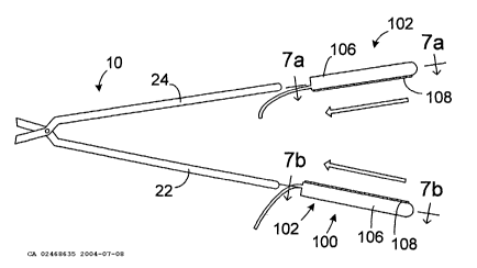
As illustrated for example in Figures 4 and 5, an apparatus 100 for
converting the clamp 10 (which has had the inserts 28 and 30 removed) into a
bi-polar tissue coagulation device includes a pair of energy transmission
assemblies 102 and 104. Each of the energy transmission assemblies includes
a base member 106 that may be removably secured to one of the clamp
members 22 and 24 and an energy transmission device 108. [The energy
transmission devices 108 are discussed in greater detail in Section II below.]
Although the configuration of the energy transmission assemblies 102 and 104
may vary from application to application to suit particular situations, the
energy
transmission assemblies in the exemplary embodiment are configured such that
they will abut one another in the same manner as the inserts 28 and 30
(Figures
1-3) when the clamp 10 is in the closed orientation illustrated in Figure 5.
Such
an arrangement will allow the energy transmission assemblies 102 and 104 to
grip a bodily structure in the same manner as the inserts 28 and 30.
The exemplary base members 106 are preferably formed from a soft,
resilient, low durometer material that is electrically insulating. Suitable
materials
include polyurethane, silicone and polyurethane/silicone blends having a
hardness of between about 20 Shore D and about 72 .Shore D. Referring to
Figures 7a, 7b and 8, each of the exemplary base members 106 includes a
longitudinally extending aperture 110 into which one of the clamp members 22
and 24 may be inserted. The apertures 110 should be sized and shaped such
that the base members 106 will be forced to stretch when the clamp members
22 and 24 are inserted. If, for example, the apertures 110 have the same cross-
sectional shape as the clamp members 22 and 24 (e.g. both are elliptical),
then
the apertures should be slightly smaller in their cross-sectional dimensions
than
the corresponding clamp members. The stretching of the apertures 110 creates
a tight interference fit between the base members 106 and clamp members 22
and 24. Additionally, although the apertures 110 have a semi-circular cross-
section in the exemplary embodiment, the apertures may have a round,
rectangular, square or elliptical cross-section, or define any other cross-
sectional
shape, depending on the particular application.
The exemplary base members 106 also include slots 112 (Figure 8) that
secure the energy transmission devices 108 in place. The configuration of a
slot
9
CA 02468635 2004-07-08
WO 03/077779 PCT/US02/38092
112 will, of course, depend on the configuration of the energy transmission
device 108 that it is holding. The illustrated energy transmission device 108
is
generally cylindrical in shape and the slot 112 has a corresponding arcuate
cross-sectional shape. The arc is preferably greater than 180 degrees so that
the base member 106 will deflect when the energy transmission device 108 is
inserted into the slot 112 and then snap back to hold the energy transmission
device in place. Adhesive may also be used to secure the energy transmission
devices 108, especially in those instances where the arc is less than 180
degrees.
Another exemplary apparatus for converting the clamp 10 (which has had
the inserts 28 and 30 removed) into a bi-polar tissue coagulation device is
illustrated in Figures 9a and 9b. The apparatus includes a pair of energy
transmission assemblies 114 and 116 which are substantially similar to the
energy transmission assemblies 102 and 104 and similar elements are
represented by similar reference numerals. Each of the energy transmission
assemblies 114 and 116 includes a base member 106' that may be removably
secured to one of the clamp members 22 and 24 and an energy transmission
device 108. Here, however, the base members 106' are secured to the clamp
members 22 and 24 with mating structures 118 that mechanically engage the
clamp members.
° The exemplary mating structures 118, which are preferably integral
with
the base members 106' and formed from the same resilient material, include a
relatively narrow portion 120 and a relatively wide portion 122. The
relatively
narrow portions are approximately the same size as the clamp member
apertures 36 and the relatively wide portions 122 are slightly larger than the
clamp member apertures. A removable connection is made by urging the mating
structures 118 into one end of the apertures 36, thereby deforming the
relatively
wide portions 122, and then urging the base members 106' against the clamp
members 22 and 24 until the relatively wide portions exit through the other
end
of the apertures and reassume their original shape.
The exemplary mating structures 118 may also be reconfigured by
eliminating the relatively wide portions 122 and enlarging the relatively
narrow
portions 120 such that the relatively narrow portions will create an
interference fit
CA 02468635 2004-07-08
WO 03/077779 PCT/US02/38092
within the clamp member apertures 36. Alternatively, as discussed below with
reference to Figure 12, longitudinally extending mating structures, which also
create an interference fit, may be employed when longitudinally extending
slots
are formed in the clamp members. Another alternative is to provide the clamp
members with one or more small mating structures that extend outwardly
therefrom. The clamp member mating structures will be received within
apertures or slots formed in the base member.
Turning to Figures 10 and 11, an energy transmission assembly 124 may
be used to convert the clamp 10 (which has had the inserts 28 and 30 removed)
into a uni-polar tissue coagulation device. The energy transmission assembly
124 includes a base member 126, which may be removably secured to both of
the clamp members 22 and 24, and a plurality of spaced energy transmission
devices 108. Although the configuration of the energy transmission assembly
124 may vary from application to application to suit particular situations,
the
energy transmission assembly in the exemplary embodiment is configured such
that it will abut each of the clamp members when the clamp 10 is in the closed
orientation illustrated in Figure 10.
The exemplary base member 126 is preferably formed from a soft,
resilient, low durometer material that is electrically insulating. Suitable
materials
include polyurethane, silicone and polyurethanelsilicone blends having a
hardness of between about 20 Shore D and about 72 Shore D. A slot 128 '
secures the energy transmission devices 108 in place. Although the
configuration of the slot 128 will depend on the configuration of the energy
transmission devices 108, the exemplary slot has an arcuate cross-sectional
shape that conforms to the shape of the exemplary cylindrical energy
transmission devices. The arc is preferably greater than 180 degrees so that
the
base member 126 will deflect when the energy transmission devices 108 are
inserted into the slot 128 and then snap back to hold the energy transmission
devices in place. Adhesive may also be used to secure the energy transmission
devices 108 in place, especially in those instances where the arc is less than
180 degrees.
The base member 126 is removably secured to the clamp members 22
and 24 with two sets of the mating structures 118 that are described above
with
11
CA 02468635 2004-07-08
WO 03/077779 PCT/US02/38092
reference to Figures 9a and 9b (with or without the relatively wide portions
122).
Alternatively, and as illustrated for example in Figure 12, in those instances
where the clamp members 22' and 24' include longitudinally extending slots 38
instead of the apertures 36, the energy transmission assembly 124 may be
provided with longitudinally extending mating structures 130 that extend
outwardly from the base member 126'. The longitudinally extending mating
structures 130, which are preferably integral with the base member 126' and
formed from the same resilient material, are sized and shaped to create an
interFerence fit with the slots 38. Still another alternative is to provide
the clamp
members with one or more small mating structures that are received within
apertures or slots formed in the base member.
Another energy transmission assembly that may be used to convert the
clamp 10 into a uni-polar tissue coagulation device is generally represented
by
reference numeral 132 in Figures 13 and 14. The energy transmission assembly
132 includes a base member 134 that is preferably formed from a soft,
resilient,
low durometer material and a plurality of energy transmission devices 108. The
material which forms the base member 134 should also be electrically
insulating. Suitable materials include polyurethane, silicone and
polyurethane/silicone blends having a hardness of between about 20 Shore D
and about 72 Shore D. A slot 128, which secures the energy transmission
devices 108 in place in the manner described above with reference to Figures
10 and 11, is also provided.
The exemplary base member 134 includes a longitudinally extending
aperture 136 into which both of the clamp members 22 and 24 may be inserted.
The aperture 136 should be sized and shaped such that the base member 134
will be forced to stretch when the clamp members 22 and 24 are inserted with
the clamp 10 in a closed orientation. The stretching creates a tight
interference
fit between the base member 134 and the clamp members 22 and 24.
Additionally, although the apertures 110 have an elliptical cross-section in
the
exemplary embodiment, the apertures may have a round, rectangular, square or
semi-circular cross-section, or define any other cross-sectional shape,
depending on the particular application.
12
CA 02468635 2004-07-08
WO 03/077779 PCT/US02/38092
The length of the base members in the exemplary energy transmission
assemblies will vary according to the intended application. In the area of
cardiovascular treatments, it is anticipated that suitable lengths will range
from,
but are not limited to, about 2 cm to about 10 cm.
The exemplary energy transmission assemblies described above may
also be modified in a variety of ways. For example, the energy transmission
assembly illustrated in Figures 10 and 11 may be converted into a bi-polar
device by simply adding a second slot 128 that is preferably spaced apart from
and parallel to the existing slot. The second slot 128 could, for example,
include
a single return energy transmission device 108 or a plurality of spaced return
energy transmission devices. Additionally, as illustrated for example in
Figures
7a and 13, the base members and energy transmission devices in the illustrated
embodiments are configured such that the energy transmission devices are
generally linear and parallel to the longitudinal axis of the base members
(when
the assemblies are in a relaxed state and not being urged against a body
structure). The base members and/or energy transmission devices may be
reconfigured such that the energy transmission devices, or a portion thereof,
are
curved and/or non-parallel to the longitudinal axis of the base members when
in
the relaxed state.
II. Energy Transmission Devices, Temperature Sensing and Power
Control
In the exemplary embodiments illustrated in Figures 4-16b, the energy
transmission devices are electrodes. More specifically, the energy
transmission
devices are preferably in the form of wound, spiral coil electrodes that are
relatively flexible. The coils are made of electrically conducting material,
like
copper alloy, platinum, or stainless steel, or compositions such as drawn-
filled
tubing (e.g. a copper core with a platinum jacket). The electrically
conducting
material of the coils can be further coated with platinum-iridium or gold to
improve its conduction properties and biocompatibility. A preferred coil
electrode
configuration is disclosed in U.S. Patent No. 5,797,905. Although the diameter
of the electrodes will very from application to application, the diameter
preferably
ranges from about 1 mm to about 3 mm for cardiovascular applications.
13
CA 02468635 2004-07-08
WO 03/077779 PCT/US02/38092
As an alternative, the electrodes may be in the form of solid rings of
conductive material, like platinum, or can comprise a conductive material,
like
platinum-iridium or gold, coated upon the base member using conventional
coating techniques or an ion beam assisted deposition (IBAD) process. For
better adherence, an undercoating of nickel or titanium can be applied. The
electrodes can also be in the form of helical ribbons. The electrodes can also
be
formed with a conductive ink compound that is pad printed onto a non-
conductive tubular body. A preferred conductive ink compound is a silver-based
flexible adhesive conductive ink (polyurethane binder), however other metal-
based adhesive conductive inks such as platinum-based, gold-based, copper-
based, etc., may also be used to form electrodes. Such inks are more flexible
than epoxy-based inks.
When a single flexible coil electrode is carried by a base member (see,
for example, Figure 7a), the length will depend on the length of the base
member and the intended application. When a plurality of spaced flexible coil
electrodes are carried by a base member (see, for example, Figure 10), the
electrodes will preferably be about 10 mm to about 40 mm in length.
Preferably,
the electrodes will be 25 mm in length with 1 mm to 2 mm spacing, which will
result in the creation of continuous lesion patterns in tissue when
coagulation
energy is applied simultaneously to adjacent electrodes. For rigid electrodes,
the
length of the each electrode can vary from about 3 mm to about 10 mm. Using
multiple rigid electrodes longer than about 10 mm each adversely effects the
overall flexibility of the device, while electrodes having lengths of less
than about
2 mm do not consistently form the desired continuous lesion patterns.
It should also be noted that other energy transmission devices, such as
laser arrays, ultrasonic transducers, microwave electrodes, and ohmically
heated hot wires, may be substituted for the electrodes. Another type of
energy
transmission device that may be ~ substituted for the electrodes is
cryotemperature elements. Here, the energy transmission is the removal of heat
from the tissue. Still another type of energy transmission device that may be
substituted for the electrodes is needle projections for chemical ablation
(which
are preferably about 1 to 2 mm in length). Here, the energy transmission is
the
transmission of chemical energy.
14
CA 02468635 2004-07-08
WO 03/077779 PCT/US02/38092
Referring for example to Figures 5-8, each energy transmission device
108 is individually coupled to a wire 137 (Figure 8) that conducts coagulating
energy. The wires 137 pass in conventional fashion through cables 138 to an
associated connector (140 or 142). The connectors 140 and 142 are configured
to plug into an electrosurgical unit ("ESU") 144 that supplies and controls
power,
such RF power. A suitable ESU is the Model 4810 ESU sold by EP
Technologies, Inc. of San Jose, California. The exemplary ESU 144 illustrated
in
Figure 6 includes a plurality of displays and buttons that are used to control
the
level of power supplied to the energy transmission devices) 108 and the
temperature at the energy transmission device(s). When a plurality of spaced
energy transmission devices 108 are employed, the ESU 144 may also be used
to selectively control which of the energy transmission devices receive power.
The amount of power required to coagulate tissue ranges from 5 to 150 w.
The exemplary ESU 144 illustrated in Figure 6 is operable in a bi-polar
mode, where tissue coagulation energy emitted by the energy transmission
devices) 108 on one energy transmission assembly is returned through the
energy transmission devices) on another energy transmission assembly, and a
uni-polar mode, where the tissue coagulation energy emitted by the energy
transmission devices) on an energy transmission assembly is returned through
one or more indifferent electrodes (not shown) that are externally attached to
the
skin of'the patient with a patch or one or more electrodes (not shown) that
are
positioned in the blood pool. To that end, the exemplary ESU 144 is provided
with a power output connector 141 and a pair of return connectors 143. In a
preferred implementation, the ESU output and return connectors 141 and 143
have different shapes to avoid confusion and the connectors 140 and 142 have
corresponding shapes. As such, in the exemplary bi-polar arrangement
illustrated in Figure 5, the connector 140 associated with energy transmission
assembly 102 has a shape corresponding to the ESU output connector 141 and
the connector 142 associated with energy transmission assembly 104 has a
shape corresponding to the ESU return connector 143.
The connector (not shown) associated with the energy transmission
assembly 124 illustrated in Figure 10, which is intended to be operated in the
uni-polar mode, would have a shape corresponding to the ESU output
CA 02468635 2004-07-08
WO 03/077779 PCT/US02/38092
connector 141. In those instances where it is desirable to clamp the
indifferent
electrode within the patient, as opposed to positioning the indifferent
electrode
on the patient's skin, a second energy transmission assembly may be provided
with a connector having a shape corresponding to the ESU return connector
143. Additionally, in those instances where the energy transmission assembly
124 has been modified to includes space electrodes (or spaced groups of
longitudinally spaced electrodes) that operated in bi-polar fashion, the
assembly
would be provided with a pair of connectors. One would have a shape
corresponding to the ESU output connector 141 and the other would have a
shape corresponding to the ESU return connector 143.
With respect to power and temperature control, one or more temperature
sensors 146, such as thermocouples or thermistors, may be located on, under,
abutting the longitudinal end edges of, or in between, the energy transmission
devices 108. A reference thermocouple (not shown) may also be provided. For
temperature control purposes, signals from the temperature sensors 146 are
transmitted to the ESU 144 by way of wires 148 (Figure 8) that are connected
to
the connector 140 and, in some instances, the connector 142. The wires 137
and 148 (which are not shown in all of the Figures for clarity purposes) run
through wire apertures 150 and small holes 152, which are formed in the base
members 106, 126, 126', 134 and 134'. Suitable temperature sensors and
power control schemes that are based on a sensed temperature are disclosed in'
U.S. Patent Nos. 5,456,682, 5,582,609 and 5,755,715.
The actual number of temperature sensors 146 may be varied in order to
suit particular applications. In the bi-polar arrangement illustrated in
Figures 7a
and 7b, for example, both of the energy transmission assemblies 102 and 104
include a single energy transmission device 108 and the energy transmission
assembly 102 includes a plurality of spaced temperature sensors 146. Here, the
level of power supplied to the energy transmission device 108 on the energy
transmission assembly 102 would be controlled based on the highest
temperature measured by the temperature sensors 146. Alternatively, the
energy transmission assembly 104 (which is being used as the return) may also
provided with a plurality of spaced temperature sensors 146. Here, the level
of
power supplied to the energy transmission device 108 on the energy
16
CA 02468635 2004-07-08
WO 03/077779 PCT/US02/38092
transmission assembly 102 would be controlled based on the highest
temperature measured by any of the temperature sensors 146, whether on the
transmitting assembly 102 or the return assembly 104.
In those instances where a plurality of spaced energy transmission
devices 108 are provided, such as in the uni-polar arrangement illustrated in
Figure 13, a temperature sensor 146 may be associated with each of the energy
transmission devices. Here, power to the energy transmission devices 108 may
be individually controlled based on the temperature measured by the associated
temperature sensor 146.
Another exemplary bi-polar arrangement, which is illustrated in Figures
16a and 16b,~is substantially similar to the arrangement illustrated in
Figures 7a
and 7b and similar reference numerals are used to represent similar elements.
Here, however, the energy transmission assembly 102' includes a plurality of
spaced energy transmission device 108, each having a temperature sensor 146
associated therewith, and the energy transmission assembly 104' includes a
single energy transmission device 108 and a plurality of temperature sensors
146. The temperature sensors 146 are preferably positioned such that, when in
use, the temperature sensors on the energy transmission assembly 102' will be
aligned with the temperature sensors on the energy transmission assembly
104'. Such an arrangement allows power to the energy transmission devices
108 on the assembly 102' to be individually controlled based on the highest of
two temperatures, i.e. the temperature measured by the temperature sensor 146
associated with the particular energy transmission device and the temperature
measured by the temperature sensor directly across from the particular energy
transmission device.
III. Tissue Cooling Apparatus
Energy transmission devices in accordance with the present inventions
may also include apparatus that cools the tissue during tissue coagulation
procedures. Examples of suitable cooling apparatus are illustrated in Figures
13-
15. Such tissue cooling apparatus may also be used in conjunction with the
exemplary devices illustrated in Figures 4, 5, 7a-12, 16a and 16b. The tissue
cooling apparatus disclosed herein employ conductive fluid to cool tissue
during
coagulation procedures. More specifically, and as described below and in U.S.
17
CA 02468635 2004-07-08
WO 03/077779 PCT/US02/38092
application Serial No. 09/761,981, heat from the tissue being coagulated is
transferred to ionic fluid to cool the tissue while energy is transferred from
an
electrode or other energy transmission device to the tissue through the fluid
by
way of ionic transport. The conductive fluid may be pumped through the tissue
cooling apparatus (Figures 13 and 14) or the tissue cooling apparatus may be
saturated with the fluid prior to use (Figure 15). In either case, cooling
tissue
during a coagulation procedure facilitates the formation of lesions that are
wider
and deeper than those that could be realized with an otherwise identical
device
which lacks tissue cooling apparatus.
Referring first to Figures 13 and 14, an exemplary tissue cooling
apparatus 154 includes a nanoporous outer casing 156 through which ionic fluid
(represented by arrows F) is transferred. The ionic fluid preferably flows
from
one longitudinal end of the tissue cooling apparatus 154 to the other. The
outer
casing 156 is secured to the base member 134 over the energy transmission
devices 108 such that a fluid transmission space 158 is defined therebetween.
More specifically, the proximal and distal ends of the outer casing 156 are
secured to the base member 134 with anchoring devices (not shown) such as
lengths of heat shrink tubing, Nitinol tubing or other mechanical devices that
form an interference fit between the casing and the base member. Adhesive
bonding is another method of securing the outer casing 156 to the base member
134. The fluid transmission space will typically be about 0.5 mm to about 2.0
mm high and slightly wider than the associated energy transmission devices)
108.
The ionic fluid is supplied under pressure from a fluid source (not shown)
by way of a supply line 160 and is returned to the source by way of a return
line
162. The supply line 160 is connected to a fluid lumen 164 that runs from the
proximal end of the base member 134 to the distal region of the outer casing
156. The fluid lumen 164 is connected to the fluid transmission space 158 by
an
aperture 166.
The electrically conductive ionic fluid preferably possesses a low
resistivity to decrease ohmic loses, and thus ohmic heating effects, within
the
outer casing 156. The composition of the electrically conductive fluid can
vary. In
the illustrated embodiment, the fluid is a hypertonic saline solution, having
a
18
CA 02468635 2004-07-08
WO 03/077779 PCT/US02/38092
sodium chloride concentration at or near saturation, which is about 5% to
about
25% weight by volume. Hypertonic saline solution has a relatively low
resistivity
of only about 5 ohm-cm, as compared to blood resistivity of about 150 ohm-cm
and myocardial tissue resistivity of about 500 ohm-cm. Alternatively, the
ionic
fluid can be a hypertonic potassium chloride solution.
With respect to temperature and flow rate, a suitable inlet temperature for
epicardial applications (the temperature will, of course, rise as heat is
transferred to the fluid) is about 0 to 25°C with a constant flow rate
of about 2 to
20 ml/min. The flow rate required for endocardial applications where blood is
present would be about three-fold higher (i.e. 6 to 60 ml/min.). Should
applications so require, a flow rate of up to 100 ml/min. may be employed. In
a
closed system where the fluid is stored in a flexible bag, such as the
Viaflex~
bag manufactured by Baxter Corporation, and heated fluid is returned to the
bag, it has been found that a volume of fluid between about 200 and 500 rnl
within the bag will remain at room temperature (about 22°C) when the
flow rate
is between about 2 ml/min. and 20 ml/min. Alternatively, in an open system,
the
flexible bag should include enough fluid to complete the procedure. 160 ml
would, for example, be required for a 20 minute procedure where the flow rate
was 8 ml/min.
The fluid pressure within the outer casing 156 should be about 30 mm Hg
in order to provide a structure that will resiliently conform to the tissue
surface in
response to a relatively small force normal to the tissue. Pressures above
about
100 mm Hg will cause the outer casing 156 to become too stiff to properly
conform to the tissue surface. For that reason, the flow resistance to and
from
the outer casing 156 should be relatively low.
The pores in the nanoporous outer casing 156 allow the transport of ions
contained in the fluid through the casing and into contact with tissue. Thus,
when an energy transmission device 108 transmit RF energy into the ionic
fluid,
the ionic fluid establishes an electrically conductive path through the outer
casing 156 to the tissue being coagulated. Regenerated cellulose membrane
materials, typically used for blood oxygenation, dialysis or ultrafiltration,
are a
suitable nanoporous material for the outer casing 156. The thickness of the
material should be about 0.05 mm to 0.13 mm. Although regenerated cellulose
19
CA 02468635 2004-07-08
WO 03/077779 PCT/US02/38092
is electrically non-conductive, the relatively small pores of this material
allow
effective ionic transport in response to the applied RF field. At the same
time,
the relatively small pores prevent transfer of macromolecules through the
material, so that pressure driven liquid perfusion is less likely to accompany
the
ionic transport, unless relatively high pressure conditions develop within the
outer casing 156.
Hydro-FluoroTM material, which is disclosed in U.S. Patent No. 6,395,325,
is another material that may be used. Materials such as nylons (with a
softening
temperature above 100°C), PTFE, PEI and PEEK that have nanopores
created
through the use of lasers, electrostatic discharge, ion beam bombardment or
other processes may also be used. Such materials would preferably include a
hydrophilic coating. Nanoporous materials may also be fabricated by weaving a
material (such as nylon, polyester, polyethylene, polypropylene, fluorocarbon,
fine diameter stainless steel, or other fiber) into a mesh having the desired
pore
size and porosity. These materials permit effective passage of ions in
response
to the applied RF field However, as many of these materials possess larger
pore diameters, pressure driven liquid perfusion, and the attendant transport
of
macromolecules through the pores, are also more likely to occur.
The electrical resistivity of the outer casing 156 will have a significant
influence on lesion geometry and controllability. Low-resistivity (below about
500
ohm-cm) requires more RF power and results in deeper lesions, while
high-resistivity (at or above about 500 ohm-cm) generates more uniform heating
and improves controllability. Because of the additional heat generated by the
increased body resistivity, less RF power is required to reach similar tissue
temperatures after the same interval of time. Consequently, lesions generated
with high-resistivity structures usually have smaller depth. The electrical
resistivity of the outer casing can be controlled by specifying the pore size
of the
material, the porosity of the material, and the water adsorption
characteristics
(hydrophilic versus hydrophobic) of the material. A detailed discussion of
these
characteristics is found in U.S. Patent No. 5,961,513. A suitable electrical
resistivity for epicardial and endocardial lesion formation is about 1 to 3000
ohm-
cm measured wet.
CA 02468635 2004-07-08
WO 03/077779 PCT/US02/38092
Generally speaking, low or essentially no liquid perfusion through the
nanoporous outer casing 156 is preferred. When undisturbed by attendant liquid
perfusion, ionic transport creates a continuous virtual electrode at the
tissue
interface. The virtual electrode efficiently transfers RF energy without need
for
an electrically conductive metal surface.
Pore diameters smaller than about 0.1 ~,m retain macromolecules, but
allow ionic transfer through the pores in response to the applied RF field.
With
smaller pore diameters, pressure driven liquid perfusion through the pores is
less likely to accompany the ionic transport, unless relatively high pressure
conditions develop within the outer casing 156. Larger pore diameters (up to 8
~m)'can also be used to permit ionic current flow across the membrane in
response to the applied RF field. With larger pore diameters, pressure driven
fluid transport across the membrane is much higher and macromolecules (such
as protein) and even small blood cells (such as platelets) could cross the
membrane and contaminate the inside of the probe. Red blood cells would
normally not cross the membrane barrier, even if fluid perfusion across the
membrane stops. On balance, a pore diameter of 1 to 5 gm is suitable for
epicardial and endocardial lesion formation. Where a larger pore diameter is
employed, thereby resulting in significant fluid transfer through the porous
region, a saline solution having a sodium chloride concentration of about 0.9%
weight by volume would be preferred.
With respect to porosity, which represents the volumetric percentage of
the outer casing 156 that is composed of pores and not occupied by the casing
material, the magnitude of the porosity affects electrical resistance. Low-
porosity
materials have high electrical resistivity, whereas high-porosity materials
have
low electrical resistivity. The porosity of the outer casing 156 should be at
least
1 % for epicardial and endocardial applications employing a 1 to 5 ~m pore
diameter.
Turning to water absorption characteristics, hydrophilic materials are
generally preferable because they possess a greater capacity to provide ionic
transfer of RF energy without significant liquid flow through the material.
The exemplary tissue cooling apparatus 168 illustrated in Figure 15
consists of a wettable fluid retention element 170 that is simply saturated
with
21
CA 02468635 2004-07-08
WO 03/077779 PCT/US02/38092
ionic fluid (such as saline) prior to use, as opposed to having the fluid
pumped
through the apparatus in the manner described above with reference to Figures
13 and 14. The energy transmission devices) 103 are carried within the fluid
retention element 170. Suitable materials for the fluid retention element 170
include biocompatible fabrics commonly used for vascular patches (such as
woven Dacron~), open cell foam materials, hydrogels, nanoporous balloon
materials (with very slow fluid delivery to the surface), and hydrophilic
nanoporous materials. The effective electrical resistivity of the fluid
retention
element 170 when wetted with 0.9% saline (normal saline) should range from
about 1 S2,-cm to about 2000 S2-cm. A preferred resistivity for epicardial and
endocardial procedures is about 1000 S2-cm.
IV. Probe Support Devices
Probe support devices in accordance with a present invention may be
used to covert a conventional clamp, or a clamp in accordance with the
inventions described in Section V below, into a tissue coagulation device by
securing one or more conventional catheters, surgical probes, or other
apparatus that support energy transmission devices, to the clamp. Although the
configuration of the probe support devices may vary from application to
application to suit particular situations, the exemplary probe support devices
are
configured such that the probes being supported will abut one another in the
same manner as the inserts 28 and 30 (Figures 1-3) when the associated clamp
is in the closed orientation. Such an arrangement will allow the energy
transmission devices on the probes to face one another in the manner similar
to
that described in Section I above.
As illustrated for example in Figures 17 and 18, a probe support device
172 in accordance with one embodiment of a present invention includes a base
member 174, a slot 176 configured to receive an electrode supporting device
such as a catheter or surgical probe, and a plurality of mating structures 173
that
mechanically engage a clamp member. The exemplary base member 174 is
preferably formed from a soft, resilient, low durometer material that is
electrically
insulating. Suitable materials include polyurethane, silicone and
polyurethane/silicone blends having a hardness of between about 20 Shore D
and about 72 Shore D.
22
CA 02468635 2004-07-08
WO 03/077779 PCT/US02/38092
The size and shape of the slot 176 will, of course, depend on the size
and shape of the probe that it is holding. Many probes are generally
cylindrical in
shape and, according, the exemplary slot 176 has a corresponding arcuate
cross-sectional shape. The arc is preferably greater than 180 degrees so that
the base member 174 will deflect when a probe is inserted into the slot 176
and
then snap back to hold the probe in place.
The exemplary mating structures 178, which are preferably integral with
the base member 174 and formed from the same resilient material, include a
relatively narrow portion 180 and a relatively wide portion 182. The
relatively
narrow portions 180 are approximately the same size as the clamp member
apertures 36 (Figure 3) and the relatively wide portions 182 are slightly
larger
than the clamp member apertures. A removable connection is made by urging
the mating structures 178 into one end of the apertures 36, thereby deforming
the relatively wide portions 182, and then urging the base members 174 against
the clamp member until the relatively wide portions exit through the other end
of
the apertures and reassume their original shape.
Turning to Figures 19 and 20, a pair of the exemplary probe support
devices 172 may be used in conjunction with a pair of probes 184 to convert
the
clamp 10 (which has had the inserts 28 and 30 removed) into a bi-polar tissue
coagulation device. Although the present inventions are not limited to use
with
an particular type of probe, each probe 184 in the exemplary implementation
includes a shaft 186, a plurality of spaced electrodes 188, and a plurality of
temperature sensors (not shown) respectively associated with the electrodes.
Once the probe support devices 172 have been secured to the clamp members
22 and 24, the probes 184 may be snapped into the slots 176 by moving the
probes from the dash line positions illustrated in Figure 19 to the solid line
positions. One of the probes 184 may be connected to the output connector of
an ESU, while the other probe may be connected to the return connector to
complete the bi-polar arrangement.
Another exemplary probe support device 190 is illustrated in Figures 21
and 22. The probe support device 190 is similar to the probe support device
172
illustrated in Figures 17 and 18 and similar structural element are
represented
by similar reference numerals. The exemplary probe support device 190 may
23
CA 02468635 2004-07-08
WO 03/077779 PCT/US02/38092
also be used in the manner described above with reference to Figures 19 and
20. Here, however, the mating structures 178 have been eliminated and the
base member 172 is provided with a longitudinally extending aperture 192 into
which one of the clamp members 22 and 24 may be inserted.
The aperture 192 should be sized and shaped such that the base
member 174' will be forced to stretch when one of the clamp members 22 and
24 is inserted. If, for example, the apertures 192 have the same cross-
sectional
shape as the clamp members 22 and 24 (e.g. both are elliptical), then the
apertures should be slightly smaller in their cross-sectional dimensions than
the
corresponding clamp members. The stretching of base member 174' creates a
tight interference fit between the base ~ member and the clamp member.
Additionally, although the aperture 192 has a semi-circular cross-section in
the
exemplary embodiment, the apertures may have a round, rectangular, square or
elliptical cross-section, or define any other cross-sectional shape, depending
on
the particular application.
Alternatively, and as illustrated for example in Figure 23, in those
instances where the clamp members include longitudinally extending slots
instead of apertures (such as the slots 38 described above with reference to
Figure 12), the probe support device 172 may be provided with a longitudinally
extending mating structure 194 that extends outwardly from the base member
174. The longitudinally extending mating structure 194, 'which is preferably
integral with the base member 174 and formed from the same resilient material,
is sized to create an interference fit with a slot. Still another alternative
is to
provide the clamp members with one or more small mating structures that are
received within apertures or slots formed in the base member 174.
An exemplary probe support device 196 that may be used in conjunction
with a probe 184 to convert the clamp 10 (which has had the inserts 28 and 30
removed) into a uni-polar tissue coagulation device is illustrated in Figures
24
and 25. Although the configuration of the probe support device 196 may vary
from application to application to suit particular situations, the probe
support
device in the exemplary embodiment is configured such that it will abut each
of
the clamp members 22 and 24 when the clamp is in the closed orientation
illustrated in Figure 25.
24
CA 02468635 2004-07-08
WO 03/077779 PCT/US02/38092
The exemplary probe support device 196 includes a base member 198, a
slot 200 configured to receive a probe 184 or other electrode supporting
device,
and a plurality of mating structures 178 that mechanically engage a clamp
members 22 and 24 in the manner described above. The exemplary base
member 198 is preferably formed from a soft, resilient, low durometer material
that is electrically insulating. Suitable materials include polyurethane,
silicone
and polyurethane/silicone blends having a hardness of between about 20 Shore
D and about 72 Shore D. The size and shape of the slot 200 will depend on the
size and shape of the probe that it is intended to hold. The exemplary probe
184
is generally cylindrical in shape and, according, the exemplary slot 200 has a
corresponding arcuate cross-sectional shape. The arc is preferably greater
than
180 degrees so that the base member 198 will deflect when the probe 184 is
inserted into the slot 200 and then snap back to hold the probe in place.
Another exemplary probe support device that may be used in conjunction
with a probe 184 to convert the clamp 10 into a uni-polar tissue coagulation
device is generally represented by reference numeral 202 in Figure 26. The
probe support device 202 includes a base member 204, a slot 206 configured to
receive a probe 184 or other electrode supporting device, and a longitudinally
extending aperture 208 into which both of the clamp members 22 and 24 may
be inserted. The exemplary base member 204 is preferably formed from a soft,
resilient, low durometer material that is electrically insulating. Suitable
materials
include polyurethane, silicone and polyurethane/silicone blends having a
hardness of between about 20 Shore D and about 72 Shore D. The size and
shape of the slot 206 will depend on the size and shape of the probe that it
is
intended to hold, as is described above with reference to slot 200. The
aperture
208 should be sized and shaped such that the base member 204 will be forced
to stretch when the clamp members 22 and 24 are inserted with the clamp 10 in
a closed orientation. The stretching creates a tight interference fit between
the
base member 204 and the clamp members 22 and 24. Additionally, although the
aperture 208 has an elliptical cross-section in the exemplary embodiment, the
aperture may have a round, rectangular, square or semi-circular cross-section,
or define any other cross-sectional shape, depending on the particular
application.
CA 02468635 2004-07-08
WO 03/077779 PCT/US02/38092
The length of the base members in the exemplary probe support devices
will vary according to the intended application. In the area of cardiovascular
treatments, it is anticipated that suitable lengths will range from, but are
not
limited to, about 3 cm to about 10 cm.
V. Clamp With Malleable Clamp Members
This portion of the specification refers to rigid and malleable structures. A
rigid structure is a structure than cannot be readily bent by a physician. A
malleable structure can be readily bent by the physician to a desired shape,
without springing back when released, so that it will remain in that shape
during
the surgical procedure. Thus, the stiffness of a malleable structure must be
low
enough ~to allow the structure to be bent, but high enough to resist bending
when
the forces associated with a surgical procedure are applied to the structure.
Rigid structures are preferably formed from stainless steel, while malleable
structure are preferably formed from annealed stainless steel or titanium.
Additional information concerning malleable structures may be found in U.S.
Patent No. 6,142,994.
As illustrated for example in Figure 27, a clamp 210 in accordance with a
preferred embodiment of a present invention includes a pair of malleable clamp
members 212 and 214. The malleable clamp members 212 and 214 are carried
at the distal ends of a pair of arms 216 and 218. The arms 216 and 218 are
pivotably secured to one another by a pin 220 to allow the clamp members 212
and 214 to be moved towards and away from one another between opened and
closed positions. The arms 216 and 218 are preferably formed from rigid
material, but may also be malleable if desired. When rigid, the arms 216 and
218 may be linear or have a preformed curvature.
A pair of handles 222 and 224 are mounted on the proximal ends of the
arms 216 and 218. A locking device 226 locks the clamp 210 in the closed
orientation illustrated in Figure 27. The locking device 226 also prevents the
clamp members 212 and 214 from coming any closer to one another than is
illustrated in Figure 27, thereby defining a predetermined spacing between the
clamp members.
The malleability of the clamp members 212 and 214 allows them to be
re-shaped by the physician as needed for particular procedures and body
26
CA 02468635 2004-07-08
WO 03/077779 PCT/US02/38092
structures. As such, a single clamp 210 is capable of taking the place of a
number of conventional clamps with rigid clamp members. In some
implementations, the clamp members 212 and 214 will be more malleable (i.e.
easier to bend) at their distal end than at their proximal end. This may be
accomplished by gradually decreasing the cross-sectional area of each clamp
member 212 and 214 from the proximal end to the distal end.
The clamp members 212 and 214 may also be provided with holes 228
(Figure 31 ) that allow soft deformable inserts, such as the conventional
inserts
28 and 30 described above with reference to Figures 1-3. The exemplary clamp
210 may also be used in conjunction with the energy transmission assemblies,
probe support devices, and probes described in Sections I-IV above.
There will be many instances where it will be important to maintain the
predefined spacing between the malleable clamp members 212 and 214 during
the bending process in order to insure that the predefined spacing will remain
when the bending process is complete. To that end, the exemplary clamp 210 is
provided with a malleable insert 230 that is sized and shaped (rectangular in
the
exemplary implementation) to be held between the malleable clamp members
212 and 214 when the clamp is closed and locked. The friction between the
clamp members 212 and 214 and insert 230 will hold the insert in place during
bending. Nevertheless, if desired, the insert 230 may be provided with small
protrusions that will be received by th'e holes 228. The malleable insert 230,
which is preferably formed from the same material as the malleable clamp
members 212 and 214, will bend with the clamp members during the bending
process, thereby maintaining the predetermined spacing. [Note Figure 32.]
The exemplary mandrel 232 illustrated in Figures 28 and 29 may be used
to bend the malleable clamp members 212 and 214. The exemplary mandrel
232 includes a base 234 and a pair of cylindrical posts 236 and 238. Posts of
other shapes, such as elliptical posts, may also be employed to achieve
particular bends. The mandrel 232 should also be formed from material that is
harder than the malleable clamp members 212 and 214, such as stainless steel
or titanium.
The exemplary mandrel 232 may be used to bend the malleable clamp
members 212 and 214 in the manners illustrated in Figures 30 and 31. Referring
27
CA 02468635 2004-07-08
WO 03/077779 PCT/US02/38092
first to Figure 30, once the malleable clamp members 212 and 214 and
malleable insert 230 have been placed between the posts 236 and 238, the
clamp 210 may be rotated in the direction of the arrow (or in the opposite
direction) until the clamp members 212 and 214 are bent the desired amount.
The clamp 210 may then moved longitudinally and the bending process
repeated until the desired bend, such as the exemplary bend illustrated in
Figure
32, has been achieved. Alternatively, or in addition, the clamp 210 can be
rotated about its longitudinal axis and bent in other planes, as is
illustrated for
example in Figures 31 and 33. It should also be noted that, if desired, the
malleable clamp members 212 and 214 may be bent independently of one
another and/or into different shapes. Preferably, the physician will simply
place
the mandrel 232 on a suitable surface and press down the base 234 during a
bending procedure. Alternatively, structure may be provided to secure the
mandrel 232 to the surface.
Another example of a clamp in accordance with a preferred embodiment
of a present invention is generally represented by reference numeral 240 in
Figure 34. Clamp 240 is similar to clamp 210 and similar elements are
represented by similar reference numerals. The exemplary clamp 240 includes
malleable clamp members 212 and 214, pivotable arms 216 and 218, handles
222 and 224, and a locking device 226. Here, however, the arms 216 and 218
are pivotably carried by one end of an elongate housing 242 and the malleable
clamp members 212 and 214 are carried by a pair of supports 244 and 246 that
are pivotably carried the other end of the housing. A suitable mechanical
linkage
(not shown) located within the housing 242 causes the supports 244 and 246
(and clamp members 212 and 214) to move relative to one another in response
to movement of the arms 216 and 218. The housing 242 may be rigid or
malleable
The present clamps with malleable clamp members (such as exemplary
clamps 210 and 240) have a wide variety of applications. One example is the
formation of transmural epicardial lesions to isolate the sources of focal (or
ectopic) atrial fibrillation and, more specifically, the creation of
transmural lesions
around the pulmonary veins. Energy transmission devices may be permanently
affixed to the malleable clamp members. Energy transmission devices may also
28
CA 02468635 2004-07-08
WO 03/077779 PCT/US02/38092
be added using the structures described in Sections I-IV above and the clamp
may be used a clamp or as a surgical probe, depending on the structure being
used in combination with the clamp. Access to the heart may be obtained via a
thoracotomy, thoracostomy or median sternotomy. Ports may also be provided
for cameras and other instruments.
Lesions may be created around the pulmonary veins individually or,
alternatively, lesions may be created around pairs of pulmonary veins. For
example, a first transmural epicardial lesion may be created around the right
pulmonary vein pair and a second transmural epicardial lesion may be created
around the left pulmonary vein pair. Thereafter, if needed, a linear
transmural
epicardial lesion may be created between the right and left pulmonary vein
pairs.
A linear transmural lesion that extends from the lesion between the right and
left
pulmonary vein pairs to the left atrial appendage may also be formed.
Alternatively, a single lesion may be formed around all four of the pulmonary
veins.
Although the present inventions have been described in terms of the
preferred embodiments above, numerous modifications and/or additions to the
above-described preferred embodiments would be readily apparent to one
skilled in the art. It is intended that the scope of the present inventions
extend to
all such modifications and/or additions and that the scope of the present
inventions is limited solely by the claims set forth below.
29