Note: Descriptions are shown in the official language in which they were submitted.
CA 02469209 2004-06-02
WO 03/048336 PCT/US02/38848
CULTURED CELLS FROM PANCREATIC ISLETS
FIELD OF THE INVENTION
The present invention pertains to the field of diabetes and pancreatic islets
and
more particularly relates to endocrine progenitor/precursor cells from
pancreatic islet
cells that have the potential to be differentiated into functioning insulin-
producing
beta-cells.
BACKGROUND OF THE INVENTION
Diabetes mellitus is a significant health problem, affecting approximately 16
million people in the United States. Loss of sufficient insulin production by
the
pancreatic islet beta cell is a hallmark of both type I and type II diabetes.
Replacement of these cells through regeneration or transplantation could offer
lifelong
treatment for diabetics. However, a major problem in implementing treatment is
the
lack of sufficient islet cell tissue for transplantation. It has been reported
that in the
U.S. only about 3,000 human donor pancreases are available each year, yet over
35,000 new cases of type I diabetes are diagnosed each year. There is a
continuing
need for a method of treating a diabetic patient by transplantation of cells
that will
function as insulin-producing pancreatic islet cells.
SUMMARY OF THE INVENTION
The invention is a cell composition comprising endocrine progenitor/precursor
cells from a mammalian pancreas, preferably a human pancreas, and typically an
adult
pancreas, that have been cultured in serial passages in a defined culture
medium and
that express islet progenitor markers pdxl and nestin. The endocrine
progenitor/precursor cells are cultured in a defined culture medium over
multiple
passages to expand cell numbers. As the cells expand, they become more
proliferative
CA 02469209 2011-07-12
and less differentiated. When a sufficient number of cells are obtained, the
cell
composition of the invention comprising the endocrine progenitor/precursor
cells may
be used to make living cell implants to treat one or more patients with
insulin deficient
diabetes.
BRIEF DESCRIPTION OF THE FIGURES
Figure 1 shows the graph of cumulative population doubling of
human islet-derived cells H297.
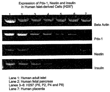
Figure 2 shows RT-PCR analysis of pdxl, nestin and insulin expression in
human islet-derived cells H297.
DETAILED DESCRIPTION
One feature of the invention is a cell composition of endocrine
progenitor/precursor cells from mammalian pancreatic islets, typically adult
pancreas
islet cells, characterized by less differentiation than the initial derived
cells prior to
culturing in serial passages in defined medium.
Another feature of the invention is a defined culture medium formulation for
the culture of endocrine precursor cells.
A further feature of the invention is a method for culturing cells from
mammalian pancreatic islets in serial passages resulting in endocrine
progenitor/precursor cells that are less differentiated than the initial cells
of the
culture.
The cells used to initiate the cell composition of the invention are derived
from
mammalian pancreatic islets, preferably human pancreatic islets, and typically
adult
pancreas islet cells. Following the described culturing methods of the
invention, these
initial pancreatic islet cells are cultured in defined culture medium and
expanded
2
CA 02469209 2004-06-02
WO 03/048336 PCT/US02/38848
through serial passages in defined culture medium, resulting in less
differentiated
endocrine progenitor/precursor cells that express islet progenitor markers pdx-
1 and
nestin. The endocrine progenitor/precursor cells have the potential to be
differentiated
into functioning insulin-producing beta-cells. As used herein, "endocrine
progenitor/precursor cells," "endocrine progenitor cells," and "endocrine
precursor
cells" are all intended to refer to cells derived from mammalian pancreatic
islets that
are capable of serial passages in defined culture medium, are less
differentiated than
the initial cells prior to culturing, and that express markers pdxl and
nestin. In the cell
composition of the invention, the endocrine progenitor cells differentiate
into
functioning insulin-producing beta cells when implanted into a patient to
treat insulin
deficient diabetes.
The medium used to culture the initial pancreatic cells and serial passage
into
endocrine precursor cells is chemically defined, meaning that it contains no
serum or
organ extracts. The medium is able to culture and maintain endocrine precursor
cells
over several passages to expand the cell numbers of the population. The
ability to
expand the cell numbers is beneficial where human pancreatic tissue is
limited. An
additional benefit is that a number of therapeutic cell compositions can be
produced
from a single pancreas.
The defined culture medium is comprised of a nutrient base usually further
supplemented with other components. The skilled artisan can determine
appropriate
nutrient bases in the art of animal cell culture with reasonable expectations
for
successfully producing a tissue construct of the invention. Many commercially
available nutrient sources are useful on the practice of the present
invention. These
include commercially available nutrient sources which supply inorganic salts,
an
energy source, amino acids, and B-vitamins such as Dulbecco's Modified Eagle's
Medium (DMEM); Minimal Essential Medium (MEM); M199; RPMI 1640; Iscove's
3
CA 02469209 2011-07-12
Modified Dulbecco's Medium (EDMEM). Minimal Essential Medium (MEM) and
M199 require additional supplementation with phospholipid precursors and non-
essential amino acids. Commercially available vitamin-rich mixtures that
supply
additional amino acids, nucleic acids, enzyme cofactors, phospholipid
precursors, and
inorganic salts include Ham's F-12, Ham's F-10, NCTC 109, and NCTC 135. Albeit
in varying concentrations, all basal media provide a basic nutrient source for
cells in
the form of glucose, amino acids, vitamins, and inorganic ions, together with
other
basic media components.
The preferred base medium of the invention comprises a nutrient base of either
calcium-free or low calcium Dulbecco's Modified Eagle's Medium (DMEM), without
glucose, magnesium, and with L-glutamine at 4.0 mM, without sodium pyruvate,
and
with Ham's F-12 (with 5 mM glucose) in a 3-to-1 ratio. The final glucose
concentration of the base is adjusted to between about 2 mM to about 8 mM,
more
preferably between about 3 mM to about 7 mM, and most preferably at about 5
mM.
The base medium is supplemented with components such as amino acids,
growth factors, and hormones. Defined culture media for the culture of cells
of the
invention are described in United States Patent No. 5,712,163 to Parenteau and
in
International PCT Publication No. WO 95/31473.
Other media are known in the art such as those
disclosed in Ham and McKeehan, Methods in Enzymology, 58:44-93 (1979), or for
other appropriate chemically defined media, in Bottenstein et al., Methods in
Enzymology, 58:94-109 (1979).
In the preferred embodiment, the base medium is supplemented with the
following components known to the skilled artisan in animal cell culture:
insulin,
transferrin, triiodothyronine (T3), either or both ethanolamine and o-
phosphoryl-
ethanolamine, epidermal growth factor, hydrocortisone, selenium, adenine,
strontium
4
CA 02469209 2004-06-02
WO 03/048336 PCT/US02/38848
chloride, sodium pyruvate, non-essential amino acids, soybean trypsin
inhibitor
(SBTI), and glucose. Concentrations and substitutions for the supplements may
be
determined by the skilled artisan by carrying out titration experiments.
Insulin is a polypeptide hormone that promotes the uptake of glucose and
amino acids to provide long term benefits over multiple passages.
Supplementation of
insulin or insulin-like growth factor (IGF) is necessary for long term culture
as there
will be eventual depletion of the cells' ability to uptake glucose and amino
acids as
well as possible degradation of the cell phenotype. Insulin supplementation is
advisable for serial cultivation and is provided to the media at a
concentration range of
preferably between about 0.5 gg/ml to about 50 g/ml, more preferably between
about
5 gg/ml to about 15 g/ml, and most preferably at about 10 gg/ml. Appropriate
concentrations for the supplementation of insulin-like growth factor, such as
IGF-l or
IGF-2, used in place of insulin may be easily determined by one of skill in
the art by
carrying out a simple titration experiment for the cell types chosen for
culture.
Transferrin is in the medium for iron transport regulation. Iron is an
essential
trace element found in serum. As iron can be toxic to cells in its free form,
in serum it
is supplied to cells bound to transferrin at a concentration range of
preferably between
about 0.05 g/ml to about 50 g/ml, more preferably between about 5 g/ml to
about
15 g/ml, and most preferably at about 5 g/ml.
Triiodothyronine (T3) is a basic component and is the active form of thyroid
hormone that is included in the medium to maintain rates of cell metabolism.
Triiodothyronine is supplemented to the medium at a concentration range
between
about 0 to about 400 pM, more preferably between about 2 pM to about 200 pM,
and
most preferably at about 20 pM.
5
CA 02469209 2004-06-02
WO 03/048336 PCT/US02/38848
Either or both ethanolamine and o-phosphoryl-ethanolamine, which are
phospholipids, are added whose function is an important precursor in the
inositol
pathway and fatty acid metabolism. Supplementation of lipids that are normally
found in serum is necessary in a serum-free medium. Ethanolamine or o-
phosphoryl-
ethanolamine, or both, are provided to media at a concentration range between
about
10-6 M to about 10-2 M, more preferably at about 1 x 10⁻⁴ M.
Hydrocortisone has been shown to have benefits when culturing other epithelial
cell types, to promote phenotype and therefore enhance differentiated
characteristics
(Rubin et al., J. Cell Physiol., 138:208-214 (1986)). Hydrocortisone may be
provided at
a concentration range of about 0.04 gg/ml to about 4.0 gg/ml, preferably at
about 0.4
g/m1.
Selenium is added to serum-free media to resupplement the trace elements of
selenium normally provided by serum. Selenium may be provided at a
concentration
range of about 10-9 M to about 10-7 M; most preferably at about 5.3 x 10"8 M.
The amino acid L-glutamine is present in some nutrient bases and may be
added in cases where there is none or insufficient amounts present. L-
glutamine may
also be provided in stable form such as that sold under the mark, G1utaMAX-1TM
(Gibco BRL, Grand Island, NY). GlutaMAX-1TM is the stable dipeptide form of L-
alanyl-L-glutamine and may be used interchangeably with L-glutamine and is
provided in equimolar concentrations as a substitute to L-glutamine. The
dipeptide
provides stability to L-glutamine to protect it from degradation over time in
storage
and during incubation that can lead to uncertainty in the effective
concentration of L-
glutamine in medium. Typically, the base medium is supplemented with glutamine
at
a concentration preferably between about 1 mM to about 10 mM, more preferably
between about 2 mM to about 8 mM, and most preferably 6 mM L-glutamine.
6
CA 02469209 2004-06-02
WO 03/048336 PCT/US02/38848
Growth factors such as epidermal growth factor (EGF) may also be added to
the medium to aid in the establishment of the cultures through cell scale-up
and
seeding. EGF in native form or recombinant form may be used. Human forms,
native
or recombinant, of EGF are preferred for use in the medium when fabricating a
skin
equivalent containing no non-human biological components. EGF is an optional
component and may be provided at a concentration between about 1 to 15 ng/mL,
more preferably between about 5 to 10 ng/mL.
The defined medium described above is typically prepared as set forth below.
However, it should be understood that the components of the defined medium may
be
prepared and assembled using any conventional methodology compatible with
their
physical properties. It is well known in the art to substitute certain
components with
an appropriate analogue or functionally equivalent acting agent for the
purposes of
availability or economy and arrive at a similar result. Naturally occurring
growth
factors may be substituted with recombinant or synthetic growth factors that
have
similar qualities and results when used in culturing. The optimal
concentration for the
supplements may have to be adjusted slightly for cells derived from different
mammalian species and cell lines from different donors will vary in their
performance
due to its age, size, and health. Titration experiments are performed with
varying
concentrations of a component to arrive at the optimal concentration for that
component.
Media in accordance with the present invention are sterile. Sterile components
are bought or rendered sterile by conventional procedures, such as filtration,
after
preparation. Proper aseptic procedures were used throughout the following
Examples.
DMEM and F-12 are combined and the individual components are then added to
complete the medium. Stock solutions of all components can be stored at -200C,
with
the exception of nutrient source that can be stored at 4 C. All stock
solutions are
7
CA 02469209 2004-06-02
WO 03/048336 PCT/US02/38848
prepared at 500X final concentrations listed above. A stock solution of
insulin,
transferrin and triiodothyronine (all from Sigma) is prepared as follows:
triiodothyronine is initially dissolved in absolute ethanol in 1N hydrochloric
acid
(HCQ) at a 2:1 ratio. Insulin is dissolved in dilute HCl (approximately 0. iN)
and
transferrin is dissolved in water. The three are then mixed and diluted in
water to a
500X concentration. Ethanolamine and o-phosphoryl-ethanolamine are dissolved
in
water to 500X concentration and are filter sterilized. Hydrocortisone is
dissolved in
absolute ethanol and diluted in phosphate buffered saline (PBS). Selenium is
dissolved in water to 500X concentration and filter sterilized. EGF is
purchased
sterile and is dissolved in PBS. Adenine is difficult to dissolve but may be
dissolved
by any number of methods known to those skilled in the art. Human serum
albumin
(HSA) or bovine serum albumin (BSA) may be added for prolonged storage to
maintain the activity of the EGF stock solutions. The medium can be either
used
immediately after preparation or, stored at 4 C. If stored, EGF should not be
added
until the time of use.
A more preferred culture medium formulation for serial culture of the
endocrine precursor cells of the invention comprises: a base 3:1 mixture of
Dulbecco's Modified Eagle's Medium (DMEM) (no glucose, no calcium, with 4 mM
L-glutamine) and Hams F-12 medium, and the base is supplemented with the
following components with the final concentration of each component indicated:
6
mM L-glutamine (or equivalent), 10 ng/ml epidermal growth factor, 0.4 g/ml
hydrocortisone, 1 x 10⁻⁴ M ethanolamine, 1 x 10⁻⁴ M o-phosphoryl-ehanolamine,
5
g/ml insulin, 5 g/mL transferrin, 20 pM triiodothyronine, 6.78 ng/ml
selenium, 24.4
g/mL adenine, 266.6 g/mL strontium chloride, 100 mM sodium pyruvate, 10 mM
non-essential amino acids, 12.5 mg/mL soybean trypsin inhibitor (SBTI), and 5
mM
glucose
8
CA 02469209 2011-07-12
The endocrine precursor cells are cultured in a vessel suitable for animal
cell
or tissue culture, such as a culture dish, flask, or roller-bottle, which
allows for the
formation of a three-dimensional tissue-like structure. Suitable cell growth
surfaces
on which the cells can be grown can be any biologically compatible material to
which
the cells can adhere and -provide an -anchoring means for the cell-matrix
construct to
form. Materials such as glass, stainless steel, polymers, including
polycarbonate,
polystyrene, polyvinyl chloride, polyvinylidene, polydimethylsiloxane,
fluoropolymers, and fluorinated ethylene propylene; and silicon substrates,
including
fused silica, polysilicon, or silicon crystals may be used as a cell growth
surfaces. The
cell growth surface material may be chemically treated or modified,
electrostatically
charged, or coated with biologicals such as with peptides. An example of a
peptide
coating is RGD peptide.
While the cells of the invention may be grown on a solid cell growth surface
or
a cell growth surface with pores, such as a porous membrane, that communicate
both
top and bottom surfaces of the membrane to allow bilateral contact of the
medium to
the culture. Bilateral contact allows medium to contact both the top and
bottom
surfaces of the culture for maximal surface area exposure to the nutrients
contained in
the medium. The pores in the growth surface allow for the passage of culture
media
for providing nutrients to the underside of the culture through the membrane,
thus
allowing the cells to be fed bilaterally. Culture vessels incorporating a
porous
membrane are known in the art and are preferred for carrying out the invention
and are
described in a number United States Patents in the field, some of which have
been
made commercially available, including for instance: 5,766,937, 5,466,602,
5,366,893, 5,358,871, 5,215,920, 5,026,649, 4,871,674, 4,608,342.
A preferred pore size is one that is small
enough that it does not allow for the growth of cells through the membrane,
yet large
9
CA 02469209 2004-06-02
WO 03/048336 PCT/US02/38848
enough to allow for free passage of nutrients contained in culture medium to
the
bottom surface of the cell culture, such as by capillary action. Preferred
pore sizes are
about less than 3 microns but range between about 0.1 microns to about 3
microns,
more preferably between about 0.2 microns to about 1 micron and most
preferably
about 0.4 micron to about 0.6 micron sized pores are employed.
The cultures are maintained in an incubator to ensure sufficient environmental
conditions of controlled temperature, humidity, and gas mixture for the
culture of
cells. Preferred conditions are between about 34 C to about 38 ~C, more
preferably
37 1~C with an atmosphere between about 5-10 1% CO2 and a relative
humidity
(Rh) between about 80-90%.
The defined culture medium allows for establishing primary cultures and serial
passaging of the cultures, thus providing for an expanded number of cells for
using the
cells for testing or as a therapeutic. One of the hurdles in human islet cell
culture is
fibroblast overgrowth that could overshadow the growth of the targeted
epithelial
cells, a sub-population with characteristics of islet progenitor/precursor
cells.
Culturing the cells with the defined medium has overcome this problem. The
cells
grown from human islets using this defined medium have shown predominantly
epitheloid-like morphology and expressed the cytokeratin epithelial marker.
At each passage of the cells, the markers specific to both progenitor cells
and
endocrine precursor cells continue to be exhibited by the cells, including pdx
1 and
nestin. The cultured cells exhibit a decrease in the expression of islet cell
markers
indicating the cells may dedifferentiate with each passage; however, the cells
maintain
progenitor phenotype throughout each passage.
Pdxl, a transcription factor also known as IDX-1, is a known marker of
pancreatic differentiation and regulator of pancreatic development. (Jonsson
et al.
Nature 371:606 (1994) and Offield et al. Development 122:983 (1996)).
CA 02469209 2004-06-02
WO 03/048336 PCT/US02/38848
Nestin is a cellular marker for developing pancreatic islet cells. (Lendahl,
et
al. Cell 60:585-595 (1990) and Zulewski et al. Diabetes. 2001 Mar;50(3):521-
33. )
The endocrine precursor cells may be induced to differentiate using chemical
or physical means, such as by supplementing the culture medium with an agent
that
promotes differentiation to insulin-producing beta cells or by way of forming
cell
clusters in a matrix, such as an extracellular matrix. Implant-induced
differentiation
(in vivo) of the cells in the right environment will induce the cells to
differentiate. The
cells may be implanted subcutaneously, in the submucosa of the small
intestine, or
under the kidney capsule.
The following examples are provided to better explain the practice of the
present invention and should not be interpreted in any way to limit the scope
of the
present invention. Those skilled in the art will recognize that various
modifications
can be made to the methods described herein while not departing from the
spirit and
scope of the present invention.
EXAMPLES
Example 1: Isolation of Pancreatic Small Cells from Cadaveric Human Pancreata
Human pancreatic islet isolation was performed by the semi-automated method
originally proposed by Ricordi (Ricordi C, Lacy PE, Finke EH, et al. Automated
method for isolation of human pancreatic islets. Diabetes 1988 37:413-420).
Procured pancreases were distended by intra-ductal infusion of a Liberase HI
(Roche
Molecular Biochemicals, Indianapolis, IN) or Serva Collagenase (Cresent
Chemical,
Brooklyn, NY) (Linetsky E, Bottno R, Ehmann R, et al. Improved human islet
isolation using a new enzyme blend, Liberase. Diabetes 1997 46:1120-1123), and
then dissociated using the automated method (Ricordi C, Lacy PE, Finke EH, et.
Al.
Automated method for isolation of human pancreatic islets. Diabetes 1988
37:413-
11
CA 02469209 2011-07-12
420). The separation occurs during a process of continuous digestion lasting
approximately 12-30 minutes, after which the digestion circuit was cooled and
the
tissue collected into approximately 8 liters of cold Hanks solution and
washed.
Liberated islets were separated from non-islet tissue on a continuous gradient
of
Euroficoll in a Cobe 2991 cell separator.
Preparations of partially purified islets from the Cobe cell separator were
then
passed through a series of different size steel mesh screens (100 to 25 p
pores), and
the tissue that is retrieved was placed into culture directly on plastic in
culture medium
and permitted to spread out.
Example 2: Isolation of Porcine Islet Cells
Pancreatic islet cells were isolated from a porcine donor and plated using the
defined medium to obtain a culture with an epithelial-like phenotype. The
isolation of
porcine islet cells procedure is as follows. Two Nalgene containers, several
50 mL
round bottom centrifuge tubes, trays, and screens were autoclaved. Two
solutions
were prepared, UW-D organ preservation solution and three concentrations, 27%,
24.6
TM
%, and 11 %, of FICOLL solution.
The UW-D organ preservation solution was made according to the
specifications given by Sumimoto et al (Transplantation 1989 July; 48(1): 1-
5). One
liter of 1X UW-D organ preservation solution consisted of 35.83 g of
lactobionic acid
(Aldrich, Milwaukee, WI), 17.83 g raffinose (Sigma, St. Louis, MO), 1.23 g
MgSO4
(Sigma, St. Louis, MO), 0.92 g glutathione (Sigma, St. Louis, MO), 0.136 g
allopurinol (Sigma, St. Louis, MO), and 3.40 g monobasic potassium phosphate
(Sigma, St. Louis, MO) and double-distilled water. This solution was then
filter-
sterilized using a 0.2 u filter and stored at 4 C until needed.
12
CA 02469209 2004-06-02
WO 03/048336 PCT/US02/38848
The FICOLL solutions were prepared from a Eurocollins base. Eurocollins
base solution (pH 7.3) consisted of 4.1 g monobasic potassium phosphate
(Sigma, St.
Louis, MO), 14.8 g dibasic potassium phosphate (Sigma, St. Louis, MO), 2.24 g
potassium chloride (Sigma, St. Louis, MO), 1.68 g sodium bicarbonate (Sigma,
St.
Louis, MO), 70 g -D-Glucose (Sigma, St. Louis, MO) and an adequate amount of
double-distilled water to bring it up to 2 liters. One liter of Eurocollins
base solution
was added to 500 g of FICOLL (Sigma, St. Louis, MO). The FICOLL was allowed to
go into solution, a process that took about 2 hours. Another 500 mL of
Eurocollins
was added. The solution was analyzed for BRIX and Refractive index ranges, 28-
28.4
and 1.3774-1.3779n0 respectively. Additional Eurocollins base was added as
needed.
The FICOLL solution was then filtered sterilized using a MILLIPORE-MILLIPACK
(Millipore, 'Bedford, MA) and distributed into sterile 1L bottles. To prepare
the 24.6
% FICOLL solution, 456 mL of the stock (27%) FICOLL was diluted with 44 mL
Eurocollins solution. To create the 11% FICOLL solution, 204 mL stock FICOLL
was diluted with 296 mL Eurocollins. The FICOLL solutions were stored at 5 C
until
needed.
The pancreas was obtained from a mixed breed pig weighing more than 40
pounds (_ 20 kg). The pig had been fed a normal diet and was fasted for 24
hours
prior to surgery. A cooler filled with ice, 500 mL of cold UW solution, 10 and
30 c.c.
syringes, and 20-gauge angiocatheters were used in the harvest and transport
of the
pancreas. Once the pancreas was removed, it was perfused with cold UW solution
until swollen. It was then placed in a 250 mL Nalgene container and put on
ice.
During collection of the pancreas, a water bath was heated to 41 C and a
filter-
sterilized Liberase PI solution (Roche Molecular Biochemicals, Indianapolis,
IN)
prepared. In order to facilitate the liberase infusion of the pancreas,
dissection trays,
large and small forceps, extra angiocaths, 30 and 60 c.c. syringes, and
Nalgene
13
CA 02469209 2004-06-02
WO 03/048336 PCT/US02/38848
containers were placed in the sterile field of the biological safety cabinet.
The organ
was then removed from the ice and put onto the dissection tray. Liberase PI
solution
was perfused into the organ. This step was done slowly to avoid disturbing the
cannulae placed there during surgery and also to prevent backflow. Once the
organ
was full, it was placed into another Nalgene container with some additional
Liberase
solution. The container was sealed and placed in the 41 C water bath to
incubate.
The pancreas was then digested until it appeared to begin separating, a
process
that took between 15-30 minutes. Before returning the organ to the sterile
biological
safety cabinet, the Nalgene container was sprayed with ethyl alcohol to insure
sterility.
The organ was then placed on the separating screen and gently scraped with
cell
scrapers for 5-10 minutes. Wash media was frequently added to facilitate the
dissociation of the tissue. The wash media consisted of modified Hank's
balanced salt
solution (HBSS) (with calcium and magnesium, no phenol red) (JRH Biosciences,
Lenexa, KS), donor herd horse serum (JRH Biosciences, Lenexa, KS),
streptomycin
10,000 ug/mL (Invitrogen Life Technologies, Carlsbad, CA), gentamycin sulfate
50
mg/mL (01 P/N 100-50), fungizone 250 mg/mL (Invitrogen Life Technologies
Carlsbad, CA), Amphotericin B (Invitrogen Life Technologies, Carlsbad, CA),
and
sodium desoxycholate 205 mg/mL (Invitrogen Life Technologies, Carlsbad, CA).
The underside of the screen was scraped to ensure that no islet cells were
left
behind. The wash/cell solution was then placed in large centrifuge bottles and
then
spun down at 700 rpms for a minute and a half. The supernatant was then
carefully
aspirated off. The content of each bottle was resuspended using wash media
that was
consolidated into one centrifuge bottle. Wash media was added until the bottle
was
full and then centrifuged again. The supernatant was then aspirated off and
the
volume of tissue determined.
14
CA 02469209 2004-06-02
WO 03/048336 PCT/US02/38848
To begin the density separation, 5 mL of 24.6 % FICOLL was added for each
mL tissue. The suspension was then mixed well and added to a 50 mL round
bottom
tube. In each tube, there should be no more than 12 mL of this suspension. A
second
layer of 27% FICOLL was added to the top of the suspension. A third layer of
11%
FICOLL was added to the top of the gradient. Special care was taken to ensure
that
the layers did not mix. The tubes were then loaded into a centrifuge and spun
down at
1700 rpms for 18 minutes. In order to maintain the gradient, the acceleration
of the
centrifuge was slowed and the brake disengaged.
To collect the islet cells, the 11-24.6 % interface layer was removed. The
islet
cells and wash media were added to a wash tube and spun down for 5 minutes at
1000
rpm. The supernatant was removed and the islet cells resuspended with more
wash
media. This resuspension and centrifugation was repeated three times. The
islet cells
were then resuspended with culture media and plated.
Example 3: Islet Cell Culture
The islets cells acquired by the method of Example 1 were then plated to 60
mm tissue-culture treated culture dishes. The medium used in this example
included
the following: a base 3:1 mixture of Dulbecco's Modified Eagle's Medium (DMEM)
(no glucose, no calcium, with 4 mM L-glutamine) and Hams F-12 medium, and the
base is supplemented with the following components with the final
concentration of
each component indicated: 2 mM L-glutamine (or equivalent), 10 ng/ml epidermal
growth factor, 0.4 g/ml hydrocortisone, 1 x 10⁻⁴ M ethanolamine, 1 x 10⁻⁴ M o-
phosphoryl-ethanolamine, 5 g/ml insulin, 5 g/ml transferrin, 20 pM
triiodothyronine, 6.78 ng/ml selenium, 24.4 g/mL adenine, 266.6 g/mL
strontium
chloride, 100 mM sodium pyruvate, 10 mM non-essential amino acids, 12.5 mg/mL
soybean trypsin inhibitor (SBTI), and 5 mM glucose.
CA 02469209 2004-06-02
WO 03/048336 PCT/US02/38848
Human islet cells were cultured from primary cultures derived from the
pancreatic tissue as described in Example 1 and passaged to passage 8 in the
defined
culture medium (identified as "H297" in the Figures). Figure 1 shows the
cumulative
population doublings for each passage.
Human islet cells were plated to the culture dishes (previously coated with
0.05 mg/mL collagen for 30 minutes) and spread out from the islet clusters as
early as
day 1 after the plating, and grew slowly during the first week. Around day 10,
small,
mitotically active cells started to emerge and form colonies. These colonies
expanded
quickly and eventually merged together to form a population with epithelial
morphology within 3-4 days. After splitting the cell culture and passing them
to new
culture dishes, the sub-cultured cells proliferated very fast with doubling
time around
30 hours. These cells maintained proliferative capability for at least 7
passages, and'
the total population doubling reached up to 9.
Example 4: Characterization Studies
To characterize the expanded cell population, the expression of islet
stem/progenitor markers pdx 1 and nestin as well as islet hormone insulin was
examined by RT-PCR, as shown in Figure 2. H297 cells from passages 0, 2, 4,
and 8
were all positive for pdxl expression. The level of pdxl expression seems to
be
relatively constant throughout the culture period. Similar expression pattern
of nestin
has also been detected in the cells from all these passages. The continued
expression
of both pdxl and nestin in the expanded cells suggests the possibility of
existence of
islet stem/progenitor cells in the culture, and indicates the potential of the
expansion
strategy for cell based therapy. The expression of insulin, as expected, can
only be
detected from the cells from early passages. At passage 4, virtually no
insulin mRNA
signal can be detected. This result is consistent with the inimunfluorescence
result in
16
CA 02469209 2004-06-02
WO 03/048336 PCT/US02/38848
which few insulin-positive cells were observed in cells from passage 4 (data
not
shown). The decrease of insulin signal suggests that the expanding cells are
more
proliferative and less differentiated.
17