Note: Descriptions are shown in the official language in which they were submitted.
CA 02470023 2004-06-04
INTRA~THOACIC C LLATEAL ~IENTILAT10N YpASS SYSTEIUi
CROSS REFERENCE TO RELATED APPLICATIONS
T his application claims the benefit of Provisional Application Number
60/x.75,990 filed June 5, 2~3.
Background of the Invention
1. Field of the Invention
The present invention relates to systems and methods for removing
trapped air in emphysematous lungs, and more particularly, to systems arid
methods for removing trapped air in emphysematous hyperinflated lungs by
bypassing rson-patent airways via a conduit through tl7e outer pleural layer
of
the lung to a containment/trap device. The present invention also relates to a
collateral vewtilation bypass system that utilizes the trachea for expelling
2~ trapped air rather than a containment/trap device. The present invention
also
relates to a device and methodology to assist in pulmonary decompression and
non-surgical/resection lung volume reduction. The present invention also
relates to systems and met~°ods for chemical pleurodesis.
2. Discussion of the Related Art
As a result of studies that date back to the ~ 9~~'s and particularly
studies conducted in the 19~a(3's and early 1370's, it has been determined
that
tong-term continuous oxygeF~ therapy is beneficial in tl ~e treatment of
hypoxemic patients with chronic obstructive pulmonary disease. tn other
words, a patient's life and quality of life carp be unproved lby providing a
constant supplemental supply of oxygen to the patient's lungs.
CA 02470023 2004-06-04
However, with the desire to contain medical costs, thorn is a growing
concern that the additional cost of providing continuous oxygen therapy for
chronic lung disease will create an excessive increa:>e in the annual cost of
oxygen therapy. Thus, it is desirable that oxygen th~;rapy, when provided, be
as cost effective as possible.
t~ Various devices and methods have been devised fo;° performing
emergency cricothyroidotornies and for providing a trache~torrsy tube so that
a
patient whose airway is oth~:rwise blocked may continue to breath. Such
devices are generally intended only for use with a patient wwho is not
breathing
spontaneously and are not :~G~itable for the long term t~ eafrr~ent of chronic
lung
20 disease. Typically, such devices are installed by pur,nturinc~ the skin to
create
Other devices which have been found satisfactory fc~r err~ergency or
ventilator use are described in E~.S. I~aten~ IVos. 958,822 to I~ogers;
2,8'78,742
3C~ to Shelden; 3,384,087 to rur~nmelkamp; 8,511,243 to T oy; 8,556,108 to
Calhoun; 2,991, i 87 to Shelden, et al; 8,688,78 to eiss; 8,81 x,250 to
~lleiss,
et al.; and 8,916,903 to F'c~~~i.
CA 02470023 2004-06-04
Although tracheotorr3y tubes are satisfactory for their intended purpose,
they are not intended for chronic usage by outpatients as a rr~eans for
delivering supplemental oxygen to spo~taneousfy breathing patients with
chronic obstructive pulmon;~ry disease. Such trache~c~tomy tubes are generally
designed so as to provide the total air supply to the patient for a relatively
short
period of titres. The tracheotomy tubes are generally of rigid or semi-rigid
construction and of caliber ranging from 2.5 mm outside diameter in infants to
mm outside diameter ire adults. They are normally inserted in an operating
room as a surgical procedure or during emergency situations, through the
1~ crico-thyroid membrane where the tissue is less vascular and the
possibility of
bleeding is reduced. These devices are intended to permits passage of air in
both directions until normal breathing has been restored by other means.
Another type of tracheotomy tube is disclosed in Jacobs, tJ.S. J'at. Nos.
1:5 3,682,166 and 3,788,326. Tl~e catheter described therein is placed over
14. or
16 gauge needle and inserted through the crico-thyroid membrane for
supplying air or oxygen arid vacuum on an emergency basis to restore the
breathing of a non-breathing patient. The air or oxygen is supplied at 30 to 1
J~
psi for inflation and deflation of the patient's lungs. TJ~e Jacobs catheter,
Jike
2a the other tracheotomy tubes previously used, is not ;suitable for long term
outpatient use, and coultt riot easily be adapted to such use.
Due to the limited fulctionaGty of tracheotomy tubes, transtracheaJ
catheters have been propo:5ed arid used for long term supplemental oxygen
2~ therapy. For example the small diameter transtracheal catheter (16 gauge)
developed by Dr. Henry J. I-leimlich ddescribed in TF-IF ANNALS OF
OTOLOGY, RHINOLOGY LARYNGOLOGY, November-December 1982;
Respiratory Rehabilitation with Transtracheal Oxygen ;ystem) has been used
by the insertion of a relativE9ly large cutting needle (1 ~ gauge} into the
trachea
30 at the mid-point between the cricothyroid membrane and the eternal notch.
This catheter size can supply oxygen up to about 3 liters per minute at low
pressures, such as 2 psi which may be insufficient for patients who reguire
higher flow rates. It does not, however, lend itself to outpatient use and
s
CA 02470023 2004-06-04
maintenance, such as periodic removal and cleaning, primarily because the
connector between the catheter and the oxygen supply ho:je is adjacent and
against the anterior portion of the trachea and cannot be easily seen and
manipulated by the patient, f=urthermore, the catheter ise not provided with
positive means to protect against kinking or collapsing whicah would prevent
its
effective use on an outpatient basis. such a feature is not only desirable but
necessary for long temp outpatient and home care use. Also, because of its
structure, i.e. only one exit opening, the oxygen from the catheter is
directed
straight down the trachea toward the bifurcation between the bronchi. Because
of the normal anatomy of the bronchi wherein the left bronchus is at a more
acute angle to the trachea than the right bronchus, more of the oxygen from
that catheter tends to be directed into the right bronchus rather than being
directed or rr~ixed for mor;: equal utifizatior: by both bronchi. Also, as
structured, the oxygen can .strike the carina, resulting in an undesirable
tickling
sensation and cough. fn addition, in such devices, if a substantial portion of
the oxygen is directed against the back wall of the trachea causing erosion of
the mucosa in this area which rr~ay cause chapping and bleeding. Overall,
because of the limited output from the device, it may not operate to supply
sufficient supplemental oxygen when the patient is exercising or otherwirise
quite active or has severe disease.
Diseases associated with chronic obstructive pulmonary disease include
chronic bronchitis and emphyserr~a. One aspect of an emphysematous lung is
that the communicating flow of air between neighboring air sacs is much more
~5 prevalent as compared to healthy lungs. ~Chis phenomenon is known as
collateral ventilation. Another aspect of an emphysematous lung is that air
cannot be expelled from the native airways due to the loss of tissue elastic
recoil and radial support of the airways. ~ssentiaify, the loss of elastic
recoil of
the lung tissue contributes to the inability of individuals to exhale
completely.
The loss of radial support of the airways also allows a collapsing phenomenon
to occur during the expiratory phase of breathing. This collapsing phenomenon
also intensifies the inability 'for individuals to exhale completely. As the
inability
to exhale completely increases, residual volume in the lungs also increases.
CA 02470023 2004-06-04
This then causes the lung ~~o establish in a hyperinflated si:ate where an
individual can only take short shallow breaths. EsSE=r~tially; air is not
effectively
expelled and stale air accumulates in the lungs. ance the stale air
accumulates in the lungs, the individual is deprived o~ oxygen.
currently, treatments ror chronic obstructive ~'ralrr~or~ary disease include
bronchodilating drugs, oxygen therapy as described above, and lung volume
reduction surgery. ~ronchodilating drugs only work on a percentage of patients
with chronic obstructive pulmonary disease and generally only provides short
term relief. t~xygen therap&~ is impractical for the reasons described above,
and lung volume reduction surgery is an extremely traumatic procedure that
involves removing part of tl-~e lung. The long term benefits of lung volume
reduction surgery are no~ fully known.
l~ Accordingly, there exists a need for increasing the expiratory flow from
an individual suffering from chronic obstructive pulmonarydisease. In
addition,
there exists a need for a minimally invasive means for removing trapped air
from the lung or lungs that ~~ould allow healthy lung i:issue to better
ventilate.
There also exists a need for a minimally invasive means for allowing trapped
air from the lung or lungs to escape that would allow healthy lung tissue to
better ventilate.
summary of the Invention
2~ The present invention overcomes the disadvantages associated with
treating chronic obstructive pulmonary disease, as briefly described above, by
utilizing the phenomenon of collateral ventilation to increase the expiratory
flow
from a diseased lung. The ~~resent invention also provides a means for
assisting in or facilitating pulmonary decompression to compress the diseased
3G area or area of the lung or lungs to a smaller volume.,
The intra-thoracic collateral ventilation bypass system of the present
invention removes trapped air in an emphysematous hyperinflated lung by
s
CA 02470023 2004-06-04
bypassing non-patent airways via a conduit through the outer pleural layer of
the lung to a more proximal airway closer to the trachma.
fn accordance with a first aspect, the present inventi~cn is directed to an
.5 intra-thoracic collateral ventilation bypass system. The system comprising
at
least one conduit having first and second ends, a first sealing device and a
second sealing device. The first end of the conduit is. in fluid
cornmunicaticn
with an airway in proximity to a trachea of a patient a'~d the second end is
in
fluid communication with the inner volume of a lung o~f a pa~:ient at a
1~ predetermined site. The first sealing device is utilizet3 for' establishing
an
airtight sea! between the conduit and the proxirr~ate air~ray. The second
seating device is utilized for establishing an airtight sE=a! between the
conduit
and the lury.
15 !n accordance with another aspect, the present invention is directed to a
method for decornpresslng a hyperinffated portion of a lung of a patient. The
method comprising deterr~ireing a site of hyperinflation in a hatfent's lung,
and
bypassing non-patent airways utilizing a device in cornrn~anication with a
hyperinflated portion of a patient's lung and an airway proximate a patient's
2~ trachea.
s
CA 02470023 2004-06-04
Essentially, stale air accumulates in the lungs, thereby depriving the
individual
of oxygen. ilarious methods may be utilized to determine the location or
locations of the diseased tissue, for example, computerized axial tomography
or CAT scans, magnetic resonance imaging or MRI, ~pc~sitron emission
tomograph or PET, andfor standard ~C-ray imaging. t~r~ce tire location or
locations of the diseased tissue are located, anastomotic openings are made in
the thoracic cavity and lung or lungs and one or more' oxygen carrying
conduits
are positioned and seated therein. The one or more oxygen carrying conduits
are connected to an oxygen source which supplies o;~ygen under elevated
tf~ pressure directly to the diseased portion or portions of the lung or
lungs. The
pressurized oxygen essentially displaces tl~e accumulated air and is thus more
easily absorbed by the alveoli tissue. In addition, the long term oxygen
therapy
system may be configured in such a way as to provide collateral ventilation
bypass in addition to direct taxygen therapy. In this configuration, an
additional
I5 conduit may be connected between the main conduit and the individual's
trachea with the appropriate valve arrangement. In this configuration, stale
air
may be removed through the trachea when the individual exhales since the
trachea is directly linked with the diseased site or sites in the lung via the
conduits.
2~
The long term oxygen' therapy system of fihe present invention improves
oxygen transfer efficiency in the lungs thereby reducing oxygen supply
requirements, which in turn reduces the patient's medical costs. The system
also allows for improved self-image, improved mobility, greeter exercise
2~ capability and is easily maintained.
The above-described song term oxygen therapy system may be utilized
to effectively treat hypoxia caused by chronic obstructive pulmonary disease;
however, other means may Ibe desirable to treat other aspects of the disease.
30 As set forth above, emphysema is distinguished as irreversible damage to
lung
tissue. The breakdown of lung tissue leads to the reduced ability for the
lungs
to recoil. The tissue breahdo~rvn also leads to the loss of radial support of
the
airways. Consequently, the loss of elastic recoil of the lung tissue
contributes
CA 02470023 2004-06-04
to the inability for individual s trvith emphysema to exhale completely. The
loss
of radial support of the airways also allows a collapsing phenomenon to occur
during the expiratory phase of breathing. This collapsing phenomenon also
intensifies the inability for individuals to exhale completely. As the
inability to
exhale increases, residua! volume in the lungs also increases. This then
causes the lung to establish in a hyperinflated state vvherein an individual
can
only take short shallow brea.tl-~s.
The collateral ventila~:ion bypass trap system ~sf the present invention
utilizes the above-described collateral venfiilation phenorr~er~on to increase
the
expiratory flow from a diseased lung or lungs, thereby treating another aspect
of chronic obstructive pulmonary disease. Essentiallt~, the ~~ost collaterally
ventilated area of the Fang or lungs is determined utilizincl tf~e scanning
techniques described above. once this area or area:3 are located, a conduit or
1~ conduits are positioned in a passage or pe.ssages theft access the outer
pleural
layer of the diseased lung o:~~ lungs. The conduit or conduits utilize the
collateral ventilation of the lung or lungs and allow th~~ entra.pped air to
bypass
the native airways and be e;~pelied to a containment :>yster~~ outside of the
body.
z~
In an alternate embodiment, the trachea, or other prc'ximal airrrvays,
including the bronchus, rnay be utilized for expelling trapped air rather than
a
containmentitrap device.
The lung reduction d~:voce of the present invention allows trapped air
from hyperinflated regions of the lung or iur~gs of a patient to vent to the
s
CA 02470023 2004-06-04
external environment through a ono-way valve. The valve prevents air from
flowing back into the lung or lungs.
In order for the system to be effective, the corr~ponents of the system
are preferably sealed to the lung. Rccordingly, the Icnali.~ed pleurodesis
chemical delivery system of the present ir~ver~tion is utilized to create a
pleurodesis in the area or areas of the lung that are ra~ost collaterally
ventilated.
Various chemicals, agents and/or compounds rnay be delivered via catheter
based delivmy systems or via implantabfe medical d~:vices,.
~0
brief ~escrintion of the ~ra~~in s
The foregoing anc~ other features arid advantages of the invention wil! be
apparent from the following, rryore particular description of preferred
15 embodiments ofi the invention, as illustrated in the accor~ipanying
drawings.
Figure 1 is a diagrarrrr~atic representation of a virst exemplary
embodiment of the long terrn oxygen Therapy system in accordance with the
present invention.
Figure ~ is a diagrar~rr~atic representation of a first exemplary
embodiment of a sealing device utilized in con~ur~ctior~ with the long term
oxygen therapy system of the present invention.
Figure 3 is a diagrat~matic representation of a second exemplary
embodiment of a sealing device utilized in con~unctior~ with the long terra~a
oxygen therapy system of tl-~e present invention.
Figure 4. is a diagrarr~rnatic representation of a Third E=xemplary
3~ embodiment of a sealing device utilized in con~uractior~ with the long term
oxygen therapy system of the present invention.
s
CA 02470023 2004-06-04
Figure 5 is a diagrarxamatic representation of a. fourth exemplary
embodiment of a seating device utilized in canjunction with the Gong term
oxygen therapy system of the present invention.
Figure 6 is a diagrammatic representation of a second exemplary
embodiment of the tong terra oxygen therapy syster~~ in accordance with the
present invention.
Figure 7 is a diagrammatic representation of a first exemplary
1~ embodiment of a collateral ventilation bypass trap system in accordance
with
the present invention.
Figure is is a diagrammatic representation of a second exemplary
embodiment of a collateral ventilation bypass systems in accordance with the
1~ present invention.
Figure 9 is a diagram:m~atic representation of a. third exemplary
embodiment of a collateral ~sentilation bypass system in accordance with the
present invention.
2C~
Figure 10 is a diagrarr~matic representation of a fourth exemplary
embodiment of a collateral ventilation bypass system in accordance with the
present inVentiOn.
25 Figure 11 is a diagrammatic representation of an exemplary
embodiment of an intra-thoracic collateral ventilation bypass system in
accordance with the present invention.
Figure 12 is a diagrammatic representation of an exemplary pulmonary
30 decompression device in accaordance with the present invention.
Figures 13a and 13b are diagrammatic representations of the effects on
lung volume in accordance with the present invention.
~o
CA 02470023 2004-06-04
Figures 14a and 14b are diagrammatic representations of the effects on
lung volume reduction utilizing the lung reduction system in accordance with
the present invention.
Figure 15 is a diagrammatic representation of a first/ exemplary
embodiment of a localized pig=urodesis chemical delivery system.
Figure 1~ is a diagrammatic representation of a. second exemplary
embodiment of a localized pleurodesis chemical delivery system.
~etailed ~escription of the I~referred Embodiments
Air typically eaters the mammalian body througf ~ the nostrils and flows
into the nasal cavities. As the air passes through the nostrils and nasal
I5 cavities, it is filtered, moistened and raised or lowered to approximately
body
temperature. The back of the nasal cavities is continuous vwith the pharynx
(throat region; therefore, air may reach the pharynx from the nasal cavities
or
from the mouth. Accordlagly, if equipped, the mammal may breath through its
nose or mouth. Generally air from the mouth is not as filtered or temperature
regulated as air from the nostrils. The air in the phar~rnx flo~rvs from an
opening
in the floor of the pharynx arid into the larynx (voice box). The epiglottis
automatically closes off the larynx during swallov~ing ;~o that solids andlor
liquids enter the esophagus rather than the lower air passageways or airways.
From the larynx, the air passes into the trachea, which divides into two
branches, referred to as the bronchi. The bronchi are connected to the lungs.
The lungs are large, paired, spongy, elastic organs, which are positioned
in the thoracic cavity. The lungs are in contact e~ith the walls sat the
thoracic
cavity. In humans, the right lung comprises three lobes and the left lung
3t) comprises two lobes. Lungs are paired in al! mammals, but the number of
lobes or sections of lungs varies from mammal to mammal. hlealthy lungs, as
discussed below, have a tremendous surface area for gas/air exchange. both
the left and right lung is covered with a pleural membrane. Essentially, the
pleural membrane around each lung forms a continuous sac that encloses the
CA 02470023 2004-06-04
lung. A pleural membrane also forms a lini~~g for the thoracic; cavity. 'The
space between the pleural membrane forming the lining of the thoracic cavity
and the pleural membranes enclosing the lungs is referred to as the pleural
cavity. The pleural cavity comprises a film of fluid that serves as a
lubricant
between the lungs and the chest wall.
!n the lungs, the bronchi branch into a multipliciay of smaller vessels
referred to as bronchioles. ~'ypically, there are more than one million
bronchioles in each lung. each bronchiole ends in a cluste~~ of extremely
srnail
1C~ air sacs referred to as alveoli. ~r~ extremely thin, single layer of
epithelia8 cells
lining each alveolus wall and an extremely thin, single layer of epithelial
cells
lining the capillary walls separate the air/gas in the alveolus from the
blood.
~xygen molecums in higher concentration, pass by simple diffusion through the
two thin layers from the alveoli into the blood in the p~slmor~ary
capillaries.
I:~ Simultaneously, carbon dioxide molecules in higher r~oncentration pass by
simple diffusion through the two thin layers from the blood in the pulmonary
capillaries into the alveoli.
breathing is a mechanical process involving inspiration and expiration.
20 The thoracic cavity is normally a closed system and air caenot enter or
leave
the lungs except through the trachea. If the chest v~rall is somehow
compromised and airlgas enters the pleural cavity, the lungs will typically
collapse. Ullhen the volume of the thoracic cavity is increased by the
contraction of the diaphragm, the volume of the lungs is also increased. ~s
the
25 volume of the lungs increase, the pressure of the air in the lungs falls
slightly
below the pressure of the air external to the body (ambient air pressure.
accordingly, as a result of this slight pressure differential, external or
ambient
air flows through the respiratory passageways dese~ribed above and fills the
lungs until the pressure eguaiizes. This process is inspiration. i~lhen the
3Q diaphragm is relaxed, the volume of the thoracic cavity decreases, which in
turn decreases the volume of the lungs. As the volume of the lungs decrease,
the pressure of the air in the lungs rises slightly above the pressure of the
air
exten~al to the body. accordingly, as a resulf of this slight pressure
differential,
~2
CA 02470023 2004-06-04
the air in the alveoli is expelled through the respiratory pass~~geways until
the
pressure equalizes. This process is expiration.
Continued insult to the respiratory system may resc,~lt in variods
diseases, for example, chronic obstructive pulmonary disease. chronic
obstructive pulmonary disease is a persistent obstruction of i:he airways
caused
by chronic bronchitis and pulmonary emphysema. In the United States atone,
approximately fourteen million people suffer from some form of chronic
obstructive pulmonary disease and it is in the top ten leadincl causes of
death.
chronic bronchitis anc acute bronchitis share certain similar
characteristics; however, they are distinct diseases. Soth cf~~ronic and acute
bronchitis involve infiamrnation and constriction of the broncf~ia! tubes and
the
bronchioles; however, acute bronchitis is generally associated with a viral
tS andlor bacterial infection and its duration is typically much shorter tha-n
chr onic
bronchitis. In chronic bronchitis, the bronchial tubes secrete too much mucus
as part of the body's defensi~re mechanisms to inhaled foreign substances.
tviucus membranes comprising ciliated cells (hair like structures) line the
trachea and bronchi. The ciliated cells or cilia continuously push or sweep
the
2~ mucus secreted from the mucus membranes in a direction away from the lungs
and into the pharynx, where it is periodically swallowed. 'lehis sweeping
action
of the cilia functions to keep foreign matter from reaching the lungs. Foreign
matter that is not filtered by the nose and larynx, as descr~ibE:d above,
becomes
trapped in the mucus arid is propelled by the cilia into the pharynx. When too
25 much mucus is secreted, thc~ ciliated cells rnay become darr'aged, leading
to a
decrease in the efficiency of the cilia to sweep the brc~nchiaf tubes and
trachea
of the mucus containing the voreign matter. This in turn causes the
bronchioles
to become constricted and inflamed and the individual becomes short of
breath. In addition, the individual will develop a chronic cough as a means of
3~ attempting to clear the airways of excess mucus.
Individuals who suffer from chronic bronchitis rr~ay develop pulmonary
emphysema. Pulmonary emphysema is a disease in which the alveoli wails,
13
CA 02470023 2004-06-04
which are normally fairly rigid structures, are destroyed. The destruction of
the
alveoli waifs is irreversible. pulmonary emphysema may be caused by a
number of factors, including chronic bronchitis, long term exposure to inhaled
irritants, e.g. air pollution, which damage the cilia, enzyme deficiencies and
other pathofogica) conditions. In pulmonary errjphyser~°~a, thf:
alveoli of the
lungs lose their elasticity, ar~ci eventually the walls between adjacent
alveoii are
destroyed. Accordingly, as more and more alveoli walls are lost, the air
exchange (oxygen and carbon dioxide) surface area of the lungs is reduced
until air exchange becomes seriously impaired. T he combination of mucus
hypersecretion and dynamic airway compression are rr~echanisms of airflow
limitation in chronic obstructive pulmonary disease, dynamic airway
compression results from the loss of tethering forces exerted on the airway
due
to the reductiorf i~~ lung tissue elasticity. f~lucus hypersecretion is
described
above with respect to bronchitis. In other words, the breakdown of lung tissue
leads to the reduced ability of the lungs to recoil and the loss of radial
support
of the airways. ~onsequentler, the loss of elastic recoil of the: lung tissue
contributes to the inability of individuals to exhale completely. The loss of
radial support of the airways also allows a collapsing Kal~errornenori to
occur
during the expiratory phase of breathing. This collapsing phenomenon also
2~ intensifies the ir~abiiity for individuals to exhale completely. As the
inability to
exhale completely increases, residual volume in the lungs also increases. This
then causes the lung to establish in a hyperinfiated state where an individual
can only take short shallow breaths. Essentially, air is not ei~fectively
expelled
and stale air accumulates in the lungs. Once the stale air accumulates in the
lungs, the individual is deprived of oxygen. There is no cure for pulmonary
emphysema, only various treatments, including exercise, drug therapy, such as
bronchodilating agents, lung volume reduction surgery and Kong term oxygen
therapy.
3~ As described above, long term oxygen therapy is widely accepted as the
standard treatment for hypoxia caused by chronic obst~~uctive pulmonary
disease. Typically, oxygen therapy is prescribed using a nasal cannula. There
are disadvantages associated with using the nasal canoula. One disadvantage
14
CA 02470023 2004-06-04
associated with utilizing nasal cannula is the significant loss of oxygen
between
the cannula and the nose, uv!~ich in turn equates to more frequent changes in
the oxygen source, or higher energy requirements to generate more oxygen.
Another disadvantage associated with utilizing nasal c;a~ir,ula is the fact
that the
cannuias may cause the nasal passages to become dry, cr~ccked and sore.
Transtracheal oxygen therapy has become a viable alternative to long
term oxygere therapy. Trans~tracheal oxygen therapy delivers oxygen directly
to
the lungs using a catheter that is placed through and down the trachea. due to
t0 the direct nature of the oxygen delivery, a number of advantages are
achieved.
These advantages include lower oxygen requirements due t:o greater
efficiency, increased mobility, greater exercise capability and improved self
image.
The long term oxyger~n therapy system and method of the present
invention may be utilized to deliver oxygen directly into the lung tissue in
order
to optimize oxygen transfer efficiency in the lungs. In other words, improved
efficiency may be achieved if oxygen were to be delivered directly into the
alveolar tissue in the lungs. In emphysema, alveoli wills are destroyed,
~0 thereby causing a decrease in air exchange surface area. As more alveoli
walls are destroyed, collateral ventilation resistance is lowered. In other
words,
pulmonary emphysema causes an increase in collateral verytilation and to a
certain extent, chronic bron~:hitis also causes an increase ire collateral
ventilation. Essentially, in an emphysematous lung, the e;ornmunicating flow
of
air between neighboring air sacs (alveoli), known as collate~°al
ventilation, is
much more prevalent as compared to a normal lung. ~ince~ air cannot be
expelled from the native airways due to the loss of tissue elastic recoil and
radial support of the airways dynamic collapse during exha.lation), the
increase
in collateral ventilation does not significantly assist are individual in
breathing.
The individual develops dsypnea. Accordingly, if it can be determined where
collateral ventilation is occurring, then the diseased lung tissue may be
isolated
and the oxygen delivered to this precise location or locations. l9arious
methods
may be utilized to determine the diseased tissue locations, for example,
CA 02470023 2004-06-04
computerized axis( tomography or ~~T scans, magnetic resonance imaging or
!VlRI, positron emission tomograph or ~E'~, and/or standard ~~-ray imaging.
Once the diseased tissue is located, pressurized oxygen rnay be directly
delivered to these diseased areas and more effectively and efficiently forced
into the lung tissue for air exchange.
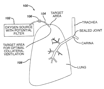
Figure 1 illustrates a first exemplary long term oxygen therapy system
100. The system 100 co~apr~ises an oxygen source 102, are oxygen carrying
conduit 104 and a one-way valve 108. The oxygen source 102 may comprise
fi0 any suitable device for supp(y(ng filtered oxygen under adjustably
regulated
pressures and flow rates, inc(ud(ng pressurszed oxygen tanks, liquid oxygen
reservoirs, oxygen concentrators and the associated devices for controlling
pressure and f iavf rafie e.g. r~;gu(ators. The oxygen carry(ng conduit 10~
may
comprise any suitable biocornpatib(e tubing having a high resistance to
fi5 damage caused by continuous oxygen exposure. The oxygen carrying conduit
i04 comprises tubing having arf inside diameter in the range from about 1/16
inch to about 1I2 inch and acre preferably from about 1/8 inch to about
1/4 inch. The one-way valve 106 may comprise ar~y suitable, in-(ire
mechanical valve which a((ovvs oxygen to f(:ow into the lungs 10~ through the
20 oxygen carrying conduit 104, but not from the (ur~gs 108 back into the
oxygen
source 102. For example, a simple check valve may be uti((zed. As illustrated
in Figure 1, the oxygen carrying conduit 104 passes thr~ugr~ the lung 108 at
the site determined to have fibs highest degree of collateral ventilation.
25 The exemplary system 100 described above rr~a~ be modified in a
number of ways, including tt-~e use of an in-line finer. (n this exemplary
embodiment, both oxygen acrd air may f(ov~r through the system. Ire other
words, during inhalation, oxygen is delivered to the lungs through the oxygen
carrying conduit 104 and dug°ing exhalation, air from tf~e (unc~s flow
through the
30 oxygen carrying conduit 10~. T he in-line filter would trap mucus and other
contaminants, thereby prev~:nting a blockage in the oxygen source i02. In this
exemplary embodiment, no valve 106 would be utilized. The flow of oxygen
is
CA 02470023 2004-06-04
into the lungs and the flow of air from the lungs is based on pressure
differentials.
In order for the exemplary song term oxygen therapy system 100 to
S function, an airtight seal is preferably maintained where the oxygen
carrying
conduit 104 passes through the thoracic cavity and lung. This seas is
maintained in order to sustain the inflation/functionality of the lungs. if
the seal
is breached, air can enter the cavity and cause the lungs to collapse as
described above.
~ method to create this seal comprises forming adt~e;~ions between the
visceral pleura of the lung and the inner waif of the thoracic cavity. This
may
be achieved usir~,g either chemical methods, including irritants such as
Doxycyciine and/or Sieomycin, surgical methods, including pleurectomy or
IS horoscope Laic pleurodesis, or radiotherapy methods, including radioactive
gold
or external radiation. Ail of ttlese methods are known in the relevant art for
creating pleurodesis. With a seal created at the site for the ventilation
bypass,
an intervention rnay be safely performed without the clanger of creating a
pneumothorax of the lung.
Similarly to ostomy pouches or bags, the oxygen carrying conduit 104
may be sealed to the skin at the site of the ventilation bypass. in one
exemplary embodiment, illustrated in figure 2, the oxygen carrying conduit 104
may be sealed to the skin of the thoracic wall utilizing an adhesive. As
illustrated, the oxygen carrying conduit 104 comprises a flange 200 having a
blocompatible adhesive coating on the skin contacting surfsce. The
biocompatible adhesive woc~ld provide a fluid tight seal between the flange
200
and the skin or epidermis of the thoracic wall. in a preferred embodiment, the
biocompatible adhesive provides a temporary fluid tigi~t seal such that the
oxygen carrying conduit 104 may be disconnected from the ventilation bypass
site. This would allow for th~~ site to be cleaned and for the long term
oxygen
therapy system 100 to undergo periodic maintenance.
CA 02470023 2004-06-04
Figure 3 illustrates another exemplary embodiment fc~r sealing the
oxygen carrying conduit 104 to the skin of the thoracic wall at the site of
the
ventilation bypass. In this exemplary embodiment, a coupling plate 300 is
sealed to the skin at the site of the ventilation bypass by a biocompatible
adhesive coating or any other suitable means. the oxygen carrying conduit
104 is then connected to the coupling plate 300 by any suita.bie means,
including threaded couplings and locking rings. the exemplary embodiment
also allows for cleaning of the site and mai~~tenance of the system 100.
t0 Figure 4 illustrates yet another exemplary embodiment for sealing the
oxygen carrying conduit ~ 04 to the skin of the thoracic wail at the site of
the
ventilation bypass. In this exemplary embodiment, balloon flanges 400 may be
utilized to c,reage the seal. 'The balloon flanges 400 may be attached to the
oxygen carrying conduit 104 siuch that in the deflated state, the oxygen
carrying
t5 conduit 104 and one of the balloon flanges passes through the ventilation
bypass anastomosis. ~'he balloon flanges 400 are spaced apart a sufficient
distance such that the balloon flanges remain on opposite sides of the
thoracic
wall. l~Ihen inflated, the ballaons expand and form a fluid tight sea! by
sandwiching the thoracic wall. ~nce again, this exemplary embodiment allows
20 for easy removal of the oxygen carrying conduit 104.
Figure 5 illustrates yet another exemplary embodiment for sealing the
oxygen carrying conduit 104 to the skin of the thoracic wall at the site of
the
ventilation bypass. In this exemplary embodiment, a single balloon flange 500
25 is utilized in combination inrith a fixed flange 502. 'The balloon flange
500 is
connected to the oxygen carr;ring conduit '104 in the same manner as
described above. In this exc~rnplary embodiment, the balloon flange 500, when
inflated, fom~s the fluid tight seal. 'The fixed flange 50~, which is
maintained
against the skin of the thoracic wall, provides the structure! support against
30 which the balloon exerts pressure to form the seal.
If an individual has difficulty exhaling and requires additional oxygen,
collateral ventilation bypass rr~ay be combined with direct oxygen therapy.
18
CA 02470023 2004-06-04
Figure 6 illustrates an exemplary embodiment of a collateral ventilation
bypass/direct oxygen therap~~ system 600. The system 600 comprises an
oxygen source 602, an oxygen carrying conduit 604 having two branches 606
and 606, and a control valve 610. The oxygen source 602 a.nd oxygen carrying
conduit 604 may comprise components similar to the above-described
exemplary embodiment illustrated in Figure 1. In this exemplary embodiment,
when the individual inhales, the valve 610 is open and oxygE:n flows into the
lung 612 and into the bronchial tube 614. In an alternate exemplary
embodiment, the branch 606 may be connected to the trachea 616.
Accordingly, during inhalation oxygen flows to the diseased site in the iung
or
lungs and to other parts of the lung through the normal bronchial passages.
During exhalation, the valve 610 is closed so that no oxygen is delivered and
air in the dijcas~:d portion of the lung may flow from the lung 612, through
one
branch 606 and into the second branch 606 and finally into the bronchial tube
l~ 616. In this manner, stale air is removed and oxygen is directly delivered.
Cnce again, as described above, the flo~rv of oxygen a.nd air is regulated by
simple pressure differentials.
The connection and sealing of the oxygen carr~~ing conduit 604 and
branches 606, 606 to the lung 612 and bronchial tube ~i14 may be made in a
manner similar to that described above.
The above-described long term oxygen therapy system may be utilized
to effectively treat hypoxia caused by chronic obstructive pulmonary disease;
however, other means may be desirable to treat other aspects of the disease.
As set forth above, emphysema is distinguished as irreversible damage to sung
tissue. The breakdown of lung tissue leads to the reduced ability for the
lungs
to recoil. The tissue breakdown also leads to the loss of radial support of
the
native airways. Consequently, the loss of elastic recoil of the lung tissue
contributes to the inability fo~~ individuals with emphysema to exhale
completely. The loss of radial support of the native ai~gays also allows a
collapsing phenomenon to occur during the expiratory phase of breathing. 'This
collapsing phenomenon alsr3 intensifies the Inability for individuals to
exhale
19
CA 02470023 2004-06-04
completely. ~1s the inability to exhale increases, residual volume in the
lungs
also increases. This then causes the lung to establish it a hyperinflated
state
wherein an individual can only take short shallow breaths.
The collateral ventiiati$~r~ bypass trap system of the present invention
utilizes the above-described collateral ventilation phenomenon to increase the
expiratory flow from a diseased lung or fangs, thereby treating another aspect
of chronic obstructive pulmonary disease. Essentially, the rr~ost collaterally
ventilated area of the lung or lungs is determined utili~.ing the scanning
techniques described above. once this area or areas are located, a conduit or
conduits are positioned in a passage or passages that: access the outer
pleural
layer of the diseased lung or lungs. 'fhe conduit or conduits utilize the
collateral ver~ti9aiion of the lu~7g or lungs and allows the entrapped air to
bypass
the native airways and be expelled to a containment system outside of the
body.
Figure 7 illustrates a first exemplary collateral ventilation bypass trap
system 700. The system '~00 comprises a trap 702, an air carrying conduit x'04
and a filter/one-way valve ?06. The air carrying conduit X04 creates a fluid
communication between an individual's lung ~0~ and the trap 702 through the
filterlone-way valve 706. It i s important to note that aithougt~ a single
conduit
704 is illustrated, multiple conduits may be utilized in each lung X08 if it
is
determined that there is more than one area of high collateral ventilation.
'The trap 702 may cor°x~prise any suitable device for collecting
discharge
from the individual's lung or 'lungs X06. Essentially, tine trap 702 is simply
a
containment vessel for temporarily storing discharge from the lungs, for
example, raucous and other fluids that may accumulate in the lungs. The trap
702 may comprise any suitable shape and may be formed from any suitable
metallic ar non-metallic matE;rials. Preferably, the trap X02 :should be
formed
from a lightweight, non-corrosive material. in additior°~, the trap
'702 should be
designed in such a manner as to allow for effective and efficient cleaning. in
one exemplary embodiment,. the trap 702 may comprise disposable liners that
CA 02470023 2004-06-04
may be removed when the trap 702 is full. The trap 702 may be formed from a
transparent material or comprise an indicator window so that it may be easily
determined when the trap ~G2 should be emptied or cleaned. A lightweight
trap x'02 increases the patient's mobility.
S
The filterlone-way vahre '~06 may be attached to the trap 702 by s.ny
suitable means, including threaded fittings or compres;sior~ type fittings
commonly utilized in compressor connections. The filter/one-way valve BOG
serves a number of functionv,. The filter/one-way valve 706 allows the air
from
t0 the individual's lung or lungs 708 to exit the trap '~02 while maintaining
the fluid
discharge and solid particulate matter in the trap '7~2. This filter/one-way
valve
706 would essentially maintain the pressure in the trap 702 below that of the
pressure ir~sici:: ape indi°,~ic~ual's lung or lungs X08 so that the
flow of air from the
lungs 708 to the trap 702 is c~aaintained in this one direction. The filter
portion
15 of the filterlone-way valve '~Ga may be designed to capture particulate
matter of
a particular size which is suspended in the air, but allc~v~s thE: clean air
to pass
therethrough and be vented ~:o the ambient environment. Tf de filter portion
r~nay
also be designed in such a ~~anner as to reduce the moisture content of the
exhaled air.
The air carrying conduit 70~ connects the trap 7G2 to the lung or lungs
708 of the patient through the filterlone-way valve ~Of. The air carrying
conduit 704 may comprise any suitable biocompatibie tubing having a
resistance to the gases contained in air. The air carrying conduit 704
comprises tubing having an inside diameter in the range frorra about ~/~ 6
inch
to about 1l2 inch, and more preferably frorrr about ~/8 inch to about 1/.4
inch.
The filterlone-way valve X06 may comprise any suitable valve which allows air
to flow from the lung or lung48 a 08 through the air carrr~ing conduit 7G4,
but not
from the trap 7G2 back to th~~ lungs 7G8. For example, a simple check valve
may be utilized. The air carrying conduit 704 may be connected to the
filterlone-way valve 706 by arty suitable means. l3referably, a quick release
mechanism is utilized so that the trap may be easily removed for maintenance.
2~
CA 02470023 2004-06-04
As illustrated in Figure 7, the air carrying conduit 704 passes through the
lung
708 at the site determined to have the highest degree of collateral
ventilation.
(f more than one site is determined, multiple air carrying conduits 704 may be
utilized. The connection of multiple air carrying condt,cits 704 to the
filter/one-
way valve 706 may be accomplished by aray suitable mean;>, including ars
octopus device similar to that utilized in scuba diving regulators.
The air carrying conduit 704 is preferably able to v~itp~stand and resist
collapsing once in place. Since air will travel through the conduit 704, if
the
la conduit is crushed and unable to recover, the effectiveness of the system
is
diminished. Accordingly, a crush recoverable material may be incorporated
into the air carrying conduit 704 in order to make it crush recoverable. Any
number of suiac~ie matermals may be utilized. For example, Nitinol
incorporated
into the conduit 704 gill give the conduit collapse resistance and collapse
1S recovery properties.
Expandable features at the end of the conduit 734 rnay be used to aid in
maintaining contact and sealing the conduit 704 to the lung pleura. Nitinol
incorporated into the conduit 704 gill provide the ability to deliver the
conduit
20 704 in a compressed state and then deployed in an expanded state to secure
it
in place. Shoulders at the end of the conduit may also provide a mechanical
stop for insertion and an ar ea for an adhesiveisealant to loin as described
in
detail subsequently.
~5 In order for the exemplary collateral ventilation bypass trap system 700
to function, an airtight seal is preferably maintained where the air carrying
conduit 704 passes through the thoracic cavity and lungs 708. This seal is
maintained in order to sustain the inflation/functionality of the lungs. if
the seal
is breached, air can enter the cavity and cause the lungs 'to collapse. one
:30 exemplary method for creating the seal comprises dorming adhesions between
the visceral pleura of the lung and the inner wall of the thoracic cavity.
This
may be achieved using eii~her chemical methods, including irritants such as
~oxycycline andlor Sleomycin, surgical methods, ir~cludir~g p9eurectomy or
2~
CA 02470023 2004-06-04
thorascopic talc pleurodesis, or radiotherapy methods, in chiding radioactive
gold or external radiation. All of these methods are known ire the relevant
art
for creating pleurodesis. In another alternate exemplary embodiment, a sealed
joint between the air carrying conduit 704 and the outer pleural layer
includes
using various glues to rsefp ~,~~ith the adhesionlseafing of the .air carrying
conduit
704. currently, Focal Inc. markets a sealant available unde!~ the tradename
FocaI/Seal-I~ which is indicated for ise on a hung for sealing perirposes.
FocalISeah-L is activated by light in order to cure the sealant. Another seal
available under the tradename Thorex, which is manufactured by Surgical
Seahants Inc., is currently conducting a clinical trial for' ring sealing
indications.
Thorex is a two-part sealant that has a set curing time after the two parts
are
mixed.
The creation of the opening in the chest cavity may be accomplished in
t5 a number of ways. For exar-nple, the procedure may !k~e acc:omplished using
an
open chest procedure, aterr~otomy or thoracotomy. Alternately, the procedure
may be accomplished ising a laproscopic technique, which is less invasive.
Regardless of the procedure itilized, the seal should be established while the
lung is at least partially inflated in order to maintain a solid adhesive
surface.
2d The opening may then be made after the joint has been adequately created
between the conduit component and the lung pleural surface. The opening
should be adequate in cross-sectional area in order to provide sufficient
decompression of the hyperinflated hung. This opening, as stated above, may
be created using a number of different techniques sich as cutting, piercing,
25 dilating, blunt dissection, radio frequency energy, ultrasonic energy,
microwave
energy, or cryoblative energy.
The air carrying conduit 704 may be sealed to the skin at the site by any
of the means and methods described above with respect to the oxygen
3a carrying conduit 704 and ill~rstrated in Figures 2 through ~.
In operation, when a~°~ individual exhales, the pressure in the
lungs is
greater than the pressure in the trap 702. Accordingly, the air in the highly
2s
CA 02470023 2004-06-04
collaterilized areas of the lung will travel through the air carrying conduit
704 to
the trap 702. 'This operation wi!! allow the individual to more easily and
completely exhale.
Figure 8 illustrates another exemplary collateral ventilation bypass
system 800. In this exemplary embodiment, the trachea is utilized to remove
trapped air rather than the native airways. As illustrated, a first conduit
802
extends from the patient's trachea 804, or other proxirna! airways, including
the
bronchus, to a position externs! of the patient's body. ~ second conduit 80E~
is
conrgected to the first conduit 802 via a fitting 808 and passes through the
thoracic wall 810 and passes through the Sung 812 at the sil:e determined to
have the highest degree of collateral ventilation. If more than one site is
determined to nave a high: degree of collateral ventilation, rr~ultipie
conduits
may be utilized. !n operation, when fhe patient exhales, the pressure in the
lungs is greater than the pressure in the trachea 804; accordingly, the air in
the
highly collaterilized areas of the lung will travel through the first and
second
conduits 802,806 to the trachea 804 and out of the patient°s nose and
mouth
with the normally exhaled air.
2~ The first and second conduits 802, 806 may com~arise any suitable
biacompatibie tubing havincl a resistance to the various gases and other
constituents contained in inhaled and exhaied~air. As in previously described
embodiments, the first and second conduits 802, 806 comprise tubing having
an inside diameter in the range from about 1116 inch to about 1l2 inch, and
more preferably from about 1l8 inch to about 1l4 inchs.
The connection of the first conduit 802 to the trachea 804 may comprise
any suitable airtight seal. F°or example, a fluid comrr~unicakion
between the
trachea 804 and the first conduit 802 may be established in a manner identical
3~a to that established for a tracheotomy. !n addition, as stated above, in
order for
the collateral ventilation bypass system 800 to functlan, are airtight sea! is
preferably maintained where the second conduit 80E~ passes through the
thoracic wall 810 and into tire lungs 812. An exemplary method for creatiryg
24.
CA 02470023 2004-06-04
this airtight sea! comprises farming adhesians between the visceral pleura of
fhe lung and the parietal pleura. This may be achieved using either chemical
methods, including irritants, surgical methods, inciudir~g p(eurectomy or
thorascopic talc pieurodesis, or radiotherapy methods, in~:lu.~ing radioactive
gold or external radiation.
The creation of the opening in the thoracic wail may be accomplished in
a number of ways. For example, the procedure may be accomplished using an
open chest procedure, aternotomy or thoracotomy. Aiterr~ately, the procedure
may be accomplished using a laproscopic technique, which is less invasive.
Regardless of the procedure utilized, the seal should Ibe established while
the
lung is at least partially infiatc~d in order fo maintain a solid adhesive
surface.
The opening c ~~ay then be made after the point has bean adequately created
between the conduit comporsent and the lung pleural surface. The opening
should be adequate in cr~ss~-sectional area in order to provide sufficient
decompression of the f-iyperinflated lung. This opening, a.s :Mated above, may
be created using a number of different techniques such as cutting, piercing,
dilating, blunt dissection, radio frequency energy, ultrasonic energy,
microwave
energy, or cryobfative energ~~.
The conduits X02, ~t~~~ may be sealed to the sl~:in at the sites by any
known methods, including those described above with respE:ct to Figures 2
through 5. '~'he connection c~f the extrathoracic component, conduit ~0~, may
comprise a drug; chemical, agent, or other means for preve,r~ting or
substantially reducing the risk of infection.
The fitting X08 connecting the first and second conduits X02, ~a~ may
comprise any suitable de~pice for creating an airtight seal. The fitting ~t3~
may
comprise any type of tfnreadc:d or non~threaded union, compression fittings
similar to compressor type fittings or any other suitable device far
establishing
an airtight seal and providinc; for quick release between the two ends of the
fitting ~0~. This type of design would allow easy access for periodic
maintenance of the system 0~, for example, cleaning the conduits 8~2, 80fi.
CA 02470023 2004-06-04
Since the fitting 808 is external to the body, access to the inner body
component of the system 800 would be easier. Essentially, access ofi the
system 800 from outside the body woaaid allow for maintenance and
diagnosislobservatiorZ of the system 800 without subjecting the patient to
additional stress and risk. It would also be less time consuming fior the
doctor.
Figure 9 illustrates an alternate exemplary embodiment of the exemplary
collateral ventilation bypass system 800 described above. In this exemp~ary
embodiment, the system 900 comprises an externally positioned access porft
i~ 908. As illustrated, a conduit 902 extends from the patient's trachea 90~,
or
other proximal airways, including the bronchus, through a suitable passageway
internal to the patient's body arid then passes t#~rough the lung 912 at the
site
determined to 5 eave the highest degree of collatera~ ventilation. As set
forth
above, if more than one site is determined to have a high degree of collateral
ver~tilatior~, multiple conduits :nay be utilised. At the desired location
within the
body, the access port 908 may be placed in-line with the conduit 902 such that
at least a portion of the access port 90~ is accessible outsidE3 of the body.
Essentially, the access port 908 shoo~d allow the patient or a doctor to open
the port and access the system 900 within the patient's body for maintenance
and diagnosislobservation ofi the system 900 as described above.
The access port 908 may comprise any suitable device for providing an
airtight seal when closed anti easy access to the conduit 90~~ when open. The
access port 908 may comprise various valve arrangements and connectors for
connecting other components which may be utilised for various functions. For
example, oxygen may be supp~ied directly to the patient's lungs 912 if needed.
In this instance, a valve may be needed to prevent the oxygen from bypassing
the lungs 912 and go straight to the trachea 904..
All the remaining components may be the same as described above. In
addition, ail seals may be accomplished as described above.
2s
CA 02470023 2004-06-04
!n yet another alternate exemplary embodiment, the extrathoracic
access port 908, illustrated in P~igure 9, may be positioned just under the
skin
so that it is accessible percutaneously. Essentially, tt~e access port would
riot
truly be extratharacic, but rather just located under the skin and accessible
extrathoracically. In this exemplary embodiment access would not be as easily
accessible; however, the access point would remain r~orb discrete than the
previously described exemplary embodiments. Figur~s 10 illustrates this
exemplary embodiment.
t6 As illustrated in Figure ~10, the collateral ventilation bypass system 1000
comprises a conduit ~ 002 t~~av extends from the patient's trachea ~ 004, or
other proximal airways, including the bronchus, through a suitable passageway
internal to a~; paiient's bodyr and then passes througl-r the lung ~ 0~ 2 at
the site
determined to have the highest degree of collateral ventilation. As set forth
t5 above, if more than one site is determined to have a high degree of
collateral
ventilation, multiple conduits may be utilized. At the desired location within
the
body, an internal access port ti 008 may be placed in-line with the conduit
1002.
The access port 1008 may c;ompoise any suitable device that allows access via
percutaneous means. ill rEmaining components may be thre same as
2G described above. In addition, all seals may be accomplished as described
above.
It is important to note that in each of the above;-described exemplary
embodiments, additional components may be added t~rat function to prevent
25 flow from the trachea end of the conduit to the lung. For example, one or
more
valves may be incorporated throughout the systems to prevent mucus and
other substances from entering or re-entering the lung. The main function of
the system is to allow exhalation. In theory, patients vvitl-r emphysema have
increased resistance to expiration and not inhalation. Arry ;suitable valves
rnay
30 be utilized, for example, one:-way check valves.
Figure 11 illustrates stet another alternate exemplary collateral ventilation
bypass system 1 ~ 00. In this exemplary embodiment, like the exemplary
2Z
CA 02470023 2004-06-04
embodiments illustrated in 1=figures 8-10, the trachea or other proximal
airvJays,
including the bronchus, is utilized to remove air trapped in the lung or
lungs.
.~s illustrated, a conduit 1102 extends from the patieni:'s bronchus 1104 and
passes directly into the lung '3100 at the site determined to !-lave the
highest
degree of collateral ventilation. If more than one site is determined to have
a
high degree of collateral ventilation, multiple conduits r~~ay be utilized. In
operation, when the patient exhales, the pressure in the lungs is greater than
the pressure in the bronchus 1104; accordingly, the air in the highly
collateralized area or areas of the lung will travel through the conduit 1102
to
lfi the bronchus 1104, into the trachea 1108 and out of the patient's nose and
mouth, not shown, with the normally exhaled air
The cor rauit 1 i 02 in this exemplary embodiment doer not leave the
patient's body. The conduit '1102 may comprise any suitably: biocompatible
tubing having a resistance to the various gases and oi:her cc,nstituents
contained in inhaled and exhaled air. As in previously described exemplary
embodiments, the conduit 110 comprises tubing having an inside diameter in
the range from about 1110 inch to about ~/2 inch, and more preferably in the
range from about 1~8 inch to about'/ inch.
The conduit 1102 prei~erably is able to withstand and resist collapsing.
Since air will travel through tl~e conduit 1102, if the conduit J 102 is
crushed
and is unable to recover, the effectiveness of the pro;:edure may be
substantially reduced. Therefore, various materials may be incorporated into
the conduit 1102 to make it crush recoverable. for example, materials
exhibiting super elastic or shape memory properties or characteristics may be
utilized. Nitinol incorporated into the conduit 1102 wili give the component
collapse resistance and collapse recovery properties. The conduit 1102 may
comprise a polymeric coating over a suitably arranged nitinol base structure.
The polymeric coating or cover layer may be formed from any suitable
polymeric materials, including polytetrafluoroethylene, silicone and
polyurethanes.
28
CA 02470023 2004-06-04
The conduit 1102 may also comprise modified ends. For example,
expandable features at each end may be utilized to maintain contact and
sealing between the conduit 1102 and/or the bronchus 1104, the trachea 1106,
and the lung 1106 pleura. Once again, nitinol or other similar property
materials may be incorporated into the conduit 1102 and thus provide the
conduit 1102 to be delivered in a smaller diameter compressed state and then
deployed in a larger diameter expanded state to help secure it in piece.
Alternately, shoulders at each end of the conduit 1102 may also provide a
mechanical stop for insertion and an area for an adhesive/:~ealant to join.
lU
The conduit 1102 m~ly be introduced into the body of the patient ire a
number of ways. In one exemplary embodiment, the conduit 1102 may be
introduced utilizing an openuchest procedure, for example, a sternotomy or
thoracotomy. in al alternate= exemplary embodiment, the conduit 1102 may be
1~ introduced utilizing a lapros~~opic technique to make ilhe procedure less
invasive. It is important to r~o~te that the conduit 110'. rrray be
incorporated into
the opening creating device. If the conduit 1102 is incorporated with the
opening creating device, the conduit 1102 may be inserted and established in
the same step as the opening creation.
2U
As stated in the above-described exemplary embodiments, in order for
the collateral ventilation by~~ass system 1100 to function, are airtight seal
is
preferably made between the conduit 1102 and the outer pleural layer of the
lung 1106. This seal is maintained in order to sustain the
inflation/functionality
2'~ of the lungs. If the seal is breached, air can enter the pleural space and
cause
the lungs to collapse. one method for creating the sE;al involves pleuroderi~,
or
forming adhesions betweer°: the visceral pleura of thE; lung and the
inner wall of
the thoracic cavity as briefly described above and in more detail
subsequently.
In another alternate exemplary embodiment, a sealed joint between the conduit
30 1102 and the outer pleural layer includes using various glues to help with
the
adhesionlsealing of the conduit 1102 as described above. Regardless of the
procedure utilized, the seal should be established while thE: lung is at least
partially inflated in order to r~naintain a solid adhesives surfac>e. The
opening
~9
CA 02470023 2004-06-04
may then be made after the;pint has been adequately created between the
conduit 1102 and the lung pleural surface. The opening should be adequate in
cross-sectional area in arder to provide sufficient decamprEassion of the
hyperinflated lung.
The connection of the conduit 1102 to the tracf~ea or bronchus 1104
should also be an airtight seal. For example, fBuid communication between the
bronchus 1104 and the conduit 110 may be established in a manner identical
to that established for a tracheotomy.
1~
The conduit 110 may be positioned at any suitable location within the
patient's body. Preferably, the conduit 1102 is positioned such that it will
not
affect the patient's ability to function normally.
15 It is important to note that in the above-described exemplary
embodiment, additional cc~r~rponenfs may be added that ~fur~ction to prevent
flow from the bronchus to the lung. For example, one or mere valves or filters
may be incorporated into the conduit to prevent mucus and other substances
from entering or re-entering the lung. The main function of the collateral
2C9 ventilation bypass system is to allow exhalation. In theory, patients with
emphysema have increased resistance to expiration and not inspiration. Any
suitable valves may be utilised, for example, one-way checi~ valves.
As described above, pulmonary emphysema leads to the breakdown of
25 lung tissue, which in turn leads to the reduced ability of the lungs to
recoil and
the loss of radial support of the airways. consequently, the Boss of elastic
recoil of the lung tissue con~:ributes to the inability of individuals to
exhale
completely. The loss of radial support of the airways also allows a collapsing
phenomenon to occur during the expiratory phase of breathing. This collapsing
3C~ phenomenon also intensifies the inability for individuals 'to exhale
completely.
As the inability to exhale completely increases, residual volume in the lungs
also increases. This then causes the lung or lungs to establish in a
hyperinflated state where an individual can only take short :shallow breaths.
so
CA 02470023 2004-06-04
Essentially, air is not effectively expelled and stale air accumulates in the
lungs. Once the stale air accumulates in the lungs, the individual is deprived
of
oxygen.
Lung volume reduction surgery is an extremely traumatic procedure that
involves removing part or paints of the lung or lungs. fly removing the
portion of
the lung or lungs which is hyperinflated, pulmonary function may improve due
to a number of mechanisms, including enhanced elastic recoil, correction of
Venttlation/perfusion mismatch and improved efficiency of respiratory work.
Essentiaily, as the emphysematous tissue volume is reduced, the healthier
tissue is better ventilated. I-~~~wever, lung volume reduction :surgery
possesses
a number of potential risks as described in more detail subsequently.
The collateral ventilation bypass trap system 700, illustrated in I=figure 7,
and the collateral ventilation bypass system 800, illustrated in Figure ~,
ut'li~e
the collateral ventilation phenomenon to allow the air entrapped in the lung
or
lungs to bypass the native airways and be expelled either to a containment
vessel or to the ambient environment. I~iowever, in an alternate exemplary
embodiment, a device, which works similarly to collateral ventilation bypass
and provides results comrne~lsurate with lung volume reduction surgery, is
disclosed herein. Essentially, in this exemplary embodimen~k, the invention is
directed to a device and ass~~ciated method for assisting pulmonary
decompression. In other words, the present invention is directed to pulmonary
decompression assist device and method that would provide a means for the
removal of trapped air in the emphysematous lung and the maintenance of the
emphysematous area compressed to a smaller volume, with the result being
that healthier lung tissue will have more volume in the thoracric cavity to
ventilate. The effects of this device may be similar to that of lung volume
reduction surgery.
~0
The exemplary pulrr~onary decompression assist device of the present
invention may be strategically positioned in the body of a patient such that
it is
in fluid communication with tile patient's lung or lungs and t~~e external
31
CA 02470023 2004-06-04
environment. The device would allow air to be exhaled out from the lung or
lungs through the native airways while assisting in removing trapped air in
the
hyperinflated portion of the lur;g or lungs. l..ung volume reduction surgery
is an
extremely invasive and traurraatic procedure that in a substantially high
number
of cases causes the patients ~,qndergoing the procedure to become excluded
from being a candidate for lung transplantation. The device of the present
invention provides for a minimally invasive procedure for causing the lung
volume to reduce similarly to lung volume reduction surgery while allowing the
patient to remain a viable candidate for lung transplaroation.
The exemplary pulmonary decompression device may utilize any
number of known technique: far creating a sufficient prevsa~re differential
between the inside of the lung or lungs and an area external of the lung or
lungs to allow the trapped air to exit the lung or lungs. The device may
comprise arty suitable device such as pumps or fans or any other means to
create the pressure differential. If the collateral airflow and areas of
emphysema are situated so that air may reinflate that area, the device may be
configured to continuously draw air from the lung or lungs tc> maintain a
smaller
lung volume of the emphysematous tissue. The device may be left in the
patient's body indefinitely ire order to maintain the compression of the
emphysematous tissue in the lung or lungs. In addition, in order to maintain
the cleanliness of the device and the safety of the patient, the device may be
constructed as a disposable device and be replaced at various intervals. In
addition, portions of the device that are easily accessible may be made
disposable. Alternately, the device may be constructed for easy removal, easy
cleaning and easy replacement.
deferring to Figure t~~., there is illustrated an e;~eernplary pulmonary
decompression device 12~~~ ire accordance with the present invention. As
3~ described herein, there is generally an optimal location to penetrate the
eater
pleura of the lung to access tt-~e mast callateraliy ventilated area or areas
of the
lung and a variety of techniques to locate the area or areas. (7nce the
desired
location is determined, the de; ampression device 1 ~~G may be inserted into
3~
CA 02470023 2004-06-04
the lung 1202. ~n insertion and placement of the decompression device 1200
into the lung 1202, it is particularly advantageous to establish an airtight
seal of
the parietal and visceral pleurae. !f a proper airtight seal is not created
between the decompression device, parietal and visceral pleurae, then a
pneumothorax may occur.
!t is important to note that one or more devices may be utilized in each
lung to remove trapped air from highly coilateralized areas. Alternately, a
single device with multiple conduits may be utilized. ~4s illustrated in
Figure 12,
the decompression device 1200 is placed in the lung 1202 in the area of
highest collateral ventilation 1204.. In one exemplary er~bodimeni, only a
first
section 1208 of the decompression device 1200 is positionE:d within the lung
1202 while a second section 1208 of the decompression device 1200 is
secured external to the lung 1202. The sealing of the device 1200 may be
t5 made in accordance with any of tl~ce devices and methodologies described
herein.
At least a portion of tl~e second section 1208 is external to the patient's
body. The portion of the second section 1208 that is external to the patient's
body may exit the body at any suitable location. In one exemplary
embodiment, the portion of the second section 1208 exists the body through
the chest and thus may be sealed in accordance with any of the devices and
methodologies descrihed h~;rein.
The first section 1208 may comprise any suitable biocompatible material
configured to facilitate the flow of air from the lung 1202. For example, the
first
section 1206 may comprise a conduit similar in size, material and construction
as the other conduits described herein. The second section 1208 may be
connected to the first section 1206 by any suitable means, including threaded
3Q unions or compression type fittings. The second section 1 a?08 comprises a
housing far an apparatus that draws air from the hyperiwflated portion of the
lung 1204 through the first section 120fi and directs it out of the patient's
body.
The apparatus may include any suitable device for creating a pressure
ss
CA 02470023 2004-06-04
differential between the inside and outside of the lung 12(32 such that air
will
easily flow from the lung i 20'?. The apparatus may include a miniature pump
or fan. The miniature pump or fan may be powered by any :suitable means,
including batteries or rechargeable batteries. In the al~ove~c~escribed
exemplary embodiment, the miniature pump or fan and its power supply may
be housed completely in the housing. In other alternate exemplary
embodiments, one or more of ~t#~e pumplfar' or power supply may be located
remotely from the second section 1208. For example, the second section 1208
may simply comprise a second conduit removably corrnected on one end to the
t0 first conduit and on a second end to the apparatus that draws air from the
diseased section of the lung 1204.
In the exemplary embodiment illustrated in Figure 12~ the apparatus that
draws air from the diseased section of the lung 1204. and its associated power
15 supply are housed within the second section 1208. This design provides the
most freedom for the patient. ~larious known miniature vacuum pumps or fans
may be used to cflntinuously draw air from the diseased ser;tion of the lung
1204, thereby reducing the emphysematous tissue volume and allowing the
healthier tissue to ventilate better. The miniature fanfpump and associated
20 power supply may be separate components or a single corr~ponent. '~ hese
miniature devices may comprise microelectromechanical systems or E11~~, or
any other suitable device for drawing air from one location and venting it to
a
second location. The decompression device 1200 should be designed to be
easily maintained. For exa~~~ple, the second section 1208 nnay be made such
25 that it can be removed, the power supply recharged and the other
compon~;nts
cleaned and then replaced. Alternately, the second section 1208 may simply
be disposable.
The power supply may comprise any suitable means for supplying
3G power continuously for extended periods of time. The power supply may
comprise batteries, rechargeable batteries, piezoelectr is devices that
generate
electrical power from mechanical strain or any other suitable device. In
34
CA 02470023 2004-06-04
addition, other than a fan or pump for creating a vacuum, soma type of
switching elements may be utilized for creating a slight pressure
differential.
Accordingly, rather than a resection of the lung tissuE:, the
decompression device removes trapped air from the emphysematous section
of the lung and maintains th~a emphysemai:ous section in a =:~ornpressed state
or smaller volume, thereby ~~llo~ving the healthier lung tissue more volume in
the thoracic cavity to ventilate. Figure 13a illustrates the decompression
device
1200 removing air from the llyperinflated portion 1302 of the lung 1300. As
IO illustrated, in this lung, the hyperinflated or emphysematou~> portion 1302
of the
lung 1300 is larger than the healthy section or portior~i 1304. of the lung
1300.
As the devise 1300 continues to remove the accumulated or trapped air, the
volume of the hyperinflated portion 1302 of the lung 1300 shrinks, thereby
allowing the healthier portion 130. more room to fully ventilate, thereby
I~ increasing in volume as illustrated in 1~'igure 13b.
In an alternate exemplary embodiment, a more passive device may be
utilized for reducing the size of the lung. A lung reduction device may be
strategically positioned alcout the body of a patient arid access the
patient's
2~0 lung or lungs. The device ~~~ould allow air to be expelled from the lung
or lungs
while preventing air from re-entering therethrough. essentially, the device
would comprise at least one component that accesses the outer pleural layer
of the emphysematous portion or portions of the patient's lung or lungs. This
at least one component will utilize the collateral ventilation of the lung or
6ur'gs
2~ and allow the entrapped air in the emphysematous portion or portions of the
lung or lungs to bypass the native airways and expel thrauc)h to the outside
of
the body through a second r~omponent. The second component includes a
feature that allows air'to tlo~~ from the lung or lungs to the ambient
environment, but not from tl~e ambient environment back into the lung or
lungs.
3C3 If the collateral airflow and areas of emphysema are situatEid so that air
cannot
reinfiate these portions of the lung or lungs, then a size reduction of that
area
of the lung should occur.
ss
CA 02470023 2004-06-04
Referring to Figures 1 ~4a and 14b, there is illustrated an exemplary lung
reduction device 1400 in accordance with the present i~ svention. As described
herein, there is generally an optimal Bocation to penetrate the outer pleura
of
the lung to access the most e;oiiaterally ventilated area or arcras of the
lung or
S lungs and a variety of techniques to locate these areas. C3nce the desired
location or locations are determined, the lu~,g reductior& device 1400 may be
inserted into the lung 1402. °The insertion or introducti~an of the
device 14DD
may be accomplished utilizing a number of minimally invasive techniques, for
example, percutaneousfy or endoscopicaliy, thereby substantially reducing the
risk to the patient and trauma to the lung ~r lungs. It is important to note
that
all of the systems and devices described herein are preferably implanted
utilizing minimally invasive techniques. (Jn insertion and placement of the
ic.mg
reduction device 1400 into the lung 1402, it is particularly advantageous to
establish an airtight seal of tl~'e parietal and viscera! pleurae utilizing
any of the
1S techniques, devices and processes described herein. if an airtight seas is
raot
established between the lung reduction device 1400, parleta.l and visceral
pleurae, then a pneurnothorax may occur.
It is ir~aportant to note that one or more lung rediuction devices may be
utilized in each lung to remove trapped air from highly coilat~~raiized areas.
Alternately, a single lung reduction device in fluid communication, through
conduits or ofher similar me2us, with multiple locations ray be utilized. For
case of explanation, a single device and single diseased portion is described
and illustrated. ~nce again, r°eferring to Figures 14a and 14k~9 the
lung
reduction device 1400 is implanted in the lung 1402 ir!~ the sires of highest
collateral ventilation 1404. in the exemplary embodiment illustrated, a first
section 1406 of the lung reduction device 1400 is posiitioned within the inner
volume of the lung 1402 while a second section 3403 of the lung reduction
device 1400 is secured to thf: patient's body external f:o the lung 1402. The
first section 1406 of the device 1400 accesses the parenchyma of the lung
1402. The parenchyma are 'the cells in tissues that are concerned with
function rather than structure. in other words, the first: section 1406
accesses
the alveoli of the lung 14D2. The attainmer't of an airtight seal of the lung
ss
CA 02470023 2004-06-04
reduction device 1400 may be made in accordance with any of the devices and
methodologies described herein.
>~t least a portion of tf~e second section 1408 is external to the patient's
body. The portion of tree second section 1408 that is external to the
patient's
body may exit or extend from the body at any suitably; location. Preferably,
the
portion of the second section 1408 exits at a location that proves to be of
minimum burden to the patient and allows for easy access for maintenance,
repair or replacement. In ore exemplary embodiment, the portion of the
second section 1408 exits t~~e body through the chess: and thus may be sealed
in accordance with any of th~~ devices and methodologies d4dscribed herein.
The first section 140~~ may comprise any suitable device for facilitating
the flow of air from the lung 1402. For example, the fig°st section
1406 may
t5 comprise a conduit similar ire size, material and construction or any of
the other
conduits described herein. -~ he second section 1408 may be connected to the
first section 1406 by any suitable means, including threaded connectors,
unions or compression type fittings.
The second section 1408 may comprise any suitable means for allowing
one-way airflow. In one exemplary embodiment, the second section 1408
comprises a housing 1410 and a one-way valve 141 ~. The housing 1410 may
be formed from any suitable biocompatible material. portion of the housing
1410 houses the one-way valve 14.12 while another portion of the housing
~5 1410 forms the portion that is external to the body. The one:-way valve141
~
may comprise any suitable pressure actuated valve, which allows air to flew
from one lung 1402 to the ambient environment. The onL-way valve 141 ~ may
comprise a check valve, a reed valve, needle valves, flappE:r check valves or
any other suitable device. Ire preferred embodiments, the ore-way valve ~ 412
requires only a slight pressure differential to open and allo~nr air flow from
the
lung 1402 to the ambient or external environment, bct does not allow air flow
back into the lung 1402 eves under substantial rever:~e pressure.
sz
CA 02470023 2004-06-04
in operation, when thf~ person inhales, the volume of the Thoracic caFvity
increases by the contraction of the diaphragm and thus the volume of the
lClrlgS
also increases. As the volume of the lungs increase, the pressure of the air
in
the lungs falls slightly below t~se pressure of the air e~;iernal to the body
and
thus air flows through the re:~piratory passageways into the lungs until the
pressure edualizes. UVhen the person exhales, the diaphragm is relaxed, the
volume of the thoracic cavit~~ decreases, which in turn decreases The volume
of
the lungs. As the volume of the lungs decrease, the pressure of the air in the
lungs rises slightly above the pressure of the air external to the body.
t0 Accordingly, as a result of this slight pressure differential, the air in
the alveoli
is expelled through the respiraTory passageways until the pressure egualizes.
I-lowever, in the diseased ar~:ea 1404 of the lung 1402, normal exhalation
does
not work for the reasons described herein and thus thm increased pressure in
the fang 1402 opens the one-way valve 1412 and air flows from the diseased
t5 portion 1404 through the first section 1406, through the one-way valve 1412
and out of the body.
The lung reduction device 1400 may be left in the lung indefinitely to
maintain the compression of the emphysematous tiss~~e Bung 1400 as
20 described above with resperat to the decompression dfevice. In order to
maintain cleanliness and safety, the lung reduction device 1400 or at feast
portions thereof may be made disposable and thus bE~ repia.ced at regular
intervals or when needed. has the lung reduction device 1400 continues to
allow the trapped air to exit the lung 1402, the volume of the hyperinflated
or
2S diseased portion 1404 of the lung 1400 shrinks, thereby allowing the
healthier
portion of the lung 1400 moe°e room to fully ventilate, thereby
increasing in
volume as illustrated in I~igure 14b.
The lung reduction device 1400 may be left in the body until the area of
30 the compressed emphysematous tissue has permanently compressed,
atelectasis. At this point, th~r Fang reduction device 1400 m,~y potentially
be
removed safely. If healing of The insertion site of the reduction device 1400.
has occurred, the fistula created may be permanently sealed.
88
CA 02470023 2004-06-04
In operation, when the person inhales, the volume of the thoracic cavity
increases by the contraction of the diaphragm and thus the volume of the fangs
also increases. As the volume of the lungs increase, the pressure of the air
in
the lungs falls slightly below the pressure of the air external to the body
and
S thus air flows through the respiratory passageways into tl'~e lungs until
the
pressure equalizes. When the person exhales, the diaphragm is relaxed, the
volume of the thoracic cavity decreases, which in turn decrE:ases the volume
of
the lungs. As the volume of the lungs decrease, the pressure of the air in the
lungs rises slightly above the pressure of the air external to the body.
Accordingly, as a result of this slight pressure differential, the air in the
alveoli
is expelled through the respiratory passageways until the pressure equalises.
However, in the diseased area 14x4 of the lung 1402, nomnal exhalation does
not work for the reasons described herein and thus the incrE=used pressure in
the fang 1402 opens the one-way valve 1412 and air flows from the diseased
portion 1404 through the tirst section 1406, through the one-way valve 1412
and out of the body.
T'he fang reduction device 1400 may be left in the lung indefinitely to
maintain the compression ct the emphysematous tissue lung 1400 as
described above with respect to the decompression device. In order to
maintain cleanliness and sar~ety, the lung reduction dE=vice 1440 or at least
portions thereof may be made disposable and thus be replaced at regular
intervals or when needed. ass the lung reduction device 14C)0 continues to
allow the trapped air to exit the lung 1402, the volume of the hyperinflated
or
diseased portion 1404 of the lung 1400 shrinks, thereby allowing the healthier
portion of the lung 1400 more room to fully ventilate, thereby increasing in
volume as illustrated in Figure 14b.
The lung reduction d~=.vice 1400 may be left in 'the body until the area of
3~ the compressed emphysematous tissue has permanently compressed,
atelectasis. At this point, the lung reduction device 14tJ0 may potentially be
removed safely. If heating of the insertion site of the reduction device 1400
has occurred, the fistula created rr~ay be permanently seated.
3~
CA 02470023 2004-06-04
fn the above-described exemplary apparatus ,~r~d procedure for
increasing expiratory flow from a diseased lung using the phenomenon of
collateral ventilation, there will be an optimal location to penetrate the
outer
pleura of the lung to access the most collaterally ventilated area or areas of
the
lung. In addition, in the above-described exemplary pulrr~onary decompre~=sion
assist device, there is an optimal location for decompressing the
hyperinflated
lung or lungs. As described above, there are a variet~~ oil tE:chniques to
locate
the most collaterally ventilated area or areas of the lungs. Since a device or
component of the apparatue> functions to allow the air entrapped in the lung
to
bypass the native airways and be expelled outside of the body, it is
particularly
advantageous to provide arc airtight seal of the pariet:ai (thoracic wail; and
visceral (lung) pleurae. if a proper airtight seal is not created between the
device, parietal and visceral pleurae, then a pneumothorax (collapsed fang)
I5 may occur. essentially, in any circumstance where tll~e lung is punctured
and a
device inserted, an airtight seal should preferably be maintained.
Cane way to achieve an airtight seal is through pleurc>desis, i,e. an
obliteration of the pleural space. Where are a number of pleurodesis methods,
including chemical, surgical and radiologicaB. In chemical pleurodesis, an
agent such as tetracycline, doxycycline, bieomycin oir nitrogen mustard may be
utilized. In surgical pieurodesis, a pleurectomy or a thorascopic talc
procedure
may be performed. 1n radiological procedures, radioactiore gold or external
radiation may be utilized. Ire the present invention, chemical pleurodesis is
utilized.
exemplary devices and methods for delivering a chemicals) or agents)
in a localized manner for ensuring a proper airtight seal of t;he above-
described
apparatus is described below. The chemical(s), ager~t(s) and/or compounds)
are used to create a pleurodesis between the parietal and visceral pleura so
that a component of the apparatus may penetrate through the particular area
and not result in a pneumothorax. There are a numk>er of c;hemical(s), agents)
and/or compounds) that may be utilized to create a pleurodesis in the pleural
39
CA 02470023 2004-06-04
space. The chemicals}, agent{s} and/or compounds) include talc,
tetr acycline, doxycyclirce, ble,omycin and rr~inocycline.
In one exemplary embodiment, a modified drug delivery catheter may be
utilized to deliver chemicals}, agent(s} and/or compounds) to a localized area
for creating a pleurodesis in that area. In this exempllary ennbodiment, the
pleurodesis is formed and then the conduit 704, as illustrated in Figure 7, is
positioned in the lung 708 through the area of the pleurodesis. l°he
drug
delivery catheter provides a minimally invasive means for creating a localized
pleurodesis. Referring to Figure '15, there is illustrated an exemplary
embodiment of a drug delivery catheter that may be utilized in accordance with
the present invention. Any number of drug delivery catheters may be utilized.
In addition. the distal tip of tl°re catheter may comprisE: any
~>uitable size, shape
or configuration thereby enabling the for oration of a pleurodesis having any
size, shape or configuration.
As illustrated in Figure ~ 5, tile catheter 1500 i~> inserted into the
patiE:nt
such that the distal end 1502 is positioned in the pleural space 1504 between
the thoracic wall ~ 508 and tile lung 1505. In the illustrated exemplary
2C> embodiment, the distal end 1502 of the catheter 150t) comprises a
substantially circular shape that would allow the chemical(s), agents} andlor
compounds} to be released towards the inner diameter of ~:he substantially
circular shape as indicated by arrows 1510. The distal end 1502 of the
catheter 1500 comprising a plurality of holes or openings 1512 through which
2~ the chemical{s), agent{s) and/or compounds) are released. As stated above,
the distal end 1502 may cornprise any suitable size, :shape or configuration.
once the chemical(s), agents) and/or compounds) e~.re delivered, the catheter
1500 may be removed to allow for implantation of the conduit '~04 (Figure 7).
Alfernately, the catheter 1500 may be utilized to facilitate delivery of the
co~lduit
30 704.
The distal end or tip ~3 502 of the catheter 1500 should preferably
maintain its desired size, shape andlor configuration once deployed in the
ao
CA 02470023 2004-06-04
pleural space. This may be accomplished in a number o~f ways. For example,
the material forming the distal end 1502 of the catheter ~5Q0 may be selected
such that it has a certain degree of flexibility for insertion of the catheter
X00
and a certain degree of shape memory such that it resume: its original or
programmed shape once ds~pioyed. Any number of biocompatible polymers
with these properties may be utilized. In an alternate embodiment, another
material may be utilized. For example, a metallic material having shape
memory characteristics may be integrated into the distal end 1502 of the
catheter 1500. This metallic material may include nitir aol or stainless
steel. In
addition, the metallic material may be radiopaque or comprise radiopaque
markers. ~y having a radiopaque material or radiopapue rrnarkers, the catheter
1500 may be viewed under ;~c-ray fluoroscopy and aid in determining when the
catheter 1500 is at the location of the highest collateral ventilation.
In another alterrvate exemplary embadiment, a local drug delivery device
may be utilized to deliver thE; pleurodesis chemical{s), agents) and/or
compound{s}. In this exemplary embodiment, the pleurodesis is formed and
then the conduit 704, as illustrated in Figure 7, is positioned in the lung
70~
through the pieurodesis, in this exemplary embodiment, chemical(s), agents)
20~ andfor compounds) may be affixed to an impiantable medical device. The
medical device is then implm~ted in the pleural cavity at a particular site
and the
chemical(s), agents) andlor~ compound{s) are released therefrom to forr~~ or
create the pleurodesis.
Any of the above-described chemical(s), agen~t(s) andlor compound{s)
may be affixed to the medical device. The chem'scal(s), agents} andlor
compounds) may be affixed to the medical device in any suitable manner. For
example, the chemical{s}, awgent(s) and/or compound{s) rns.y be coated on the
device utilizing any number of well known techniques including, spin coating,
spraying or dipping, they may be incorporated into a polymeric matrix that is
affixed to the surface of the medics! device, they may be irrppregnated into
the
outer surface of the medical device, they may be incorporai:ed into holes or'
chambers in the medical device, they may be coated onto the surface of the
CA 02470023 2004-06-04
medical device and then coG~ted ~rith a polymeric layer that acts as a
diffusion
barrier for controlled release of the chemical(s), agents) and/or compound(s),
they may be incorporated directly into the material forming the medical
device,
or any combination of the above-described techniques. In another alternate
embodiment, the medical device may be formed from a biodegradable material
which elutes the chemica!(s), agents) and/or cor~pour~d(s) as the device
degrades.
The implantable medical device may comprise any suitable size, shape
and/or configuration, and may be formed using any suitable biocompatible
material. Figure 16 illustrates one exemplary embodiment of an implantable
medical device 1600. In this embodiment, the impiantabie medical device
1600 comprises a substantially cylindrical disk 1600. ~'he disk 1600 is
positioned in the pleural space 1602 between the thoiracic gall 1604 and ths~
lung 1606. C)nce in positior°~, the disk 1600 elutes or catherwise
releases the
chemical(s), agents) andlc~r compounds) that form the plecarodesis. The
release rate may be precisely controlled by using any of the various
techniques
described above, for example, a polymeric diffusion barrier. Also, as stated
above, the disk 1600 may be formed from a biodegradable material that elutes
the chemical(s), agents) an:~/or compounds) as the disk-1 ~a00 itself
disintegrates or dissolves. C)epending upon the material utilized in the
construction of the disk 16f~~), a non-biodegradable disk 1 X00 may or may not
require removal from the pleural cavity 1602 once the iJleurodesis is formed.
For example, it may be desirable that the disk 1600 is a permanent implant
that
becomes integral with the pleurodesis.
As described in the previous exemplary embodiment, the disk 1600 may
comprise a radiopaque marker or be formed from a radiopaque material. The
radiopaque marker or material allows the disk 1600 to be seen under
fluoroscopy and then positioned accurately.
in yet another altQrnate exemplary embodiment, the fluid characteristics
of the chemical(s), agents) andlor compounds) may be altered. For example,
CA 02470023 2004-06-04
the chemical(s), agents) andior compounds) may be made more viscous.
V'ilith a more viscous chemical agent and/or compound, there would be less
chance of the chemical, agent and/or compound moving from the desired
location in the pleural space. the chemical(s), agent(;s) andlor compounds)
may also comprise radiopague constituents. IVfaking 'the =;hemical(s), agents)
and/or compounds radiopaque would allow the confirmation of the location of
the chemical(s), agents) and; or compounds) with regard to the optimal
location of collateral ventilation.
t0 The chemical(s), agents) and/or compounds) as modified above may
be utilized in conjunction witi~ standard chemical pieurodesis devices and
processes or in conjunction ~ruith the exemplary embociiments set forth above.
ilithough shown and described is what is believed to be the most
tS practical and preferred er~°3bodirnents, it is apparent that
departures from
specific designs and methods described and shown ~riil suggest themselves to
those skilled in the art and rnay be used without departinci from the spirit
and
scope of the invention. 'The present invention is not restricted to the
particular
constructions described and illustrated, but should be constructed to cohere
20 with all modifications that may fall within the scope of the appended
clairrss.
4~