Note: Descriptions are shown in the official language in which they were submitted.
CA 02470068 2004-06-11
WO 03/053669 PCT/US02/39743
TEXTURED SURFACE HAVING UNDERCUT MICRO RECESSES IN A SURFACE
S
BACKGROUND OF THE INVENTION
1 S 1. Field of the Invention
The invention relates to the production of textured surfaces for medical,
industrial,
and commercial applications and is directed more particularly to surfaces
having undercut
micro recesses.
2. Description of the Prior Art
It is known to use textured surfaces on surgical implants for the purpose of
encouraging bone adhesion and thus stabilizing the location of the implant
relative to the
bone. For example, in an artificial hip, including a femoral sub-assembly for
positioning in a
patient's femur, and an acetabular sub-assembly for positioning in the
patient's acetabulum,
the femoral sub-assembly includes an artificial stem which is typically
provided with a
1
CA 02470068 2004-06-11
WO 03/053669 PCT/US02/39743
textured surface, and the acetabular sub-assembly includes an acetabular cup
which is
typically provided with a textured surface, the textured surfaces being
provided to promote
bone in-growth.
The desirability of roughened, textured, bone-engaging surfaces to assure
stable
S positioning of surgical implants has been recognized in U.S. Patent No.
5,298,115, issued
March 29,1994, in the name of Ian Leonard, U.S. Patent No. 5,456,723, issued
October 10,
1995, in the name of Samuel G. Steinemann, U.S. Patent No, 5,603,338, issued
February 18,
1997, in the name of Keith D. Beaty, U.S. Patent No. 5,853,561, issued
December 29, 1998,
in the name of Bruce A. Banks, and U.S. Patent No. 5,965,006, issued October
12, 1999, in
the names of Roland Baege et al.
To produce such textured surfaces, one known method is to provide a mass of
titanium spheres vacuum fused onto the datum surface of the implant. This
method is
described in U.S. Patent No. 4,834,756, issued May 30, 1989, to Robert V.
Kenna. In a
similar procedure, described in U.S. Patent No. 4,644,942, issued February 24,
1987 to
1 S Kenneth R. Sump, an extractable component and titanium spheres are
densified as a coating,
which is fused onto a datum surface of the implant, and the extractable
component
subsequently is extracted. While an improvement over untreated metal,
questions have arisen
over the longevity of usefulness of the implanted devices utilizing such
surfaces. It is
questionable whether there is substantial genuine adhesion. It is believed
that the voids
formed by the spheres are not sufficient for long-term nourishment of
ingrowing tissue and/or
bone. Further, there have been failures of prosthetics treated in this manner
because of the
fusing process adversely affecting metallurgical properties. of the implant
material, and
because of difficulties in removing manufacturing contaminants, such as
cutting oils, from
the fused sphere network. Still further, the original datum surface, which can
be accurately
determined, is lost by the application of the coating spheres.
2
CA 02470068 2004-06-11
WO 03/053669 PCT/US02/39743
The formation of perforated thin metallic sheets or plates by means of
chemical
milling and/or photo-chemical etching techniques has been described in U.S.
Patent No.
3,359,192, issued December 19, 1967, in the names of Hans-Joachim Heinrich et
al., U.S.
Patent No. 5,606,589, issued February 25, 1997, in the names of Anthony J.
Pellegrino et al.,
and U.S. Patent No. 5,814,235, issued September 29, 1998, in the names of
Anthony J.
Pellegrino et al. The processes therein described have been found lacking in
precise control
over the degree and extent of roughness or texturing.
An acetabular cup is a hemispherical device that is implanted in the
acetabulum in
hip-replacement surgery. The cup serves as a "socket" in a ball-and-socket
joint of the hip.
Generally, a lining in the cup consists of a biologically inert anti-friction
bearing surface,
such as high molecular weight polyethylene. The external portion of the cup is
usually made
of a biocompatible metal, such as cobalt-chromium or titanium alloys that have
the stiffness
needed to support the bearing surface, and the dimensional stability needed to
prevent
deflection or displacement of the bond formed to the surface of the host bone.
A strong
mechanical bond to the bone is needed because, in use, the joint is subjected
to strong
mechanical forces. Commonly, the exterior of the cup is textured by diffusion
bonding metal
spheres so as to form a complex network on the exterior surface, in hopes that
bone ingrowth
will generate a mechanical bond. Some manufacturers machine patterns of
grooves in these
surfaces. Because of metallurgical annealing in the fusion process, and
limitations in
machining, the surfaces so generated have not been optimal and have not
reached the desired
installed-life duration. When the cup is installed, an accompanying reamer is
furnished to
machine the bone surface to close dimensional fit to the replacement device.
Accordingly, there is a need for an acetabular implant having an improved
exterior
surface that effects a short-term bond with the bone to which the implant is
affixed, and that
CA 02470068 2004-06-11
WO 03/053669 PCT/US02/39743
provides for long-term increased bonding between the implant and the bone, and
that further
requires no reamer or other bone-machining device.
SUMMARY OF THE PREFERRED EMBODIMENTS
A preferred embodiment of the invention is a textured surface which is adapted
to
interlock with an adjacent body and method of producing a textured surface.
A further embodiment is to provide a texture having an undercut micro recesses
in a
surface of a body and method of producing a textured surface.
A still further embodiment provides recesses in a desired pattern which is
measurable
and predictable, and which can be duplicated and repeated precisely in any
selected number
of surfaces and method of producing a textured surface.
A still further embodiment is a surgical implant device wherein the material
of the
device retains its metallurgical properties throughout production and method
of producing a
textured surface.
A still further embodiment is a surgical implant, with a textured surface that
promotes
1 S the in-growth of tissue and/or bone to securely interconnect the implant
and the tissue and/or
bone and method of producing a textured surface.
A still further object is to provide an implant with surfaces that include
undercut and
interconnecting recesses which promote and facilitate ingrowth of bone and
which, upon
implantation, facilitate a "scratch fit" with bone, to stabilize the position
of the surface on the
bone and to initiate an interconnection process between the implant and the
bone. The
"scratch fit" is accomplished by the textured surface scraping bone from the
implant site
during a press fit implantation, thereby producing autografted bone in the
voids of the
textured surface.
A still further embodiment is a surgical implant for attachment to tissue
(e.g., bone).
A still further embodiment is a surgical implant for attachment to bone.
4
CA 02470068 2004-06-11
WO 03/053669 PCT/US02/39743
A still further embodiment is a surgical implant facilitating bone harvesting
and
seeding of the surgical implant with particulate bone matter during attachment
of the implant
to the bone.
A still further embodiment is a surgical implant which exhibits a precise fit
with a
bone implant site, to reduce micro-motion between the implant and the bone
site.
A still further embodiment of the invention is to provide a surgical implant
having
undercut micro recesses with sharply defined edges in a bone-engaging surface
thereof.
Yet another embodiment of the invention is the provision of an article, having
a
surface that includes a multiplicity of undercut microrecesses in the surface,
such that the
article thereby exhibits a greater fractal area at a level below the surface
than is exhibited at
the surface, the article produced by a method comprising the steps of applying
a maskant
layer to substantially an entirety of the article surface, removing the
maskant layer in selected
loci to expose underlying portions of the article surface in a selected,
predictable, and
reproducible pattern, applying an etchant to the exposed underlying surface
portions for a
time sufficient to etch the exposed surface portions and to enable the etchant
to etch beneath
remaining portions of the maskant layer and produce a multiplicity of undercut
recesses, and
removing the remaining maskant layer portions to provide the article surface
in exposed
condition with the multiplicity of recesses undercut and comprising
interconnected recesses,
to provide an engineered pattern of the recesses.
There is, furthermore, an article having a multiplicity of undercut micro
recesses in a
surface thereof, the recesses being in a selected pattern which can be
repeated in any selected
number of surfaces and produced by a method having the steps of: applying a
maskant layer
to substantially an entirety of a selected surface of the article; removing
the maskant layer by
computer-directed laser ablation in programmed loci to expose underlying
portions of the
surface of the article in a programmed pattern; applying an etchant to the
exposed underlying
CA 02470068 2004-06-11
WO 03/053669 PCT/US02/39743
surface portions for a time sufficient to etch the exposed surface portions
and to enable the
etchant to etch beneath remaining portions of the maskant layer and produce
the multiplicity
of undercut recesses; and removing the remaining maskant layer to provide the
selected
surface in exposed condition with the multiplicity of undercut recesses
therein.
In accordance with a still another embodiment of the invention, there is
provided a
surgical implant having facility for stimulating ingrowth of bone upon
attachment of the
implant to a bone that is produced by a method that includes the steps of:
providing a rigid
article; applying a maskant layer to substantially an entirety of a datum
surface of the article;
removing portions of the maskant layer in selected loci to expose underlying
portions of the
surface of the article; applying an etchant to the exposed underlying surface
portions for a
time sufficient to etch the exposed surface portions and to enable the etchant
to etch beneath
remaining portions of the maskant layer and produce a multiplicity of undercut
recesses
having sharp edges at their intersections with the datum surface; and removing
the remaining
portions of the maskant layer to provide the datum surface in exposed
condition with the
sharp edges for shaving particulate matter from the bone, and with the
recesses for receiving
and retaining the bone particulate matter for stimulating ingrowth of bone.
In accordance with a still further embodiment of the invention, there is
provided a
textured surface in a surgical implant produced by a method that includes the
steps of:
applying a maskant layer to substantially an entirety of a datum surface of
the implant;
removing portions of the maskant layer in selected loci to expose underlying
portions of the
datum surface of the implant; applying an etchant to the exposed underlying
datum surface
portions for a time sufficient to etch the exposed surface portions and to
enable the etchant to
etch beneath remaining portions of the maskant layer and produce a
multiplicity of undercut
recesses having sharp edges at their intersections with the datum surface; and
removing the
remaining portions of the maskant layer to provide the datum surface in
exposed condition
6
CA 02470068 2004-06-11
WO 03/053669 PCT/US02/39743
with the sharp edges for shaving particulate matter from the bone, and with
the recesses for
receiving and retaining the bone particulate matter for stimulating in-growth
of bone.
In accordance with a still further embodiment of the invention, there is
provided a
surgical implant that is attached to a bone, in accordance with a method
comprising the steps
of providing a surgical implant having a datum surface, a multiplicity of
micro recesses in
the datum surface, and bone milling structure on the datum surface; pressing
the datum
surface against a surface of the bone; and urging the implant along the bone
surface to mill
particulate bone matter from the bone, wherein the recesses receive and retain
the particulate
bone matter to stimulate ingrowth of the bone into the datum surface.
In accordance with a still further embodiment of the invention, there is
provided a
surgical implant having a datum surface and a multiplicity of undercut
microrecesses in the
datum surface, such that the implant exhibits a greater fractal area at the
level below the
datum surface than is exhibited at the datum surface, intersections of the
datum surface and
the recesses defining sharp edges; pressing the datum surface against a
surface of the bone,
and urging the implant along the bone surface, to cause the sharp edges to
shave particulate
bone matter from the bone, wherein the recesses receive and retain the
particulate bone
matter to stimulate ingrowth of the bone to attach the surgical implant to the
bone.
In accordance with a still further embodiment of the invention, there is
provided a
method for bone harvesting and seeding of a surgical implant with particulate
bone matter
during attachment of the implant to the bone, the method comprising the steps
of providing a
surgical implant having a surface for engagement with a bone surface, the
implant having a
multiplicity of undercut micro recesses and bone milling structure in the
surface thereof,
wherein moving the implant along the bone, such that the milling structure
dislocates
particulate bone matter from the bone, the bone matter falling into the micro
recesses and
retained thereby to stimulate ingrowth of the bone into the undercut recesses
harvests the
7
CA 02470068 2004-06-11
WO 03/053669 PCT/US02/39743
bone and seeds the surgical implant with particulate bone matter during
attachment of the
implant to the bone.
In accordance with a still further embodiment of the invention, there is a
surgical
implant having generally opposed datum surfaces spaced from each other by a
predetermined
distance, each of the datum surfaces being adapted to interlock with a bone
surface that is
made by a process comprising the steps of providing an article having first
and second datum
surface portions adapted to respectively engage first and second bone
surfaces, the datum
surface portions being spaced from each other by the predetermined distance
which is
substantially equal to a distance between the first and second bone surfaces;
applying a
maskant layer to substantially an entirety of each of the datum surfaces;
removing the
maskant layers in selected loci to expose underlying portions of the datum
surfaces in a
selected pattern; applying an etchant to the exposed underlying datum surface
portions for a
time sufficient to etch the exposed portions of the datum surfaces and to
enable the etchant to
etch beneath the remaining maskant layers and produce undercut recesses; and
removing the
remaining maskant to provide the opposed datum surfaces in exposed condition
with the
multiplicity of undercut recesses and devoid of structure protruding
therefrom.
In accordance with a still further embodiment of the invention, there is
provided a
surgical implant comprising an article having a datum surface for abutting
engagement with a
bone, and a multiplicity of undercut micro recesses in the datum surface, such
that the body
exhibits a greater fractal area at a level below the surface than is exhibited
at the surface.
Intersections of the recesses and the datum surface define sharp edges adapted
to cut the bone
and produce bone particulates. The recesses are adapted to receive and retain
the bone
particulates cut from the bone by the edges, to stimulate ingrowth of the bone
into the
recesses.
8
CA 02470068 2004-06-11
WO 03/053669 PCT/US02/39743
For all objects of the invention that describe a device or a structure, the
invention
includes a method for producing the described devices or structures.
The above and other embodiments of the invention, including various novel
details of
components and method steps, will now be more particularly described with
reference to the
accompanying drawings and pointed out in the claims. It will be understood
that the
particular methods and devices embodying the invention are shown and described
by way of
illustration only and not as limitations of the invention. The principles and
features of this
invention may be employed in various and numerous embodiments without
departing from
the scope of the invention.
A further object of the invention is, therefore, to provide an acetabular cup
having an
outer bone-engaging surface provided with a multiplicity of sharp-edged
undercut recesses
for receiving bone particulates milled from the bone by the sharp edges during
the mounting
of the cup.
A further object of the invention is to provide a method for attaching such
acetabular
cup to a host bone.
With the above and other objects in view, a feature of the invention is the
provision of
an acetabular implant having a datum surface for abutting engagement with a
bone, and a
multiplicity of undercut micro recesses in the datum surface, such that the
body exhibits a
greater fractal area at a level below the surface than is exhibited at the
surface. Intersections
of the recesses and the datum surface define sharp edges adapted to cut the
bone and produce
bone particulates. The recesses are adapted to receive and retain the bone
particulates cut
from the bone by the edges, to stimulate ingrowth of the bone into the
recesses.
In accordance with a further feature of the invention, there is provided a
method for
attaching an acetabular orthopedic surgical implant to a host bone. The method
comprises
providing an acetabular cup having a datum surface, a multiplicity of micro
recesses in the
9
CA 02470068 2004-06-11
WO 03/053669 PCT/US02/39743
datum surface, and a bone-milling structure on the datum surface, pressing the
datum surface
against a surface of the host bone, and urging the implant along the host-bone
surface to ream
the host bone and to mill particulate bone matter, from the host bone. The
recesses are
adapted to receive and retain the particulate bone matter which stimulates
ingrowth of the
host bone.
The above and other features of the invention, including various novel details
of
components and method steps, will now be more particularly described with
reference to the
accompanying drawings and pointed out in the claims. It will be understood
that the
particular methods and devices embodying the invention are shown and described
by way of
illustration only and not as limitations of the invention. The principles and
features of this
invention may be employed in various and numerous embodiments without
departing from
the scope of the invention.
BRIEF DESCRIPTION OF THE DRAWINGS
Reference is made to the accompanying drawings in which are shown illustrative
1 S embodiments of the invention, from which its novel features and advantages
will be apparent.
In the drawings:
FIG. 1 is a diagrammatic sectional view of an article having a surface in
which it is
desired to provide a multiplicity of undercut micro recesses;
FIG. 2 depicts the article of FIG. 1 with a layer of maskant material
deposited on the
aforesaid surface;
FIG. 3 depicts the article and maskant layer of FIG. 2 with the maskant layer
in part
removed;
FIG. 4 is similar to FIG. 3 and showing portions of the article not covered by
maskant
etched away to provide undercut and interconnected recesses;
FIG. S is similar to FIG. 4, but showing the remaining maskant layer stripped
away;
CA 02470068 2004-06-11
WO 03/053669 PCT/US02/39743
FIGS. 6-10 are progressive diagrammatic sectional views showing positioning of
the
article adjacent a bone and interconnection of the article and the bone;
FIG. 11 is a diagrammatic sectional view of a surgical implant having a
plurality of
surfaces treated as illustrated in FIGS. 2-10;
FIG 12 is a diagrammatic sectional view of structural features of the a
surface texture;
FIG 13 is a three-dimensional illustration of a textured pattern;
FIG 14 is a diagrammatic sectional view of a complex ellipsoid;
FIGS. 15a-1 Sc illustrate diagrammatic sectional views off structural features
of a
textured surface;
FIG. 16 is a diagrammatic sectional view of a complex ellipsoid;
FIG. 16A is a diagrammatic sectional view showing an embodiment of complex
ellipsoids with an oblique orientation;
FIG. 16B illustrates an exemplary textured structure;
FIG. 16C - 16D are diagrammatic sectional views of a textured structure being
inserted into a bone channel;
FIGS. 17A - 17E are three-dimensional illustrations of exemplary textures;
FIGs. 18 - 27 are diagrammatic cross-sectional views of successive stages in
the
making of a mesh-and-plate implant in accordance with an embodiment of the
invention;
FIG. 28 is a top plan view of a mesh-and-plate implant made in accordance with
the
method illustrated in FIGS. 18-27;
FIG. 29 is similar to FIG. 28, but illustrative of an alternative implant;
FIG. 30 is an enlarged illustration of the mesh portions of the implants of
FIGS. 28
and 29;
FIG. 31 is a three-dimensional illustration of a textured implant;
FIG. 32 is a diagrammatic cross-section of an implant;
11
CA 02470068 2004-06-11
WO 03/053669 PCT/US02/39743
FIG. 33 is an illustration of an implant with protrusions;
FIG. 34 is a diagrammatic cross-sectional view of an implant with protrusions;
FIG. 35 is a diagrammatic cross sectional view of a textured implant;
FIG. 36 is a diagrammatic cross sectional view of an implant with recesses;
FIGS. 37A-37C illustrate implants with apertures;
FIGs. 38A-38D illustrate implants with ribs;
FIGS. 39A - 39C are diagrammatic cross-sections illustrating textured
implants;
FIG. 40 illustrates a two dimensional pattern of the present invention;
FIG. 41 is a diagrammatic illustration of a barbed implant;
FIG. 42A is a diagrammatic cross-sectional illustration of an implant having a
directionally impinged textured surface; and
FIG. 42B is a diagrammatic cross-sectional illustration of an implant having a
non-
directionally impinged textured surface.
FIG. 43 is a side elevational view of a hip replacement assembly, including an
acetabular cup illustrative of an embodiment of the invention;
FIG. 44 is an exploded perspective view of the assembly of FIG. 43;
FIGS. 45-49 are progressive diagrammatic sectional views illustrating a method
for
making an acetabular cup datum surface having undercut micro recesses; and
FIGS. SO-54 are progressive diagrammatic sectional views showing positioning
of the
acetabular cup adjacent a bone, reaming of the bone, and interconnection of
the cup and the
bone.
DESCRIPTION OF THE PREFERRED EMBODIMENTS
Undercutting occurs, for example, when the chemical etchant removes metal
beyond
the boundary of a maskant, or resist layer. Often, such undercutting limits
the fine resolution
needed for many processes, such as the production of electronic devices,
rotogravure plates,
12
CA 02470068 2004-06-11
WO 03/053669 PCT/US02/39743
and other fine parts. However, predetermined and controlled undercutting may
be exploited
and utilized to produce useful and novel three-dimensional geometries by
allowing the
undercutting effect to expand deeper regions of a chemically applied pattern,
so that the
resulting treatment layer is an engineered pattern of undercut recesses. This
provides sharp
geometries when desired, and produces a higher void volume and larger fractal
dimensions
than are obtainable by other methods. Further, it permits retention of a
predetermined area of
original surface to afford an engineered and repeatable "datum surface," or
surface intended
to abut another body to which the undercut surface will be attached. The metal
of the
complex pattern is identical and contiguous with the base metal of the treated
body, because
it is generated in the body, and not later applied, such as the fused metal
spheres mentioned
hereinabove.
While the methods and products described herein are described in terms of
producing
textured metal surfaces, and while it is expected that the method will find
substantial utility in
metal bodies, and while the method produces deeply textured surfaces in
metals, such as
titanium, zirconium, stainless steel and alloys thereof, tantalum, refractory
metals, metal
carbides, oxidized zirconium and cobalt/chromium, it will be appreciated that
the method is
readily adapted for use with bodies of other materials including ferrous and
non-ferrous
metals, and alloys thereof, and ceramics, plastics and glass, and composites
of metals,
ceramics, plastics, and glass or any other material.
Application of the present invention is particularly useful in materials that
are
susceptible to so-called "nick-bend failures" or "notch failures." An example
of a notch
failure is crack propagation in metal surfaces. Such propagation is known to
occur, for
example, in materials having an applied surface texture. An example of a
testing method for
the measurement of creep crack growth rates in metals is provided in ASTM
Standard E1457-
00, which is herein incorporated by reference in its entirety.
13
CA 02470068 2004-06-11
WO 03/053669 PCT/US02/39743
One material that is particularly suited for implant applications is titanium.
The
susceptibility of titanium to crack propagation is well documented. Annealing
is one method
to relieve strain from a material surface such as titanium. Annealing,
however, can have
other deleterious effects on metals (e.g., increased softness). Another
documented method
S for mitigating nick-bend failures is to strain relieve the material by, for
example, removing
the "rind", "skin" or outer surface of the metal that has been strained.
Examples of
techniques for removing rind includes machining, grinding, laser welding or
laser machining
involving thermal shock and sudden phase changes. These techniques may be
employed to
expose the pristine metal beneath the surface. In some cases, however,
removing the rind
from a textured surface has the unwanted effect of removing a portion of the
texture. For .
example, where lasers are used to machine a metal (e.g., applying a texture to
a surface using
laser etching techniques), there is created an area of increased strain where
the laser acted
upon the surface of the material. One technique for strain release of that
affected area is to
remove the surface of the material. Where laser machining was performed for
the purpose of
applying a texture, removing that affected area would, in some cases, reduce
or eliminate the
effectiveness of the texture.
The methods and products of the present invention avoid this problem. To the
extent
that the laser ablation of maskant has a deleterious effect on the material
being treated, in one
embodiment of the invention, those effects are minimized or eliminated during
the etching
process resulting in a strain-reduced/relieved textured surface. That is, the
strained regions of
the surface are etched-away to relieve the strain, without forming a non-
strained region.
Furthermore, it has been found that strained metal (such as where a laser has
been
applied to a surface) tends to etch, corrode or dissolve faster than metal in
its pristine state.
In a preferred embodiment, the laser ablation of maskant (e.g., to expose the
surface of the
metal for later etching) increases localized strain on the surface of the
metal, thereby focusing
14
CA 02470068 2004-06-11
WO 03/053669 PCT/US02/39743
future etching to produce a precisely textured strain-relieved surface.
Furthermore, it has
been found that the strained areas etch somewhat more quickly thereby
promoting the
preferentially accelerated resolution of these strains.
In preferred embodiments, the textures described herein are useful in the
medical,
industrial, consumer product, computer, electrical, and mechanical fields. For
example, in
the medical field, textures are useful in orthopeadic implants (e.g., in
artificial hips, knees,
acetabular cups, ankles, shoulders, and interbody fusion devices); spinal
implants (e.g., spinal
fusion devices, articulating intravertebrae devices, and external spinal
fixation devices);
neurocranial and maxillofacial implants (e.g., fracture plates and mesh,
scafolds, and
bridges); dental implants (e.g., osseointegration posts); joint replacement
implants, cemented
and cementless applications, and any medically implanted device where there is
a need for
improved fixation. Such improved fixation is useful between metal to tissue
(e.g., bone),
metal to plastic, metal to adhesive, soft tissue to bone, ligament to bone,
soft tissue to
implant, positional stability of implant (e.g., rough surface to hold implant
in place during a
surgical procedure) and for providing increased vascular flow (e.g., textured
surface provides
space between the implant and bone to allow for greater vascular flow between
bone and
implant).
In other fields, textures have use in any component device in which a material
requires a surface conditioning to promote adhesion or increase friction.
Exemplary fields
include aerospace (e.g., fusilage bonding and fasteners); automotive (e.g.,
brake shoes to
brake pads), sports gloves (e.g., rock climbing, football and golf gloves),
composites (e.g.,
golf club heads, and any other contact surfaces where increased grip is
desired), and tool
sharpening. Other applications in industry and manufacturing will be apparent
for the
textured pattern of the present invention, including cutting surfaces (e.g.,
rasps, dental drills,
medical files, burrs and orthopeadic cutters). Further utility will be found
where improved
CA 02470068 2004-06-11
WO 03/053669 PCT/US02/39743
adhesion is desired (e.g., metal to metal adhesion, polymer to polymer
adhesion, metal to
polymer adhesion, and on layers of material that are laminated to one
another).
Among the characteristic features of the present invention are engineered
patterns
(e.g., application specific custom patterns and textures, repetitive or random
patterns, patterns
created on complex geometries, with no metallurgical changes in material, a
chemically pure
resulting surface, and a pattern that is integral with the parent material
(e.g., not a coating).
Application of these features achieves exemplary benefits such as: precise
control of
micropore size, accurate maintenance of a percentage of original surface;
reduction of
micromotion, and/or retention of bone chips shaved from undercut edges in
texture pockets.
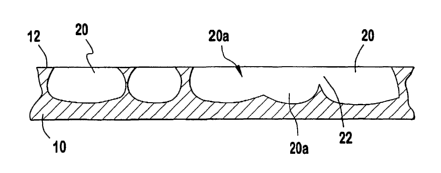
Refernng to FIG. 1, it will be seen that there first is provided an article 10
of one of the
above-mentioned materials, or a material similar thereto. The article 10 is
provided with a
datum surface 12 in which it is desired to provide a multiplicity of undercut
recesses.
As shown in FIG. 2, a layer 14 of maskant material is deposited on
substantially the
entirety of the surface 12. The maskant is a suitable acrylic, epoxy, or
polyester resist, or the
like. The layer 14 may be applied by dipping, spray coating, or electrostatic
depositing, or
any other coating method to produce a layer thickness of about 0.001-0.010
inch. The coated
article of FIG. 2 preferably is baked at 200°F (t10°F) for about
15-17 minutes, or any
sufficient combination of time, pressure (such as vacuum-baking) and
temperature to insure
the removal of water, as is customarily used in the art. Kodak Thin Film
Resist~ has been
found to be a quite suitable maskant. To the Kodak Resist is added 2%, by
weight, carbon
black pigment, or other pigment described hereinbelow.
In one embodiment, the adhesion of the resist or masking agent to a metal
surface of
the object to be textured preferably includes of an actual chemical, ionic, or
molecular bond
to the metal itself. In one embodiment, the undercutting process preferably is
conducted via
spray impingement of the etchant, or other agitation, such as turbulence or
ultrasonic
16
CA 02470068 2004-06-11
WO 03/053669 PCT/US02/39743
cavitation, often for periods of time that are more prolonged than is
generally encountered in
common photofabrication. The duration of the undercutting process is dependant
upon the
substrate selected and the etchant system chosen. Preferably, etching lasts up
to 10-15
minutes. It may exceed 20 minutes. In some embodiments of the present
invention use a
layer 14 of common photopolymerized polyester resist, requires cleaning and
abrading of the
datum surface prior to application of layer 14. As is common good practice in
the metal
finishing field, in some embodiments, pre-etching and pre-baking of the metal
surface is
sometimes required to insure the integrity of the maskant-metal bond. In one
embodiment, it
is desirable to remove a minute amount of surface material (preferably 0.001
inches to 0.005
inches) to insure a chemically clean and pristine metal surface. Additional
embodiments
include the use of a layer 14 of epoxy dip coatings, electrostatic coatings,
electrophoresis
coatings or other electro-deposited coatings, and spray coatings of resist or
masking agents.
Dispersing an appropriate pigment or dye into a maskant layer can render the
maskant
laser receptive. The maskant is selected based on the wavelength of the laser,
or any
projected light source, to be used to produce the desired pattern of maskant
14 on surface 12.
In one embodiment, in the case of an infrared laser, the resulting local
heating from the
absorption of laser energy selectively removes tiny areas of the resist or
maskant layer 14,
thereby exposing the underlying metal surface 12 of the article to the action
of an etchant.
Ordinary conventional photographic image and development techniques may be
used with
these photosensitive materials and methods. They are less suitable, however,
for contoured
parts, as artwork negatives cannot easily be laid upon them for exposure.
As noted above, a preferred maskant is Kodak Resist, to which is added about
2%
carbon black pigment, or other pigment more particularly suited to the laser
wavelength to be
employed. The pigment is dispersed into the maskant in a high shear mixer
until fully
dispersed, or until a temperature rise of 15-20°C is reached. The
resulting maskant is applied
17
CA 02470068 2004-06-11
WO 03/053669 PCT/US02/39743
by dipping or by spraying, spinning, brushing or electrostatically depositing
onto the surface
to be treated.
Selected areas 16 of the layer 14 are then removed to expose portions 18 of
the datum
surface 12. In one embodiment, the use of computer-directed direct laser
ablation to generate
programmed patterns in the maskant layer allows the application of such
patterns to
irregularly shaped finished goods, parts, or items which have surfaces of
compound curves or
radii. Such shapes are routinely encountered in implantable medical devices,
such as dental
post implants, hip joint assemblies, and maxillofacial prosthetics.
To generate a selected image, or array of recesses, or a fractal pattern, in a
laser
receptive maskant, the use of a computer-directed laser to directly ablate the
maskant or etch
resist layer in selected loci is preferred.
FIG. 40 illustrates a preferred two-dimensional pattern 410. In one
embodiment, the
pattern is transferred to the surface maskant by laser ablating black regions
412. The black
regions 412 are then etched with undercutting until the etched regions reach
the desired
complex 3-dimensional arrays of cavities. In one embodiment, the resulting
patterns does not
superficially resemble the starting pattern, though the resulting pattern is a
derivative of the
starting pattern, and/or the starting patterns basic fractal elements.
In a preferred embodiment of the method, ablations are made by direct writing
with a
neodymium-doped YAG laser with a wavelength of 1.06 microns, to which carbon
black is
receptive. A pattern is selected which optimizes the effects of undercutting.
The pattern
chosen is saved in Tagged Image File Format (TIFF) or as a plot (PLT) graphics
file, and
used to direct a laser marker.
In one embodiment, an Electrox, Scriba Nd:YAG laser marker may be used, with
patterns stored in digital file format. Upon laser exposure, the underlying
surface portions 18
are exposed in those areas in which the maskant absorbs the laser beam.
18
CA 02470068 2004-06-11
WO 03/053669 PCT/US02/39743
The pattern produced by laser ablation is predictable and can be accurately
duplicated
and repeated from implant to implant. While the aforementioned YAG laser has
been found
effective in the present invention, so also have C02, diode pump, and green
lasers. Any laser
capable of ablating, or thermally vaporizing, the maskant to generate a
desired pattern of
exposed surface may be used in carrying out the method described herein. Other
methods of
removing maskant include: mechanical tools, chemical milling, photo-chemical
etching and
laser eradication.
The pattern can be generated on a Computer Aided Design (CAD) system using any
compatible file type, or generated as a phototool for imaging. The pattern can
be scanned
from a drawing, print, photograph, or the like, and converted into any file
type compatible
with the laser system employed.
An alternative method of manufacture is to use a photo sensitive maskant,
which is
applied to the device as stated above, or applied as a dry film which is
laminated to the
surface. The maskant is then exposed, using a light source of an appropriate
wavelength
(typically 280-550 nanometers). Portions of the maskant are cross-linked
and/or bonded to
the surface during the exposing process (in the case of negative working
resist). The other
areas of the maskant are dissolved or washed away in a developing process that
utilizes a
compatible developer solution, such as sodium or potassium carbonate, or
stoddard solvents,
thereby exposing the underlying material.
The exposed portions 18 of the surface 12 are etched, preferably using a spray
etcher
at 100°F spray temperature and 10 lbs/in2 spray pressure, in a Nitric
and Hydrofluoric Acid
solution for about 20 minutes. Sufficient "fresh" etchant is continuously
impinged upon the
etch surfaces 18 to encourage lateral, as well as vertical etching. It will be
understood that
alternative etching processes, such as immersing ultrasonics and electrolytic
etching, can
produce similar results.
19
CA 02470068 2004-06-11
WO 03/053669 PCT/US02/39743
In one embodiment of the present invention, the methods of replenishing the
etchant
at the surface being textured is required to successful practice of the
invention. At the
etchant/metal interface a depletion condition exists as some of the active
species in the
etchant are consumed by the etching action and the formation of metal salts.
Because
aggressive and uniform etching action is preferable to ensure development of
the desired
elliptical geometry of the etched cavity, a spray impingement method is
frequently used. In
one embodiment, such a spray method allows an off axis or directional control
to achieve the
resulting textured surface and causes the undercutting to be in one or more
desired directions
or axes of the part (e.g., device 10) rather than another.
In a preferred embodiment, the spray etching system can be arranged so as not
to
impinge from all angles, or not to rotate with respect to the workpiece. For
example, if a
spray nozzle is affixed so as to impinge the work at a constant oblique angle
to the piece
being etched, then surface flow effects will control the etching process in
the microscopic
regions of the ablated maskant in such a way that "tilting" of the etched
cavities (described in
more detail below) is a consequence of the process.
The etching produces recesses 20 which are undercut, as shown in FIG. 4, and
which
are, in part, interconnected, as at 22. In the preferred embodiment, the metal
is etched in such
a manner as to deliberately cause undercutting of the pattern, and to permit
connection,
joining, or "breakthrough" of some of the recesses so as to produce a sharply
defined
complex network structure, including an interconnecting pattern in which the
size of most of
the recesses is smaller at the surface 12 than at a plane some distance below
the surface 12 of
the article 10. The recesses 20 may, in at least some instances, interconnect
at and near the
surface 12, as at 22 in FIG. 4, to provide enlarged surface recesses 20a (FIG.
5).
The etching of the metal surface 12 is thus carried out in one step, as
opposed to
repetitive etching suggested in some of the prior art references cited
hereinabove. In the
CA 02470068 2004-06-11
WO 03/053669 PCT/US02/39743
preferred one-step etching process, non-spherical ovoid shaped recesses are
created featuring
desired sizes and depths which are repeatable from implant to implant.
The remaining resist may be removed by immersing the body surface in a
NJ/Phase
23 Stripper bath at about 180°F for about 10 minutes. Alternatively,
the maskant layer may
be removed (FIG. S) by solvation or emulsification. If desired, the article 10
may be lightly
post-etched.
In one embodiment, in a titanium hip joint, for example, the metal was first
washed
with an alkaline degreasing detergent (e.g., an Oaktite~ solution), water-
rinsed in de-ionized
water and briefly pre-etched in a hydrofluoric/nitric acid etchant solution,
so as to produce a
chemically clean, freshly-exposed metal surface suited to maskant adhesion,
water-rinsed
again thoroughly, and oven dried at 110°C prior to coating with the
selected resist agent
(e.g., Kodak KPFR, or any other resist, including other polymer classes, such
as reactive
epoxy or urethan systems, or lacquers and varnishes). A polyester resist
coating was then
applied by dipping, air-drying for 15 minutes, and baking at 100°C for
20 minutes.
Alternatively, an epoxy e-coating (e.g., PPG Powercron~ CF-665) can be
successfully used.
A desired pattern may then be laser-imaged onto the surface with a 90 watt
neodymium-
doped YAG laser at 85% power, and at a machine setting of 3 frequency units to
achieve a
shallow penetration to and below the surface of the base metal. The typical
depth of
penetration is 10 microns. This assures the cleaning of the metal base layer
to remove
maskant ablation residues from the regions to be etched. The hip joint was
then again baked
at 100°C prior to etching to insure full cross-linking of the polymer,
and to remove low-
molecular-weight pyrolysis products from the maskant polymer.
The surface of the hip joint was then etched to the desired depth. The
broadest depth
range is O.lmm to 2mm, with a preferred depth range of 0.4 to 0.6 mm. Certain
patterns may,
in extreme situations on some metals require shallower or deeper etching than
the preferred
21
CA 02470068 2004-06-11
WO 03/053669 PCT/US02/39743
range, in order to develop a desired three-dimensional pattern. For example,
more delicate
pattern designs, may require very light etching in order to preserve the
original pattern
details. However, nearly all the patterns used to date have been well-formed
by etch depths
of 0.4 to 0.6 mm.
The depth range preferably depends on pattern coarseness. In one embodiment,
coarser patterns have wider land areas (discussed below), and therefore can
tolerate deeper
etch depths before they are cut off by undercutting in a vertical spray etcher
at 20 to 30
minutes (depending on desired depth and pattern coarseness) at 100°F.
After etching to the desired depth the article is rinsed well in running water
and air
dried. The article is then inspected for proper etching, sufficient
undercutting, and general
quality. The resist or maskant was then stripped from the hip joint using a
nuphase stripper
solution such as a concentrated caustic stripping solution (e.g., Oakite~
Eurostrip~ 704
manufactured by PC&E), at a temperature of 160°F, for 15 minutes. After
stripping, the
article was rinsed in deionized water and oven dried at 220°F.
In one embodiment, there is thus provided a method for producing a complex, at
least
in part interconnecting pattern, or similar 3-dimensional surface treatment,
to enhance the
attachment of biological matter to a surface of an implantable device, or the
interconnection
of other bodies to be bonded, made by selective etching and undercutting of a
surface so as to
form a network of at least in part interconnected recesses. The pattern is
formed by the direct
laser ablation of an etch resist or maskant layer, allowing the textured
surface to be applied to
items with complex or curved surface geometries. The pattern is stored in a
CAD or other
computer-based system which controls the maskant ablations and is predictable
and subject to
repetitive duplication. The article is chemically etched to form the complex
pattern. The
metallurgical properties of the material of the article are not altered by
heat, but remain
substantially consistent during the process. Soft tissue or bone may in-grow
the surface so
22
CA 02470068 2004-06-11
WO 03/053669 PCT/US02/39743
produced, resulting in an interpenetrating network that offers superior
mechanical adhesion
and resistance to degradation. Further, the sharp edges at the intersections
of the undercut
recesses and the original datum surface facilitate an initial "scratch-fit"
between the implant
surface and a bone.
In operation, to produce a textured surface on a surgical implant, a selected
pattern of
undercut and at least in part interconnected recesses is effected in a surface
of the surgical
implant (FIG. 5). In implantation, the implant surface 12 is pressed against
the bone B, (FIG.
6) such that sharp edges 24 of the recesses effect the "scratch fit" with the
bone B, which
involves shaving off, or milling, particulate segments b of the bone B, which
segments b
enter the ovoid recesses 20 wherein, in due course, the bone segments b
stimulate in-growth
of the bone B (FIG. 7) to securely lock the implant to the bone B (FIG. 8).
Thus, the scratch-fit securely adjoins the implant article 10 to the bone B,
to prevent
or minimize micro motion between the body 10 and bone B. The presence of such
motion
would discourage the ingrowth of bone into the implant and thereby discourage
the long-term
1 S interconnection of the implant and bone.
Further, the scratch-fit application of the implant to the bone harvests bone
particulate
matter which falls into the surface recesses and is retained by the recesses
to encourage and
stimulate ingrowth of the bone into the recesses. Inasmuch as the recesses are
of an ovoid
configuration, they provide a greater sub-surface fractal area than
spherically shaped
recesses, and thereby a greater area for engagement of the bone material and
the implant.
Refernng to FIG. 1 l, it will be seen that for bones B accepting an implant 10
having a
plurality of datum surfaces 12, including opposed surfaces 12a and 12b, the
accurate location
of the datum surfaces is most critical, inasmuch as any build-up of implant
material above the
datum surfaces causes the implant not to be accepted by the bone B. Texturing
the surfaces
12 below the surfaces 12 does not add material to surfaces 12. Whereas,
texturing the
23
CA 02470068 2004-06-11
WO 03/053669 PCT/US02/39743
surfaces 12, by adding texture above the surfaces increases the space required
between the
opposed bone surfaces to accept the implant and leads to rejection of the
implant. Known
methods of texturing by adding to a surface lack the required precise control
to determine the
deviation of the peaks of the added material. The method presented herein
facilitates
accurate and precise location of datum surfaces of surgical implants.
The milling of the host bone B may further serve to ream the bone B to the
precise
size and configuration of the article 10, insuring the best possible fit.
Accordingly,
appropriately shaped and sized burrs of the texture on article 10 are
preferably furnished to
pre-form a receptacle in the bone to properly receive the article 10. The
scratch-fit securely
adjoins the implant article 10 to the bone B, to prevent or minimize micro
motion between
the implant 10 and bone B. The presence of such motion would discourage the
ingrowth of
bone into the implant and thereby discourage the long-term interconnection of
the implant 10
and bone B.
In some embodiments, a device with a textured surface according to the present
invention, can affect a self fitting function. For example, it will be
appreciated that where a
textured surface, according to the present invention, is applied to an
acetabular cup, the cup
itself can be used as a reaming tool, effecting a perfect fit to the host bone
and shortening
healing time. Further, in the process of self fitting, there is milled, or
harvested, fine bone
particulates, or pulp, from the patients' own body, for example, as shown in
FIGS. 6-10. The
resulting material is forced into the recesses to serve as a nucleation host
for a spontaneous
homograft completed by the attraction and growth of the patient's osteoblasts,
providing a
strong bond and longer installed lifetime.
Some embodiments of the present invention may incorporate sharp-edged geometry
such as at undercut 3 (See FIG. 12). It may be desirable for the sharp-edged
undercut
geometry to be later modified by a subsequent dulling process so as to causes
sharp edges to
24
CA 02470068 2004-06-11
WO 03/053669 PCT/US02/39743
be rounded. For example, abrasive blasting, glass bead blasting, or a
subsequent acid etch all
may be used to slightly dull or "break" the original sharp edges, if desired.
Such a
subsequent dulling process may include, for example, etching, polishing (e.g.,
electropolishing), abrasive reduction, buffing, and honing. Through this
process, at least
some of the walls 7 (shown on Fig. 14 and described in more detail below) may
become
perpendicular to the surface of the article, or may actually diverge outward
from each other as
they approach the surface of the texture pattern. In some embodiments, post-
processing (e.g.,
cleaning or polishing) defeats the benefits of the textured surface. Still,
some practitioners
may incorporate a post-processing step.
Looking in more detail at formed features according to some exemplary
embodiments
of the present invention, FIG. 14 illustrates a preferred geometry of a cavity
20 which has
been deliberately produced with the geometry of a complex ellipsoid. A complex
ellipsoid is
preferably derived from two or more ellipsoids. In a preferred embodiment, the
complex
ellipsoid is derived from at least two non-spherical ellipsoids. Alternative
embodiments may
include combinations of spherical and non-spherical ellipsoids. The complex
ellipsoids may
be a combination of ellipsoid 30a and ellipsoid 30b. The complex ellipsoids
may
alternatively combine three or more ellipsoids with three or more different
angular
projections. More particularly, a geometric shape of the illustrated cavity 20
may be
described as being defined, at least in part, by two non-spherical ellipsoids
sharing a major
axis 9 and having varying angular projections (e.g., an ellipse having a 40
degree protection,
superimposed on an ellipse having a 50 degree projection, or any combination
of ellipses
from about 9 degrees to about 89 degrees so as to produce a species of the
complex ellipse
shape).
Ellipsoid 30b with a narrower angular projection defines a bottom 4 of cavity
20.
Ellipsoid 30a with a wider angular projection defines an arched undercut 3
which is
CA 02470068 2004-06-11
WO 03/053669 PCT/US02/39743
contiguous with wall 7 and bottom 4. In the preferred embodiment wall 7 and
bottom 4 are
concave and contiguous. Cavity 20 (FIG. 16) may be further characterized as
having a ratio
of cavity width, w, to cavity depth, d, that is greater than or equal to
unity. The preferable
width:depth ratio ranges from 1:1 to 9:1. In a preferred embodiment, the ratio
is
S approximately 4:1.
Referring now to FIGS. 12, 14, lSa, 15b, and 15c, protrusions 2a, 2b, and 2c
have
differing geometries each of which is preferably defined by the relationship
between at least
two complex ellipsoids. Protrusions 2a, 2b, and 2c extend from bottom surface
4 of cavity 20
toward datum 12. Protrusion 2a has a peak Sa in the form of a plateau or mesa
which is
coincident with outer surface 12. Complex ellipsoids l la and 1 lb define the
lateral
dimensions of protrusion 2a. Cavity 20 is also defined by complex ellipsoids l
la and l lb
which intersect datum 12 to create cavity 20 in outer surface 12. The portion
of outer surface
12 remaining untouched by the ellipsoids l la, 1 lb defines peak Sa. An
ellipsoid forming
protrusion 2a preferably forms an undercut 3 at the face of the protrusion.
Protrusion 2c has a sub-peak Sc located between datum 12 and bottom 4. Sub-
peak
Sc is defined by the intersection point of at least two complex ellipsoids 11.
In a preferred
embodiment, protrusion 2c is formed from the at least two complex ellipsoids l
la, l lb,
which overlap to the extent that no overhang is created in wall 7 of
protrusion 2c. In some
embodiments the overlapping of ellipsoids may result in an overhang on one
face of the
protrusion but not on another face of the same protrusions.
Protrusion 2b has an intermediate peak Sb which is also located between datum
12a
and bottom 4. Intermediate-peak Sb is preferably defined by two or more
complex ellipsoids
l la and l lb that do not overlap. The complex ellipsoids l la, l lb that at
least partially define
intermediate-peak Sc, however, are in close enough proximity to each other
that the portion
140 of article 10 that previously existed between intermediate-peak Sc and
datum 12a can be
26
CA 02470068 2004-06-11
WO 03/053669 PCT/US02/39743
predicted to break-off during manufacture such as, for example, during
polishing of the
etched product, leaving an intermediate peak Sc. In one embodiment, the
surface is
"softened" by common buffing or polishing methods (as described above). Thus,
in one
embodiment, the surface is modified such that the sharp edges will be removed,
as well as
some of the tops of the projections.
For clarity, FIGS. 14 and 16 shows the major axis 9 substantially parallel
with datum
surface 12. Orientation of the major axis may vary from perpendicular to
parallel depending
on the desired finished texture, the pattern of the desired texture and
direction of the texture.
Preferred embodiments have an orientation of axis 9 that varies between 90
degrees and 70
degrees relative to datum 12. In one embodiment, for example, axis 9 is
oblique to datum
surface 12 (shown in Fig. 16A). This is preferably achieved by impinging
etchant at an acute
angle to datum surface 12. In one embodiment there is achieved an asymmetrical
undercut
pattern that is characterized by an undercut face 165, of ellipsoid 166, that
projects over
cavity 20 a distance greater than the projection of the opposing face 164. In
one embodiment,
opposing face 164 does not overhang cavity 12. In one embodiment, the
impingement of
etchant at an angle produces a tilted saw-tooth or tiger-tooth structure 169
shown in FIG.
16B. Such structure allows relatively easy insertion into a bone channel 168
(e.g., when
insertion is in a direction such that undercut 165 is not urged into bone B as
in FIG. 16C), but
strongly resists tensile force urging dislodgment of the implant (e.g., when
removal is in a
direction such that undercut 165 is urged into bone B as in FIG. 16D). In one
embodiment,
cavities 20 are aligned in series so that a cross-section of the textured
surface has a regular
pattern which may also be a repeating pattern (see, e.g., FIGs. 16C, 16D), 42A
and 42B).
FIGs. 42A and 42B illustrate a comparison between the affects of directional
and
nondirectional impingement. FIG. 42B illustrates an embodiment of the
invention where
nondirectional impingement has been performed. In a preferred embodiment
employing
27
CA 02470068 2004-06-11
WO 03/053669 PCT/US02/39743
nondirectional impingement, the major axis 9 of complex ellipsoid 30 is
substantially parallel
with datum surface 12, and undercuts are present at the point 432 where the
cross section of
ellipsoid 30 intersects datum 12. In another embodiment illustrated in FIG.
42A, major axis 9
is oriented oblique to datum surface 12, there is an undercut 432 at the
distal end 431 of
cavity 20, and face 434 has no undercut at the proximate end 433 of cavity 20.
In one
embodiment, cavity 20, formed by directional etching, is defined by a concave
surface 432
and a convex surface 434. Alternatively, opposing surfaces of cavity 20 may
each have
concave configurations (FIGs. 16C and 16D).
In one embodiment of implant 40 (illustrated in FIG. 41), unidirectional barbs
422 are
produced in the surface that can be inserted into bone or tissue. The
directional orientation of
barbs 422 make it difficult to withdraw from the bone or tissue. In conditions
where a
directional or anisotropic geometry are not desired, simpler non-directional
agitation such as
a turbulently-flowing etchant bath, or ultrasonic cavitation can be employed.
FIG. 16 illustrates a preferred geometric relationship between surface 12 and
ellipsoids 30a and 30b. The dimensions are typical and descriptive of a
commonly achieved
pattern, but are not restrictive. The skilled practitioner may vary these
dimensions greatly
depending on the desired outcome. In the embodiment illustrated, depth d of
cavity 20 as
measured from the datum 12a to bottom 4 is substantially 0.0210 inches. Major
diameter a of
ellipsoids 30a and 30b is substantially 0.0474. The distance b from datum 12
to the major
axis 9 is substantially 0.0055 inches. Distance c along the major axis 9
between the perimeter
of complex ellipsoid 30a to a point x projecting from the intersection of
complex ellipsoid
30a with datum 12a is substantially 0.0012 inches. It follows, that distance a
along major
axis 9 between projection points x and y is substantially 0.0450 inches.
Returning to FIG. 13, there is illustrated preferred three dimensional texture
130.
Texture 130 is embodied in an article 10 after the etching process described
above. In this
28
CA 02470068 2004-06-11
WO 03/053669 PCT/US02/39743
embodiment, while three dimensional pattern 130 may be of an irregular pattern
to the extent
that the structural features of the pattern are unevenly distributed on a
micro-level, this
pattern is both repeatable across the device, and reproducible between
devices.
The repeatable and reproducible nature of texture 130 is achievable through
the
employment of patterns (e.g., pattern 400 in FIG. 40) which may be seen by an
observer. In
one embodiment, the patterns are written into or through the maskant or etch
resist layer by a
laser as described herein. In another embodiment, the pattern is displayed as
an image that
can be seen by an observer on a computer monitor, printed output, or other
such viewing
device.
In one embodiment, pattern 400 may be derived from an initial pattern having a
regular series of elements that include one or more regular or irregular
geometric
configurations such as circular dots, squares, prisms, parallelepipeds,
trapezoids, triangles,
hexagons, and other such geometric shapes that the practitioner deems suitable
to generate
the third-dimension development of the desired finished texture 130. In one
embodiment, a
pattern of repeated fractals or other pseudorandom network of elements are
employed to
generate the initial pattern. In practice when patterns are developed into
three-dimensional
patterns 130 by etching, they generally regularize rather than randomize. For
example, an
array of dots or other shapes will form boundaries as the surrounding material
is removed by
etching. When a connection density of portions of the etched surface peaks,
sub-peaks, and
intermediate peaks of approximately 58% is reached, a phenomenon known as
"percolation
threshold" forms, where networks of connections form.
FIGS. 17A, 17B, 17C, 17D, and 17E illustrate variations on the textured
surface of the
preferred embodiment. FIG. 17A illustrates textured surface 170 having a three-
dimensional
irregular pattern that is characterized by multiple intersections of complex
non-spherical
ellipsoids that form peaks Sa and Sc. There is illustrated sub-peak 161 which
is defined by a
29
CA 02470068 2004-06-11
WO 03/053669 PCT/US02/39743
the intersection of at least two complex ellipsoids. A first complex ellipsoid
defining sub-
peak 161 further defines, at least a portion of cavity 162. A second complex
ellipsoid
defining at least a portion of cavity 163, intersects the first complex
ellipsoid to define sub-
peak 161. This configuration further illustrates a resulting texture where
intersecting
complex ellipsoids of varying angular projections are oriented with major axes
at an offset
angle to one another. For example, if the initial pattern used includes
elongated elements, or
sets of two or more dots, a first set oriented at one angle to a grid, and a
second set oriented at
another angle, then the etching and undercutting process will generate a
series of ovoid
shapes as viewed from the top, with long axes aligned to these grid lines. For
example, one
set of grid lines may be at an angle of 40 degrees to the other, resulting in
a lozenge-shaped
array.
Also illustrated in FIG. 17A is the effect of a scratch-fit where soft tissue
shavings
160 have migrated into the voids 20 and are growing to fill the voids.
FIG. 17B illustrates textured surface 171 having a regular pattern. Each
protrusion 2a
in FIG. 17B has peak Sa coincident with outer surface 12 (not illustrated).
There is
furthermore, a geometry characterizing protrusion 2a that is defined by four
(4) complex
ellipsoids which have been uniformly oriented. Further characterizing this
embodiment is a
distance, f, between protrusions that is uniform among transverse and
longitudinally
adjoining protrusions. In this embodiment, distance f between protrusion 165a
and 165b is
substantially the same as the distance, g, between 165b and 165c. In
alternative uniform
embodiments, the distance between transversely adjacent protrusions may be
different from
the distance between longitudinally adjacent protrusions thus forming a
"rectangular pattern"
as opposed to a "square pattern". Any other geometrically uniform patterns are
also within
the scope of this invention. These include, as examples, ellipsoidal cavities
substantially
arranged on a hexagonal, pentagonal, triangular, or other regular geometric
lattice.
CA 02470068 2004-06-11
WO 03/053669 PCT/US02/39743
FIG. 17C illustrates a uniform repeating pattern that combines a plurality of
protrusion geometries including protrusions 166a defined by five (5) complex
ellipsoids;
protrusions 166b defined by four complex ellipsoids; and sub-peaks 166c
defined by three
complex ellipsoids 166c. In this embodiment, the horizontal orientation of
adjoining
protrusions varies in a substantially non-random pattern.
FIG. 17D illustrates protrusion 167b that is defined by a multiplicity of
complex
ellipsoids. Protrusion 167b has peak 167a that has been formed to project a
scalloped surface
177. There is also shown intermediate peak 175 and sub-peak 176. In this
embodiment,
intermediate peak 175 has one face that is not undercut, or with undercut
regions have been
etched away in the process, and another face that is undercut.
FIG. 17E illustrates protrusion 168 that has been defined by a multiplicity of
complex
ellipsoids. In one embodiment, protrusion 168 extends for a predetermined
length of the
surface. In one embodiment, the predetermined length forms a rib. The top of
rib 168 is
preferably coincident with the datum surface 12. In other embodiments rib 168
has a sub
peak or an intermediate peak. Alternatively, rib 168 has a combination of
peaks, sub-peaks,
and intermediate peaks. In this embodiment, each face 17 of protrusion 168 has
an undercut
surface. There is also illustrated protrusion 169 which is characterized by an
intermediate
peak Sb that was formed when the base material above the peak was broken-off,
or where a
top section became isolated or cut away by the interception of undercut
regions.
While some textured surface applications benefit from an undercut textured
embodiments, the methods of the present invention can be used to create
engineered articles.
For example, certain embodiments are suited for creating an implant article
having a mesh-
and plate-surface (described below). In other embodiments, undercut textured
surfaces are
combined with mesh-and-plate surfaces.
To make a mesh-and-plate surgical implant, there is provided a thin sheet 180
(FIG.
31
CA 02470068 2004-06-11
WO 03/053669 PCT/US02/39743
18) of tissue and bone compatible metal, such as titanium. A maskant layer 192
(FIG. 19) is
applied to a first face 194 of the sheet 180 and a maskant layer 196 is
applied to a second face
198 of the sheet 180. The maskant layers 192, 196 cover substantially the
entirety of the first
and second faces 194, 198, respectively. The maskant layers 192, 196 are
resistant to
chemical attack. It has been found that a photo-chemical resist, such as
duPont Riston~, or
Kodak Thin Film Resist~, serve as appropriate materials for the maskant layers
192, 196.
The maskant layers 192, 196 are then in part ablated from selected portions of
the
metal faces 194, 198 (FIG. 20), as by mechanical tools, chemical milling,
photo-chemical
etching, or by laser eradication, to expose portions 202, 204, 206 of the
respective metal faces
194, 198 in desired patterns, ready for etching.
Refernng to FIG. 21, it will be seen that the exposed portions 204 of the
first face 194
and the maskant layer 192 on the first face 194 are covered with a protective
tape 210,
leaving exposed only the region 202 where a central through-hole is desired
for acceptance of
a mounting screw (not shown). Similarly, the exposed portions 206 of the
second face 198
and the maskant layer 196 on the second face 198 are covered with a protective
tape 212.
The tapes 210, 212 may be 3M Brand Type #1280 Platers Tape.
The through-hole region 202 is then subjected to etching, for example, as by
spray or
immersion, using an acid bath of a mixture of nitric and hydrofluoride acid.
It is preferred,
during the etching process, to periodically remove sheet 180 from the etching
process and
rinse, dry and bake the sheet to maintain the integrity of the maskant and
allow for in-process
inspections.
When the etchant reaching the exposed surface 202 has created a shallow crater
222
(FIG. 22), the protective tape 210 is removed (FIG. 23) and the etching of the
crater 222 is
resumed, and etching of the exposed portions 204, constituting the mesh
portion of the
implant, is undertaken. As etching proceeds, the exposed metal regions 222 and
204 are
32
CA 02470068 2004-06-11
WO 03/053669 PCT/US02/39743
progressively removed by the etchant (FIG. 24). The etching continues until
the removal of
metal from the first face 194 and crater 222 has reached the predetermined
extent desired
(FIG. 25).
The second tape 212 is then removed, exposing the maskant layer 196 and
exposed
portions 206 on the second face 198, including an area 252 opposite the crater
222.
Etching of the through-hole area 252 in the sheet face 198 breaks through to
the crater
222 to effect a counter-sunk through-hole 262 (FIG. 26) and second face
openings 264 in
communication with the newly etched first face 266.
The first and second maskant layers 192, 196 are then removed (FIG. 27),
leaving an
implant device having the mesh portion 272, a plate portion 274, and at least
one through-
hole 262 for receiving a mounting screw.
In FIG. 28 there is shown, for illustrative purposes, a dog-leg plate portion
274 having
one or more through-holes 272 therein, the plate portion 274 being bounded by
the mesh
portion 272. In FIG. 29 there is shown a divided plate 292 having through-
holes 262 therein,
and bounded by the mesh portion 272.
Refernng to FIG. 30, it will be seen that through-holes 262 may be provided in
mesh
portions 262, such through-holes preferably being surrounded by rim collars
302 comparable
in thickness to a plate portion 274. The through-holes 262 preferably are
countersunk to
receive mounting screws.
In an alternative embodiment, the maskant layers 192, 196 may be exposed to a
movable laser beam which is moved in accordance with a path governed by a CAD
data file,
wherein the beam removes unwanted maskant. After the laser removes the
maskant, the
sheet 180 is exposed to heat and/or ultraviolet light to cure and harden the
remaining
maskant.
The mesh portions 272 preferably are of a thickness of about .5 mm and are
readily
33
CA 02470068 2004-06-11
WO 03/053669 PCT/US02/39743
flexed to follow the curvature of a bone.
There is thus provided an improved method for making an article such as a mesh-
and-
plate surgical implant including both bendable perforated mesh portions and
relatively rigid
plate portions, wherein the bendable or comfortable perforated portions are
integral with and
kinematically related to the rigid plate portions. The improved method further
provides
through-holes for receiving mounting screws during implantation.
The performance of tissue implants may be enhanced by textured surface having
undercut characteristics and/or mesh-and-plate characteristics. For example,
where a textured
surface is specified to promote osseointegration an undercut texture may be
specified.
Alternatively, where there is need to join the implant with a mechanical
securement (e.g., a
screw or bolt), the mesh-and-plate texture may be specified. It should be
recognized that
combining one or more of such textures on a single implant is a viable
alternative within the
scope of this invention.
FIG. 31 illustrates tissue implant 310, constructed from a bio-compatible
material
such as metal, glass, plastic, and ceramic. In one preferred embodiment, the
bio-compatible
material is chemically pure (CP) titanium. Implant 310 is useful in any
application in which
a secure attachment to an object is desired and would be enhanced by using a
textured surface
such as discussed herein. Though dental applications and spinal applications
have different
mechanical objectives, the present invention is useful in both applications.
Spinal devices,
for example, preferably separate vertebrae in permanent.alignment and transfer
compressive
forces between adjacent vertebrae. Bone ingrowth secures these devices into
place,
especially with the object of resisting forces that would tend to dislodge
them. Dental
devices, for example, are preferably screwed into a hole bored in the
mandible, whereupon
bone ingrowth locks them into place so that teeth or other appliances can be
affixed to them.
34
CA 02470068 2004-06-11
WO 03/053669 PCT/US02/39743
In this application, dental devices must resist tensile and compressive forces
and transfer
those forces to the bone sufficiently that the bone-to-appliance bond strength
is not exceeded.
For example, implant 310 is useful for implantation and/or attachment to hard
tissue
such as bone in spinal and dental applications and soft tissue. In one
embodiment, implant
310 may be adapted for use in a commercial spinal fusion device. Another
embodiment of
implant 310 may be adapted for use in dental applications, such as with a
dental implant in
tissue. In one embodiment, implant 310 is particularly well suited as a dental
implant post
onto which prosthetic teeth or other appliances may be attached.
Implant 310 has a longitudinal axis 311. Distal end 312 of implant 310
typically is
inserted into tissue such as a bone (e.g., a jaw bone). In one embodiment,
proximal end 314
may be adapted to receive a prosthesis such as an artificial tooth or other
dental appliance.
As shown in FIG. 32, in one embodiment, implant 310 preferably has a wall 320
that
is disposed about longitudinal axis 311. In one embodiment, wall 320 is
radially disposed
about axis 311, and wall 320 has an outer face 322, and an inner face 321. In
one
embodiment, one or both of outer face 322 and inner face 321 may be
substantially parallel to
axis 311. In a preferred embodiment, wall 320 is tapered such that distal end
312 of wall 320
has a smaller diameter about axis 311 than at proximal end 314 of wall 320. In
one
embodiment, wall 320 defines cavity 324.
It was known to use perforations in tissue implants to promote tissue inter-
growth
necessary to lock an implant to tissue, and to use perforations in tissue
implants to vent heat
and tissue during installation, as set forth in U.S. Patent No. 4,960,381, the
entire contents of
which are incorporated herein by reference. In an embodiment of the present
invention, the
textured surfaces described below are believed to satisfy one or both of those
functions and
implant 310 does not include any perforations in wall 320.
CA 02470068 2004-06-11
WO 03/053669 PCT/US02/39743
In other embodiments, as shown in FIGS. 31, 35, 37A, 37B, and 37C, wall 320
includes one or more apertures 318 which permit the transmission of fluids
(e.g., blood),
tissue, and tissue fragments (e.g., bone fragments) into cavity 324 to
promote, for example,
tissue inter-growth (i.e., osseointegration) and/or a heat and/or tissue
venting. Aperture 318
may be formed by any method including the methods described herein or other
mechanical
methods (e.g., drilling), chemical methods(e.g., chemical etching), and/or
heat methods(e.g.,
laser). In one embodiment, apertures 318 are formed by chemical etching of one
or both of
outer surface 322. Apertures 318 may be more specifically formed by a chemical
etching
process applied to outer surface 322. In another embodiment, aperture 318 is
formed by laser
ablation and chemical etching process applied to both outer surface 322 and
inner surface 321
such as the method used to create countersinks, mesh geometries and textures
as described in
U.S. Patent application Nos. 09/976,722 to Amrich et al., and 10/021,616 to
Amrich; the
entirety of each being incorporated herein by reference.
As shown in FIGs. 31 and 32, in preferred embodiments, there is at least one
aperture
318 that is defined by aperture surface 323, extending between inner face 321
and outer
surface 322. Aperture surface 323 may be of any geometry. In one embodiment,
aperture
surface 323 is substantially flat (e.g., is substantially in a two dimensional
plane). In another
embodiment, aperture surface 323 is curved such as concave, convex or a
compound curve
(e.g., a curve with both a concave and a convex component, and multiple
concave or convex
curves of varying radii and combinations thereof). In one embodiment, aperture
surface 323
is disposed normal to axis 311. In another embodiment, aperture surface 323 is
oblique to
axis 311. Where aperture surface 323 is oblique to axis 311, that angle may be
either acute or
obtuse. Aperture surface 323 may also be disposed at varying angles to inner
face 321 and
outer surface 322.
36
CA 02470068 2004-06-11
WO 03/053669 PCT/US02/39743
In a preferred embodiment, aperture surface 323 is at least partially a
textured surface
351 as illustrated in FIG. 35 and as disclosed herein. In one embodiment, the
textured
surface 351 of aperture surface 323 has a texture that is characterized by
undercut recesses
such as is created from the methods described herein.
Aperture 318 may be of varying sizes depending on their intended function.
Aperture
318 can have any shape including curved (e.g., round aperture 371), angular
(e.g., square
aperture 372 and triangular (not shown)), complex geometric shapes (e.g.,
mufti-curved
aperture 373) and combinations thereof within any one aperture 318 or among
different
apertures 318 of the same implant 310 (as illustrated in FIG. 37A). In a
preferred
embodiment, aperture 318 has an oblong geometry with the major axis of
aperture 318
longitudinally oriented and the minor axis transversely oriented to axis 311
as illustrated in
FIG. 37C. Implant 310 may have one or more aperture 318. In one embodiment,
where
more than one aperture is included, apertures 318 are either angularly
disposed to one another
(e.g., less than 180° apart along face 321), longitudinally disposed
(e.g., aligned along the
longitudinal axis), disposed across from one another (e.g., at 180°
relative to face 321), and in
geometric combinations thereof. Apertures 318 may extend into the self tapping
region
(discussed below) of implant 310.
In one embodiment, as shown in FIG. 36, one or both of outer wall 322 and
inner wall
321 include one or more recesses 362 that do not extend through wall 320.
Recesses 362
may be formed by any method as described with respect to aperture 318 and may
include any
attribute of size, shape, and/or configuration discussed with respect to
aperture 318.
Furthermore, any portion or all of recess 362 may include a texture surface as
described.
As shown in FIGS. 31, 32, 33, 34, 35, 38A, 38B, 39A, 39B, 39C, 38C, and 38D,
in
preferred embodiments, implant 310 may also include one or more protrusions
325 from
outer face 322. In one embodiment, protrusions 325 may be one or more ribs
326. In one
37
CA 02470068 2004-06-11
WO 03/053669 PCT/US02/39743
embodiment, ribs 326 may be helically disposed about longitudinal axis 311 to
form threads
316 as illustrated in FIG. 31. An implant 310, having threads 316 may
typically be installed
by screwing implant 310 into tissue (e.g., bone). In one embodiment, thread
316 is
continuous from a point approximately at distal end 312 to a point
approximately at proximal
end 314. In another embodiment, threads 316 may be discontinuous. Where thread
316 is
discontinuous, the discontinuity may be at a location proximate to aperture
318 as illustrated
in FIG. 31, or the discontinuity may be located where there is no aperture as
illustrated in
FIG. 38C.
In one embodiment, protrusions 325 are in the form of longitudinal ribs 382
(as
illustrated in FIG. 38A and 33) longitudinally disposed on implant 310 such
that ribs 382 run
generally between proximal end 314 and distal end 312. In one embodiment ribs
382 run
generally parallel to axis 311. In another embodiment, angular ribs 383 may be
disposed at
an angle relative to axis 311, as illustrated in FIG. 38B. In another
embodiment, transverse
ribs 384 are disposed to form substantially concentric rings that are radially
disposed about
axis 311 as illustrated in FIG. 38D. Transverse ribs 384 can have any desired
pitch angle. In
one embodiment, transverse ribs 384 have a low pitch angle. In another
embodiment,
transverse ribs 384 have a pitch angle of substantially 0°. While
longitudinal ribs 382,
angular ribs 383, and transverse ribs 384 are shown to be substantially
continuous from distal
end 312 to proximal end 314, such ribs having discontinuities are also within
the scope of the
present invention. In one embodiment, ribs 326 may be in the form of discrete
segments 335
protruding from wall 320. In one embodiment, discrete segments 335 are evenly
distributed
about axis 311. In one embodiment evenly spaced discrete segments 335 are of
substantially
uniform size. In another embodiment, discrete segments 335 are of varying
sizes. In another
embodiment, discrete segments are unevenly spaced.
38
CA 02470068 2004-06-11
WO 03/053669 PCT/US02/39743
Ribs 326 may have any cross sectional geometry including v-shaped, keystone
shaped
(e.g., rib 343), curved (e.g., rib 344), trapezoidal with an exterior short
face (e.g., rib 348),
trapezoidal with an exterior long face (e.g., rib 347), undercut (e.g., rib
345), hourglass (e.g.,
rib 349) and square (e.g., rib 346), as shown in FIG. 34. In one embodiment,
ribs 326 on
S implant 310 are in the form of longitudinal ribs T32 having one or more of
several geometries
as shown in Figs 32, 33, 34.
It may be necessary, in some procedures, to drill at least one pilot hole or
successively
larger pilot holes to accommodate implant 310 prior to screwing implant 310
into the tissue.
In another embodiment, it may be desirable to drill a large-enough hole where
implant 310
can be tapped into place with a mallet. Typically, for implant 310, those
embodiments
having non-helical ribs are suited for tapping in place while those
embodiments including
threaded ribs are suited for screwing in place.
In a preferred embodiment, implant 310 may be configured to include a self
tapping
thread 317 that is helically disposed about longitudinal axis 311, as shown in
FIG. 31. Where
self tapping threads 317 are employed, installation may be accomplished with
no pilot hole or
with smaller diameter pilot holes than if the self tapping thread 317 was not
included. Self
tapping threads 317 of a preferred embodiment, has a thinner web thickness and
more root
dimension than crest spacing. The ribs may therefore, appear to be more widely
spaced apart.
Where self tapping threads are used, sufficient anchoring can be achieved
while reducing the
amount of material (e.g., tissue) that is cut or displaced. The tapping force
requirement to
install a device with the self tapping features is lower than is necessary for
non self tapping
threads.
In one embodiment, threads T13 on implant 310 include one or more portions
that are
non self tapping threads. For example, in one embodiment, only a distal
portion of the
threaded area of implant 310 is self tapping.
39
CA 02470068 2004-06-11
WO 03/053669 PCT/US02/39743
As shown in a preferred embodiment of FIG. 32, protrusions according to the
present
invention, have a height, h, suitable to restrict removal of implant 310, for
example, by the
application of axial, longitudinal, and/or radial force.
In one embodiment, outer face 322 and ribs 326 are textured over substantially
their
entire surface. In another embodiment, outer surface 322 and ribs 326 have
textured portions
and portions that are relatively smooth (e.g., not textured). More
particularly, where a
portion of ribs 326 are relatively smooth, that portion may be the crests 392
of ribs 326 as
shown in FIG. 39A. In another embodiment, as illustrated in FIG. 39A, outer
face 322 is at
least partially textured, and ribs 326 are non-textured. Leaving crests 392
without texture is
believed to facilitate the installation of implant 310, for example, by
avoiding excessive
fragmentation to tissue (e.g., bone) surrounding the distal end of implant
310. In a preferred
embodiment, outer surface 322 is textured and ribs 326 are not textured. In
other
embodiments regions of texturing and non-texturing may be used on inner face
321, outer
surface 322, aperture surface 323, and ribs 326. In one embodiment, inner face
321 is
textured as illustrated in FIG. 39B. In another embodiment, inner face 321 is
textured and
there are alternating regions of outer surface 322 that are textured as
illustrated in FIG. 39C.
Known devices, incorporate an anti-rotation fixture (which may include a
hollow
cross-drilled structure) that locks the screw device into permanent immobility
when bone
ingrows the fixtures Such a structure may further serve as a vent during
installation. In one
embodiment of the present invention, the locking mechanism is integral to the
surface of a
portion of the threads thus eliminating the need for an additional anti-
rotation fixture. In
Niznick (U.S. Pat. No. 5,571,017), for example, an anti-rotation fixture
characterizes
approximately one-third of the device.
In one embodiment, a textured surface may be described as having a higher
fractal
dimension than a non-textured surface. Depending on the nature of the implant,
the fractal
CA 02470068 2004-06-11
WO 03/053669 PCT/US02/39743
dimension may be specified based upon the nature of the implant. For example,
where the
scratch-fit properties of the implant (e.g., as described above) is of primary
importance, a
greater fractal dimension may be specified to improve the tissue harvest
during implantation.
Alternatively, where strength of the bond created by the thread alone is of
primary
importance, a lower fractal dimension may be specified to limit the disruption
of host tissue
during implantation. Furthermore, single implant may have regions of varying
fractal
dimensions that may be dictated by the function of each region on the implant.
In a preferred
embodiment, a distal region of the implant, that anchors the implant in a
tissue, has a lower
fractal dimension than a more proximal region that is designed with a
preference toward the
promotion of tissue growth over short term strength. In another embodiment,
regions of
higher and lower fractal dimension may alternate along the longitudinal axis
311.
Where a textured component on implant 310 is designed to promote a scratch-fit
with
tissue that accepts implant 310, the scratch-fit is believed to promote faster
healing time by,
for example, auto-grafting the patient's bone into the interstices of the
surface geometry and
1 S causing less compressive trauma to the bone. In one embodiment, the
interference fit of the
appliance 310 into the prepared site causes the patient's bone B to be shaved
by the sharp
edges of the surface texture. Since the bone matter is "donated" by the
patient, the resulting
autograft allows faster healing than is possible with bone matter provided by
bone bank
donors. The bone matter in one embodiment, originates from the patient and
does not need to
be denatured, or otherwise have its vitality compromised. In a further
embodiment, the bone
matter is instantly used as a growth lattice with no local immune reactions.
The bone matter
may thus be regarded as "alive". Even where a textured surface does not
scratch the tissue, it
provides a bedding upon which tissue fragments may accumulate and seed. In a
preferred
embodiment, the elliptical cavities of the textured surface becomes filled
with the shavings
from the patient's bone without compromising the vitality of shavings and thus
not requiring
41
CA 02470068 2004-06-11
WO 03/053669 PCT/US02/39743
a denaturing step. This bone matter is, therefore, instantly available for
growth lattice
without local immune reactions. In addition to promoting faster healing times,
the higher
fractal dimension of the textured surface (relative to a non-textured surface)
is believed to
ensure a higher shear strength of the bond between the implant and the host
tissue. During
installation of a preferred embodiment, where ribs 393 are not textured and
surface 322 is
textured, ribs 393 are believed to initially retain the implant. Where implant
310 is tapered
about axis 311, once implant 310 is seated (e.g., pressed or driven to its
intended installation
depth, or to the depth of the preliminary reaming or drilling), the taper
promotes harvesting of
bone into the textured surface. In one embodiment, as the device is rotated
(e.g., screwed
into the host bone), the widening shape generates radial forces that act as a
progressively
tightening fit.
One may evaluate the benefits gained from implementing these preferred
embodiments by observing the correlation between the fractal information
dimension and the
"pin pushout strength" of a textured body. "Pin pushout strength" refers to a
test where
candidate textures are applied to small metal dowels. Holes are bored into
femur bone in a
test subject (canine), and the test sample dowels are pressed into the bores,
using a fixture to
assure straight and uniform placement. After sufficient time is allowed for
healing and bone
growth, the bone is removed by pressing the dowel with a pin. The amount of
force required
to press out the dowel with the pin is measured and recorded by an Instron~
tester or similar
apparatus. This measures the shear strength of the bone-to-implant bond. Of
the samples
tested, those embodying the present invention did not fail at the bond line,
but rather at some
distance outward from the bond line into the vital bone. This result
demonstrates that the
strength of the implant-to-bone bond was greater than the strength of the host
bone itself. In
general terms, the greater the fractal information dimension the greater the
pin pushout
42
CA 02470068 2004-06-11
WO 03/053669 PCT/US02/39743
strength which is a quantifiable measurement of bond between a prosthetic body
and the
tissue to which it is attached.
Tests were performed to compare some embodiments of the present textured
surfaces
with those of known textured surfaces. The tests include quantifying the
degree of roughness
attributable to these competing embodiments and comparing that roughness to
the
effectiveness of an implant bonded with the embodied textured surfaces. The
degree of
roughness was quantified using the fractal analysis program BENOIT v.1.3,
(developed by
Trusoft International, Inc. of St. Petersburg, Florida).
In the study of natural biological growth and many other applications of
fractal
analysis, it is often appropriate to employ a variation of the "Box Dimension
Method" where
not only the number of boxes penetrated by the pattern are counted, but where
the degree of
penetration, or extent of box contents, is weighted in the calculation. This
modified method
is called the "Information Dimension Method." Both the Box Dimension Method
and the
Information Dimension Method are described in the literature accompanying the
BENOIT
software, which is incorporated by reference in its entirety herein.
The simplest method of measuring the fractal dimension of a surface is to
photograph
a cross section of the prepared surface, and render the outline as a simple
white line on a
black background in an image stored as a bitmap (BMP) file for processing by
the program.
If such a line is superimposed on a grid, and the number of boxes penetrated
by the line is
counted, and repeated at successively smaller box sizes, then plotted in log-
log axes, a scale-
independent number is reached.
As described in Benoit's software, the box dimension is defined as the
exponent Db
in the relationship:
Eq. ( 1 a) N(d)~ 1 /d ~Db
43
CA 02470068 2004-06-11
WO 03/053669 PCT/US02/39743
where N(d) is the number of boxes of linear size, d, necessary to cover a data
set of
points distributed in a two-dimensional plane. The basis of this method is
that, for objects
that are Euclidean, Eq. (la) defines their dimension. One needs a number of
boxes
proportional to 1/d to cover a set of points lying on a smooth line,
proportional to 1/d~2 to
cover a set of points evenly distributed on a plane, and so on.
This dimension is sometime called grid dimension because for mathematical
convenience the boxes are usually part of a grid. One could define a box
dimension where
boxes are placed at any position and orientation, to minimize the number of
boxes needed to
cover the set. It is obviously a very difficult computational problem to find,
among all the
possible ways to cover the set with boxes of size d, the configuration that
minimizes N(d).
Also, if the overestimation of N(d) in a grid dimension is not a function of
scale (i.e., we
overestimate N(d) by, say, S% at all box sizes d), which is a plausible
conjecture if the set is
self similar, then using boxes in a grid or minimizing N(d) by letting the
boxes take any
position is bound to give the same result. This is because a power law such as
Eq. (la) is such
that the exponent does not vary if we multiply N(d) or d by any constant.
In practice, to measure Db, one counts the number of boxes of linear size d
necessary
to cover the set for a range of values of d; and plot the logarithm of N(d) on
the vertical axis
versus the logarithm of d on the horizontal axis. If the set is indeed
fractal, according to
BENOIT, this plot will follow a straight line with a negative slope that
equals minus Db. To
obtain points that are evenly spaced in log-log space, it is best to choose
box sizes d that
follow a geometric progression (e.g., d = l, 2, 4, 8,...), rather than use an
arithmetic
progression (e.g., d = 1, 2, 3, 4,...).
A choice to be made in this procedure is the range of values of d. Trivial
results are
expected for very small and very large values of d. A conservative choice may
be to use as
the smallest d ten times the smallest distance between points in the set, and
as the largest d
44
CA 02470068 2004-06-11
WO 03/053669 PCT/US02/39743
the maximum distance between points in the set divided by ten. Alternatively,
one may
exceed these limits and discard the extremes of the log-log plot where the
slope tends to zero.
In theory, for each box size, the grid should be overlaid in such a way that
the
minimum number of boxes is occupied. This is accomplished in Benoit by
rotating the grid
for each box size through 90 degrees and plotting the minimum value of N(d).
Benoit
permits the user to select the angular increments of rotation.
In the study of natural biological growth and many other applications of
fractal
analysis, it is often more appropriate to employ a variation of the Box
Dimension method
where not only the number of boxes penetrated by the pattern are counted, but
where the
degree of penetration, or extent of box contents is weighted in the
calculation. This is the
Information Dimension Method.
The Information Dimension is often encountered in physics literature, and is
generally
different from the box dimension. In the definition of box dimension, a box is
counted as
occupied and enters the calculation of N(d) regardless of whether it contains
one point or a
relatively large number of points. The information dimension effectively
assign weights to
the boxes in such a way that boxes containing a greater number of points count
more than
boxes with less points.
CA 02470068 2004-06-11
WO 03/053669 PCT/US02/39743
The information entropy I(d) for a set of N(d) boxes of linear size d is
defined as
N(d)
Eq. ( 1 b) I (d ) _ - ~ m; log(m; )
r=~
where m; is:
Eq.(2) m; = M'
where M; is the number of points in the i-th box and m is the total number of
points in
the set.
Consider a set of points evenly distributed on the two-dimensional plane. In
this case,
we will have
Eq. (3) N(d ) ~ d2
m ~ dz
so that Eq. (2) can we written as:
Eq. (4) : I(d) ~ -N(d)[d Zlog(d2)] ~ - ~2 [2d21og(d)] _ -2log(d)
I(d) ~ -log(d)
For a set of points composing a smooth line we would find:
Therefore, we can define the information dimension D; as in
Eq. (5) I(d) ~ D;log(d)
In practice, to measure D; one covers the set with boxes of linear size d
keeping track
of the mass m; in each box, and calculates the information entropy I(d) from
the summation
in [Eq. (lb)]. If the set is fractal, a plot of I(d) versus the logarithm of d
will follow a straight
line with a negative slope equal to -D;.
46
CA 02470068 2004-06-11
WO 03/053669 PCT/US02/39743
It was noted above that the information dimension differs from the box
dimension in
that it weighs more heavily boxes containing more points. To see this, let us
write the number
of occupied boxes N(d) and the information entropy I(d), in terms of the
masses m; contained
in each box:
Eq. (6) N(d ) _ ~ m,.° ; I (d ) _ -~ m; log(m; )
The first expression in Eq. (6) is a somewhat elaborate way to write N(d), but
it shows
that each box counts for one, if m; > 0. The second expression is taken
directly from the
definition of the information entropy Eq. (lb). The number of occupied boxes,
N(d), and the
information entropy I(d) enter on different ways into the calculation of the
respective
dimensions, it is clear from [Eq. (6)] that:
Eq~ (7) D6 ~ Dr
The condition of equality between the dimensions [Eq. (7)] is realized only if
the data
set is uniformly distributed on a plane.
The following table illustrates the relationship between the Information
Dimension of
certain textural products and the Pin Pushout strength for selected products.
Also illustrated
in the following table is the standard deviation of the Information Dimension
as calculated by
the Benoit software. Higher standard deviations number are illustrative of a
more "random",
less ordered surface texture.
TABLE 1:
Textured SurfaceInformationStandard Pin Pushout,
kN(')
Image DimensionDeviation
Zirconium 1.91496 0.0002102 2.83
Tecotex~ Sam
. 1
Zirconium 1.93619 0.0002025
Tecotex~ Sam
. 2
Zirconium 1.90834 0.0001329
Tecotex~ Sam
. 3
47
CA 02470068 2004-06-11
WO 03/053669 PCT/US02/39743
Titanium 1.906 0.00011
Tecotex~
Stainless 1.9329 0.00011
Tecotex~
C cam 555surface1.77720 0.0036506 1.56
Cycam555, 1.69434 0.0015109
Sam le 2
Sintered S 1.9236 0.000022 2.67
heres
CrCo Tecotex~ 1.86023 0.00000
In any of the above embodiments, one or more of the elements described may be
included in implant 310. For example, textured portion 351 has any desired
texture
depending on the particular use of implant 310. For example, textured portions
351 may
include patterns formed from the methods described herein. Textured portions
may also
include random patterns as described in U.S. Patent No. 5,258,098, U.S. Patent
No.
5,507,815, U.S. Patent No. 5,922,029, and U.S. Patent No. 6,193, 762 each
issued to Wagner
et al., the entire contents of which are hereby incorporated by reference. The
textures may
also include the patterns described in U.S. Patent No. 5,975,903 issued to
Shoher et al., U.S.
Patent No. 6,095,817 issued to Wagner et al., U.S. Patent No. 6,217,333 B1
issued to Ercoli,
U.S. Patent No. 5,571,017 issued to Niznick, U.S. Patent No. 5,639,237 issued
to Fontenot,
and U.S. Patent No. 4,960,381 issued to Niznick the entire contents of which
are hereby
incorporated by reference.
Referring to FIGS. 43 and 44, it will be seen that a typical hip-replacement
assembly,
or prosthesis, includes a femoral stem 4302 having a neck portion 4304 to
which there is
fixed a ball-like head 4306. The head is received in a lining 4308, which is
disposed in an
acetabular cup 4510. The acetabular cup 4510 is provided with a datum surface
12, which is
adapted for engagement with a bone. The datum surface 12 is provided with a
multiplicity of
undercut micro recesses 20, described hereinbelow.
The micro recesses 20 may be provided in accordance with methods set forth
above.
In one embodiment, there is provided a complex, at least in part
interconnecting pattern, or
48
CA 02470068 2004-06-11
WO 03/053669 PCT/US02/39743
similar 3-dimensional surface, which enhances the attachment of bone to the
surface of the
implant.
Briefly, the undercut micro recesses 20 are produced as follows:
There first is provided the acetabular cup 4510 of one of titanium, zirconium,
stainless
steel and alloys thereof, tantalum, refractory metals, metal carbides, cobalt-
chromium, and
alloys thereof, and ceramics, plastics and glass, and composites of metals,
ceramics, plastics,
and glass, or a material similar thereto. The cup 4510 is provided with the
datum surface 12
(FIG. 45) in which it is desired to provide a multiplicity of undercut
recesses 20.
As shown in FIG. 46, a layer 14 of maskant material is deposited on
substantially the
entirety of the datum surface 12. The maskant is a suitable acrylic, epoxy, or
polyester resist,
or the like, or any other maskant such as identified herein. The layer 14 may
be applied by
dipping, spray-coating, or electrostatic-depositing to produce a layer
thickness of about
0.001-0.010 inch.
Selected areas 16 of the layer 14 (FIG. 47) are then removed to expose
portions 18 of
1 S the datum surface 12. The use of computer-directed direct laser ablation
to generate
programmed patterns in the maskant layer 14 allows the application of such
patterns to the
compound curves of the acetabular cup 4510.
To generate a selected image, or array of recesses, or a fractal pattern, in a
laser
receptive maskant, the use of a computer-directed laser to directly ablate the
maskant or etch
resist layer in selected loci is preferred. The pattern produced by laser
ablation is predictable
and can be accurately duplicated and repeated from implant to implant.
The exposed portions 18 of the surface 12 are etched, preferably using a spray
etcher
at 100°F spray temperature and 10 lbs/inz spray pressure, in a Nitric
and Hydrofluoric Acid
solution for about 20 minutes. Sufficient "fresh" etchant is continuously
impinged upon the
etch surfaces 18 to encourage lateral, as well as vertical, etching. It will
be understood that
49
CA 02470068 2004-06-11
WO 03/053669 PCT/US02/39743
alternative etching processes, such as immersing ultrasonics and electrolytic
etching, can
produce similar results. The etching produces recesses 20 that are undercut,
as shown in FIG.
48, and that are, in part, interconnected, as at 22. The metal is etched in
such a manner as to
deliberately cause undercutting of the pattern, and to permit connection,
joining, or
"breakthrough" of some of the recesses 20 so as to produce a sharply defined
complex
network structure, including an interconnecting pattern in which the size of
most of the
recesses 20 is smaller at the surface 12 than at a plane some distance below
the surface 12 of
the article 4510. The recesses 20 may, in at least some instances,
interconnect at and near the
surface 12, as at 22 in FIG. 48, to provide enlarged surface recesses 20a
(FIG. 49).
The etching of the metal surface 12 is thus carned out in one step, wherein
non-
spherical ovoid-shaped recesses 20 are created, featuring desired sizes and
depths that are
repeatable from implant to implant.
The remaining resist may be removed (FIG. 49) by immersing the body surface 12
in
an ND/Phase 23 Stripper bath at about 180°F for about 10 minutes.
Alternatively, the
maskant layer may be removed by solvation or emulsification. If desired, the
article 4510
may be lightly post-etched.
In implantation, the implant surface 12 is pressed against the bone B (FIGS.
50 and
S 1 ) such that sharp edges 24 of the recesses 20 effect the "scratch fit"
with the bone B, which
involves shaving off, or milling, particulate segments b of the bone B, which
segments b
enter the ovoid recesses 20 (FIG. 46) wherein, in due course, the harvested
bone segments b
stimulate ingrowth of the bone B (FIGS. 52-54) to securely lock the implant to
the bone B
(FIG. 54).
The milling of the host bone B further serves to ream the bone B to the
precise size
and configuration of the cup 4510, ensuring the best possible fit.
CA 02470068 2004-06-11
WO 03/053669 PCT/US02/39743
The scratch-fit securely adjoins the implant article 4510 to the bone B, to
prevent or
minimize micro motion between the body 4510 and bone B. The presence of such
motion
would discourage the ingrowth of bone into the implant and thereby discourage
the long-term
interconnection of the implant and bone.
Further, the scratch-fit application of the implant to the bone harvests bone
particulate
matter that falls into the surface recesses and is retained by the recesses to
encourage and
stimulate ingrowth of the bone into the recesses. Inasmuch as the recesses are
of an ovoid
configuration, they provide a greater subsurface fractal area than spherically
shaped recesses,
and thereby a greater area for engagement of the bone material and the
implant.
Thus, the cup itself can be used as a reaming tool, effecting a perfect fit to
the host
bone and shortening healing time. Further, in the process of self fitting,
there are milled, or
harvested, fine bone particulates, or pulp, from the patients' own body. The
resulting
material is forced into the recesses to serve as a nucleation host for a
spontaneous homograft
completed by the attraction and growth of the patient's osteoblasts, providing
a strong bond
and longer installed lifetime.
It will be understood that many additional changes in the details, materials,
steps and
arrangement of parts, which have been herein described and illustrated in
order to explain the
nature of the invention, may be made by those skilled in the art within the
principles and
scope of the invention. For example, while the recesses are shown with a
central axis
generally normal to the curved datum surface, it will be apparent that the
recess axes can be
"tilted" to provide asymmetrical undercutting. By etching out the recesses at
an angle, a
tilted saw-toothed structure (not shown) can be realized. Such a structure
allows relatively
easy insertion into a bone cavity, but strongly resists tensile force urging
dislodgment of the
cup.
51
CA 02470068 2004-06-11
WO 03/053669 PCT/US02/39743
Although the foregoing description is directed to the preferred embodiments of
the
invention, it is noted that many additional changes, variations and
modifications in the
details, materials, steps and arrangement of parts, which have been herein
described and
illustrated in order to explain the nature of the invention, will be apparent
to those skilled in
S the art, and may be made without departing from the spirit or scope of the
invention.
All references referred to herein are hereby incorporated by reference in
their entirety.
52