Note: Descriptions are shown in the official language in which they were submitted.
CA 02470577 2004-06-18
WO 03/051214 PCT/US02/39936
CRYO-TEMPERATURE MONITORING
Baclc~,round of the Invention
1. Field of the Invention
The present invention pertains generally to the field of cryo therapy. More
particularly, the present invention pertains to cryo balloon therapy catheters
for use in
causing cold-induced necrosis.
2. Description of the Related Art
A number of medical conditions may be treated using ablative techniques or
devices. Ablative techniques, generally, result in the necrosis of abnormal
tissue at an
area of interest. Ablation of the abnormal tissue may result in an efficacious
treatment for
a medical condition. For example, atrial fibrillation may be the result of
abnormal
electrical activity in the left atrium and the pulmonary vein, and may be
treatable by
ablation of the abnormal tissue within the left atrium andlor the pulmonary
vein.
Atrial fibrillation is a serious medical condition that is the result of
abnormal
electrical activity within the heart. This abnormal activity may occur at
regions of the
heart including the sino-atrial (SA) node, the atriovenricular (AV) node, the
bundle of
His, or within other areas of cardiac tissue. Moreover, atrial fibrillation
may be caused by
abnormal activity within a isolated focal center within the heart. It is
believed that these
foci can originate within the pulmonary vein, particularly the superior
pulmonary veins.
Minimally invasive techniques have been described that use ablation catheters
to
target the pulmonary vein with the hope of ablating foci having abnormal
electrical
activity. The techniques typically are characterized by application of energy
to cause
lesions within the foci or other areas possessing abnormal electrical
activity.
-1-
CA 02470577 2004-06-18
WO 03/051214 PCT/US02/39936
Some ablation devices utilize radio frequency (RF) energy for ablation,
including
the device disclosed in U.S. Patent No. 6,024,740 to Lesh et al. The RF energy
devices
may be used to ablate an area of interest with heat. The use of RF energy for
ablation
may, however, lead to untoward healing responses such as collagen build up at
the area of
interest after treatment. Moreover, RF ablation of within an atrium may
decrease atrial
output. A need, therefore, exists for ablative devices and methods that
include improved
healing responses.
An alternative treatment strategy has been developed that uses cooling energy
for
ablation. This method, termed cryoplasty or cryo balloon therapy, may be used
to cool
the lesion to freeze a portion of the affected area. For example, cryo balloon
therapy may
be used to freeze a lesion within a blood vessel that might otherwise lead to
restenosis or
recoil.
In addition to its potential utility in preventing and slowing restenosis and
addressing recoil, cryo balloon therapy may be used for ablation techniques.
For
example, cryo balloon therapy may be efficacious in varicose vein treatment of
incompetent valves, valvular disease, mitral valve regurgitation therapy,
atrial fibrillation,
gastric reflux disease, gastro esophageal reflux disease, GURD, esophageal
disease,
cancer treatment including stomach or uterine cancer, etc.
Uses of cryo balloon therapy include cold-induced necrosis of cells within the
body. When the target area is located within the heart or pulmonary
vasculature, it may
be important to precisely control the cryo balloon therapy catheter to
necrosis only the
desired tissue. Precise temperature regulation may be required to necrosis
target tissues
while minimizing damage to healthy tissue. Moreover, precise temperature
monitoring
-2-
CA 02470577 2004-06-18
WO 03/051214 PCT/US02/39936
may be useful in target areas that have an uneven surface, such as trabeculae
within the
heart. A need, therefore, exists for cryoplasty catheters with precise
temperature
monitoring capabilities.
Brief Summary of the Invention
The present invention pertains to a refinement to cryo therapy catheters that
may
incorporate some of the needs described above. More particularly, the present
invention
comprises a temperature monitoring device for use with a cryo balloon therapy
catheters.
The temperature monitoring device may be coupled to a cryo therapy catheter
and may be
used to measure temperature while performing a medical procedure, for example
cryo
balloon therapy or cryoplasty. The temperature monitoring device may comprise
a
tubular member having a temperature monitoring member coupled thereto.
The temperature monitoring member may comprise a retractable needle slidably
disposed within a lumen of the tubular member. Alternatively, the temperature
monitoring member comprises an infrared optic sensor, an ultrasound
transmitter, or a
sheath that encircles the cryo therapy apparatus having a plurality of thermal
spikes. In
addition, one or more tubular members may be disposed about the cryo therapy
apparatus
in an array.
Brief Description of the Several Views of the Drawings
Figure 1 is a plan view of a cryo therapy apparatus including a retractable
needle;
Figure 2 is an alternate cryo therapy apparatus including an infrared sensor;
Figure 3 is a cross-sectional view of a quartet array arrangement of tubular
members;
-3-
CA 02470577 2004-06-18
WO 03/051214 PCT/US02/39936
Figure 4 is a cross-sectional view of an octet array arrangement of tubular
members;
Figure 5 is a second alternate embodiment of a cryo therapy apparatus
including
an ultrasound transmitter;
Figure 6 is a detailed view of an arrangement of the tubular members shown in
Figure 5;
Figure 7 is a third alternate embodiment of a cryo therapy apparatus including
a
plurality of thermal spikes;
Figure ~ is a fourth alternate embodiment of a cryo therapy apparatus.
Detailed Description of the Invention
The following description should be read with reference to the drawings
wherein
like reference numerals indicate like elements throughout the several views.
The detailed
description and drawings represent select embodiments and are not intended to
be
limiting.
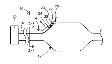
Figure I is a plan overview of a temperature monitoring device 10 for use with
a
cryo therapy apparatus 12 according to an embodiment of the invention.
Temperature
monitoring device 10 may include a tubular member 14 having a temperature
monitoring
member 20 coupled thereto. Temperature monitoring member 20 may be used to
measure temperature while performing a medical procedure, for example cryo
therapy,
cryo balloon therapy, or cryoplasty.
Tubular member 14 includes a proximal end 16 and a distal end 18. Tubular
member I4 may be coupled to cryo therapy apparatus 12, for example along the
length or
proximate an external surface of cryo therapy apparatus 12. Tubular member 14
may be
-4-
CA 02470577 2004-06-18
WO 03/051214 PCT/US02/39936
comprised of materials including, but not limited to, metals, stainless steel,
nickel alloys,
nicleel-titanium alloys, thermoplastics, high performance engineering resins,
fluorinated
ethylene propylene (FEP), polymer, polyethylene (PE), polypropylene (PP),
polyvinylchloride (PVC), polyurethane, polytetrafluoroethylene (PTFE),
polyether-ether
lcetone (PEEK), polyimide, polyamide, polyphenylene sulfide (PPS),
polyphenylene
oxide (PPO), polysufone, nylon, perfluoro (propyl vinyl ether) (PFA),
combinations
thereof, or other suitable materials.
Cryo therapy apparatus 12 is substantially similar to analogous devices (cryo
therapy apparatuses, cryoplasty catheters, etc.) disclosed within U.S. Patent
No.
5,868,735 to Lafontaine and U.S. Patent Application Serial No. 09/849,892 to
Lafontaine,
the entire disclosures of which are hereby incorporated by reference. Briefly,
cryo
therapy apparatus 12 may include a shaft with a cryoplasty device (e.g., a
cryoplasty
balloon) disposed at a distal end thereof. The shaft may include an inflation
tube, a drain
tube, and may further comprise an outer sheath defining an annular lumen
between the
outer sheath and the shaft. The annular lumen may be sealed such that a vacuum
may be
maintained therein. The cryoplasty device may include a single balloon or
multiple
balloons (i.e., a first balloon within a second balloon).
In use, coolant may pass through the inflation lumen into the cryoplasty
device.
The cryoplasty device may then be used for heat transfer with an area of
interest. Coolant
may be removed from the cryoplasty device through the drain tube following
heat
transfer.
Temperature monitoring member 20 may comprise a retractable thermocoupled
needle 22 slidably disposed within a lumen 24 of tubular member 14. According
to this
-5-
CA 02470577 2004-06-18
WO 03/051214 PCT/US02/39936
embodiment, the inside diameter of tubular member I4 is sized appropriately
for having
temperature monitoring member 20 disposed within lumen 24. Thermocoupled
retractable needle 22 is understood to include temperature sensing means that
measure
temperature in a mariner that is quantifiable by a clinician. Alternatively,
retractable
needle 22 may include a temperature sensor coupled thereto. Retractable needle
22 may
include a sharpened distal point 26 and at least one marker band 28. Distal
point 26 may
be adapted to penetrate and/or cut into tissue during a medical procedure.
From Figure 1 it can be appreciated that temperature monitoring member 20 (and
others described below) have a length along the longitudinal axis and extends
away from
cryo therapy apparatus 12 along the longitudinal axis. Moreover, an angle may
be
defined between temperature monitoring member 20 and cryo therapy apparatus
12. This
angle may be about 90°, acute, or obtuse. It can also be seen in Figure
1 that the length of
temperature monitoring member 20 that extends from cryo therapy apparatus is
greater
than its width (measured along the axis perpendicular to the longitudinal
axis).
Marker band 24 may produce a relatively bright image on a fluoroscopy screen
during a medical procedure. This relatively bright image aids the user of
marlcer band 24
in determining the location of temperature monitoring member 20. Marker band
24 may
comprise a number of radiopaque materials including, but not limited to, gold,
platinum,
and plastic material loaded with a radiopaque filler. Temperature monitoring
member 20
may further comprise additional marker bands or may comprise a marker band
disposed
at a different location. Fox example, marlcer band 24 may comprise a first
marlcer band
(e.g., marker band 24) a fixed distance from distal point 22 of temperature
monitoring
member 20. A second marker band may be disposed on temperature monitoring
member
-6-
CA 02470577 2004-06-18
WO 03/051214 PCT/US02/39936
20 proximally a distance that is approximately equal to the distance the first
marleer band
is from distal point 22.
Proximal end 16 of tubular member 14 may be connected to a manifold 30.
Manifold 30 comprises means for controlling temperature monitoring member 20.
More
specifically, manifold 30 may comprise means for quantifying temperature as
measured
by, for example, thermocoupled needle 20. Means for quantifying temperature
may
include an analog temperature reading or display, a digital temperature
reading or display,
a connector for coupling to a computerized system for measuring temperature, a
computerized system for processing other data, and combinations thereof.
In use, temperature monitoring device 10 may be advanced to an area of
interest.
The area of interest may be, for example, an artery including the pulmonary
artery, a vein
including the pulmonary vein, a blood vessel, the heart, trabeculae within the
heart, a
body organ, or other areas where cryoplasty may prove beneficial. Cryo therapy
apparatus 12 may be used to cool the area of interest while temperature
monitoring
member 20 may be used to quantify temperature by inserting distal point 26
into tissue at
the area of interest. In an embodiment, distal point 26 may contact the
surface of the
tissue at the area of interest or it may penetrate and/or cut into the tissue
to measure
temperature below the surface. Marlcer band 28 may be used to determine the
location of
distal point 26 during heat transfer. Accurately determining the location of
distal point 26
may allow more precise cooling and prevent possible tissue damage due to over-
cooling.
Alternatively, the needles may have pre-determined depth marker band/stops
which allow
tissue penetration to a fixed depth such as 1-3mm, etc.
CA 02470577 2004-06-18
WO 03/051214 PCT/US02/39936
Figure 2 is a plan view of an alternate temperature monitoring device 110
according to an embodiment of the invention. Temperature monitoring device 110
is
substantially similar to temperature monitoring device 10 except that
temperature
monitoring member 120 comprises an infrared or optic sensor 132. At least a
portion of
infrared or optic sensor 132 may be disposed within a lumen of tubular member
114, i.e.,
the equivalent of lumen 24 of tubular member 14. Alternatively, infrared or
optic sensor
132 may be disposed proximate distal end 218 of tubular member 214.
Temperature monitoring device 110 may be used to measure temperature at an
area of interest by detecting infrared energy at the area of interest with
infrared sensor 32.
Quantification of infrared energy may comprise a measurement of heat and/or
temperature. Manifold 30 comprises means for quantifying temperature. For
example,
manifold 30 may comprise means for quantifying infrared energy.
Cooling may result in the formation of ice or ice balls adjacent cryo therapy
apparatus 12 and/or the treatment site. As a result, alternative temperature
monitoring
members may be used. For example, in order to monitor or otherwise visualize
ice or ice
ball formation, optical sensing may be used. Optic sensing may be looking at
the ice ball
visually by color change or appearance of ice. Other methods may be used as
described
herein to monitor ice formation as well as methods known to those in the art.
Figure 3 and Figure 4 depict a plan overview of arrangements of tubular
members
14 taken through section 3/4-3/4 of Figure 1 and depicting additional tubular
members 14.
More than one tubular member 14 may be disposed about cryo therapy apparatus
12 in an
array. Four tubular members 14 may be disposed about cryo therapy apparatus 12
in a
quartet array as shown in Figure 3. Similarly, Figure 4 depicts eight tubular
members 14,
_g_
CA 02470577 2004-06-18
WO 03/051214 PCT/US02/39936
evenly spaced between, and disposed about cryo therapy apparatus 12 in an
octet array.
Although Figure 3 and Figure 4 depict arrangements of tubular member 14, it
should be
understood that any of the tubular members, temperature monitoring devices,
and
analogous structures disclosed herein may be substituted without departing
from the spirit
of the invention.
Figure 5 is a plan view of an alternate temperature monitoring device 210
according to an embodiment of the invention. Temperature monitoring device 210
is
substantially similar to temperature monitoring device 10 except that
temperature
monitoring member 220 comprises an ultrasound transmitter 234. Similar to what
is
disclosed above, at least a portion of ultrasound transmitter 234 may be
disposed within a
lumen of tubular member 214 or ultrasound transmitter 234 may be disposed at
distal end
218 of tubular member 214.
Temperature monitoring device 210 may be used to measure temperature at an
area of interest by transmitting ultrasound energy from ultrasound transmitter
234.
Manifold 30 may comprise means for quantifying temperature including means for
accumulating ultrasound images, ultrasound energy, and other ultrasound data.
Analysis
of ultrasound images, ultrasound energy, and other ultrasound data may provide
an
indirect measurement of temperature. For example, an ultrasound image may be
used to
view a phase change within the area of interest. The phase change may indicate
a
quantifiable level of cooling.
Figure 6 is a detailed view of a tubular members 214 taken through line 6-6 of
Figure 5 and depicting addition tubular members 214. In an embodiment, tubular
members 214 may be disposed about cryo therapy apparatus 12 in an array. In an
-9-
CA 02470577 2004-06-18
WO 03/051214 PCT/US02/39936
exemplary embodiment, the array may be a circular array. The circular array
may enable
a user to more precisely measure temperature and determine the location of
temperature
monitoring device 210. In addition, tubular members 214 may be arranged in a
quartet or
octet array as disclosed above, and tubular members 14 and 114 may also be
arranged in a
circular array.
Figure 7 is a plan view of an alternate temperature monitoring device 310
according to an embodiment of the invention. Temperature monitoring device 310
is
substantially similar to temperature monitoring device 10 with a number of
refinements
described below.
Tubular member 314 comprises a sheath that, encircles cryo therapy apparatus
12.
Temperature monitoring member 320 comprises a stmt 36 disposed at distal end
318 of
tubular member 314. Stent 36 may further comprise a plurality of thermal
spikes 38.
Stent 36 is comprised of a shape memory alloy (e.g., nickel-titanium alloy).
Alternatively, stmt 36 may be comprised of materials similar to those listed
above
including metals and polymers.
Thermal spikes 38 may be capable of measuring temperature at an area of
interest.
According to this embodiment, thermal spikes 38 may be coupled to manifold 30
such
that a user may quantify temperature. Manifold 30 may comprise means for
quantifying
temperature including those listed above.
In addition, thermal spikes 38 may be used to facilitate heat transfer to an
area of
interest. For example, trabeculae within the heart may not allow cryo therapy
apparatus
12 to evenly cool the heart. The result may be uneven or incomplete heat
transfer.
Thermal spikes 38 may be capable of reaching, contacting, and penetrating
surfaces of an
-10-
CA 02470577 2004-06-18
WO 03/051214 PCT/US02/39936
area of interest. For example, thermal spilees 38 may be capable of contacting
trabeculae
within the heart and may, thus, facilitate heat transfer to these areas.
In an embodiment, stmt 36 may be collapsed at body temperature and be
expanded when cooled. A collapsed state at body temperature will minimize the
outside
diameter of stmt 36, which may facilitate delivery of temperature monitoring
device 310
to an area of interest. Cooling, for example cooling initiated by cryo therapy
apparatus
12, may expand stmt 36 in order to move thermal spikes 38 proximate the area
of
interest.
Figure 8 illustrates another alternative temperature monitoring device 410.
Device 410 includes cryo therapy apparatus 412 coupled to manifold 30
essentially as
described above. In addition, tubular member 414 may be coupled to cryo
therapy
apparatus 412. For example, tubular member 414 may be disposed at least
partially
within cryo therapy apparatus 12 or within the shaft portion of cryo therapy
apparatus 12.
When disposed within cryo therapy apparatus 12, tubular member 414 may be
substantially coaxial with or proximate an interior wall of apparatus 12.
Temperature monitoring member 420 may be disposed within tubular member
441 and may extend into cryo therapy apparatus 12. In an alternative
embodiment,
temperature monitoring member 420 may be disposed within cryo therapy
apparatus 12
without the use of tubular member 441. For example, temperature monitoring
member
420 may be disposed within the shaft of cryo therapy apparatus 12.
Temperature monitoring member 420 includes an optical imaging apparatus 434
including an emitter 440 and a detector 442. Emitter 440 is adapted and
configured to
emit energy (e.g., light, infrared energy, ultrasonic energy, etc.) from
within cryo therapy
-11-
CA 02470577 2004-06-18
WO 03/051214 PCT/US02/39936
apparatus. Detector 442 is adapted to collect data by detecting energy. A
number of
different arrangements of emitters and/or detectors may be used without
departing from
the spirit of the invention.
Cryo therapy apparatus 412 is essentially the same in fornl and function as
cryo
therapy apparatus 12 but further includes an inner cooling chamber 444 and an
outer
cooling chamber 446. A dual-chamber cooling apparatus (such as apparatus 412)
may
provide additional safety or cooling advantages. For example, outer cooling
chamber 446
may prevent loss of coolant into the body if inner cooling chamber 444 failed.
It can be
appreciated that the dual chamber cooling apparatus 412 can be substituted
into any of the
other embodiments described herein.
Numerous advantages of the invention covered by this document have been set
forth in the foregoing description. It will be understood, however, that this
disclosure is,
in many respects, only illustrative. Changes may be made in details,
particularly in
matters of shape, size, and arrangement of steps without exceeding the scope
of the
invention. The invention's scope is, of course, defined in the language in
which the
appended claims are expressed.
-12-