Note: Descriptions are shown in the official language in which they were submitted.
CA 02471582 2004-06-23
WO 03/057845 PCT/US02/41788
AUTOMATED SYSTEMS AND METHODS FOR ANALYSIS OF
PROTEIN POST-TRANSLATIONAL MODIFICATION
Reference to Related Applications
The present application claims priority to U.S. Provisional application
60/343,645, filed on December 28, 2001, and U.S. Provisional application
60/361,236, filed on March l, 2002, the entire contents of which are all
incorporated
by reference herein.
Field of the Invention
This invention is in the field of proteomics, and applies mass spectrometry to
the analysis of peptides and amino acids. More particularly, the invention
relates to a
mass spectrometry-based method for detection of amino acid modifications, such
as
phosphorylation.
Background to the Invention
With the availability of a burgeoning sequence database, genomic
applications demand faster and more efficient methods for the global screening
of
protein expression in cells. However, the complexity of the cellular proteome
expands substantially if protein post-translational modifications are also
taken into
account.
Dynamic post-translational modification of proteins is important for
maintaining and regulating protein structure and function. Among the several
hundred different types of post-translational modifications characterized to
date,
protein phosphorylation plays a prominent role. Enzyme-catalyzed
phosphorylation
and dephosphorylation of proteins is a key regulatory event in the living
cell.
Complex biological processes such as cell cycle, cell growth, cell
differentiation,
and metabolism are orchestrated and tightly controlled by reversible
phosphorylation
events that modulate protein activity, stability, interaction and
localization.
Perturbations in phosphorylation states of proteins, e.g. by mutations that
generate
constitutively active or inactive protein lcinases and phosphatases, play a
prominent
-1-
CA 02471582 2004-06-23
WO 03/057845 PCT/US02/41788
role in oncogenesis. Comprehensive analysis and identification of
phosphoproteins
combined with exact localization of phosphorylation sites in those proteins
('phosphoproteomics') is a prerequisite for understanding complex biological
systems and the molecular features leading to disease.
It is estimated that 1/3 of all proteins present in a mammalian cell are
phosphorylated and that lcinases, enzymes responsible for that
phosphorylation,
constitute about 1-3% of the expressed genome. Organisms use reversible
phosphorylation of proteins to control many cellular processes including
signal
transduction, gene expression, the cell cycle, cytoslceletal regulation and
apoptosis.
A phosphate group can modify serine, threonine, tyrosine, histidine, arginine,
lysine,
cysteine, glutamic acid and aspartic acid residues. However, the
phosphorylation of
hydroxyl groups at serine (90%), threonine (10%), or tyrosine (0.05%) residues
are
the most prevalent, and are involved among other processes in metabolism, cell
division, cell growth, and cell differentiation. Because of the central role
of
phosphorylation in the regulation of life, much effort has been focused on the
development of methods for characterizing protein phosphorylation.
The identification of phosphorylation sites on a protein is complicated by the
facts that proteins are often only partially phosphorylated and that they are
often
present only at very low levels. Therefore techniques for identifying
phosphorylation
sites should preferably work in the low picomole to sub-picomole range.
Traditional methods for analyzing O-phosphorylation sites involve
incorporation of 32P into cellular proteins via treatment with radiolabeled
ATP. The
radioactive proteins can be detected during subsequent fractionation
procedures (e.g.
two-dimensional gel electrophoresis or high-performance liquid chromatography
[HPLC]). Proteins thus identified can be subjected to complete hydrolysis and
the
phosphoamino acid content determined. The sites) of phosphorylation can be
determined by proteolytic digestion of the radiolabeled protein, separation
and
detection of phosphorylated peptides (e.g. by two-dimensional peptide
mapping),
followed by peptide sequencing by Edman degradation. These techniques can be
tedious, require significant quantities of the phosphorylated protein and
involve the
use of considerable amounts of radioactivity.
_2_
CA 02471582 2004-06-23
WO 03/057845 PCT/US02/41788
In recent years, mass spectrometry (MS) has become an increasingly viable
alternative to more traditional methods of phosphorylation analysis. The most
widely used method for selectively enriching phosphopeptides from mixtures is
immobilized metal affinity chromatography (IMAC). In this technique, metal
ions,
usually Fe3+ or Ga3+, are bound to a chelating support. Phosphopeptides are
selectively bound because of the affinity of the metal ions for the phosphate
moiety.
The phosphopeptides can be released using high pH or phosphate buffer, the
latter
usually requiring a fiu-ther desalting step before MS analysis. Limitations of
this
approach include possible loss of phosphopeptides because of their inability
to bind
to the IMAC column, difficulty in the elution of some multiply phosphorylated
peptides, and background from unphosphorylated peptides (typically acidic in
nature) that have affinity for immobilized metal ions. Two types of chelating
resin
are commercially available, one using iminodiacetic acid and the other using
nitrilotriacetic acid. Some groups have observed that iminodiacetic acid resin
is less
specific than nitrilotriacetic acid, whereas another study reported little
difference
between the two. Several studies have examined off line MS analysis of IMAC-
separated peptides.
Recently, two groups have described protocols to achieve this goal. Oda et
al. (Nat Biotechnol. 2001 19:379-82) start with a protein mixture in which
cysteine
reactivity is removed by oxidation with performic acid. Base hydrolysis is
used to
induce -elimination of phosphate from phosphoserine and phosphothreonine,
followed by addition of ethanedithiol to the alkene. The resulting free
sulfhydryls
are coupled to biotin, allowing purification of phosphoproteins by avidin
affinity
chromatography. Following elution of phosphoproteins and proteolysis,
enrichment
of phosphopeptides is carried out by a second rotuld of avidin purification.
Disadvantages of this approach include the failure to detect phosphotyrosine
containing peptides and generation of diastereoisomers in the derivatization
step.
The approach suggested by the Zhou et al. (Nat Bioteclmol 2001 19:375-
378) circumvents these problems but involves a six step
derivatization/purification
protocol for tryptic peptides that requires more than 13 hrs to complete and
affords
only a 20% yield fiom picomoles of phosphopeptide starting material. The
method
begins with a proteolytic digest that has been reduced and alkylated to
eliminate
-3-
CA 02471582 2004-06-23
WO 03/057845 PCT/US02/41788
reactivity from cysteine residues. Following N-terminal and C-terminal
protection,
phosphoramidate adducts at phosphorylated residues are formed by carbodiimide
condensation with cystamine. The free sulfhydryl groups produced from this
step are
covalently captuxed onto glass beads coupled to iodoacetic acid. Elution with
trifluoroacetic acid then regenerates phosphopeptides for analysis by mass
spectrometry.
-4-
CA 02471582 2004-06-23
WO 03/057845 PCT/US02/41788
Summary of the Invention
One aspect of the present provides a method for identifying modified amino
acids within a protein by combining affinity purification and mass
spectroscopy in a
manner which is amenable to high throughput and automation. In general, the
S subject method makes use of affinity capture reagents for isolating, from a
protein
sample, those proteins which have been post-translationally modified with a
moiety
of interest. In order to improve the selectivity/efficiency of the affinity
purification
step, the protein samples to be analyzed are chemically modified at at least
one of
the C-terminal carboxyl, the N-terminal amine and amino acid side chains of
the
proteins which may interfere with the selectively of the affinity purification
step for
the post-translational modification of interest. Proteins which are isolated
based on
post-translational modifications are than analyzed by mass spectroscopy in
order to
identify patterns of modification across a proteome, and/or to provide the
identity of
proteins in the sample which are modified or shows changes in modification
status
between two different samples.
In certain preferred embodiments, the proteins are cleaved into smaller
peptide fragments before, after or during the chemical modification step. For
instance, the proteins can be fragmented by enzymatic hydrolysis to produce
peptide
fragments having carboxy-terminal lysine or arginine residues. In certain
preferred
embodiments, the proteins are fragmented by treatment with trypsin.
In certain embodiments, the proteins are mass-modified with isotopic labels
before, after or during the chemical modification step.
In certain embodiments, the proteins are further separated by reverse phase
chromatography before analysis by mass spectroscopy.
'there are a variety of mass spectroscopy techniques which can be employed
in the subject method. In certain preferred embodiments, the isolated proteins
are
identified from analysis using tandem mass spectroscopy techniques, such as
LC/MS/1VIS. Where the proteins have been further fragmented with trypsin or
other
predictable enzymes, the molecular weight of a fragment as determined from the
mass spectroscopy data can be used to identify possible matches in molecular
weight
-5-
CA 02471582 2004-06-23
WO 03/057845 PCT/US02/41788
databases indexed by predicted molecular weights of protein fiagments which
would
result under similar conditions as the fragments generated in the subject
method.
However, the subject method can be carried out using mass spectroscopy
techniques
which produce amino acid sequence mass spectra for the isolated proteins or
peptide
fragments. The sequence data can be used to search one or more sequence
databases.
In certain preferred embodiments, the method is used to identify
phosphorylated proteins or changes in the phosphorylation pattern amongst a
group
of proteins. In such embodiments, the affinity capture reagent can be an
immobilized
metal affinity chromatography medium, and the step of processing the protein
samples includes chemically modifying the side chains of glutamic acid and
aspartic
acid residues to neutral derivatives, such as by alkyl-esterification.
The subject method is amenable to analysis of multiple different protein
samples, particularly in a multiplex fashion. In such embodiments, the
proteins or
fragments thereof are isotopically labeled in a manner which permits
discrimination
of mass spectroscopy data between protein samples. That is, a mass spectra on
the
mixture of various protein samples can be deconvoluted to determine the sample
origin of each signal observed in the spectra. In certain embodiments, this
technique
can be used to quantitated differences in phosphorylation (or other
modification)
levels between samples prepared under different conditions and admixed prior
to
MS analysis.
In certain embodiments, the subject method is used for analyzing a
phosphoproteome. For example, the proteins in the sample can be chemically
modify at glutamic acid and aspartic acid residues, such as by allcyl-esterif
ration, to
generate neutral side chains at those positions. The phosphorylated proteins
in the
same are then isolated by immobilized metal affinity chromatography, and
analyzed
by mass spectroscopy. In preferred embodiments, the proteins are cleaved,
e.g., by
trypsin digestion or the like, into smaller peptide fragments before, after or
during
the step of chemically modify the glutamic acid and aspartic acid residues. In
one
embodiment, the subject method is carried out on multiple different protein
samples,
and proteins which a differentially phosphorylated between two or more protein
-6-
CA 02471582 2004-06-23
WO 03/057845 PCT/US02/41788
samples are identified. That data can, for instance, be used to generate or
augment
databases with the identity of proteins which are determined to be
phosphorylated.
Another aspect of the invention provides a method for identifying a treatment
that modulates a modification of amino acid in a target polypeptide. In
general, this
method is carried out by providing a protein sample which has been subjected
to a
treatment of interest, such as treatment with ectopic agents (drugs, growth
factors,
etc). The protein samples can also be derived from normal cells in different
states of
differentiation or tissue fate, or derived from normal and diseased cells.
Following
the affinity purificationlMS method set forth above, the identity of proteins
which
are differentially modified in the treated protein sample relative to an
untreated
sample or control sample can determined. From this identification step, one
can
determine whether the treatment results in a pattern of changes in protein
modification, relative to the untreated sample or control sample, which meet a
pre-
selected criteria. Thus, one can use this method to identify compounds likely
to
mimic the effect of a growth factor by scoring for similarities in
phosphorylation
patterns when comparing proteins from the compound-treated cells with proteins
from the growth factor treated cells. The treatment of interest can include
contacting
the cell with such compounds as growth factors, cytolcines, hormones, or small
chemical molecules. In certain embodiments, the method is carried out with
various
members of a chemically diverse library.
Another aspect of the invention provides a diagnostic method. In general,
using any one of the suitable methods of the instant invention, one can
generate
profiles of phosphopeptides of related biological samples (for example,
disease
tissue vs. normal tissue, or stem / progenitor cells vs. differentiated cells,
cells
treated by certain agents (such as pharmaceutical drugs or drug candidates)
~s. those
untreated control, or cells at different developmental stages, etc.), and
compare these
profiles of phosphopeptides. If a statistically significant difference in the
profile is
present between the samples being compared, a conclusion can be made about the
status of these samples. In this regard, the instant invention can be used to
diagnose
the presence of a certain disease state, using a biopsy obtained from a
patient. The
instant invention can also be used to sort biological samples into different
categories
based on the similarity of their respective profiles.
CA 02471582 2004-06-23
WO 03/057845 PCT/US02/41788
It should be understood that profiles of amino acid modifications other than
phosphorylation can also be obtained using the subject method, and thus such
methods also fall within the scope of the invention.
Yet another aspect of the present invention provides a method of conducting
a drug discovery business. Using the assay described above, one determines the
identity of a compound that produces a pattern of changes in protein
modification,
relative to the untreated sample or control sample, which meet a preselected
criteria.
Therapeutic prafiling of the compound identifed by the assay, or further
analogs
thereof, can be carried out for determining efficacy and toxicity in animals.
Compounds identified as having an acceptable therapeutic profile can then be
formulated as part of a pharmaceutical preparation. In certain embodiments,
the
method can include the additional step of establishing a distribution system
for
distributing the pharmaceutical preparation for sale, and may optionally
include
establishing a sales group fox marketing the pharmaceutical preparation. In
other
embodiments, rather than carry out the profiling and/or formulation steps, one
can
license, to a third party, the rights for further drug development of
compounds that
axe discovered by the subject assay to alter the level of modification of the
taxget
polypeptide.
Yet another aspect of the present invention provides a method of conducting
a drug discovery business in which, after determining the identity of a
protein that is
post-translationally modified under the conditions of interest, the identity
of one or
more enzymes which catalyze the post-translational modification of the
identified
protein under the conditions of interest is determined. Those enzymes) are
then
used as targets in drug screening assays for identifying compounds which
inlubit or
potentiate the enzymes and which, therefore, can modulate the post-
translational
modification of the identified protein under the conditions of interest.
_g_
CA 02471582 2004-06-23
WO 03/057845 PCT/US02/41788
Reference to the Drawings
Figure 1. Five nonphosphorylated proteins; glyceraldehyde 3-phosphate
dehydrogenase, bovine serum albumin, carbonic anhydrase, ubiquitin, and (3-
lactoglobulin (Sigma Chemical Co., St. Louis, MO) (I00 nmol each) in I.I mI of
100 mM ammonium bicarbonate (pH 8) were digested with trypsin (20 pg)
(Promega, Madison, WI) for 24 h at 37oC. The reaction was quenched with 65 ~,l
of
glacial acetic acid, and the mixture was then diluted to final volume of 50 ml
with
0.1 % acetic acid. To this solution was added 500 pmol of HPLC purified
phosphopeptide, DRVpYIHPF (SEQ ID No: 1) (Novabiochem, San Diego, CA), in
0.1% acetic acid (2 ~L of a 250 pmol/~,L stock solution). An aliquot of the
standard
mixture (100 ~.l) was lyophilized and redissolved in 100 ~.l of 2 N methanolic
HCI.
This latter solution was prepared by dropwise addition of 160 ~,1 of acetyl
chloride
with stirring to 1 ml of methanol. Esterification was allowed to proceed for
2h at
room temperature. Solvent was removed by lyophilization and the resulting
sample
was redissolved in 100 p,l of solution containing equal volumes of methanol,
water
and acetonitrile. Phosphate methyl esters are not observed under these
conditions.
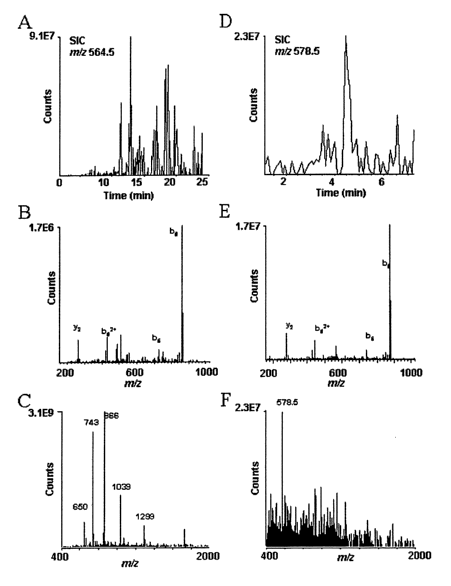
Mass spectra recorded by a combination of immobilized metal affinity
chromatography (IMAC) and nano-flow HPLC microelectrospray ionization mass
spectrometry on the phosphopeptide, DRVpYIHPF (SEQ ID No: 1), present at the
level of 10 fmol/p,l in a mixture containing tryptic peptides fiom 5 proteins
at the
level of 2 pmol/~.1. Aliquots corresponding to 0.5 ~.1 of the above solutions
(tryptic
peptides from 1 pmol of each protein plus 5 fmol of phosphopeptide, DRVpYIHPF,
SEQ ID No: I) were analyzed by mass spectrometry. (A) Selected ion
chromatogram, SIC, or plot of the ion current vs scan number for m/z 564.5
corresponding to the (M+2H)~ of the phosphopeptide, DRVpYIHPF (SEQ ID No:
1). (B) MS/MS spectrum characteristic of the sequence, DRVpYIHPF (SEQ ID No:
1), recorded on ions of mlz 564.5 in scans 610-616. (C) Electrospray
ionization mass
spectrum recorded during this same time interval. Abundant ions from tryptic
peptides non-specifically bound to the IMAC column obscure the signal at m/z
564.5 for DRVpYIHPF (SEQ ID No: I). (D) SIC for m/z 578.5 corresponding to the
-g_
CA 02471582 2004-06-23
WO 03/057845 PCT/US02/41788
(M+2H)++ ion for the dimethyl ester of DRVpYIPF (SEQ ID No: 1). (E) MS/MS
spectrum characteristic of the sequence, DRVpYIPF (SEQ ID No: 1), recorded in
on
ions of m/z 578.5 in scans 151-163. (F) Electrospray ionization mass spectrum
recorded in scan 154 showing the parent ion, m/z 578.5 for the phosphopeptide
dimethyl ester and the absence of signals for tryptic peptides non
specifically bound
to the IMAC column.
Fi~,ure 2. The phosphopeptide B-casein was analyzed using the subject
invention. Top: extracted ion chromatograph from the HPLC separation showing
the
B-casein peak at 85.33 min. Bottom: Casein MS/MS scan at m/z = 1031.6, showing
individual peptide fragments of the phosphopeptide components of Casein.
Fi-gore 3. Schematic of exemplary system for automating the subject method.
-10-
CA 02471582 2004-06-23
WO 03/057845 PCT/US02/41788
Detailed Description of the Invention
Definitions
For convenience, certain terms employed in the specification, examples, and
appended claims are collected here.
"Affinity capture reagent" as used herein means reagents that has affinity for
proteins, including their backbone and side-chains, either modified or
naturally
occurring, due to, for example, electrostatic, hydrophobic, ionic and/or
hydrogen-
bond interactions under physiological / experimental conditions. An exemplary
affinity capture reagent is resin used in IMAC.
"Binding," "bind" or "bound" refers to an association, which may be a stable
association between two molecules, e.g., between a modified protein ligand an
affinity capture reagent, due to, for example, electrostatic, hydrophobic,
ionic and/or
hydrogen-bond interactions under physiological conditions.
"Cells," "host cells" or "recombinant host cells" are terms used
interchangeably herein. It is understood that such terms refer not only to the
particular subject cell but to the progeny or potential progeny of such a
cell. Because
certain modifications may occur in succeeding generations due to either
mutation or
environmental influences, such progeny may not, in fact, be identical to the
parent
cell, but are still included within the scope of the term as used herein.
A "chimeric protein" or "fusion protein" is a fusion of a first amino acid
sequence encoding a polypeptide with a second amino acid sequence defining a
domain foreign to and not substantially homologous with any domain of the
protein.
A chimeric protein may present a foreign domain which is found (albeit in a
different protein) in an organism which also expresses the first protein, or
it may be
an "interspecies", "intergenic", etc. fusion of protein structures expressed
by
different kinds of organisms.
The terms "compound", "test compound" and "molecule" are used herein
interchangeably and are meant to include, but are not limited to, peptides,
nucleic
-11-
CA 02471582 2004-06-23
WO 03/057845 PCT/US02/41788
acids, carbohydrates, small organic molecules, natural product extract
libraries, and
any other molecules (including, but not limited to, chemicals, metals and
organometallic compounds).
The phrases "conserved residue" "or conservative amino acid substitution"
refer to grouping of amino acids on the basis of certain common properties. A
functional way to define common properties between individual amino acids is
to
analyze the normalized frequencies of amino acid changes between corresponding
proteins of homologous organisms (Schulz, G. E. and R. H. Schirmer.,
Principles of
Protein Structure, Springer-Verlag). According to such analyses, groups of
amino
acids may be defzned where amino acids within a group exchange preferentially
with
each other, and therefore resemble each other most in their impact on the
overall
protein structure (Schulz, G. E. and R. H. Schirmer., Principles of Protein
Structure,
Springer-Verlag). Examples of amino acid groups defined in this manner
include:
(i) a charged group, consisting of Glu and Asp, Lys, Arg and His,
(ii) a positively-charged group, consisting of Lys, Arg and His,
(iii) a negatively-charged group, consisting of Glu and Asp,
(iv) an aromatic group, consisting of Phe, Tyr and Trp,
(v) a nitrogen ring group, consisting of His and Trp,
(vi) a large aliphatic nonpolar group, consisting of Val, Leu and Ile,
(vii) a slightly-polar group, consisting of Met and Cys,
(viii) a small-residue group, consisting of Ser, Thr, Asp, Asn, Gly, Ala, Glu,
Gln and Pro,
(ix) an aliphatic group consisting of Val, Leu, Ile, Met and Cys, and
(x) a small hydroxyl group consisting of Ser and Thr.
-12-
CA 02471582 2004-06-23
WO 03/057845 PCT/US02/41788
In addition to the groups presented above, each amino acid residue may form
its own group, and the group formed by an individual amino acid may be
referred to
simply by the one and/or three letter abbreviation for that amino acid
commonly
used in the art.
The term "DNA sequence encoding a polypeptide" may refer to one or more
genes within a particular individual. As is well laiown in the art, genes for
a
particular polypeptide may exist in single or multiple copies within the
genome of an
individual. Such duplicate genes may be identical or may have certain
modifications,
including nucleotide substitutions, additions or deletions, which all still
code for
I O polypeptides having substantially the same activity. Moreover, certain
differences in
nucleotide sequences may exist between individual organisms, which are called
alleles. Such allelic differences may or may not result in differences in
amino acid
sequence of the encoded polypeptide yet still encode a protein with the same
biological activity.
15 The term "domain" as used herein refers to a region within a protein that
comprises a particular structure or function different from that of other
sections of
the molecule.
"Exogenous" means caused by factors or an agent from outside the organism
or system, or introduced from outside the organism or system, specifically:
not
~0 normally synthesized within the organism or system. A fusion l tagged
protein
expressed from an introduced plasmid may be considered exogenous to the host
cell
expressing the fusion protein, although the host itself may express an
endogenous
version of the same protein.
"Extracellular factor" includes a molecule or a change in the environment
25 that is transduced intracelhilarly via cell surface proteins (e.g. cell
surface receptors)
that interact, directly or indirectly, with a signal. An extracellular factor
includes any
compound or substance that in some manner specifically alters the activity of
a cell
surface protein. Examples of such signals or factors include, but are not
limited to
growth factors, that bind to cell surfaces and/or intracellular receptors and
ion
-13-
CA 02471582 2004-06-23
WO 03/057845 PCT/US02/41788
channels and modulate the activity of such receptors and channels. The signals
and
factors include analogs, derivatives, mutants, and modulators of such growth
factors.
"Intracellular factor" includes a molecule or a change in the cell environment
that is transduced in the cell via cytoplasmic proteins that interact,
directly or
indirectly with a signal. An intracellular factor includes any compound or
substance
that in some manner specifically alters the activity of a cytoplasmic protein
involved
in a biological or signal transduction pathway.
"High throughput" refers to the ability to process large amount of samples in
a given process, method, or assay, etc. In a preferred embodiment, the high
throughput process is conducted with an automated maclune(s), which is
optionally
controlled by computer software or human or both.
"Homology" or "identity" or "similarity" refers to sequence similarity
between two peptides or between two nucleic acid molecules, with identity
being a
more strict comparison. Homology and identity can each be determined by
comparing a position in each sequence which may be aligned for purposes of
comparison. When a position in the compared sequence is occupied by the same
base or amino acid, then the molecules are identical at that position. A
degree of
homology or similarity or identity between nucleic acid sequences is a
function of
the number of identical or matching nucleotides at positions shared by the
nucleic
acid sequences. A degree of identity of amino acid sequences is a function of
the
number of identical amino acids at positions shared by the amino acid
sequences. A
degree of homology or similarity of amino acid sequences is a function of the
number of amino acids, i.e. structurally related, at positions shared by the
amino acid
sequences. An "unrelated" or "non-homologous" sequence shares less than 40
identity, though preferably less than 25 % identity, with one of the--
sequences of the
present invention.
As used herein, the term "gene" or "recombinant gene" refers to a nucleic
acid comprising an open reading frame encoding a polypeptide of the present
invention, including both exon and (optionally) intron sequences. A
"recombinant
gene" refers to nucleic acid encoding a polypeptide and comprising exon coding
-14-
CA 02471582 2004-06-23
WO 03/057845 PCT/US02/41788
sequences, though it may optionally include intron sequences derived from a
chromosomal gene. The term "intron" refers to a DNA sequence present in a
given
gene which is not translated into protein and is generally found between
exons.
The term "GI" or "GI Number" or "GI No." refers to database access number
(such as gene banlc) for genes and/or proteins useful for retrieving sequence
and
other related information.
"Homology" or "identity" or "similarity" refers to sequence similarity
between two peptides or between two nucleic acid molecules. Homology and
identity can each be determined by comparing a position in each sequence which
may be aligned for purposes of comparison. When an equivalent position in the
compared sequences is occupied by the same base or amino acid, then the
molecules
are identical at that position; when the equivalent site occupied by the same
or a
similax amino acid residue (e.g., similar in steric and/or electronic nature),
then the
molecules can be referred to as homologous (similar) at that position.
Expression as
a percentage of homology/similarity or identity refers to a function of the
number of
identical or similar amino acids at positions shared by the compared
sequences. A
sequence which is "unrelated" or "non-homologous" shares less than 20%
identity,
though preferably less than 15% identity with a sequence of the present
invention.
Similarly, "homology" or "homologous" refers to sequences that are at least
20%,
25%, 30%, 35%, 40%, 45%, 50%, 55%, 60%, 65%, 70%, 75%, 80%, 85%, 90%, or
even 95% to 99% identical to one another.
The term "homology" describes a mathematically based comparison of
sequence similarities which is used to identify genes or proteins with similar
functions or motifs. The nucleic acid and protein sequences of the present
invention
may be used as a "query sequence" to perform a seaxch' against public
databases to, .
for example, identify other family members, related sequences or homologs.
Such
searches can be performed using the NBLAST and XBLAST programs (version 2.0)
of Altschul, et al. (1990) J Mol. Biol. 215:403-10. BLAST nucleotide searches
can
be performed with the NBLAST program, score=100, wordlength=12 to obtain
nucleotide sequences homologous to nucleic acid molecules of the invention.
-15-
CA 02471582 2004-06-23
WO 03/057845 PCT/US02/41788
BLAST protein searches can be performed with the XBLAST program, score=50,
wordlength=3 to obtain amino acid sequences homologous to protein molecules of
the invention. To obtain gapped alignments for comparison purposes, Gapped
BLAST can be utilized as described in Altschul et al., (1997) Nucleic Acids
Res.
25(17):3389-3402. When utilizing BLAST and Gapped BLAST programs, the
default parameters of the respective programs (e.g., XBLAST and BLAST) can be
used.
As used herein, "identity" means the percentage of identical nucleotide or
amino acid residues at corresponding positions in two or more sequences when
the
sequences are aligned to maximize sequence matching, i.e., talcing into
account gaps
and insertions. Identity can be readily calculated by known methods, including
but
not limited to those described in Computational Molecular Biology, Leslc, A.
M.,
ed., Oxford University Press, New Yorlc, 1988; Biocomputing: Informatics and
Genome Projects, Smith, D. W., ed., Academic Press, New York, 1993; Computer
Analysis of Sequence Data, Part I, Griffin, A. M., and Griffin, H. G., eds.,
Humana
Press, New Jersey, 1994; Sequence Analysis in Molecular Biology, von Heinje,
G.,
Academic Press, 1987; and Sequence Analysis Primer, Gribskov, M. and Devereux,
J., eds., M Stockton Press, New Yorlc, 1991; and Carillo, H., and Lipman, D.,
SIAM
J. Applied Math., 48: 1073 (1988). Methods to determine identity are designed
to
give the largest match between the sequences tested. Moreover, methods to
determine identity are codified in publicly available computer programs.
Computer
program methods to determine identity between two sequences include, but axe
not
limited to, the GCG program package (Devereux, J., et al., Nucleic Acids
Research
12(1): 387 (1984)), BLASTP, BLASTN, and FASTA (Altschul, S. F. et al., J.
Molec. Biol. 215: 403-410 (1990) and Altschul et al. Nuc. Acids Res. 25: 3389-
3402
(1997)). The BLAST X program is publicly available from NCBI and other sources
(BLAST Manual, Altschul, S., et al., NCBI NLM NIH Bethesda, Md. 20894;
Altschul, S., et al., J. Mol. Biol. 215: 403-410 (1990). The well known Smith
Waterman algorithm may also be used to determine identity.
The term "percent identical" refers to sequence identity between two amino
acid sequences or between two nucleotide sequences. Identity can each be
-16-
CA 02471582 2004-06-23
WO 03/057845 PCT/US02/41788
determined by comparing a position in each sequence which may be aligned for
purposes of comparison. When an equivalent position in the compared sequences
is
occupied by the same base or amino acid, then the molecules are identical at
that
position; when the equivalent site occupied by the same or a similar amino
acid
residue (e.g., similar in steric and/or electronic nature), then the molecules
can be
referred to as homologous (similar) at that position. Expression as a
percentage of
homology, similarity, or identity refers to a function of the number of
identical or
similar amino acids at positions shaxed by the compared sequences. Expression
as a
percentage of homology, similarity, or identity refers to a function of the
number of
IO identical or similar amino acids at positions shared by the compared
sequences.
Various alignment algorithms and/or programs may be used, including FASTA,
BLAST, or ENTREZ. FASTA and BLAST are available as a part of the GCG
sequence analysis package (University of Wisconsin, Madison, Wis.), and can be
used with, e.g., default settings. ENTREZ is available through the National
Center
for Biotechnology Information, National Library of Medicine, National
Institutes of
Health, Bethesda, Md. In one embodiment, the percent identity of two sequences
can
be determined by the GCG program with a gap weight of 1, e.g., each amino acid
gap is weighted as if it were a single amino acid or nucleotide mismatch
between the
two sequences.
Other techniques for alignment are described in Methods in EnzymologY,
vol. 266: Computer Methods for Macromolecular Sequence Analysis (1996), ed.
Doolittle, Academic Press, Inc., a division of Harcourt Brace & Co., San
Diego,
California, USA. Preferably, an alignment program that permits gaps in the
sequence is utilized to align the sequences. The Smith-Waterman is one type of
algorithm that permits gaps in sequence alignments. See Meth. Mol. Biol. 70:
173-
187 (1997). Also, the GAP program using the Needleman and Wunsch alignment
method can be utilized to align sequences. An alternative search strategy uses
MPSRCH software, which runs on a MASPAR computer. MPSRCH uses a Smith-
Waterman algorithm to score sequences on a massively parallel computer. This
approach improves ability to pick up distantly related matches, and is
especially
tolerant of small gaps and nucleotide sequence errors. Nucleic acid-encoded
amino
acid sequences can be used to search both polypeptide and DNA databases.
-17-
CA 02471582 2004-06-23
WO 03/057845 PCT/US02/41788
Databases with individual sequences are described in Methods in
Enzymolo~y, ed. Doolittle, supra. Some exemplary public databases include
GenBanlc, EMBL, DNA Database of Japan (DDBJ), SwissProt, PIR and other
databases derived therefrom. In comparing a new nucleic acid with lenown
sequences, several alignment tools are available. Examples include Pileup,
which
creates a multiple sequence alignment, and is described in Feng et al., J.
Mol. Evol.
(1987) 25:351-360. Another method, GAP, uses the alignment method of
Needleman et al., J. Mol. Biol. (1970) 48:443-453. GAP is best suited for
global
alignment of sequences. A third method, BestFit, functions by inserting gaps
to
maximize the number of matches using the local homology algorithm of Smith and
Waterman, Adv. Appl. Math. (1981) 2:482-489. Alternatively, certain commercial
software packages such as LaserGene from DNAStar inc. can be used for certain
aspects of sequence analysis. Multiple software and databases may be used in
any
analysis.
The term "Interacting Protein" is meant to include polypeptides that interact
either directly or indirectly with another protein. Direct interaction means
that the
proteins may be isolated by virtue of their ability to bind to each other
(e.g. by
coimmunoprecipitation or other means). Indirect interaction refers to proteins
which
require another molecule in order to bind to each other. Alternatively,
indirect
interaction may refer to proteins which never directly bind to one another,
but
interact via an intermediary.
The term "isolated", as used herein with reference to the subject proteins and
protein complexes, refers to a preparation of protein or protein complex that
is
essentially free from contaminating proteins that normally would be present in
association with the protein or complex, e.g., in the cellular milieu in which
the
protein or complex is found endogenously. Thus, an isolated protein complex is
isolated from cellular components that normally would "contaminate" or
interfere
with the study of the complex in isolation, for instance while screening for
modulators thereof. It is to be understood, however, that such an "isolated"
complex
may incorporate other proteins the modulation of which, by the subject protein
or
protein complex, is being investigated.
-18-
CA 02471582 2004-06-23
WO 03/057845 PCT/US02/41788
Polypeptides referred to herein as "mammalian homologs" of a protein refers
to other mammalian paralogs, or other mammalian orthologs.
"Analyzing a protein by mass spectrometry" or similar wording refers to
using mass spectrometry to generate information which may be used to identify
or
aid in identifying a protein. Such information includes, for example, the mass
or
molecular weight of a protein, the amino acid sequence of a protein or protein
fragment, a peptide map of a protein, and the purity or quantity of a protein.
The term "motif' as used herein refers to an amino acid sequence that is
commonly found in a protein of a particular structure or function. Typically a
consensus sequence is defined to represent a particular motif. The consensus
sequence need not be strictly defined and may contain positions of
variability,
degeneracy, variability of length, etc. The consensus sequence may be used to
search
a database to identify other proteins that may have a similar structure or
function due
to the presence of the motif in its amino acid sequence. For example, on-line
databases such as GenBanlc or SwissProt can be searched with a consensus
sequence
in order to identify other proteins containing a particular motif. Various
search
algorithms and/or programs may be used, including FASTA, BLAST or ENTREZ.
FASTA and BLAST are available as a part of the GCG sequence analysis package
(University of Wisconsin, Madison, Wis.). ENTREZ is available through the
National Center for Biotechnology Information, National Library of Medicine,
National Institutes of Health, Bethesda, Md.
"Proteome" refers to all the proteins that can be encoded by a given genome,
which is in turn all the genetic material (including all the genes) of a given
organism. Not all proteins within a given proteome are necessarily expressed
at the
same time, in the same cell type / tissue origin. Due to changes in conditions
such as
developmental, environmental, physiological, or pathological conditions, any
given
tissue / cell type may only express a fraction of the total number of proteins
that can
be encoded by a given genome (or, a fraction of the total proteome).
"Proteome"
may also refer to the entire complement of proteins expressed by a given
tissue or
cell type.
-19-
CA 02471582 2004-06-23
WO 03/057845 PCT/US02/41788
The term "purified protein" refers to a preparation of a protein or proteins
which are preferably isolated from, or otherwise substantially free of, other
proteins
normally associated with the proteins) in a cell or cell lysate. The term
"substantially free of other cellular proteins" (also referred to herein as
"substantially
free of other contaminating proteins") is defined as encompassing individual
preparations of each of the component proteins comprising less than 20% (by
dry
weight) contaminating protein, and preferably comprises less than 5%
contaminating
protein. Functional forms of each of the component proteins can be prepared as
purified preparations by using a cloned gene as described in the attached
examples.
By "purified", it is meant, when referring to component protein preparations
used to
generate a reconstituted protein mixture, that the indicated molecule is
present in the
substantial absence of other biological macromolecules, such as other proteins
(particularly other proteins which may substantially mask, diminish, confuse
or alter
the characteristics of the component proteins either as purified preparations
or in
their function in the subject reconstituted mixture). The term "purified" as
used
herein preferably means at least 80% by dry weight, more preferably in the
range of
9S-99% by weight, and most preferably at least 99.8% by weight, of biological
macromolecules of the same type present (but water, buffers, and other small
molecules, especially molecules having a molecular weight of less than 5000,
can be
present). The term "pure" as used herein preferably has the same numerical
Limits as
'°purified" immediately above. "Isolated" and "purified" do not
encompass either
protein in its native state (e.g. as a part of a cell), or as part of a cell
Iysate, or that
have been separated into components (e.g., in an acrylamide gel) but not
obtained
either as pure (e.g. lacking contaminating proteins) substances or solutions.
The term
isolated as used herein also refers to a component protein that is
substantially free of
cellular material or culture medium when produced by recombinant DNA
techniques, or chemical precursors or other chemicals when chemically
synthesized.
The term "recombinant protein" refers to a protein of the present invention
which is produced by recombinant DNA techniques, wherein generally DNA
encoding the expressed protein is inserted into a suitable expression vector
wluch is
in turn used to transform a host cell to produce the heterologous protein.
Moreover,
the phrase "derived from", with respect to a recombinant gene encoding the
-20-
CA 02471582 2004-06-23
WO 03/057845 PCT/US02/41788
recombinant protein is meant to include within the meaning of "recombinant
protein" those proteins having an amino acid sequence of a native protein, or
an
amino acid sequence similar thereto which is generated by mutations including
substitutions and deletions of a naturally occurring protein.
Genetic techniques, which allow for the expression of transgenes can be
regulated via site-specific genetic manipulation in vivo, are lcnown to those
skilled in
the art. For instance, genetic systems are available which allow for the
regulated
expression of a recombinase that catalyzes the genetic recombination of a
target
sequence. As used herein, the phrase "target ,sequence" refers to a nucleotide
sequence that is genetically recombined by a recombinase. The target sequence
is
flanked by recombinase recognition sequences and is generally either excised
or
inverted in cells expressing recombinase activity. Recombinase catalyzed
recombination events can be designed such that recombination of the target
sequence results in either the activation or repression of expression of one
of the
subject target gene polypeptides. For example, excision of a target sequence
which
interferes with the expression of a recombinant target gene, such as one which
encodes an antagonistic homolog or an antisense transcript, can be designed to
activate expression of that gene. This interference with expression of the
polypeptide
can result from a variety of mechanisms, such as spatial separation of the
target gene
from the promoter element or an internal stop codon. Moreover, the transgene
can be
made wherein the coding sequence of the gene is flanked by recombinase
recognition sequences and is initially transfected into cells in a 3' to 5'
orientation
with respect to the promoter element. In such an instance, inversion of the
target
sequence will reorient the subject gene by placing the 5' end of the coding
sequence
in an orientation with respect to the promoter element which allows for
promoter
driven transcriptional activation.
"Phospho-protein" is meant a polypeptide that can be potentially
phosphorylated on at least one residue, which can be either tyrosine or serine
or
threonine or any combination of the three. Phosphorylation can occur
constitutively
or be induced.
-21-
CA 02471582 2004-06-23
WO 03/057845 PCT/US02/41788
"Post-translational modification" is meant any changes/modifications that
can be made to the native polypeptide sequence after its initial translation.
It
includes, but are not limited to, phosphorylation/dephosphorylation,
prenylation,
myristoylation, palmitoylation, limited digestion, irreversible conformation
change,
methylation, acetylation, modification to amino acid side chains or the amino
terminus, and changes in oxidation, disulfide-bond formation, etc.
"Sample" as used herein generally refers to a type of source or a state of a
source, for example, a given cell type or tissue. The state of a source may be
modified by certain treatments, such as by contacting the source with a
chemical
compound, before the source is used in the methods of the invention. It should
be
noted that protein interaction networlc data based on "a sample" does not
necessarily
comprise results obtained from a single experiment. Rather, to completely
determine
a protein interaction networl~, multiple experiments are often needed, and the
combined results of which are used to construct the protein interaction
networlc data
for that particular sample.
By "semi-purified", with respect to protein preparations, it is meant that the
proteins have been previously separated from other cellular or viral proteins.
For
instance, in contrast to whole cell lysates, the proteins of reconstituted
conjugation
system, together with the substrate protein, can be present in the mixture to
at least
50°/~ purity relative to all other proteins in the mixture, more
preferably are present
at least 75% purity, and even more preferably are present at 90-95% purity.
The term "semi-purified cell extract" or, alternatively, "fractionated
lysate",
as used herein, refers to a cell lysate which has been treated so as to
substantially
remove at least one component of the whole cell lysate, or to substantially
enrich at
least one component of the whole cell lysate. "Substantially remove", as used
herein,
means to remove at least 10%, more preferably at least 50%, and still more
preferably at least ~0%, of the component of the whole cell lysate.
"Substantially
enrich", as used herein, means to enrich by at least 10%, more preferably by
at least
30%, and still more preferably at least about 50%, at least one component of
the
whole cell lysate compared to another component of the whole cell lysate. The
term
_22_
CA 02471582 2004-06-23
WO 03/057845 PCT/US02/41788
"semi-purified cell extract" is also intended to include the lysate from a
cell, when
the cell has been treated so as to have substantially more, or substantially
less, of a
given component than a control cell. For example, a cell which has been
modified
(by, e.g., recombinant DNA techniques) to produce none (or very little) of a
component of a signaling pathway, will, upon cell lysis, yield a semi-purified
cell
extract.
The terms "signal transduction," "signaling," "signal transduction pathway,"
"signaling pathway," etc. are used herein interchangeably and refer to the
processing
of physical or chemical signals from the cellular environment through the cell
membrane, and may occur through one or more of several mechanisms, such as
activation/inactivation of enzymes (such as proteases, or other enzymes which
may
alter phosphorylation patterns or other post-translational modifications),
activation
of ion channels or intracellular ion stores, effector enzyme activation via
guanine
nucleotide binding protein intermediates, formation of inositol phosphate,
activation
or inactivation of adenylyl cyclase, direct activation (or inhibition) of a
transcriptional factor and/or activation, etc.
"Small molecule" as used herein, is meant to refer to a composition, which
has a molecular weight of less than about 5 1cD and most preferably less than
about
2.5 kD. Small molecules can be nucleic acids, peptides, polypeptides,
peptidomimetics, carbohydrates, lipids or other organic (carbon containing) or
inorganic molecules. Many pharmaceutical companies have extensive libraries of
chemical and/or biological mixtures comprising arrays of small molecules,
often
fungal, bacterial, or algal extracts, which can be screened with any of the
assays of
the invention.
"Solid support" or "carrier," used interchangeably, refers to a material which
is an insoluble matrix, and may (optionally) have a rigid or semi-rigid
surface. Such
materials may take the form of small beads, pellets, disks, chips, dishes,
mufti-well
plates, wafers or the lilce, although other forms may be used. In some
embodiments,
at least one surface of the substrate will be substantially flat.
-23-
CA 02471582 2004-06-23
WO 03/057845 PCT/US02/41788
As applied to polypeptides, "substantial sequence identity" means that two
mammalian peptide sequences, when optimally aligned, such as by the programs
GAP or BESTFIT using default gap which share at least 90 percent sequence
identity, preferably at least 95 percent sequence identity, more preferably at
least 99
percent sequence identity or more. Preferably, residue positions which are not
identical differ by conservative amino acid substitutions. For example, the
substitution of amino acids having similar chemical properties such as charge
or
polarity are not likely to effect the properties of a protein. Examples
include
glutamine fox aspaxagine or glutamic acid for aspartic acid.
As used herein, the term "vector" refers to a nucleic acid molecule capable of
transporting another nucleic acid to which it has been linked. One type of
preferred
vector is an episome, i.e., a nucleic acid capable of extra-chromosomal
replication.
Preferred vectors are those capable of autonomous replication and/expression
of
nucleic acids to which they are linked. Vectors capable of directing the
expression of
genes to which they are operatively linked are referred to herein as
"expression
vectors". In general, expression vectors of utility in recombinant DNA
techniques
are often in the form of "plasmids" which refer to circular double stranded
DNA
loops which, in their vector form are not bound to the chromosome. In the
present
specification, "plasmid" and "vector" are used interchangeably as the plasmid
is the
most commonly used form of vector. However, the invention is intended to
include
such other forms of expression vectors which serve equivalent functions and
which
become lcnown in the art subsequently hereto.
Overview
The current progression from genomics to proteomics is fueled by the
realization that many properties of proteins (e.g., interactions, post-
translational
modifications) cannot be predicted from DNA sequence. The present invention
provides a method useful to identify modified amino acid sites within peptide
analytes. These modified amino acids are amino acids that incorporate
conjugating
groups including but not limited to those conjugating groups are that
incorporated
naturally by the cell, typically as post-translational modifications. Such
conjugating
-24-
CA 02471582 2004-06-23
WO 03/057845 PCT/US02/41788
groups include saccharide moieties, such as monosaccharides, disaccharides and
polysaccharides. Such conjugating groups further include lipids and
glycosaminoglycans. Other modified amino acids containing various types of
conjugating groups cm also be detected by the present method, including amino
acids modified by iodination, bromination, nitration and sulfation, and
particularly
amino acids modified by phosphorylation. In certain preferred embodiments, the
subject method is used to identify phosphate modified serine, threonine,
tyrosine,
histidine, arginine, lysine, cysteine, glutamic acid and aspartic acid
residues, more
preferably to identify phosphoserine, phosphothreonine and phosphotyrosine
containing peptides.
The subject invention provides apparatus and methods for automating the use
of mass spectroscopy for identifying post-translationally modified
polypeptides. In
particular, the subject method provides for automation of a process including
affinity
chromatography capture of post-translationally modified proteins, and
processing
the modified proteins for analysis by mass spectroscopy. Unlike the prior art
methods which require conversion of the modified amino acid residue to another
chemical entity which can be used to purify a particular peptide, the subject
method
is based on affinity capture by way of the originally modified amino acid
residue
after treatment of the peptide with agents that modify other residues in the
peptide
which might otherwise interfere with the affinity capture of the peptide.
The salient advantage of the subject method is that it can be incorporated in
an automated system that reduces the amount of tedious manual labor associated
with the traditional method of phosphopeptide analysis. Using methods taught
in the
prior art, the complete process generally takes at least 2 hours to caxry out
and
requires significant vigilance on the part of the experimentalist. An
experienced
researcher can generally do no more than 3-4 runs in a day. An automated
system
(or a series of such systems) can dramatically increase the amount of samples
processed per day since most human resource limits are eliminated. Other
advantages include:
-25-
CA 02471582 2004-06-23
WO 03/057845 PCT/US02/41788
~ Efficiency and reproducibility are also increased as the automated
components deliver consistent performance not possible with manual
methods.
~ The automated system also allows for multiple column switching
abilities. This multiplexing ability can dramatically increase the
number of samples analyzed per day.
~ The incorporation of automated HPLC pumps in the automation
process allows the use of gradient elution of the IMAC column, a
process not possible by manual methods.
~ The amount of sample handling is reduced.
The subject method can be illustrated by the example of its use in identifying
phosphorylated polypeptides. Phosphopeptides bind Fe(III) with high
selectivity, so
are amenable to affinity purification using Fe(III) immobilized metal-ion
affinity
chromatography (IMAC) techniques. However, the presence of hydroxyl and
carboxyl groups in the sample peptides, e.g., due to a free carboxyl terminus
and the
presence of side chains such glutamic acid and aspartic acid, can reduce the
efficiency of purification by contributing to non-specific binding to the
metal
column. Conversion of these side chains to neutral derivatives, such as by
alkyl-
esterification (which converts Glu and Asp to their neutral, allcyl ester
derivatives,
and also converts the C-terminal carboxyl group to an alkyl ester) can be used
to
reduce non-specific binding. The phosphate groups, if any, are not neutralized
under
the reaction conditions, and are accordingly still available for coordinating
a metal
ion. Thus, the resulting peptide mixture is contacted with a metal affinity
column or
resin which retains only peptides which bear the phosphate groups. The other
peptides "flow through" the column. The phosphopeptides can then be eluted in
a
second step and analyzed by mass spectrometry, such as LC/MS/MS. Sequencing of
the peptides can reveal both their identity and the site of phosphorylation.
To further illustrate, alkyl esters of free carboxyl groups in a peptide can
be
formed by reaction with alkyl halides and salts of the carboxylic acids, in an
amide-
type solvent, particularly dimethylformamide, in the presence of an iodine
-26-
CA 02471582 2004-06-23
WO 03/057845 PCT/US02/41788
compound. In other embodiments, the reaction can be carried out with
equimolecular amounts of an alkyl halide and a tertiary aliphatic amine.
In yet another embodiment, the method of the present invention can include
esterification of the free carboxylic groups by reacting a salt of the
carboxylic acid
with a halogenated derivative of an aliphatic hydrocarbon, a cycloaliphatic
hydrocarbon or an aliphatic hydrocarbon bearing a cyclic substituent in an
aqueous
medium, and in the presence of a phase transfer catalyst. By the expression
"phase
transfer catalyst" is intended a catalyst which transfers the carboxylate
anion from
the aqueous phase into the organic phase. The preferred catalysts for the
process of
the invention are the onium salts and more particularly quaternary ammonium
and/or
phosphonium salts.
The alkyl ester of the dipeptide is most preferably a methyl ester and may
also be an ethyl ester or alkyl of up to about four carbon atoms such as
propyl,
isopropyl, butyl or isobutyl.
In still other embodiments, the carboxyl groups can be modified using
reagents which are traditionally employed as carboxyl protecting groups or
cross-
coupling agents, such as 1,3-dicyclohexylcarbodiimide (DCC), 1,1'
carbonyldiimidazole (CDI), 1-ethyl-3-(3- dimethylaminopropyl) carbodiimide
hydrochloride (EDC), benzotriazol-1-yl-oxytris(dimethylamino) phosphonium
hexafluorophosphate (BOP), and 1,3-Diisopropylcarbodiimide (DICD).
It will be appreciated by those skilled in the art that the subject method can
be extended to other types of protein modifications, particularly those which
result
in modifications) which change the protein's susceptibility to metal ion
affinity
purification in a manner dependent on the presence of the modified residues
and
which difference is enhanced by further chemical modification of other amino
acid
side chains and/or terminal groups of the protein. Exemplary post-translation
modifications for which the subject method can used include glycosylation,
acylation, methylation, phosphorylation, sulfation, prenylation, hydroxylation
and
carboxylation. For example, the automated analysis of glycopeptides could be
accomplished by substituting a boronate-type column into the system.
Alternatively,
a thiol-containing column could be used to purify cysteine-containing
peptides. As
-27-
CA 02471582 2004-06-23
WO 03/057845 PCT/US02/41788
in the case of phosphorylation, the method can include steps for treating
protein
samples with agents that selectively react with certain groups that are
typically
found in peptides (e.g., suhfliydryl, amino, caxboxy, hydroxyl groups and the
like).
In certain embodiments, the proteins or protein mixtures are processed, e.g.,
cleaved either chemically or enzymaticalhy, to reduce to the proteins to
smaller
peptides fragments. In certain preferred embodiments, the amide baclcbone of
the
proteins are cleaved through enzymatic digestion, preferably treatment of the
proteins with an enzyme which produces a carboxy terminal lysine and/or
arginine
residue, such as selected from the group of trypsin, Arg-C and Lys-C, or a
combination thereof. This digestion step may not be necessary, if the proteins
are
relatively small.
In certain embodiments, the reactants and reaction conditions can be selected
such that differential isotopic labeling can be carried out across multiple
different
samples to generate substantially chemically identical, but isotopically
distinguishable peptides. In this way, the source of particular samples can be
encoded in the label. This technique can be used to quantitate differences in
phosphorylation patterns and/or levels of phosphorylation between two or more
samples. Merely to illustrate, the esterification reaction can be performed on
one
sample in the matter described above. In another sample, esterification is
performed
by deuterated or tritiated alkyl alcohols, e.g., D3COD (D4 methyl-alcohol),
leading
to the incorporation of three deuterium atoms instead of hydrogen atoms for
each
site of esterification. Likewise, 180 can be incorporated into peptides. The
peptide
mixtures from the two samples are then mixed and analyzed together, for
example
by LC/MS/MS. The phosphopeptides will be detected as light and heavy forms,
and
the relative ratio of peak intensities can be used to calculate the rehative
ratio of the
phosphorylation in the two cases.
It can also be advantageous to perform one methyl-esterification reaction on
the whole protein with methyl-alcohol for both samples. Subsequent to
enzymatic
digestion, one of the samples is then further esterified with D4 Methyl-
alcohol. This
leads to the incorporation of three deuterium atoms in each peptide rather
than a
variable number depending on the number of acidic residues in the peptide.
-28-
CA 02471582 2004-06-23
WO 03/057845 PCT/US02/41788
To complete the analysis, the sample may be further separated by reverse
phase chromatography and on-line mass spectrometry analysis using both MS and
MS/MS. To illustrate, the sequence of isolated peptides can be determined
using
tandem MS (MSn) techniques, and by application of sequence database searching
techniques, the protein from which the sequenced peptide originated can be
identified. In general, at least one peptide sequence derived from a protein
will be
characteristic of that protein and be indicative of its presence in the
mixture. Thus,
the sequences of the peptides typically provide sufficient information to
identify one
or more proteins present in a mixture.
Quantitative relative amounts of proteins in one or more different samples
containing protein mixtures (e.g., biological fluids, cell or tissue lysates,
etc.) can be
determined using isotopic labeling as described above. In this method, each
sample
to be compared is treated with a different isotopically labeled reagent. The
treated
samples are then combined, preferably in equal amounts, and the proteins in
the
combined sample are enzymatically digested, if necessary, to generate
peptides. As
described above, peptides are isolated by affinity purification based on the
post-
translation modification of interest and analyzed by MS. The relative amounts
of a
given protein in each sample is determined by comparing relative abundance of
the
ions generated from any differentially labeled peptides originating from that
protein.
More specifically, the method can be applied to screen for and identify
proteins
which exhibit differential levels of modification in cells, tissue or
biological fluids.
A schematic configuration of equipment which can be used to automate the
subject method is shown in Figure 3. Basic components include an autosampler,
a
loading pump, two 6-port valves, a binary pump, a pre-column, an IMAC column,
and an ion source capable of interfacing with any commercially available mass
spectrometer. The autosampler preferably has pre-treatment capability and the
ability to hold at least 6 reagent bottles for liquid handling capability. In
the illustrate
embodiment, the user is only required to prepare the samples and place them in
the
autosampler.
In operation, at the beginning of the process Valve No.l is at such position
that solvent stream through the IMAC column goes directly to waste, while at
the
-29-
CA 02471582 2004-06-23
WO 03/057845 PCT/US02/41788
same time, solvent from the binary pump goes to the nano HPLC assembly. Valve
No. 2 is at such position that flow from the binary pump is allowed to vent at
the
pressure restrictor, which generates back pressure for nanoliter per minute
flow at
the column tip.
When analysis starts, the autosampler injects condition buffers according to
the sequence previously described. The sample solution is then injected. A
wash
solution is injected to remove peptides that non-specifically bind to the IMAC
column. The last step of the process is elution : when elution buffer is
injected by the
autosampler, Valve No.l switches to the position that allows eluting solvent
containing phosphorylated peptides to be connected directly to the precolumn.
At
the same time, Valve No.2 switches to a position such that solvent flow going
through the precolumn is directed to waste.
After all phosphorylated peptides are loaded onto the precolumn, Valves
No.l and No.2 switch back to their original positions. A solvent composition
gradient is started on the binary pump to complete the analysis.
The method of the present invention is useful for a variety of applications.
For example, it permits the identification of enzyme substrates which are
modified
in response to different environmental cues provided to a cell. Identification
of those
substrates, in turn, can be used to understand what intracellular signaling
pathways
are involved in any particular cellular response, as well as to identify the
enzyme
responsible for catalyzing the modification. To further illustrate, changes in
phosphorylation states of substrate proteins can be used to identify lcinases
and/or
phosphatases which are activated or inactivated in a manner dependent on
particular
cellular cues. In turn, those enzymes can be used as drug screening targets to
find
agents capable of altering their activity and, therefore, altering the
response of the
cell to particular environmental cues. So, for example, kinases andlor
phosphatases
which are activated in transformed (tumor) cells can be identified through
their
substrates, according to the subject method, and then used to develop anti-
proliferative agents which are cytostatic or cytotoxic to the tumor cell.
In other embodiments, the present method can be used to identify a treatment
that can modulate a modification of amino acid in a target protein without any
-30-
CA 02471582 2004-06-23
WO 03/057845 PCT/US02/41788
lcnowledge of the upstream enzymes which produce the modified target protein.
By
comparing the level of a modification before and after certain treatments, one
can
identify the specific treatment that leads to a desired change in level of
modification
to one or more target proteins. To illustrate, one can screen a library of
compounds,
for example, small chemical compounds from a library, for their ability to
induce or
inhibit phosphorylation of a target polypeptide. While in other instances, it
may be
desirable to screen compounds for their ability to induce or inhibit the
dephosphorylation of a target polypeptide (i.e., by a phosphatase).
Similar treatments are not limited to small chemical compounds. For
example, a large number of lazown growth factors, cytolcines, hormones and any
other known agents known to be able to modulate post-translational
modifications
axe also within the scope of the invention.
In addition, treatments are not limited to chemicals. Many other
environmental stimuli are also lcnown to be able to cause post-translational
modifications. For example, osmotic shock may activate the p3~ subfamily of
MAPK and induce the phosphorylation of a number of downstream targets. Stress,
such as heat shock or cold shock, many activate the JNK/SAPK subfamily of MAPK
and induce the phosphorylation of a number of downstream targets. Other
treatments
such as pH change may also stimulate signaling pathways characterized by post-
translational modification of key signaling components.
hi another respect, the instant invention also provides a means to
characterize
the effect of certain treatments, i.e., identifying the specific post-
translational
modification on specific polypeptides as a result of the treatment.
To illustrate, one may wish to identify the effect of treating cells with a
growth factor. More specifically, one may desire to identify the specific
signal
transduction pathways involved downstream of a growth factor. By comparing
post-
translational modification levels of certain candidate polypeptides before and
after
the growth factor treatment, one can use the method of the instant invention
to
determine precisely what downstream signaling pathways of interest are
activated or
down regulated. This in turn also leads to the identification of potential
drug screen
targets if such signaling pathways are to be modulated.
-31-
CA 02471582 2004-06-23
WO 03/057845 PCT/US02/41788
In connection with those methods, the instant invention also provides a
method for conducting a drug discovery business, comprising: i) by suitable
methods mentioned above, determining the identity of a compound that modulates
a
modification of amino acid in a target polypeptide; ii) conducting therapeutic
profiling of the compound identified in step i), or further analogs thereof,
for
efficacy and toxicity in animals; and, iii) formulating a pharmaceutical
preparation
including one or more compounds identified in step ii) as having an acceptable
therapeutic profile. Such business method can be further extended by including
an
additional step of establishing a distribution system for distributing the
pharmaceutical preparation for sale, and may optionally include establishing a
sales
group fox marketing the pharmaceutical preparation.
The instant invention also provides a business method comprising: i) by
suitable methods mentioned above, determining the identity of a compound that
modulates a modification of amino acid in a target polypeptide; ii) licensing,
to a
third party, the rights for further drug development of compounds that alter
the level
of modification of the target polypeptide.
The instant invention also provides a business method comprising: i) by
suitable methods mentioned above, determining the identity of the polypeptide
and
the nature of the modification induced by the treatment; ii) licensing, to a
third party,
the rights for further drug development of compounds that alter the level of
modification of the polypeptide.
The following sections describe in detail about certain aspects of the
invention.
-32-
CA 02471582 2004-06-23
WO 03/057845 PCT/US02/41788
Affinity Capture of Polypentide Samples
Polypeptide separation and isolation schemes can achieved based on
differences in the molecular properties such as size, charge and solubility.
Protocols
based on these parameters include SDS-PAGE (SDS-PolyAcrylamide Gel
Electrophoresis), size exclusion chromatography, ion exchange chromatography,
differential precipitation and the lilce. SDS-PAGE is well-known in the art of
biology, and will not be described here in detail. See Molecular Cloning A
Laboratory Manual, 2nd Ed., ed. by Sambrook, Fritsch and Maniatis (Cold Spring
Harbor Laboratory Press: 1989).
Size exclusion chromatography, otherwise known as gel filtration or gel
permeation chromatography, relies on the penetration of macromolecules in a
mobile phase into the pores of stationary phase particles. Differential
penetration is a
function of the hydrodynamic volume of the particles. Accordingly, under ideal
conditions the larger molecules are excluded from the interior of the
particles while
the smaller molecules are accessible to this volume and the order of elution
can be
predicted by the size of the polypeptide because a linear relationship exists
between
elution volume and the log of the molecular weight. Size exclusion
chromatographic
supports based on cross-linked dextrans e.g. SEPHADEX®, spherical agarose
beads e.g. SEPHAROSE® (both commercially available from Pharmacia AB.
Uppsala, Sweden), based on cross-linked polyacrylamides e.g. BIO-GEL®
(commercially available from BioRad Laboratories, Richmond, Calif.) or based
on
ethylene glycol-methacrylate copolymer e.g. TOYOPEARL HW65S (commercially
available from ToyoSoda Co., Tokyo, Japan) are useful in the practice of this
invention.
Precipitation methods are predicated on the fact that in crude mixtures of
polypeptides the solubilities of individual polypeptides are likely to vary
widely.
Although the solubility of a polypeptide in an aqueous medium depends on a
variety
of factors, for purposes of this discussion it can be said generally that a
polypeptide
will be soluble if its interaction with the solvent is stronger than its
interaction with
polypeptide molecules of the same or similar kind. Without wislung to be bound
by
-33-
CA 02471582 2004-06-23
WO 03/057845 PCT/US02/41788
any particular mechanistic theory describing precipitation phenomena, it is
nonetheless believed that the interaction between a polypeptide and water
molecules
occur by hydrogen bonding with several types of charged groups, and
electrostatically as dipoles with uncharged groups, and that precipitants such
as salts
of monovalent cations (e.g., ammonium sulfate) compete with polypeptides for
water molecules, thus at high salt concentrations, the polypeptides become
"dehydrated" reducing their interaction with the aqueous environment and
increasing
the aggregation with like or similar polypeptides resulting in precipitation
from the
medium.
One form of affinity capture reagent can be used in ion exchange
chromatography, which involves the interaction of charged functional groups in
the
sample with ionic functional groups of opposite charge on an adsorbent
surface.
Two general types of interaction axe lcnown. Anionic exchange chromatography
mediated by negatively charged amino acid side chains (e.g. aspartic acid and
glutamic acid) interacting with positively charged surfaces and cationic
exchange
chromatography mediated by positively charged amino acid residues (e.g. lysine
and
arginine) interacting with negatively charged surfaces.
ore recently affinity chromatography and hydrophobic interaction
chromatography techniques have been developed to supplement the more
traditional
size exclusion and ion exchange chromatographic protocols. Affinity
chromatography relies on the interaction of the polypeptide with an
immobilized
ligand. The ligand can be specific for the particular polypeptide of interest
in which
case the ligand is a substrate, substrate analog, inhibitor or antibody.
Alternatively,
the ligand may be able to react with a number of polypeptides. Such general
ligands
as adenosine monophosphate, adenosine diphosphate, nicotine adenine
dinucleotide
or certain dyes may be employed to recover a particular class of polypeptides.
One
of the least biospecific of the affinity chromatographic approaches is
immobilized
metal affinity chromatography (IMAC), also referred to as metal chelate
chromatography. IMAC introduced by Porath et al.(Nature 258:598-99(I975)
involves chelating a metal to a solid support and then forming a complex with
electron donor amino acid residues on the surface of a polypeptide to be
separated.
-34-
CA 02471582 2004-06-23
WO 03/057845 PCT/US02/41788
Hydrophobic interaction chromatography was first developed following the
observation that polypeptides could be retained on affinity gels which
comprised
hydrocarbon spacer arms but lacked the affinity ligand. Although in this field
the
term hydrophobic chromatography is sometimes used, the term hydrophobic
interaction chromatography (HIC) is preferred because it is the interaction
between
the solute and the gel that is hydrophobic not the chromatographic procedure.
Hydrophobic interactions are strongest at high ionic strength, therefore, this
form of
separation is conveniently performed following salt precipitations or ion
exchange
procedures. Elution from HIC supports can be effected by alterations in
solvent, pH,
ionic strength, or by the addition of chaotropic agents or organic modifiers,
such as
ethylene glycol. A description of the general principles of hydrophobic
interaction
chromatography can be found in U.S. Pat. No. 3,917,527 and in U.S. Pat. No.
4,000,098. The application of HIC to the purification of specific polypeptides
is
exemplified by reference to the following disclosures: human growth hormone
(U.S.
Pat. No. 4,332,717), toxin conjugates (U.S. Pat. No. 4,771,128), antihemolytic
factor
(U.S. Pat. No. 4,743,680), tumor necrosis factor (LJ.S. Pat. No. 4,894,439),
interleulcin-2 (LJ.S. Pat. No. 4,908,434), human lymphotoxin (LT.S. Pat. No.
4,920,196) and lysozyme species (Fausnaugh, J. L. and F. E. Regnier, J.
Chromatog.
359:131-146 (1986)). '
The principles of IMAC are generally appreciated. It is believed that
adsorption is predicated on the formation of a metal coordination complex
between a
metal ion, immobilized by chelation on the adsorbent matrix, and accessible
electron
donor amino acids on the surface of the polypeptide to be bound. The metal-ion
microenvironment including, but not limited to, the matrix, the spacer arm, if
any,
the chelating ligand, the metal ion, the properties of the surrounding liquid
medium
and the dissolved solute species can be manipulated by the skilled artisan to
affect
the desired fractionation.
Not wishing to be bound by any particular theory as to mechanism, it is
further believed that the more important amino acid residues in terms of
binding are
histidine, tryptophan and probably cysteine. Since one or more of these
residues are
generally found in polypeptides, one might expect all polypeptides to bind to
IMAC
-35-
CA 02471582 2004-06-23
WO 03/057845 PCT/US02/41788
columns. However, the residues not only need to be present but also accessible
(e.g.,
oriented on the surface of the polypeptide) for effective binding to occur.
Other
residues, for example poly-histidine tails added to the amino terminus or
carboxyl
terminus of polypeptides, can be engineered into the recombinant expression
systems by following the protocols described in U.S. Pat. No. 4,569,794.
The nature of the metal and the way it is coordinated on the column can also
influence the strength and selectivity of the binding reaction. Matrices of
silica gel,
agarose and synthetic organic molecules such as polyvinyl-methacrylate co-
polymers can be employed. The matrices preferably contain substituents to
promote
chelation. Substituents such as iminodiacetic acid (IDA) or its tris
(carboxymethyl)
ethylene diamine (TED) can be used. IDA is preferred. A particularly useful
IMAC
material is a polyvinyl methacrylate co-polymer substituted with IDA available
commercially, e.g., as TOYOPEARL AF-CHELATE 650M (ToyoSoda Co.; Tolcyo.
The metals are preferably divalent members of the first transition series
through to
I S zinc, although Coy+, Nip, Cdr and Fey can be used. An important selection
parameter is, of course, the affinity of the polypeptide to be purified for
the metal.
Of the four coordination positions around these metal ions, at least one is
occupied
by a water molecule which is readily replaced by a stronger electron donor
such as a
histidine residue at slightly alkaline pH.
In practice the IMAC column is "charged" with metal by pulsing with a
concentrated metal salt solution followed by water or buffer. The column often
acquires the color of the metal ion (except for zinc). Often the amount of
metal is
chosen so that approximately half of the column is charged. This allows for
slow
leakage of the metal ion into the non-charged area without appearing in the
eluate. A
pre-wash with intended elution buffers is usually carried out. Sample buffers
may
contain salt up to 1M or greater to minimize nonspecific ion-exchange effects.
Adsorption of polypeptides is maximal at higher pHs. Elution is normally
either by
lowering of pH to protonate the donor groups on the adsorbed polypeptide, or
by the
use of stronger complexing agent such as imidazole, or glycine buffers at pH
9. In
these latter cases the metal may also be displaced from the column. Linear
gradient
elution procedures can also be beneficially employed.
-36-
CA 02471582 2004-06-23
WO 03/057845 PCT/US02/41788
As mentioned above, IMAC is particularly useful when used in combination
with other polypeptide fractionation techniques. That is to say it is
preferred to apply
IMAC to material that has been partially fractionated by other protein
fractionation
procedures. A particularly useful combination chromatographic protocol is
disclosed
in U.S. Pat. No. 5,252,216 granted 12 Oct. 1993, the contents of which are
incorporated herein by reference. It has been found to be useful, for example,
to
subject a sample of conditioned cell culture medium to partial purification
prior to
the application of IMAC. By the term "conditioned cell culture medium" is
meant a
cell culture medium which has supported cell growth and/or cell maintenance
and
contains secreted product. A concentrated sample of such medium is subjected
to
one or more polypeptide purification steps prior to the application of a IMAC
step.
The sample may be subjected to ion exchange chromatography as a first step. As
mentioned above various anionic or cationic substituents may be attached to
matrices in order to form anionic or cationic supports for chromatography.
Anionic
exchange substituents include diethylaminoethyl (DEAE), quaternary aminoethyl
(QAE) and quaternary amine (Q) groups. Cationic exchange substituents include
carboxymethyl (CM), sulfoethyl (SE), sulfopropyl (SP), phosphate (P) and
sulfonate
(S). Cellulosic ion exchange resins such as DE23, DE32, DE52, CM-23, CM-32
alld
CM-52 are available from Whatman Ltd. Maidstone, Kent, U.K.
SEPHADEX®-based and cross-linked ion exchangers are also known. For
example, DEAE-, QAE-, CM-, and SP-dextran supports under the tradename
SEPHADEX® and DEAF-, Q-, CM-and S-agarose supports under the
tradename SEPHAROSE® are all available from Pharmacia AB. Further both
DEAE and CM derivatized ethylene glycol-methacrylate copolymer such as
TOYOPEARL DEAF-6505 and TOYOPEARL CM-6505 are available from Toso
Haas Co., Philadelphia, Pa. Because elution from ionic supports sometimes
involves
addition of salt and IMAC may be enhanced under increased salt concentrations.
The introduction of a IMAC step following an ionic exchange chromatographic
step
or other salt mediated purification step may be employed. Additional
purification
protocols may be added including but not necessarily limited to HIC, further
ionic
exchange chromatography, size exclusion chromatography, viral inactivation,
concentration and freeze drying.
-37-
CA 02471582 2004-06-23
WO 03/057845 PCT/US02/41788
Hydrophobic molecules in an aqueous solvent will self associate. This
association is due to hydrophobic interactions. It is now appreciated that
macromolecules such as polypeptides have on their surface extensive
hydrophobic
patches in addition to the expected hydrophilic groups. HIC is predicated, in
part, on
the interaction of these patches with hydrophobic ligands attached to
chromatographic supports. A hydrophobic ligand coupled to a matrix is
variously
referred to herein as an HIC support, HIC gel or HIC column. It is further
appreciated that the strength of the interaction between the polypeptide and
the HIC
support is not only a function of the proportion of non-polar to polar
surfaces on the
polypeptide but by the distribution of the non-polar surfaces as well.
A number of matrices may be employed in the preparation of HIC columns,
the most extensively used is agarose. Silica and organic polymer resins may be
used.
Useful hydrophobic ligands include but are not limited to alkyl groups having
from
about 2 to about 10 carbon atoms, such as a butyl, propyl, or octyl; or aryl
groups
such as phenyl. Conventional HIC products for gels and columns may be obtained
commercially from suppliers such as Pharmacia LKB AB, Uppsala, Sweden under
the product names butyl-SEPHAROSE®, phenyl-SEPHAROSE® CL-4B,
octyl-SEPHAROSE® FF and phenyl-SEPHAROSE® FF; Tosoh
Corporation, Tokyo, Japan under the product names TOYOPEARL Butyl 650,
Ether-650, or Phenyl-650 (FRACTOGEL TSK Butyl-650) or TSK-GEL phenyl-
SPW; Miles-Yeda, Rehovot, Israel under the product name ALKYL-AGAROSE,
wherein the alkyl group contains fiom 2-10 carbon atoms, and J. T. Balcer,
Phillipsburg, N.J, under the product name BAKERBOND WP-HI-propyl.
Ligand density is an important parameter in that it influences not only the
strength of the interaction but the capacity of the column as well. The ligand
density
of the commercially available phenyl or octyl phenyl gels is on the order of
40
~M/ml gel bed. Gel capacity is a function of the particular polypeptide in
question
as well pH, temperature and salt concentration but generally can be expected
to fall
in the range of 3-20 mg/ml of gel.
-38-
CA 02471582 2004-06-23
WO 03/057845 PCT/US02/41788
The choice of a particular gel can be determined by the skilled artisan. In
general the strength of the interaction of the polypeptide and the HIC ligand
increases with the chain length of the of the alkyl ligands but ligands having
from
about 4 to about 8 carbon atoms are suitable for most separations. A phenyl
group
has about the same hydrophobicity as a pentyl group, although the selectivity
can be
quite different owing to the possibility of pi-pi interaction with aromatic
groups on
the polypeptide.
Adsorption of the polypeptides to a HIC column is favored by high salt
concentrations, but the actual concentrations can vary over a wide range
depending
on the nature of the polypeptide and the particular HIC ligand chosen. Various
ions
can be arranged in a so-called soluphobic series depending on whether they
promote
hydrophobic interactions (salting-out effects) or disrupt the structure of
water
(chaotropic effect) and lead to the weakening of the hydrophobic interaction.
Canons are ranked in terms of increasing salting out effect as Ba <Ca~ <Mg~
<Li+ <Cs+ <Na <I~+ <Rb+ <NH4+. While anions may be ranked in terms of
increasing chaotropic effect as PO4 -- <SO4 - <CH3C00- <Cl- <Br <N03- <CIO4 <I-
<SCN-.
Accordingly, salts may be formulated that influence the strength of the
interaction as given by the following relationship:
NaaSO4 >NaCI >(NH4)2SO4 >NH4C1 >NaBr >NaSCN
In general, salt concentrations of between about 0.75 and about 2M
ammonium sulfate or between about l and 4M NaCI are useful.
The influence of temperature on HIC separations is not simple, although
generally a decrease in temperature decreases the interaction. However, any
benefit
that would accrue by increasing the temperature must also be weighed against
adverse effects such an increase may have on the activity of the polypeptide.
Elution, whether stepwise or in the form of a gradient, can be accomplished
in a variety of ways: (a) by changing the salt concentration, (b) by changing
the
polarity of the solvent or (c) by adding detergents. By decreasing salt
concentration
-39-
CA 02471582 2004-06-23
WO 03/057845 PCT/US02/41788
adsorbed polypeptides are eluted in order of increasing hydrophobicity.
Changes in
polarity may be affected by additions of solvents such as ethylene glycol or
(iso)propanol thereby decreasing the strength of the hydrophobic interactions.
Detergents function as displacers of polypeptides and have been used primarily
in
connection with the purification of membrane polypeptides.
When the eluate resulting from HIC is subjected to further ion exchange
chromatography, both anionic and cationic procedures may be employed.
As mentioned above, gel filtration chromatography affects separation based
on the size of molecules. It is in effect a form of molecular sieving. It is
desirable
that no interaction between the matrix and solute occur, therefore, totally
inert
matrix materials are preferred. It is also desirable that the matrix be rigid
and highly
porous. For large scale processes rigidity is most important as that parameter
establishes the overall flow rate. Traditional materials such as crosslinked
dextran or
polyacrylamide matrices, commercially available as, e.g., SEPHADEX® and
BIOGEL®, respectively, were sufficiently inert and available in a range of
pore
sizes, however these gels were relatively soft and not particularly well
suited for
large scale purification. More recently, gels of increased rigidity have been
developed (e.g. SEPHACRYL®, ULTROGEL®, FRACTOGEL®
and SUPEROSE®). All of these materials are available in particle sizes
which
are smaller than those available in traditional supports so that resolution is
retained
even at higher flow rates. Ethylene glycol-methacrylate copolymer matrices,
e.g.,
such as the TOYOPEARL HW series matrices (Toso Haas) are preferred.
Phosphoproteins can be isolated using IMAC as described above. However,
they can also be isolated by other means. Specifically, phosphoproteins with
phosphorylated tyrosine residues can be isolated with phospho-tyrosine
specific
antibodies, which may be affixed to affinity columns using well-known routine
procedure. Likewise, phospho-serine/threonine specific antibodies can be used
to
isolate phosphoproteins with phosphorylated serine/threonine residues. Many of
these antibodies are available as affinity purified forms, either as
monoclonal
antibodies or antisera or mouse ascites fluid. For example, phospho-Tyrosine
-40-
CA 02471582 2004-06-23
WO 03/057845 PCT/US02/41788
monoclonal antibody (P-Tyr-102) is a high-affinity IgG1 phospho-tyrosine
antibody
clone that is produced and characterized by Cell Signaling Technology
(Beverly,
MA). As determined by ELISA, P-Tyr-102 (Cat. No. 9416) binds to a larger
number
of phospho-tyrosine containing peptides in a manner largely independent of the
surrounding amino acid sequences, and also interacts with a broader range of
phospho-tyrosine containing polypeptides as indicated by 2D-gel Western
analysis.
P-Tyr-I02 is highly specific for phospho-Tyr in peptides/proteins, shows no
cross-
reactivity with the corresponding nonphosphorylated peptides and does not
react
with peptides containing phospho-Ser or phospho-Thr instead of phospho-Tyr. It
is
IO expected that P-Tyr-102 will react with peptides/pxoteins containing
phospho-Tyr
from all species.
Phospho-threonine antibodies are also available. For example, Cell Signaling
Technology also offer an affinity-purified rabbit polyclonal phospho-threonine
antibody (P-Thr-Polyclonal, Cat. No. 9381) which binds threonine-
phosphorylated
I S sites in a manner largely independent of the surrounding amino acid
sequence. It
recognizes a wide range of threonine-phosphorylated peptides in ELISA and a
large
number of threonine-phosphorylated polypeptides in 2D analysis. It is specific
for
peptides/proteins containing phospho-Thr and shows no cross-reactivity with
corresponding nonphosphorylated sequences. Phospho-Threonine Antibody (P-Thr-
20 Polyclonal) does not cross-react with sequences containing either phospho-
Tyrosine
or phospho-Serine. It is expected that this antibody will react with threonine-
phosphorylated peptides/proteins regardless of species of origin. Upstate
Biotechnology (Lake Placid, NY) also provides an anti-phospho-sexine/threonine
antibody with broad immunoreactivity for polypeptides containing
phosphorylated
25 serine and phosphorylated threonine residues.
Many other similar products are also available on the market. These
antibodies can be readily coupled to supporting matrix materials to generate
affinity
columns according to standard molecular biology protocols (for details and
general
means of antibody production, see Using Antibodies : A Laboratory Manual
30 Pof°table Protocol N~. I, Harlow and Lane, Cold Spring Harbor
Laboratory Press:
-41-
CA 02471582 2004-06-23
WO 03/057845 PCT/US02/41788
1998; also see Antibodies : A Laboratory Manual, edited by Harlow and Lane,
Cold
Spring Harbor Laboratory Press: 1988).
A similar approach can be applied towards the isolation of any specific
polypeptide, against which specific antibodies are available.
Isolation of membrane-associated polypeptides can be casTied out using
appropriate methods as described above (for example, hydrophobic interaction
chromatography). Alternatively, it can be performed with other standard
molecular
biology protocols. See, for example, Molecular Cloning A Laboratory Manual,
2nd
Ed., ed. by Sambrook, Fritsch and Maniatis (Cold Spring Haxbor Laboratory
Press:
1989); B. Perbal, A Practical Guide To Molecular Cloning (1984); the treatise,
Methods In Enzymology (Academic Press, Inc., N.Y.); Methods In Enzymology,
Vols. 154 and 155 (Wu et al. eds.), Immunochemical Methods In Cell And
Molecular Biology (Mayer and Walker, eds., Academic Press, London, 1987).
For example, cells can be lysed in appropriate buffers and the membrane
portions can be isolated by centrifugation. Depending on particular cases,
cells
preferably can be lysed in hypotonic buffer by homogenization. Cell debris and
nuclei can then be removed by low speed centrifugation, followed by high speed
centrifugation (such as under centrifugation conditions of 100,000 x g or
more) to
pellet membrane portions. Membrane polypeptides can then be extracted by
organic
solvents such as chloroform and methanol.
Alternatively, membrane polypeptides can be isolated by extraction of
membrane portions with extraction buffer containing detergents. Depending on
specific occasions, the detergent used can be SDS or other ionic or non-ionic
detergents. Different choices of detergent or extraction buffer in general may
facilitate global non-biased extraction of membrane polypeptides or isolation
of
specific membrane polypeptides of interest. The reduced complexity of
polypeptide
mixtures resulting from the use of specific extraction protocols may be
beneficial for
the following digestion, separation, and analysis procedures.
-42-
CA 02471582 2004-06-23
WO 03/057845 PCT/US02/41788
One method of isolating hydrophobic membrane proteins is strong cation
exchange (SCX) chromatography. Strong cation exchange (SCX) chromatography is
particularly suited for isolating / purifying hydrophobic proteins, such as
membrane
proteins. Many SCX chromatographic columns are commercially available. For
illustration purpose only, details regarding one type of SCX column, the
PolySulfoethyl Aspartamide Strong Cation Exchange Columns manufactured by
The Nest Group, Inc. (45 Valley Road, Southborough, MA), are described below.
It
is to be understood that the recommendations below are by no means limiting in
any
respect. Many other commercial SCX columns are also available, and should be
used according to the recommendation of respective manufacturers.
According to the manufacturer, aspartamide canon exchange chemistries are
some of the best materials available for the HPLC separation of peptides.
These are
wide-pore (3000 silica packings with a bonded coating of hydrophilic,
sulfoethyl
anionic polymer. With the PoIySULFOETHYL Aspartamide SCX column, mobile
phase modifiers can be used to help improve peptide solubility or to mediate
the
interaction between peptide and stationary phase. By varying the pH, ionic
strength
or organic solvent concentration in the mobile phase, chromatographic
selectivity
can be significantly enhanced. For more strongly hydrophobic peptides, a non-
ionic
surfactant (at a concentration below its CMC) and/or acetonitrile or n-
propanol as
mobile phase modifiers, can substantially improve resolution and recovery over
conventional reverse phase methods. Additional selectivity can be obtained by
simply changing the slope of the ICI or (NH4)~SO4 gradient.
Using this column at pH 3 is better for retention of neutral to slightly
acidic
peptides. Use of a higher pH may be considered for basic hydrophobic peptides.
The
addition of MeCN or propanol to the A8~B solvents (see below) changes the
mechanism of separation and results in a separation based not only on positive
charge, but also on hydrophobicity.
These columns are quite useful for neuropeptides, growth factors, CNBr
peptide fragments, and synthetic peptides as a complement to RPC (Reverse
Phase
-43-
CA 02471582 2004-06-23
WO 03/057845 PCT/US02/41788
Chromatography), or to remove organic reagents from peptide samples which
would
cause smearing on a RPC column.
The operating conditions for these applications for an analytical column are:
Buffer A: SrnM K-P04 + 25% MeCN;
Buffer B: SmM K-PO4 + 25% MeCN + 300-SOOmM KCI;
Linear gradient, 30 min at 1 ml/min.
The peptides are retained on the column by the positive charge of at least the
terminus amino and elute by total charge, charge distribution and
hydrophobicity. If
the peptide does not stick to the column, prepare the peptide in a small
amount of
buffer, or decrease the concentration of organic in the A&B solvents to 5 or
10%.
Organic solvent concentration is empirically determined and n-propanol can be
substituted for MeCN for more hydrophobic species.
Since the total binding capacity of these columns is on the order of 100
mg/gm of paclcing (for nonresolved materials) there will be a considerable
Donan
effect present. It will be necessary to have the sample in 5-15 mM of salt or
buffer to
prevent exclusion from the column. Additionally, the gradient at the outlet of
the
column will be much more concave than that observed on the chart paper. It is
recommended that an upper load limit of 1 milligram for an analytical column.
For a
guard column used as a methods development column, a load limit of one-tenth
of a
milligram is recommended.
Flow rates of 0.7 to 1.0 ml/min with a 30 minutes gradient should be used for
the analytical column. If using the 4.6 x 20 mm guard column as a methods
development column, gradient times should be shortened to 8-10 min at the same
flow rate since the void volume is only 0.3 ml. The semiprep columns, 9.4 mm
ID,
require flow rates and equilibration volumes 4x that of the analytical
columns.
Typically, for the first run, equilibrate the analytical column in the high
salt
(or final pH) solution (at least 25 ml, or for a guard column used as a
methods
-44-
CA 02471582 2004-06-23
WO 03/057845 PCT/US02/41788
development column use 8 ml, or on the semiprep column use 100 ml), and inject
the sample under these isocratic conditions to observe the elution profile.
The
protein should elute at the void volume. Then equilibrate the column in low
salt (or
low pH if doing a pH gradient) conditions and run the gradient to the final
conditions. Comparison of the chromatograms will assure that the proteins will
elute
in a predictable fashion. To decrease elution times increase the salt
concentration (in
a convex or step manner), increase the pH, or shorten the equilibration times
between gradient runs. Exposure to a pH above 7 should be avoided since this
will
affect the silica support and will shorten colum life, as will temperatures
above
45°C. For buffer gradients, phosphate or bis-tris are good buffers to
use since they
allow monitoring in the low UV range. For salt gradients, acetate salts are
frequently
used. However, it may be necessary to use sulfate or chloride if the buffering
capacity of acetate is undesirable or if the absorbance is to be monitored
below 235
nm. When chloride has been used for salt gradient elution, flush the column
with at
least 30 ml of deionized water at the end of the day to prevent corrosion. If
a
denaturant such as 4M urea is used in the mobile phase to increase the
accessibility
of the ionizable groups, be sure to have a silica saturator column in line in
front of
the injector, to minimize attack of the silica on the ion exchange column.
New columns should be condition before use, preferably according to the
following protocol. Specifically, columns are filled with methanol when
shipped so
the (analytical) column should be flushed with at least 40 ml water before
elution
with salt solution to prevent precipitation. The hydrophilic coating imbibes a
layer of
water. The resultant swelling of the coating leads to a slight and
irreversible increase
in the column back pressure. Some additional swelling occurs with extended use
of
the column. Since the swelling increases the surface area of the coating, the
capacity
of the column for proteins increases as well. Thus, retention times may
increase by
up to 10%. This process should be hastened by eluting the column with a strong
buffer for at least one hour prior to its initial use. A convenient solution
to use is 0.2
M monosodium phosphate + 0.3 M sodium acetate.
The conditioning process is reversed by exposing the column to pure organic
solvents. Accordingly, to minimize the time to start the column after a 1-2
day
-45-
CA 02471582 2004-06-23
WO 03/057845 PCT/US02/41788
storage, the column should be flushed with at least 40 ml of deionized water
(not
methanol), and the ends should be plugged. For extended storage it is
recommended
that a 100% methanol storage be used to prevent bacterial growth and
contamination. Exercise care when using organic solvents to prevent
precipitation of
salts.
It is recommended that a new column be conditioned with two injections of
an inexpensive protein (e.g. BSA) before it is used to analyze very dilute or
expensive samples since new HPLC columns sometimes absorb small quantities of
proteins in a nonspecific maimer. The sintered metal frits have been
implicated in
this process. Fortunately these sites are quickly saturated. Mobile phases
should be
filtered before use, as should samples. Failure to do so may cause the inlet
frit to
plug. A guard column, P410-2SEA, will prevent damage to the analytical or
preparative columns. Use of 0.1 % TFA or high concentrations of formic acid in
the
mobile phase is not recommended.
For use in normal phase and HILIC polarity, the following should be talcen
into consideration. By adding even more organic solvent to the mobile phase,
these
columns offer enough flexibility so that they may be used in a normal or
Hydrophilic Interaction (HILIC) mode. Here, more polar peptides having little
or no
retention under conventional reverse-phase or even ion-exchange conditions are
retained, and very hydrophobic peptides may have enhanced solubility and thus
chromatograph better. There are two approaches to this mode: 1) using
isocratic
HILIC conditions or 2) using a sodium perchlorate gradient. The key to
achieving
HILIC conditions is to use greater than 70% organic solvent with the SCX
column.
Care should be taken to assure solubility of salts under these conditions.
Mass Spectrometers, Detection Methods and Seguence Analysis
In certain embodiments, the isolated proteins are subjected to protease
digestion followed by mass spectrometry. During the past decade, new
techniques in
mass spectrometry have made it possible to accurately measure with high
sensitivity
the molecular weight of peptides and intact proteins. These techniques have
made it
-46-
CA 02471582 2004-06-23
WO 03/057845 PCT/US02/41788
much easier to obtain accurate peptide masses of a protein for use in
databases
searches. Mass spectrometry provides a method, of protein identification that
is both
very sensitive (10 fmol - 1 pmol) and very rapid when used in conjunction with
sequence databases. Advances in protein and DNA sequencing technology are
resulting in an exponential increase in the number of protein sequences
available in
databases. As the size of DNA and protein sequence databases grows, protein
identification by correlative peptide mass matching has become an increasingly
powerful method to identify and characterize proteins.
Mass Spectrometry
Mass spectrometry, also called mass spectroscopy, is an instrumental
approach that allows for the gas phase generation of ions as well as their
separation
and detection. The five basic parts of any mass spectrometer include: a vacuum
system; a sample introduction device; an ionization source; a mass analyzer;
and an
ion detector. A mass spectrometer determines the molecular weight of chemical
compounds by ionizing, separating, and measuring molecular ions according to
their
mass-to-charge ratio (m/z). The ions are generated in the ionization source by
inducing either the loss or the gain of a charge (e.g. electron ejection,
protonation, or
deprotonation). Once the ions are formed in the gas phase they can be
electrostatically directed into a mass analyzer, separated according to mass
and
finally detected. The result of ionization, ion separation, and detection is a
mass
spectrum that can provide molecular weight or even structural information.
A common requirement of all mass spectrometers is a vacuum. A vacuum is
necessary to permit ions to reach the detector without colliding with other
gaseous
molecules. Such collisions would reduce the resolution and sensitivity of the
instrument by increasing the kinetic energy distribution of the ion°s
inducing
fragmentation, or preventing the ions from reaching the detector. In general,
maintaining a high vacuum is crucial to obtaining high quality spectra.
The sample inlet is the interface between the sample and the mass
spectrometer. One approach to introducing sample is by placing a sample on a
probe
which is then inserted, usually through a vacuum lock, into the ionization
region of
-47-
CA 02471582 2004-06-23
WO 03/057845 PCT/US02/41788
the mass spectrometer. The sample can then be heated to facilitate thermal
desorption or undergo any number of high-energy desorption processes used to
achieve vaporization and ionization.
Capillary infusion is often used in sample introduction because it can
efficiently introduce small quantities of a sample into a mass spectrometer
without
destroying the vacuum. Capillary columns are routinely used to interface the
ionization source of a mass spectrometer with other separation techniques
including
gas chromatography (GC) and liquid chromatography (LC). Gas chromatography
and liquid chromatography can serve to separate a solution into its different
components prior to mass analysis. Prior to the 1980's, interfacing liquid
chromatography with the available ionization techniques was unsuitable because
of
the low sample concentrations and relatively high flow rates of liquid
chromatography. However, new ionization techniques such as electrospray were
developed that now allow LC/MS to be routinely performed. One variation of the
technique is that high performance liquid chromatography (HPLG) can now be
directly coupled to mass spectrometer for integrated sample separation /
preparation
and mass spectrometer analysis.
In terms of sample ionization, two of the most recent techniques developed
in the mid 1980's have had a significant impact on the capabilities of Mass
Spectrometry: Electrospray Ionization (ESI) and Matrix Assisted Laser
Desorption/Ionizatiori (MALDI). ESI is the production of highly charged
droplets
which are treated with dry gas or heat to facilitate evaporation leaving the
ions in the
gas phase. MALDI uses a laser to desorb sample molecules from a solid or
liquid
matrix containing a highly IJV-absorbing substance.
The MALDI-MS technique is based on the discovery in the late 1980s that
an analyte consisting of, for example, large nonvolatile molecules such as
proteins,
embedded in a solid or crystalline "matrix" of laser light-absorbing molecules
can be
desorbed by laser irradiation and ionized from the solid phase into the
gaseous or
vapor phase, and accelerated as intact molecular ions towards a detector of a
mass
spectrometer. The "matrix" is typically a small organic acid mixed in solution
with
-48-
CA 02471582 2004-06-23
WO 03/057845 PCT/US02/41788
the analyte in a 10,000:1 molar ratio of matrix/analyte. The matrix solution
can be
adjusted to neutral pH before mixing with the analyte.
The MALDI ionization surface may be composed of an inert material or else
modified to actively capture an analyte. For example, an analyte binding
partner
may be bound to the surface to selectively absorb a target analyte or the
surface may
be coated with a thin nitrocellulose film for nonselective binding to the
analyte. The
surface may also be used as a reaction zone upon which the analyte is
chemically
modified, e.g., CNBr degradation of protein. See Bai et al, Anal. Chem. 67,
1705-
1710 (1995).
Metals such as gold, copper and stainless steel are typically used to form
MALDI ionization surfaces. However, other commercially-available inert
materials
(e.g., glass, silica, nylon and other synthetic polymers, agarose and other
carbohydrate polymers, and plastics) can be used where it is desired to use
the
surface as a capture region or reaction zone. The use of Nation and
nitrocellulose-
coated MALDI probes for on-probe purification of PCR-amplified gene sequences
is
described by Liu et al., Rapid Commun. Mass Spec. 9:735-743 (1995). Tang et
al.
have reported the attachment of purified oligonucleotides to beads, the
tethering of
beads to a probe element, and the use of this technique to capture a
complimentary
DNA sequence for analysis by MALDI-TOF MS (reported by I~. Tang et al., at the
May 1995 TOF-MS workshop, R. J. Cotter (Chairperson); K. Tang et al., Nucleic
Acids Res. 23, 3126-3131, 1995). Alternatively, the MALDI surface may be
electrically- or magnetically activated to capture charged analytes and
analytes
anchored to magnetic beads respectively.
Aside from MALDI, Electrospray Ionization Mass Spectrometry (ESI/MS)
has been recognized as a significant tool used in the study of proteins,
protein
complexes and bio-molecules in general. ESI is a method of sample introduction
for
mass spectrometric analysis whereby ions are formed at atmospheric pressure
and
then introduced into a mass spectrometer using a special interface. Large
organic
molecules, of molecular weight over 10,000 Daltons, may be analyzed in a
quadrupole mass spectrometer using ESI.
-49-
v
CA 02471582 2004-06-23
WO 03/057845 PCT/US02/41788
In ESI, a sample solution containing molecules of interest and a solvent is
pumped into an electrospray chamber through a fine needle. An electrical
potential
of several lcilovolts may be applied to the needle for generating a fine spray
of
charged droplets. The droplets may be sprayed at atmospheric pressure into a
chamber containing a heated gas to vaporize the solvent. Alternatively, the
needle
may extend into an evacuated chamber, and the sprayed droplets are then heated
in
the evacuated chamber. The fine spray of highly charged droplets releases
molecular
ions as the droplets vaporize at atmospheric pressure. In either case, ions
are focused
into a beam, which is accelerated by an electric field, and then analyzed in a
mass
spectrometer.
Because electrospray ionization occurs directly from solution at atmospheric
pressure, the ions formed in this process tend to be strongly solvated. To
carry out
meaningful mass measurements, solvent molecules attached to the ions should be
efficiently removed, that is, the molecules of interest should be
"desolvated."
Desolvation can, for example, be achieved by interacting the droplets and
solvated
ions with a strong countercurrent flow (6-91/m) of a heated gas before the
ions enter
into the vacuum of the mass analyzer.
Other well-known ionization methods may also be used. For example,
electron ionization (also known as electron bombardment and electron impact),
atmospheric pressure chemical ionization (APCI), fast atom Bombardment (FAB),
or chemical ionization (CI).
Immediately following ionization, gas phase ions enter a region of the mass
spectrometer known as the mass analyzer. The mass analyzer is used to separate
ions
within a selected range of mass to charge ratios. This is an important part of
the
instrument because it plays a large role in the instrument's accuracy and mass
range.
Ions are typically separated by magnetic fields, electric fields, and/or
measurement
of the time an ion takes to travel a fixed distance.
If all ions with the same charge enter a magnetic field with identical kinetic
energies a definite velocity will be associated with each mass and the radius
will
depend on the mass. Thus a magnetic field can be used to separate a
monoenergetic
-50-
CA 02471582 2004-06-23
WO 03/057845 PCT/US02/41788
ion beam into its various mass components. Magnetic fields will also cause
ions to
form fragment ions. If there is no kinetic energy of separation of the
fragments the
two fragments will continue along the direction of motion with unchanged
velocity.
Generally, some Icinetic energy is lost during the fragmentation process
creating
non-integer mass peak signals which can be easily identified. Thus, the action
of the
magnetic field on fragmented ions can be used to give information on the
individual
fragmentation processes taking place in the mass spectrometer.
Electrostatic fields exert radial forces on ions attracting them towards a
common center. The radius of an ion's trajectory will be proportional to the
ion's
kinetic energy as it travels through the electrostatic field. Thus an electric
field can
be used to separate ions by selecting for ions that travel within a specific
range of
radii wluch is based on the kinetic energy and is also proportion to the mass
of each
ion.
Quadrupole mass analyzers have been used in conjunction with electron
ionization sources since the 1950s. Quadrupoles are four precisely parallel
rods with
a direct current (DC) voltage and a superimposed radio-frequency (RF)
potential.
The field on the quadrupoles determines which ions are allowed to reach the
detector. The quadrupoles thus function as a mass filter. As the field is
imposed, ions
moving into this field region will oscillate depending on their mass-to-charge
ratio
and, depending on the radio frequency field, only ions of a particular m/z can
pass
through the filter. The m/z of an ion is therefore detemnined by correlating
the field
applied to the quadrupoles with the ion reaching the detector. A mass spectrum
can
be obtained by scanning the RF field. Only ions of a particular m/z are
allowed to
pass through.
Electron ionization coupled with quadrupole mass analyzers can be
employed in practicing the instant invention. Quadrupole mass analyzers have
found
new utility in their capacity to interface with electrospray ionization. This
interface
has three primary advantages. First, quadrupoles are tolerant of relatively
poor
vacuums (~5 x 10'S torr), which makes it well-suited to electrospray
ionization since
the ions axe produced under atmospheric pressure conditions. Secondly,
quadrupoles
-51-
CA 02471582 2004-06-23
WO 03/057845 PCT/US02/41788
are now capable of routinely analyzing up to an m/z of 3000, which is useful
because electrospray ionization of proteins and other biomolecules commonly
produces a charge distribution below m/z 3000. Finally, the relatively low
cost of
quadrupole mass spectrometers malces them attractive as electrospray
analyzers.
The ion trap mass analyzer was conceived of at the same time as the
quadrupole mass analyzer. The physics behind both of these analyzers is very
similar. In an ion trap the ions are trapped in a radio frequency quadrupole
field. One
method of using an ion trap for mass spectrometry is to generate ions
externally with
ESI or MALDI, using ion optics for sample injection into the trapping volume.
The
quadrupole ion trap typically consist of a ring electrode and two hyperbolic
endcap
electrodes. The motion of the ions trapped by the electric field resulting
from the
application of RF and DC voltages allows ions to be trapped or ejected from
the ion
trap. In the normal mode the RF is scanned to higher voltages, the trapped
ions with
the lowest m/z and are ejected through small holes in the endcap to a detector
(a
mass spectrum is obtained by resonantly exciting the ions and thereby ejecting
from
the trap and detecting them). As the RF is scanned further, higher m/z ratios
become
are ejected and detected. It is also possible to isolate one ion species by
ejecting all
others from the trap. The isolated ions can subsequently be fragmented by
collisional
activation and the fragments detected. The primary advantages of quadrupole
ion
traps is that multiple collision-induced dissociation experiments can be
performed
without having multiple analyzers. Other important advantages include its
compact
size, and the ability to trap and accumulate ions to increase the signal-to-
noise ratio
of a measurement.
Quadrupole ion traps can be used in conjunction with electrospray ionization
MS/MS experiments in the instant invention.
The earliest mass analyzers separated ions with a magnetic field. In magnetic
analysis, the ions are accelerated (using an electric field) and are passed
into a
magnetic field. A charged particle traveling at high speed passing through a
magnetic field will experience a force, and travel in a circular motion with a
radius
depending upon the m/z and speed of the ion. A magnetic analyzer separates
ions
-52-
CA 02471582 2004-06-23
WO 03/057845 PCT/US02/41788
according to their radii of curvature, and therefore only ions of a given mlz
will be
able to reach a point detector at any given magnetic field. A primary
limitation of
typical magnetic analyzers is their relatively low resolution.
In order to improve resolution, single-sector magnetic instruments have been
replaced with double-sector instruments by combining the magnetic mass
analyzer
with an electrostatic analyzer. The electric sector acts as a kinetic energy
filter
allowing only ions of a particular lcinetic energy to pass through its field,
irrespective of their mass-to-charge ratio. Given a radius of curvature, R,
and a field,
E, applied between two curved plates, the equation R = 2V/E allows one to
I O determine that only ions of energy V will be allowed to pass. Thus, the
addition of
an electric sector allows only ions of uniform kinetic energy to reach the
detector,
thereby increasing the resolution of the two sector instnunent to 100,000.
Magnetic
double-focusing instrumentation is commonly used with FAB and EI ionization,
however they are not widely used for electrospray and MALDI ionization sources
primarily because of the much higher cost of these instruments. But in theory,
they
can be employed to practice the instant invention.
ESI and MALDI-MS commonly use quadrupole and time-of flight mass
analyzers, respectively. The limited resolution offered by time-of flight mass
analyzers, combined with adduct formation observed with MALDI-MS, results in
accuracy on the order of 0. I % to a high of 0.0I %, while EST typically has
an
accuracy on the order of 0.01 %. Both ESI and MALDI are now being coupled to
higher resolution mass analyzers such as the ultrahigh resolution (>105) mass
analyzer. The result of increasing the resolving power of ESI and MALDI mass
spectrometers is an increase in accuracy for biopolymer analysis.
Fourier-transform ion cyclotron resonance (FTMS) offers two distinct
advantages, high resolution and the ability to tandem mass spectrometry
experiments. FTMS is based on the principle of a charged particle orbiting in
the
presence of a magnetic field. While the ions are orbiting, a radio frequency
(RF)
signal is used to excite them and as a result of this RF excitation, the ions
produce a
detectable image current. The time-dependent image current can then be Fourier
-53-
CA 02471582 2004-06-23
WO 03/057845 PCT/US02/41788
transformed to obtain the component frequencies of the different ions which
correspond to their m/z.
Coupled to ESI and MALDI, FTMS offers high accuracy with errors as low
as X0.001 %. The ability to distinguish individual isotopes of a protein of
mass
29,000 is demonstrated.
A time-of flight (TOF) analyzer is one of the simplest mass analyzing
devices and is commonly used with MALDI ionization. Time-of flight analysis is
based on accelerating a set of ions to a detector with the same amount of
energy.
Because the ions have the same energy, yet a different mass, the ions reach
the
detector at different times. The smaller ions reach the detector first because
of their
greater velocity and the larger ions take longer, thus the analyzer is called
time-of
flight because the mass is determine from the ions' time of arrival.
The arrival time of an ion at the detector is dependent upon the mass, charge,
and kinetic energy of the ion. Since kinetic energy (KE) is equal to 1/2 mv2
or
velocity v = (2I~EE/m)1~2, ions will travel a given distance, d, within a
time, t, where t
is dependent upon their m/z.
The magnetic double-focusing mass analyzer has two distinct parts, a
magnetic sector and an electrostatic sector. The magnet serves to separate
ions
according to their mass-to-charge ratio since a moving charge passing through
a
magnetic field will experience a force, and travel in a circular motion with a
radius
of curvature depending upon the m/z of the ion. A magnetic analyzer separates
ions
according to their radii of curvature, and therefore only ions of a given m/z
will be
able to reach a point detector at any given magnetic field. A primary
limitation of
typical magnetic analyzers is their relatively low resolution. The electric
sector acts
as a lcinetic energy filter allowing only ions of a particular kinetic energy
to pass
through its field, irrespective of their mass-to-charge ratio. Given a radius
of
curvature, R, and a field, E, applied between two curved plates, the equation
R =
2V/E allows one to determine that only ions of energy V will be allowed to
pass.
Thus, the addition of an electric sector allows only ions of uniform kinetic
energy to
reach the detector, thereby increasing the resolution of the two sector
instrument.
-54-
CA 02471582 2004-06-23
WO 03/057845 PCT/US02/41788
The new ionization techniques are relatively gentle and do not produce a
significant amount of fragment ions, this is in contrast to electron
ionization (EI)
which produces many fragment ions. To generate more information on the
molecular ions generated in the ESI and MALDI ionization sources, it has been
necessary to apply techniques such as tandem mass spectrometry (MS/MS), to
induce fragmentation. Tandem mass spectrometry (abbreviated MSn - where n
refers
to the number of generations of fragment ions being analyzed) allows one to
induce
fragmentation and mass analyze the fragment ions. This is accomplished by
collisionally generating fragments from a particular ion and then mass
analyzing the
fragment ions.
Tandem mass spectrometry or post source decay is used for proteins that
cannot be identified by peptide-mass matching or to confirm the identity of
proteins
that are tentatively identified by an error-tolerant peptide mass search,
described
above. This method combines two consecutive stages of mass analysis to detect
secondary fragment ions that are formed from a particular precursor ion. The
first
stage serves to isolate a particular ion of a particular peptide (polypeptide)
of interest
based on its m/z. The second stage is used to analyze the product ions formed
by
spontaneous or induced fragmentation of the selected ion precursor.
Interpretation of
the resulting spectrum provides limited sequence information for the peptide
of
interest. However, it is faster to use the masses of the observed peptide
fragment
ions to search an appropriate protein sequence database and identify the
protein as
described in Griffin et al, Rapid Commun. Mass. Spectrom. 1995, 9: 1546.
Peptide
fragment ions are produced primarily by breakage of the amide bonds that join
adjacent amino acids. The fragmentation of peptides in mass spectrometry has
been
well described (Faliclc et al., J. Am Soc. Mass Spectrom. 1993, 4, 882-893;
Bieniann, K., Biomed. Environ. Mass Spectrum. 1988, 16, 99-111).
For example, fragmentation can be achieved by inducing ion/molecule
collisions by a process known as collision-induced dissociation (CID) or also
known
as collision-activated dissociation (CAD). CID is accomplished by selecting an
ion
of interest with a mass filter/analyzer and introducing that ion into a
collision cell. A
collision gas (typically Ar, although other noble gases can also be used) is
-55-
CA 02471582 2004-06-23
WO 03/057845 PCT/US02/41788
introduced into the collision cell, where the selected ion collides with the
argon
atoms, resulting in fragmentation. The fragments can then be analyzed to
obtain a
fragment ion spectrum. The abbreviation MSn is applied to processes which
analyze
beyond the initial fragment ions (MS2) to second (MS3) and third generation
S fragment ions (MS4). Tandem mass analysis is primarily used to obtain
structural
information, such as protein or polypeptide sequence, in the instant
invention.
In certain instruments, such as those by JEOL USA, Inc. (Peabody, MA), the
magnetic and electric sectors in any JEOL magnetic sector mass spectrometer
can be
scanned together in "linked scans" that provide powerful MS/MS capabilities
without requiring additional mass analyzers. Linked scans can be used to
obtain
product-ion mass spectra, precursor-ion mass spectra, and constant neutral-
loss mass
spectra. These can provide structural information and selectivity even in the
presence of chemical interferences. Constant neutral loss spectrum essentially
"lifts
out" only the interested peaks away from all the background pealcs, hence
removing
1 S the need for class separation and purification. Neutral loss spectrum can
be routinely
generated by a number of commercial mass spectrometer instruments (such as the
one used in the Example section). JEOL mass spectrometers can also perform
fast
linked scans for GC/MS/MS and LC/MS/MS experiments.
Once the ion passes through the mass analyzer it is then detected by the ion
detector, the final element of the mass spectrometer. The detector allows a
mass
spectrometer to generate a signal (current) from incident ions, by generating
secondary electrons, which are further amplified. Alternatively some detectors
operate by inducing a current generated by a moving charge. Among the
detectors
described, the electron multiplier and scintillation counter are probably the
most
ZS commonly used and convert the lcinetic energy of incident ions into a
cascade of
secondary electrons. Ion detection can typically employ Faraday Cup, Electron
Multiplier, Photomultiplier Conversion Dynode (Scintillation Counting or Daly
Detector), High-Energy Dynode Detector (HED), Array Detector, or Charge (or
Inductive) Detector.
-S 6-
CA 02471582 2004-06-23
WO 03/057845 PCT/US02/41788
The introduction of computers for MS woxlc entirely altered the manner in
which mass spectrometry was performed. Once computers were interfaced with
mass spectrometers it was possible to rapidly perform and save analyses. The
introduction of faster processors and larger storage capacities has helped
launch a
new era in mass spectrometry. Automation is now possible allowing for
thousands
of samples to be analyzed in a single day. The use of computer also helps to
develop
mass spectra databases which can be used to store experimental results.
Software
packages not only helped to melee the mass spectrometer more user friendly but
also
greatly expanded the instxument's capabilities.
The ability to analyze complex mixtures has made MALDI acid ESI very
useful for the examination of proteolytic digests, an application otherwise
known as
protein mass mapping. Through the application of sequence specific proteases,
protein mass mapping allows for the identification of protein primary
structure.
Performing mass analysis on the resulting proteolytic fragments thus yields
information on fragment masses with accuracy approaching ~5 ppm, or X0.005 Da
for a 1,000 Da peptide. The protease fragmentation pattern is then compared
with
the patterns predicted for all proteins within a database and matches are
statistically
evaluated. Since the occurrence of Arg and Lys residues in proteins is
statistically
high, trypsin cleavage (specific for Arg and Lys) generally produces a large
number
of fragments which in turn offer a reasonable probability for unambiguously
identifying the target protein.
The primary tools in these protein identification experiments are mass
spectrometry, proteases, and computer-facilitated data analysis. As a result
of
generating intact ions, the molecular weight information on the
peptides/proteins are
quite unambiguous. Sequence specific enzymes can then provide protein
fragments
that can be associated with proteins within a database by correlating observed
and
predicted fxagment masses. The success of this strategy, however, relies on
the
existence of the protein sequence within the database. With the availability
of the
human genome sequence (which indirectly contain the sequence information of
all
the proteins in the human body) and genome sequences of other organisms
(mouse,
-57-
CA 02471582 2004-06-23
WO 03/057845 PCT/US02/41788
rat, Drosophila, C. elegans, bacteria, yeasts, etc.), identification of the
proteins can
be quiclcly determined simply by measuring the mass of proteolytic fragments.
Representative mass spectrometry instruments useful for practicing the
instant invention are described in detail in the Examples. 'A skilled artisan
should
readily understand that other similar instruments with equivalent function /
specification, either commercially available or user modified, are suitable
for
practicing the instant invention.
Protease digestion
Prior to analysis by mass spectrometry, the protein may be chemically or
enzymatically digested. For protein bands from gels, the protein sample in the
gel
slice may be subjected to in-gel digestion. (see Shevchenko A. et al., Mass
Spectrometric Sequencing of Proteins from Silver Stained Polyacrylamide Gels.
Analytical Chemistry 1996, 58: 850).
One aspect of the instant invention is that peptide fragments ending with
lysine or axginine residues can be used for sequencing with tandem mass
spectrometry. While trypsin is the preferred the protease, many different
enzymes
can be used to perform the digestion to generate peptide fragments ending with
Lys
or Arg residues. For instance, in page 886 of a 1979 publication of Enzymes
(Dixon,
M. et al. ed., 3rd edition, Academic Press, New Yorlc and San Francisco, the
content
of which is incorporated herein by reference), a host of enzymes are listed
which all
have preferential cleavage sites of either Arg- or Lys- or both, including
Trypsin
[EC 3.4.21.4], Thrombin [EC 3.4.21.5], Plasmin [EC 3.4.21.7], Kallikrein [EC
3.4.21.8], Acrosin [EC 3.4.21.10], and Coagulation factor Xa [EC 3.4.21.6].
Particularly, Acrosin is the Trypsin-like enzyme of spermatoza, and it is not
inhibited by a,l-antitrypsin. Plasmin is cited to have higher selectivity than
Trypsin,
while Thrombin is said to be even more selective. However, this list of
enzymes are
for illustration purpose only and is not intended to be limiting in any way.
Other
enzymes known to reliably and predictably perform digestions to generate the
polypeptide fragments as described in the instant invention are also within
the scope
of the invention.
-58-
CA 02471582 2004-06-23
WO 03/057845 PCT/US02/41788
BLAST Search
The raw data of mass spectrometry will be compared to public, private or
commercial databases to determine the identity of polypeptides.
BLAST search can be performed at the NCBI's (National Center for
Biotechnology Information) BLAST website. According to the NCBI BLAST
website, BLAST° (Basic Local Alignment Search Tool) is a set of
similarity search
programs designed to explore all of the available sequence databases
regardless of
whether the query is protein or DNA. The BLAST programs have been designed for
speed, with a minimal sacrifice of sensitivity to distant sequence
relationships. The
scores assigned in a BLAST search have a well-defined statistical
interpretation,
mal~ing real matches easier to distinguish from random background hits. BLAST
uses a heuristic algorithm which seelcs local as opposed to global alignments
and is
therefore able to detect relationships among sequences which share only
isolated
regions of similarity (Altschul et al., 1990, J. Mol. Biol. 215: 403-10). The
BLAST
website also offer a "BLAST course," which explains the basics of the BLAST
algorithm, for a better understanding of BLAST.
For protein sequence search, several protein-protein BLAST can be used.
Protein BLAST allows one to input protein sequences and compare these against
other protein sequences.
"Standard protein-protein BLAST" takes protein sequences in FASTA
format, GenBank Accession numbers or GI numbers and compares them against the
NCBI protein databases (see below).
"PSI-BLAST" (Position Specific Iterated BLAST) uses an iterative search
in which sequences found in one round of searching are used to build a score
model
for the next round of searching. Highly conserved positions receive high
scores and
weakly conserved positions receive scores near zero. The profile is used to
perform
a second (etc.) BLAST search and the results of each "iteration" used to
refine the
profile. This iterative searclung strategy results in increased sensitivity.
-59-
CA 02471582 2004-06-23
WO 03/057845 PCT/US02/41788
"PHI-BLAST" (Pattern Hit Initiated BLAST) combines matching of regular
expression pattern with a Position Specific iterative protein search. PHI-
BLAST can
locate other protein sequences which both contain the regular expression
pattern and
are homologous to a query protein sequence.
"Search for short, nearly exact sequences" is an option similar to the
standard protein-protein BLAST with the parameters set automatically to
optimize
for searching with short sequences. A short query is more Iilcely to occur by
chance
in the database. Therefore increasing the Expect value threshold, and also
lowering
the word size is often necessary before results can be returned. Low
Complexity
filtering has also been removed since this filters out larger percentage of a
short
sequence, resulting in little or no query sequence remaining. Also for short
protein
sequence searches the Matrix is changed to PAM-30 which is better suited to
finding
short regions of high similarity.
The databases that can be searched by the BLAST program is user selected,
and is subject to frequent updates at NCBI. The most commonly used ones are:
Nr: All non-redundant GenBank CDS translations + PDB + SwissProt + PIR
+ pRF;
Month: All new or revised GenBank CDS translation + PDB + SwissProt +
PIR + pRF released in the last 30 days;
Swissprot: Last major release of the SWISS-PROT protein sequence
database (no updates);
Drosophila genome: Drosophila genome proteins provided by Cetera and
Berkeley Drosophila Genome Project (BDGP);
S. cerevisiae: Yeast (Saccharomyces cerevisiae) genomic CDS translations;
Ecoli: Escherichia coli genomic CDS translations;
Pdb: Sequences derived from the 3-dimensional structure from Brookhaven
Protein Data Banlc;
-60-
CA 02471582 2004-06-23
WO 03/057845 PCT/US02/41788
Alu: Translations of select Alu repeats from REPBASE, suitable for
masking Alu repeats from query sequences. It is available by anonymous FTP
from
the NCBI website. See "Alu alert" by Claverie and Malcalowslci, Nature vol.
371,
page 752 (1994).
Some of the BLAST databases, lilce SwissProt, PDB and Kabat are complied
outside of NCBI. Other like ecoli, dbEST and month, are subsets of the NCBI
databases. Other "virtual Databases" can be created using the "Limit by Entrez
Query" option.
The Welcome Trust Sanger Institute offer the Ensembl software system
which produces and maintains automatic annotation on eulcaryotic genomes. All
data
and codes can be downloaded without constraints from the Sanger Centre
website.
The Centre also provides the Ensembl's International Protein Index databases
which
contain more than 90% of all known human protein sequences and additional
prediction of about 10,000 proteins with suppouting evidence. All these can be
used
for database search purposes.
In addition, many commercial databases axe also available for search
purposes. For example, Celera has sequenced the whole human genome and offers
commercial access to its proprietary annotated sequence database (DiscoveryTM
database).
Various software programs can be employed to search these databases. The
probability search software Mascot (Matrix Science Ltd.). Mascot utilizes the
Morose search algorithm and scores the hits using a probabilistic measure
(Perkins et
aL, 1999, Electrophoresis 20: 3551-3567, the entire contents are incorporated
herein by reference). The Mascot score is a function of the database utilized,
and the
score can be used to assess the null hypothesis that a particular match
occurred by
chance. Specifically, a Mascot score of 46 implies that the chance of a random
hit is
less than 5 %. However, the total score consists of the individual peptide
scores, and
occasionally, a high total score can derive from many poor hits. To exclude
this
possibility, only "high quality" hits - those with a total score > 46 with at
least a
single peptide match with a score of 30 ranking number 1 - are considered.
-61-
CA 02471582 2004-06-23
WO 03/057845 PCT/US02/41788
Other similar softwares can also be used according to manufacturer's
suggestion.
PubMed, available via the NCBI Entrez retrieval system, was developed by
the National Center for Biotechnology Information (NCBI) at the National
Library
of Medicine (NLM), located at the National Institutes of Health (NIH). The
PubMed
database was developed in conjunction with publishers of biomedical literature
as a
search tool for accessing literature citations and linking to full-text
journal articles at
web sites of participating publishers.
Publishers participating in PubMed electronically supply NLM with their
IO citations prior to or at the time of publication. If the publisher has a
web site that
offers full-text of its journals, PubMed provides links to that site, as well
as sites to
other biological data, sequence centers, etc. User registration, a
subscription fee, or
some other type of fee may be required to access the full-text of articles in
some
j ournals.
In addition, PubMed provides a Batch Citation Matcher, which allows
publishers (or other outside users) to match their citations to PubMed
entries, using
bibliographic information such as journal, volume, issue, page number, and
year.
This permits publishers easily to link from references in their published
articles
directly to entries in PubMed.
PubMed provides access to bibliographic information which includes
MEDLINE as well as:
~ The out-of scope citations (e.g., articles on plate tectonics or
astrophysics) from
certain MEDLINE journals, primarily general science and chemistry journals,
for which the life sciences articles are indexed for MEDLINE.
~ Citations that precede the date that a journal was selected for MEDL1NE
indexing.
~ Some additional life science journals that submit full text to PubMed
Central and
receive a qualitative review by NLM.
-62-
CA 02471582 2004-06-23
WO 03/057845 PCT/US02/41788
PubMed also provides access and links to the integrated molecular biology
databases included in NCBI's Entrez retrieval system. These databases contain
DNA
and protein sequences, 3-D protein structure data, population study data sets,
and
assemblies of complete genomes in an integrated system.
MEDL1NE is the NLM's premier bibliographic database covering the fields
of medicine, nursing, dentistry, veterinary medicine, the health caxe system,
and the
pre-clinical sciences. MEDLINE contains bibliographic citations and author
abstracts from more than 4,300 biomedical journals published in the United
States
and 70 other countries. The file contains over 11 million citations dating
back to the
mid-1960's. Coverage is worldwide, but most records are from English-language
sources or have English abstracts.
PubMed's in-process records provide basic citation information and abstracts
before the citations are indexed with NLM's MeSH Terms and added to MEDLINE.
New in process records axe added to PubMed daily and display with the t'ag
[PubMed - in process]. After MeSH terms, publication types, GenBank accession
numbers, and other indexing data are added, the completed MEDLINE citations
are
added weekly to PubMed.
Citations received electronically from publishers appear in PubMed with the
tag [PubMed - as supplied by publisher]. These citations are added to PubMed
Tuesday through Saturday. Most of these progress to In Process, and later to
MEDLINE status. Not all citations will be indexed for MEDLINE and are tagged,
[PubMed - as supplied by publisher].
The Batch Citation Matcher allows users to match their own list of citations
to PubMed entries, using bibliographic information such as journal, volume,
issue,
page number, and year. The Citation Matcher reports the corresponding PMID.
This
number can then be used to easily to link to PubMed. This service is
frequently used
by publishers or other database providers who wish to link from bibliographic
references on their web sites directly to entries in PubMed.
-63-
CA 02471582 2004-06-23
WO 03/057845 PCT/US02/41788
As used herein, nr database includes all non-redundant GenBanlc CDS
translations + PDB + SwissProt + PIR + PRF according to the BLAST website.
The E-value for an alignment score "S" represents the number of hits with a
score equal to or better than "S" that would be "expected" by chance (the
background noise) when searching a database of a particular size. In BLAST
2.0, the
E-value is used instead of a P-value (probability) to report the significaxlce
of a
match. The default E-value for blastn, blastp, blastx and tblastn is 10. At
this setting,
hits with scores equal to or better than the defined alignment score, S, are
expected to occur by chance (in a search of the database using a random query
with
10 similar length). The E-value can be increased or decreased to alter the
stringency of
the search. Increase the E-value to 1000 or more when searching with a short
query,
since it is likely to be found many times by chance in a given database. Other
information regarding the BLAST program can be found at the NCBI BLAST
website.
The invention also uses standard laboratory techniques, including but are not
limited to recombination-based molecular cloning, yeast cell culture,
immunoprecipitation, SDS-PAGE electrophoresis, protein complex isolation, in-
gel
protease digestion, etc. Such information can be readily found in a number of
standard laboratory manuals such as Cuy~~~ent Protocols in Cell Biology (CD-
ROM
Edition, ed, by Juan S. Bonifacino, Jennifer Lippincott-Schwartz, Joe B.
Harford,
and Kenneth M. Yamada, John Wiley & Sons, 1999).
Example: Phosphoproteome Analysis by Mass Suectrometry
Following the methodology of the present invention, it is now possible to
characterize most, if not all, phosphoproteins from a whole cell lysate in a
single
experiment. Proteins were digested with trypsin and the resulting peptides are
then
converted to methyl esters, enriched for phosphopetpides by immobilized metal
affinity chromatography (IMAC), and analyzed by nanoflow HPLC/electrospray
ionization mass spectrometry.
-64-
CA 02471582 2004-06-23
WO 03/057845 PCT/US02/41788
Initial experiments were conducted using a prototype version of the
invention. In one such experiment, B-casein was digested with trypsin and
analyzed
using the invention. Results of these experiments are shown in Figure 2.
More than a 1,000 phosphopeptides were detected when the methodology
was applied to the analysis of a whole cell lysate from S. cerevisiae.
Sequences,
including 383 sites of phosphorylation derived from 216 peptides were
determined.
Of these 60 were singly phosphorylated, 145 doubly phosphorylated, and 11
triply
phosphorylated. To validate the approach, the results were compared with the
literature, revealing 18 previously identified sites, including the doubly
phosphorylated motif pTXpY derived from the activation loop of two MAP
lcinases.
We note that the methodology can easily be extended to display and quantitate
differential expression of phosphoproteins in two different cell systems, and
therefore demonstrates an approach for "phosphoprofiling" as a measure of
cellular
states.
We prepared a standard mixture of tryptic peptides containing a single
phosphopeptide and then analyzed the mixture before and after converting the
peptides to the corresponding methyl esters (using methanol and acetyl or
thionyl
chloride, for example). This rendered the IMAC selective for phosphopeptides,
and
eliminated confounding binding through carboxylate groups. Equimolar
quantities of
glyceraldehyde 3-phosphate dehydrogenase, bovine serum albumin, carbonic
anydrase, ubiquitin, and ~3-lactoglobulin were digested with trypsin
(approximately
125 predicted cleavage sites) and then combined with the phosphopeptide
DRVpYIHPF (SEQ ID No: 1) (lower case p precedes a phosphorylated residue), to
give a mixture that contained tryptic peptides at the 2 pmol/ql level and
phosphopeptide at the IO finol/~I level. All experiments were performed on
0.5,1
aliquots of this solution.
Shown in Figure 1 are the results obtained when a 0.5 ~.1 aliquot of the
standard mixture was analyzed by a combination of IMACS°6 and nanoflow-
HPLC
on the LCQ ion trap mass spectrometer. In this experiment, the instrument was
set to
cycle between two different scan functions every 2 sec throughout the HPLC
gradient. Electrospray ionization spectra were recorded in the first of the
two scans.
-65-
CA 02471582 2004-06-23
WO 03/057845 PCT/US02/41788
MS/MS spectra on the (M+2H)~ ion of the phosphopeptide, DRVpYIHPF (SEQ ID
No: 1, m/z S64.S) were recorded in the second scan of the cycle. Figure lA
shows a
selected-ion-chromatogram (SIC) or plot of the ion current observed for m/z
S64.S
as a function of scan number. Note that a signal at this m/z value is observed
at
S numerous points in the chromatogram. Only ions at m/z S64.S in scans 610-616
fragment to generate MS/MS spectra characteristic of the phosphopeptide,
DRVpYIHPF (SEQ ID No: l, Figure 1B). We conclude that DRVpYIHPF (SEQ ID
No: 1) elutes from the HPLC column in scans 610-616.
Displayed in Figure 1 C is an electrospray ionization mass spectrum recorded
during this same time period. Note that the spectrum contains signals of high
intensity (ion currents of 1-3 x 10⁹) corresponding to nonphosphorylated
tryptic
peptides in the mixture but no signal above the chemical noise level for the
phosphopeptide (m/z 564.5). We conclude that tryptic peptides containing
multiple
carboxylic acid groups can bind efficiently to the IMAC colurml, elute during
the
1S HPLC gradient, and suppress the signal from trace level phosphopeptides in
the
mixture.
To prevent binding of non-phosphorylated peptides to the IMAC column, all
peptides in the standard mixture were converted to the coiTesponding peptide
methyl
esters (again, using methanol and acetyl or thionyl chloride) and a O.S~,1
aliquot was
then analyzed by the protocol outlined above. To detect the phosphopeptide in
which
both carboxylic acid groups had been esterified, MS/MS spectra were recorded
on
the (M+2H)~ ion at m/z S78.S. The SIC for xn/z S78.S (Fig. 1D) suggests that
the
phosphopeptide dimethyl ester elutes during scans 1 S 1 - 163. Indeed, MSIMS
spectra (Fig.lE) recorded in this time window all contain the predicted
fragments
2S expected for the dimethyl ester of DRVpYIHPF (SEQ ID No: 1). Fig. 1F shows
an
electrospray ionization mass spectrum recorded in the same area of the
chromatogram (scan #1S4). Note that the parent ion, rn/z S78.S for the
phosphopeptide dimethyl ester is now observed with a signal/noise of 3/1 and
an ion
current of 2 x 10⁷. This signal level on the LCQ is not atypical for
phosphopeptpide
samples at the 3-S finol level. Note also that signals above the chemical
noise (ion
current of 1 x 10⁷) for nonphosphorylated tryptic peptides no longer appear in
this
-66-
CA 02471582 2004-06-23
WO 03/057845 PCT/US02/41788
electrospray ionization spectrum or in any other spectrum recorded throughout
the
entire chromatogram. We conclude that conversion of carboxylic acid groups to
methyl esters reduces nonspecific binding by at least two orders of magnitude
and
allows detection of phosphopeptides in complex mixtures down to the level of
at
least 5 finol with the LCQ instrument.
To further evaluate the above protocol, we next analyzed a protein pellet
(500 ~,g) obtained from a whole cell lysate of S. ce~evisiae. If the average
mol. wt.
of yeast proteins is 25 kDa and half the genome is expressed and isolated in
the
pellet, then the average quantity each protein in the sample is expected to be
approximately 5 pmol. If one makes the further assumption that 30% of
expressed
proteins contain at least one covalently bound phosphate, the total number of
phosphoproteins in the sample could easily exceed 1,000. To evaluate this
possibility, the pellet was digested with trypsin and the resulting peptides
were
converted to peptide methyl esters. One fifth of the resulting mixture was
then
fractionated by IMAC and analyzed by nano-flow HPLC on the LCQ ion trap mass
spectrometer. Spectra were acquired with the instrument operating in the data-
dependent mode throughout the HPLC gradient. Every 12-15 sec, the instrument
cycled through acquisition of a full scan mass spectrum and 5 MS/MS spectra
recorded sequentially on the 5 most abundant ions present in the initial MS
scan.
More than 1,500 MS/MS spectra were recorded in this mode of operation during
the
chromatographic separation.
Data acquired in the above experiment were analyzed both by a computer
algorithm, the Neutral Loss Tool, and also by SEQUEST. The Neutral Loss Tool
searches MS/MS spectra for fragment ions formed by loss of phosphoric acid,
32.6,
49 or 98 Da from the (M+3H)~, (M+2H)~ and (M+H)+ ions, respectively.
Phosphoserine and phosphothreonine, but not phosphotyrosine, lose phosphoric
acid
readily during the collision activation dissociation process in the ion trap
mass
spectrometer. Thus, appearance of fragment ions 32.6, 49 or 98 Da below the
triply,
doubly or singly charged precursor ions in peptide MS/1VIS spectra strongly
suggests
that the peptide contains at least one phosphoserine or phosphothreonine
residue. In
the above experiment, more than 1,000 different phosphoserine or
phosphothreonine
-67-
CA 02471582 2004-06-23
WO 03/057845 PCT/US02/41788
containing peptides were detected in the yeast whole cell lysate with the
Neutral
Loss Tool.
To identify phosphopeptides in the above sample, MS/MS spectra were
searched with the SEQUEST algorithm against yeast protein database (obtained
from the Saccha~omyces Genome Database (SGD) http://~enome-
www.stanford.edu/ Saccharomyces~. Of the 216 sequence confirmed, 60 (28%) are
singly phosphorylated, 145 (67%) are doubly phosphorylated, and 11 (5%) are
triply
phosphorylated.
This clearly indicates the potential of the phosphoprofiling approach as a
measure of cellular activation states. In fact, we identified 171 different
proteins,
including abundant species such as the heat shock proteins and those involved
in
carbohydrate metabolism, and protein synthesis. Rare proteins such as the cell
cycle
regulatory molecules and cytoplasmic proteins, were also observed. Of the 216
confirmed peptide sequences, 66 have sequences that correspond to a codon bias
of
less than 0.1 and are therefore lilcely to be expressed in low copy number.
Eighty-five additional phosphopeptides were identified by recording MS/MS
on the sample eluted from the IMAG column after it had been treated with
alkaline
phosphatase to remove covalently bound phosphate. In this experiment, peptide
methyl esters were eluted from the IMAC column directly to a second column
paclced with F7m Polyvinyl spheres containing immobilized allcaline
phosphatase.
Dephosphorylated peptides were then eluted to the standard nano-flow HPLC
column and analyzed on the LCQ using the data dependent scan protocol
described
above. This approach has the advantage that the resulting MS/MS spectra
usually
contain a larger number of abundant, sequence-dependent, fragment ions than
those
recorded on the corresponding phosphorylated analogs. This, in turn, improves
the
likelihood that the SEQUEST algorithm will find a unique match in the protein
database. The disadvantage of the protocol is that the resulting MS/MS spectra
no
longer contain information on the number and location of the phosphorylated
residues within the peptide.
Also, we note that the above methodology can be modified easily to allow
quantitation and/or differential display of phosphoproteins expressed in two
different
-68-
CA 02471582 2004-06-23
WO 03/057845 PCT/US02/41788
samples. For this experiment, peptides are converted to methyl esters from one
sample with do-methanol and from the other sample with d3-methanol. The two
samples are combined, fractionated by IMAC, and the resulting mixture of
labeled
and unlabeled phosphopeptides is then analyzed by nanoflow HPLC/electrospray
ionization on a newly constructed Fourier transform mass spectrometer. This
instrument operates with a detection limit in the low attomole level. Signals
for
peptides present in both samples appear as doublets separated by n(3Da)/z (
where n
= the number of carboxylic acid groups in the peptide and z = the charge on
the
peptide). The ratio of the two signals in the doublet changes as a function of
expression level of the particular phosphoprotein in each sample. Peptides of
interest
axe then targeted for sequence analysis in a subsequent analysis performed on
the ion
trap instrument as discussed above.
Fractionation of peptides on these columns is based on their affinity for Fe+3
that is coordinated to chelating agents covalently attached to the packing
material'.
Protein extraction from S. cerevisiae. Yeast strain 2124 MATa ade2-l,
ade6-1, leu2-3,112, ura3-52, his3~l, trill-289, canl cyh2 barl::KAN (40 ml)
was
grown in YPD at 23°C to a density of 1x10' cells/ml. The cell pellet
was
resuspended in 1.5 ml of Trizol (Gibco-BRL) and cell lysis was performed by
homogenization with glass beads in 3 consecutive sessions of 45 sec each in a
Fastprep FP120 shaker (Savant). Total yeast protein, free of nucleic acids,
was
extracted from this yeast lysate using Trizol according to the manufacturer's
directions (Gibco-BRL). The protein pellet was resuspended in 1% SDS, and
dialyzed against 1% SDS using a Slyde-A-Lyzer, 10,000 MW cutoff (Pierce) to
remove small molecules and stored at -80°C. To follow the removal of
nucleotide,
0.1 ~l of a P32 CTP (Amersham-Pharmacia) was added to a 10 ml equivalent of
lysed cells. Aliquots were removed after each step in purification and the
amount of
nucleotide was quantitated by Scintillation with Scintisafe EconoF (Fischer).
Yeast
protein, 500 ~,g (approximately 10 nmol), in 500 ~,l of 100 mM ammonium
acetate
(pH 8.9), was digested with trypsin (20 ~,g)(Promega) overnight at
37°C. Solvent
was removed by lyophilization and the residue was reconstituted in 400 ~.1 of
2N
methanolic HCl and allowed to stand at room temperature for 2h. Solvent was
-69-
CA 02471582 2004-06-23
WO 03/057845 PCT/US02/41788
lyophilized and the resulting peptide methyl esters were dissolved in 120 ~.1
of a
solution containing equal parts of methanol, water and acetonitrile. An
aliquot
corresponding to 20% of this material (2 nmol of yeast protein) was subjected
to
chromatography and mass spectrometry as described below.
Chromatography. Construction of immobilized metal affinity
chromatography (IMAC) columns has been described previously9. Briefly, 360 ~,m
O.D. x 100 yn LD. fused silica (Polymicro Technologies, Phoenix, AZ) was
packed
with 8 cm POROS 20 MC (PerSeptive Biosystems, Framingham, MA). Columns
were activated with 200 ~,1 100 mM FeCl3 (Aldrich, Milwaukee, WI and loaded
with
either 0.5 ~,1 of the above standard mixture or sample corresponding to
peptides
derived from 100 ~,g (10 nmol) of protein extract from S. cerevisiae. To
remove
non-specific binding peptides, the column was washed with a solution
containing
100 mM NaCI (Aldrich) in acetonitrile (Mallinkrodt, Paris, KY), water, and
glacial
acetic acid (Aldrich) (25:74:1, v/v/v). For sample analysis by mass
spectrometry, the
affinity column was connected to a fused silica pre-column (6 cm of 360 pm
O.D. x
100 ~,m LD.) packed with 5-20 ~Cm C18 particles (YMC, Wilmington, NC). A11
column connections were made with 1 cm of 0.012" LD. x 0.060" O.D. Teflon
tubing (Zeus, Orangeburg, SC). Phosphopeptides were eluted to the pre-column
with
10 ~,1 50 mM Na2HP04 (Aldrich) (pH 9.0) and the pre-column was then rinsed
with
several column volumes of 0.1% acetic acid to remove Na2HP04. The pre-column
was connected to the analytical HPLC column (360 ~m O.D. x 100 ~.m LD. fused
silica) packed with 6-8 cm of S~,m C18 particles (YMC, Wilmington, NC). One
end
of this column contained an integrated laser pulled ESI emitter tip (2-4 ~,m
in
diameter)14. Sample elution from the HPLC column to the mass spectrometer was
accomplished with a gradient consisting of 0.1 % acetic acid and acetonitrile.
For
removal of phosphate from the tryptic peptides, the IMAC column was coimected
to
a fritted 360 p,m O.D. x 200 ~m LD. fused silica capillary packed with F7m
(Polyvinyl spheres), containing immobilized allcaline phosphatase (MoBiTech,
Marco Island, Fl). Phosphopeptides were eluted from the IMAC column through
the
phosphatase column onto a precolumn with 25 ~L of 1 mM
ethylenediaminetetraacetic acid (EDTA) (pH 9.0), and the pre-column was then
-70-
CA 02471582 2004-06-23
WO 03/057845 PCT/US02/41788
rinsed with several column volumes of 0.1 % acetic acid to remove EDTA. The
pre-
column was connected to an analytical HPLC column. Sample elution from the
HPLC column to the mass spectrometer was accomplished with a gradient
consisting of 0.1 % acetic acid and acetonitrile.
Mass Spectrometry. All samples were analyzed by nanoflow-
HPLC/microelectrospray ionization on a Finnigan LCQ~ ion trap (San Jose, CA).
A
gradient consisting of 0-40% B in 60 min, 40-100% B in 5 min (A=100 mM acetic
acid in water, B= 70% acetonitrile, 100 mM acetic acid in water) flowing at
approximately 10 nL/min was used to elute peptides from the reverse-phase
column
to the mass spectrometer through an integrated electrospray emitter tipl4.
Spectra
were acquired with the instrument operating in the data-dependent mode
throughout
the HPLC gradient. Every 12-15 sec, the instrument cycled through acquisition
of a
full scan mass spectrum and 5 MS/MS spectra (3 Da window; precursor m/z +/-
1.5
Da, collision energy set to 40%, dynamic exclusion time of 1 minute) recorded
sequentially on the 5 most abundant ions present in the initial MS scan. To
perform
targeted analysis of the phosphopeptide in the standard mixture, the ion trap
mass
spectrometer was set to repeat a cycle consisting of a full MS scan followed
by an
MS/MS scan (collision energy set to 40%) on the (M+2H)~ of DRVpYIHPF (SEQ
ID No: 1) or its methyl ester (m/z 564.5 and 578.5, respectively). The
gradient
employed for this experiment was 0-100% B in 30 minutes for the underivatized
sample, 0-100°/~ B in 17 minutes for derivatized sample (A=100 mM
acetic acid in
water, B = 70% acetonitrile, 100 mM acetic acid in water).
Database Analysis. All MS/MS spectra recorded on tryptic phosphopeptides
derived from the yeast protein extract were searched against the S ce~evisiae
protein
database by using the SEQUEST algoritlnnl°. Search parameters included
a
differential modification of +80 Da (presence or absence of phosphate) on
serine,
threonine and tyrosine and a static modification of +14 Da (methyl groups) on
aspartic acid, glutamic acid, and the C-terminus of each peptide.
-71-
CA 02471582 2004-06-23
WO 03/057845 PCT/US02/41788
References:
1. Hubbard, M.J. and Cohen, P. On target with a new mechanism for the
regulation of protein phosphorylation. Trends Biochem. Sci. 18, 172-177
(1993).
2. Annan, R., Huddleston, M., Verma, R., Deshaies, R. & Carr, S. A
Multidimensional Electrospray MS-Based Approach to Phosphopeptide
Mapping. Anal. Chem. 73, 393-404 (2001).
3. Oda, Y., Nagasu, T. & Chait, B. Enrichment analysis of phosphorylated
proteins as a tool for probing the phosphoproteome. Nat. Biotechnol. 19,
379-382 (2001).
4. Zhou, H., Watts, J. & Aebersold, R. A systematic approach to the analysis
of
protein phosphorylation. Nat. Biotechnol. 19, 375-378 (2001).
5. Andersson, L. and Porath, J. Isolation of phosphoproteins by immobilized
metal (Fe3+) affinity chromatography. Anal. Biochem. 154, 250-2S4 (1986b)
6. Michel, H., Hunt, D.F., Shabanowitz, J. and Bennett, J. Tandem mass
spectrometry reveals that three photosystem II proteins of spinach
chloroplasts contain -O-phosphothreonine at their NH2 termini. J. Biol.
Chem. 263, 1123-1130 (1988).
7. Muszynska, G., Dobrowolska, G., Medin, A., El~nan, P. & Porath, J.O.
Model studies on iron(III) ion affinity chromatography. II. Interaction of
immobilized iron(III) ions with phosphorylated amino acids, peptides and
proteins. J. Chrom. 604, 19-28 (1992).
8. Nuwaysir, L. 8~ Stults, J. Electrospray ionization mass spectrometry of
phosphopeptides isolated by on-line immobilized metal-ion affinity
chromatography. J. Amer. Soc. Mass Spectrom. 4, 662-669 (1993).
_72_
CA 02471582 2004-06-23
WO 03/057845 PCT/US02/41788
9. Zarling, A.L. et al. Phosphorylated peptides are naturally processed and
presented by major histocompatibility complex class I molecules in vivo. J.
Exp. Med. 192, 1755-1762 (2000).
10. Eng, J., McCormaclc, A.L. and Yates, J.R. An approach to correlate tandem
mass spectral data of peptides with amino acid sequences in a protein
database. J. Amer. Soc. Mass Spectrom, 5, 976-989 (1994).
11. Bennetzen, J.L. & Hall, B.D. Codon selection in yeast. J Biol Chem 257,
3026-3031 (1982).
12. Zhang, X. et al. Identification of phosphorylation sites in proteins
separated
by polyacrylamide gel electrophoresis. Anal Chem 70, 2050-2059 (1998). ,,
13. Amankwa, L.N., Harder, I~., Jirilc, F. & Aebersold, R. High-sensitivity
determination of tyrosine-phosphorylated peptides by on-line enzyme reactor
and electrospray ionization mass spectrometry. Prot. Sci. 4, 113-125 (1995).
14. Martin, S.E., Shabanowitz, J., Hunt, D.F. 8~ Marto, J.A. Subfemtomole ms
and ms/ms peptide sequence analysis using nano-hplc micro-esi fourier
transform ion cyclotron resonance mass spectrometry. Anal Chem 72, 4266-
4274 (2000).
-73-