Note: Descriptions are shown in the official language in which they were submitted.
CA 02472207 2004-06-30
WO 03/059151 PCT/US02/41358
APPARATUS AND METHOD FOR ENDOSCOPIC COLECTOMY
CROSS-REFERENCE TO RELATED APPLICATIONS
[0001] This application claims the benefits of priority to U.S. Provisional
Patent
Application Serial No. 60/347,674 filed January 9, 2002, the entirety of which
is
incorporated herein by reference.
FIELD OF THE INVENTION
[0002] The present invention relates generally to surgical methods and
apparatus.
More particularly, it relates to methods and apparatus for performing
endoscopic
colectomy.
BACKGROUND OF THE INVENTION
[0003] Endoscopy studies the intralumenal aspects of hollow organs of the
upper
and lower intestine including the esophagus, stomach and the colon through
cannulation of
the lumen via the mouth or anus. Endoscopic polypectomy is presently limited
to a
submucosal resection. The endoscopist is often unable to completely resect a
sessile polyp
or lesion and therefore the patient is subjected to subsequent definitive
surgery, i.e.
resection of the base of the tumor. Endoscopic polypectomy can be used to
debulk sessile
masses but it is unable to resect mural disease. Incomplete resection of a
sessile polyp may
destroy the biopsy specimen and alter the relationship of the gross specimen
given to the
pathologist thereby resulting in the pathologist possibly providing incorrect
or incomplete
study results. The endoscopist is also unable to correct uncommon, but life
threatening,
procedural complications such as perforations. Other cases where resection is
required are
invasive tumors, perforation from different causes, inflammatory bowel
disease,
diverticulosis and others.
[0004] Surgical approaches for resecting diseased tissue are largely practiced
by
making large laparotomy incisions or using minimally invasive techniques such
as
laparoscopic surgery in which tissues are resected and repaired through small
incisions.
[0005] There are numerous surgical devices enabling surgeons to resect
diseased
tissue and subsequently anastomose remaining tissue either through a
conventional incision
CA 02472207 2004-06-30
WO 03/059151 PCT/US02/41358
or using a laparoscope and making one or more relatively small incisions.
Additionally,
endoscopically assisted stapling devices are known which enable surgeons to
remotely
anastomose lumenal structures such as the bowel. Endoscopically assisted bowel
anastomosis nevertheless typically requires extralumenal assistance via a
traditional
laparotomy incision or use of a laparoscope.
[0006] Trends in surgery are towards minimally invasive procedures as
evidenced
by developments including laparoscopic cholecystectomy, laparoscopic
appendectomy and
laparoscopically assisted partial colectomies and hernia repairs. All of these
minimally
invasive procedures involve introducing a laparoscope through the abdominal
wall and
creating other associated openings to gain access to the peritoneal cavity in
order to
perform the necessary surgical procedure. Typically, general anesthesia is
required.
Endoscopically possible procedures include polypectomy, mucosectomy, and
cauterization.
During "laparoscopic colectomy" today the colon is separated from its omentum
laparoscopically and then the colon is exteriorized out of the abdominal
cavity, through a
laparotomy incision where the resection and anastomosis are performed
extracorporeally.
[0007] Disadvantages of the laparoscopic method include the need to traverse
the
abdominal wall, increased operating time secondary to the lack of exposure for
the
procedure and possibly the need to convert to an "open" laparotomy in the
course of
performing the procedure.
[0008] Present stapling techniques in surgery are for the most part
functionally
adequate but limited. Devices exist including the GIA and EEA staplers which
can be used
to transect tissue in a linear or circular fashion, respectively, with
subsequent anastomosis
with staples. The linear GIA is relatively versatile. The EEA is primarily
suited for lower
colonic circular anastomosis after a lesion has been surgically removed (via
laparotomy or
laparoscopically) or during a colostomy takedown procedure.
[0009] The rigid post of the EEA stapler severely limits its use, as well as
requiring
that an open procedure be utilized. The steerable endoscopic stapler is useful
in allowing
for more bowel accessibility; however, it remains dependent upon
transabdominal surgical
exposure prior to utilization. While laparoscopic surgical instruments have
been used for
bowel anastomosis, in such procedures the bowel is exteriorized through the
laparoscopic
incision and anastomosed extracorporeally or in an augmented stapled side-to-
side fashion.
[0010] U.S. Patents 5,868,760 and 6,264,086 describe a method and apparatus
for
performing endolumenal resection of tissue, in particular for removal of
diseased portions
2
CA 02472207 2004-06-30
WO 03/059151 PCT/US02/41358
of a patient's colon. This purely endolumenal approach to colostomy does not
fully
address the surgical anatomy of the colon. As is well known, the colon and
other viscera
are connected and supported within the abdomen by the omentum, a membranous
extension of the peritoneum that carries the blood supply to the colon.
Resection of more
than a small portion of the colon requires mobilization of the colon from the
omentum and
ligation or cauterization of the blood vessels supplying that portion of the
colon. This
aspect is not addressed by the endolumenal approach described; therefore it
would be
suitable for resecting only small portions of the colon.
[0011] Commonly owned and copending U.S. Patent Application Serial Nos.
09/790,204 filed February 20, 2001 (now U.S. Patent No. 6,468,203); 09/969,927
filed
October 2, 2001; and 10/229,577 filed August 27, 2002, describe steerable
colonoscopes
that uses serpentine motion to facilitate rapid and safe insertion of the
colonoscope into a
patient's colon. The technology described therein can also be used in
conjunction with the
methods and apparatus of the present invention to facilitate endoscopic
colectomy or
resection of any other part of the gastrointestinal system including, but not
limited to, the
esophagus, duodenum, jejunum and ileum or any other tubular organ like the
bronchus.
These patents and patent applications, and all other patents and patent
applications referred
to herein, are hereby incorporated by reference in their entirety.
SUMMARY OF THE INVENTION
[0012] In keeping with the foregoing discussion, the present invention takes
the
form of methods and apparatus for performing endoscopic colectomy that combine
the
advantages of the laparoscopic and endolumenal approaches. The diseased
portion of the
colon to be resected is identified using either laparoscopic and/or
colonoscopic techniques
or using another imaging modality. A colectomy device mounted on a colonoscope
grasps
the colon wall at two sites adjacent to a diseased portion of the colon. Using
laparoscopic
techniques, the diseased portion of the colon is separated from the omentum
and the blood
vessels supplying it are ligated or cauterized. The colon wall is transected
to remove the
diseased portion and the excised tissue is removed using the laparoscope or
drawn into the
colectomy device for later removal upon withdrawal of the colonoscope. The
colectomy
device approximates the two ends of the colon and performs an end-to-end
anastomosis. If
the part to be resected is a tumor, prior to the resection, the edges of the
segment to be
CA 02472207 2004-06-30
WO 03/059151 PCT/US02/41358
resected will be stapled to seal it and prevent spillage of malignant cells to
the healthy
tissue.
[0013] The methods and apparatus of the present invention provide a number of
benefits not realized by the prior art approaches to colectomy. As stated
above, the purely
endolumenal approach does not provide for separation of the colon from the
omentum,
which is necessary when resecting more than just a small portion of the colon
wall. By
combining laparoscopic techniques with a colonoscope-mounted colectomy device,
the
present invention overcomes this deficiency in the prior art allowing a more
comprehensive
approach to colectomy. Unlike prior art laparoscopic techniques, however, the
colon does
not need to be exteriorized for excision of the diseased portion or
anastomosis of the
remaining colon. The colonoscope-mounted colectomy device approximates the
ends of
the colon and performs an anastomosis from the interior of the lumen of the
colon. The
excised tissue can be drawn into the colectomy device for removal through the
lumen of the
colon along with the colonoscope or can be taken out by the laparoscope, which
can be
done through a very small incision in the patient's skin. The prior art
approach also does
not protect from leaking of malignant cells to the periphery. This idea will
enable sealing
of the tissue with staples at its ends to prevent such leakage. Optionally, it
will be done
with the help of a laparoscopic device that will serve as an anvil. Unlike the
prior art
procedure, the present invention will optionally use a balloon inflated in the
lumen of the
colon or any other resected organ before stapling, and by this assure the
anastomosis will
be ideal with the best possible approximation of the edges.
[0014] The use of colonoscopic techniques in the present invention provides
another benefit not realized by a purely laparoscopic approach. Since
colonoscopic
examination is at present the most definitive diagnostic method for
identifying diseases of
the colon, locating the lesions through the exterior of the colon by
laparoscopy or even by
direct visualization can be somewhat problematic. Using the colonoscope to
identify and
isolate the diseased portion of the colon from within the lumen helps assure
that the correct
portions of the colon wall are excised and makes clean surgical margins
without residual
disease more assured as well.
[0015] In a preferred embodiment, the present invention utilizes a steerable
colonoscope as described in U.S. Patent Application Serial Nos. 09/790,204
(now U.S.
Patent No. 6,468,203); 09/969,927; and 10/229,577, which have been
incorporated by
reference. The steerable colonoscope described therein provides a number of
additional
4
CA 02472207 2004-06-30
WO 03/059151 PCT/US02/41358
benefits for performing endoscopic colectomy according to the present
invention. The
steerable colonoscope uses serpentine motion to facilitate rapid and safe
insertion of the
colonoscope into the patient's colon, which allows the endoscopic colectomy
method to be
performed more quickly and more safely. Beyond this however, the steerable
colonoscope
has the capability to create a three-dimensional mathematical model or map of
the patient's
colon and the location of any lesions identified during the initial
examination. Lesions
found during a previous examination by CT, MRI or any other imaging technology
can also
be mapped onto the three-dimensional mathematical model of the colon. By
generating a
three-dimensional map of the colon, the system knows where each part of the
endoscope is
in the colon and will be able to localize the two parts of the dissecting and
stapling system
exactly in the desired location. During surgery, this information can be used
to quickly and
accurately return the colonoscope to the location of the identified lesions
where the
colonoscope-mounted colectomy device will be used to complete the endoscopic
colectomy
procedure.
BRIEF DESCRIPTION OF THE DRAWINGS
[0016] FIG 1 is a phantom drawing illustrating a diseased portion of the colon
being separated from the omentum using laparoscopic techniques through a small
incision
in a patient's abdomen.
[0017] FIG 2 is a cutaway drawing illustrating a steerable colonoscope with a
colectomy device mounted thereon being inserted through the lumen of a
patient's colon.
[0018] FIG 3 is a cutaway drawing showing the gripping mechanism of the
colonoscope-mounted colectomy device expanded within the lumen of the colon.
[0019] FIG 4 illustrates the colon after the diseased portion has been excised
and
removed with the colonoscope-mounted colectomy device in position to
approximate the
transected ends of the colon.
[0020] FIG 5 illustrates the colonoscope-mounted colectomy device performing
an
end-to-end anastomosis to complete the endoscopic colectomy procedure.
CA 02472207 2004-06-30
WO 03/059151 PCT/US02/41358
DETAILED DESCRIPTION OF THE INVENTION
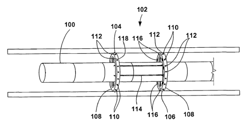
[0021] FIG 2 is a cutaway drawing illustrating a steerable colonoscope 100
with a
colectomy device 102 mounted thereon being inserted through the lumen of a
patient's
colon. As mentioned before, the same technique may apply for every other
tubular shaped
organ. Preferably, the steerable colonoscope 100 is constructed as described
in U.S. Patent
Application Serial Nos. 09/790,204 (now U.S. Patent No. 6,468,203);
09/969,927; and
10/229,577, with multiple articulating segments that are controlled to move
with a
serpentine motion that facilitates insertion and withdrawal of the colonoscope
with a
minimum of contact and stress applied to the colon walls. In addition, the
control system
of the steerable colonoscope 100 has the capability to construct a three-
dimensional
mathematical model or map of the colon as it advances through lumen under
control of the
operator. The three-dimensional mathematical model of the colon and the
location and
nature of any lesions identified in the course of an initial colonoscopic
examination can be
stored and used in performance of the endoscopic colectomy procedure. In
alternate
embodiments, the colectomy device 102 of the present invention may be mounted
on a
colonoscope of a different design and construction.
[0022] The colectomy device 102 can be permanently or removably mounted on the
steerable colonoscope 100. The colectomy device 102 has a distal component 104
and a
proximal component 106. The distal component 104 and the proximal component
106 each
have an expandable member 108 and a gripping mechanism 110 for gripping the
wall of
the colon. The expandable member 108 may be an inflatable balloon or a
mechanically
expandable mechanism. The gripping mechanism 110 may comprise a plurality of
circumferentially located ports within which attachment points 112, e.g.,
needles, hooks,
barbs, etc., may be retractably positioned about an exterior surface of the
expandable
member 108. Alternatively, the gripping mechanism 110 may utilize a vacuum
gripper
through a plurality of circumferentially located ports around the distal
component 104
and/or the proximal component 106 or other known gripping mechanisms. In the
case of
the vacuum gripper, gripping mechanism 110 is in fluid communication through
the ports
and through the colonoscope 100 to the proximal end of the colonoscope 100 to
a vacuum
pump (not shown). At least one, and optionally both, of the distal component
104 and the
proximal component 106 are movable longitudinally with respect to the body of
the
steerable colonoscope 100. Rails, grooves or the like 114 may be provided on
the body of
6
CA 02472207 2004-06-30
WO 03/059151 PCT/US02/41358
the steerable colonoscope 100 for guiding the longitudinal movement of the
distal
component 104 and the proximal component 106.
[0023] In addition, the colectomy device 102 includes a surgical stapler 116
or
other anastomosis mechanism. The surgical stapler 116 is carried on either the
distal
component 104 or the proximal component 106 and a stapler anvil 118 is carried
on the
other of these components. The surgical stapler 116 may be configured
similarly to any
number of conventional stapling devices which are adapted to actuate staples
into tissue.
Another option is that there is a stapler and an anvil on both components for
stapling and
sealing the edges. Optionally, the colectomy device 102 may include a cutting
device
and/or electrocautery and/or a laser device for transecting the colon wall.
Optionally, the
colectomy device 102 may also include a vacuum mechanism or the like for
drawing the
excised tissue into the colectomy device 102 for later removal along with the
steerable
colonoscope 100.
[0024] FIG 2 shows the steerable colonoscope 100 with the expandable members
108 of the distal component 104 and the proximal component 106 in a contracted
or
deflated condition for easy passage through the lumen of the patient's colon.
The control
system of the steerable colonoscope 100 monitors the position of each segment
of the
colonoscope 100 as it is advanced within the colon and can signal to the
operator when the
segments carrying the distal component 104 and the proximal component 106 of
the
colectomy device 102 are correctly positioned with respect to a previously
detected lesion
in the colon. Alternatively, the control system of the steerable colonoscope
100 can be
programmed to advance the colonoscope 100 automatically through the lumen of
the colon
and to stop it when the distal component 104 and the proximal component 106 of
the
colectomy device 102 are correctly positioned with respect to the lesion in
the colon.
Alternatively, the control system will be able to automatically guide and
deliver the two
components to the desired location after the colonoscope has been inserted to
the colon.
[0025] FIG 3 is a cutaway drawing showing the expandable members 108 of the
distal component 104 and the proximal component 106 of the colonoscope-mounted
colectomy device 102 expanded within the lumen of the colon so that the
gripping
mechanism 110 grips the wall of the colon. The distal component 104 and the
proximal
component 106 may be expanded through any number of expansion devices. For
instance,
they may be radially expanded upon spoke-like support structures or they may
be
configured to radially expand in a rotational motion until the desired
expansion diameter is
7
CA 02472207 2004-06-30
WO 03/059151 PCT/US02/41358
attained. At this point, with the diseased portion of the colon identified and
isolated by the
colonoscope-mounted colectomy device 102, the diseased portion is separated
from the
omentum and the blood vessels supplying it are ligated and/or cauterized using
laparoscopic techniques. FIG 1 is a phantom drawing illustrating a diseased
portion of the
colon being separated from the omentum using laparoscopic techniques through a
small
incision in a patient's abdomen.
[0026] Next, the diseased portion of the colon is excised by transecting the
colon at
the proximal and distal end of the diseased portion. The colon may be
transected using
laparoscopic techniques or using a cutting mechanism and/or electrocautery
device
mounted on the colectomy device 102. The excised tissue is removed using the
laparoscope or drawn into the colectomy device 102 for later removal upon
withdrawal of
the steerable colonoscope 100. FIG 4 illustrates the colon after the diseased
portion has
been excised and removed with the colonoscope-mounted colectomy device 102 in
position
to approximate the transected ends of the colon.
[0027] The remaining ends of the colon are approximated one to the other by
moving the distal component 104 and/or the proximal component 106
longitudinally with
respect to the body of the steerable colonoscope 100, as shown by the arrows.
Optionally,
the proximal component 106 may be longitudinally translated towards the distal
component
104 or both components 104,106 may be approximated simultaneously towards one
another. The ends of the colon are stapled to one another to create an end-to-
end
anastomosis 120 using the surgical stapler 116 and stapler anvil 118 on the
colectomy
device 102. Once the ends of the tissue have been approximated, staples or
other fastening
devices, e.g., clips, screws, adhesives, sutures, and combinations thereof,
etc., may be
actuated through the surgical stapler 116 such that they pierce both ends of
the tissue
against the stapler anvil 118. FIG 5 illustrates the colonoscope-mounted
colectomy device
performing an end-to-end anastomosis 120 to complete the endoscopic colectomy
procedure. Once the anastomosis 120 is complete, the expandable members 108 of
the
distal component 104 and the proximal component 106 are deflated or contracted
and the
steerable colonoscope 100 and the colectomy device 102 are withdrawn from the
patient's
body. The expanded members will assure a very accurate end-to-end anastomosis
and
prevent stenosis that can happen as a result of inaccurate approximation of
the two ends.
[0028] In an alternative method using the colonoscope-mounted colectomy device
102, the diseased portion of the colon may be excised using a cutting device
within the
8
CA 02472207 2004-06-30
WO 03/059151 PCT/US02/41358
colectomy device 102 after the ends of the diseased portion have been
approximated and
anastomosed. The excised tissue is drawn into the colectomy device 102 and
removed
when the steerable colonoscope 100 is withdrawn from the patient.
[0029] In another alternative method, the colectomy procedure may be performed
entirely from the endolumenal approach using the colonoscope-mounted colectomy
device
102 without laparoscopic assistance. This method would be particularly
advantageous for
resection of small portions of the colon where it may not be necessary to
mobilize an
extended portion of the colon from the omentum to achieve successful
approximation and
anastomosis. The three-dimensional mapping capability of the steerable
colonoscope 102
would be used to locate previously identified lesions without laparoscopic
assistance.
[0030] While the present invention has been described herein with respect to
the
exemplary embodiments and the best mode for practicing the invention, it will
be apparent
to one of ordinary skill in the art that many modifications, improvements and
subcombinations of the various embodiments, adaptations and variations can be
made to
the invention without departing from the spirit and scope thereof.
9