Note: Descriptions are shown in the official language in which they were submitted.
CA 02472594 2004-07-07
WO 03/059214 PCT/US02/36320
DEVICES AND METHODS USING AN EXPANDABLE BODY
WITH INTERNAL RESTRAINT FOR COMPRESSING
CANCELLOUS BONE
FIELD OF THE INVENTION
This invention relates to the treatment of bone
conditions in human and other animals.
BACKGROUND OF THE INVENTION
When cancellous bone becomes diseased, for example,
because of osteoporosis, avascular necrosis, or cancer, the
surrounding cortical bone becomes more prone to compression
fracture or collapse. This is at least in part because the
cancellous bone no longer provides interior support for the
surrounding cortical bone. The bone disease may also affect
the strength and integrity of the surrounding cortical bone,
further disposing the bone to fracture and/or collapse.
There are 2 million fractures each year in the
United States, of which about 1.3 million are caused by
osteoporosis alone. There are also other bone diseases
involving infected bone, poorly healing bone, or bone fractured
2 0 by severe trauma. Moreover, the use of various drugs, such as
steroids, tobacco and/or the excessive intake of alcohol, can
significantly degrade bone quality. Any of these conditions,
if not successfully addressed, can result in fracture and/or
collapse of bone, causing deformities, chronic complications,
and an overall adverse impact upon the quality of life.
CA 02472594 2004-07-07
WO 03/059214 PCT/US02/36320
- 2 -
U.S. Patent Nos. 4,969,888 and 5,108,404 disclose
apparatus and methods for the fixation of fractures or other
conditions of human and other animal bone systems, both
osteoporotic and non-osteoporotic. Among other inventions,
these patents disclose devices and methods that employ an
expandable body to compress cancellous bone and/or create an
interior cavity within the targeted bone. The cavity receives
a filling material, which hardens and provides renewed interior
structural support for cortical bone.
The better and more efficacious treatment of bone
disease that these patents promise can be more fully realized
with improved systems and methods for making and deploying
expandable bodies in bone.
SUMMARY OF THE INVENTION
One aspect of the invention provides devices and
methods for compressing cancellous bone. In one arrangement,
the devices and methods make use of an expandable body that
includes an internal restraint coupled to the body. The
internal restraint directs expansion of the body. In one
arrangement, a method for treating bone inserts the device
having the internal restraint inside bone and causes directed
expansion of the body in cancellous bone. Cancellous bone is
compacted by the directed expansion.
Another aspect of the invention provides devices
and methods for compacting cancellous bone. In one
arrangement, the devices and methods make use of a body adapted
to be inserted into bone and undergo expansion in cancellous
bone to compact cancellous bone. The body includes material
that, during the expansion in cancellous bone, applies a force
capable of moving fractured cortical bone, and further includes
an interior membrane to constrain the expansion in cancellous
bone. In one arrangement, a method for treating bone inserts
the device having the internal membrane inside bone and causes
restrained expansion of the body in cancellous bone. Cancellous
bone is compacted by the restrained expansion.
CA 02472594 2009-05-07
50749-20
-2a-
Another aspect of the invention provides a device for compressing
cancellous bone comprising an elongated expandable body having an elongated
axis, the expandable body including an internal membrane that directs
expansion
of the body more in a first direction relative to the elongated axis than in a
second
direction relative to the elongated axis.
CA 02472594 2004-07-07
WO 03/059214 PCT/US02/36320
- 3 -
Features and advantages of the invention are set
forth in the following Description and Drawings, as well as in
the appended Claims.
BRIEF DESCRIPTION OF THE DRAWINGS
FIG. 1 is a perspective view of a first embodiment
of a balloon constructed in accordance with the teachings of
the present invention, the embodiment being in the shape of a
stacked doughnut assembly.
FIG. 2 is a vertical section through the balloon of
FIG. 1 showing the way in which the doughnut portions of the
balloon of FIG. 1 fit into a cavity of a vertebral body.
FIG. 3 is a schematic view of another embodiment of
the balloon of the present invention showing three stacked
balloons and string-like restraints for limiting the expansion
of the balloon in various directions of inflation.
FIG. 4 is a top plan view of a spherical balloon
having a cylindrical ring surrounding the balloon.
FIG. 5 is a vertical section through the spherical
balloon and ring of FIG. 4.
FIG. 6 shows an oblong-shaped balloon with a
catheter extending into the central portion of the balloon.
FIG. 6A is a perspective view of one way in which a
catheter can be arranged relative to the inner tubes for
inflating the balloon of FIG. 6.
FIG. 7 is a suction tube and a contrast injection
tube for carrying out the inflation of the balloon and removal
of debris caused by expansion from the balloon itself.
FIG. 8 is a vertical section through a balloon
after it has been deflated and as it is being inserted into the
vertebral body of a human.
FIG. 9, 9A, and 9B are side elevational views of a
cannula showing how the protective sleeve or guard member can
expand when leaving the cannula.
FIG. 10 is a perspective view of another embodiment
of a balloon of the present invention formed in the shape of a
CA 02472594 2004-07-07
WO 03/059214 PCT/US02/36320
- 4 -
kidney bean.
FIG. 11 is a perspective view of the vertebral bone
showing the kidney shaped balloon of FIG. 10 inserted in the
bone and expanded.
FIG. 12 is a top view of a kidney shaped balloon
formed of several compartments by a heating element or branding
tool.
FIG. 13 is a cross-sectional view taken along line
13-13 of FIG. 12 but with two kidney shaped balloons that have
been stacked.
FIG. 14 is a view similar to FIG. 11 but showing
the stacked kidney shaped balloon of FIG. 13 in the vertebral
bone.
FIG. 15 is a top view of a kidney balloon showing
outer tufts holding inner strings in place interconnecting the
top and bottom walls of the balloon.
FIG. 16 is a cross-sectional view taken along line
16-16 of FIG. 15.
FIG. 17A is a dorsal view of a humpback banana
balloon in a right distal radius.
FIG. 17B is a cross-sectional view of FIG. 17A
taken along line 17B-17B of FIG. 17A.
FIG. 18 is a spherical balloon with a base in a
proximal humerus viewed from the front (anterior) of the left
proximal humerus.
FIG. 19A is the front (anterior) view of the
proximal tibia with the elliptical cylinder balloon introduced
beneath the medial tibial plateau.
FIG. 19B is a three-quarter view of the balloon of
FIG. 19A.
FIG. 19C is a side elevational view of the balloon
of FIG. 19A.
FIG. 19D is a top plan view of the balloon of FIG.
19A.
FIG. 20 is a spherically shaped balloon for
CA 02472594 2004-07-07
WO 03/059214 PCT/US02/36320
- 5 -
treating avascular necrosis of the head of the femur (or
humerus) as seen from the front (anterior) of the left hip.
FIG. 20A is a side view of a hemispherically shaped
balloon for treating avascular necrosis of the head of the
femur (or humerus).
FIG. 21 is a balloon for preventing and/or treating
hip fracture as seen from the anterior (front) of the left hip.
FIGS. 22A-C are schematic illustrations of a
representative method and system for delivering a therapeutic
substance to a bone according to the present invention.
FIG. 23 is another embodiment of an expandable
structure incorporating an internal expansion restraint.
FIGS. 24A-C are cross-sectional views of the
expandable structure of FIG. 23 undergoing expansion in air.
FIG. 25A is a front view of another embodiment of
an expandable structure for use in compressing cancellous bone
and/or displacing cortical bone.
FIG. 25B is a side view of the structure of FIG.
25A.
FIG. 25C is a perspective view of the structure of
FIG. 25A.
FIG. 26A is side view of a cavity forming device
carrying an expandable structure of the type shown in Figs. 23
and 24A to 24C.
FIG. 26B is a perspective view of the distal end of
the cavity forming device shown in Fig. 26A, showing the
assembly of the proximal end of the expandable structure to the
distal end of the outer catheter body of the device.
FIG. 26C is a perspective view of the distal end of
the cavity forming device shown in Fig. 26A, after the proximal
and distal ends of the expandable structure have been secured,
respectively, to the distal end of the outer catheter body and
the distal end of the inner catheter body of the device.
FIG. 27 is another embodiment of an expandable
structure.
CA 02472594 2004-07-07
WO 03/059214 PCT/US02/36320
- 6 -
FIG. 28 is a side view of the distal tip of a
cavity-forming device.
DETAILED DESCRIPTION OF THE PREFERRED EMBODIMENT
2. Balloons for Anatomical Structures
The present invention is dir`ected to a balloon that can
be used to treat bones predisposed to fracture or collapse.
These balloons comprise one or more inflatable balloon bodies
for insertion into said bone. The body has a preferred shape
and size when substantially inflated sufficient to compress at
least a portion of the inner cancellous bone to create a cavity
in the cancellous bone and/or to restore the original position
of the outer cortical bone, if fractured or collapsed. In
various embodiments, the balloon body is restrained to create
said preferred shape and size so that the fully inflated
balloon body is desirably inhibited from applying substantial
pressure to a single point on the inner surface of the outer
cortical bone if said bone is unfractured or uncollapsed.
In addition to the shape of the inflatable device itself,
another important aspect is the construction of the wall or
walls of the balloon such that the proper inflation of the
balloon body is achieved to provide for optimum compression of
the cancellous bone. The material of the balloon is also
desirably chosen so the balloon can be inserted quickly and
easily into a bone through a cannula, yet can also withstand
high pressures when inflated. For example, the material could
be chosen to facilitate folding of the balloon. Alternatively,
the material could desirably allow plastic, elastic and/or
semi-elastic deformation of the balloon during inflation. The
material will also desirably resist abrasion and/or puncture of
the balloon when in contact with cortical and/or cancellous
bone during introduction and inflation of the balloon. The
balloon can also include optional ridges or indentations which
are imparted to the cavity, desirably remaining in the cavity
walls after the balloon has been removed, to enhance the
stability of the bone void filler. Also, the inflatable device
CA 02472594 2004-07-07
WO 03/059214 PCT/US02/36320
- 7 -
can be made to have an optional, built-in suction catheter.
This may be used to remove any fat or fluid extruded from the
bone during balloon inflation in the bone. Also, the balloon
body can be protected from puncture (by the surrounding bone or
cannula) by being covered while inside the cannula and/or bone
with an optional protective sleeve of suitable materials, such
as Kevlar fiber products or polyethylene tetraphthalate (PET)
or other polymer or substance that can protect the balloon.
This covering material may also provide the additional
advantage of reducing friction between the balloon and cannula,
or it can incorporate a lubricating material, such as silicone,
to reduce friction. One important purpose of the inflatable
device, therefore, is the forming or enlarging of a cavity or
passage in a bone, especially in, but not limited to, vertebral
bodies.
In one aspect, the invention provides an improved
balloon-like inflatable device for use in carrying out a
surgical protocol of cavity formation in bones to enhance the
efficiency of the protocol, to minimize the time required to
performing the surgery for which the protocol is designed, and
to improve the clinical outcome. If desired, these balloons
may approximate the inner shape of the bone they are inside of
in order to maximally compress cancellous bone. They may also
have additional design elements to achieve specific clinical
goals. In various embodiments, they are made of inelastic,
semi-elastic, elastomeric or plastically deformable materials
and kept in their defined configurations when inflated, by
various restraints, including, but not limited to, use of
inelastic, semi-elastic, elastomeric or plastically deformable
materials in conjunction with the balloon body, seams in the
balloon body created by bonding or fusing separate pieces of
material together, or by fusing or bonding together opposing
sides of the balloon body, woven material bonded inside or
outside the balloon body, strings or bands placed at selected
points in the balloon body, and stacking balloons of similar or
CA 02472594 2004-07-07
WO 03/059214 PCT/US02/36320
- 8 -
different sizes or shapes on top of each other by gluing or by
heat fusing them together. Optional ridges or indentations
created by the foregoing structures, or added on by bonding
additional material, can increase stability of the bone void
filler. The ridges or indentations may also help keep the bone
filler material in a desired position during subsequent loading
and/or healing of the treated bone. Optional suction devices,
preferably placed so that if at least one such device is
located approximate the lowest point of the cavity being
formed, will desirably allow the cavity to be cleaned and/or
permit fluid or solids to be removed from and/or introduced
into the cavity before filling.
Among the various embodiments of the present invention
are the following:
1. A doughnut (or torus) shaped balloon with an
optional built-in suction catheter to remove fat and other
products extruded during balloon expansion.
2. A balloon with a spherical outer shape surrounded
by a ring-shaped balloon segment for body cavity formation.
3. A balloon which is kidney bean shaped in
configuration. Such a balloon can be constructed in a single
layer, or several layers stacked on top of each other. This
embodiment can also be a square or a rectangle instead of a
kidney bean.
4. A spherically shaped balloon approximating the size
of the head of the femur (i.e. the proximal femoral epiphysis).
Such a balloon can also be a hemisphere.
5. A balloon in the shape of a humpbacked banana or a
modified pyramid shape approximating the configuration of the
3 0 distal end of the radius (i.e. the distal radial epiphysis and
metaphysis).
6. A balloon in the shape of a cylindrical ellipse to
approximate the configuration of either the medial half or the
lateral half of the proximal tibial epiphysis. Such a balloon
can also be constructed to approximate the configuration of
CA 02472594 2004-07-07
WO 03/059214 PCT/US02/36320
- 9 -
both halves of the proximal tibial epiphysis.
7. A balloon in the shape of a sphere on a base to
approximate the shape of the proximal humeral epiphysis and
metaphysis with a plug to compress cancellous bone into the
diaphysis, sealing it off. Such an embodiment can also be a
cylinder.
8. A balloon in the shape of a boomerang to
approximate the inside of the femoral head, neck and lesser
trochanter, allowing a procedure to prevent hip fracture.
9. A balloon in the shape of a cylinder to approximate
the size and shape of the inside of the proximal humerus or of
the distal radius.
10. A balloon in the shape of a peanut or hourglass
with an internal membrane. to constrain expansion
preferentially along one or more axes.
11. A balloon in the shape of a disk.
12. A balloon device with an optional suction device.
13. Protective sheaths to act as puncture guard members
optionally covering each balloon inside its catheter.
.20 The present invention, therefore, provides improved,
inflatable devices for creating or enlarging a cavity or
passage in a bone wherein the devices are inserted into the
bone. In various embodiments, the configuration of each device
can be defined by the surrounding cortical bone and adjacent
internal structures, and is designed to occupy up to 70-90% of
the volume of the inside of the bone, although balloons that
are as small as about 40% (or less) and as large as about 99%
are workable for fractures. In various other embodiments, the
inflated balloon size may be as small as 10% of the cancellous
3 0 bone volume of the area of bone being treated, such as for the
treatment of avascular necrosis and/or cancer, due to the
localized nature of the fracture, collapse and/or treatment
area. The fully expanded size and shape of the balloon is
desirably regulated by material in selected portions of the
balloon body whose resistance to expansion creates a restraint
CA 02472594 2004-07-07
WO 03/059214 PCT/US02/36320
- 10 -
as well as by either internal or external restraints formed in
the device including, but not limited to, mesh work, webbing,
membranes, partitions or baffles, a winding, spooling or other
material laminated to portions of the balloon body, continuous
or non-continuous strings across the interior of the balloon
held in place at specific locations by bonding to the inside of
the balloon (by glue, welding, etc.) or by threading these
strings through to the outside, and seams in the balloon body
created by bonding two pieces of body together or by bonding
opposing sides of a body through glue or heat. 'Aside from the
use of different materials, the objectives of the present
invention could similarly be accomplished by utilizing
different thicknesses of materials to regulate the expansion of
the balloon body. Moreover, the use of similar materials of
differing elasticity, for example a polyurethane plastic
balloon having discrete sections that are cross-linked by gamma
radiation exposure and which are thus less prone to expansion,
could accomplish the objectives of the present invention as
well.
Spherical portions of balloons may be restrained by using
inelastic, semi-elastic, elastic and elastomeric materials in
the construction of the balloon body, or may be additionally
restrained as just described. The material of the balloon can
be a non-elastic material, such as polyethylene tetraphthalate
(PET), nylon, Kevlar@) or other patented or nonpatented medical
balloon materials. It can also be made of semi-elastic
materials, such as silicone, rubber, thermoplastic rubbers and
elastomers or elastic materials such as latex or polyurethane,
if appropriate restraints are incorporated. The restraints can
be continuous or made of discrete elements of a flexible,
inelastic high tensile strength material including, but not
limited to, the materials described in U.S. Pat. No. 4,706,670,
which is incorporated herein by reference. The thickness of
the balloon wall is typically in the range of 2/1000ths to
25/1000ths of an inch, although other thicknesses that can
CA 02472594 2004-07-07
WO 03/059214 PCT/US02/36320
- 11 -
withstand increased pressures, such as 250-400 psi or greater,
even up to 500, 1000 or 2000 psi, may be used.
One important goal of percutaneous vertebral body
augmentation of the present invention is to provide a balloon
which can create a cavity inside the vertebral body whose
configuration is optimal for supporting the bone. Another
important goal is to move the top and bottom of the vertebral
body (otherwise known as the upper and lower endplates) toward
a more normal anatomical position to restore height where
possible. Both of these objectives, however, are desirably
achieved without significantly altering the outer dimensions of
the sides of the vertebral body, either by fracturing the
cortical sidewalls of the vertebral body or by moving already
fractured bone in the sidewalls.
The present invention satisfies these goals through the
design of inflatable devices to be described. Inflating such a
device desirably creates a cavity within the calcium-containing
soft cancellous bone (such as by compressing the cancellous
bone) and/or desirably displaces surrounding cortical bone
towards a more normal anatomical position.
In one embodiment, the balloon body desirably recreates
the shape of the inside of an unfractured vertebral body, and
optimally grows no more than a maximum of 70 to 90% of the
inner volume. The balloons of these embodiments are designed
such that maximally inflating them will desirably recreate the
predetermined shape and size. However, conventional balloons
become spherical when inflated. Spherical shapes do not
typically permit the hardened bone void filler to support the
spine adequately, because they can create a generally spherical
cavity which, when filled with filler material, makes single
points of contact on the vertebral body surfaces (the
equivalent of a circle inside a square, or a sphere inside a
cylinder). In contrast, various embodiments of the balloons of
the present invention more generally recreate the flat surfaces
of the vertebral body by incorporating restraints that maintain
CA 02472594 2004-07-07
WO 03/059214 PCT/US02/36320
- 12 -
the balloon in desired shapes. These desired shapes create
cavities which, when filled with filler material, desirably
distribute the load transferred from the vertebral body
surfaces to the bone void fillers, which ultimately strengthens
the spine. In addition, the volume of bone void filler that
fills these cavities desirably creates a thick mantle of cement
(for example a thickness of 4 mm or greater), which increases
the compressive strength of the filler material. Another
useful feature of various embodiments is the incorporation of
ridges in the balloons which can leave one or more imprints in
the walls of the cavity created within the compressed
cancellous bone. The resulting bone void filler "fingers"
which will ultimately fill these imprints can provide enhanced
stability, and reduce the opportunity for the filler material
to shift or displace within the vertebral body under
compressive loading of the spine.
Balloons which can optimally compress cancellous bone in
vertebral bodies include the balloons listed as balloon types
1-3, 10 and 12 above. Some of these balloons are desirably
configured to approximate the shape of the vertebral body.
Since the balloon can be chosen to occupy less than the total
inner volume (prior to fracture) of the targeted vertebral
body, inflation of the balloon will desirably not exert undue
pressure on the surrounding cortical sidewalls of the vertebral
body (the sidewalls of the vertebral body will desirably not be
expand beyond their existing size - either fractured or
unfractured). However, since the upper and lower end plates of
the vertebral body are typically depressed in a compression
fracture, and the balloon can be approximately the height of an
unfractured vertebral body, inflation of the balloon can move
the top and bottom end plates back towards their pre-fractured
position and/or orientation. Moreover, a plurality of
individual balloons can be utilized inside the vertebral body,
such as by being stacked, and stacks containing any of the
disclosed balloon types can be mixed in shape and/or size to
CA 02472594 2004-07-07
WO 03/059214 PCT/US02/36320
- 13 -
provide greater flexibility and/or control.
A primary goal of percutaneous femoral (or humeral) head
augmentation (balloon type 4), percutaneous distal radius
augmentation (balloon type 5), percutaneous proximal tibial
augmentation (balloon type 6), and percutaneous proximal
humeral augmentation (balloon type 7) is to create a cavity
whose configuration is optimal to support the bone to be
treated. Another important goal is to compress avascular (or
aseptic) necrotic bone or to support avascular necrotic bone.
Yet another important goal is to help realign the fracture
fragments. These goals are generally achieved by exerting
pressure primarily on the cancellous bone which may be
transferred to the surrounding cortical bone. Pressure
directly on a small section of the cortical bone could
conceivably cause worsening of the fracture, which, while not
precluded, is desirably avoided. The design of various
embodiments of the inflatable devices approximates the shape of
the bone to be treated. The approximate volume of the cavity
made by the inflatable device(s) can be as much as 70 to 90% of
the volume of the bone to be treated. In the case of avascular
necrosis, depending upon the extent of the avascular necrosis,
a smaller or larger cavity inside bone will be formed. In some
cases, if the area of avascular necrosis is small, a small
balloon will be utilized which might create a cavity only 10 to
15% of the total volume. If larger areas are involved with
avascular necrosis, then one or more larger balloons could be
utilized which might create a much larger cavity, including
cavities as large as 80 to 90% of the volume of the bone (or
greater). The present invention satisfies these goals through
the design of the inflatable devices to be described.
For example, percutaneous hip augmentation (as shown in
connection with balloon type 8) is designed to prevent and/or
treat hip fracture by compacting weak cancellous bone in the
femur where hip fractures occur and replacing it with an
appropriate supporting material. The present invention
CA 02472594 2004-07-07
WO 03/059214 PCT/US02/36320
- 14 -
satisfies this goal through the design of the inflatable
devices to be described.
The present invention discloses improved systems for
deployment in bone comprising structures adapted to assume
expanded geometries having a desired configuration when used.
These expandable structures include material that allows the
structure to differentially expand when under internal
pressure. These structures, when in use, are able to expand
preferentially along one or more axes so as to deliver a
greater force and/or displacement of cancellous bone towards
one direction versus another. Furthermore, such structures,
when distended, can generally match the geometry of the
interior bone space in which the structure is deployed, if
desired. For example, such structures could optimally expand
to a desired shape rather than simply towards areas of lowest
bone density, i.e. expansion of the structure is can be
controlled even when encountering areas in the bone of varying
resistance.
Moreover, the exposure of the expandable structure to
cancellous bone also typically requires materials having
significant resistance to surface abrasion, puncture and/or
tensile stresses. For example, structures incorporating
elastomer materials, which have been preformed to a desired
shape, e.g., by exposure to heat and pressure, can undergo
controlled expansion and further distention in cancellous bone,
without failure, while exhibiting resistance to surface
abrasion and puncture when contacting cancellous bone.
The present invention further discloses inflatable
devices that have one or more biased directions of inflation.
For example, inflatable devices having reduced lateral growth
may provide improved fracture reduction because such devices
can exert a greater vertical force and/or displacement within
the treated bone. Such inflatable devices may also protect the
lateral and anterior/posterior sidewalls of the vertebral body
by minimizing expansion towards these sidewalls and directing
CA 02472594 2004-07-07
WO 03/059214 PCT/US02/36320
- 15 -
expansion to a greater degree along the longitudinal axis of
the spine. In situations where a surgical procedure is
terminated when the inflatable device contacts a lateral
cortical wall of the targeted bone, such biased expansion could
permit improved fracture reduction prior to reaching this
procedure endpoint.
Due to the nature of the injury, disease or other
treatments, as well as the health and age of the patient
suffering from these injuries, it may be preferable to treat a
bone with the devices of this invention during an open or semi-
open surgical procedure. In addition, a goal of the surgery
may be to replace the diseased or injured bone with materials
(such as bone fillers or certain drugs) which do not flow, and
which thus are not well suited for a more minimally invasive
procedure.
A. Balloons for Vertebral Bodies
A first embodiment of the balloon (FIG. 1) constructed in
accordance with the teachings of the present invention is
broadly denoted by the numeral 10 and includes a balloon body
11 having a pair of hollow, inflatable parts 12 and 14
comprised of flexible material, including (but not limited to)
non-elastic materials such as PET, mylar or Kevlarelastic
materials such as polyurethane, latex or rubber, semi-elastic
materials such as silicone, or other materials. Parts 12 and
14 have a suction tube 16 therebetween for drawing fats and
other debris by suction into tube 16 for transfer to a remote
disposal location. Catheter 16 has one or more suction holes
so that suction may be applied to the open end of tube 16 from
a suction source (not shown).
In this embodiment, the parts 12 and 14 are connected
together by an adhesive which can be of any suitable type for
adhering such materials as well as by bonding, i.e. thermal
bonding (laser, radio-frequency (RF)/induction, heated dies),
ultrasonic welding, solvent bonding, etc. Parts 12 and 14 are
doughnut-shaped as shown in FIG. 1 and have tubes 18 and 20
CA 02472594 2009-05-07
50749-20
- 16 -
which communicate with and extend away from the parts 12 and
14, respectively, to a source of inflating fluid under pressure
(not shown). The inflating fluid is preferably a liquid. The
c
liquid inflates the balloon 10, particularly parts 12 and 14
thereof after the balloon has been inserted in a collapsed
condition (FIG. 8) into a bone to be treated, such as a
vertebral bone 22 in FIG. 2. The above-mentioned U.S. Pat.
Nos. 4,969,888 and 5,108,404 disclose the use of a guide pin
and cannula for inserting the balloon irito bone to be treated
when the balloon is deflated and has been inserted into a tube
and driven by the catheter into the cortical bone where the
balloon is inflated.
FIG. 8 shows a deflated balloon 10 being inserted through
a cannula 26 into bone. The balloon in cannula 26 is deflated
and is forced through the cannula by exerting manual force on
the catheter 21 which extends into a passage 28 extending into
the interior of the bone. The catheter is slightly flexible
but is sufficiently rigid to allow the balloon to be forced
into the interior of the bone where the balloon is then
inflated by directing fluid into the tube 88 whose outlet ends
are coupled to respective parts 12 and 14.
In use, the balloon 10 is initially deflated and, after
the bone to be filled with the balloon has been prepared to
receive the balloon (such as by punching, drilling or otherwise
displacing a small amount of the cancellous bone directly
beyond the opening of the cannula), the deflated balloon is
advanced into the bone in a collapsed condition through the
cannula 26. (The bone is shown in FIG. 2.) In this
embodiment, the balloon is oriented preferably in the bone such
that the balloon expansion permits minimum pressure to be
exerted on the cortical bone if there were no fracture or
collapse of the bone. Where such fracture or collapse has not
occurred, such pressure would desirably compress the bone
marrow and/or cancellous bone against the inner wall of the
cortical bone, thereby compacting the bone marrow of the bone
CA 02472594 2004-07-07
WO 03/059214 PCT/US02/36320
- 17 -
to be treated and to further enlarge the cavity in which the
bone marrow is to be replaced by a biocompatible, flowable bone
material.
The balloon is inflated to compact the bone marrow and/or
cancellous bone in the cavity and, after compaction of the bone
marrow and/or cancellous bone, the balloon is deflated and
removed from the cavity. While inflation of the balloon and
compaction occurs, fats and other debris may be removed from
the space between and around parts 12 and 14 by applying a
suction force to catheter tube 16, if desired. Following this,
and following the compaction of the bone marrow, the balloon is
deflated and pulled out of the cavity by applying a manual,
pulling force to the catheter tube 21.
Another embodiment of an inflatable device constructed in
accordance with the teachings of the present invention is
broadly denoted by the numeral 60 and is shown in FIGS. 4 and
5. The balloon 60 includes a central spherical part 62 which
is hollow and which receives an inflating liquid under pressure
through a tube 64. The spherical part is provided with a
2 0 spherical outer surface 66 and has an outer periphery which is
surrounded substantially by a ring shaped part 68 having tube
segments 70 for inflation of part 68. A pair of passages 69
interconnect parts 62 and 68. A suction tube segment 72 draws
liquid and debris from the bone cavity being formed by the
balloon 60.
Provision can be made for'a balloon sleeve 71 for the
balloon 60 as well as for all balloons disclosed herein. A
balloon sleeve 71 (FIG. 9) is shiftably mounted in an outer
tube 71a and can be used to insert the balloon 60 when deflated
3 0 into a cortical bone. The sleeve 71 has resilient fingers 71b
which bear against the interior of the entrance opening 71c of
the vertebral bone 22 (FIGS. 9A and 9B) to prevent rearing or
bunching of the balloon 60. Upon removal of the balloon
sleeve, liquid under pressure will be directed into the tube 64
which will inflate parts 62 and 68 so as to compact the bone
CA 02472594 2004-07-07
WO 03/059214 PCT/US02/36320
- 18 -
marrow within the cortical bone. Following this, the balloon
60 is deflated and removed from the bone cavity.
FIGS. 6 and 6A show views of a modified doughnut shape
balloon 80 of the type shown in FIGS. 1 and 2, with one
difference being the doughnut shapes of the balloon 80 are not
stitched onto one another. In FIG. 6, the balloon 80 has a
pear-shaped outer convex surface 82 which is made up of a first
hollow part 84 and a second hollow part 85. A tube 88 is
provided for directing liquid into the two parts along branches
90 and 92 to inflate the parts after the parts have been
inserted into the medullary cavity of a bone. A catheter tube
16 is inserted into the space 96 between two parts of the
balloon 80. An adhesive bonds the two parts 84 and 85 together
at the interface thereof.
FIG. 6A shows one way in which the catheter tube 16 is
inserted into the space or opening 96 between the two parts of
the balloon 80.
FIG. 7 shows the tube 88 of which, after directing
inflating liquid into the balloon 80, can inject contrast
material into the balloon 80 so that x-rays can be taken of the
balloon with the inflating material therewithin to determine
the proper placement of the balloon. Alternatively, the
inflation liquid could comprise a radiopaque inflation liquid,
such as Conray contrast medium (commercially available from
Mallinckrodt Inc. of St. Louis, Mo.), such that inflation and
visualization can be done currently, allowing monitoring of the
balloon position and condition during the inflation step. Tube
16 is also shown in FIG. 6, it being attached in some suitable
manner to the outer side wall surface of tube 88.
Still another embodiment of the invention is shown in
FIG. 3, which is similar to FIG. 1(although one difference is
that it is not a doughnut) and includes an inflatable device
109 having three balloon units 110, 112 and 114 which are
inflatable and which have string-like restraints 117 which
limit the expansion of the balloon units in a direction
CA 02472594 2004-07-07
WO 03/059214 PCT/US02/36320
- 19 -
transverse to the longitudinal axes of the balloon units. If
desired, the restraints can comprise the same or a similar
material as the balloon, or the restraints can comprise a
material having a reduced, little or no substantial expansion
capability.
A tube system 115 can be provided to direct liquid under
pressure into the balloon units 110, 112 and 114 so that liquid
can be used to inflate the balloon units when placed inside the
bone in a deflated state. Following the proper inflation and
compaction of the bone marrow, the balloon(s) can be removed by
deflating it/them and pulling it/them outwardly of the bone
being treated. The restraints desirably keep the opposed sides
77 and 79 substantially flat and parallel with respect to each
other.
In FIG. 10, another embodiment of the inflatable balloon
is shown. The device comprises a kidney shaped balloon body
130 having a pair of opposed kidney shaped side walls 132 which
are adapted to be collapsed and to cooperate with a continuous
end wall 134 so that the balloon 130 can be forced into a bone
136 shown in FIG. 11. A tube 138 is used to direct inflating
liquid into the balloon to inflate the balloon and cause it to
assume the dimensions and location shown in the vertebral body
136 in FIG. 11. The balloon 130 will desirably compress the
cancellous bone if there is no fracture or collapse of the
cortical bone. The restraints for this action are principally
due to the side and end walls of the balloon.
FIG. 12 shows a balloon 140 which is also kidney shaped
and has a tube 142 for directing an inflatable liquid into the
tube for inflating the balloon. The balloon is initially
formed in a single chamber bladder but the bladder can
subsequently be branded and/or melted along curved lines or
strips 141 to form attachment lines 144 which take the shape of
side-by-side compartments 146 which are kidney shaped as shown
in FIG. 13. The branding desirably causes a welding and/or
bonding of the two sides of the bladder - the material can be
CA 02472594 2004-07-07
WO 03/059214 PCT/US02/36320
- 20 -
standard medical balloon material, which is typically plastic
that can be formed and/or bonded using heat.
FIG. 14 is a perspective view of a vertebral body 147
containing the balloon of FIG. 12, showing a double stacked
balloon 140 when it is inserted in vertebral bone 147.
FIG. 15 is a view similar to FIG. 10 except that tufts
155, which can be string-like restraints or other structures
between the opposing inner walls of the balloon, extend between
and are connected to the side walls 152 of the inflatable
device 150 and limit the expansion of the side walls with
respect to each other. In this embodiment, the tufts desirably
render the side walls generally parallel with each other. Of
course, tufts which merely limit and/or reduce the displacement
between opposing walls of the balloon will similarly accomplish
various objectives of the present invention to some degree.
Tube 88 is used to fill the kidney shaped balloon with an
inflating liquid in the manner described above.
The dimensions for a vertebral body balloon can vary
across a broad range, depending upon the size, location, and
condition of the targeted vertebral body as well as the
objectives of the treatment. For example, the height (H, FIG.
11) of a vertebral body balloon for both lumbar and thoracic
vertebral bodies can typically range from 0.5 cm to 3.5 cm.
The anterior to posterior (A, FIG. 11) vertebral body balloon
dimensions for both lumbar and thoracic vertebral bodies can
typically range from 0.5 cm to 3.5 cm. The side to side (L,
FIG. 11) vertebral body dimensions from thoracic vertebral
bodies will often range from 0.5 cm to 3.5 cm. The side to
side vertebral body dimensions for lumbar vertebral bodies will
typically range from 0.5 cm to 5.0 cm. Of course, depending
upon the objectives of the treatment and the actual dimensions
of the patient's bones, the use of balloons having larger or
smaller dimension than these disclosed ranges may be
appropriate.
The eventual selection of the appropriate balloon for,
CA 02472594 2004-07-07
WO 03/059214 PCT/US02/36320
- 21 -
for instance, a given vertebral body is based upon several
factors. One major factor affecting the choice of balloon size
is the objectives of the treatment. For example, if the
principal treatment objective is simply the repair and/or
augmentation of a collapsed vertebral body, then the
appropriate balloon size (and desired cavity size) may be a
balloon which approximates the size of the interior of the
vertebral body in an unfractured and/or uncollapsed condition.
Alternatively, two or more balloons could be used concurrently
within a single vertebral body, which together create a desired
size cavity within the vertebral body. As another alternative,
if the objective of treatment is more localized within the
bone, such as the creation of a smaller cavity to augment
and/or repair a smaller section of the bone, then the use of a
smaller balloon size (and desired cavity size) may be desirous.
Similarly, where the cancellous bone is relatively strong
and/or resistant to compression, the use of a smaller balloon
may be warranted to accomplish the objective of displacing
cortical bone (to reduce the fracture) without significantly
compressing the cancellous bone (thus creating a smaller
cavity) . Moreover, smaller balloons may also be suited for use
in the treatment of bone tumors, etc., where the balloon can be
used to create a small cavity adjacent to the tumor - this
small cavity will simplify the use of other minimally invasive
tools to directly visualize the treatment area as well as
morselize and/or excise the tumor from the bone.
The anterior-posterior (A-P) balloon dimension is
measured from the internal cortical wall of the anterior cortex
to the internal cortical wall of the posterior cortex of the
vertebral body. In general, for augmentation and/or
reinforcement of a collapsed vertebral body, the appropriate A-
P balloon dimension will often be approximately 5 to 7
millimeters less than this measurement.
The appropriate side to side balloon dimensions for a
given vertebral body is selected from the CT scan or from a
CA 02472594 2004-07-07
WO 03/059214 PCT/US02/36320
- 22 -
plain film x-ray view of the vertebral body to be treated. The
side to side distance can be measured from the internal
cortical walls of the side of the vertebral bone. In one
embodiment, the appropriate side to side balloon dimension may
be 5 to 7 millimeters less than this measurement. In alternate
embodiments, the appropriate side to side balloon dimensions
may be significantly smaller, such as where multiple balloons
are introduced into a single vertebral body or where the
displacement of cortical bone is a primary objective of the
treatment. In general, lumbar vertebral bodies tend to be much
wider in their side to side dimension than in their A-P
dimension. In contrast, thoracic vertebral bodies are
typically approximately equal in their the side to side
dimensions and their A-P dimensions.
The height dimensions of the appropriate vertebral body
balloon for a given vertebral body may be chosen by the CT scan
or x-ray views of the vertebral bodies above and below the
vertebral body to be treated. The height of the vertebral
bodies above and below the vertebral body to be treated can be
2 0 measured and averaged. This average may be used to determine
the appropriate height dimension of the chosen vertebral body
balloon. Of course, as previously mentioned, various other
balloon sizes may be desirous based upon the objectives of the
treatment, as well as the actual patient's anatomy.
B. Balloons for Long Bones
Long bones which can be treated with the use of balloons
of the present invention include (but are not limited to) the
distal radius (larger arm bone at the wrist), the proximal
tibial plateau (leg bone just below the knee), the proximal
humerus (upper end of the arm at the shoulder), and the
proximal femoral head (leg bone in the hip).
C. Distal Radius Balloon
For the distal radius, one embodiment of a balloon 160 is
shown in the distal radius 152 has a shape which approximates a
pyramid but more closely can be considered the shape of a
CA 02472594 2004-07-07
WO 03/059214 PCT/US02/36320
- 23 -
humpbacked banana in that it substantially fills the interior
of the space of the distal radius to force cancellous bone 154
against the inner surface 156 or cortical bone 158.
The balloon 160 has a lower, conical portion 159 which
extends downwardly into the hollow space of the distal radius
152, and this conical portion 159 increases in cross section as
a central distal portion 161 is approached. The cross section
of the balloon 160 is shown at a central location (FIG. 17B)
and this location is near the widest location of the balloon.
The upper end of the balloon, denoted by the numeral 162,
converges to the catheter 88 for directing a liquid into the
balloon for inflating the same to compress the cancellous bone
and/or force the cancellous bone against the inner surface of
the cortical bone. The shape of the balloon 160 is desirably
predetermined and can be restrained by tufts formed by string
restraints 165, as well as various other types of restraints
described herein. These restraints are optional and provide
additional strength to the balloon body 160, but are not
absolutely required to achieve the desired configuration. The
balloon is placed into and taken out of the distal radius in
the same manner as that described above with respect to the
vertebral bone.
The dimensions of the distal radius balloon vary as
follows:
The proximal end of the balloon (i.e. the part nearest
the elbow) is cylindrical in shape and will vary from 0.5 x 0.5
cm to 1.8 x 1.8 cm.
The length of the distal radius balloon will vary from
1.0 cm to 12.0 cm.
The widest medial to lateral dimension of the distal
radius balloon, which occurs at or near the distal radio-ulnar
joint, will measure from 1.0 cm to 2.5 cm.
The distal anterior-posterior dimension of the distal
radius balloon will vary from 0.5 cm to 3.0 cm.
CA 02472594 2004-07-07
WO 03/059214 PCT/US02/36320
- 24 -
In an alternate embodiment also suited for use in
treating a distal radius fracture, a balloon can take the shape
of a toroidal or disk-like shape, such as shown in Figures 25A-
25C.
D. Proximal Humerus Fracture Balloon
The selection of the appropriate balloon size to treat a
given fracture of the distal radius will often depend on the
radiological size of the distal radius and the location of the
fracture, as well as the treatment goals.
In the case of the proximal humerus 169, one embodiment
of a balloon 166 shown in FIG. 18 is spherical and has a base
design. It can optimally compact the cancellous bone 168 in a
proximal humerus 169. A mesh 170, embedded, laminated and/or
wound, may be used to form a neck 172 on the balloon 166, and a
second mesh 170a may be used to conform the bottom of the base
172a to the shape of the inner cortical wall at the start of
the shaft. These restraints provide additional strength to the
balloon body, but the configuration can be achieved through
various methods, including molding of the balloon body or
various other restraints described herein. This embodiment
desirably compresses the cancellous bone to create a compacted
region surrounding the balloon 166 as shown in FIG. 18. The
cortical bone 173 is desirably relatively wide at the base 174
and is thin-walled at the upper end 175. The balloon 166 has a
feed tube 177 into which liquid under pressure is forced into
the balloon to inflate it to compact the cancellous bone in the
proximal humerus. The balloon is inserted into and taken out
of the proximal humerus in the same manner as that described
above with respect to the vertebral bone.
In this embodiment, the dimensions of the proximal
humerus fracture balloon vary as follows:
The spherical end of the balloon will vary from 1.0 x 1.0
cm to 3.0 x 3.0 cm.
The neck of the proximal humeral fracture balloon will
CA 02472594 2004-07-07
WO 03/059214 PCT/US02/36320
- 25 -
vary from 0.8 x 0.8 cm to 3.0 x 3.0 cm.
The width of the base portion or distal portion of the
proximal humeral fracture balloon will vary from 0.5 x 0.5 cm
to 2.5 x 2.5 cm.
The length of the balloon will vary from 4.0 cm to 14.0
cm.
The selection of the appropriate balloon to treat a given
proximal humeral fracture depends on the radiologic size of the
proximal humerus and the location of the fracture as well as
the treatment goals.
E. Proximal Tibial Plateau Fracture Balloon
The tibial fracture is shown in FIG. 19A in which one
embodiment of a balloon 180 is placed in one side 182 of a
tibia 183. Desirably, the balloon, when inflated, compacts the
cancellous bone in the layer 184 surrounding the balloon 180.
A cross section of this embodiment of a balloon is shown in
FIG. 19C wherein the balloon has a pair of opposed sides 185
and 187 which are interconnected by restraints 188 which can be
in the form of strings or flexible members of any suitable
construction. In this embodiment, the restraints desirably
maintain the sides 185 and 187 substantially parallel with each
other and non-spherical. A tube 190 is coupled to the balloon
180 to direct inflation liquid into and out of the balloon.
The ends of the restraints are shown in FIGS. 19B and 19D and
denoted by the numeral 191. The balloon is inserted into and
taken out of the tibia in the same manner as that described
above with respect to the vertebral bone. FIG. 19B shows a
substantially circular configuration for the balloon; whereas,
FIG. 19D shows a substantially elliptical version of the
balloon.
The dimensions of this embodiment of a proximal tibial
plateau fracture balloon vary as follows:
The thickness or height of the balloon will vary from 0.5
cm to 5.0 cm.
CA 02472594 2004-07-07
WO 03/059214 PCT/US02/36320
- 26 -
The anterior-posterior (front to back) dimension will
vary from 1.0 cm to 6.0 cm.
The side to side (medial to lateral) dimension will vary
from 1.0 cm to 6.0 cm.
The selection of the appropriate balloon to treat a given
tibial plateau fracture will depend on the radiological size of
the proximal tibial and the location of the fracture, as well
as the treatment goals.
F. Femoral Head Balloon
In the case of the femoral head, one embodiment of a
balloon 200 is shown as having been inserted inside the
cortical bone 202 of the femoral head which is thin at the
outer end 204 of the femur and which can increase in thickness
at the lower end 206 of the femur. The cortical bone surrounds
the cancellous bone 207 and this bone is desirably compacted by
the inflation of the balloon 200. The tube for directing
liquid for inflation purposes into the balloon is denoted by
the numeral 209. It extends along the femoral neck and is
directed into the femoral had which is generally spherical in
configuration. FIG 20A shows that the balloon, denoted by the
numeral 200a, can be hemispherical as well as spherical, as
shown in FIG. 20. The balloon 200 is inserted into and taken
out of the femoral head in the same manner as that described
with respect to the vertebral bone. The hemispherical shape is
maintained in this example by bonding overlapping portions of
the bottom, creating pleats 200b as shown in FIG. 20A.
The dimensions of the femoral head balloon may vary as
follows - the diameter of the femoral head balloon will vary
from 1.0 cm to up to 4.5 cm or greater. The appropriate size
of the femoral head balloon to be chosen depends on the
radiological or CT scan size of the head of the femur and the
location and size of the avascular necrotic bone. The
dimensions of the hemispherical balloon are similar to those of
the spherical balloon, except that approximately one half of
the balloon is provided.
CA 02472594 2004-07-07
WO 03/059214 PCT/US02/36320
- 27 -
G. Prevention of Hip Fracture
FIG. 21 illustrates one embodiment of a "boomerang"
balloon 210 adapted for preventing and/or treating hip
fracture. When inflated, the "boomerang" balloon 210 is a
cylinder which gradually bends in the middle, like a boomerang,
and extends from about 0.5 cm from the end of the femoral head
211 through the femoral neck 212 and down into the proximal
femoral diaphysis 213 about 5-7 cm past the lesser trochanter
214. This embodiment of a balloon 210 preferably maintains its
shape by rings of inelastic material (215 is one of them) held
closer together on one side by attachment to a shorter
inelastic band 216 running the length of the side of balloon
and further apart by attachment to a longer inelastic band 217
bonded on the opposite side, although various other restraints
disclosed herein would also suffice.
After and prior to inflation, the balloon 210 may be
folded back (shown in dotted lines at 218) against the
inflation tube 219. Prior to inflation, the balloon 210 can
also be rolled up and held against the inflation tube with
2 0 loose attachments that break when the balloon is inflated. To
insert the balloon on its inflation tube into the hip, the
surgeon can use a power drill under radiographic guidance to
create a cavity 220 that is usually 4-6 mm wide starting at the
lateral femoral cortex 221 and proceeding into the femoral head
211. Inflation of the balloon 210 into the greater
trochanteric region 222 instead of down the femoral diaphysis
213 is less desirable and is typically avoided by proper
choices in the shape of the balloon as well as by its placement
and correct orientation (the deflated balloon desirably facing
the lesser trochanter). After the balloon 210 has been
inflated within the cavity 220 (see the dotted lines in
FIG. 21), the predetermined size and shape of the balloon
biases the proximal portion of the balloon downward into the
lesser trochanter. Optionally, a second cavity can be drilled
down into the diaphysis, starting from the same entry point or
CA 02472594 2004-07-07
WO 03/059214 PCT/US02/36320
- 28 -
from the other side.
Patients with bone density in the hip below a threshold
value are at increased risk of hip fracture, and lower
densities create greater risk. Patient selection may be done
through a bone density scan or other methods of determining
bone quality well known in the art. Such selection could also
result from a previous and/or concurrent fracture of the other
hip, or some other type and/or location of osteoporotic
fracture. The balloon length can be chosen by the surgeon to
extend about 0.5 cm from the end of the femoral head, through
the femoral neck and into the proximal femoral diaphysis,
usually about 4-8 cm below the lesser trochanter. The balloon
diameter can be chosen by measuring the inner cortical diameter
of the femoral neck (the most narrow area) and subtracting 0.5
cm. The preferred dimensions of the "boomerang" balloon are a
total length of 10-20 cm and a diameter of 1.0-2.5 cm. (A
"humpback banana" balloon with appropriate length may also be
useful in hip fracture prevention, where the "humpback" width
does not exceed the desired femoral neck dimensions.)
Patients having the lowest bone densities in the femoral
head may require greater compacting in the femoral head, which
may, for example, be provided by using two balloons, one after
the other: the "boomerang" followed by the femoral head balloon
(inserted at the same point and expanded prior to inserting any
supporting material.) Alternatively, the "boomerang" balloon
may be adapted to have a distal portion that approximates the
shape of the femoral head balloon.
The various balloons described herein could also be used
in conjunction with the replacement of various structures
within human and animal bodies. For example, the balloons
described herein could be used to compress cancellous bone in a
femur in preparation for the implantation of an artificial hip
stem. Similarly, the balloons described herein could be used
in conjunction with various other joint replacement procedures,
including artificial knee and ankle joints.
CA 02472594 2004-07-07
WO 03/059214 PCT/US02/36320
- 29 -
H. All Balloons
It should be understood that the various embodiments of
balloons disclosed herein are by no means limited in their
utility to use in a single treatment location within the body.
Rather, while each embodiment may be disclosed in connection
with an exemplary treatment location, these embodiments can be
utilized in various locations within the human body, depending
upon the treatment goals as well as the anatomy of the targeted
bone. For example, the embodiment of a balloon previously
disclosed as useful in treating a fracture of the distal radius
could similarly be used in the treatment of fractures in
various other areas within the body, including but not limited
to fractures and/or impending fractures of the femur, the
radius, the ulna, the tibia, the humerus, the calcaneus or the
spine. Similarly, the various other disclosed embodiments can
be utilized throughout the body, with varying results depending
upon treatment goals and/or the anatomy of the targeted bone.
II. The Inflatable Device
A. Complex Expandable Structures
Sometimes it can be difficult to achieve a desired
uniformity and area of compaction within a given cancellous
bone region using an expandable body having a single expansion
region. Figure 27 shows a complex preformed structure 280
which includes expanded segmented regions 282 and 284 spaced
along its length. The structure 280 provides a longer profile
along which volume can be increased.
The complex expandable structure is created by extruding
or molding a tube 286 of plastic or other elastomer material.
In a preferred embodiment, the tube is comprised of plastic
material. The tube has a normal extruded wall thickness (T5)
and a normal extruded outside diameter (D5) (as shown in Figure
27).
The segmented shaped regions 282 and 284 are created by
exposing an intermediate region of the tube to heat, positive
interior pressure and/or stretching inside a fixture or mold
CA 02472594 2004-07-07
WO 03/059214 PCT/US02/36320
- 30 -
(not shown). In one embodiment, the fixture could possess two
cavity regions separated by a reduced diameter region or
intermediate channel. The cavity regions and the channel can
be exposed to a source of heat, to soften the material of the
region. When heat-softened (in the manner previously
described), the interior of the tube 286 is stretched and
subjected to positive pressure from a source. The material in
the region 288 will desirably expand or extend within the
cavities and the channel.
Once cooled and removed from the fixture, the structure
280 can be attached to the distal end of an outer catheter tube
250. (See Fig. 28.) The structure of the outer catheter tube
250 (as well as the inner catheter tube 258) can vary, and the
outer catheter tube 250 can comprise various flexible
materials, including medical grade plastic materials like
vinyl, polyethylenes, ionomer, polyurethane, and
polytetrapthalate (PET) as well as less flexible materials such
as Kevlar , PEBAX"m, stainless steel, nickel-titanium alloys,
and other metals and/or ceramics. The outer catheter tube 250
desirably incorporates an interior bore 260, into which an
inner catheter tube 258 extends. It should be appreciated that
the outer catheter tube 250 can have one or more interior
lumens. In the illustrated embodiment, the inner catheter tube
258 extends through the interior bore 260 and beyond the distal
end 254 of the catheter tube 250. A distal end region of the
structure 280 is secured to the to the distal end region 254 of
the outer catheter tube 250, while a proximal end region of the
structure 280 is secured to the distal end region 262 of the
inner catheter tube 258. The end regions can be secured, e.g.,
using adhesive or thermal bonding, etc.
The structure 280 possesses, in an open air environment,
a normal expanded shape, having diameter D7 (shown in phantom
lines of Fig. 27). The normal shape and diameter D7 for the
regions 282 and 284 generally correspond with the shape and
dimension of the cavities, respectively.
CA 02472594 2004-07-07
WO 03/059214 PCT/US02/36320
- 31 -
When an interior vacuum is drawn, removing air and/or
fluid from the structure 280, the structure 280 assumes a
substantially collapsed, and not inflated, geometry, shown as
lines D6 in Figure 27. Due to the application of heat and
pressure upon the intermediate region 288, the diameter D6 for
each region 282 and 284 is larger than the normally extruded or
molded outside diameter D5 of the original extruded tube.
The regions 282 and 284 are separated by a tubular neck
298, which segments the structure 280 into two expandable
regions 282 and 284. When substantially collapsed under vacuum
or not inflated, the structure 280 exhibits a low profile,
ideal for the insertion into and/or removal from the targeted
cancellous bone region.
The introduction of fluid volume back into the tube 286
will cause each region 282 and 284 to return from the collapsed
diameter D6 to the normal, enlarged, but not distended,
geometry, having the shape and diameter shown in phantom lines
D7 in Figure 27.
In the illustrated embodiment, the first and second
shaped regions 282 and 284 have generally the same radius of
expansion and thus the same non-distended shape and diameter
D7. Alternatively, each region 282 and 284 can have a
different radius of expansion, and thus a different non-
distended shape and diameter. Regardless, when in the normal,
non-distended diameter D7, the material of the structure 280 in
the region 288 is not significantly stretched or stressed,
because the regions 282 and 284 have been expanded in a stress-
relieved condition into these geometries in the cavities.
As before explained in conjunction with the structure,
the regions 282 and 284 can be shaped by heat and/or interior
pressure within different cavities to assume different
geometry's, e.g., cylindrical or elliptical geometry, or a non-
spherical, non-cylindrical, or non-elliptical geometry, with
either uniform or complex curvature, and in either symmetric or
asymmetric forms. Of course, more than two segmented regions
CA 02472594 2004-07-07
WO 03/059214 PCT/US02/36320
- 32 -
282 and 284 can be formed along the length of the tube. In
addition, the normally expanded shape characteristics of the
structure can be achieved by other techniques. For example,
and not by way of limitation, the structure can be formed by
dipping, lost wax casting, or injection molding.
Each shaped region 282 and 284 possesses a minimum wall
thickness (designated T7 in Figure 27) when in the normally
enlarged but not distended geometry D7. Due to expansion of
heat-softened material under pressure in the cavities, the wall
thickness is not uniform, i.e., T7 is less than the normal
extruded or molded wall thickness T5 of the tube. The minimum
wall thickness T7 for the regions 282 and 284 can be the same
or different.
When in the enlarged, but not distended geometry, the
neck region 298 has an outside diameter (designated D9 in
Figure 27), which is equal to or greater than the normal
extruded or molded diameter D5 of the tube. The size of the
channel in the fixture determines the magnitude of the diameter
D9. Due to expansion of heat-softened material in the adjacent
regions 282 and 284 under pressure in the cavities, the neck
region 298 (which expands under pressure in the channel) has a
wall thickness (designated T9 in Figure 27) which is less than
or equal to the normal extruded or molded wall thickness T5 of
the tube 286, but still greater than the minimum wall thickness
T7 of either fully shaped region 282 or 284.
The formed complex structure 280 thus possesses regions
of non-uniform minimum wall thickness along its length; that
is, T5ZT9aT7. The formed complex structure 280 also provides
multiple expandable regions 282 and 284 of the same or
3 0 different enlarged outside diameters (D7), segmented by a neck
region 298, in which D6>D5; D7>D6; and D7>D9.
By continuing to apply fluid volume at a constant
pressure at a threshold amount P(t), and thereby increasing the
volume within the structure 280, the shaped regions 282 and 284
of the structure 280 will continue to enlarge beyond diameter
CA 02472594 2004-07-07
WO 03/059214 PCT/US02/36320
- 33 -
D7 to a distended shape and geometry, designated D8 in Figure
27. The wall thickness T7 further decreases and approaches T8.
As the regions 282 and 284 approach diameter D8, the diameter
D9 of the neck region 298 will likewise increase toward
diameter D10, as Figure 27 shows, providing more uniform,
elongated surface contact with cancellous bone.
Enlargement of the structure 280 beyond diameter D7
stretches the material in the regions 282, 284 and 298 beyond
their stress-relieved condition, although the distended
geometry of the regions 282 and 284 will, in important
respects, maintain the preformed shape dictated by the
cavities.
The degree of stretching at a substantially constant
incremental pressure condition can be tailored to achieve a
desired, fully distended diameter D8. The final, fully
distended diameter D8 can be selected to match the dimensions
of the targeted cancellous bone region. The controlled
stretching of the segmented regions 282 and 284 in tandem can
provide an equal volume compression of cancellous bone with a
2 0 major diameter that is less than a single non-segmented region
(i.e., one without the neck region 298). Stated another way,
segmented regions 282 and 284, when expanded to a given
inflation volume, have a diameter less than a sphere expanded
to an equal inflation volume.
While expanding in the region between D7 and D8, the
structure 280, when inside bone, assumes an increasingly larger
surface area and volume, thereby compacting surrounding
cancellous bone. Inflation in cancellous bone may occur at the
same threshold pressure P(t) as outside bone. However, an
increase in the threshold pressure P(t) inside bone is
typically required, due to the density of the cancellous bone
and resistance of the cancellous bone to compaction.
B. Assembly of an Expandable Balloon Device
with an Internal Membrane
Figures 23 and 24A - 24C depict cross-sectional views of
CA 02472594 2004-07-07
WO 03/059214 PCT/US02/36320
- 34 -
another alternate embodiment of a cavity-forming device
constructed in accordance with the teachings of the present
invention. Because many of the features of this embodiment are
similar to those described in connection with the previous
embodiment, like reference numerals will be used to describe
similar components.
In this embodiment the cavity-forming device incorporates
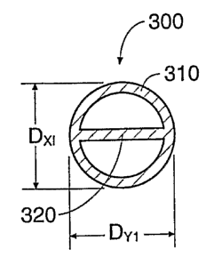
a balloon 300 comprising a section of dual lumen tubing having
an outer wall 310 and an internal membrane 320. The balloon
300 will desirably comprises a material that is commonly used
for balloon catheters including, but not limited to,
polyethylene, mylar, rubber or polyurethane. Even more
desirably, the balloon 300 will comprise an elastomer material,
which also possess the capability of being preformed, i.e., to
acquire a desired shape by exposure, e.g., to heat and
pressure, e.g., through the use of conventional thermoforming,
blow molding and/or dip coating techniques. Candidate materials
that meet this criteria include polyurethane, silicone,
thermoplastic rubber, nylon, and thermoplastic elastomer
materials.
In the illustrated embodiment, the balloon 300 comprises
a plastic material. This material can be processed and
extruded in a tubular shape, which can then be cut into
individual lengths for further processing. The balloon 300 can
be formed by exposing a cut tube length to heat and then
enclosing the heated tube within a mold while positive interior
pressure is applied to the tube length. The mold can of course
be part of a conventional balloon forming machine.
In the present embodiment, after the balloon is formed
the proximal end of the balloon 300 can be attached to the
distal end of an outer catheter body 250 and the distal end of
the balloon 300 can be attached to the distal end of an inner
catheter body 258. The outer and inner catheters may each
comprise extruded tubing made, e.g., from plastic material, and
each can extruded in a tubular shape.
CA 02472594 2004-07-07
WO 03/059214 PCT/US02/36320
- 35 -
In assembling the cavity-forming device, the proximal end
of the balloon 300 is desirably bonded to the distal end of an
outer catheter body 250, as Figure 26A shows. In one preferred
embodiment (as Figure 26B shows), a razor blade or other
cutting instrument can be used to split approximately 5mm of
the distal end of the outer catheter body 250, creating a pair
of slots 360, as best shown by "A" in Figure 26B. The proximal
end of the balloon 300 can then be slid over the distal end of
the outer catheter body 250, with the outer wall 310 positioned
around the distal tip of the outer catheter body 250 and the
internal membrane 320 positioned within the slots 360 (as
Figure 26C shows). To maintain the flow channels (for the
inflation fluid) through the outer catheter body 250 and into
the balloon 300, a pair of mandrels or inserts (not shown) can
be introduced into the outer catheter body and balloon in a
manner well known in the art. The distal end of the outer
catheter body 250 and the proximal end of the balloon 300 can
then be bonded together using various means including heat
bonding, adhesives, or the like. After the bond is formed, the
2 0 mandrels can be removed. Desirably, the splitting of the outer
catheter body 250 increases the mechanical strength of the bond
between the catheter body 250 and the balloon 300 and permits
the balloon to be more securely bonded to the outer catheter
body 250, reducing the opportunity for a proximal bond failure
of the balloon 300.
The distal end of the balloon 300 is also bonded to the
distal end of an inner catheter body 258. If desired, the
distal end of the inner catheter body 258 may be split in a
similar manner to increase the mechanical strength of the
distal bond. Desirably, the inner catheter body 258 will
extend through the outer catheter body 250 and the balloon 300
along one side the internal membrane 320.
As Figure 26A shows, the proximal end of the outer
catheter body 250 can be secured to a distal end of a y-shaped
luer fitting 400. The inner catheter body 258 desirably
CA 02472594 2004-07-07
WO 03/059214 PCT/US02/36320
- 36 -
extends through an inner lumen of the luer fitting 400, and may
be bonded to a proximal end of the fitting 400. Desirably, an
inflation fitting 402 of the y-shaped luer fitting 400 will be
in fluid communication with the lumen 404 (see Figure 26C)
formed between the inner and outer catheter bodies 250 and 258,
which will in turn be in fluid communication with the interior
of the balloon 300, such that an inflation fluid introduced
into the inflation fitting 402 will inflate the balloon 300.
Desirably (as Figures 26A to 26C show), the outer
catheter body 250 and/or y-shaped luer fitting 400 will
incorporate a marker 406 or other externally viewable indicia
which shows a physician the orientation of the internal
membrane 320 when the balloon 300 is in a desired position
within the patient. Such indicia could include colored markers
or stripes 406, indentations and/or protrusions on the outer
catheter body 250 or y-shaped luer fitting 400 as well as the
orientation of the luer fitting itself. By utilizing such
indicia 406, the physician can easily rotate the balloon 300 to
a desired orientation within the vertebral body. Because the
materials used in constructing medical balloons are typically
radio-lucent, it would be difficult to gage the orientation of
the internal membrane 320 once the balloon 300 is in position
within the targeted bone. Alternatively, or in combination
with external indicia 406, the internal membrane 320 could
incorporate one or more marker bands or other radiopaque
substances 408 (see Figure 26C) to depict the orientation of
the membrane 320 within the targeted vertebral body.
Various materials can be selected for the component parts
of the cavity-forming device. Furthermore, the dimensions of
the component parts of the cavity-forming device can also vary,
according to its intended use. It should also be understood
that, while one described embodiment incorporates dual lumen
tubing, various other embodiments could incorporate other types
of multi-lumen tubing (including, but not limited to triple,
quadruple, etc., lumen tubing), as well as could incorporate
CA 02472594 2004-07-07
WO 03/059214 PCT/US02/36320
- 37 -
membrane(s) having varying orientations and/or positions within
the tubing (e.g., symmetrical or asymmetrical).
C. Exemplary Performance Features of the Expandable
Balloon
Figures 24A, 24B and 24C show cross-sectional views of
the previously-described embodiment of a balloon 300 during its
deployment in air. Desirably, the balloon 300 will expand in a
similar fashion within the targeted bone such as a vertebral
body.
Figure 24A depicts a cross-sectional view of the balloon
300 when filled with a small amount of inflation fluid, such
that the balloon desirably assumes the approximate size and
shape of the mold in which the balloon was previously formed,
with minimal stresses experienced by the internal membrane 320.
In this condition, the expansion of the balloon is
substantially circular in cross-section. Accordingly, the
vertical and horizontal dimensions of the cross-section of the
expanded balloon 300 are approximately equal, or Dxl = DY1.
Figure 24B depicts the balloon 300 of Figure 24A when
further filled with a pressurized inflation fluid. In this
figure, the balloon 300 has assumed a further distended shape,
with the wall material of the balloon 300 typically undergoing
elastic and/or plastic deformation to assume this enlarged
geometry. The balloon 300 desirably does not assume a
completely circular cross-sectional shape, principally because
the internal membrane resists lateral expansion of the outer
walls 310. While some elongation of the internal membrane 320
typically occurs (due to elastic and/or plastic deformation of
the membrane), the resulting cross-sectional shape is generally
ovoid or somewhat similar to a figure-8. The balloon 300,
however, is not as significantly restrained from growing in the
vertical direction. This combination of restraints results in
a balloon which substantially expands or grows more in the
vertical direction than in the horizontal direction.
Accordingly, the vertical dimension of the expanded balloon 300
CA 02472594 2004-07-07
WO 03/059214 PCT/US02/36320
- 38 -
is larger than the horizontal dimension of the balloon 300, or
D%2 > DY2.
Figure 24C depicts the balloon 300 of Figures 24A and 24B
when further filled with a pressurized inflation liquid. In
this figure, the balloon 300 has assumed an even more distended
shape, with the wall material typically having undergone both
elastic and significant plastic deformation in order to assume
this enlarged geometry. At this point, the balloon 300 is
clearly in a non-circular shape, with the internal membrane 320
significantly resisting lateral growth of the balloon (although
some additional elastic stretching and/or plastic deformation
of the membrane 320 has likely occurred). Accordingly, the
vertical dimension of the expanded balloon 300 is significantly
larger than the horizontal dimension of the balloon 300, or Dx3
>> DY3.
For the above-described embodiment, an experimental
inflation of the balloon with inflation fluid with volumes of
0cc to 2cc and 2cc to 4cc produced the following results:
0cc:
* Balloon Minor diameter (DX1 - width): 7.7mm
* Balloon Major diameter (DY1 - height): 7.7mm
Inflation to 2cc (Fluid):
* Balloon Minor diameter (DXZ - width): 9.2 mm
* Increase in minor (horizontal) diameter: 1.5 mm (width) -
[19.5 % total increase]
* Balloon Major diameter (DY2 - height) : 10.9 mm
* Increase in major (vertical) diameter: 2.2 mm (height) -
[28.6 % total increase]
Inflation to 4cc (Fluid):
* Balloon Minor diameter (DX3 - width) : 12.7 mm
* Increase in minor (horizontal) diameter: 5mm (width) -
[65% total increase]
* Balloon Major diameter (DY3 - height) 15.4 mm
* Increase in major (vertical) diameter: 7.7mm (height) -
[100% total increase]
CA 02472594 2004-07-07
WO 03/059214 PCT/US02/36320
- 39 -
In addition to axial growth of the balloon 300 as the
balloon expands (as previously described), the longitudinal
length of a balloon also tends to increase during inflation.
This is because the stresses experienced by the balloon
material are typically acting in more than one dimension
(resulting in material deformation along more than a single
axis), causing the overall longitudinal length of the balloon
300 to expand in response to the increased internal pressure.
In the present embodiment, however, the internal membrane 320
also tends to reduce the longitudinal growth of the balloon
during inflation. For example, for the previously described
embodiment of a balloon 300, a volumetric increase from 2cc to
4cc results=in a longitudinal length increase for the balloon
of only 27.1%. For a similarly constructed balloon that does
not incorporate an interior membrane, a volumetric increase
from 2cc to 4cc results in a longitudinal length increase of
37.1%. Accordingly, the interior membrane 320 of the present
invention restrains not only'certain aspects of circumferential
expansion, but also restrains aspects of longitudinal expansion
as well.
The internal membrane 320 of the present embodiment also
significantly reduces the opportunity for the balloon 300 to
experience a complete radial failure and/or fragment within the
patient. During a surgical procedure, if the balloon is
punctured or torn, the balloon failure may propagate through a
significant amount of the balloon material. If this failure
propagates around the entire radius of the balloon, then the
distal section of the balloon is in danger of becoming
completely separated from the proximal end of the balloon, with
only the inner catheter body 258 connecting the distal section
of the balloon to the cavity-forming device. in such a case,
upon removal of the cavity forming device from the patient, it
is possible for the inner catheter body 258 to fail, leaving
the distal section and any balloon fragments in the patient.
The internal membrane 320 of the present embodiment
CA 02472594 2004-07-07
WO 03/059214 PCT/US02/36320
- 40 -
desirably reduces any opportunity for a complete radial failure
of the balloon 300, and also significantly reduces the
opportunity for balloon fragments to separate from the cavity-
forming device. Where the interior membrane 320 joins the
expandable wall, the geometry and/or additional thickness of
balloon material at this junction 410 (see Figure 26C)
significantly increases the balloon's resistance to fracture at
his location. A fracture which propagates towards such a
junction 410 will typically be redirected by the junction -
typically the fracture will either terminate, will rebound from
the junction and/or will be redirected along the junction.
In the disclosed embodiment, a radial fracture which
propagates towards the junction 410 will generally be
redirected towards the longitudinal axis of the balloon 300.
Moreover, the interior membrane 320 serves to connect the
proximal and distal ends of the balloon 300, which will
reinforce the inner catheter body 258 in the unlikely event of
a complete radial failure of the balloon. Accordingly, because
the present embodiment incorporates at least two longitudinally
extending junctions (i.e., the internal membrane 320 of the
balloon 300 and the inner catheter body 258 to which the distal
end of the balloon 300 is secured), a fracture of this
embodiment is unlikely to result in a complete radial tear of
the balloon material and/or fragmentation of the cavity forming
device.
III. Implant Creation and Performance
Once the balloon 300 is in a desired position within a
targeted bone (in this example a vertebral body), an inflation
medium can be introduced into the balloon, which desirably
expands the balloon within the targeted bone. The balloon will
desirably assume a similar shape within the targeted bone as it
would in air, thereby creating a cavity within the bone that is
substantially the same shape and size as the inflated balloon.
It must be understood, however, that variations in cancellous
bone density and quality may distort the final expanded size
CA 02472594 2004-07-07
WO 03/059214 PCT/US02/36320
- 41 -
and shape of the inflated balloon, such that the expanded
balloon may be significantly different in size and shape than
it would be when expanded in air.
While the restraints described herein may not absolutely
guarantee that the final shape and size of the balloon (and
thus the cavity) will be identical to the shape and size of the
balloon in air, the restraints described herein significantly
increase the potential for creating an optimally sized and
shaped cavity to achieve one or more desired treatment goals.
For example, if the desired treatment goal is the reinforcement
and/or repair of a targeted vertebral body, a balloon may be
chosen that incorporates restraints to maximize vertical growth
of the balloon (in this context, the vertical orientation can
be assumed to be parallel to the longitudinal axis of the
spine) while minimizing horizontal and/or longitudinal growth
of the balloon. If desired, this balloon could also
incorporate restraints that reduce and/or minimize balloon
expansion along its longitudinal axis.
Alternatively, a physician may desire a balloon that
incorporates restraints to maximize horizontal growth of the
balloon (in this context, horizontal growth can be assumed to
be transverse to the longitudinal axis of the spine) while
minimizing vertical growth of the balloon. Such a balloon
(which could simply be the previously described embodiment when
rotated 90 about its longitudinal axis) could be used to
initially create a cavity extending across substantially the
entire vertebral body. After removal of the first balloon, a
second balloon (of the same or different design) could
subsequently be introduced into the horizontal cavity and
expanded. If desired, the second balloon could substantially
fill the horizontal cavity prior to inflation (thereby
maximizing the surface area of the balloon facing the upper and
lower endplates) and, when expanded, could maximize the
vertical forces which ultimately act on the endplates of the
vertebral body (in an attempt to displace the surrounding
CA 02472594 2004-07-07
WO 03/059214 PCT/US02/36320
- 42 -
cortical bone).
If desired, a balloon chosen for treatment of a vertebral
body may further incorporate restraints that cause the balloon
to expand into an irregular shape. In one embodiment disclosed
herein, best shown in Fig. 23, the balloon desirably expands to
a "peanut-like" shape when viewed from the side. This
embodiment will desirably create a cavity that is similarly
"peanut-shaped", with the cavity essentially comprising a pair
of enlarged cavity lobes that are separated by a region of
reduced cavity size - in other words, the cavity is dumb-bell
shaped. Desirably, the filler material which occupies this
cavity will harden, set and/or solidify into an implant having
substantially the shape of the cavity into which it was
introduced. By forming the implant into this dumb-bell shape,
the region of reduced width of the implant will desirably help
to anchor the implant within the cancellous bone, thereby
reducing the opportunity for the implant to displace along the
longitudinal axis of the implant and/or migrate within or
outside the treated bone.
Furthermore, if desired a balloon used for treatment of a
vertebral body could incorporate additional restraints that
alter the outer shape of the expanded balloon to further reduce
the opportunity and/or tendency of an implant to migrate within
and/or outside of a treated bone. For example, in one
embodiment described above, the balloon incorporates an
internal membrane which desirably causes the expanded balloon
to assume an indented or elongated "figure-8" shape in cross-
section (see Figure 24c). This shape, if formed into the
cavity walls and ultimately assumed by the filler material,
will desirably create an implant of similar cross-section. By
forming the implant into this figure-8 shape, the implant will
desirably be anchored within the cancellous bone, thereby
reducing the opportunity for the implant to rotate about the
longitudinal axis of the implant and/or migrate within or
outside the treated bone.
CA 02472594 2004-07-07
WO 03/059214 PCT/US02/36320
- 43 -
In addition to creating a desired shape and size to the
cavity, which will desirably act as a mold to bound and shape
the filler material, the physician can further customize the
shape of the implant in various ways. For example, after the
initial cavity formation, but prior to the introduction of the
filler material, the physician could use other surgical
instruments to alter the shape and/or size of the cavity, such
as by removing additional cancellous bone and/or scoring the
compressed cancellous bone along the walls of the cavity.
Similarly, prior to introducing the filler material the
physician could introduce one or more additional balloons into
the cavity to alter the existing cavity dimensions and/or
create additional cavities of unique and/or desired shape. The
physician could alternatively choose to introduce two or more
different bone filler materials into a single cavity, with
different materials occupying different portions of the cavity
and/or being intertwined, mixed or separated in some manner, if
desired. In addition, after the filler material has filled the
entire cavity, the physician could continue introducing an
additional amount of bone filler material, which would
desirably cause small amounts of the bone filler material to
interdigitate or flow into various gaps and/or cracks in the
walls of the cavity, thereby further anchoring the resulting
implant within the cancellous bone. For example, the injection
of an additional % cc, 1 cc or l%cc of bone filler material
(beyond the volume of the cavity created within the cancellous
bone) can significantly increase the interdigitation of bone
filler material with the surrounding cancellous bone matrix.
IV. Other Uses, Methods and Balloons
The cavity created by the balloon can be filled with a
medically-appropriate formulation of a drug or a growth factor.
As an example of delivering a drug, a typical dose of the
antibiotic, gentamicin, to treat a local osteomyelitis (bone
infection), is 1 gram (although the therapeutic range for
gentamicin can be far greater, from 1 nanogram to 100 grams,
CA 02472594 2004-07-07
WO 03/059214 PCT/US02/36320
- 44 -
depending on the condition being treated and the size of the
area to be covered). A medically-suitable gel formulated with
appropriate gel materials, such a polyethylene glycol, can
contain 1 gram of gentamicin in a set volume of gel, such as
10 cc. A balloon with this volume whose shape and size is
appropriate for the site being treated (that is, the balloon
desirably will not break the cortical bone when inflated at the
chosen site) can be used to compact the infected cancellous
bone. This creates a space that can be filled with the
antibiotic gel in an open or minimally invasive procedure.
This places and holds the required amount of drug right at the
site needing treatment, and protects the drug from being
quickly washed away by blood or other fluids. Not only can the
dose be optimized, but additional doses can be applied at later
times without open surgery, enhancing the therapeutic outcome.
If the required cavity for the optimal drug dose weakens the
bone, the bone can be protected from future fractures with a
cast or with current internal or external metal or plastic
fixation devices. The therapeutic substance put,into bone may
be acting outside the bone as well. A formulation containing
chemotherapeutic agent could be used to treat local solid
osteosarcoma or other tumor near that bone.
As an alternative, to deliver therapeutic substances,
balloons can be dipped in a medical formulation (often a dry
powder, liquid or gel) containing a medically-effective amount
of any desired antibiotic, bone growth factor or other
therapeutic agent to coat the balloon with the above-mentioned
substance before it is inserted into a bone being treated.
Optionally, the balloon can be wholly or partially inflated
with air or liquid before the coating is performed.
Optionally, the coated balloon can be dried with air or by
other means when the applied formulation is wet, such as a
liquid or a gel. The balloon is refolded as required and
either used immediately or stored, if appropriate and desired.
Coated on the balloon, therapeutic substances can be delivered
CA 02472594 2004-07-07
WO 03/059214 PCT/US02/36320
- 45 -
while cancellous bone is being compressed, or with an
additional balloon once the cavity is made.
The methods described above can also be used to coat
Gelfoam absorbable gelatin powder or other agents onto the
balloon before use. Such agents may also comprise substances
that desirably promote coagulation and/or thickening of body
fluids. inflating a Gelfoam-coated balloon inside bone may
further fill any cracks in fractured bone not already filled by
the compressed cancellous bone.
FIGS. 22A-C schematically illustrate one system and
method for delivering a therapeutic substance to the bone
according to the present invention. As shown in FIG. 22A, an
inflated balloon 229 attached to an inflating tube 230 is
stabilized with a clip 231 that couples tube 230 to a wire 232.
As shown in FIG. 22B, a measured amount of gel formulation
containing the desired amount of substance 233 is uniformly
dispensed from a container 234, preferably in thin lines 235,
onto the outer surface of a balloon 236. As shown in FIG. 22C,
the coated balloon 23 is then deflated and allowed to dry until
the gel sets. The coated balloon 237 is then ready for
packaging for use by the surgeon. Of course, the balloon can
also be coated without prior inflation. In addition, the
coating substance can be the desired compound alone in its
natural state (solid, liquid or gas) or in an appropriate
formulation, including a dry powder, an aerosol or a solution.
The optional drying time will, of course, depend on the nature
of the compound and its formulation.
Delivering a therapeutic substance on the outside of the
balloon used to compact the bone or with a second (possibly
slightly larger) balloon after the bone is compacted, is
qualitatively different than putting formulated drug into the
cavity. When delivered while compressing the bone, the
substance becomes incorporated into the compacted bone. This
can serve as a way to instantly formulate a slow release
version of the substance. It simultaneously allows the surgeon
CA 02472594 2004-07-07
WO 03/059214 PCT/US02/36320
- 46 -
to fill the cavity with an appropriate supporting material,
like acrylic bone cement or biocompatible bone substitute, so
no casting or metal fixation is required. Such a combination
allows the surgeon, for example, to percutaneously fix an
osteoporotic fracture while delivering a desired therapeutic
substance (like an antibiotic, bone growth factor or
osteoporosis drug) to the site. Thus, casts or metal fixation
devices may not be required in such instances.
Medically-effective amounts of therapeutic substances are
typically defined by their manufacturers or sponsors and are
generally in the range of 10 nanograms to 50 milligrams per
site, although more or less may be required in a specific case.
Typical antibiotics include gentamicin and tobramycin. Typical
bone growth factors are members of the bone morphogenetic
factor, osteogenic protein, fibroblast growth factor, insulin-
like growth factor, and transforming growth factor alpha and
beta families. Chemotherapeutic and related agents include
compounds such as cisplatin, doxorubicin, daunorubicin,
methotrexate, taxol and tamoxifen. Osteoporosis drugs include
estrogen, calcitonin, diphosphonates, and parathyroid hormone
antagonists.
The balloons described in this invention can be used in
open surgical procedures at the sites discussed above to
provide an improved space for inserting orthopedic implants,
bone graft, bone substitutes, bone fillers or therapeutic
substances. The size and shape of balloon chosen will be
determined depending upon the site being treated as well as the
size, shape or amount of material that the surgeon wants to
insert into the remaining bone. Square and rectangular
balloons can be used at any site for the placement of bone
substitutes like hydroxyapatites which are available in those
shapes. Balloons would desirably be made to match those
predetermined sizes, and the surgeon would chose the balloon to
fit the size of material chosen.
To insert materials which do not flow into the balloon-
CA 02472594 2004-07-07
WO 03/059214 PCT/US02/36320
- 47 -
made cavity, like hydroxyapatite granules or bone mineral
matrix, the surgeon can push them down a tube with a long pin
whose diameter is slightly more narrow than the inner diameter
of the cannula through procedures in which the minimally-
invasive procedure is taking place. During open surgery, the
surgeon can approach the bone to be treated as if the procedure
is percutaneous, except that here is no skin and other tissues
between the surgeon and the bone being treated. This desirably
keeps the cortical bone as intact as possible. If the material
to be inserted does not flow and should not be pushed into the
cavity through a cannula (as in the case of the hydroxyapatite
block, because that may result in significant damage to the
patient), the surgeon can make the cavity using the "minimally
invasive" approach, then punch a hole using standard tools
(such as a punch, gouge or rasp) into one side of the cortical
bone to allow insertion of the block. This same approach can
be used for implanting a metal prosthesis, such as the metal
tibial component of a total knee replacement system.
Different sizes and/or shapes of balloons may be used at
sites not specified above, such as the jaw bones, the midshaft
of the arm and leg bones, the cervical vertebral bodies, the
foot and ankle bones, the ribs and the like. One of the keys
to choosing balloon shape and size in treating or preventing
bone fracture is the teaching of this application that,
optimally, up to 70-90% (or greater) of the cancellous bone can
be compacted in cases where the bone disease causing fracture
(or the risk of fracture) is the loss of cancellous bone mass
(as in osteoporosis). Compacting less than 70-90% of the
cancellous bone at the site being treated could possibly leave
an extensive amount of the diseased cancellous bone at the
treated site. The diseased cancellous bone could remain weak
and later collapse, causing fracture despite treatment. With
this principle, the allowed shapes and minimum sizes for any
chosen bone are explained and defined.
Of course, there are many exceptions to this 70-90%
CA 02472594 2004-07-07
WO 03/059214 PCT/US02/36320
- 48 -
cavity size, as generally described in this specification. One
exception is when the bone disease being treated is localized,
as in avascular necrosis, where local loss of blood supply is
killing bone in a limited area. In that case, the balloons can
be smaller, because the disease area requiring treatment is
often smaller. A second exception is in the use of the devices
to improve insertion of solid materials in defined shapes, like
hydroxyapatite and components in total joint replacement. In
these cases, the balloon shape and size is generally defined by
the shape and size of the material being inserted. Another
exception is the delivery of therapeutic substances. In this
case, the cancellous bone may or may not be affected. If it is
not, some of the cancellous bone can be sacrificed by
compacting it to improve the delivery of a drug or growth
factor which has an important therapeutic purpose. In this
case, the bone with the drug inside is supported while the drug
works and then the bone heals through casting or current
fixation devices. Another exception can involve the treatment
of bone tumors, where the creation of a small cavity in
cancellous bone adjacent the tumor could facilitate the
minimally invasive manipulation and/or removal of the tumor.
Another exception could be where the quality of the cancellous
bone is generally good, but the bone has fractured and/or
collapsed in some manner. In such a case, the creation of a
small cavity within the stronger cancellous bone may displace
the cortical bone fragments to a position at or near the
fragments' normal anatomic positions without significantly
compressing the cancellous bone.
Another key to choosing balloon shape and size is one
teaching of this invention - that inelastic, elastic and/or
semi-elastic balloon restraints can be utilized and that
inelastic or semi-elastic balloon materials are often
preferred. Such materials can safely and easily prevent the
balloon from expanding beyond its predetermined shape and size
which can be defined by the limits of the normal dimensions of
CA 02472594 2004-07-07
WO 03/059214 PCT/US02/36320
- 49 -
the outside edge of the cancellous bone (which is inside of the
cortical bone) . A balloon which expands too much, for example,
can create the risk of immediate fracture, so in one embodiment
this defines the upper limits of balloon sizes at each site.
With many typical angioplasty balloons, surgeons usually rely
on monitoring pressure (instead of the balloon design features
of this invention) to prevent their balloons from inflating too
much. This often requires greater surgical skill than the
teachings of the present application, which are to take an X-
ray of the site to be treated and measure the important
dimensions as described herein. In addition, in bone
treatment, relying on pressure can often result in an inferior
clinical outcome. The surgeon generally will not know in
advance what pressure is required to completely compact the
cancellous bone, because this varies depending on the thickness
of the cancellous bone and the extent to which it has lost
density due to its disease. The surgeon is often likely to
under inflate the balloon to avoid the potential consequences
of overinflation and/or cortical bone fracture.
Another teaching of this application is that, while
maximal pressures equally exerted in all directions can
typically compress the weakest areas of cancellous bone, the
use of restraints in a balloon body will desirably control
balloon expansion to some degree. If the balloon design does
not incorporate restraints, it may not compress cancellous bone
in an optimal manner for reinforcement and/or repair of a
fractured vertebral. The shape of the cancellous bone to be
compressed, and the local structures that could be harmed if
bone were moved inappropriately, are generally understood by
medical professionals using textbooks of human skeletal anatomy
along with their knowledge of the site and its disease or
injury. Ranges of shapes and dimensions are defined by the
site to be treated. Precise dimensions for a given patient can
be determined by X-ray of the site to be treated, the
therapeutic goal and safety constraints at the site. For
CA 02472594 2004-07-07
WO 03/059214 PCT/US02/36320
- 50 -
diseased bone, replacement of most of the cancellous bone may
be desired, so a balloon whose shape and size will compress
around 70-90% (or greater) of the volume of the cancellous bone
in the treated region can be chosen. However, as previously
noted balloons that are smaller or larger may be appropriate,
particularly where localized bone treatments and/or delivery of
a therapeutic substance is the main goal. If desired, the
balloon size can be chosen by the desired amount of therapeutic
substance, keeping in mind that the balloon should desirably
not displace the cortical bone beyond its normal unfractured
dimensions.
While the new devices and methods have been more
specifically described in the context of the treatment of human
vertebrae, it should be understood that other human or animal
bone types can be treated in the same or equivalent fashion.
By way of example, and not by limitation, the present systems
and methods could be used in any bone having bone marrow
therein, including the radius, the humerus, the vertebrae, the
femur, the tibia or the calcaneus. In addition, other
embodiments and uses of the invention will be apparent to those
skilled in the art from consideration of the specification and
practice of the invention disclosed herein. All documents
referenced herein are specifically and entirely incorporated by
reference. The specification and examples should be considered
exemplary only with the true scope and spirit of the invention
indicated by the following claims. As will be easily
understood by those of ordinary skill in the art, variations
and modifications of each of the disclosed embodiments can be
easily made within the scope of this invention as defined by
the following claims.