Note: Descriptions are shown in the official language in which they were submitted.
CA 02473135 2011-06-14
1
STABILIZED mRNA TUMOUR VACCINE
The present invention relates to a pharmaceutical
composition comprising at least one mRNA comprising at
least one coding region for at least one antigen from a
tumour, in combination with an aqueous solvent and
preferably a cytokine, e.g. GM-CSF, and a process for the
preparation of the pharmaceutical composition. The
pharmaceutical composition according to the invention is
used in particular for therapy and/or prophylaxis against
cancer.
Gene therapy and genetic vaccination are molecular medicine
methods which, when used in the therapy and prevention of
diseases, will have considerable effects on medical
practice. Both methods are based on the introduction of
nucleic acids into cells or into tissues of the patient and
on subsequent processing of the information coded by the
nucleic acids introduced, i.e. expression of the desired
polypeptides.
The conventional procedure of methods of gene therapy and
of genetic vaccination to date is the use of DNA to insert
the required genetic information into the cell. Various
methods for introducing DNA into cells have been developed
in this connection, such as e.g. calcium phosphate
CA 02473135 2004-07-22
2
transfection, polyprene transfection, protoplast fusion,
electroporation, microinjection and lipofection, whereas
lipofection in particular having emerged as a suitable
method.
A further method which has been proposed in particular in
the case of genetic vaccination methods is the use of DNA
viruses as DNA vehicles. Such viruses have the advantage
that because of their infectious properties a very high
transfection rate can be achieved. The viruses used are
genetically modified, so that no functional infectious
particles are formed in the transfected cell. In spite of
this safety precaution, however, a certain risk of
uncontrolled propagation of the genes having a gene therapy
action and the viral genes introduced cannot be ruled out
because of possible recombination events.
The DNA introduced into the cell is conventionally
integrated into the genome of the transfected cell to a
certain extent. On the one hand this phenomenon can exert a
desired effect, since a long-lasting action of the DNA
introduced can thereby be achieved. On the other hand, the
integration into the genome results in a substantial risk
of gene therapy. Thus e.g. the DNA introduced may be
inserted into an intact gene, which represents a mutation
which interferes or even completely switches off the
function of the endogenous gene. On the one hand enzyme
systems which are essential for the cell may be switched
off by such integration events, and on the other hand there
is also the danger of a transformation of the cell modified
in this way into a degenerated state if a gene which is
decisive for regulation of cell growth is modified by the
CA 02473135 2004-07-22
3
integration of the foreign DNA. A risk of the development
of cancer therefore cannot be ruled out when using DNA
viruses as gene therapeutics and vaccines. In this
connection it is also to be noted that for effective
expression of the genes introduced into the cell, the
corresponding DNA vehicles contain a strong promoter, e.g.
the viral CMV promoter. Integration of such promoters into
the genome of the treated cell can lead to undesirable
changes in the regulation of gene expression in the cell.
A further disadvantage of the use of DNA as gene
therapeutics and vaccines is the induction of pathogenic
anti-DNA antibodies in the patient, causing a possibly
fatal immune response.
In contrast to DNA, the use of RNA as a gene therapeutic or
vaccine is to be classified as substantially safer. In
particular, RNA does not involve the risk of being
integrated into the genome of the transfected cell in a
stable manner. Furthermore, no viral sequences, such as
promoters, are necessary for effective transcription.
Moreover, RNA is degraded considerably more easily in vivo.
Apparently because of the relatively short half-life of RNA
in the blood circulation compared with DNA, no anti-RNA
antibodies have been detected to date. RNA can therefore be
regarded as the molecule of choice for molecular medicine
therapy methods.
Nevertheless, medical methods based on RNA expression
systems still require a solution to some fundamental
problems before they are used more widely. One of the
problems of using RNA is reliable cell- or tissue-specific
CA 02473135 2004-07-22
4
efficient transfer of the nucleic acid. Since RNA usually
proves to be very unstable in solution, it has not hitherto
been possible, or has been possible only in a very
inefficient manner, to use RNA as a therapeutic or vaccine
by the conventional methods which are used with DNA.
RNA-degrading enzymes, so-called RNAases (ribonucleases),
are responsible for the instability. Even the smallest
impurities of ribonucleases are sufficient to degrade RNA
in solution completely. The natural degradation of mRNA in
the cytoplasm of cells is very finely regulated. Several
mechanisms are known in this respect. Thus, the terminal
structure is of decisive importance for a functional mRNA.
At the 5'-end is the so-called "cap structure" (a modified
guanosine nucleotide), and at the 3'-end a sequence of up
to 200 adenosine nucleotides (the so-called poly-A tail).
The RNA is recognized as mRNA and the degradation is
regulated via these structures. Moreover, there are further
processes which stabilize or destabilize RNA. Many of
these processes are still unknown, but an interaction
between the RNA and proteins often appears to be decisive
for this. For example, an "mRNA surveillance system" has
recently been described (Hellerin and Parker, Annu. Rev.
Genet. 1999, 33: 229 to 260), in which incomplete or
nonsense mRNA is recognized by certain feedback protein
interactions in the cytosol and is rendered accessible to
degradation, the majority of these processes being
performed by exonucleases.
Some measures for increasing the stability of RNA and
thereby rendering possible its use as a gene therapeutic or
RNA vaccine have been proposed in the prior art.
CA 02473135 2004-07-22
To solve the abovementioned problems of the instability of
RNA ex vivo, EP-A-1083232 proposes a process for
introduction of RNA, in particular mRNA, into cells and
5 organisms, in which the RNA is in the form of a complex
with a cationic peptide or protein.
WO 99/14346 describes further processes for stabilizing
mRNA. In particular, modifications of the mRNA which
stabilize the mRNA species against the degradation by
RNases are proposed. Such modifications concern on the one
hand stabilization by sequence modifications, in particular
reduction of the C and/or U content by base elimination or
base substitution. On the other hand, chemical
modifications, in particular the use of nucleotide
analogues, and 5'- and 31-blocking groups, an increased
length of the poly-A tail and complexing of the mRNA with
stabilizing agents and combinations of the measures
mentioned, are proposed.
The US patents US 5,580,859 and US 6,214,804 disclose,
inter alia, mRNA vaccines and therapeutics in the context
of "transient gene therapy" (TGT). Various measures for
increasing the translation efficiency and the mRNA
stability based above all on untranslated sequence regions
are described.
Bieler and Wagner (in: Schleef (ed.), Plasmids for Therapy
and Vaccination, chapter 9, pages 147 to 168, Wiley-VCH,
Weinheim, 2001) report on the use of synthetic genes in
connection with gene therapy methods using DNA vaccines and
lentiviral vectors. The construction of a synthetic gag
CA 02473135 2004-07-22
6
gene derived from HIV-1, in which the codons were modified
(alternative codon usage)compared with the wild-type
sequence such that they corresponded to the use of codons
which are to be found in highly expressed mammalian genes,
is described. By this means, the A/T content in particular
was reduced compared with the wild-type sequence. The
authors find in particular an increased expression rate of
the synthetic gag gene in transfected cells. Furthermore,
in mice an increased formation of antibodies against the
gag protein was observed in mice immunized with the
synthetic DNA construct, and also an increased cytokine
release in vitro in transfected spleen cells of mice.
Finally, an induction of a cytotoxic immune response was to
be found in mice immunized with the gag expression plasmid.
The authors of this article attribute the improved
properties of their DNA vaccine substantially to a change,
caused by the optimized codon usage, to the nucleo-
cytoplasmic transporation of the mRNA expressed by the DNA
vaccine. In contrast, the authors consider the effect of
the modified codon usage on the translation efficiency to
be low.
The present invention is therefore based on the object of
providing a new system for gene therapy and genetic
vaccination for tumours which overcomes the disadvantages
associated with the properties of DNA therapeutics and
vaccines.
This object is solved by the embodiments of the present
invention characterized in the claims.
CA 02473135 2010-04-01
7
In one particular embodiment there is provided a process
for the preparation of a pharmaceutical composition
comprising the steps: (a) preparation of a part of a cDNA
library from tumour tissue of a patient, wherein the part
is a subtraction library of the entire cDNA library and
codes for tumour-specific antigens, the sequences of the
tumour specific antigens are determined before step (a) and
wherein the determination of the sequences of the tumour-
specific antigens comprises an alignment with a cDNA
library from healthy tissue; (b) preparation of a matrix
for in vitro transcription of mRNAs with the aid of the
part of the cDNA library; and (c) in vitro transcribing of
the matrix.
According to the invention, the expression "antigen from a
tumour" means that the corresponding antigen is expressed
in cells associated with a tumour. According to the
invention, antigens from tumours are therefore in
particular those which are produced in the degenerated
cells themselves. These are preferably antigens located on
the surface of the cells. Furthermore, however, antigens
from tumours are also those which are expressed in cells
which are (were) not themselves (or originally themselves)
degenerated but are associated with the tumour in question.
These also include e.g. antigens which are connected with
tumour-supplying vessels or (re)formation thereof, in
particular those antigens which are associated with
neovascularization or angiogenesis, e.g. growth factors,
such as VEGF, bFGF etc. Such antigens connected with a
tumour furthermore also include those from cells of the
CA 02473135 2010-04-01
7a
tissue embedding the tumour. Corresponding antigens of
connective tissue cells, e.g. antigens of the extracellular
matrix, are to be mentioned here.
According to the invention, in the pharmaceutical
composition one (or more) mRNAs is used for therapy or
inoculation, i.e. vaccination, for treatment or prevention
(prophylaxis) of cancer diseases. The vaccination is based
on the introduction of an antigen (or several antigens) of
a tumour, in the present case the genetic information for
the antigen in the form of the mRNA which codes for the
CA 02473135 2004-07-22
8
antigen(s), into the organism, in particular into the cell.
The mRNA contained in the pharmaceutical composition is
translated into the (tumour) antigen, i.e. the polypeptide
or antigenic peptide coded by the modified mRNA is
expressed, as a result of which an immune response directed
against this polypeptide or antigenic polypeptide is
stimulated. In the present case of the use as genetic
vaccines for treatment of cancer, the immune response is
therefore achieved by introduction of the genetic
information for antigens from a tumour, in particular
proteins which are expressed exclusively on cancer cells,
in that a pharmaceutical composition according to the
invention which comprises an mRNA which codes for such a
cancer antigen is administered. By this means, the cancer
antigen(s) is (are) expressed in the organism, as a result
of which an immune response which is directed effectively
against the cancer cells is provoked.
In its use as a vaccine, the pharmaceutical composition
according to the invention is to be considered in
particular for treatment of cancer diseases (the mRNA
preferably coding for a tumour-specific surface antigen
(TSSA), e.g. for treatment of malignant melanoma, colon
carcinoma, lymphomas, sarcomas, small-cell pulmonary
carcinoma, blastomas etc. Specific examples of tumour
antigens are, inter alia, 707-AP, AFP, ART-4, BADE, R-
catenine/m, Bcr-abl, CAMEL, CAP-1, CASP-8, CDC27/m, CDK4/m,
CEA, CT, Cyp-B, DAM, ELF2M, ETV6-AML1, G250, GAGE, GnT-V,
GplOO, HAGE, HER-2/neu, HLA-A*0201-R170I, HPV-E7, HSP70-2M,
HAST-2, hTERT (or hTRT), iCE, KIAA0205, LAGE, LDLR/FUT,
MAGE, MART-1/melan-A, MC1R, myosine/m, MUC1, MUM-1, -2, -3,
NA88-A, NY-ESO-1, p190 minor bcr-abl, Pml/RARa, PRAME, PSA,
CA 02473135 2004-07-22
9
PSM, RAGE, RU1 or RU2, SAGE, SART-1 or SART-3, TEL/AML1,
TPI/m, TRP-1, TRP-2, TRP-2/INT2 and WT1.
According to a further preferred embodiment, the antigen(s)
from a tumour is or are a polyepitope of the antigen(s)
from a tumour. A "polyepitope" of an antigen or several
antigens is an amino acid sequence in which several or many
regions of the antigen(s) which interact with the antigen-
binding part of an antibody or with a T cell receptor are
represented. In this context, the polyepitope can be
complete and non-modified. However, according to the
present invention it can also be modified, in particular to
optimize the antibody/antigen and T cell receptor/antigen
interaction, respectively. A modification compared with the
wild-type polyepitope can include e.g. a deletion, addition
and/or substitution of one or more amino acid residues.
Accordingly, in the mRNA of the present invention which
codes for the modified polyepitope, one or more nucleotides
is/are removed, added and/or replaced, compared with the
mRNA which codes for the wild-type polyepitope.
In order to increase the stability of the (m)RNA contained
in the pharmaceutical composition of the present invention,
each (m)RNA contained in the pharmaceutical composition
preferably has one or more modifications, in particular
chemical modifications, which contribute towards increasing
the half-life of the (m)RNA (one or more) in the organism
or improve the transfer of the (m)RNA (one or more) into
the cell.
For example, in the sequences of eukaryotic mRNAs, there
are destabilizing sequence elements (DSE) to which signal
CA 02473135 2004-07-22
proteins bind and regulate the enzymatic degradation of the
mRNA in vivo. For further stabilization of the modified
mRNA preferably contained in the pharmaceutical composition
according to the invention, where appropriate in the region
5 which codes for at least one antigen from a tumour one or
more modifications compared with the corresponding region
of the wild-type mRNA are carried out, so that no
destabilizing sequence elements are present. According to
the invention, it is of course also preferable, where
10 appropriate, to eliminate from the mRNA DSEs present in the
untranslated regions (3'- and/or 5'-UTR).
Such destabilizing sequences are e.g. AU-rich sequences
("AURES"), which occur in 3'-UTR sections of numerous
unstable mRNAs (Caput et al., Proc. Natl. Acad. Sci. USA
1986, 83: 1670 to 1674). The RNA molecules contained in the
pharmaceutical composition according to the invention are
therefore preferably modified compared with the wild-type
mRNA such that they contain no such destabilizing
sequences. This also applies to those sequence motifs which
are recognized by possible endonucleases, e.g. the sequence
GAACAAG, which is contained in the 3'-UTR segment of the
gene which codes for the transferrin receptor (Binder et
al., EMBO J. 1994, 13: 1969 to 1980). These sequence motifs
are also preferably eliminated in the modified mRNA of the
pharmaceutical composition according to the invention.
A skilled person in the art is familiar with various
processes which are suitable for substitution of codons in
the modified mRNA according to the invention. In the case
of relatively short coding regions (which code for
biologically active or antigenic peptides) e.g. the total
CA 02473135 2004-07-22
11
mRNA can be synthesized chemically using standard
techniques.
Nevertheless, base substitutions are preferably introduced,
using a DNA matrix for the preparation of the modified mRNA
with the aid of techniques of the usual targeted
mutagenesis; Maniatis et al., Molecular Cloning: A
Laboratory Manual, Cold Spring Harbor Laboratory Press, 3rd
ed., Cold Spring Harbor, NY, 2001.
In this process, for the preparation of the mRNA, a
corresponding DNA molecule is therefore transcribed in
vitro. This DNA matrix has a suitable promoter, e.g. a T7
or SP6 promoter, for the in vitro transcription, which is
followed by the desired nucleotide sequence for the mRNA to
be prepared and a termination signal for the in vitro
transcription. According to the invention, the DNA molecule
which forms the matrix of the RNA construct to be prepared
is prepared by fermentative proliferation and subsequent
isolation as part of a plasmid which can be replicated in
bacteria. Plasmids which may be mentioned as suitable for
the present invention are e.g. the plasmids pT7TS (GenBank
Access Number U26404; Lai et al., Development 1995, 121:
2349 to 2360; cf. also fig. 8), pGEMO serie, e.g. pGEMC-1
(GenBank Access Number X65300; from Promega) and pSP64
(GenBank Access Number X65327); cf. also Mezei and Storts,
Purification of PCR Products, in: Griffin and Griffin
(ed.), PCR Technology: Current Innovation, CRC Press, Boca
Raton, FL, 2001.
Using short synthetic DNA oligonucleotides which contain
short single-stranded transitions at the cleavage sites
CA 02473135 2004-07-22
12
formed or genes prepared by chemical synthesis, the desired
nucleotide sequence can thus be cloned into a suitable
plasmid by molecular biology methods with which a skilled
person in the art is familiar (cf. Maniatis et al., see
above). The DNA molecule is then excised the plasmid, in
which it can be present in one or multiple copy, by
digestion with restriction endonucleases.
The modified mRNA contained in the pharmaceutical
composition according to the invention can moreover have a
5'-cap structure (a modified guanosine nucleotide).
Examples of cap structures which may be mentioned are
m7G(5')ppp (5' (A,G(5')ppp(5' )A and G(5')ppp(5' )G.
According to a further preferred embodiment of the present
invention, the modified mRNA contains a poly(A+) tail of at
least about 25, in particular at least about 30, preferably
at least about 50 nucleotides, more preferably at least
about 70 nucleotides, particularly preferably at least
about 100 nucleotides. However, the poly(A+) tail can also
comprise 200 and more nucleotides.
For efficient translation of the mRNA, effective binding of
the ribosomes to the ribosome binding site (Kozak sequence:
GCCGCCACCAUGG, AUG forms the start codon) is necessary. In
this respect, it has been found that an increased A/U
content around this site renders possible a more efficient
ribosome binding to the mRNA.
It is furthermore possible to insert one or more so-called
IRES ("internal ribosomal entry site) into the mRNA. An
IRES can thus function as the single ribosome binding site,
CA 02473135 2004-07-22
13
but it can also serve to provide an mRNA which codes
several peptides or polypeptides which are to be translated
by the ribosomes independently of one another
("multicistronic" or "polycistronic" mRNA). Examples of
IRES sequences which can be used according to the invention
are those from picornaviruses (e.g. FMDV), pestviruses
(CFFV), polioviruses (PV), encephalomyocarditis viruses
(ECMV), foot and mouth disease viruses (FMDV), hepatitis C
viruses (HCV), classical swine fever viruses (CSFV), mouse
leukoma virus (MLV), simian immunodeficiency viruses (SIV)
or cricket paralysis viruses (CrPV).
According to a further preferred embodiment of the present
invention, the mRNA has, in the 5'-and/or 31-untranslated
regions, stabilizing sequences which are capable of
increasing the half-life of the mRNA in the cytosol.
These stabilizing sequences can have a 100 % sequence
homology to naturally occurring sequences which occur in
viruses, bacteria and eukaryotes, but can also be partly or
completely of synthetic nature. Examples of stabilizing
sequences which can be used in the present invention and
which may be mentioned are the untranslated sequences (UTR)
of the P-globin gene, e.g. from Homo sapiens or Xenopus
laevis. Another example of a stabilizing sequence has the
general formula (C/U) CCAN,,CCC (U/A) Py,,UC (C/U) CC, which is
contained in the 3'-UTR of the very stable mRNA which codes
for a-globin, a-(I)-collagen, 15-lipoxygenase or for
tyrosine hydroxylase (cf. Holcik et al., Proc. Natl. Acad.
Sci. USA 1997, 94: 2410 to 2414). Such stabilizing
sequences can of course be used individually or in
combination with one another and also in combination with
CA 02473135 2004-07-22
14
other stabilizing sequences known to a skilled person in
the art.
For further stabilization of the mRNA, it is moreover
preferred to contain at least one analogue of naturally
occurring nucleotides. This is based on the fact that the
RNA-degrading enzymes occurring in the cells preferentially
recognize naturally occurring nucleotides as a substrate.
The degradation of RNA can therefore be made difficult by
insertion of nucleotide analogues, whereby the effect on
the translation efficiency on insertion of these analogues,
in particular in the coding region of the mRNA, can have a
positive or negative effect on the translation efficiency.
In a list which is in no way conclusive, examples which may
be mentioned of nucleotide analogues which can be used
according to the invention are phosphoroamidates,
phosphorothioates, peptide nucleotides, methylphosphonates,
7-deazaguanosine, 5-methylcytosine and inosine. The
preparation of such analogues is known to a skilled person
in the art e.g. from the US patents 4,373,071, US
4,401,796, US 4,415,732, US 4,458,066, US 4,500,707, US
4,668,777, US 4,973,679, US 5,047,524, US 5,132,418, US
5,153,319, US 5,262,530 and 5,700,642. According to the
invention, such analogues can occur in untranslated and
translated regions of the modified mRNA.
Furthermore, effective transfer of the preferably modified
mRNA into the cells to be treated or the organism to be
treated can be improved if the mRNA is associated with a
cationic or polycationic agent, in particular a
corresponding peptide or protein, or bound thereto. The
CA 02473135 2004-07-22
mRNA is therefore present in the pharmaceutical composition
according to the invention preferably in a form complexed
or condensed with such an agent. In particular, the use of
protamine as a polycationic, nucleic acid-binding protein
5 is particularly effective in this context. The use of other
cationic peptides or proteins, such as poly-L-lysine, poly-
L-arginine or histones, is furthermore also possible. This
procedure for stabilizing the modified mRNA is described in
EP-A-1083232, the disclosure content of which in this
10 respect is included in its full scope in the present
invention.
The mRNA modified according to the invention can moreover
also contain, in addition to the peptide or polypeptide
15 which is antigenic or active in gene therapy, at least one
further functional section which e.g. codes for a cytokine
which promotes the immune response, (monokine, lymphokine,
interleukin or chemokine, such as IL-1, IL-2, IL-3, IL-4,
IL-5, IL-6, IL-7, IL-8, IL-9, IL-10, IL-12, IFN-a, IFN-y,
GM-CFS, LT-a or growth factors, such as hGH).
The pharmaceutical composition according to the invention
can further comprise one or more adjuvants to increase the
immunogenicity. "Adjuvant" here is to be understood as
meaning any chemical or biological compound which promotes
a specific immune response. Various mechanisms are possible
in this respect, depending on the various types of
adjuvants used. For example, compounds which promote
endocytosis of the modified mRNA contained in the
pharmaceutical composition by dendritic cells (DC) form a
first class of adjuvants which can be used. Other compounds
which allow the maturation of the DC, e.g.
CA 02473135 2004-07-22
16
lipopolysaccharides, TNF-a or CD40 ligand, are a further
class of suitable adjuvants. Generally, any agent which
influences the immune system of the nature of a "warning
signal" (LPS, GP96, oligonucleotides with the CpG motif) or
cytokines, in particular GM-CSF, can be used as an adjuvant
which allow an immune response against an antigen which is
coded by the modified mRNA to be increased and/or
influenced in a targeted manner. In particular, the
abovementioned cytokines are preferred in this context.
Further known adjuvants are aluminium hydroxide, Freund's
adjuvant and the abovementioned stabilizing cationic
peptides or polypeptides, such as protamine. Lipopeptides,
such as Pam3Cys, are also particularly suitable for use as
adjuvants in the pharmaceutical composition of the present
invention; c.f. Deres et al., Nature 1989, 342: 561-564.
Further particularly suitable adjuvants are moreover
(other) RNA or also mRNA species, which can be added to the
pharmaceutical composition of the present invention to
increase the immunogenicity. Such adjuvant RNA is
advantageously chemically modified for stabilization ("cis
modification" or "cis stabilization"), for example by the
abovementioned nucleotide analogues, in particular
phosphorothioate-modified nucleotides, or by the above
further measures for stabilization of RNA. A further
advantageous possibility of stabilization is complexing or
association ("trans association" or "trans modification"
and "trans stabilization", respectively) with the
abovementioned cationic or polycationic agents, e.g. with
protamine.
CA 02473135 2004-07-22
17
According to a further advantageous embodiment, the
stability of the RNA molecules contained in the
pharmaceutical composition (mRNA, coding for a tumour
antigen, and optionally adjuvant (m)RNA) is increased by
one or more RNase inhibitors. Preferred RNase inhibitors
are peptides or proteins, in particular those from the
placenta (e.g. from the human placenta) or pancreas. Such
RNase inhibitors can also be in a recombinant form. A
specific example of an RNase inhibitor is RNasin , which is
commercially obtainable, e.g. from Promega. Such RNase
inhibitors can be used generally for stabilizing RNA. A
pharmaceutical composition comprising at least one RNA, in
particular mRNA, which codes for at least one antigen, and
at least one RNase inhibitor as defined above, optionally
in combination with a pharmaceutically acceptable solvent,
carrier and/or vehicle, is therefore also provided
generally according to the invention. Corresponding
antigens in a general form and solvents, carriers and
vehicles are defined below. In respect of preferred tumour
antigens, reference is made to the statements in this
respect concerning the preferred pharmaceutical composition
comprising at least one mRNA which codes for at least one
antigen from a tumour.
The pharmaceutical composition according to the invention
preferably comprises, in addition to the aqueous solvent
and the mRNA, one or more further pharmaceutically
acceptable carrier(s) and/or one or more further
pharmaceutically acceptable vehicle(s). Corresponding
routes for suitable formulation and preparation of the
pharmaceutical composition according to the invention are
disclosed in "Remington's Pharmaceutical Sciences" (Mack
CA 02473135 2004-07-22
18
Pub. Co., Easton, PA, 1980), which is a constituent in its
full content of the disclosure of the present invention.
Possible carrier substances for parenteral administration
are e.g., in addition to sterile water or sterile saline
solutions as aqueous solvents, also polyalkylene glycols,
hydrogenated naphthalene and, in particular, biocompatible
lactide polymers, lactide/glycolide copolymers or
polyoxyethylene/polyoxypropylene copolymers. Compositions
according to the invention can comprise filler substances
or substances such as lactose, mannitol, substances for
covalent linking of polymers, such as e.g. polyethylene
glycol, to inhibitors according to the invention,
complexing with metal ions or inclusion of materials in or
on particular preparations of a polymer compound, such as
e.g. polylactate, polyglycolic acid or hydrogel, or on
liposomes, microemulsions, micelles, unilamellar or
multilamellar vesicles, erythrocyte fragments or
spheroplasts. The particular embodiments of the
compositions are chosen according to the physical
properties, for example in respect of solubility,
stability, bioavailability or degradability. Controlled or
constant release of the active compound component according
to the invention in the composition includes formulations
based on lipophilic depots (e.g. fatty acids, waxes or
oils). Coatings of substances according to the invention or
compositions comprising such substances, that is to say
coatings with polymers (e.g. polyoxamers or polyoxamines)
are also disclosed in the context of the present invention.
Substances or compositions according to the invention can
furthermore have protective coatings, e.g. protease
inhibitors or permeability-increasing agents. Preferred
aqueous carrier materials are e.g. water for
CA 02473135 2004-07-22
19
injection (WFI) or water buffered with phosphate, citrate
or acetate etc., whereby the pH typically being adjusted to
5.0 to 8.0, preferably 6.0 to 7Ø The aqueous solvent or
the further carrier(s) or the further vehicle(s) will
additionally preferably comprise salt constituents, e.g.
sodium chloride, potassium chloride or other components
which render the solution e.g. isotonic. Aqueous solvents
or the further carrier(s) or the further vehicle(s) can
furthermore comprise, in addition to the abovementioned
constituents, additional components, such as human serum
albumin (HSA), Polysorbate 80, sugars or amino acids.
The method and mode of administration and the dosage of the
pharmaceutical composition according to the invention
depend on the disease to be treated and the stage of
advancement thereof, and also the body weight, the age and
the sex of the patient.
The concentration of the modified mRNA in such formulations
can therefore vary within a wide range from 1 pg to
100 mg/ml. The pharmaceutical composition according to the
invention is preferably administered to the patient
parenterally, e.g. intravenously, intraarterially,
subcutaneously or intramuscularly. It is also possible to
administer the pharmaceutical composition topically or
orally. The pharmaceutical composition according to the
invention is preferably administered intradermally. A
transdermal administration with the aid of electric
currents or by osmotic forces is furthermore possible. The
pharmaceutical composition of the present invention can
moreover be injected locally into a tumour.
CA 02473135 2004-07-22
Thus, a method for treatment or a vaccination method for
prevention of cancer diseases or the abovementioned
diseases which comprises administration of the
pharmaceutical composition according to the invention to a
5 patient, in particular a human, is thus also provided
according to the invention.
According to a preferred embodiment of the treatment or
vaccination method or in the use, defined above, of the
10 mRNA according to the invention which codes for at least
one antigen from a tumour for the preparation of a
pharmaceutical composition for treatment and/or prevention
of cancer diseases one or more cytokine(s) is administered
to the patient, in addition to the pharmaceutical
15 composition according to the invention.
A treatment or vaccination method comprising administration
of at least one RNA, preferably mRNA, which code(s) for at
least one antigen from a tumour (in accordance with the
20 above definition) and is (are) optionally stabilized in
accordance with the above statements, and at least one
cytokine, e.g. one or more of the abovementioned cytokines,
in particular GM-CSF, to a patient, in particular a human,
is therefore also provided generally according to the
invention. The method is used in particular for treatment
and/or prevention of corresponding cancer diseases (e.g.
the above cancer diseases). The present invention is
accordingly also directed generally to a pharmaceutical
composition comprising at least one RNA, preferably mRNA,
which code(s) for at least one antigen from a tumour
(according to the above definition) and is (are) optionally
stabilized in accordance with the above statements, and at
CA 02473135 2004-07-22
21
least one cytokine, e.g. one or more of the abovementioned
cytokines, such as GM-CSF, preferably in combination with a
pharmaceutically acceptable carrier and/or vehicle, e.g. an
aqueous solvent, or one or more of the carriers, solvents
or vehicles defined above. The use of cytokines, e.g. one
or more of the abovementioned cytokines, in particular GM-
CSF, in combination with one or more RNA molecule(s) as
defined above, for treatment and/or prevention of cancer
diseases (e.g. cancer diseases listed above) is thus also
disclosed according to the invention.
According to a further preferred embodiment of the present
invention, the cytokine, e.g. GM-CSF, is administered
simultaneously with or, which is more preferable, before or
after the pharmaceutical composition comprising the mRNA
which codes for at least one antigen from a tumour (or is
used for the preparation of a corresponding medicament for
simultaneous administration with or for administration
before or after the abovementioned (m)RNA). The
administration of the cytokine, in particular GM-CSF, is
very particularly preferably carried out shortly before
(e.g. about 15 min or less, e.g. about 10 or about 5 min)
or a relatively short time (e.g. about 5, 10, 15, 30, 45
or 60 min) after or a longer time (e.g. about 2, 6, 12, 24
or 36 h) after the administration of the pharmaceutical
composition defined above or generally after the (m)RNA of
at least one which codes for at least one antigen from a
tumour.
The application of the cytokine, e.g. GM-CSF, can be
carried out in this context by the same route as the
pharmaceutical composition according to the invention or
CA 02473135 2004-07-22
22
the at least one (m)RNA which codes for at least one
antigen from a tumour or in a manner separate from this.
Suitable administration routes and also the suitable
formulation possibilities in respect of the cytokine(s) can
be found from the above statements in respect of the
pharmaceutical compositions according to the invention. In
the case of a human patient, a GM-CSF dose of
100 micrograms/m2 in particular is advisable. The
administration of the cytokine, e.g. GM-CSF, is
particularly preferably carried out by an s.c. injection.
The pharmaceutical compositions of the present invention or
the RNA which codes for an antigen from a tumour and where
appropriate, in association therewith, the cytokine(s) are
preferably administered in the form of interval doses. For
example, a dose of a pharmaceutical composition according
to the invention can be administered in relatively short
intervals, e.g. daily, every second day, every third day
etc., or, which is more preferable, in longer intervals,
e.g. once weekly, once in two weeks, once in three weeks,
once a month etc. The intervals can also be changeble in
this context, whereby it being necessary in particular to
take into account the immunological parameters of the
patient. For example, the administration of a
pharmaceutical composition according to the invention (and
where appropriate, in association therewith, also the
administration of the cytokine(s)) can follow a treatment
plan in which the interval is shorter, e.g. once in two
weeks, at the start of the treatment and then, depending on
the course of treatment or the appropriately determined
immunological parameters of the patient, the interval is
lengthened to e.g. once a month. A therapy plan tailor-made
CA 02473135 2004-07-22
23
to the particular individual can thus be applied according
to the patient, in particular his condition and his
immunological parameters.
The present invention also provides a process for the
preparation of the pharmaceutical composition defined
above, comprising the steps:
(a) preparation of a cDNA library, or a part thereof, from
tumour tissue of a patient,
(b) preparation of a matrix for in vitro transcription of
RNA with the aid of the cDNA library or a part thereof
and
(c) in vitro transcribing of the matrix.
The tumour tissue of the patient can be obtained e.g. by a
simple biopsy. However, it can also be provided by surgical
removal of tumour-invaded tissue. The preparation of the
cDNA library or a part thereof according to step (a) of the
preparation process of the present invention can moreover
be carried out after the corresponding tissue has been
deep-frozen for storage, preferably at temperatures below
-70 C. For preparation of the cDNA library or a part
thereof, isolation of the total RNA, e.g. from a tumour
tissue biopsy, is first carried out. Processes for this are
described e.g. in Maniatis et al., supra. Corresponding
kits are furthermore commercially obtainable for this, e.g.
from Roche AG (e.g. the product "High Pure RNA Isolation
Kit"). The corresponding poly(A+) RNA is isolated from the
total RNA in accordance with processes known to a person
skilled in the art (cf. e.g. Maniatis et al., supra).
Appropriate kits are also commercially obtainable for this.
An example is the "High Pure RNA Tissue Kit" from Roche AG.
CA 02473135 2004-07-22
24
Starting from the poly(A') RNA obtained in this way, the
cDNA library is then prepared (in this context cf. also
e.g. Maniatis et al., supra). For this step in the
preparation of the cDNA library also, commercially
obtainable kits are available to a person skilled in the
art, e.g. the "SMART PCR cDNA Synthesis Kit" from Clontech
Inc. The individual sub-steps from the poly(A+) RNA to the
double-stranded cDNA is shown schematically in fig. 11 by
the example of the process in accordance with the "SMART
PCR cDNA Synthesis Kit" from Clontech Inc.
According to step (b) of the above preparation process,
starting from the cDNA library (or a part thereof), a
matrix is synthesized for the in vitro transcription.
According to the invention, this is effected in particular
by cloning the cDNA fragments obtained into a suitable RNA
production vector. The suitable DNA matrix and the plasmids
which are preferred according to the invention are already
mentioned above in connection with the preparation of the
mRNA for the pharmaceutical composition according to the
invention.
For in vitro transcription of the matrix prepared in
step (b) according to the invention, these are first
linearized with a corresponding restriction enzyme, if they
are present as circular plasmid (c)DNA. Preferably, the
construct cleaved in this way is purified once more, e.g.
by appropriate phenol/chloroform and/or
chloroform/phenol/isoamyl alcohol mixtures, before the
actual in vitro transcription. By this means it is ensured
in particular that the DNA matrix is in a protein-free
form. The enzymatic synthesis of the RNA is then carried
CA 02473135 2004-07-22
out starting from the purified matrix. This sub-step takes
place in an appropriate reaction mixture comprising the
linearized, protein-free DNA matrix in a suitable buffer,
to which a ribonuclease inhibitor is preferably added,
5 using a mixture of the required ribonucleotide
triphosphates (rATP, rCTP, rUTP and rGTP) and a sufficient
amount of a RNA polymerase, e.g. T7 polymerase. The
reaction mixture is present here in RNase-free water.
Preferably, a CAP analogue is also added during the actual
10 enzymatic synthesis of the RNA. After an incubation of an
appropriately long period, e.g. 2 h, at 37 C, the DNA
matrix is degraded by addition of RNase-free DNase,
incubation preferably being carried out again at 37 C.
15 Preferably, the RNA prepared in this way is precipitated by
means of ammonium acetate/ethanol and, where appropriate,
washed once or several times with RNase-free ethanol.
Finally, the RNA purified in this way is dried and,
according to a preferred embodiment, is taken up in RNase-
20 free water. The RNA prepared in this way can moreover be
subjected to several extractions with phenol/chloroform or
phenol/chloroform/isoamyl alcohol.
According to a further preferred embodiment of the
25 preparation process defined above, only a part of a total
cDNA library is obtained and converted into corresponding
mRNA molecules. According to the invention, a so-called
subtraction library can therefore also be used as part of
the total cDNA library in order to provide the mRNA
molecules according to the invention. A preferred part of
the cDNA library of the tumour tissue codes for the tumour-
specific antigens. For certain tumours, the corresponding
CA 02473135 2004-07-22
26
antigens are known. According to a further preferred
embodiment, the part of the cDNA library which codes for
the tumour-specific antigens can first be defined (i.e.
before step (a) of the process defined above). This is
preferably effected by determining the sequences of the
tumour-specific antigens by an alignment with a
corresponding cDNA library from healthy tissue.
The alignment according to the invention comprises in
particular a comparison of the expression pattern of the
healthy tissue with that of the tumour tissue in question.
Corresponding expression patterns can be determined at the
nucleic acid level e.g. with the aid of suitable
hybridization experiments. For this e.g. the corresponding
(m)RNA or cDNA libraries of the tissue can in each case be
separated in suitable agarose or polyacrylamide gels,
transferred to membranes and hybridized with corresponding
nucleic acid probes, preferably oligonucleotide probes,
which represent the particular genes (northern and southern
blots, respectively). A comparison of the corresponding
hybridizations thus provides those genes which are
expressed either exclusively by the tumour tissue or to a
greater extent therein.
According to a further preferred embodiment, the
hybridization experiments mentioned are carried out with
the aid of a diagnosis by microarrays (one or more
microarrays). A corresponding DNA microarray comprises a
defined arrangement, in particular in a small or very small
space, of nucleic acid, in particular oligonucleotide,
probes, each probe representing e.g. in each case a gene,
the presence or absence of which is to be investigated in
CA 02473135 2004-07-22
27
the corresponding (m)RNA or cDNA library. In an appropriate
microarrangement, hundreds, thousands and even tens to
hundreds of thousands of genes can be represented in this
way. For analysis of the expression pattern of the
particular tissue, either the poly(A+) RNA or, which is
preferable, the corresponding cDNA is then marked with a
suitable marker, in particular fluorescence markers are
used for this purpose, and brought into contact with the
microarray under suitable hybridization conditions. If a
cDNA species binds to a probe molecule present on the
microarray, in particular an oligonucleotide probe
molecule, a more or less pronounced fluorescence signal,
which can be measured with a suitable detection apparatus,
e.g. an appropriately designed fluorescence spectrometer,
is accordingly observed. The more the cDNA (or RNA) species
is represented in the library, the greater will be the
signal, e.g. the fluorescence signal. The corresponding
microarray hybridization experiment (or several or many of
these) is (are) carried out separately for the tumour
tissue and the healthy tissue. The genes expressed
exclusively or to an increased extent by the tumour tissue
can therefore be concluded from the difference between the
signals read from the microarray experiments. Such DNA
microarray analyses are described e.g. in Schena (2002),
Microarray Analysis, ISBN 0-471-41443-3, John Wiley & Sons,
Inc., New York, the disclosure content in this respect of
this document being included in its full scope in the
present invention.
However, the establishing of tumour tissue-specific
expression patterns is in no way limited to analyses at the
nucleic acid level. Methods known from the prior art which
CA 02473135 2004-07-22
28
serve for expression analysis at the protein level are of
course also familiar to a person skilled in the art. There
may be mentioned here in particular techniques of 2D gel
electrophoresis and mass spectrometry, whereby these
techniques advantageously also can be combined with protein
biochips (i.e., microarrays at the protein level, in which
e.g. a protein extract from healthy or tumour tissue is
brought into contact with antibodies and/or peptides
applied to the microarray substrate). With regard to the
mass spectroscopy methods, MALDI-TOF ("matrix assisted
laser desorption/ionization-time of flight") methods are to
be mentioned in this respect. The techniques mentioned for
protein chemistry analysis to obtain the expression pattern
of tumour tissue in comparison with healthy tissue are
described e.g. in Rehm (2000) Der Experimentator:
Proteinbiochemie/Proteomics [The Experimenter: Protein
Biochemistry/Proteomics], Spektrum Akademischer Verlag,
Heidelberg, 3rd ed., to the disclosure content of which in
this respect reference is expressly made expressis verbis
in the present invention. With regard to protein
microarrays, reference is moreover again made to the
statements in this respect in Schena (2002), supra.
The figures show:
Fig. 1 shows a graphical view of the results of a tumour
vaccination, with RNA, of mice (rat Her-2/neu
transgenic animals) which develop mammary
carcinomas spontaneously. The tumour multiplicity
is plotted on the y-axis against the age of the
mice on the x-axis. Untreated mice (n = 4), which
served as a control, all had tumours at an age of 6
CA 02473135 2004-07-22
29
months. Three mice were injected with DNA which
codes for Her-2/neu, one mouse being tumour-free
after 10 months. As a further negative control, 4
mice received an antisense mRNA complementary to
the mRNA for Her-2/neu. These mice also all had
tumours after 6 months (not shown). In contrast,
one of 4 mice which were injected with mRNA which
codes for Her-2/neu (i.e., the sense strand) was
tumour-free after 9 months.
Fig. 2 shows a graphical view of the results of
experiments relating to beta-galactosidase (beta-
Gal)-specific CTL (cytotoxic T lymphocyte) activity
by immunization with an mRNA which codes for beta-
Gal, under the influence of GM-CSF. BALE/c mice
were immunized with 25 pg of mRNA which codes for
beta-Gal by injection into the inner auricula. The
splenocytes were stimulated with beta-Gal protein
in vitro and the CTL activity was determined 6 days
after the in vitro stimulation using a standard 51Cr
release test. The target cells were P815 (H2d) cells
which were charged (^) with the synthetic peptide
TPHPARIGL, which corresponds to the H2d epitope of
beta-Gal, or were not charged (A) . In each case
three or two animals were treated per group.
Animals which were injected i.d. in both auriculae
with only injection buffer served as a negative
control. Animals which were injected i.d. in both
auriculae with 10 pg of a plasmid which codes for
beta-Gal in PBS served as a positive control
("DNA"). The test groups received RNA which codes
for beta-Gal by itself or in combination with
CA 02473135 2004-07-22
GM-CSF, which was injected 24 h ("GM-CSF t-1"), 2 h
before the RNA injection ("GM-CSF t0") or 24 h
after the RNA injection ("GM-CSF t+l") into the
same site (into the auriculae) or at another site
5 (s.c. on the back). In each case three different
effector/target cell ratios (200, 44, 10) were
tested.
Fig. 3 shows further graphical views of the results of
10 ELISA standard tests specific for IFN-gamma (A) and
IL-4 (B), which document the corresponding cytokine
production of splenocytes which were restimulated
with beta-Gal protein in vitro. BALE/c mice were
immunized as already described above for fig. 2.
15 The splenocytes were stimulated with beta-Gal
protein in vitro, the corresponding culture
supernatants were obtained and the IFN-gamma or
IL-4 concentration was determined using an ELISA
standard test.
Fig. 4 shows further graphical views which demonstrate the
antibody response of mice immunized according to
the invention. BALE/c mice were immunized as
described for fig. 2. Two weeks after the boost,
blood was taken and the blood serum was obtained
therefrom. Beta-Gal-specific IgGl (A) and IgG2a
antibodies (B) were determined with the aid of an
ELISA test. In each case the extinction (OD) at 405
nm which results from the conversion of the
substrate ARTS in the ELISA test is shown on the y-
axis. The extinctions shown are the values from
CA 02473135 2004-07-22
31
which the corresponding values of mice treated with
injection buffer are subtracted.
Fig. 5 shows microscope sections, stained with X-Gal, of
the auricula of mice which have been injected i.d.
into the auricula with mRNA which codes for beta-
galactosidase. 12 hours after the injection of
25 pg RNA in HEPES-NaCl injection buffer, the ears
were removed and sections stained with X-Gal were
prepared. Blue cells indicate a beta-galactosidase
activity. As can be seen from the two sections,
only few blue cells are present.
Fig. 6 shows a section, corresponding to fig. 5, through
an auricula of a mouse which was injected into the
auricula with mRNA which codes for beta-
galactosidase and was stabilized with protamine.
The microscope section stained with X-Gal show a
few cells stained blue.
Fig. 7 shows two further sections through the auricula of
mice, two images being produced per section in
order to represent a larger area. In this case,
mRNA which codes for beta-galactosidase, in a
buffer, to which 10 U RNasin, an enzymatic RNase
inhibitor from the pancreas (obtainable from Roche
or Promega) was added directly before the
injection, was injected into the auricula. Compared
with the sections of fig. 5 and fig. 6,
significantly more blue-stained regions of cells
with beta-galactosidase activity are to be
recognized.
CA 02473135 2004-07-22
32
Fig. 8 shows a schematical view of the plasmid pT7TS,
which was used for the in vitro transcription.
Constructs according to the invention were cloned
into the BglII and Spel sites, the relative
position of which to one another is shown. The
region shaded in black contains the 5' untranslated
region of the beta-globin gene from Xenopus laevis,
while the region shaded in grey represents a
corresponding 3' untranslated region of the beta-
globin gene from X. laevis. The relative position
of the T7 promoter, the PstI site used for
sequencing, the poly (A+) tail (A30C30) and, with an
arrow, the transcription direction are furthermore
indicated.
Fig. 9 shows in a flow chart, by way of example, the
course of an RNA vaccination therapy according to
the invention with assisting administration of
GM-CSF. The mRNA molecules which code for one or
more tumour antigens (MUC1, Her-2/neu, tilomerase,
MAGE-1) or a mRNA which codes for a control antigen
(influenza matrix protein (IMP), a viral antigen)
are administered i.d. to the patient on days 0, 14,
28 and 42. In addition, one day after the RNA
inoculation the patient is injected s.c. with GM-
CFS (Leucomax (100 pg/m2) from Novartis/Essex
Pharma). When the course is stable or there is an
objective tumour response (complete remission (CR)
or partial remission (PR)), the patients receive
the vaccinations s.c. once a month. After the
fourth injection (day 49), the response of the
CA 02473135 2004-07-22
33
tumour is evaluated radiologically, by laboratory
chemistry or sonographically, and the immunological
phenomena induced by the therapy are evaluated.
From day 70, the immunization therapy is continued
at intervals of 4 weeks. On day 0, 14, 28, 42 and
49, blood samples are taken for determination of
appropriate laboratory parameters, the differential
blood count (Diff-BB), FACS analysis and cytokines.
Restaging of the patient takes place from day 49
and where appropriate every further 4 to 8 weeks.
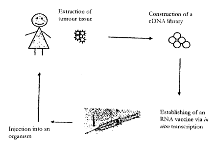
Fig. 10 shows a flow chart of the construction of
autologous, stabilized RNA according to the
preparation process of the present invention.
Tumour tissue is first obtained, e.g. by biopsy.
The total RNA is extracted from this. A cDNA
library is constructed with the aid of the poly(A+)
RNA obtained from the RNA extraction. Starting from
this, after preparation of a corresponding DNA
matrix, the autologous, stabilized RNA is obtained
by means of in vitro transcription.
Fig. 11 shows a reaction scheme of the steps for
preparation of a cDNA library, starting from
poly(A+) RNA, for the SMART PCR cDNA Synthesis Kit
from Clontech Inc. by way of example.
Fig. 12 shows a photograph of an agarose gel which shows
the typical size fractionation of a cDNA library
compiled from human placenta tissue. A length
marker with fragments of the length shown on the
left is plotted in track M. The "DS cDNA" track
CA 02473135 2004-07-22
34
contains the cDNA library. Those fragments which
correspond to the expected size fraction (about
200 bp to 4,000 bp) are used for the in vitro
transcription.
Fig. 13 shows by way of example a treatment plan for the
tumour therapy according to the invention by
injection of a tumour mRNA library, here in
combination with GM-CSF, for patients with
malignant melanoma. Autologous, stabilized RNA
prepared from the patient's own tumour tissue is
used for this. This amplified autologous tumour RNA
is administered to the patient i.d. on days 0, 14,
28 and 42. In addition, one day after the RNA
injection the patient is injected s.c. with GM-CSF
(Leucomax 100 pg/m2 Novartis/Essex Pharma). Two
weeks after the fourth injection (day 56), the
response of the tumour is evaluated by a staging
analysis (inter alia sonography, thorax X-ray, CT
etc.) and by assessment of the immunological
parameters induced by the therapy. When the course
of the disease is stable or there is an objective
tumour response (CR or PR), the patient receives in
each case a further vaccination every four weeks.
Further restaging analyses are carried out on
day 126 and then at intervals of 12 weeks.
Fig. 14 shows once more schematically of the general course
of a therapy with the pharmaceutical composition
according to the invention with autologous,
amplified tumour RNA, i.e. the RNA contained in the
pharmaceutical composition represents a cDNA
CA 02473135 2004-07-22
library of the tumour tissue. A sample of the
tumour tissue is first obtained, e.g. via a biopsy.
The total and then the poly (A) RNA are prepared
from the tissue by appropriate extractions.
5 Starting from the poly(A+) RNA, a cDNA library is
constructed and is cloned into a vector suitable
for subsequent in vitro transcription. An RNA
vaccine is then obtained by in vitro transcription,
and is injected into the patient from whom the
10 tumour tissue has been taken to combat the tumour.
The following embodiment examples explain the present
invention in more detail, without limiting it.
15 EXAMPLES
Example 1: Tumour vaccination with RNA in an animal model
Materials and methods
Capped mRNA which codes for a shortened version of the Her-
2/neu protein of the rat ("ECD-TM-neu-rat", containing the
extracellular domain and the transmembrane region, but not
the cytoplasmic region) was prepared, using the "SP6
mMessagemMachine" (Ambion), with the aid of a plasmid which
substantially corresponded to the structure shown in
fig. 8, but contained an SP6 promoter instead of the T7
promoter and in which the ECD-TM-neu-rat construct was
inserted after the SP6 RNA polymerase promoter. The mRNA
prepared was dissolved in injection buffer (150 mM NaCl,
10 mM HEPES) at a concentration of 0.8 mg/1 and the
solution was mixed with protamine sulfate (Sigma) (1 mg
CA 02473135 2004-07-22
36
protamine per 1 mg RNA). 50 pl of this solution were
injected into the auriculae (in each case 25 pl per ear)
of mice. Eight injections were performed, in each case one
at the age of 6, 8, 13, 15, 20, 22, 27 and 29 weeks. Mice
to which corresponding injections with injection buffer,
with plasmid DNA which codes for ECD-TM-neu rat or with an
antisense mRNA corresponding to the mRNA according to the
invention were administered served as controls.
Results
Female BalB-neu T mice (Ba1B/c mice which express the
oncogene Her-2/neu of the rat; cf. Rovero et al. (2000) J.
Immunol. 165(9):5133-5142) which develop mammary carcinomas
spontaneously were immunized with RNA which codes for a
shortened version of the Her-2/neu protein ("ECD-TM-neu-
rat", containing the extracellular domains and the
transmembrane region, but not the cytoplasmic region). Four
mice treated with injection buffer served as a negative
control. A further group of three mice was injected with
DNA which codes for the shortened Her-2/neu. Four mice
received the mRNA which codes, according to the invention,
for the tumour antigen Her-2/neu (shortened version of ECD-
TM, see above). Four mice which were injected with the
corresponding antisense RNA served as a further control
group. As shown in fig. 1, in the animals of the untreated
control group a tumour multiplicity of on average 10 was
observed after 26 weeks, whereby all animals having
palpable breast tumours at the age of about 20 weeks. In
contrast, in the case of immunization with the mRNA which
codes for ECD-TM-neu-rat, a significant slowing down of the
formation of carcinomas is to be observed, in particular a
CA 02473135 2004-07-22
37
tumour multiplicity of 10 is achieved only at the age of 30
weeks. Furthermore, the size of the tumours is also reduced
(not shown). Of the 4 mice treated with the mRNA according
to the invention, one was still tumour-free after 9 months.
That group of mice which had been injected with the
antisense mRNA all showed tumours at the age of 6 months.
The comparison group of mice injected with plasmid DNA
which codes for the shortened version of Her-2/neu also
showed a carcinoma formation which was slowed down compared
with the untreated control group (cf. also in respect of
corresponding plasmid DNA experiments on intramuscular
injection: Di Carlo et al. (2001) Clin. Cancer Res. 7 (3rd
supplement): 830s-837s), but the formation of carcinomas up
to the 27th week was not slowed down to the same extent as
in the case of immunization with mRNA according to the
invention which codes for the shortened version of Her-
2/neu. Furthermore, in the case of immunization with DNA,
the abovementioned disadvantages, in particular the risk of
integration of the DNA into the genome, the formation of
anti-DNA antibodies etc., are to be taken into account.
Example 2: Influence of GM-CSF on RNA vaccination
Materials and methods
Mice
BALB-c AnNCr1BR (H-2d) mice (female) 6-10 weeks old were
obtained from Charles River (Sulzfeld, Germany).
CA 02473135 2004-07-22
38
Plasmids and preparation of RNA
The ORF (LacZ) which codes for beta-galactosidase, flanked
by 5'-and 3'-untranslated sequences from the beta-globin
gene of X. Laevis, was into the plasmid pT7TS (P.A. Creek,
Austin, TX, USA), in order to prepare the plasmid pT7TS-
kozak-5' beta gl-lacZ-3' beta gl-A30C30 (cf. Hoerr et al.
(2000) Eur. J. Immonol. 30: 1-7). A schematical view of the
general structure of the plasmid pT7TS with the flanking 5'
and 3' untranslated sequences from the beta-globin gene of
X. Laevis is shown in fig. 8.
The plasmid prepared in this way was linearized with PstI
and transcribed in vitro using the m-MessagemMachineT7 Kit
(Ambion, Austin, TX USA). The RNA prepared in this way was
purified by means of LiCl precipitation, phenol/chloroform
extraction and ammonium acetate precipitation. Finally, the
purified RNA was resuspended in injection buffer (150 mM
NaCl, 10 mM HEPES) in a concentration of 0.5 mg/ml.
Media and cell culture
P815 and P13.1 cells were cultured in RPMI 1640 (Bio-
Whittaker, Verviers, Belgium), supplemented with 10% heat-
inactivated foetal calf serum (FCS) (PAN systems, Germany),
2 mM L-glutamine, 100 U/ml penicillin and 100 mg/ml
streptomycin.
CTL cultures were kept in RPMI 1640 medium, supplemented
with 10 % FCS, 2 mM L-glutamine, 100 U/ml penicillin,
100 mg/ml streptomycin, 0.05 pM beta-mercaptoethanol,
50 mg/ml gentamycin, MEM non-essential amino acids (100 x)
CA 02473135 2004-07-22
39
and 1 mm sodium pyruvate. The CTL were restimulated for one
week with 1 mg/ml beta-galactosidase protein (Sigma,
Taufkirchen, Germany). On day 4, 4 ml of culture
supernatant were carefully pipetted off and replaced by
fresh medium containing 10 U/ml rIL-2 (final
concentration).
Immunization
3 BALB/c mice per group were anesthetized with 20 mg
pentobarbital i.p. per mouse. The mice were then injected
i.d. in both auriculae with 25 mg of mRNA which codes for
beta-galactosidase (beta-Gal) in injection buffer (150 mM
NaCl, 10 mM HEPES). In some cases, granulocyte macrophage
colony-stimulating factor (GM-CSF) was additionally
injected into the same site or into an injection site away
from this (into the auricula or s.c. into the back) 24 h or
2 h before or 24 h after the RNA injection. As a positive
control, animals were injected i.d. in both auriculae with
in each case 10 mg of a DNA plasmid which codes for beta-
gal in PBS. A group of animals to which only injection
buffer was administered i.d. into both auriculae served as
a negative control. Two weeks after the first injection, a
boost injection was performed in each case in the same
manner as the first injection. Two weeks after the boost
injection, blood was taken, the mice were sacrificed and
the spleen was removed.
51Cr release test
Splenocytes obtained from the spleen were stimulated with
beta-gal protein in vitro and the CTL activity was
CA 02473135 2004-07-22
determined after 6 days using a 6-hours 51Cr standard test
as described in Rammensee et al. (1989) Immunogenetics 30:
296 - 302. Summarized briefly, target cells were marked
with 51Cr and charged with the peptide TPHPARIGL for 20 min
5 at room temperature. After co-incubation of effector and
target cells (at in each case three different ratios of
effector:target cells: 200, 44 and 10) in circular plates
with 96 wells for 6 h, 50 ml of 200 ml of culture
supernatant were pipetted into a Luma scintillation plate
10 (Packard) with 96 wells and, after drying, the
radioactivity was measured with a scintillation counter
(1405 Microbeta Plus). The percentage specific release was
determined from the amount of 51Cr released into the medium
(A) minus the spontaneous release (B) divided by the total
15 release (C) (using Triton X-100) minus the spontaneous
release (B): Per cent specific lysis = 100 (A-B)/(C-B).
Cytokine ELISA
20 After 4 days of restimulation with beta-gal protein, the
supernatant of the splenocyte culture was pipetted off and
stored at -50 C until used. 100 ml anti-mouse-anti-IFN-
gamma or -IL-4 scavenger antibodies (Becton Dickenson,
Heidelberg, Germany) were pipetted out overnight at 4 C on
25 MaxiSorb plates (Nalge Nunc International, Nalge, Denmark)
at a concentration of 1 mg/ml in coating buffer (0.02 %
NaN3, 15 mM Na2CO3, 15 mM NaHCO3 , pH 9.6) . After washing
three times with washing buffer (0.05 % Tween 20 in PBS),
the plates were saturated with 200 ml of blocking buffer
30 (0.05 % Tween 20, 1 % BSA in PBS) for 2 h at 37 C. After
washing three times with washing buffer, 100 ml of the cell
culture supernatants were incubated for 5 h at 37 C. The
CA 02473135 2004-07-22
41
plates were then washed four times with washing buffer, 100
ml of biotinylated anti-mouse-anti-IFN-gamma or -IL-4
detection antibodies (Becton Dickenson, Heidelberg,
Germany) per well at a concentration of 0.5 mg/ml in
blocking buffer were pipetted and incubation was carried
out for 1 h at room temperature. After washing three times
with washing buffer, 100 ml of a 1/1,000 dilution of
streptavidin-HRP (BD Biosciences, Heidelberg, Germany) were
added into each well. After 30 min at room temperature, the
plates were washed three times with washing buffer and
twice with bidistilled water. Thereafter, 100 ml of the
ARTS substrate were added into each well. After is -
30 min at room temperature, the extinction at 405 nm was
measured with a Sunrise ELISA reader (Tecan, Crailsheim,
Germany).
Antibody ELISA
Two weeks after the boost injection, blood was taken from
the mice via the orbital vein and blood serum was prepared.
100 ml of beta-gal protein at a concentration of 100 mg/ml
in coating buffer (0.05 M Tris-HC1, 0.15 M NaCl, 5 mM CaC12,
pH 7.5) were pipetted out for 2 h at 37 C on to MaxiSorb
plates (Nalge Nunc International, Nalge, Denmark). The
plates were then washed three times with 200 ml of washing
buffer (0.05 M Tris-HC1, 0.15 M NaCl, 0.01 M EDTA, 0.1 %
Tween 20, 1 o BSA, pH 7.4) and saturated with protein with
200 ml of washing buffer overnight at 4 C. The plates were
washed three times with washing buffer and blood sera were
added in a dilution of 1/10, 1/30 or 1/90 in washing
buffer. After 1 h at 37 C, the plates were washed three
times with washing buffer and 100 ml of 1/1,000 dilutions
CA 02473135 2004-07-22
42
of goat anti-mouse IgGl or IgG2a antibodies (Caltag,
Burlington, CA, USA) were added. After 1 h at room
temperature, the wells were washed three times with washing
buffer and 100 ml of ABTS substrate per well were added.
After 15 - 30 min at room temperature, the extinction at
405 nm was measured with a Sunrise ELISA reader (Tecan,
Crailsheim, Germany).
Results and discussion
It was confirmed that direct injection of RNA which codes
for beta-galactosidase into the auricula of mice induces an
anti-beta-galactosidase immune response, substantially of
the Th2 type. Production of anti-beta-galactosidase
immunoglobulins of the IG1 type (fig. 3A) and secretion of
IL-4 (fig. 3B) was found in splenocytes, stimulated with
beta-galactosidase, from mice which had been injected with
the RNA which codes for beta-galactosidase. To increase the
efficiency of the RNA vaccine, the cytokine GM-CSF was
additionally administered. This cytokine increases the
efficiency of some DNA vaccines. It was furthermore found
that the time of the GM-CSF injection influences the type
of the immune response, compared with DNA injection
(Kusakabe (2000) J. Immunol. 164: 3102-3111). It was found
according to the invention that GM-CSF can enhance the
immune response brought about by an RNA vaccination. The
injection of GM-CSF one day before the injection of RNA
shows scarcely any influence on the strength or the type of
the immune response. In contrast, injection of GM-CSF
2 hours before injection of the RNA enhances the immune
response (cf. the IL-4 release in fig. 3B in the 2 mice
injected with GM-CSF at time T = 0), but does not influence
CA 02473135 2004-07-22
43
the Th2 polarity. On the other hand, if GM-CSF is injected
one day after the RNA vaccine into the same site or into a
site away from this (not shown), not only is the immune
response enhanced overall (cf. the antibody response
according to fig. 3), the immune response is polarized to
the Thi type (cf. the IFN-gamma production by splenocytes
stimulated with beta-gal protein according to fig. 3A, the
production of IgG2a antibodies against beta-Gal according
to fig. 3B and the production of activated CTL according to
fig. 1). The injection of GM-CSF some minutes or some hours
after the RNA injection should result in the same effect
(enhancement and polarization) on the immune response.
Example 3: Effect of an RNase inhibitor on mRNA expression
in vivo
Naked or protamine-associated or -complexed mRNA which
codes for beta-galactosidase (prepared as described in
example 2) was injected into the auricula of mice in an
amount of 25 mg of RNA in injection buffer (150 mM NaCl,
10 mM HEPES). Further mice were injected with the mRNA
which codes for beta-galactosidase, together with 10 U of
the RNase inhibitor RNasin (an enzymatic RNase inhibitor
extracted from the pancrease, obtainable from Roche or
Promega). The RNase inhibitor was mixed with the RNA
solution directly before the injection. After 12 hours, the
ears were in each case removed from the mice. Thin
microscope sections of the auriculae were prepared and were
stained with X-gal. Injection of naked or protamine-
associated mRNA leads to a detectable beta-galactosidase
activity in a few cells in the corresponding thin sections
(blue cells in fig. 5 and 6). Some cells have thus taken up
CA 02473135 2004-07-22
44
the exogenous RNA here and translated it into the protein.
When the mRNA which codes for beta-galactosidase was in the
form protected with the RNase inhibitor RNasin, very many
more blue cells were observed than in the case of the naked
or protamine-associated RNA (fig. 7). Since RNasin inhibits
RNases, the half-life of the injected mRNA molecules in
vivo is prolonged, where the environment (interstitial
tissue) is contaminated with RNases. Such a stabilization
of the RNA leads to an increased uptake by the surrounding
cells and therefore to an increased expression of the
protein coded by the exogenous RNA. This phenomenon can
therefore also be utilized for an enhanced immune response
to an antigen coded by the mRNA injected.
Example 4: RNA vaccination of patients with malignant
diseases
Introduction
Cytotoxic T lymphocytes (CTL) recognize antigens as short
peptides (8-9 amino acids) which are expressed bound to MHC
class 1 glycoproteins on the cell surface (1). These
peptides are fragments of intracellular protein molecules.
However, there are indications that antigens taken up
exogenously by macropinocytosis or phagocytosis can lead to
the CD8+ T cell-mediated immune response. The proteins are
cleaved into proteosomes and the peptides formed by this
means are transported out of the cytosol into the lumen of
the endoplasmic reticulum and bound to MHC class I
molecules.
CA 02473135 2004-07-22
The proteins processed in this way are transported as
peptide/MHC class I complex to the cell surface and
presented to the CTL. This process takes place in every
cell and in this way makes it possible for the immune
5 system to monitor accurately each individual cell for the
presence of proteins which are foreign to the body or
modified or embryonic, regardless of whether they originate
from intracellular pathogenic germs, oncogenes or
dysregulated genes. By this means, cytotoxic lymphocytes
10 are capable of recognizing and lysing infected and
neoplastic cells, respectively (2, 3).
In recent years various tumour-associated antigens (TAA)
and peptides which are recognized by CTL and therefore lead
15 to lysis of tumour cells have been successfully isolated
(21-27). These TAA are capable of stimulating T cells and
inducing antigen-specific CTL, if they are expressed as a
complex of HLA molecule and peptide on antigen-presenting
cells (APC).
In numerous studies carried out mainly on patients with
malignant melanoma, it has been possible to demonstrate
that malignant cells lose the expression of TAA as the
tumour disease proceeds. Similar circumstances are also
observed with vaccinations with individual tumour antigens.
Under vaccination therapies, selection of tumour cells may
also occur, which renders possible an escape from the
immune system and a progression of the disease in spite of
therapy. The use of several different tumour antigens as
envisaged in the treatment plan according to the invention
of the present example should prevent selection of tumour
CA 02473135 2004-07-22
46
cells and escape of the malignant cells from the immune
system due to loss of antigens.
A method with which DC can be transfected with RNA from a
plasmid which codes for a tumour antigen has recently been
developed (Nair et al., 1998, Nair et al., 2000).
Transfection of DC with RNA for CEA or telomerase led to
induction of antigen-specific CTL. This process renders it
possible to induce CTL and T helper cells against several
epitopes on various HLA molecules from a tumour antigen. A
further advantage of this strategy is the fact that neither
the characterization of the tumour antigens or epitopes
used nor definition of the HLA haplotype of the patient is
a prerequisite. By a polyvalent vaccine of this type, the
probability of the occurrence of so-called clonal "tumour
escape" phenomena could be reduced significantly.
Furthermore, T cell-mediated immune responses against
antigens processed and presented by the natural route and
with possibly a higher immune dominance could be induced by
this approach. By additional participation of MHC class II-
restricted epitopes, the induced tumour-specific immune
response could be enhanced and maintained for longer.
A treatment sheme according to the invention for tumour
vaccination of patients with advanced malignant diseases
(mammary, ovarian, colorectal, pancreatic and renal cell
carcinomas) is provided by way of example. In this, RNA
which has been prepared from plasmids which code for MUC1,
Her-2/neu, telomerase and MAGE-1 tumour antigens and
influenza matrix protein (IMP) (positive control) is
administered intradermally to patients with the
abovementioned malignant diseases. A CTL induction in vivo
CA 02473135 2004-07-22
47
is thereby rendered possible, in order to prevent the
progression of the disease or to effect the regression
thereof in this way. The tumour antigens mentioned are
expressed on the malignant cells of mammary, ovarian,
colorectal, pancreatic and renal cell carcinomas.
According to the treatment plan (cf. the following
statements in this respect and fig. 9), the RNA species
prepared in the laboratory which code for CEA, MUC1,
Her-2/neu, telomerase, Mage-1 and IMP are administered to
the patient"i.d., initially 4 x on days 0, 14, 28 and 42.
In addition, GM-CSF (Leucomax0, 100 pg/m2, Novartis/Essex
Pharma) is administered s.c. to the patient in each case
one day after the RNA inoculation.
The treatment according to the invention is an immunisation
approach which requires only minimal interventions on the
patient (injection). Therapy is conducted ambulant and is
suitable for many tumour patients, without the limitation
to particular HLA types or defined T cell epitopes.
Furthermore, polyclonal CD4'-T helpers and also CD8+-CTL can
be induced by this therapy.
Treatment plan
The RNAs for several tumour antigens (MUC1, Her-2/neu,
telomerase, MAGE-1) and for a control antigen, influenza
matrix protein (IMP, a viral antigen) are administered i.d.
to the patient on days 0, 14, 28 and 42. In addition, the
patients receive GM-CSF (Leucomax(D (100 pg/m2)
Novartis/Essex Pharma) s.c. in each case one day after the
RNA inoculation. When the course of the disease is stable
CA 02473135 2004-07-22
48
or there is an objective tumour response (complete
remission (CR) or partial remission (PR)), where
appropriate the patients receive the vaccinations s.c. once
a month. After the fourth injection (day 49), the response
of the tumour is evaluated radiologically, by laboratory
chemistry and/or sonographically, and the immunological
phenomena induced by the therapy are evaluated.
From day 70, the immunization therapy is continued at
intervals of 4 weeks.
On days 0, 14, 28, 42 and 49, in each case blood samples
are taken for laboratory parameters, Diff-BB, FACS analysis
and cytokines (50 ml in total). Restaging of the patients
takes place from day 49 and where appropriate every further
4 to 8 weeks.
The treatment plan is shown schematically in fig. 9.
Laboratory: clotting, electrolytes, LDH, R2-M, CK, liver
enzymes, bilirubin, creatinine, uric acid, total
protein, CRP, tumour markers (Ca 12-5, Ca 15-3,
CEA, Ca 19-9): 15 ml of blood.
Diff-BB:differential blood count with smear (5 ml of EDTA
blood).
Cytokines: 10 ml of serum
FACS: 10 ml of heparin blood.
ELlspot:20 ml of heparin blood.
Multitest: analysis of the DTH reaction.
DTH: ("delayed type hypersensitivity", delayed T cell-
mediated reaction) analysis of the reaction to
intradermally administered RNA. In addition a skin
CA 02473135 2004-07-22
49
biopsy should be performed in the event of a
positive DTH reaction (local anaesthesia is not
necessary for this).
Preparation of RNA from plasmids
For production of a vaccine based on mRNA, only precursors
which are chemically synthesized and purified from bacteria
are required. This is preferably effected in a specially
equipped RNA production unit. This is in a sealed-off room
which is declared an RNase-free zone, i.e. work with RNase
(e.g. purification of plasmids) must not be carried out.
Contamination with naturally occurring RNases is also
constantly checked. This room is fitted out with new
apparatuses (4 C and -20 C refrigerators, heating block,
sterile bench, centrifuges, pipettes) which have never been
used for biological or clinical work. This RNA production
unit is used exclusively for enzymatic production (in vitro
transcription) of mRNA (without bacterial, viral or cell
culture work). The end product comprises a sterile RNA
solution in HEPES/NaCl buffer. Quality analyses are carried
out on a formaldehyde-agarose gel. In addition, the RNA
concentration and the content of proteins are determined
photometrically (OD320 < 0.1; ratio of OD260/OD280 > 1.8 in
pure RNA). Possible contamination by LPS is analysed in the
LAL test. All RNA samples are subjected to sterile
filtration before administration.
Plasmid constructs
The chosen genes (CEA, mucinl, Her-2/neu, telomerase,
Mage-Al and influenza matrix) are amplified via a PCR using
CA 02473135 2004-07-22
a heat-stable high-performance enzyme (pfu, Stratagene).
The genes originate from tumour cDNA (mucinl, Her-2/neu,
telomerase), or they have been cloned into bacterial
vectors (influenza matrix and MAGE-Al). The PCR fragments
5 are cleaved with restriction enzymes (mucinl: Bglll-Spel;
Her-2/neu: HinDlllblunt-Spel; telomerase: BglII-SpeI;
MAGE-Al: BamHI-Spel; influenza matrix protein: BglII-SpeI)
and cloned into the T7TS-Plasmid (cf. fig. 8) via the BglII
and Spel restriction sites. Plasmids of high purity are
10 obtained via the Endo-free Maxipreparation Kit (Qiagen,
Hilden, Germany). The sequence of the vector is controlled
via a double-strand sequencing from the T7 promoter up to
the PstI site and documented. Plasmids with a correct
inserted gene sequence without mutations are used for the
15 in vitro transcription. (Control via the published
sequences: Accession Numbers: M11730 for Her-2/neu,
NM 002456 for MUC1, NM 003219 for telomerase TERT, V01099
for influenza matrix and M77481 for MAGE-Al).
20 in vitro Transcription
Production of linear, protein-free DNA
500 pg of each plasmid are linearized in a volume of 0.8 ml
25 via digestion with the restriction enzyme PstI in a 2 ml
Eppendorf reaction vessel. This cleaved construct is
transferred into the RNA production unit. 1 ml of a mixture
of phenol/chloroform/isoamyl alcohol is added to the
linearized DNA. The reaction vessel is vortexed for
30 2 minutes and centrifuged at 15,000 rpm for 3 minutes. The
aqueous phase is removed and mixed with 0.7 ml 2-propanol
in a 2 ml reaction vessel. This vessel is centrifuged at
CA 02473135 2004-07-22
51
15,000 rpm for 15 minutes, the supernatant is discarded and
1 ml 75% ethanol is added. The reaction vessel is
centrifuged at 15,000 rpm for 10 minutes and the ethanol is
removed. The vessel is centrifuged for a further 2 minutes
and the residues of the ethanol are removed with a
microlitre pipette tip. The DNA pellet is then dissolved in
1 pg/ml in RNase-free water.
Enzymatic synthesis of the RNA
The following reaction mixture is prepared in a 50 ml
Falcon tube: 100 .g linearized protein-free DNA, 1 ml 5x
buffer (200 mM Tris-HC1 (pH 7.9), 30 mM MgC12, 10 mM
spermidine, 50 mM NaCl, 50 mM DTT), 200 p1 ribonuclease
(RNase) inhibitor (recombinant, 5,000 U), 1 ml rNTP mix (in
each case 10 mM ATP, CTP, UTP; 2 mM GTP), 1 ml CAP analogue
(8 mM), 150 pl T7 polymerase (3,000 U) and 2.55 ml RNase-
free water. The total volume is 5 ml. The mixture is
incubated at 37 C for 2 hours in a heating block.
Thereafter, 100 U of RNase-free DNase are added and the
mixture is incubated again at 37 C for 30 minutes. The DNA
matrix is enzymatically degraded by this procedure.
Description and origin of the individual components
T7 polymerase: purified from an E coli strain which
contains a plasmid with the gene for the polymerase. This
RNA polymerase uses as the substrate only promoter
sequences of the T7 phage; Fermentas.
NTPs: synthesized chemically and purified via HPLC. Purity
more than 96 %; Fermentas.
CA 02473135 2004-07-22
52
CAP analogue: synthesized chemically and purified via HPLC.
Purity more than 90 %; Institute of Organic Chemistry of
the University of Tubingen.
RNase inhibitor: RNasin, for injection, prepared
recombinantly (E. coli); Promega.
DNase: Pulmozymc' ("dornase alfa"); Roche
Purification
The RNA treated with DNase is mixed with 20 ml of a
solution of 3.3 ml 5 M NH4OAc plus 16.65 ml of ethanol. The
mixture is incubated at -20 C for 1 hour and centrifuged
at 4,000 rpm for 1 hour. The supernatant is removed and the
pellet is washed with 5 ml of 75 % RNase-free ethanol. The
vessel is centrifuged again at 4,000 rpm for 15 minutes and
the supernatant is removed. The vessel is centrifuged again
under the previous conditions and the ethanol which remains
is removed with a microlitre pipette tip. The reaction
vessel is opened and the pellet is dried under a sterile
bench in the sterile environment.
1 ml of RNase-free water is added to the dried RNA. The
pellet is incubated at 4 C for at least 4 hours. 2 pl of
the aqueous solution are subjected to a quantitative
analysis (determination of the UV absorption at 260 nm).
2 ml of a phenol/chloroform/isoamyl alcohol solution are
added to 1 ml of aqueous RNA solution. The mixture is
vortexed for 2 minutes and centrifuged at 4,000 rpm for
2 minutes. The aqueous phase is removed with a microlitre
pipette and transferred into a new reaction vessel. 4 ml of
a solution of 0.66 ml 5 M NH4OAc plus 3.33 ml ethanol are
added. The mixture is incubated at -20 C for 1 hour and
CA 02473135 2004-07-22
53
centrifuged at 4,000 rpm for 1 hour. The supernatant is
removed and the pellet is washed with 75 % RNase-free
ethanol. The vessel is centrifuged again at 4,000 rpm for
15 minutes and the supernatant is removed. The vessel is
centrifuged again under the previous conditions and the
ethanol which remains is removed with a microlitre pipette
tip. The reaction vessel is opened and the pellet is dried
under a sterile bench in the sterile environment.
The RNA is dissolved in RNase-free water and adjusted to a
concentration of 10 mg/ml. It is incubated for 12 hours at
4 C. A final concentration of 2 mg/ml is achieved by
addition of injection buffer (150 mM NaCl, 10 mN HEPES).
The end product is preferably subjected to sterile
filtration under GMP conditions before use.
Application of the RNA
Each patient receives at two different sites an intradermal
(i.d.) injection of in each case 150 pl of the injection
solution in which in each case 100 ug of antigen-coding
mRNA (CEA, Her-2/neu, MAGE-Al, mucin 1, telomerase,
influenza matrix protein) are present in solution.
After the primary immunization, a booster immunization is
carried out every 14 days, for the inoculations then to be
repeated at a monthly interval. In each case one day after
the RNA injection, GM-CSF (Leucomax , Sandoz/Essex Pharma)
is administered subcutaneously (s.c.) to the patient.
If a clinical response is present or the disease is
stabilized, this therapy is continued at monthly intervals.
CA 02473135 2004-07-22
54
Further immunological investigations in vitro (optional)
Flow cytometry analyses of PBMC for quantification of CTL
precursors;
51Cr release tests;
Soluble receptor and cytokine levels in the serum;
DTH reaction (skin reaction to intradermally injected RNA,
"delayed type hypersensitivity", T lymphocyte-mediated
reaction); and
Skin biopsy samples from the injection site for
histological analysis for T cell infiltration (pathology).
Parameters for evaluation of the efficacy
To be able to answer the question of the efficacy of this
immunotherapy, the induction of tumour-specific T cells and
a measurable tumour remission is used. Parameters are
T cell reactions measured in vitro and in vivo and changes
in the size of bidimensionally recordable tumour
manifestation or laboratory chemistry parameters of the
course of the disease.
Objective remission is defined as the best response in the
form of a complete or partial remission, corresponding to
the criteria listed below. The remission rate is calculated
from the ratio of the number of patients with objective
remission and the total number of evaluable patients.
A change in the immune status, determined by immunotyping
of peripheral mononuclear cells, an increase in the
antigen-specific CTL precursor frequency in the peripheral
CA 02473135 2004-07-22
blood and the induction of a persistent tumour-specific
T cell activity are assessed as the immunological response
to the therapy. For this purpose, in vitro induction
cultures are established for activation of tumour-specific
5 CTL.
Remission criteria (acc. to UICC)
Complete remission (CR): Complete regression of all
10 measurable tumour manifestation,
documented by 2 control
investigations at least 4 weeks
apart.
Partial remission (PR): Decrease in size of the total
15 area dimensions (product of two
tumour diameters or linear
measurement of one-dimensionally
measurable lesions of all tumour
findings by 50 % for at least 4
20 weeks). No new occurrence of
tumour manifestations or
progression of a tumour finding.
"No Change" (NC): Decrease of all the measurable
tumour manifestations by less
25 than 50 % or increase in a tumour
finding.
Progression (PD): Increase in size of the tumour
parameters in at least one focus
or new occurrence of a tumour
30 manifestation.
CA 02473135 2004-07-22
56
References
1. Rammensee HG, Falk K, Rotzschke 0: Peptides naturally
presented by MHC class I molecules. Annu Rev Immunol 11:
213, 1993.
2. Bevan M.J: Antigen presentation to cytotoxic T
lymphocytes in vivo. J Exp Med 182: 639, 1995.
3. Rock K.L: A new foreign policy: MHC class I molecules
police the outside world. Immunol Today 17:131, 1996.
4. Steinman, A.M: The dendritic cell system and its role in
immunogenicity. Annu. Rev Immunol 9:271, 1991.
5. Steinman RM, Witmer-Pack M, Inaba K: Dendritic cells:
antigen presentation, accessory function and clinical
relevance. Adv Exp Med Biol 329:1, 1993.
6. Inaba K, Metlay JP, Crowley MT, Steinman RM: Dendritic
cells pulsed with protein antigens in vitro can prime
antigen-specific, MHC-restricted T cells in situ. J Exp
Med 172:631, 1990.
7. Austyn JM: New insight into the mobilisation and
phagocytic activity of dendritic cells. J Exp Med
183:1287, 1996.
8. Romani N, Koide S, Crowley M, Witmer-Pack M, Livingstone
AM, Fathman CG, Steinman RM: Presentation of exogenous
protein antigens by dendritlc cells to T cell clones. J
Exp Med 169:1169, 1989.
CA 02473135 2004-07-22
57
9. Nair S, Zhou F, Reddy R, Huang L, Rouse BT: Soluble
proteins delivered to dendritic cells via pH-sensitive
liposomes induce primary cytotoxic T lymphocyte
responses in vitro. J ExpiMed 175:609, 1992.
10. Cohen PJ, Cohen PA, Rosenberg SA, Katz SI, Mule JJ:
Murine epidermal Langerhans cells and splenic dendritic
cells present tumor-associated antigens to primed T
cells. Eur J Immunol 24:315, 1994.
11. Porgador A, Gilboa E: Bone-marrow-generated dendritic
cells pulsed with a class I-restricted peptide are
potent inducers of cytotoxic T lymphocytes. J Exp Med
182:255, 1995.
12. Celluzzi CM, Mayordomo JI, Storkus WJ, M. T. Lotze MT,
and L. D. Falo LD: Peptide-pulsed dendritic cells induce
antigen-specific, CTL-mediated protective tumor
immunity. J Exp Med 183:283, 1996.
13. Zitvogel L, Mayordomo JI, Tjandrawan T, DeLeo AB,
Clarke MR, Lotze MT, Storkus WJ: Therapy of murine
tumors with tumor peptide-pulsed dendritic cells:
dependence on T cells, B7 costimulation, and T helper
cell 1-associated cytokines. J Exp Med 183:87, 1996.
14. Porgador A, Snyder D, Gilboa E: Induction of
antitumor immunity using bone marrow-generated dendritic
cells. J Immunol 156:2918, 1996.
CA 02473135 2004-07-22
58
15. Paglia P, Chiodoni C, Rodolfo M, Colombo MP: Murine
dendritic cells loaded in vitro with soluble protein
prime cytotoxic T lymphocytes against tumor antigen in
vivo. J Exp Med 183:317, 1996.
16. Brossart P, Goldrath AW, Butz EA, Martin S, Bevan MJ:
Adenovirus mediated delivery of antigenic epitopes into
DC by a means of CTL induction. J Immunol 158: 3270,
1997.
17. Fisch P, Kohler G, Garbe A, Herbst B, Wider D, Kohler
H, Schaefer HE, Mertelsmann R, Brugger W, Kanz L:
Generation of antigen-presenting cells for soluble
protein antigens ex vivo from peripheral blood
CD34+hematopoetic progenitor cells in cancer patients.
Eur J Immunol 26: 595, 1996.
18. Sallusto F, Cella M, Danieli C, Lanzavecchia A:
Dendritic cells use macropinocytosis and the mannose
receptor to concentrate macromolecules in the Major
Histocompatibility Complex class II compartment: Down
regulation by cytokines and bacterial products. J Exp
Med 182:389, 1995.
19. Bernhard H, Disis ML, Heimfeld S, Hand S, Gralow JR,
Cheever MA: Generetion of immunostimulatorry dendritic
cells from human CD34+ hematopoetic progenitor cells of
th bone marrow and peripheral blood. Cancer Res 55:
1099, 1995.
20. Hsu FJ, Benike C, Fagnoni F, Liles TM, Czerwinski D,
Taidi B, Engelman EG, Levy R: Vaccination of patients
CA 02473135 2004-07-22
59
with B-cell lympnoma using autologous antigen-pulsed
dendritic cells. Nat Med 2: 52, 1996.
21. Robbins PF, Kawakami Y: Human tumor antigens recognized
by T cells. Curr Opin Immunol 8: 628, 1996.
22. Linehan DC, Goedegebuure PS, Peoples GE, Rogers SO,
Eberlein TJ: Tumor-specific and HLA-A2 restricted
cytolysis by tumor-associated lymphocytes in human
metastatic breast cancer. J Immunol 155: 4486, 1995.
23. Peoples GE, Goedegebuure PS, Smith R, Linehan DC,
Yoshino I, Eberlein TJ: Breast and ovarian cancer
specific cytotoxic T lymphocytes recognize the same HER-
2/-neu derived peptide. Proc Natl Acad Sci USA 92: 432,
1995.
24. Fisk B, Blevins TL, Wharton JT, Ioannides CG:
Identification of an immunodomonant peptide of HER-2/neu
protooncogene recognized by ovarian tumor-specific
cytotoxic t lymphocyte lines. J Exp Med 181: 2109, 1995.
25. Brossart P, Stuhler G, Flad T, Stevanovic S,
Rammensee H-G, Kanz L and Brugger W. HER-2/neu
derived peptides are tumor-associated antigens
expressed by human renal cell and colon carcinoma
lines and are recognized by in vitro induced
specific cytotoxic T lymphocytes. Cancer Res. 58:
732-736, 1998.
CA 02473135 2004-07-22
26. Apostolopoulos, V. and McKenzie, I. F. C., Cellular
mucins: targets for immunotherapy. Crit. Rev.
Immunol. 14: 293-302, 1995.
5 27. Brossart P, Heinrich KS, Stevanovic S, Stuhler G,
Behnke L, Reichardt VL, Muhm A, Rammensee H-G, Kanz L,
Brugger W. Identification of HLA-A2 restricted T cell
epitopes derived from the MUC1 tumor antigen for broadly
applicable cancer vaccines. Blood 93: 4309-4317, 1999
28.Brossart P, Wirths S, Stuhler G, Reichardt VL, Kanz
L, Brugger W. Induction of CTL responses in vivo
after vaccinations with peptide pulsed dendritic
cells. Blood 96:3102-8, 2000
29. Kugler A, Stuhler G, Walden P, Zoller G, Zobywalski A,
Brossart P, Trefzer U, Ullrich S, Muller CA, Becker V,
Gross AJ, Hemmerlein B, Kanz L, Muller GA, Ringert RH.
Regression of human metastatic renal cell carcinoma
after vaccination with tumor cell-dendritic cell
hybrids. Nature Med 3: 332-336, 2000 (IF 25,58)
30. Nestle FO, Alijagic S, Gilliet M, Sun Y, Grabbe S,
Dummer R, Burg G, Schadendorf D (1998) Vaccination of
melanoma patients with peptide- or tumor lysate-pulsed
dendritic cells. Nat.Med. 4:328
31.Schuler-Thurner B, Dieckmann D, Keikavoussi P, Bender A,
Maczek C, Jonuleit H, Roder C, Haendle I, Leisgang W,
Dunbar R, Cerundolo V, von Den DP, Knop J, Brocker EB,
Enk A, Kampgen E, Schuler G (2000) Mage-3 and influenza-
matrix peptide-specific cytotoxic T cells are inducible
CA 02473135 2004-07-22
61
in terminal stage HLA-A2.1+ melanoma patients by mature
monocyte-derived dendritic cells. J.Immunol. 165:3492
32. Thurner B, Haendle I, Roder C, Dieckmann D, Keikavoussi
P, Jonuleit H, Bender A, Maczek C, Schreiner D, von Den
DP, Brocker EB, Steinman RM, Enk A, Kampgen E, Schuler G
(1999) Vaccination with mage-3A1 peptide-pulsed mature,
monocyte-derived dendritic cells expands specific
cytotoxic T cells and induces regression of some
metastases in advanced stage IV melanoma. J.Exp.Med.
190:1669
Example 5: Vaccination with autologous, amplified tumour
RNA in patients with malignant melanoma
Introduction
The incidence of malignant melanoma has increased sharply
worldwide in recent years. If the melanoma disease is
already in the metastased stage at the time of diagnosis,
there is currently no therapy which has a positive
influence on the further course of the disease with
sufficient certainty.
Vaccination therapies carried out to date using dendritic
cells are very labour-, cost- and time-intensive because of
the complicated culturing of the cells (GMP conditions).
Furthermore, the studies have hitherto concentrated
predominantly on known tumour-associated antigens (TAA),
such as, for example, melan-A or tyrosinase.
CA 02473135 2004-07-22
62
A number of various immunological phenomena, such as, inter
alia, the occurrence of spontaneous tumour regressions or
spontaneous involution of metastases, have made the
melanoma the prior candidate for testing immunotherapy
investigations (Parkinson et al., 1992). In addition to
experiments on non-specific stimulation of the immune
system by means of interleukin-2, mistletoe extracts, BCG
and interferons, which have so far not led to decisive
breakthroughs in the therapy of advanced tumour diseases,
the strategy of induction of various highly specific
cytotoxic T lymphocytes (CTL) has been pursued in
particular in recent years. These CTL are capable of
recognizing and killing autologous melanoma cells (Boon et
al., 1994; Houghton, 1994). Studies of this process have
shown that the CTL recognize defined peptides in
combination with MHC class I molecules. The presentation of
peptides by antigen-presenting cells (APC) is the
physiological route to generation of specific immune
responses by lymphocytes (Rammensee, 1993). Dendritic cells
have proved to be potent antigen-presenting cells which
lead to an induction of the immune response by two routes:
The first is the direct presentation of peptides towards
CD8+-T lymphocytes and activation thereof (Schuler &
Steinmann, 1985; Inaba et al., 1987; Romani et al., 1989),
and the second is the generation of a protective immune
response, which is mediated by CD4+ helper lymphocytes, and
requires a presentation of peptides via MHC class II
molecules (Grabbe et al., 1991, 1992, 1995).
By means of peptide analysis, it was therefore possible to
identify in this way various tumour-associated antigens
(TAA) which are specific for the melanoma and, after
CA 02473135 2004-07-22
63
presentation in combination with the MHC molecule and
recognition by the CTL, lead to cytolysis of the tumour
cells (Schadendorf et al., 1997, p. 21-27).
The use of autologous, dendritic cells was tested in the
context of a pilot study on melanoma patients in respect of
its potential to induce cytotoxic T lymphocytes
effectively, rapidly and reliably. In this study, 16
melanoma patients in stage IV who had already been
pretreated by chemotherapy were vaccinated with peptide-
charged dendritic cells. The response rates were above 30
(5/16 patients) (Nestle et al., 1998). In a further
independent study it was possible to demonstrate an even
higher response rate of more than 50 % (6/11 patients)
after immunization of melanoma patients who had already
been pretreated by chemotherapy with MAGE-3A1-charged
dendritic cells (Thurner et al., 1999). A significant
expansion of MAGE-A3-specific CD8+-T cells was also observed
in 8/11 patients. A regression of the metastases took place
in some cases after the DC vaccination. This was
accompanied by a CD8+-T cell infiltration. This showed that
the T cells induced were active in vivo. A disadvantage of
this strategy is the high outlay on costs and the
laboratory (in particular GMP conditions). Large amounts of
blood from the patient are required for the time-intensive
generation of the DC. In the preparation of the peptides,
on the one hand only known tumour-associated antigens can
be used, and on the other hand various peptides are
necessary, depending on the HLA haplotype.
A further development of this approach is vaccination with
RNA-transfected DC (Nair et al., 1998, Nair et al., 2000).
CA 02473135 2004-07-22
64
In the meantime, numerous studies demonstrate that DC from
mice and humans which have been transfected with mRNA can
induce an efficient CTL response in vitro and in vivo and
can lead to a significant reduction in metastases
(Boczkowski et al., 1996, 2000; Ashley et al., 1997; Nair
et al., 1998, 2000; Heiser et al., 2001; Mitchell and Nair,
2000; Koido et al., 2000; Schmitt et al., 2001). A great
advantage in the use of RNA compared with peptides is that
the most diverse peptides can be processed and presented
from one mRNA which codes for a TAA. By a polyvalent
vaccine of this type, the probability of the occurrence of
so-called clonal "tumour escape" phenomena can be reduced
significantly. Furthermore, T cell-mediated immune
responses against antigens processed and presented by the
natural route and with potentially a higher immune
dominance can be induced by this system. By additional
participation of MHC class II-restricted epitopes, the
tumour-specific immune response induced can be intensified
and maintained for longer. Nevertheless, this process also
can be carried out only with a high outlay on the
laboratory (GMP conditions) because of the necessary
culturing of the autologous DCs.
In the present strategy according to the invention,
vaccination is carried out with the RNA expression profile
present in the autologous tumour of the patient. The
specific tumour profile of the patient is thereby taken
into account, unknown TAAs also being included in the
inoculation. Expensive culture of the DCs is omitted, since
RNAs (not transfected DCs) are used in the vaccination.
CA 02473135 2004-07-22
A vaccination therapy using amplified autologous tumour RNA
on patients with metastased malignant melanoma, in
particular stage III/IV, is therefore provided according to
the invention.
5
Tumour-specific cytotoxic T cells are induced in vivo by
the vaccination, in order thus to achieve a clinico-
therapeutic effect (tumour response). This is an
immunisation system which requires only minimal
10 interventions on the patient (injection). Therapy can be
conducted ambulant and is suitable for many tumour
patients, without the limitation to particular HLA types or
defined T cell epitopes. Furthermore, polyclonal CD4+-T
helpers and also CD8+-CTL can be induced by this therapy.
15 From the point of view of the strategy, it is decisive also
that hitherto unknown TAAs are taken into account in the
vaccination protocol, and the exclusive use of autologous
material is particularly advantageous.
20 Treatment plan
The amplified autologous tumour RNA is administered to the
patient i.d. on days 0, 14, 28 and 42. In addition, the
patients receive GM-CSF (Leucomax 100 pg/m2,
25 Novartis/Essex) s.c. in each case one day after the RNA
inoculation. Each patient receives, at two different
sites, an i.d. injection of in each case 150 pl of the
injection solution, in which in each case 100 pg of
autologous tumour RNA is dissolved.
2 weeks after the fourth injection (day 56), where
appropriate the response of the tumour is evaluated by a
CA 02473135 2004-07-22
66
staging analysis (inter alia sonography, thorax X-ray, CT
etc.; in this context see the statements below) and by
assessment of the immunological parameters induced by the
therapy.
When the course of the disease is stable or there is an
objective tumour response (CR or PR), the patients receive
the vaccinations every four weeks. Further restaging
analyses can be envisaged e.g. on day 126 and then at an
interval of 12 weeks.
A diagram of the treatment plan is shown in fig. 13.
Preparation of autologous tumour RNA
The aim is the preparation of autologous poly(A+) RNA. For
this, poly(A+) RNA is isolated from the patient's own tumour
tissue. This RNA isolated is very unstable per se and its
amount is limited. The genetic information is therefore
transcribed into a considerably more stable cDNA library
and thus conserved. Starting from the patient's own cDNA
library, stabilized autologous RNA can be prepared for the
entire treatment period. The procedure according to the
invention is shown schematically in fig. 10.
Isolation of RNA
A process of Roche AG is used to isolate total RNA from a
tumour tissue biopsy. The High Pure RNA Isolation Kit
(order number 1828665) is employed here in accordance with
the manufacturer's instructions. Poly(A+) RNA is isolated
CA 02473135 2004-07-22
67
from the total RNA via a further process of Roche AG with
the High Pure RNA Tissue Kit (order number 2033674).
Preparation of a cDNA library
The cDNA library is constructed with the "SMART PCR cDNA
Synthesis Kit" (Clontech Inc., USA; order number PT3041-1)
in accordance with the manufacturer's instructions.
In this procedure, the single-stranded poly(A') RNA is
subjected to reverse transcription via a specific primer.
Via a poly-C overhang at the 31-end of the newly
synthesized DNA, a further primer can hybridize, via which
the construct can be amplified by a PCR. The double-
stranded cDNA fragments are now ready for cloning into a
suitable RNA production vector (e.g. pT7TS; cf. fig. 8).
The process for the preparation of the cDNA library from
the poly(A) RNA with the aid of the above kit is shown
schematically in fig. 11.
Plasmid constructs
The cDNA PCR fragments are cleaved with the restriction
enzymes NotI and Spel and cloned into the corresponding
restriction sites of the pT7TS vector by a procedure
analogous to that described in example 4. Plasmids of high
purity are obtained via the Endo-free Maxipreparation Kit
(Qiagen, Hilden, Germany). Plasmids with a cloned-in gene
sequence which corresponds to the expected size
fractionation (200 bp - 4,000 bp) of the cDNA library are
used for the in vitro transcription. An example of a
CA 02473135 2004-07-22
68
separation of a representative cDNA library in an agarose
gel is shown in fig. 12.
in vitro Transcription and RNA administration
The in vitro transcription and the administration of the
RNA are carried out as described in the above example 4.
Investigations during the treatment
Before each inoculation (on the day of the inoculation):
Physical examination (including RR, fever);
Blood sample for routine laboratory values
1. Blood count, differential blood count: 3 ml
2. Electrolytes, LDH, CK, liver enzymes, bilirubin,
creatinine, uric acid, total protein, CRP: 5 ml
3. Blood sedimentation: 2 ml; and
at repeat inoculations additionally: Inspection of the
injection sites.
On day 1 after each inoculation:
Physical examination (including RR, fever); and
Inspection of the injection sites.
In staging analyses on day 56 and 126 after the first
inoculation, then every 12 weeks, additionally:
Extended routine blood sample:
1. Tumour marker S100 (7 ml)
2. Clotting values (3 ml ) ;
Blood sample for immune monitoring (30 ml);
General well-being (ECOG score);
CA 02473135 2004-07-22
69
Imaging methods (thorax X-ray, sonography, skeleton
scintigram, CT abdomen, pelvis, thorax, skull); and
ECG ("EKG").
Further immunological investigations in vitro
Where appropriate, the relative incidence of antigen-
specific CTL precursor cells in the peripheral blood of the
patient in the course of time of the vaccination therapy is
measured.
On the one hand CTL precursor cells which are directed
against antigens expressed to a particular degree by
melanoma cells (tyrosinase, MAGE-3, melan-A, GP100) are
quantified here with FACS analyses (tetramer staining). On
the other hand ELlspot analyses are carried out, these
being designed such that CTL precursor cells which are
directed specifically against hitherto unknown antigens are
additionally recorded. For this, autologous dendritic cells
cultured from the peripheral blood of the patient are
incubated with the same RNA with which the inoculation has
also been carried out. These then serve as stimulator cells
in the ELlspot analysis. The measurement thus records the
total vaccine spectrum. For these analyses, blood samples
of 30 ml in total (20 ml ELlspot, 10 ml FACS analysis) can
be envisaged for the immune monitoring in the context of
the staging analyses and additionally on days 0, 14, 28 and
42, as well as a single withdrawal of 100 ml on day 70 for
culture of the DC.
CA 02473135 2004-07-22
Furthermore, skin biopsy samples from the injection site
can be obtained for histological analysis in respect of a
T cell infiltration.
5 Parameters for evaluation of the efficacy
The efficacy of the therapy according to the invention is
evaluated with the aid of the parameters described above in
example 4.
References
Anichini, A, Mortarini, R., Maccalli, C., Squarcina, P.,
Fleishhauer, K., Mascheroni, L., Parmiani, G. (1996).
Cytotoxic T cells directed to tumor antigens not
expressed on normal melanocytes dominate HLA-A2.1-
restricted immune repertoire to melanoma. J. Immunol.
156, 208-217.
Ashley, DM., Faiola, B., Nair, S., Hale, LP., Bigner, DD,
Gilboa, E. (1997). Bone marrow-generated dendritic
cells pulsed with tumor extracts or tumor RNA induce
anti-tumor immunity against central nervous system
tumors. J. Exp. Med. 186, 1177-1182.
Boczkowski, D., Nair, SK., Synder, D., Gilboa, E. (1996).
Dendritic cells pulsed with RNA are potent antigen-
presenting cells in vitro and in vivo. J. Exp. Med.
184, 465-472.
Boczkowski, D., Nair, SK., Nam, J., Lyerly, K., Gilboa, E.
(2000). Induction of tumor immunity and cytotoxic T
CA 02473135 2004-07-22
71
lymphocyte responses using dendritic cells transfected
with messenger RNA amplified from tumor cells. Cancer
Res. 60, 1028-1034.
Boon, T., Coulie,P., Marchand, M., Weynants, P., Wolfel,
T., Brichard, V. (1994). Genes coding for tumor
rejection antigens: perspectives for specific
immunotherapy. In Important Advances in Oncology 1994.
DeVita, VT, Hellman, S., Rosenberg, SA, ed.
(Philadelphia: Lippincott Co), pp. 53-69.
Garbe, C, Orfanos, CE (1989): Epidemiologie des malignen
Melanoms in der Bundesrepublik Deutschland im
internationalen Vergleich [Epidemiology of malignant
melanoma in the Federal Republic of Germany in an
international comparison]. Onkologie 12, 253-262.
Grabbe, S., Bruvers, S., Gallo, R.L., Knisely, T.L.,
Nazareno, R., and Granstein, R.D. (1991). Tumor antigen
presentation by murine epidermal cells. J Immunol.
146, 3656-3661.
Grabbe, S., Bruvers, S., Lindgren, A.M., Hosoi, J., Tan,
K.C., and Granstein, R.D. (1992). Tumor antigen
presentation by epidermal antigen-presenting cells in
the mouse: modulation by granulocyte-macrophage colony-
stimulating factor, tumor necrosis factor alpha, and
ultraviolet radiation. J Leukoc.Biol. 52, 209-217.
Grabbe, S., Beissert, S., Schwarz, T., and Granstein, R.D.
(1995). Dendritic cells as initiators of tumor immune
CA 02473135 2010-04-01
72
responses: a possible strategy for tumor
immunotherapy?. Immunol.Today 16, 117-121.
Heiser, A., Maurice, MA., Yancey, DR., Coleman, DM., Dahm,
P., Vieweg, J. (2001). Human dendritic cells
transfected with renal tumor RNA stimulate polyclonal T
cell responses against antigens expressed by primary
and metastatic tumors. Cancer Res. 61, 3388-3393.
Heiser, A., Maurice, MA., Yancey, DR., Wu, NZ., Dahm, P.,
Pruitt, SK., Boczkowski, D., Nair, SK., Ballo, MS.,
Gilboa, E., Vieweg, J. (2001). Induction of polyclonal
prostate cancer-specific CTL using dendritic cells
transfected with amplified tumor RNA. J. Immunol. 166,
2953-2960.
Hoerr, I, Obst, R, Rammensee, HG, Jung, G (2000). In vivo
application of RNA leads to induction of specific
cytotoxic T lymphocytes and antibodies. Eur J Immunol.
30, 1-7.
Houghton, AN (1994). Cancer antigens: immune recognition of
self and altered self. J.Exp.Med 180, 1-4
CA 02473135 2004-07-22
73
Inaba, K, Young, JW and Steinman, RM (1987). Direct
activation of CD8+ cytotoxic T lymphocytes by dendritic
cells. J.Exp.Med. 166, 182-194.
Koido, S., Kashiwaba, M., Chen, D., Gendler, S., Kufe, D.,
Gong, J. (2000). Induction of antitumor immunity by
vaccination of dendritic cells transfected with MUC1
RNA. J. Immunol. 165, 5713-5719.
Mitchell, DA., Nair, SK. (2000). RNA-transfected dendritic
cells in cancer immunotherapy. J. Clin. Invest. 106,
1065-1069.
Nair, S., Boczkowski, S., Synder, D., Gilboa, E. (1998).
Antigen presenting cells pulsed with unfractionated
tumor-derived peptides are potent tumor vaccines. Eur.
J. Immunol. 27, 589-597.
Nair, S., Heiser, A., Boczkowski, D., Majumdar, A., Naoe,
M., Lebkowski, JS., Vieweg, J., Gilboa, E. (2000).
Induction of cytotoxic T cell responses and tumor
immunity against unrelated tumors using telomerase
reverse transcriptase RNA transfected dendritic cells.
Nat. Med. 6, 1011-1017.
Nestle, F.O., Alijagic, S., Gilliet, M., Sun, Y., Grabbe,
S., Dummer, R., Burg, G., and Schadendorf, D. (1998).
Vaccination of melanoma patients with peptide- or tumor
lysate-pulsed dendritic cells. Nat.Med 4, 328-332.
Parkinson, DR, Houghton, AN, Hersey, P, Borden, EC (1992).
Biologic therapy for melanoma. I Cutaneous melanoma.
CA 02473135 2004-07-22
74
Balch, CM, Houghton, AN, Milton GW, Soober, AJ, Soong,
Si, ed. (Lippincott Co), pp. 522-541
Rammensee, H.G., Falk, K., and Rotzschke, O. (1993).
Peptides naturally presented by MHC class I molecules.
Annu.Rev.Immunol. 11, 213-244.
Romani N, Koide S, Crowley M, Witmer-Pack M, Livingstone
AM, Fathman CG, Steinman RM: Presentation of exogenous
protein antigens by dendritlc cells to T cell clones. J
Exp Med 169:1169, 1989.
Schadendorf, D, Grabbe, S, Nestle, FO (1997). Vaccination
with Dendritic Cells - A specific Immunomodulatory
Approach. In Strategies for Immunointervention in
Dermatoilogy. Burg, G, Dummer, RG, ed. (Heidelberg, New
York: Springer-Verlag),
Schmitt, WE., Stassar, MJJG., Schmitt, W., Littlee, M.,
Cochlovius, B. (2001). In vitro induction of a bladder
cancer-specific T-cell response by mRNA-transfected
dendritic cells. J. Cancer Res. Clin. Oncol. 127, 203-
206.
Schmoll HJ, Hoffken K, Possinger K (1997): Kompendium
internistische Onkologie [Compendium of Internal
Oncology], 2nd ed., Springer-Verlag Berlin, part 2,
1415.
Schuler G and Steinmann RM (1985). Murine epidermal
Langerhans cells mature into potent immunostimul.atory
dendritic cells in vitro. J.Exp.Med. 161, 526-546.
CA 02473135 2004-07-22
Thurner, B., Haendle, I., Roder, C. et al. (1999).
Vaccination with Mage-3A1 peptide-pulsed mature,
monocyte-derived dendritic cells expands specific
5 cytotoxic T cells and induces regression of some
metastases in advanced stage IV melanoma. J. Exp. Med.
190, 1669-1678.
CA 02473135 2004-11-22
76
SEQUENCE LISTING
<110> CUREVAC GmbH
<120> STABILISED MRNA TUMOUR VACCINE
<130> 57945-NP
<140> CA 2,473,135
<141> 2002-12-19
<150> PCT/EP02/14577
<151> 2002-12-19
<150> DE 101 62 480.8
<151> 2001-12-19
<160> 5
<170> Patentln Ver. 2.1
<210> 1
<211> 45
<212> DNA
<213> Xenopus laevis
<220>
<223> Xenopus beta-Globin 51-non-translatable region
<400> 1
gcttgttctt tttgcagaag ctcagaataa acgctcaact ttggc 45
<210> 2
<211> 157
<212> DNA
<213> Xenopus laevis
<220>
<223> Xenopus beta-Globin 31-non-translatable region
<400> 2
gactgactag gatctggtta ccactaaacc agcctcaaga acacccgaat ggagtctcta 60
agctacataa taccaactta cacttacaaa atgttgtccc ccaaaatgta gccattcgta 120
tctgctccta ataaaaagaa agtttcttca cattcta 157
<210> 3
<211> 13
<212> RNA
<213> Artificial sequence
<220>
<223> Description of artificial sequence: Kozak-sequence
<400> 3
gccgccacca ugg 13
CA 02473135 2004-11-22
77
<210> 4
<211> 9
<212> PRT
<213> Artificial sequence
<220>
<223> Description of the artificial sequence: peptide for 51Cr-
release assay
<400> 4
Thr Pro His Pro Ala Arg Ile Gly Leu
1 5
<210> 5
<211> 15
<212> RNA
<213> Artificial sequence
<220>
<223> Description of artificial sequence: consensus sequence
contained in the 3'UTR of the very stable RNA encoding
alpha-Globin among others.
<220>
<223> Position 4: repeated x-times, Position 10: repeated x-times;
n = a or g or c or t/u unknown or other;
<400> 5
yccancccwy ucycc 15