Note: Descriptions are shown in the official language in which they were submitted.
CA 02473458 2004-08-05
30765-3
1
THE COMBINATION OF CIRCULATING EPSTEIN-BARB
VIRUS (EBV) DNA IN THE SERUM OR PLASMA OF
PATIENTS AND A METHOD TO ASSESS EBV SUBTYPES
FOR THE PREDICTION AND DETECTION OF EPSTEIN-
s BARB VIRUS ASSOCIATED CANCERS
FIELD OF THE INVENTION
This invention relates to the discovery that Epstein Barr virus may be found
in the cell free fluid of a patient's blood and when such virus is found, the
patients may be
suffering from Epstein Barr virus associated cancer. A new method to detect
EBV
subtypes that are more cancer prone will enable the prediction and diagnosis
of such
cancers.
BACKGROUND OF THE INVENTION
It is known that tumour-derived DNA can be released by cancer cells of a
variety of tumours (Anker et al., Cancer Metastasis Rev. 18: 65-73 ( 1999)).
Examples
include oncogene mutations from pancreatic c arcinoma (Anker et a l., G
astroenterology.
2 0 112: 4-1120 (1997)), microsatellite alterations in lung cancer (Chen et
al., Nature
Medicine. 2: 3-1035 (1996)) and epigenetic changes from liver cancer (along et
al.,
Cancer Res. 59: 3 (1999)). In addition, virus DNA has been found in the
circulation of a
number of cancers known to be associated with virus infection. Examples
include
Epstein-Barr virus (EBV) DNA from nasopharyngeal cancer (M:utirangura et al.,
Clin
2 5 Cancer Res. 4: 665-9 (1998); L,o et al., Cancer Res. 59: 1188-91 (1999))
and certain
lymphomas (Lei et al., Br J Haematol. 111: 239-46 (2000); Gallagher et al.,
Int J Cancer.
84: 442-8 (1999); Drouet et al., J Med Virol. 57: 383-9 (1999)), and human
papillomavirus
DNA from head and neck cancer (Capone et al., Clin Cancer Res. 6: 4171-S
(2000)).
Recently, much interest has been focused on the presence of tumor-derived
3 0 DNA in the plasma and serum of cancer patients (Chen, X.Q. et al., Nat.
Med., 2: 1033
1035 (1996); Nawroz, H. et al., Nat. Med., 2: 1035-1037 (1996)). For virally-
associated
cancers, cell-free tumor-associated viral DNA has been detected in the plasma
and serum
CA 02473458 2004-08-05
30765-3
2
of p atients ( Mutirangura, A . a t a l., C ancer R es., 4 : 665-669 ( 1998);
Lo, Y.M.D. et al.,
Clin. Cancer Res. S9: 1188-1191 (1999); Capone, R.B. Clin. Cancer Res., 6:
4171-4175
(2000)). One important virus which has been associated with many types of
malignancy is
the Epstein-Barr virus (EBV) (Cohen, J.LN. Engl. J. Med., 343: 481-492
(2000)).
Epstein-Barr virus (EBV) is a human herpesvirus that infects the majority of
the human
population. EBV is commonly transmitted by saliva and established latent
infection in B
lymphocytes where it persists for the lifetime of the host. In this regard,
circulating EBV
DNA has been detected in the plasma and serum of patients with nasopharyngeal
carcinoma (NPC) (Mutirangura, A. et al., Cancer Res., 4: 665-669 (1998); Lo,
Y.M.D. et
al., Clin. Cancer Res., 59: 1188-1191 (1999)) and certain lymphoid
malignancies (Lei, K.I.
et al., Br. J. Haematol., 111: 239-246 (2000); Drouet, E. et al., J. Med.
Virol., 57: 383-389
(1999); Gallagher, A. et al., Int. J. Cancer, 84: 442-448 (1999)).
EBV infection has also been reported to be associated with a proportion of
gastric carcinomas (Shibata, D. et al., Am. J. Pathol., 140: 769-774 (1992)).
In Hong
Kong, approximately 10% of g astric caxcinorna cases have been found to be a
ssociated
with EBV infection (Yuen, S.T. et al., Am. J. Surg. Pathol., I8: I 158-1163
(1994)).
The present invention provides methods for detecting EBV DNA in the sera
of patients. If EBV DNA were found by this method and no cancer is found
clinically,
another step is taken whereby the different subtypes of EBV with single
nucleotide
2 0 polymorphism are confirmed. Patients with the benign subtype will be
unlikely to develop
into EBV associated cancer. Those with the more cancerous subtypes of EBV,
however,
will be more prone to develop such cancer later.
BRIEF SUMMARY OF THE INVENTION
2 5 In a first aspect, the present invention features methods fox diagnosing,
detecting, monitoring and determining the prognosis of EBV associated cancers
apart from
head, neck and lymphoid malignancies in a patient. The methods feature
detecting or
determining the amount of Epstein Barr Virus DNA (EBV DNA) present in the
serum or
plasma of such patients. Accordingly, the present invention have broad
applicability in
3 0 clinical medicine especially latent EBV infection occur over 90% of some
populations.
The methods according to the present invention are also applicable for
diagnosing, detecting, monitoring and determining the prognosis of any EBV
associated
cancers including lymphomas.
CA 02473458 2004-08-05
30765-3
3
The methods according to the present invention generally comprise the
steps of (1) obtaining a blood sample from a patient, (2) extracting DNA from
the blood
sample, (3) measuring the amount of circulating EBV DNA present in the blood
sample,
and (4) comparing the amount of circulating EBV DNA present in the blood
sample to a
control.
Preferably, the blood sample is a non-cellular fluid sample. By non-cellular
we mean that the sample is either blood sera where the cells are extracted by
clotting and
separation of the cells from the remaining fluid or by inhibiting clotting and
centrifuging
the fluid fraction (plasma). The EBV DNA is measured from the fluid fraction.
When
EBV is found in the fluid of a non-cellular sample, it is understood that the
infection is
active and infected cells releasing EBV.
In a second aspect, the present invention features a genotyping test for
detecting known polymozphisms that divides EBV into malignant and benign
subtypes.
In a third aspect, the present invention features diagnostic kits comprising
suitable reagents for detecting EBV DNA and EBV subtypes in the serum or
plasma of
patients. The kits according to the present invention rnay further comprise
one or more of
a device for obtaining a blood sample from a patient, a means to separate the
EBV DNA
from the blood sample and a means to quantify the amount of EBV DNA present in
the
blood sample. Such kits are use~:ul for diagnosing, detecting, monitoring and
determining
2 o the prognosis for EBV associated cancers including lymphomas.
BRIEF DESCRIPTION OF THE DRAWINGS
These and other features of the invention will become more apparent in the
following detailed description in which reference is made to the following
appended
2 5 drawing wherein:
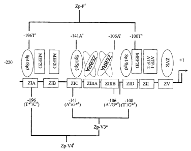
Figure one is a diagram of Zp ( in E ), the promoter of the Epstein-Barr virus
BZLF 1 gene. The different polymorphisms as described by Gutierrez et al
indicated here
by Zp-V3 (in * ) and Zp-V4 ( in # ), Zp-P and Zp-V3 (malignant subtypes) and
Zp-V4
(benign subtype).
DETAILED DESCRIPTION OF THE INVENTION
The present invention features methods for predicting, diagnosing,
detecting, monitoring and determining the prognosis of EBV associated cancers
in a
CA 02473458 2004-08-05
30765-3
4
patient. The methods feature detecting or determining the amount of EBV DNA
present in
the serum of these patients and if positive, a second method determines the
cancerous
potential of the EBV subtype in these patients. The methods according to the
present
invention h ave b road applicability i n c finical m edicine s ince 1 atent i
nfection of EBV is
prevalent and widespread. In some population, over 90% of the population has
such a
latent EBV infection.
Clinically, circulating EBV DNA is applicable in diagnosing and
monitoring gastric carcinoma patients who have EBER-positive tumors, similar
to w hat
has been achieved for nasopharyngeal cancers (Lo, Y.M.D. et al., Clin. Cancer
Res., S9:
1188-1191 (1999); Lo, Y.M.D. et al., Cancer Res., S9: S4S2-S4SS (1999)) and
certain
lymphomas (Lei, K.I. et al., Br. J. Haematol., 111: 239-246 (2000); Drouet, E.
et al., J.
Med. Virol., S7: 383-389 (1999); Gallagher, A. et al., Int. J. Cancer, 84: 442-
448 (1999)).
The recent demonstration of the prognostic significance of circulating EBV DNA
in
nasopharyngeal cancers (Lo, Y.M.D. et al., Cancer Res., 60: 6878-6881)
suggests that
EBV DNA measurement has prognostic importance for gastric carcinoma.
The methods according to the present invention are also applicable for
detecting, monitoring and determining the prognosis of EBV associated cancers
apart from
head, neck and lymphoid malignancies where those cancers are associated with
EBV.
Some of these neoplasms have been shown previously to be associated with EBV
infection
2 0 (Bonnet et al., J Natl Cancer Inst. 91: 1376-81 (1999)), as have certain
liver cancers
(Sugawara et al., Virology. 256: 196-202 (1999)).
Any of the conventional DNA amplification or signal amplification
methods may be used for detection of EBV DNA. In most instances, it is
desirable to
amplify the target sequence using any of several nucleic acid amplification
procedures
2 5 which are well known in the art. Specifically, nucleic acid amplification
is the enzymatic
synthesis of nucleic acid amplicons (copies) which contain a sequence that is
complementary to a nucleic acid sequence being amplified. Examples of nucleic
acid
amplification procedures practiced in the art include the polymerise chain
reaction (PCR),
strand displacement amplification (SDA}, ligase chain reaction (LCR), and
transcription-
3 0 associated amplification (TAA). Nucleic acid amplification is especially
beneficial when
the amount of target sequence present in a sample is very low. By amplifying
the target
sequences and detecting the amplicon synthesized, the sensitivity o:f an assay
can be vastly
improved, since fewer target sequences are needed at the beginning of the
assay to better
CA 02473458 2004-08-05
30765-3
ensure detection of nucleic acid in the sample belonging to the organism or
virus of
interest.
Methods of nucleic acid amplification are thoroughly described in the
literature. PCR amplification, for instance, is described by Mullis et al. in
U.S. Fat. Nos.
5 4,683,195 Methods of nucleic acid amplification are thoroughly described in
the literature.
PCR amplification, for instance, is described by Mullis et aI. in U.S. Pat.
Nos. 4,683,195,
4,683,202 and 4,800,159, and in Methods in Enzymology, 155: 335-350 (1987).
Examples
of SDA can be found in Walker, PCR Methods and Applications, 3: 25-30 (1993),
Walker
et al. in Nucleic Acids Res., 20: 1691-1996 (1992) and Proc. Natl. Acad. Sci.,
89: 392-396
(1991). LCR is described in U.S. Pat. Nos. 5,427,930 and 5,686,272. And
different TAA
formats are provided in publications such as Burg et al. in U.S. Pat. Nos.
5,437,990;
Kacian et al. in U.S. Pat. Nos. 5,399,491 and 5,554,516; and Gingeras et al.
in
International Application No. PCTlUS87/01966 and International Publication No.
WO
88/01302, and International Application No. PCT/US88/02108 and International
Publication No. WO 88/10315.
Real-time quantitative PCR is a preferred means to monitor EBV DNA and
is based on the continuous optical monitoring of the progress of a fluorogenic
PCR
reaction (Heide et al. Genome Res. 6: 986-694, 1996 and Lo et al. Am J. Hum.
Genet. 62:
768-775, 1998). In this system, in addition to the two amplification primers
used in
2 0 conventional PCR, a dual-labeled fluorogenic hybridization probe is also
included (Livak,
et al. PCR Methods Appl., 4357-362, 1995). One fluorescent dye serves as a
reporter
{FAM), and its emission spectrum is quenched by a second fluorescent dye
(TAMRA).
During t he a xtension p hase o f P CR, t he 5 ' t o 3 ' a xonuclease a
ctivity o f t he Taq DNA
ploymerase (9) cleaves the reporter from the probe, thus releasing it from the
quencher and
2 5 resulting in an increase in fluorescence emission at 518 nm.
The methods according to the present invention generally comprise the
steps of (1) obtaining a blood sample from a patient, (2) extracting DNA from
the blood
sample, (3) measuring the amount of circulating EBV DNA present in the blood
sample,
and (4) comparing the amount of circulating EBV DNA present in the blood
sample to a
3 0 control. Preferably, the blood sample is centrifuged, a fluid fraction is
obtained, and the
EBV DNA is measured from the fluid fraction.
Those o f s kill i n t he art w ill understand that the DNA may be extracted
from a blood sample by many means known in the art. One preferred means is
using a
CA 02473458 2004-08-05
30765-3
6
QIAamp Blood Kit. Also, the amount of circulating EBV DNA may be measured
using
one of many known or novel protocols. A protocol comprising a real time PCR
amplification system is particularly preferred. Standard procedures for
comparing the
levels of EBV DNA so detected to a control may easily be devised so as to
statistically
assess the significance of the values obtained.
The number of copies of EBV DNA may be measured over time and
correlated to disease progression or regression. Thereby, the present
invention provides a
non-invasive method that allows a good measurement of the prognosis of these
EBV
associated cancers.
On the other hand, because of the frequent presence of lytic reactivations of
EBV as well as transformation of chronic active EBV carriers, EBV DNA level
can be
found in a significant percentage of the population without cancer. In our
study quoted in
the patent claiming priority, 3.6% of the normal population is positive in
serum or plasma
EBV DNA at any one time. Fortunately, most of these cases, especially those
with EBV
DNA copies of less than S00 capies/ml, are in fact benign in origin. The level
of EBV
DNA will go down to zero in these benign cases after one to two weeks, marking
the end
of the lytic reactivation or chronic reinfection. Nevertheless such
reactivation of EBV and
resulting elevation of IgA antibodies against EBV capsid antigen and
neutralizing
antibodies against EBV Dnase are predictive of nasopharyngeal carcinoma in a
population
2 0 in Taiwan. ( Chien YC, Serologic markers of EBV infection and
nasopharyngeal
carcinoma in Taiwanese men, N England J Med, Vol 34S no26 Dec 27, 2001 )
Conclusively, it is common to find such low-grade EBV DNA infections and
patients are
obviously anxious to know their carcinogenic potential. In fact, our serum or
plasma EBV
DNA test, while extremely sensitive and useful to detect and monitor cancer
such as
2 5 nasopharyngeal carcinoma, has been partially responsible for fording such
a large portion
of the population that in fact is harboring and reactivating EBV at very
frequent intervals.
The present invention is to turn this extreme sensitivity into a useful tool
so that the
carcinogenic potential of these frequent EBV reactivators can be measured once
and for
all.
3 0 In a second aspect, the present invention features genotyping methods that
can detect the different subtypes of EBV with different carcinogenic
potentials. One of the
methods and genotyping that was published showed that polymorphisms in the
regulatory
sequences of BZLFl promoter region of the lytic regulatory gene of EBV are
differentially
CA 02473458 2004-08-05
30765-3
7
distributed among malignant and nonmalignant cells. Three polyrnorphic Zp
sequences
were detected. Among the malignant samples, all 52 samples showed
polymorphisrns
either as Zp-P (identical to the EBV B95.8 strain) or the Zp-V3 sequence. For
the benign
samples, all 52 showed a Zp-V4 polymorphism. (Gutierrez, Discrete alterations
in the
BZLFl promoter in tumor and non tumor associated EB U J of the National Cancer
Institute, hol 94, no 23 December 4 2002 )
In the third aspect, diagnostic kits comprising suitable reagents for
detecting
EBV DNA in the serum or plasma of patients and for the polymorphic subtypes
are
included. The kits according to the present invention may further comprise one
or more of
a device for obtaining a blood sample from a patient, a means to separate the
EBV DNA
from the blood sample and a means to quantify the amount of EBV DNA present in
the
blood sample. Such kits are useful for diagnosing, detecting, monitoring and
determining
the prognosis of EBV associated cancers.
All publications and patent applications cited in this specification are
herein
incorporated by reference as if each individual publication or patent
application were
specifically and individually indicated to be incorporated by .reference.
Although the foregoing invention has been described in some detail by way
of illustration and example for purposes of clarity of understanding, it will
be readily
apparent to those of ordinary skill in the art in light of the teachings of
this invention that
2 0 certain changes and modifications may be made thereto without departing
from the spirit
or scope of the appended claims.
FXAMPT .FC
The following examples are provided by way of illustration only and not by
way of limitation. Those of skill will readily recognize a variety of
noncritical parameters
2 5 which could be changed or modified to yield essentially similar results.
EXAMPLE 1
Materials and Methods
Detecting EBV DNA from Plasma Samples Patients are recruited with
3 0 known EBV associated cancers such as nasopharyngeal carcinoma. Normal
individuals are
also r ecruited t hat s erve as c ontrol f or b enign c arriers o f E BV a nd
for t he d etection o f
serum or plasma EBV DNA and :Eor EBV subtyping according to polymorphisms.
CA 02473458 2004-08-05
30765-3
8
DNA Extraction from Plasma Samples is done as followed. Peripheral
blood (5 ml) can be collected from each subject into an EDTA tube for the
isolation of
plasma. Blood samples are centrifuged at 1600 X g, and plasma carefully
removed from
the EDTA-containing tubes and transferred into plain polypropylene tubes. The
samples
are stored at -20°C until further processing. DNA form plasma samples
are extracted
using a QIAamp B food Kit ( Qiagen, H ilden, G ermany) a sing t he b lood a nd
b ody fluid
protocol as recommended by the manufacturer (2). Plasma samples (130-800
pl/column)
are used for DNA extraction. The exact amount is documented for the
calculation of the
target DNA concentration. A final elution volumn of 50 pl is used from the
extraction
columns.
Circulating EBV DNA concentrations were measured using a real time
quantitative PCR system towards the BamHI-W fragment region of the EBV genome
(Lo,
Y.M.D. et al., Cancer Res., 59: 1188-1191 (1999)). The principals of real time
quantitative PCR and reaction set-up procedures were as previously described
(Lo, Y.M.D.
et al., Cancer Res., 59: 1188-1191 (1999)). Data were collected using an ABI
Prism 7700
Sequence Detector and were analyzed using the Sequence Detection System
software
(version 1.6.3) developed by Applied Biosystems. Results were expressed as
copies of
EBV genomes per millititer of serum.
All serum DNA samples were also subjected to real time PCR analysis for
2 0 the (beta-globin gene (Lo, Y.M.D. et al., Cancer Res., 59: 1188-1191
(1999)), which gave
a positive signal on all tested samples, thus demonstrating the quality of the
extracted
DNA. Multiple negative water blanks were included in every analysis.
More specifically, two real-time quantitative PCR systems have been
developed for EBV DNA detection: (a) one toward the BamHI-W region; and (b)
the other
2 5 toward the EBNA-I region (Baer, et al Nature, 310: 207-211, 1984). The
BamHI-W
system consisted of the amplification primers (SEQ ID NO: 1) W-44F (5'-
CCCAACACTCCACCACACC-3') and (SEQ ID NO: 2) W-1198 (5'-TCTT
AGGAGCTGTCCGAGGG-3') and the dual-labeled fluorescent probe (SEQ ID NO: 3)
W-67T (5'-FAM)CACACACTACACACACCCAC-CCGTCTC(TAMRA)-3']. The
3 o EBNA-1 system consisted of the amplification primers (SEQ ID NO: 4) EBNA-
1162F (5'-
TCATCATCATCCGGGTCTCC-3') and (SEQ ID NO: 5) EBNA-12298 (5'-
CCTACAGGGT-GGAAAAATGGC-3') and the dual-labeled fluorescent probe (SEQ ID
NO: 6) EBNA-1186T [5'-(FAM)CGCAGGCCCCCTCCAGGTA-GAA(TAMRA)-3'].
CA 02473458 2004-08-05
30765-3
9
The fluorescent probes contained a 3'-blocking phosphate group to prevent
probe
extension during PCR. Primer/probe combinations were designed using Primer
Express
software (Perkin-Elmer Corp., Foster City, CA). Sequence data for the EBV
genome were
obtained from the GenBank Sequence Database (accession number V01555). Real-
time
quantitative PCR for the /3 globin gene consisted of primers and probe, as
described
previously in Lo, et al. Am J. Hum Genet 62: 768-775, 1998), and was used as a
control
for the amplifiability of plasma DNA.
Fluorogenic PCR reactions are set up in a reaction volume of 50 ~,l using
components ( except f or t he fluorescent p robes and a mplification primers)
supplied in a
TaqMan PCR Core Reagent K it ( Perkin-Elmer C orp.). Fluorescent p robes are
custom-
synthesized by Perkin-Elmer Applied Biosystems. PCR primers were synthesized
by Life
Technologies, Inc. (Gaithersburg, MD). Each reaction contained 5 pl of l Ox
buffer A; 300
nM of each of the amplification primers; 25 nM (for the EBV probes) or 100 nM
(for the
~3 globin probe) of the corresponding fluorescent probe; 4 MM MgCl2; 200 pm
each of
dATP, dCTP, and dGTP; 400 pM dUTP; 1.25 units of AmpliTaq Gold; and 0.5 unit
of
AmpErase uracil N-glycosylase.
DNA amplifications are carried out in a 96-well reaction plate format in a
Perkin-Elmer Applied Biosystems 7700 Sequence Detector. Each sample are
analyzed in
duplicate. Multiple negative water blanks were included in every analysis.
2 0 A calibration curve is run in parallel and in duplicate w ith each
analysis,
using DNA extracted from the EBV-positive cell line Namalwa (American Type
Culture
Collection CRL-1432; See Klein et al., Int J. Cancer, 10: 44-57, 1972) as a
standard.
Namalwa is a diploid cell line that contains two integrated viral
genomes/cell. A
conversion factor o f 6 .6 p g o f D NA/diploid c ell w as a sed .f or copy n
umber calculation
2 5 (Saiki et al., Science, 239: 487-491, 1988). Concentrations of circulating
cell-free EBV
DNA were expressed as copies of EBV genome/ml plasma.
An identical thermal profile was used for the EBV BamHI-W and EBNA-I
PCR systems. Thermal cycling was initiated with a 2-min incubation at
50°C for the
uracil N-glycosylase to act, followed by an initial denaturation step of 10
min at 95°C, and
3 0 then 40 cycles of 95°C for 15 s and 56°C for 1 min were
carried out.
Amplification data collected by the 7700 Sequence Detector and stored in a
Macintosh computer (Apple Computer, Cupertino, CA) is then analyzed using the
Sequence Detection System software developed by Perkin-Elmer Applied
Biosystems.
CA 02473458 2004-08-05
30765-3
The mean quantity of a ach duplicate is a sed far further c oncentration c
alculation. T he
plasma concentration of EBV DNA or the ,(3-globin gene (expressed in
copieslml) is
calculated using the following equation:
5
DNA 1
C ~ Q x .x -
~PCR text
in which C represents the target concentration in plasma (copies/ml), Q
represents the
target quantity (copies) determined by a sequence detector in a PCR, YDNA
represents the
total volume of DNA obtained after extraction (typically 50 pl/Qiagen
extraction), VPCR
1 o represents the volume of DNA solution used for PCR (typically 5 ~l, and
Vext represents
the volume of plasmalserum extracted (typically 0.13-0.80 ml)).
The presence of EBV in tumor cells was assessed by in-situ hybridization
on paraffin-embedded tissue sections using a fluorescein-conjugated
oligonucleotide probe
for EBER (Novocastra, U.K.) as previously described (Hui, P.K. et al., Hum.
Pathol., 25:
947-952 (1994)).
Detection of EBV Subtypes Patients' samples DNA can be taken fram the
cell free plasma as described in [27]. These DNA samples can also be isolated,
by using a
standard phenol/chloroform extraction technique after proteinase K digestion,
from
peripheral blood lymphocytes. Furthermore, DNA samples can also be isolated
from
2 0 suspicious areas of EBV tumors or re-activation sites.
We use the TaqMan Allelic Discrimination assay with the 5' nuclease
activity of Taq polymerase to detect a fluorescent reporter signal generated
during or after
PCR reactions. For genotyping of EBV, one pair of TaqMan probes and one pair
of PCR
primers are used. The assay uses two TaqMan probes that differ at the
polymorphic site,
2 5 with one probe complementary to the wide-type allele and the other to the
variant allele.
A 5' reporter dye and a 3' quencher dye are covalently linked to the wild-type
or variant
allele probes. When the probes are intact, fluorescence is quenched because of
the
physical proximity of the reporter and quencher dyes. During the PCR annealing
step, the
TaqMan p robes h ybridize t o the targeted polymorphic site. During the PCR
extension
3 0 phase, the 5' reporter dye is cleaved by the 5' nuclease activity of the
Taq polymerase,
leading to an increase in the characteristic fluorescence of the reporter dye.
Specific
CA 02473458 2004-08-05
30765-3
11
genotyping is determined by measuring the signal intensity of the two
different reporter
dyes after the PCR reaction. TaqMan genotyping instrumentation and reagents
are
supported by Applied Biosystems.
EBV subtypes polymorphisms tested on the 200-nt Zp BZLF1 promoter
region are listed as follows (as shown in Fig. 1):
(1) Zp-P (protype) identical to EBV strain B95-8
(2) Zp-V3 with three point changes:
(a) A to G at position -141
(b) A to G at position -106
(c) T to G at position -100
(3) Zp-V4: containing all of the Zp-V3 plus a fourth substitution, T to C
at position -196
CA 02473458 2004-08-05
30765-3
12
Clinical Results Using the Comparison of V3 (Malignant)
and P (Benign) Status of Patient Samples in
Nasophawrngeal Carcinoma (NPC)
BZLF1 real-time PCR Reacti~n Mix
Vp-P = FAM
Vp-V3 = VIC
Reagent Volume (30 ul)
1 Ox buffer A 3.00
25 mM Mg2+ 4.50
dNTP 2.50
BZLF1 (Forward primer) 1.00
BZLF1 (Reverse primer) 1.00
BZLF1-probe P 0.40
BZLF1-probe V3 0.40
U NG 0.30
Taq gold 0.15
H20 11.85
Total : 25.00
25 ul Master Mix + 5 ul ~NA sample
BZLF1 Real-time PCR Profile
50°C 2 wins
95°C 5 minx
94°C 20 secs
X 45 cycles
60°C 1 min
CA 02473458 2004-08-05
30765-3
13
EBV DNA EBonco Additional
Patient NPC ? NPC Clinical
No.
~~opieslml plasma) V3 or
P
Information
375 101 - 1000 X P
405 195 X P
709 34 X P NPC (new case)
837 1174 X P
931 1279 X P
1024 605 X P
256 2273312 X V3 Metastatic
262 127971 X V3 NPC
331 <100 X V3
Recurrence
of
NPC
348 22250 X V3
350 1050 X V3
377 33925 X V3
399 101 - 1000 X V3 Cervical LN
also
ositive
404 304 X V3
408 295 X V3
N PC post RT
427 106 X V3 with
known bone
&
lun Seconds
447 3000 X V3
735 59 X V3 Metastatic
NPC
785 13631 X V3 Metastatic
NPC
790 43 X V3
858 778 X V3
960 944 X V3
991 4650 X V3
1003 101 - 1000 X V3
1025 5131 X V3
TH589 25 X V3
CA 02473458 2004-08-05
30765-3
14
Patient EBV DNA (copieslml EBonco Results
No. lasma Normal V3 or P
622 0 X Undetermined
623 0 X U ndetermined
624 0 X Undetermined
625 0 X Undetermined
626 0 X __ Undetermined
627 0 X Undetermined
628 0 X Undetermined
629 0 X Undetermined
634 0 __ X Undetermined
635 0 X _ Undetermined
980 0 X Undetermined
981 0 X Undetermined
982 0 X _ Undetermined
1033 0 X Undetermined
1034 0 X Undetermined
1035 0 X U ndetermined
TH39 0 X Undetermined
TH355 0 X Undetermined
TH516 0 X Undetermined
TH663 0 X Undetermined
TH836 0 X Undetermined
TH 1142 0 X Undetermined
TH1164 0 X Undetermined
Conclusion
It can be shown from the above clinical study that out of 26 patients with
NPC and possible NPC (no pathological confirmation), 20 carried the more
malignant
subtype of V3 (for Chinese population, V3 is the dominant malignant subtype),
contributing to 77% of the NPC population. If the possible NPC cases were
deleted from
the study, the percentage of the V3 subtype remained the same as 77% (I7 out
of 22
patients). For normal individuals with no EBV DNA circulating in the blood,
there is no
V3 or P detected. Preliminary study shown h ere d emonstrated t hat t he
combination o r
individual blood testing of the subtype V3 of the EBV DNA may be a powerful
tool for
CA 02473458 2004-08-05
30765-3
the future estimation of cancer changes in patients with a positive EBV DNA
titer in their
blood.
Alternatively detection of EBV subtypes can be done by other methods
which enable single base discrimination, including, but not limited to single-
strand
5 conformation polymorphism, allele-specific PCR, mass spectrometric analysis
of PCR
products, a rtificial r estriction fragment 1 ength p olymorphism, Iigase
chain reaction.. The
following protocol illustrates the use of single-strand conformation
polymorphism
analysis. Five hundred nanograms of genomic DNA obtained from all tumor and
nontumor
samples was directly used as a template in polymerise chain reactions (PCRs).
PCR was
10 used to amplify the fragment from nucleotides -221 to +12 (with respect to
the
transcription start site) of the BZLFl promoter (nucleotides 103420-103182 of
the EBV
gonome, Genbank accession no. NC 001345). The primers used were 5'-
agcatgccatgcatatttc-3' and 5'-ttggcaaggtgcaatgttt-3'. PCR conditions consisted
of 5
minutes at 95°C, 30 cycles of 30 seconds at 95°C, 30 seconds at
60°C, and 1 minute at
15 72°C, followed by a long extension of 10 minutes at 72°C.
[32P]dCTP was included in the
PCR buffer. PCR products were separated by electrophoresis through 6%
nondenaturing
acrylamide gels (19:1, acrylamide to bis) containing 10% glycerol at 6W for 18
hours and
visualized by autoradiography. Autoradiograms were exposed for 2-16 hours.
Single-
strand conformation polymorphisms (SSCPs) were apparent by differences in the
patterns
2 0 of migration of the PCR product. Amplified product obtained from the EBV
strain B95.8
served as a reference control. The above protocol demonstrates that other
cancer causing
subtypes can be found and be tested similarly from blood samples as shown in
the clinical
study illustrated for V3 and P.
Conclusions
EBV samples of NPC patients in the Chinese population show that the
Zp-V3 subtype may diagnosis patients that are more cancer prone than those
with Zp-P
subtype.
CA 02473458 2004-08-05
30765-3
16
Results
Results from our past publications showed a sensitivity of 96% and a
specificity of 93% in detecting nasopharyngeal carcinoma with concentration of
EBV
DNA copies exceeding 500 copies/ml. Among the healthy control, 3.6% showed a
positive
EBV DNA titre, most of them below 500 copies/ml. This present invention can
predict
further that among the 3.6% of the health population, how many are actually
harboring a
subtype of EBV that can most likely link to the development of EBV associated
cancer.
More careful assessment of the patient, and detail follow up may be able to
pick up EBV
associated cancer at early stage, whereby a cure is expected.
Discussion
Clinically, circulating EBV DNA may have application in the diagnosis and
monitoring in the proportion of gastric carcinoma patients who have EBER-
positive
tumors, similar to what has been achieved for NPC (Lo, Y.M.D. et al., Cancer
Res., 59:
1188-1191 (1999); Lo, Y.M.D. et al., Cancer Res., 59: 5452-5455 (1999)) and
certain
lymphomas (Lei, K.I. et al., Br. J. Haematol., 111: 239-246 (2000); Drouet, E,
et al., J.
Hed. Viol. 57: 383-389 (1999); Gallagher, A. et al., Int. J. Cancel°,
84: 442-448 (1999)).
Recently, the value of circulating EBV DNA in nasopharyngeal cancer prognosis
has been
2 o demonstrated. Additional data (Lo, Y.M.D. et al., Cancer Res., 60: 6878-
6881) indicate
that EBV DNA measurement also has prognostic importance for gastric carcinoma.
There is however, a large group of patients that have been diagnosed with
low EBV DNA level and chronic or frequent EBV reactivations with no visible
sign and
symptom of cancers. Disposition and follow up of such patients is so far
unsatisfactory due
2 5 to the known carcinogenic potential of EBV with additional information
that cancer is
indeed more prevalent in this group.
One of the many methods to detect polymorphism or other genotypic
abnormality is demonstrated by Crutierrez ( JNCl, 94 no23 Dec 4th 2002 ). Her
publication
revealed the division of the promoter region of the EBV lytic gene BZLF1 into
three
3 0 subtypes with polymorphism that can be detected by PCR-single strand
confirmation
polymorphism. All cancer samples belonged to two subtypes of Zp-P and Zp-V3.
All
benign samples belonged to one subtype Zp-V4. In the Chinese population,
however, the
Zp-V3 is the dominant malignant subtype over Zp-P, while Zp-V4 cannot be
found.
CA 02473458 2004-08-05
30765-3
17
The very sensitive method described in this invention can detect cell free
plasma EBV DNA in a group of individuals that may only have apparent benign
diseases
such as lytic reactivation in the upper airways and gastritis. They usually
have lower than
500 copies/ml of EBV DNA and exhaustive examination reveals no known
malignancy.
The combination of the second part of this invention through the detection of
definitive
polymorphisms and subtypes of F?BV will further clarify the management of this
group of
individuals. One group with the malignant subtype should undergo regular
follow up and
frequent measurement of the EB V DNA level. The other group with the benign
subtype
may be relieved of their anxiety and be followed up on a as needed basis.
Data also suggest that circulating EBV DNA may be useful in many other
cancer types that are associated with EBV. Examples of such cancers include
breast
cancer (Bonnet, M. et al., J. Natl. Cancer Inst., 91: 1376-1381 (1999)) and
hepatocellular
carcinoma (Sugawara, Y. et al., Yzralogy, 256: 196-202 {1999)). As the
association
between some tumor types and EBV is still controversial, the possible
detection of EBV
DNA and the detection of cancerous subtypes in the plasma of patients with
such tumors
may contribute towards resolving these issues.
CA 02473458 2004-08-05
30765-3
18
SEQUENCE LISTING
(1) GENERAL INFORMATION:
(i) APPLICANT: YEUNG, W.AH-HIN A.
(ii) TITLE OF INVENTION: THE COMBINATION OF CIRCULATING EPSTEIN-
BARR VIRUS (EBV) DNA IN THE SERUM OR PLASMA OF PATIENTS AND A METHOD
TO ASSESS EBV SUBTYPES FOR THE PREDICTION AND DETECTION OF EPSTEIN-
BARR VIRUS ASSOCIATED CANCERS
(iii) NUMBER OF SEQUENCES: 6
(iv) CORRESPONDENCE ADDRESS:
(A) ADDRESSEE: FETHERSTONHAUGH & CO.
(B) STREET: P.O. BOX 2999, STATION D
(C) CITY: OTTAWA
(D) STATE: ONT
(E) COUNTRY: CANADA
(F) ZIP: K1P SY6
(v) COMPUTER READABLE FORM:
(A) MEDIUM TYPL: Floppy disk
2 0 (B) COMPUTER: IBM PC compatible
(C) OPERATING SYSTEM: PC-DOS/MS-DOS
(D) SOFTWARE: ASCII (text)
(vi) CURRENT APPLICATION DATA:
(A) APPLICATION NUMBER: CA
2 5 (B) FILING DATE:
(C) CLASSIFICATION:
(vii) PRIOR APPLICATION DATA:
(A) APPLICATION NUMBER:
(B) FILING DATE:
3 0 (viii) ATTORNEY/AGENT INFORMATION:
(A) NAME: FETHERSTONHAUGH & CO.
(B) REGISTRATION NUMBER:
(C) REFERENCE/DOCKET NUMBER: 30765-3
(ix) TELECOMMUNICATION INFORMATION:
3 5 (A) TELEPHONE: (613)-235-4373
(B) TELEFAX: (613)-232-8440
(2) INFORMATION FOR SEQ ID NO.: 1:
(i) SEQUENCE CHARACTERISTICS
(A) LENGTH: 19
4 0 (B) TYPE: nucleic acid
(C) STRANDEDNESS:
(D) TOPOLOGY:
(ii) MOLECULE TYPE: DNA
(vi) ORIGINAL SOURCE:
4 5 (A) ORGANISM: Artificial
(ix) FEATURE
(C) OTHER INFORMATION: BamHI-W system amplification primer W-44F
(xi) SEQUENCE DESCRIPTION: SEQ m NO.: 1:
CCCAACACTC CACCACACC 19
(2) INFORMATION FOR SEQ ID NO.: 2:
(i) SEQUENCE CHARACTERISTICS
CA 02473458 2004-08-05
30765-3
19
(A) LENGTH: 20
(B) TYPE: nucleic acid
(C) STRANDEDNESS:
(D) TOPOLOGY:
(ii) MOLECULE TYPE: DNA
(vi) ORIGINAL SOURCE:
(A) ORGANISM: .Artificial
(ix) FEATURE
(C) OTHER INFORMATION: BamHI-W system amplification primer W-1198
(xi) SEQUENCE DESCRIPTION: SEQ ID NO.: 2:
TCTTAGGAGC TGTCCGAGGG 20
(2) INFORMATION FOR SEQ ID NO.: 3:
(i) SEQUENCE CHARACTERISTICS
(A) LENGTH: 27
(B) TYPE: nucleic acid
(C) STRANDEDNESS:
2 0 (D) TOPOLOGY:
(ii) MOLECULE TYPE: DNA
(vi) ORIGINAL SOURCE:
(A) ORGANISM: Artificial
(ix) FEATURE
2 5 (C) OTHER INFORMATION: BamHI-W system dual - labeled fluorescent
probe W-67T
(ix) FEATURE
(A) NAME/KEY: mist feature
(B) LOCATION: (1)..(1)
3 0 (C) OTHER INFORMATION: N = FAM - labeled c
(ix) FEATURE
(A) NAME/KEY: misc_feature
(B) LOCATION: (27)..(27)
(C) OTHER INFORMATION: N = TAMRA - labeled c
3 5 (xi) SEQUENCE DESCRIPTION: SEQ ff~ NO.: 3:
NACACACTAC ACACACCCAC CCGTCTN 27
4 0 (2) INFORMATION FOR SEQ ID NO.: 4:
(i) SEQUENCE CHARACTERISTICS
(A) LENGTH: 20
(B) TYPE: nucleic acid
(C) STRANDEDNESS:
4 5 (D) TOPOLOGY:
(ii) MOLECULE TYPE: DNA
(vi) ORIGINAL SOURCE:
(A) ORGANISM: Artificial
(ix) FEATURE
5 0 (C) OTHER INFORMATION: EBNA-I system amplification primer EBNA-
1162F
(xi) SEQUENCE DESCRIPTTON: SEQ ID NO.: 4:
TCATCATCAT CCGGGTCTCC 20
CA 02473458 2004-08-05
30765-3
(2) INFORMATION FOR SEQ ID NO.: 5:
(i) SEQUENCE CHARACTERISTICS
(A) LENGTH: 21
(B) TYPE: nucleic acid
5 (C) STRANDEDNESS:
(D) TOPOLOGY:
(ii) MOLECULE TYPE: DNA
(vi) ORIGINAL SOURCE:
(A) ORGANISM: Artificial
10 (ix) FEATURE
(C) OTHER INFORMATION: EBNA-1 system amplification primer EBNA-
1229R
(xi) SEQUENCE DESCRIPTION: SEQ ID NO.: 5:
CCTACAGGGT GGAAAAATGG C 21
(2) INFORMATION FOR SEQ ID NO.: 6:
(i) SEQUENCE CHARACTERISTICS
2 0 (A} LENGTH: 22
(B) TYPE: nucleic acid
(C) STRANDEDNESS:
(D) TOPOLOGY:
(ii) MOLECULE TYPE: DNA
2 5 (vi) ORIGINAL SOURCE:
(A) ORGANISM: Artificial
(ix) FEATURE
(C) OTHER INFORMATION: EBNA-1 system dual-labeled fluorescent probe
EBNA-1186T
3 0 (ix) FEATURE
(A) NAME/KEY: misc_feature
(B) LOCATION: (1)..(1)
(C) OTHER INFORMATION: N= FAM - labeled c
(ix) FEATURE
3 5 (A) NAME/KEY: misc_feature
(B) LOCATION: (22)..(22)
(C) OTHER INFORMATION: N = TAMRA - labeled a
(xi) SEQUENCE DESCRIPTION: SEQ 117 NO.: 6:
NGCAGGCCCC CTCCAGGTAG AlV 22