Note: Descriptions are shown in the official language in which they were submitted.
CA 02473582 2008-04-25
AUTONOMIC NERVOUS SYSTEM MONITORING
TECHNICAL FIELD
This invention relates to medical devices, and more particularly to
physiological
monitoring methods and devices used for detection of autonomic nervous system
(ANS)
activity in the field of sleep research. The present invention discloses a
portable, simple, and
cost-effective electronic sleep diagnostic device containing hardware and
software that permits
recording and signal conditioning of a pulsatile blood volume waveform
obtained through use
of a photoplethysmographic (optical volume detecting) probe, thereby allowing
analysis pulse
transitional slope data that is representative of activity in the autonomic
nervous system (ANS).
BACKGROUND OF THE INVENTION
Cardiovascular risk is directly linked to sleep related breathing disorders
(SRBD). The
number of U.S. laboratories that study sleep, roughly 2,792, is incredibly low
when compared
to the number of Ameiicans estimated to have a chronic SRBD, just over 40
million. The
average number of beds per lab is 3.6 bringing the total number of beds in
which to do a sleep
study to roughly 10,000. This means that to test all 40 million Americans,
there would be
CA 02473582 2004-07-13
WO 2004/026132 PCT/US2003/029862
4,000 patients that would be seen per bed. If sleep tests were ran 365 days
per year, the result
is an astounding 11 years of conclusive tests needed to be run to test the
current population of
individuals suffering form SRBD. The lengtli of time increases as one
considers the actual
number of days per year sleep labs actually test patients, plus the amount of
tests that need to
be re-run due to inconclusive testing, plus the number of patients that
continually need to be
retested to see if their treatment is functioning properly. Given this
scenario, it is no shock that
wait times for patients to be scheduled for a sleep test can typically range
from six weeks to six
months. The problem will only increase, as "it is estimated that nearly 80
million Americans
will have a sleep problem by the year 2010 and 100 million will have oile by
the year 2050."
Clearly then, the problem with wait time for testing should be addressed
immediately to relieve
pent up demand.
The current "gold standard" for testing sleep related breathing disorders is
full
polysomnography. Full polysomnography is, however, quite labor intensive,
requires
considerable instrumeiltation and is therefore rather expensive to conduct. As
a result, many
sleep laboratories have found it difficult to keep up with the demand for this
test, and a long
waiting list becomes the norm. Given that obstructive sleep apnea (OSA) is
quite prevalent,
leads to serious complications and that treatment options exist, it is
important that individuals
suffering from the disease are identified.
The need to study the ANS has been realized in academia for a considerable
time. It is
known in the field of microneurography that rapid-eye moveinent (REM) sleep is
associated
with profound sympatlietic activity. It has also been found that arousals from
non-rapid-eye
movement (NREM) elicits K complexes that are associated with sympathetic
activity. The
sympatlletic division of the ANS prepares a body for movement. Arousals
require movement
and hence an arousal requires sympathetic activation.
2
CA 02473582 2004-07-13
WO 2004/026132 PCT/US2003/029862
Generally, patients with OSA, a type of SRBD, have extremely disrupted sleep
and
terribly high daytime somnolence. Obstructive sleep apnea events are always
accompanied by
an acute rise in systolic blood pressure (rises in systolic blood pressure are
associated with
sympathetic activation), even when the usual EEG criteria for arousals are not
met (a
recognizable cortical electroencephalographic arousal). The duration of the
apnea of
individuals that demonstrate EEG arousal and those that do not meet the usual
criteria for
defining an arousal have been found to be identical. The pleural pressure
peak, at the end of
apnea, is identical between the two types of arousals, as are the EEG
frequencies. These
findings suggest that monitoring the cardiac changes of sleep is a more
accurate measurement.
It has been demonstrated that apneic episodes result in progressive increases
in
sympathetic nerve activity. The increases are most marked toward the end of
the apnea, when a
patient moves. These findings are exactly what is excepted of sympathetic
activation and its
relationship to arousals in patients with SRBD.
Because cardiovascular control during sleep is primarily dictated by brain
states that
produce profound variation in ANS activity, many studies have been conducted
to monitor the
ANS. Since the data shows clearly that monitoring the ANS or cardiac changes
in sleep yields
more accurate data defining an arousal in sleep, it is clear that diagnostic
studies must include
ANS or cardiac monitoring.
It has been shown that in transitions from NREM to REM sleep, heart rate
accelerations
precede the EEG arousals marking the onset of REM. Therefore, not only does
monitoring
ANS activity give the clinician a possibly more accurate study, but also
changes in ANS
activity precede that information being observed via the EEG electrodes.
There are two existing technologies that attempt to monitor the ANS, namely
pulse
transit time (PTT) and peripheral arterial tonometry (PAT). Neither PTT nor
PAT can lay
claim to monitoring the ANS without adding additional sensors. PTT requires
the use of ECG
3
CA 02473582 2004-07-13
WO 2004/026132 PCT/US2003/029862
electrodes that may be difficult for a patient to self-apply due to skin
cleaning and shaving
requirements. PAT requires a very costly gauntlet-type device with a single-
use finger pressure
cuff. Also, the addition of extra sensors adds to noise artifact and
difficulty in patient use. It is
therefore an object of the present invention to provide an improvement over
existing PTT and
PAT technology through a more economical and more easily used device without
need of
additional sensors.
Several disclosures have been made in the prior art that teach methods and
devices for
diagnosis and monitoring of sleep breathing disorders using physiological data
obtained from
pulse oxiinetry-derived waveforms.
U.S. Patent No. 5,398,682 to Lynn (March 21, 1995) discloses a method and
apparatus
for the diagnosis of sleep apnea utilizing a single interface with a human
body part. More
specifically, a device is disclosed for diagnosing sleep apnea by identifying
the desaturation
and resaturation events in oxygen saturation of a patient's blood. The slope
of the events is
determined and coinpared against various information to determine sleep apnea.
U.S. Patent No. 6,363,270 B1 to Colla, et al. (March 26, 2002) discloses a
method and
apparatus for monitoring the occurrence of apneic and hypopneic arousals
utilizing sensors
placed on a patient to obtain signals representative of at least two
physiological variables,
including blood oxygen concentration, and providing a means for recording the
occurrence of
arousals. Obtained signals pass through conditioning circuitiy and then
processing circuitry,
where correlation analysis is performed. A coincident change in at least two
of the processed
signals are indicative of the occurrence of an arousal that in turn indicates
an apneic or
hypopneic episode has occurred. A patient thus can be diagnosed as suffering
conditions sucli
as obstructive sleep apnea.
U.S. Patent No. 6,529,752 B2 to Krausman and Allen (March 4, 2003) discloses a
method and apparatus for counting the number of sleep disordered breathing
events
4
CA 02473582 2004-07-13
WO 2004/026132 PCT/US2003/029862
experienced by a subject within a specified time period. Such a counter
comprises: (1) an
oxygen saturation level sensor for location at a prescribed site on the
subject, (2) an oximetry
conditioning and control module that controls the operation of the sensor and
converts its
output data to oxygen saturation level data, (3) a miniature monitoring unit
having a
microprocessor, a memory device, a timer for use in time-stamping data, a
display means and a
recall switch, and (4) firmware for the unit that directs: (i) the sampling
aiid temporary storage
of the oxygen saturation level data, (ii) the unit to analyze using a
specified method the
temporarily stored data to identify and count the occurrence of the subject's
disordered
breathing events, and to store the time of occurrence of each of these events,
and (iii) the
display means to display specified information pertaining to the counts in
response to the
actuation of the recall switch.
U.S. Patent No. 6,580,944 B1 to Katz, et al. (June 17, 2003) discloses a
method and
apparatus for identifying the timing of the onset of and duration of an event
characteristic of
sleep breathing disorder while a patient is awake. Chaotic processing
techniques analyze data
concerning a cardiorespiratory function, such as oxygen saturation and nasal
air flow.
Excursions of the resulting signal beyond a threshold provide markers for
delivering the
average repetition rate for such events that is useful in the diagnosis of
obstructed sleep apnea
and other respiratory dysfunctions.
The above references all malce use of oxygen saturation data obtained through
pulse
oximetry to determine arousals and/or sleep breathing disorders. Each
necessarily requires
additional analysis and calculation of blood oxygen concentrations in order to
render
information useful specifically in the diagnosis and monitoring of sleep
breathing disorders. It
is therefore another object of the present invention to provide a more
simplified method of
obtaining and analyzing physiological data that accurately represents ANS
activity.
5
CA 02473582 2007-01-19
BRIEF SUMMARY OF THE INVENTION
The present invention overcomes one or more of the problems with the prior
art. In
one preferred embodiment the present invention provides a method and apparatus
for
improved monitoring of ANS activity using a single patient sensor.
Accordingly, the present invention provides an apparatus for monitoring human
autonomic nervous system activity using pulsatile blood volume waveform
signals, said
apparatus comprising:
a photoplethysmographic probe having a light emitting element and an opposing
light detecting element, and having an output signal indicating changes in
blood volume on
at least one alpha andrenergic receptor site of a human body;
a processor element, responsive to said output signal indicating changes in
blood volume, for reducing said waveform signals to a slope value;
said processor element containing an algorithm for normalization of the slope
value;
said processor element containing an artifact rejection algorithm for
eliminating
from further processing slope values less than one; and
amplifier and filter circuitry for rendering output signals representative of
said slope
values.
The present invention also provides an apparatus for monitoring human
autonomic
nervous system activity using pulsatile blood volume waveform signals, said
apparatus
comprising:
a photoplethysmographic probe having a light emitting element and an opposing
light detecting element, and having an output signal indicating changes in
blood volume on
at least one alpha andrenergic receptor site of a human body;
a power supply having a battery with capacity for at least 12 hours;
6
CA 02473582 2007-01-19
analog circuitry for power supply voltage regulation and conditioning;
an interface for an OEM supplied finger pulse oximetry probe;
a low frequency front-end filter for conditioning a probe input signal;
an input signal pre-amplifier stage;
a high frequency filter for conditioning a probe input signal;
a gain-controlled signal amplifier stage;
a bar graph display for a visual indication of a pulse signal;
a polygraph output port for pulse signal data;
a digital processing unit, such as a microprocessor or microcontroller, to
provide
slope detection and peak to peak height determination of each systolic finger
pulse,
mathematical normalization of input signal slope, digital to analog (D/A)
conversion of the
slope value for a polysomnographic display, and digital control of finger
probe gain, and
having a status indicator LED;
a plurality of user controls comprising on/off, start/stop and transmit
functions;
a display for visual indication of slope ratio information;
a data storage unit, such as such an on-board multi-media card, to permit at
least 5
hours of data storage; and
a plurality of output ports for providing analog and digital output of the
pulsatile
waveform and a DC level representative of a normalized slope, and slope ratio
data.
In a further object the present invention provides a method for identification
of
human autonomic nervous system activity, the method comprising the steps of:
disposing a photoplethysmographic probe proximate to a single alpha
andrenergic
receptor site of a human body part;
6a
CA 02473582 2007-01-19
obtaining an electrical signal from said probe representative of pulsatile
blood
volume within said body part;
deriving a pulsatile blood volume waveform as a function of amplitude and
time;
defining a time interval for calculation of a slope of the pulsatile blood
volume
waveform;
applying an algorithm that continuously provides real-time calculation of the
slope
along said waveform within said time interval;
dividing peak amplitude values by a time constant and eliminating slope values
less
than 1, whereby artifact elimination is achieved;
normalizing slope values; and
providing information representative of slope values, whereby autonomic
nervous
system activity is monitored.
A variety of breathing disturbances may occur during sleep, including snoring,
hypoventilation, apnea, increased upper-airway resistance, and asthma related
conditions.
This project proposes development of a novel device that can noninvasively and
accurately
detect frequent brief microarousals that are not well identified by
conventional airflow,
respiratory effort, pulse oximetry and EEG methods. These subcortical events
result from
increased respiratory effort and cause disruption of nocturnal sleep, leading
to excessive
daytime somnolence.
Since microarousals have been associated with changes in autonomic system
outflow, this invention provides for a small, portable device that analyzes
the shape of the
arterial finger pulse, thereby detecting on a beat by beat basis changes in
vascular tone
directly attributable to microarousals. The present invention uses a
photoplethysmographically derived arterial blood volume waveform for
monitoring changes
6b
CA 02473582 2007-01-19
in peripheral arterial vascular tone, in conjunction with A/D converters and a
microcontroller for analyzing the morphology of the pulsatile signal.
The method of the present invention provides for detection of microarousals
that
compares favorably with detection by pulse transit time (PTT) devices, EEG
analysis, ECG
analysis, esophagal pressure (Pes) or some combination of these methods.
Although PTT
and peripheral arterial tonometry (PAT) have both been receiving much
attention as
techniques for detecting changes in the ANS during sleep studies, PAT is
relatively
expensive and PTT has implementation problems caused by motion artifact.
The present invention also provides an apparatus that utilizes transmitted
light
intensity from an existing FDA approved pulse oximeter probe so that no
6c
CA 02473582 2007-01-19
additional device is attached to the patient. Valuable diagnostic information
can then be
extracted through electronic processing of this existing data.
Normalization is a method to correct for the photoplethysmographic pulse
signal
morphological changes based on finger position (as opposed to actual changes
of autonomic
activity.) PTT and PAT lack a means for signal normalization and therefor
cannot correct for
finger position changes. Normalization provides immunity to artifact caused by
both elevation
changes of the finger probe, and changes in blood flow due to arterial
compression during
patient positional changes. Therefore, the present invention also provides a
means of normalization in order to ensure appropriate artifact suppression.
Since pulse oximeters use an alternating flashing of two different wavelength
LEDs, the
present invention is intended to synchronize with the desired LED in order to
examine the
transmitted intensity due to a single wavelength. Alternatively, certain
models of oximeter
OEM modules provide an analog or digital output that can be utilized directly
by the present
invention.
The present invention also provides algorithms for slope detection, peak to
peak height, and
normalization may be performed either with firmware within the present
invention apparatus,
or by software after the data is downloaded into a polysomnograph or other
data processing
device.
The present invention also provides a means of data storage and
transfer, and to provide a method of displaying the observed changes in slope.
Alternative
embodiments display these changes as a waveform, light bars, and/or numerical
information.
BRIEF DESCRIPTION OF THE DRAWINGS
FIG. 1 shows a schematic representation of a typical pulse oximeter sensing
configuration on a finger.
7
CA 02473582 2004-07-13
WO 2004/026132 PCT/US2003/029862
FIG. 2 shows a graphic representation of the components of vascular tissue
that
contribute to light absorption plotted as absorption versus time.
FIG. 3 shows a graphic representation of a single peripheral pulse waveform
plotted as
voluine versus time.
FIG. 4 shows comparative physiological waveforms following administration of
vasoactive agents.
FIG. 5 shows a second derivative waveform consisting of a, b, c and d waves in
systole,
and an e wave in diastole.
FIG. 6 shows a graphic representation of changes in Normalized Slope plotted
as slope
ratio versus heart beats while subject performs Valsalva maneuver.
FIG. 7 shows a sleep stage hypnogram of an hour and a quarter sleep study.
FIG. 8 shows a block diagram of the present invention apparatus.
FIG. 9 shows a block diagram of the present invention method.
DETAILED DESCRIPTION OF THE INVENTION
A variety of breathing disturbances may occur during sleep, including snoring,
hypoventilation, apnea, increased upper-airway resistance, and asthma related
conditions. The
present invention discloses a method and apparatus that can noninvasively and
accurately
detect frequent brief "inicroarousals" (small amplitude subcortical
disturbances that disrupt
normal sleep) that are not well identified by conventional airflow,
respiratory effort, pulse
oximetry and EEG methods. These subcortical events result from increased
respiratory effort
and cause disruption of nocturnal sleep, leading to excessive daytime
somnolence.
Microarousals can be detected using data obtained from the absorbance of
visible or
infrared light in a finger or other body part of a patient, and by analyzing
changes in the
obtained peripheral blood volume waveform that are indicative of
microarousals. Specifically,
8
CA 02473582 2004-07-13
WO 2004/026132 PCT/US2003/029862
sufficient information is contained in slope variations of the rising edge of
the pulsatile blood
volume waveform to allow analysis of changes in the autonomic nervous system
(ANS). This
technology is herein referred to as pulse transitional slope (PTS). Both. ANS
and hemodynamic
responses occur during obstructive sleep apnea and are influenced by apnea,
hypopnea,
hypercapnea, aiid arousal.
Analysis of the noninvasive blood pressure pulse wave has been shown to be
useful for
evaluation of vascular load and aging. Pressure transducers located at a
palpable artery, such as
the carotid, femoral, or radial artery provided a detailed waveform of
pressure versus time.
This continuous pulse wave tracing contains precise waveshape, frequency, and
inflection
information easily discemable by the human eye that is not available from only
systolic and
diastolic pressure numerics. The progression from pressure transducers to
photoplethysmography allows detection of the pulse wave at sites not easily
palpated, including
the finger and earlobe. Photoplethysmography detects the changes in the amount
of light
absorbed by hemoglobin, which corresponds to changes in blood volume. Changes
in
amplitude of the photoplethysmographic wave have been used to evaluate
arterial compliance,
but the wave contour itself was not used, as is disclosed by the present
invention.
Plethysmography is the measurement of volume changes of tissue or an organ.
Photoplethysmography measures blood volume changes in a tissue using the
fractional change
in light transmission. One of the most common applications of this technology
is the
noninvasive measurement of the oxygen saturation of the hemoglobin in red
blood cells
through a teclulique called pulse oximetry. FIG. 1 shows a typical pulse
oximeter sensing
configuration on a finger. Typically, two different wavelengths of light (e.g.
660 and 805nm)
are applied to one side of a finger and the received intensity is detected on
the opposite side
after experiencing some absorption by the intervening vascular tissues. The
amount of
9
CA 02473582 2004-07-13
WO 2004/026132 PCT/US2003/029862
absorption (and conversely transmission) is a function of the thickness,
color, and structure of
the slcin, tissue, bone, blood, and other tissues that the light traverses.
The present invention is specifically directed to alpha andrenergic receptor
sites, the
activation of these receptors at certain locations on the body resulting in
physiological
responses such as peripheral vascular resistance, mydriasis, and contraction
of pilomotor
muscles, which are representative of sympathetic nervous system activity. The
preferred
locations generally include the fingers and the big toe (other sites are under
investigation), due
to a desirable lack of beta or parasympathetic receptors at those locations on
the body.
The transmitting light comes from light emitting diodes (LEDs), typically in
the visible
red and the invisible infrared (IR) spectrums. The optical receiver may be a
photodiode,
photoresistor, or solar cell. By using two different wavelengths, each with
different absorbance
characteristics in oxygenated and deoxygenated blood, the intensity ratio
between the two
received signals can be analyzed, and not just the intensity. Therefore the
attenuating tissues
mentioned earlier do not affect the ratio of the intensities, which via a look-
up table can
determine the oxygen saturation percent in the finger vasculature.
FIG. 2 shows the components of vasculature tissue that contribute to light
absorption.
The static or dc component of the received optical signal represents light
absorption by the
tissue, venous blood, pigments and other structures. The present invention is
concerned with
the ac, or pulsatile component because the focus is on examining the wave
shape of the systolic
portion of the blood volume waveform. Electronically, the dc component is
removed with a
simple resistor-capacitor high pass circuit that has a -3dB frequency of
around one Hertz.
The amount of light passing through the finger is called transmittance, T, and
is defined
by:
T=I/Io
CA 02473582 2004-07-13
WO 2004/026132 PCT/US2003/029862
where Io is the intensity of the incident light and I is the intensity of the
transmitted
light.
The ainount of light of a specified wavelength absorbed by a substance is
directly
proportional to both the length of the light path and the concentration of the
material within the
light path. The absorbance, A, is defined as the negative logarithm of the
transmittance, or:
A = -logT = -log I/lo = aCL
where a is a constant called the extinction coefficient and is dependent on
the
wavelength of the light passing through the substance and on the chemical
nature of the
substance. C is the concentration of the substance and L is the path length of
the absorbing
material.
The present invention makes use of just one of the wavelengths from the pulse
oximeter probe, since the objective is to observe only relative changes in the
pulse wave shape,
which in tunl is derived from systolic blood voluine changes in the finger.
Since a pulse
oximeter probe is part of all portable sleep diagnostic screening devices, it
is a further object of
the present invention to tap into the received liglit inteilsity signal of an
existing probe, thereby
alleviating the need for any additional patient sensors.
FIG. 3 shows a typical peripheral pulse waveform. Pulse height is the number
of A/D
counts between the minimum and maximum excursions of each pulse, while the
slope is also
calculated in A/D counts for a fixed period of time beginning about 40ms after
a minimum is
detected.
The first and second derivative waveforms of the photoplethysmographic
waveform
have characteristic contours, and the contour of the second derivative
facilitates the
interpretation of the original waves. The analysis of the second derivative of
a fingertip
11
CA 02473582 2004-07-13
WO 2004/026132 PCT/US2003/029862
photoplethysmogram waveform has been shown to be a good indicator of the
effects of
vasoconstriction and vasodilation by vasoactive agents, as well as an index of
left ventricular
afterload as shown in FIGs. 4A, 4B and 4C.
FIGs. 4A, 4B and 4C show waveform tracings deinonstrating the results of
administration of vasoactive agents. FIG. 4A shows the ECG parameter, FIG. 4B
shows
corresponding PTG and SDPTG waveforms, and FIG. 4C shows corresponding AoP and
AoF
waveforms. An increase in the late systolic component of aortic pressure (AoP)
and PTG after
intravenous injection of 2.5 mg AGT and a deepened d-wave in relation to the
height of the a-
wave (decreased d/s) are seen in SDPTG. On the otller hand, NTG produces
marked reduction
in late systolic components of aortic pressure and PTG, with d-waves becoming
shallower in
relation to the height of a wave (increased d/a). AoF indicates ascending
aortic flow velocity.
Augmentation index (AI) is defined as the ratio of the height of the late
systolic pealc to that of
the early systolic peak, two components of the ascending aortic pressure at
the anacrotic notch.
Selected Abbreviations and AcronMs
AGT = Angiotensin
Al = Augmentation Index
NTG = Nitroglycerin
PTG = Photoplethysmography
SDPTG = Second Derivative Wave of Fingertip Photoplethysmography, where the
a through d components of the second derivative wave are described in FIG. 5.
The second
derivative waveform consists of a, b, c, and d waves in systole and an e-wave
in diastole.
Pulse transitional slope (PTS) technology as applied in the present invention
expands on
this concept of using pliotoplethysmographically derived wavefonns to assess
changes in
vascular tension, whether caused by apnaeic obstruction or the more subtle
microarousals that
are not detectable by cortical means. A normalized slope is calculated by
dividing the height
12
CA 02473582 2004-07-13
WO 2004/026132 PCT/US2003/029862
achieved during 40ms of rise time by the maximum height of the pulse waveform
(= height of
late systolic peak). A normalized slope can be calculated in real time by a
microprocessor
controlled device as opposed to the post processing (analysis after recording)
required by
second derivative methods. This will allow use of the present invention
technology in labs
perforining overnight polysomnograph studies in addition to the intended use
for home sleep
screening.
Since vasoactive drugs have a distinct and predictable affect on the AI when
measured
by photoplethysmographic methods, by extension the body's own hormonal control
of the
arterial system shows comparable changes in the pulse waveforln when measured
using similar
techniques.
The present invention provides a portable, simple, and cost effective sleep
diagnostic
metllod and apparatus capable of detecting arousals and microarousals without
adding EEG
electrodes or additional patient sensors beyond those worn during a typical
home study.
Since microarousals have been associated with changes in autonomic system
outflow,
an object of the present invention is to provide a small, portable device that
analyzes the shape
of the ar-terial finger pulse, thereby detecting on a beat by beat basis
changes in vascular tone
directly attributable to microarousals. The present invention uses a
photoplethysmographically
derived arterial blood volume wavefonn for monitoring change in peripheral
arterial vascular
tone in conjunction with A/D converters and a microcontroller for analyzing
the morphology of
the pulsatile signal.
Detection of microarousals by the present invention coinpares favorably with
results
achieved using pulse transit time (PTT) devices, EEG analysis, ECG analysis,
esophagal
pressure (Pes), and combinations of these methods. Although PTT and peripheral
arterial
tonometry (PAT) have both been receiving inuch attention as techniques for
detecting changes
in the ANS during sleep studies, PAT is relatively expensive and PTT has
implementation
13
CA 02473582 2004-07-13
WO 2004/026132 PCT/US2003/029862
problems caused by motion artifact.
Efficacy of the present invention has been verified through monitoring of test
subjects
performing a "Valsalva Maneuver," which is the quickest and most dramatic
method of
producing ANS discharge - a resulting increase in intrapulmonic pressure
produced by forcible
exhalation against the closed glottis. This produces a sympathetic discharge
with subsequent
vascular constriction.
A typical response to the Valsalva Maneuver is shown in FIG. 6. The normalized
slope
increases significantly, around 30% on the average which we postulate to be
caused by
increased rate of heart tissue conduction, increased contraction force, and
increased rigidity in
the arterioles. FIG. 6 shows changes in Normalized Slope produced by the
present invention
during a Valsalva Maneuver. The increase in ANS outflow begins around heart
beat 59,
indicated by the sharp rise in the normalized slope of he pulsatile arteriole
waveform.
Further testing was conducted using daytime nap studies - Several short
daytime nap
studies were performed on sleep deprived volunteers for the purpose of scoring
the sleep stages
during these naps and looking for correlations between the stages and recorded
normalized PTS .
slopes. None of the subjects were known to have sleep disordered breathing.
Volunteers were
monitored with two central lobe electroencephalographic EEG electrodes, two
occipital EEG
electrodes, two electrooculogram (EOG) electrodes, a chin electrode, a nasal
air flow device,
two respiratory airflow belts, and a PTS apparatus of the present invention,
which provided a
normalized slope value on a beat to beat basis.
A typical recording of the normalized slope (on a scale of 0 to 100, where 100
is
vertical) versus the sleep stages is shown in FIGs. 7A and 7B. The sleep
stages were scored by
a registered polysomnographic technologist (RPSGT) from the EEG, EOG, and
respiratory
waveforms. FIGs. 7A and 7B show a sleep stage hypnogram of an hour and a
quarter sleep
study. FIG. 7A shows sleep ratio percentages through the duration of the
study. FIG. 7B
14
CA 02473582 2004-07-13
WO 2004/026132 PCT/US2003/029862
shows a graph that has been scored from EEG, EOG, and respiratory waveforms
according to
the sleep scoring convention of the American Sleep Academy. Point A is the
beginning of
stage 3 sleep, corresponding to point B on the normalized pulse slope diagram.
Area C is stage
4, and a definitive corresponding area of reduced slope values can be seen in
the area labeled D.
As sleep becomes lighter, rising from at point E to stage 3 and then stage 2,
a corresponding
rise in slope can be seen starting at point F.
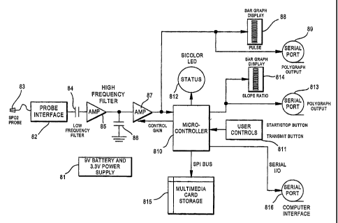
A block diagrain of a preferred embodiment of the present invention apparatus
is shown
in FIG. 8. ' The device is battery powered 81, witli sufficient capacity for a
12 hour overnight
study; analog circuitry for voltage regulation and power supply conditioning;
the device has an
'interface 82 for an OEM supplied finger pulse oximetry probe 83; a low
frequency front-end
filter 84 for probe input signals; an input signal pre-amplifier 85; a high
frequency filter 86; a
gain-controlled signal amplifier stage 87; a bar graph display for indication
of a pulse signal 88;
a polygraph output 89 for pulse signal data; a means of digital processing via
a microprocessor
or microcontroller 810 to provide slope detection and peak to peak height
determination of each
systolic finger pulse, matheinatical normalization of the slope, digital to
analog (D/A)
conversion of the slope value for polysomnographic display, and digital
control of the finger
probe gain, and having a status indicator LED 812; user controls 811 including
start/stop and
transmit functions; a polygraph output 813 for slope ratio data; a bar graph
display 814 for
visual indication of slope ratio information; means to permit 12 hours
(minimum) of data
storage, such as on-board multi-media card storage 815; and analog and/or
digital outputs 816
for providing output of the pulsatile waveform and a DC level representative
of the normalized
slope, and slope ratio data.
The present invention provides a constant excitation to the pulse oximeter
finger probe
LED to evaluate the overall concept of slope detection without actually using
the OEM's pulse
oximeter circuit board. In an alternative embodiment, a pulse oximeter printed
circuit board
CA 02473582 2004-07-13
WO 2004/026132 PCT/US2003/029862
(PCB) is incorporated as a daughter board (internal to the device).
Normalization is a method to correct for the photoplethysmographic pulse
signal
morphological changes based on finger position (as opposed to actual changes
of autonomic
activity.) Currently used ANS activity monitoring methods such as PTT and PAT
lack the
capability for normalization of incoming data and therefor can.not correct for
finger position
changes. The present invention includes a process for normalization, and thus
provides
immunity to artifact caused by both elevation changes of the finger probe, and
changes in blood
flow due to arterial compression during patient positional changes.
The obtained photoplethysmographic signal can be normalized to miniinize
changes in
peak to peak signal amplitude that are not due to ANS activity. In other
words, if there is a
vertical peak to peak percentage increase of the pulsatile waveform (and
consequent increase
in slope), but otherwise no waveform distortion, the percentage increase is
likely to have been
caused by a changing of position (relative to the heart) of the finger probe.
If this same
increase in peak to peak height occurred, but there was also a slight shift in
the waveform
shape, becoming slightly more of a square wave from its sinusoidal shape, then
the increased
slope is likely to be the effect of ANS activity, such as increased heart
contractility.
The present invention normalizes the slope of each blood pressure pulse by
dividing the
slope by the peak to peak height of that same pulse. For each pulsatile beat
with a constant
period and shape over a relatively short period of time, the normalization
will remove
variations due to height only. Both the height and peak will actually be
measured in terms of
analog-to-digital (A/D) counts in the PTS unit's microcontroller. Research on
finger vascular
tone has shown that nonnalized pulse volume, also derived
photoplethysmograpliically,
appears to be superior to the conventional pulse volume.
The present invention is intended to malce use of an existing single
photoplethysmmographic (optical volume detecting) probe, and therefore
existing pulse
16
CA 02473582 2004-07-13
WO 2004/026132 PCT/US2003/029862
oximetry technology. Since pulse oximeters use an alternating flashing of two
different
wavelength LEDs, the present invention synchronizes with the desired LED in
order to
examine the transmitted intensity due to a single wavelength. Alternatively,
certain models of
oximeter OEM modules provide an analog or digital output that can be utilized
directly by the
present device.
In a preferred embodiment, the apparatus contains an autogain circuit to
prevent the
pulsatile waveform from clipping during changes in finger height, large blood
pressure
changes, and between patients with different thickness and skin color fingers.
There are also
several peak detection algorithms for detecting the beginning of the systolic
rise time and the
beginning of diastole. These algorithms provide the minimal quantizing noise,
something that
can occur when attempting to lock onto the rounded peak of a waveform. With
sufficiently fast
sampling and the correct threshold for detecting a zero slope (peak) the
circuit was designed to
not trigger on noise and yet be sensitive enough to be very close to the peak
and not loose
accuracy due to detection well beyond the peak.
Alternative embodiments of the present invention provide algorithms for slope
detection, peak to peak height, and normalization in the form of firmware
within the device, or
by software after the data is downloaded into the polysomnograph. Alternative
methods of data
storage and transfer are also possible, including multimedia card storage,
computer hard drive
storage, serial input/output interface with other devices, and various forms
of telemetry and
phone transmission. Various embodiments for displaying pulse rate and slope
ratio can include
waveform displays, light bars, and numerical infornnation.
FIG. 9 shows a block diagram of the method of the present invention. Shown are
the
steps of disposing a photoplethysmographic probe proximal to a single body
part; deriving a
continuous pulsatile blood pressure waveform as a function of amplitude and
time; defining a
time interval for calculation of a slope of the pulsatile blood pressure
waveform; performing
17
CA 02473582 2004-07-13
WO 2004/026132 PCT/US2003/029862
continuous calculation of the slope of each blood pressure waveform over a
defined time
interval; processing input data to divide pealc amplitude values by a given
time constant;
eliminating from further calculation slope values of less than one; signal
processing,
conditioning, and artifact rejection; amplifying and filtering normalized
slope values; and
providing output infonnation representative of pulse and slope ratio in the
fonn of a display,
electronic data output, and data storage.
INDUSTRIAL APPLICABILITY
The present invention has applicability to the field of medical devices, and
more
particularly to a physiological monitoring method and device used for
detection of autonomic
nervous system (ANS) activity in the field of sleep research.
In compliance with the statute, the invention has been described in language
more or
less specific as to sleep diagnostic medical devices. It is to be understood,
however, that the
invention is not limited to the specific means or features shown or described,
since the means
and features shown or described comprise preferred ways of putting the
invention into effect.
Additionally, while this invention is described in terms of being used for
sleep
diagnostic studies, it will be readily apparent to those skilled in the art
that the invention can be
adapted to other uses for other forms of medical and non-medical monitoring of
the autonomic
nervous system as well, and therefore the invention sho.uld not be construed
as being limited to
sleep study applications. The invention is, therefore, claimed in any of its
forms or
modifications within the legitimate and valid scope of the appended claiins,
appropriately
interpreted in accordance with the doctrine of equivalents.
18