Note: Descriptions are shown in the official language in which they were submitted.
CA 02473621 2004-07-15
WO 03/071934 PCT/US03/06692
METHODS AND DEVICES FOR QUANTITATIVE ANALYSIS OF X-RAY IMAGES
Technical Field
The present invention is in the field of radiographic imaging and analysis
thereof. In
particular, network enabled analyses and analysis techniques are described.
Also described
are devices comprising calibration phantoms and methods of using these
devices.
Background
X-rays and other radiographic analysis are important diagnostic tools.
Furthermore,
Io it is common practice to transmit x-ray images via local and long-distance
networks.
Current technology, however, does not allow for the accurate determination of
quantitative
information contained in the x-ray such as the density of an anatomic
structure when x-ray
images are transmitted in a network environment.
Calibration references (also known as calibration phantoms) for use in imaging
~s technologies have also described. See, e.g., U.S. Patent No. 5,493,601 and
U.S. Patent No.
5,235,628. U.S. Patent No. 5,335,260 discloses a calibration phantom
representative of
human tissue containing variable concentrations of calcium that serves as
reference for
quantifying calcium, bone mass and bone mineral density in radiography and CT
imaging
systems. However, currently-available calibration phantoms are not always
accurate, due to
zo both the effect of structures or materials that project on or with the
calibration phantom and,
additionally, to the fact that one or more regions of the calibration phantom
do not always
appear on the x-ray image.
Thus, there remains a need for methods for quantitative assessment of
information
contained in x-ray images such as the density, structure and/or geometry, of
an anatomic
Zs structure in a network environment. There also remains a need for devices
and methods that
include dependable and accurate calibration phantoms.
Summary
The present invention meets these and other needs by providing compositions
and
3o methods that allow for the analysis of x-ray images in a network
environment. Also
provided are x-ray assemblies comprising accurate calibration phantoms
including, in
particular, calibration phantoms which act as references in order to determine
bone mineral
CA 02473621 2004-07-15
WO 03/071934 PCT/US03/06692
density from an x-ray image.
In one aspect, the invention includes a method for deriving quantitative
information
from an x-ray image in a network environment comprising, providing a local
computer for
transmitting the x-ray image, providing a remote computer for receiving the x-
ray image
s and providing a computer program to analyze and extract quantitative
information from the
x-ray image. In certain embodiments, the quantitative information is
densitometric
information, for example bone mineral density or density of selected soft-
tissues or organs.
Alternatively, the quantitative information is information on the morphology
of a structure,
for example information on the two-dimensional arrangement of individual
components
~o forming said structure or information on the three-dimensional arrangement
of individual
components forming said structure, for example cortical structure, trabecular
structure, etc..
In any of the methods described herein, the anatomic structure can be bone and
the
information can be, for example, information on trabecular thickness,
trabecular spacing
and/or estimates of the two- or three-dimensional architecture of the
trabecular network.
is Further, in any of the methods described herein, quantitative information
can be derived
with use of an external standard, for example a calibration phantom of known x-
ray density.
(e.g., a calibration phantom is included with the structure to be imaged on
the x-ray image).
In other embodiments, the quantitative information derived from the x-ray
image
includes one or more parameters relating to the acquisition of the x-ray image
(e.g., x-ray
Zo tube voltage, x-ray energy, x-ray tube current, film-focus distance, object-
film distance,
collimation, focal spot size, spatial resolution of the x-ray system, filter
technique, film
focus distance, correction factors) or combinations thereof), for instance to
improve the
accuracy of the quantitative information. The x-ray acquisition parameters can
be
transferred over the network prior to, simultaneously or after transmission of
the x-ray
is image. Furthermore, one or more of the x-ray acquisition parameters can be
entered either
manually or, alternatively, automatically into a computer.
In another aspect, a method for measuring quantitative information in an x-ray
image in a network environment is provided. In certain embodiments, the method
comprises transmitting an x-ray image from a local computer to a remote
computer and
so obtaining quantitative information from the x-ray image using a computer
program. In
certain embodiments, one or more internal standard is provided in (or with)
the x-ray image
or the computer program. The internal standard can be, for example, density of
a tissue of a
human (e.g., subcutaneous fat, bone, muscle), air surrounding an anatomic
structure or
2
CA 02473621 2004-07-15
WO 03/071934 PCT/US03/06692
combinations of tissue and air density. In other embodiments, one or more
external
standards are provided in (or with) the x-ray image or computer program. The
at least one
external standard (e.g., calibration phantom can be temporarily or permanently
physically
connected to the x-ray film with use of an attachment mechanism, for example a
mechanical
s attachment mechanism such as Velcro or adhesive. Additionally, the at least
one external
standard can be integrated into the film and/or film holder, for example by
including a
material of known x-ray density between two of the physical layers of the x-
ray film or by
including a material of know x-ray density within one of the physical layers
of the x-ray
film.
io In other aspects, any of the methods described herein further comprise the
steps of
generating a diagnostic report based on the quantitative information and,
optionally, sending
the diagnostic report (for example to a physician). Such reports can be
generated using
computer programs, for example programs on the remote computer. The diagnostic
report
can include, for example, information on a patient's state of health (e.g.,
bone mineral
is density status such as osteoporosis and/or information on fracture risk).
Other disease states
can also be analyzed from x-ray images using the teachings described herein.
In certain
embodiments, these methods further comprise generating a bill (accounting of
charges) for
the recipient of the diagnostic report. The bill can include charges for
generating the report,
profession fees, technical fees or the like. The bill can be printed and
transmitted by mail or
Zo by fax to the recipient or the bill can be electronically transmitted. The
recipient can be a
physician, a subject, a patient, the patient's employer, a health maintenance
organization,
health insurance provider, a government agency, or government representative.
The bill can
also be divided and various portions thereof sent to multiple recipients. In
certain
embodiments, the bill is generated using a computer program on the remote
computer.
Zs In another aspect, the invention includes a method of formulating one or
more x-ray
image data databases, said method comprising collecting x-ray image data
(e.g.,
densiometric information) from one or more subj ects, formulating said one or
more data
databases by associating each of said data points with one or more data
attributes (e.g., age
of subject, weight of subject, height of subject, disease state, etc. The data
can be collected
so using x-ray imaging techniques, for example by using digital or digitized x-
ray images. In
certain embodiments, the data is from a single subject while in other
embodiments, the data
is from two or more subjects. In other embodiments, the methods further
comprise
compiling multiple databases from each database where the data points are
collected from a
CA 02473621 2004-07-15
WO 03/071934 PCT/US03/06692
single individual and the data points for each single individual are
associated with one or
more relevant data attributes.
In any of the methods described herein, the quantitative information can be
densitometric information, for example bone mineral density or density of
selected sort-
s tissues or organs. Alternatively, the quantitative information is
information on the
morphology, for example information on the two-dimensional and/or three-
dimensional
arrangement of individual components making up the anatomic structure (e.g.,
bone). In
any of the methods described herein, the structure can be bone and the
information can be,
for example, information on trabecular thickness, trabecular spacing and/or
estimates of the
io two- or three-dimensional architecture of the trabecular network.
Furthermore, the
information can be encrypted in any of the methods described herein (e.g., to
hide the
subject's name or other demographic information from unauthorized users).
Furthermore, in any of the methods described herein, the x-ray image can be
derived
from x-ray film, for example using a phosphorous plate system. Preferably, the
image is
is digitized, for example, the image may be acquired digitally (e.g., using a
selenium or silicon
detector system) or digitized using a scanning unit.
In another aspect, the invention includes databases made by any of the methods
described herein, for example by formulating data points collected from x-ray
images. In
certain embodiments, the data points are associated with one or more relevant
data
zo attributes.
In another aspect, a method of manipulating an x-ray image data database,
comprising providing any of the databases comprising data points and data
attributes
described herein; and manipulating said data points via said attributes
associated with~said
data points to determine relationships between said data points and said
attributes.
zs In another aspect, the invention includes an x-ray assembly for determining
bone
mineral density comprising an x-ray film holder; x-ray film and a calibration
phantom
comprising at least one marker, for example, a line, or other geometric
pattern (e.g., circles,
stars, squares, crescents, ovals, multiple-sided objects, irregularly shaped
objects or
combinations of any of these shapes) positioned in an area of known density
and wherein
3o the calibration phantom projects free of bone tissue. The calibration
phantom can be
attached to the x-ray filin, the film holder and/or the detection system. The
attachment can
be permanent (e.g., integral to the film such as between two physical layers
of the film or
within one layer of the film, and/or integral to the film holder or detector)
or temporary
4
CA 02473621 2004-07-15
WO 03/071934 PCT/US03/06692
(e.g., via a mechanical or other attachment mechanism such as Velcro, adhesive
or the like).
Thus, in certain embodiments, the calibration phantom is reusable andlor can
be sterilized
between uses. In certain embodiments, the assembly is a dental x-ray assembly.
In any of
the x-ray assemblies described herein, the calibration phantom can be shaped,
for example,
s as a stepwedge or as a plurality of fluid-filled chambers of known
densities.
In certain aspects, a wedge-shaped calibration phantom is used to provide
reference
measurements to express the density of an anatomic structure in terms of
thickness of the
phantom material. For example, described herein are methods of generating a
calibration
curve describing the relationship between measured attenuation and material
thickness
~o wherein the data points making up the curve are derived from the image of
the phantom. In
certain embodiments, the wedge-shaped calibration phantom has a length L and a
linear
change from a maximum thickness T to thickness 0 over the length (e.g., the
calibration
phantom has two ends - one marking the thinnest point and one the thickest,
where
thickness varies linearly between the two ends). In these embodiments, each
point X in the
is image of the wedge at distance D from the thin end of the wedge results in
a point (T*L/D,
G) of the calibration curve, where T*L/D is the thickness of the wedge at X
and G the
attenuation at X.
Alternatively, calibration curves can also be generated when the distance D is
unknown, for example if the calibration wedge varies non-linearly over its
length or both
Zo ends of the phantom cannot be properly identified in the image. In certain
embodiments,
the shape of the expected entire calibration curve is determined, and the part
of the curve
that can be calculated from the identified regions of the calibration wedge is
fitted to the
expected curve. For a~known attenuation of a particular anatomic structure,
the
corresponding thickness value is determined with use of said calibration
curve.
zs Furthermore, calibration curves generated from both linear and non-linear
phantoms
can be further manipulated, for example to translate thickness data into
concentration (e.g.,
calcium concentration). In certain embodiments, the image of an aluminum step
wedge and
a calibration curve expressing thickness is translated into units of calcium
concentration by
including samples of varying calcium concentration in the image. Using this
second
3o calibration curve, values expressed in aluminum thickness can be converted
into units of
calcium concentration.
In still further embodiments, a reference calibration curve can be generated,
for
example by averaging a plurality of calibration curves obtained by any of the
methods
S
CA 02473621 2004-07-15
WO 03/071934 PCT/US03/06692
described herein (e.g., thickness and/or concentration curves). Reference
calibration curves
find use in analysis of images that do not have their own phantoms or other
standards.
In yet another aspect, the invention includes methods of obtaining accurate
bone
mineral density information using any of the calibration phantoms and/or
methods described
s herein. Thus, in certain embodiments, the methods comprise positioning any
of the
calibration phantoms described herein such that x-rays pass through a subject
and the
calibration phantom simultaneously, wherein the calibration phantom projects
free of
materials that alter its' apparent density; creating an image of the phantom
and the portion
of the subject's anatomy; and comparing the image of the phantom and the
subject's
io anatomy to determine bone mineral density of the subject. The x-ray image
can be, for
example, a dental x-ray. Any of the methods described herein can be performed
on a
computer, in a network environment, or manually.
Another aspect of the invention is a kit for aiding in assessing the condition
of bone
in a subj ect, which kit comprises a software program, which that when
installed and
~s executed on a computer reads an x-ray image (e.g. a digital or digitized
dental x-ray) and
produces a computer readout showing bone mineral density. Any of the kits
described
herein can also include a calibration phantom, x-ray film, x-ray film holders
and computer
programs (e.g., software) for displaying and/or generating a bill for the
readout regarding
bone mineral density.
Zo In other aspects, the invention includes a calibration phantom, for example
a
calibration phantom comprising a plurality of geometric shapes, wherein the
calibration
phantom is less than 2.5 cm in length and less than 5 mm in width. The
geometric shapes
may be for example, comprise rectangles that forth a step-wedge. The phantom
itself may
be stainless steel for example 0.08% carbon, 2% manganese, 1% silicon, 0.045%
is phosphorus, 0.03% sulphur; 10-14% nickel, 16-18% chromium, 2-3% molybdenum
and
iron up to 100%.
In yet another aspect, methods of diagnosing osteoporosis in a subject are
provided,
for example using any of the kits, methods and/or devices described herein. In
certain
embodiments, the methods of diagnosing osteoporosis in a subject comprise
using a
so computer program to analyze bone mineral density of an x-ray image and
comparing the
bone mineral density value obtained from the image with a reference standard
or curve,
thereby determining if the subject has osteoporosis. In certain embodiments,
the x-ray
image includes a calibration phantom, for example a calibration phantom as
described
6
CA 02473621 2004-07-15
WO 03/071934 PCT/US03/06692
herein. In other embodiments, a reference calibration curve can be used to
analyze the
image.
In still further aspects, methods of assessing bone mineral density are used
to
provide suitable treatment for a subject in need thereof. For instance, using
any of the
methods, kits, and/or devices described herein, the presence of osteoporosis
in a subject can
be diagnosed and that subject provided with appropriate therapy (e.g., one or
more anti-
resorptive agents and/or one or more anabolic agents). Additionally, over
time, the methods
described herein can be used to assess the efficacy of the selected treatment.
Thus, in
certain embodiments, diagnosis and/or treatment of osteoporosis are achieved
in a network-
io enabled environment.
These and other embodiments of the subject invention will readily occur to
those of
skill in the art in light of the disclosure herein.
Brief Description of the Figures
is Figure 1 depicts an example of network enabled quantitative x-ray analysis
useful in
monitoring osteoporosis.
Figure 2 depicts an exemplary dental x-ray film holder. The film holder
includes a
calibration phantom.
Figure 3 depicts another exemplary dental x-ray film holder. The film holder
zo includes a calibration phantom.
Figure 4 depicts an exemplary embodiment in which a calibration phantom is
attached to a dental x-ray film holder.
Figure 5 depicts an exemplary embodiment in which a calibration phantom is
attached to x-ray film.
zs Figure 6 depicts an exemplary embodiment in which a calibration phantom is
integrated into a film cover.
Figure 7 depicts an exemplary embodiment in which a calibration phantom is
integrated into a detector system.
Figure 8 depicts exemplary measurement sites in a dental x-ray that can be
used as
3o an intrinsic standard to calibrate the image.
Figure 9 depicts exemplary measurement sites in a dental x-ray that can be
used as
an intrinsic standard to calibrate the image.
Figure 10, panels A and B, depict an exemplary calibration phantom. Panel (A)
7
CA 02473621 2004-07-15
WO 03/071934 PCT/US03/06692
shows a top view and the dimensions of the device. Panel (B) shows the side
view of the
step-wedge and exemplary dimensions.
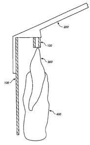
Figure 11 depicts a side-view of an exemplary x-ray system for dental x-rays
in
which a calibration phantom (104) is integrated into the film holder. Also
shown in Figure
11 are bite block (100), film (103), ring-shaped Rinn holder (102) and
stainless steel rod
(101). The ring-shaped Rinn holder can help align the x-ray tube so that it is
perpendicular
or near perpendicular to the film.
Detailed Description
io Before describing the present invention in detail, it is to be understood
that this
invention is not limited to particular formulations or process parameters as
such may, of
course, vary. It is also to be understood that the terminology used herein is
for the purpose
of describing particular embodiments of the invention only, and is not
intended to be
limiting.
is The practice of the present invention employs, unless otherwise indicated,
conventional methods of database storage and manipulation, within the skill of
the art.
Such techniques are explained fully in the literature. See, e.g., Numerical
Mathematical
Analysis, Third Edition, by J.B. Scarborough, 1955, John Hopkins Press,
publisher; System
Analysis and Design Methods, by Jeffrey L. Whitten, et al., Fourth Edition,
1997, Richard
zo D. Irwin, publisher; Modern Database Management, by Fred R. McFadden, et
al., Fifth
Edition, 1999, Addison-Wesley Pub. Co., publisher; Modern System Analysis and
Design,
by Jeffery A. Hoffer, et al., Second Edition, 1998, Addison-Wesley Pub. Co.,
publisher;
Data Processing: Fundamentals, Design, and Implementation, by David M.
Kroenke,
Seventh Edition, 2000, Prentice Hall, publisher; Case Method: Entity
Relationship
zs Modeling (Computer Aided Systems Engineering), by Richard Barker, 1990,
Addison-
Wesley Pub Co., publisher.
All publications, patents and patent applications cited herein, whether above
or
below, are hereby incorporated by reference in their entirety.
It must be noted that, as used in this specification and the appended claims,
the
so singular forms "a", "an", and "the" include plural referents unless the
content clearly
dictates otherwise. Thus, for example, reference to "a calibration phantom"
includes a one
or more such phantoms.
8
CA 02473621 2004-07-15
WO 03/071934 PCT/US03/06692
Definitions
Unless defined otherwise, all technical and scientific terms used herein have
the
same meaning as commonly understood by one of ordinary skill in the art to
which the
invention pertains. Although any methods and materials similar or equivalent
to those
s described herein can be used in the practice for testing of the present
invention, the
preferred materials and methods are described herein.
The term "subject" encompasses any warm-blooded animal, particularly including
a
member of the class Mammalia such as, without limitation, humans and nonhuman
primates
such as chimpanzees and other apes and monkey species; farm animals such as
cattle,
io sheep, pigs, goats and horses; domestic mammals such as dogs and cats;
laboratory animals
including rodents such as mice, rats and guinea pigs, and the like. The term
does not denote
a particular age or sex and, thus, includes adult and newborn subjects,
whether male or
female.
"Parameter" refers to an arbitrary constant or variable so appearing in a
~s mathematical expression that changing it gives various cases of the
phenomenon
represented (McGraw-Hill Dictionary of Scientific and Technical Terms, S.P.
Parker, ed.,
Fifth Edition, McGraw-Hill Inc., 1994). A parameter is any of a set of
properties whose
values determine the characteristics or behavior of something.
A "data point", generally, is a numeric value which corresponds to a physical
zo measurement (an "acquired" datum or data point) or to a single numeric
result calculated or
derived from one or more acquired data points (a "calculated" or "derived"
datum or data
point). Derived data include, but are not limited to, derived quantities from
original data,
such as, rate and/or magnitude of change, slope of a line (e.g., as determined
by regression
analysis), an intercept (e.g., as determined by regression analysis), and
correlation
zs coefficients.
"Data tags," also referred to as "attributes" of a data point, are various
characteristics
of the particular data point with which they are associated. For example, data
points
comprising x-ray information (and/or bone mineral density) are associated with
a number of
attributes, e.g., the date and time the image was taken; certain
identification related to the
3o particular subject from which the measurement was made (e.g., demographic
information
such as the particular user's sex, age, weight or race; medical information
e.g., the
medications used by the subject and/or type of disease suffered by the
subject).
9
CA 02473621 2004-07-15
WO 03/071934 PCT/US03/06692
A "database" is a collection of data points and data attributes associated
with each
data point. Thus, a "data points, derived data, and data attributes database"
is a database
comprising data points collected, e.g. from an x-ray image, data derived from
the original
data points and the data attributes associated with those data points or the
derived data. A
s database may be limited to data points comprising measurements of one or
more levels;
those data points may further be collected from one or more subjects. For
example, one
data point database may be created and the information in the database related
to a second
database of attributes. Such combinations of one or more databases are within
the skill of
one of ordinary skill in the art in view of the teachings of the present
specification. A "data
~o warehouse" is another term for database. The term data warehouse is
typically applied to
large databases.
"Formulation" of a database comprises collecting data points, inputting those
data
points into a desired database format, and associating various attributes with
each data point
according to the particular format employed. A wide variety of software exists
which
is provides a means for inputting data points, and associating the data points
with data
attributes, such as Excel~ (Microsoft~ Corporation, Seattle, Washington)
spreadsheet
software, Quattro~ (Corel Inc., Ottawa, Canada) spreadsheet software,
Microsoft Access
2000~ (Microsoft) software, Oracle~ (Oracle Inc., Redwood Shores, CA)
software, as well
as other database and data warehousing software.
zo "Manipulation" of a database refers to a variety of processes, e.g.,
selecting, sorting,
sifting, aggregating, clustering, modeling, exploring, and segmenting data
points using
various data attributes or tags associated with the data points. Available
systems for
generating databases and manipulating the resulting databases include but are
not limited to
Sybase~ (Sybase Systems, Emeryville, CA), Oracle~ (Oracle Inc., Redwood
Shores, CA),
zs and Sagent Design Studio~ (Sagent Technologies Inc., Mountain View,
California) systems
software. Further, statistical packages and systems for data analysis and data
mining are
also available. Illustrative examples include SAS~ (SAS Institute Inc., Cary,
NC) and
SPSS~ (SPSS Inc., Chicago, IL) systems software.
"Data mining" refers to the process of selecting, exploiting, modeling, etc.,
large
3o amounts of data to uncover previously unknown trends, patterns, and
relationships within
and among various data points and data attributes.
CA 02473621 2004-07-15
WO 03/071934 PCT/US03/06692
"Data aggregation" and "data clustering" refers to the process of grouping
data
points on the basis of one or more common attributes. Conversely, "data
segmentation"
refers to the process of differentiating data into discrete groups on the
basis of one or more
attributes.
"Bone structure" refers to two-dimensional or three-dimensional arrangement
(e.g.,
architecture or microarchitecture) of bone tissue. (See, also, International
Publication WO
02/30283). Generally, bone tissue includes two types of bone -- an outer layer
of cortical
bone that is generally mostly solid with some canals or pores therein and an
inner layer of
trabecular (or cancellous) bone that generally is sponge-like or honeycomb-
like in
io structure. Structural features or cortical and trabecular bone include, but
are not limited to,
trabecular thickness; trabecular spacing; two-dimensional or three-dimensional
spaces
between trabeculae; two-dimensional or three-dimensional architecture of the
trabecular
network, solid material (typically greater than 3000 pm), primary and/or
secondary
trabeculae (typically 75 to 200 pm), primary and secondary osteons (typically
100 to 300
is pm, plexiform, interstitial bone, trabecular packets, lamellae (typically 1
to 20 ~.m),
lacunae, cement Lines, canaliculi, collagen-mineral composite (typically 0.06
to 0.4 Vim),
cortical pores, trabecular connectivity, nodes and branch points, and the
like. One or more
of these and other structural features may be measured in the practice of the
present
invention. Preferably, measurements are the sub-millimeter range, more
typically in the 10
zo - 500 ~m range. Non-limiting examples of microarchitecture parameters
include trabecular
structure thresholded binary image parameters such as trabecular area; total
area; trabecular
area /total area; trabecular perimeter area; trabecular distance transform;
marrow distance
transform; trabecular distance transform regional maxima values (mean, min.,
max, std.
Dev); marrow distance transform regional maxima values (mean, min., max, std.
Dev); star
zs volume (see, e.g., Ikuta et. al. (2000) JBMR 18:217-277; Vesterby (1990)
Bone 11:149-155;
and Vesterby et al. (1989) Bone 10:7-13); trabecular Bone Pattern Factor (Hahn
et. al.,
(1992) Bone 13:327-330); TBPf = (Pl - P2) / (A1 - A2 ) where P1 and A1 are the
perimeter
length and trabecular bone area before dilation and P2 and A2 corresponding
values after a
single pixel dilation as well as trabecular skeleton parameters such as
connected skeleton
3o count or Trees (T); node count (I~; segment count (S); node-to-node segment
count (NIA;
node-to-free-end segment count (NF); node-to-node segment length (NNL); node-
to-free-
end segment length (NFL); free-end-to-free-end segment length (FFL); node-to-
node total
11
CA 02473621 2004-07-15
WO 03/071934 PCT/US03/06692
struts length (NN.TSL) (see, e.g., Legrand et. al., (2000) JMBR 15:13-19; free-
end-to-free-
ends total struts length( FF.TSL); total struts length (TSL); FF.TSL/ TSL;
NN.TSL/ TSL;
Loop count (Lo); Loop area; mean distance transform values for each connected
skeleton;
mean distance transform values for each segment (Tb.Th ); mean distance
transform values
s for each node-to-node segment (Tb.Th.NN); mean distance transform values for
each node-
to-free-end segment (Tb.Th.NF); orientation (angle) of each segment; angle
between
segments; length-thickness ratios (NNL/Tb.Th.NN ) and (NFL/ Tb.Th.NF); and
interconnectivity index (ICI) where ICI = (N * NN)/ ( T * (NF + 1 ).
"Macro-anatomical parameter" refers to any parameter describing the shape,
size or
~o thickness ofbone and/or surrounding structure, typically parameters that
are greater than
O.Smm in size in at least one dimension. Macro-anatomical parameters include,
for
example, in the hip joint thickness of the femoral shaft cortex, thickness of
the femoral neck
cortex, hip axis length, CCD (caput-collum-diaphysis) angle and width of the
trochanteric
region.
~s "Bone quality" refers broadly to overall characteristics of any given bone.
Thus, the
term refers to characteristics including, but not limited to, the degree of
bone density
present, the thickness of the bone, the fragility of the bone (e.g.,
susceptibility to fracture),
bone strength and/or one or macro- or micro-anatomical structural
characteristics of the
bone.
zo
General Overview
Methods and compositions useful in analyzing x-ray images are described. In
particular, the invention includes methods of obtaining and/or deriving
information from an
x-ray image in network environment. Additionally, the present invention
relates to the
zs provision of accurate calibration phantoms for X-ray systems and methods of
using these
calibration phantoms. Typically, the calibration phantom is formed of a
material that
simulates the properties of human bone tissue and is provided in an x-ray
assembly such
that improved accuracy and precision in the quantification of calcium, bone
mass and bone
density using conventional X-ray equipment is achieved.
so Advantages of the present invention include, but are not limited to, (i)
providing
fast, centralized networks for the analysis of x-ray images, particularly
analysis of x-rays for
bone mineral density; (ii) providing accessible and reliable means for
analyzing x-rays; (iii)
providing accurate calibration phantoms; (iv) providing accurate calibration
phantoms that
12
CA 02473621 2004-07-15
WO 03/071934 PCT/US03/06692
can be readily used with standard x-ray technology; and (v) providing methods
and
materials for making these network-enabled techniques and devices.
Database Formulation
s The method of formulating data points, derived data, and data attributes
database
according to the present invention may comprise the following: (1) the
collection of data
points, said data points comprising information obtained from an x-ray image,
for example,
bone mineral density information; and (2) the association of those data points
with relevant
data point attributes. The method may further comprise (3) determining derived
data points
io from one or more direct data points and (4) associating those data points
with relevant data
point attributes. The method may also comprise (5) collection of data points
using a remote
computer whereby said remote computer operates in a network environment.
In preferred embodiments, the information is obtained from an x-ray image, for
example of an anatomical structure or of a non-living structure. X-ray images
can be
~ s acquired at a local site using known techniques. If the x-ray image was
captured using
conventional x-ray film, the data points (information) of the x-ray image may
be digitized,
for example using a scanning device. The digitized x-ray image information can
then be
transmitted over the network, e.g. the Internet, into a remote computer or
server. If the x-ray
image was acquired using digital acquisition techniques, e.g. using phosphorus
plate
2o systems or selenium or silicon detector systems, the x-ray image
information is already
available in digital format. In this case the image can be transmitted
directly over the
network, e.g. the Internet. The information can also be compressed and/or
encrypted prior
to transmission. Transmission can also be by other methods such as fax, mail
or the like.
Zs Data Points
Thus, the methods of formulating data points, derived data, and data
attributes
database that forms an aspect of the present invention begins with the
collection of data sets
of measurement values, for example measurements of bone mineral density from x-
ray
images. Records may be formulated in spreadsheet-like format, for example
including data
3o attributes such as date of x-ray, patient age, sex, weight, current
medications, geographic
location, etc. The database formulation method of the present invention may
further
comprise the calculation of derived or calculated data points from one or more
acquired data
points. A variety of derived data points may be useful in providing
information about
13
CA 02473621 2004-07-15
WO 03/071934 PCT/US03/06692
individuals or groups during subsequent database manipulation, and are
therefore typically
included during database formulation. Derived data points include, but are not
limited to
the following: (1) maximum bone mineral density or maximum of one or more bone
structure measurements, determined for a selected region of bone or in
multiple samples
s from the same or different subjects; (2) minimum bone mineral density or
minimum of one
or more bone structure measurements, determined for a selected region of bone
or in
multiple samples from the same or different subjects; (3) mean bone mineral
density or
mean of selected bone structure measurements, determined for a selected region
of bone or
in multiple samples from the same or different subjects; (4) the number of
measurements
io that are abnormally high or low, determined by comparing a given
measurement data point
with a selected value; and the like. Other derived data points will be
apparent to persons of
ordinary skill in the art in light of the teachings of the present
specification. The amount of
available data and data derived from (or arrived at through analysis of) the
original data
provide provides an unprecedented amount of information that is very relevant
to
~s management ofbone related diseases such as osteoporosis. For example,
examining
subjects over time can assess the efficacy of medications.
Measurements and derived data points are collected and calculated,
respectively, and
may be associated with one or more data attributes to form a database.
Data attributes can be automatically input with the x-ray image and can
include, for
zo example, chronological information (e.g., DATE and TIME). Other such
attributes may
include, but are not limited to, the type of x-ray imager used, scanning
information,
digitizing information and the like. Alternatively, data attributes can be
input by the subject
and/or operator, for example subject identifiers, i.e. characteristics
associated with a
particular subject. These identifiers include but are not limited to the
following: (1) a
is subject code (e.g., a numeric or alpha-numeric sequence); (2) demographic
information
such as race, gender and age; (3) physical characteristics such as weight,
height and body
mass index (BMI); (4) selected aspects of the subject's medical history (e.g.,
disease states
or conditions, etc.); and (5) disease-associated characteristics such as the
type of bone
disorder, if any; the type of medication used by the subject. In the practice
of the present
3o invention, each data point would typically be identified with the
particular subject, as well
as the demographic, etc. characteristic of that subject.
Other data attributes will be apparent to persons of ordinary skill in the art
in light of
the teachings of the present specification.
14
CA 02473621 2004-07-15
WO 03/071934 PCT/US03/06692
Storage of Data Sets and Association of Data Points with Relevant Data
Attributes
A number of formats exist for storing data sets and simultaneously associating
related attributes, including but not limited to (1) tabular, (2) relational,
and (3) dimensional.
s In general the databases comprise data points, a numeric value which
correspond to physical
measurement (an "acquired" datum or data point) or to a single numeric result
calculated or
derived from one or more acquired data points that are obtained using the
various methods
disclosed herein. The databases can include raw data or can also include
additional related
information, for example data tags also referred to as "attributes" of a data
point. The
~o databases can take a number of different forms or be structured in a
variety of ways.
The most familiar format is tabular, commonly referred to as a spreadsheet. A
variety of spreadsheet programs are currently in existence, and are typically
employed in the
practice of the present invention, including but not limited to Microsoft
Excel spreadsheet
software and Corel Quattro spreadsheet software. In this format, association
of data points
is with related attributes occurs by entering a data point and attributes
related to that data point
in a unique row at the time the measurement occurs.
Further, rational, relational (Database Design for Mere Mortals, by Michael J.
Hernandez, 1997, Addison-Wesley Pub. Co., publisher; Database Design for
Smarties, by
Robert J. Muller, 1999, Morgan Kaufinann Publishers, publisher; Relational
Database
Zo Design Clearly Explained, by Jan L. Harrington, 1998, Morgan Kaufinann
Publishers,
publisher) and dimensional (Data-Parallel Computing, by V.B. Muchnick, et al.,
1996,
International Thomson Publishing, publisher; Understanding Fourth Dimensions,
by David
Graves, 1993, Computerized Pricing Systems, publisher) database systems and
management
may be employed as well.
Zs Relational databases typically support a set of operations defined by
relational
algebra. Such databases typically include tables composed of columns and rows
for the data
included in the database. Each table of the database has a primary key, which
can be any
column or set of columns, the values for which uniquely identify the rows in a
table. The
tables in the database can also include a foreign key that is a column or set
of columns, the
3o values of which match the primary key values of another table. Typically,
relational
databases also support a set of operations (e.g., select, join and combine)
that form the basis
of the relational algebra governing relations within the database.
CA 02473621 2004-07-15
WO 03/071934 PCT/US03/06692
Such relational databases can be implemented in various ways. For instance, in
Sybase~ (Sybase Systems, Emeryville, CA) databases, the tables can be
physically
segregated into different databases. With Oracle~ (Oracle Inc., Redwood
Shores, CA)
databases, in contrast, the various tables are not physically separated,
because there is one
s instance of workspace with different ownership specified for different
tables. In some
configurations, databases are all located in a single database (e.g., a data
warehouse) on a
single computer. In other instances, various databases are split between
different
computers.
It should be understood, of course, that the databases are not limited to the
foregoing
io arrangements or structures. A variety of other arrangements will be
apparent to those of
skill in the art.
Database Manipulation
Databases formulated using the methods of the present invention are useful in
that
is they can be manipulated, for example, using a variety of statistical
analyses, to produce
useful information. The databases of the present invention may be generated,
for example,
from data collected for an individual or from a selected group of individuals
over a defined
period of time (e.g., days, months or years), from derived data, and from data
attributes.
The present invention further relates to a method for manipulating data
points,
zo derived data, and data attributes database in order to provide a useful
result, said method
comprising providing data points, derived data, and data attributes database,
and
manipulating and/or analyzing the database.
For example, data sets may be aggregated, sorted, selected, sifted, clustered
and
segregated by means of the attributes associated with the data points. A
number of database
zs management systems and data mining software programs exist which may be
used to
perform the desired manipulations.
Relationships in the database can be directly queried and/or the data analyzed
by
statistical methods to evaluate the information obtained from manipulating the
database.
For example, a distribution curve can be established for a selected data set,
and the
3o mean, median and mode calculated therefrom. Further, data spread
characteristics, e.g.
variability, quartiles and standard deviations can be calculated.
16
CA 02473621 2004-07-15
WO 03/071934 PCT/US03/06692
The nature of the relationship between a particular variable and bone mineral
density
levels can be examined by calculating correlation coefficients. Useful methods
for doing so
include but are not limited to the following: Pearson Product Moment
Correlation and
Spearman Rank Order Correlation.
s Analysis of variance permits testing of differences among sample groups to
determine whether a selected variable has a discernible effect on the
parameter being
measured.
Non-parametric tests may be used as a means of testing whether variations
between
empirical data and experimental expectancies are attributable merely to chance
or to the
to variable or variables being examined. These include the Chi Square test,
the Chi Square
Goodness of Fit, the 2 x 2 Contingency Table, the Sign Test, and the Phi
Correlation
Coefficient.
There are numerous tools and analyses available in standard data mining
software
that can be applied to the analysis of the databases of the present invention.
Such tools and
~s analyses include, but are not limited to, cluster analysis, factor
analysis, decision trees,
neural networks, rule induction, data driven modeling, and data visualization.
Some of the
more complex methods of data mining techniques are used to discover
relationships that are
more empirical and data-driven, as opposed to theory-driven, relationships.
Exemplary data mining software that can be used in analysis and/or generation
of
Zo the databases of the present invention includes, but is not limited to:
Link Analysis (e.g.,
Associations analysis, Sequential Patterns, Sequential time patterns and Bayes
Networks);
Classification (e.g., Neural Networks Classification, Bayesian Classification,
k-nearest
neighbors classification, linear discriminant analysis, Memory based
Reasoning, and
Classification by Associations); Clustering (e.g., k-Means Clustering,
demographic
2s clustering, relational analysis, and Neural Networks Clustering);
Statistical methods (e.g.,
Means, Std dev, Frequencies, Linear Regression, non-linear regression, t-
tests, F-test, Chi2
tests, Principal Component Analysis, and Factor Analysis); Prediction (e.g.,
Neural
Networks Prediction Models, Radial Based Functions predictions, Fuzzy logic
predictions,
Times Series Analysis, and Memory based Reasoning); Operating Systems; and
Others
30 (e.g., Parallel Scalability, Simple Query Language functions, and C++
objects generated for
applications). Companies that provide such software include, for example, the
following:
Adaptative Methods Group at UTS (UTS City Campus, Sydney, NSW 2000), CSI~,
Inc.,
(Computer Science Innovations, Inc. Melbourne, Florida), IBM~ (International
Business
17
CA 02473621 2004-07-15
WO 03/071934 PCT/US03/06692
Machines Corporation, Armonk, NY), Oracle~ (Oracle Inc., Redwood Shores, CA)
and
SAS~ (SAS Institute Inc., Cary, NC).
These methods and processes may be applied to the databases of the present
invention, for example, databases comprising, x-ray image data sets, derived
data, and data
attributes.
For a general discussion of statistical methods applied to data analysis, see
Applied
Statistics for Science and Industry, by A. Romano, 1977, Allyn and Bacon,
publisher.
Hardware/Software and System Considerations
Io A. Hardware/Software
Various computer systems, typically comprising one or more microprocessors,
can
be used to store, retrieve, and analyze information obtained according to the
methods
described herein. The computer system can be as simple as a stand-alone
computer that is
not networked to other computers, provided the system has a form of data
storage, for
is example disk drives, removable disk storage, for example ZIP~ drives
(Iomega
Corporation, Roy, Utah), optical medium (e.g., CD-ROM), magnetic tape, solid-
state
memory, and/or bubble memory. Alternatively, the computer system can include a
networked computer system in which a computer is linked to one or more
additional
computers, for example a network server. The networked system can be an
intranet system
zo and/or a system linked to other computers via the Internet. Thus, the
computer systems can
be Internet-based systems or non-Internet based systems.
In addition, devices such as the Personal Digital Assistants (PDA), for
example
Palm PilotTM (Palm Inc., Santa Clara, CA) or HandspringTM VisorTM (Handspring,
Inc.,
Mountain View, CA) and Pocket PCs (PPC), for example Casio~ EM500 Multimedia
zs Cassiopeia Pocket PC (Casio Inc., Dover, NJ) or Compaq~ iPAQTM (Compaq
Computer
Corporation, Houston, Texas) can be used to store and retrieve patient
database information.
The PDA or PPC can be a simple stand-alone device that is not networked to
other
computers, provided the device has a form of data storage, for example solid-
state memory,
SD (secure digital) and MMC (multimedia card) cards. Alternatively, the PDA or
PPC can
3o be attached to a network in which the unit is linked to one or more
computers, for example a
network server or PC. The networked PDA or PPC can be an intranet system
and/or a
18
CA 02473621 2004-07-15
WO 03/071934 PCT/US03/06692
system linked to computers via the Internet. Thus, the PDA or PPC systems can
be Internet
attached systems or non-Internet attached systems.
For example, information regarding x-ray images and the parameters that were
used
to acquire the x-ray image (e.g., acquisition parameters) can be transmitted
with the image
over a local or long-distance network. The image acquisition parameters can be
transmitted
simultaneous with the image or before or after the image transmission over the
network.
Image acquisition parameters that can be transmitted in this fashion include
but are not
limited to x-ray tube voltage settings, energy settings, x-ray tube current,
film-focus
distance, object-film distance, collimation, focal spot, spatial resolution,
filter settings, etc.
io These parameters can be entered manually into a data registration sheet or
database that can
be transmitted before, after or simultaneous with the x-ray images.
Alternatively, at least
some of these parameters can be transmitted automatically, while others that
may be kept
constant between different patients can be stored either at the local site or
on the network.
Thus, transmission of the x-ray acquisition parameters before, after or
simultaneous
~s with the x-ray image over the network can be used to improve the accuracy
of quantitative
measurements from x-ray images. For example, information on the density of an
anatomic
structure or a non-living object included on the x-ray image can be derived
more accurately,
when the x-ray image acquisition parameters are known.
The software can be installed in a PC, a Silicon Graphics, Inc. (SGI) computer
or a
zo Macintosh computer.
B. Stand-alone System
Connection to a central network (e.g., the Internet) can be made either
directly, or
via serial interface adapter. For example, a direct connection could be made
if the readout
zs device had wireless capability; alternately, a connection through a SIA or
other sort of
docking station between the device and the network.
In some instances, a computer system includes a computer having an Intel~
Pentium~ microprocessor (Intel Corporation, Santa Clara, CA) that runs the
Microsoft~
WINDOWS~ Version 3.1, WINDOWS95~, WINDOWS98~, or WINDOWS2000~
30 operating system (Microsoft Corporation, Redmond, WA). Of course other
microprocessors such as the ATHLONTM microprocessor (Advanced Micro Devices,
Inc.,
Sunnyvale, CA) and the Intel~ CELERON~ and XEON~ microprocessors can be
utilized.
The methods and systems can also include other operating systems, for example,
UNIX,
19
CA 02473621 2004-07-15
WO 03/071934 PCT/US03/06692
LINUX, Apple MAC OS 9 and OS X (Apple, Cupertino, CA), PaImOS~ (Palm Inc.,
Santa
Clara, CA), Windows~ CE 2.0 or Windows~ CE Professional (Microsoft
Corporation,
Redmond, WA) without departing from the scope of the present invention. Also
typically
included is the storage media, for example disk drive, removable disk storage,
CD-ROM,
s required to store and retrieve patient database information.
Communication with a computer system can be achieved using a standard computer
interface, for example a serial interface or Universal Serial Bus (USB) port.
Standard
wireless interfaces, for example radio frequency (RF) technology - IEEE 802.11
and
Bluetooth, and/or infrared technologies can also be used. The data can be
encoded in the
io standard manner, for example American Standard Code for Information
Interchange
(ASCII) format - a standard seven-bit code that was proposed by ANSI in 1963,
and
finalized in 1968. ASCII is the common code for microcomputer equipment.
The computer system can store the information, for example into a database,
using a
wide variety of existing software that provides a means for inputting data
points, and
~s associating the data points with data attributes. Available systems for
generating databases
and manipulating the resulting databases include but are not limited to Excel~
(Microsoft~
Corporation, Seattle, Washington) spreadsheet software, Quattro~ (Corel Inc.,
Ottawa,
Canada), Sybase~ (Sybase Systems, Emeryville, CA), Oracle~ (Oracle Inc.,
Redwood
Shores, CA), and Sagent Design Studio~ (Sagent Technologies Inc., Mountain
View,
zo California) systems software. Further, statistical packages and systems for
data analysis and
data mining are also available (see above). Illustrative examples include but
are not limited
to SAS~ (SAS Institute Inc., Cary, NC) and SPSS~ (SPSS Inc., Chicago, IL). The
database
can be recorded on, for example a disk drive - internal or external to the
system, a
Read/Write CD-ROM drive, a tape storage system, solid-state memory or bubble
memory,
zs an SD or MMC. In addition to saving the data in a database, the information
can be
forwarded to an auxiliary readout device such as a display monitor.
C. Networked System
Networked computer systems are also suitable for performing the methods of the
3o present invention. A number of network systems can be used, for example a
local area
network (LAN) or a wide area network (WAN). The network computer system
includes the
necessary functionality for forwarding the data in established formats, for
example Ethernet
CA 02473621 2004-07-15
WO 03/071934 PCT/US03/06692
or Token Ring Packets or Frames, HTML-formatted data, or WAN digital or analog
protocols, in combination with any parameter information, for example
Destination
Address, or Cyclic Redundancy Check (CRC). CRC is a powerful and easily
implemented
technique to obtain data reliability. The CRC technique is used to protect
blocks of data
s called Frames. Using this technique, the transmitter appends an extra n- bit
sequence to
every frame called Frame Check Sequence (FCS). The FCS holds redundant
information
about the frame that helps the transmitter detect errors in the frame. The CRC
is one of the
most used techniques for error detection in data communications into a format
suitable for
transmission across a transmission line for delivery to a database server.
Further, the
io network system may comprises the necessary software and hardware to receive
the data
from the readout device, store the data, process the data, display the data in
a variety of
ways, and communicate back to the readout device as well as to allow
communication
among a variety of users and between these users to the readout device.
The networked computer system, for example an Ethernet, Token Ring or FDDI
is network, can be accessed using a standard network interface card (NIC), for
example a
3Com~ EtherLink~ NIC (3Com, Inc, Santa Clara, CA) which provide network
connections
over UTP, coaxial, or fiber-optic cabling or an Intel~ PRO/100 S Desktop
Adapter (Intel
Corporation, Santa Clara, CA) or using a standard remote access technology,
for example a
modem using a plain old telephone system (POTS) line, or a xDSL router
connected to a
Zo digital subscriber lines (DSL), or a cable modem. Additionally, the
networked computer
system can be connected to the LAN using a standard wireless interface, for
example radio
frequency (RF) technology - IEEE 802.11 and Bluetooth.
The networked computer system would have the same capability of storing data,
as
the stand-alone system, onto a storage media, for example a disk drive, tape
storage, or CD-
Zs ROM. Alternatively, the networked computer system would be able to transfer
data to any
device connected to the networked computer system for example a medical doctor
or
medical care facility using standard e-mail software, a central database using
database query
and update software (e.g., a data warehouse of data points, derived data, and
data attributes
obtained from a large number of subjects). Alternatively, a user could access
from a
3o doctor's office or medical facility, using any computer system with
Internet access, to
review historical data that may be useful for determining treatment.
If the networked computer system includes a World Wide Web application, the
application includes the executable code required to generate database
language statements,
21
CA 02473621 2004-07-15
WO 03/071934 PCT/US03/06692
for example, SQL statements. Such executables typically include embedded SQL
statements. The application further includes a configuration file that
contains pointers and
addresses to the various software entities that are located on the database
server in addition
to the different external and internal databases that are accessed in response
to a user
s request. The configuration file also directs requests for database server
resources to the
appropriate hardware, as may be necessary if the database server is
distributed over two or
more different computers.
Usually each networked computer system includes a World Wide Web browser that
provides a user interface to the networked database server. The networked
computer system
io is able to construct search requests for retrieving information from a
database via a Web
browser. With access to a Web browser users can typically point and click to
user interface
elements such as buttons, pull down menus, and other graphical user interface
elements to
prepare and submit a query that extracts the relevant information from the
database.
Requests formulated in this manner are subsequently transmitted to the Web
application that
~s formats the requests to produce a query that can be used to extract the
relevant information
from the database.
When Web-based applications are utilized, the Web application accesses data
from a
database by constructing a query in a database language such as Sybase or
Oracle SQL
which is then transferred to a relational database management system that in
turn processes
zo the query to obtain the pertinent information from the database.
Accordingly, in one aspect the present invention describes a method of
providing
data on x-ray images on a network, for example the Internet, and methods of
using this
connection to provide real-time and delayed data analysis. The central network
can also
allow access by the physician to a subject's data. Similarly, an alert could
be sent to the
zs physician if a subject's readings are out of a predetermined range, etc.
The physician can
then send advice back to the patient via e-mail or a message on a web page
interface.
Further, access to the entire database of data from all subjects may be useful
for statistical or
research purposes. Appropriate network security features (e.g., for data
transfer, inquiries,
device updates, etc.) are of course employed.
3o Further, a remote computer can be used to analyze the x-ray that has been
transmitted over the network automatically. For example, x-ray density
information or
structural information about an object can be generated in this fashion. X-ray
density
22
CA 02473621 2004-07-15
WO 03/071934 PCT/US03/06692
information can, for example, be bone mineral density. If used in this
fashion, the test can
be used to diagnose osteoporosis (See, Fig. 1).
D. Graphical User Interface
s In certain of the computer systems, an interface such as an interface screen
that
includes a suite of functions is included to enable users to easily access the
information they
seek from the methods and databases of the invention. Such interfaces usually
include a
main menu page from which a user can initiate a variety of different types of
analyses. For
example, the main menu page for the databases generally include buttons for
accessing
io certain types of information, including, but not limited to, project
information, inter-project
comparisons, times of day, events, dates, times, ranges of values, etc.
E. Computer Program Products
A variety of computer program products can be utilized for conducting the
various
~s methods and analyses disclosed herein. In general, the computer program
products
comprise a computer-readable medium and the code necessary to perform the
methods set
forth supra. The computer-readable medium on which the program instructions
are encoded
can be any of a variety of known medium types, including, but not limited to,
microprocessors, floppy disks, hard drives, ZIP drives, WORM drives, magnetic
tape and
zo optical medium such as CD-ROMs.
For example, once an x-ray is transmitted via a local or long-distance
computer
network and the data on the x-ray received by a remote computer or a computer
connected
to the remote network computer, an analysis of the morphology of the object
can be
performed, for example using suitable computer programs. This analysis of the
object's
as morphology can occur in two-dimensions, although it is also possible in
three-dimensions,
in particular when x-ray images have been acquired through the anatomic object
using
multiple different x-ray transmission angles. For example, in imaging osseous
structures,
such morphological analysis of the transmitted x-ray image can be used to
measure
parameters that are indicative or suggestive of bone loss or metabolic bone
disease. Such
3o parameters include all current and future parameters that can be used to
evaluate osseous
structures. For example, such parameters include, but are not limited to,
trabecular spacing,
trabecular thickness and intertrabecular space.
23
CA 02473621 2004-07-15
WO 03/071934 PCT/US03/06692
Information on the morphology or 2D or 3D morphology of an anatomic structure
can be derived more accurately, when x-ray image acquisition parameters such
as spatial
resolution are known. Other parameters such as the degree of cone beam
distortion can also
be helpful in this setting.
s As noted above, an x-ray image can be transmitted from a local site into a
remote
server and the remote server can perform an automated analysis of the x-ray.
Further, the
remote server or a computer connected to the remote server can then generate a
diagnostic
report. Thus, in certain embodiments, a computer program (e.g., on the remote
server or on
a computer connected to the remote server) can generate charges for the
diagnostic report.
io The remote server can then transmit the diagnostic report to a physician,
typically the
physician who ordered the test or who manages the patient. The diagnostic
report can also
be transmitted to third parties, e.g. health insurance companies. Such
transmission of the
diagnostic report can occur electronically (e.g. via e-mail), via mail, fax or
other means of
communication. All or some of the transmitted information (e.g., patient
identifying
is information) can be encrypted to preserve confidentiality of medical
records.
Typically, one or more computer programs capable of generating bills will also
be
employed, for example a bill-making program on the remote server. The charges
on the bill
will typically follow general medical reimbursement guidelines. The bill can
be transmitted
electronically (e.g. via e-mail), via mail, fax or other means of
communication. Splitting of
Zo fees can also be performed by these programs, for example where a
percentage of the fee for
the diagnostic test is transferred to the physician responsible for
interpreting the test, a
percentage of the fee for the diagnostic test is transferred to the agency,
e.g. a hospital, x-ray
clinic, women's clinic, dentist's office acquiring the x-ray image, and a
percentage of the
fee for the diagnostic test is transferred to the entity responsible for the
extraction of x-ray
2s information and automated analysis. Such fees can contain a professional
and a technical
component. Bills can be transmitted simultaneously with the transmission of
the results of
the automated network based analysis or can be transmitted after the report is
sent.
Similarly, payment can be collected using any suitable medium, for example
payment by
credit card over the Internet or by mail.
Calibration Phantoms
Although a wealth of information can be obtained from x-ray images alone, it
is
highly preferred that the networked x-ray images include accurate reference
markers, for
24
CA 02473621 2004-07-15
WO 03/071934 PCT/US03/06692
example calibration phantoms for assessing bone mineral density of any given x-
ray image.
Thus, in certain aspects, the current invention provides for methods and
devices that allow
accurate quantitative assessment of information contained in an x-ray such as
x-ray density
of an anatomic structure or morphology of an anatomic structure in a network
environment.
An x-ray image can be acquired using well-known techniques from any local
site.
For example, in certain aspects, 2D planar x-ray imaging techniques are used.
2D planar x-
ray imaging is a method that generates an image by transmitting an x-ray beam
through a
body or structure or material and by measuring the x-ray attenuation on the
other side of
said body or said structure or said material. 2D planar x-ray imaging is
distinguishable
~o from cross-sectional imaging techniques such as computed tomography or
magnetic
resonance imaging. If the x-ray image was captured using conventional x-ray
film, the x-
ray can be digitized using any suitable scanning device. The digitized x-ray
image is then
transmitted over the network, e.g. the Internet, into a remote computer or
server. It will be
readily apparent that x-ray images can also be acquired using digital
acquisition techniques,
is e.g. using phosphorus plate systems or selenium or silicon detector
systems, the x-ray image
information is already available in digital format. In this case the image can
be transmitted
directly over the network, e.g. the Internet, or alternatively, it can be
compressed prior to
transmission.
In preferred embodiments, when an x-ray of an anatomic structure or a non-
living
zo object is acquired a calibration phantom is included in the field of view.
Any suitable
calibration phantom can be used, for example, one that comprises aluminum or
other radio-
opaque materials. U.S. Patent No. 5,335,260 describes other calibration
phantoms suitable
for use in assessing bone mineral density in x-ray images. Examples of other
suitable
calibration reference materials can be fluid or fluid-like materials, for
example, one or more
zs chambers filled with varying concentrations of calcium chloride or the
like. In a preferred
embodiment, the material of the phantom is stainless steel (e.g., AISI grade
316 comprising
carbon (0.08%); manganese (2%); silicon (1%); phosphorus (0.045%); sulphur
(0.03%);
nickel (10-14%); chromium (16-18%); molybdenum (2-3%); plus iron to make up
100%).
The relative percentages of the components may be with respect to weight or
volume.
so It will be readily apparent that a calibration phantom can contain several
different
areas of different radio-opacity. For example, the calibration phantom can
have a step-like
design, whereby changes in local thickness of the wedge result in differences
in radio-
opacity. Stepwedges using material of varying thickness are frequently used in
radiology
CA 02473621 2004-07-15
WO 03/071934 PCT/US03/06692
for quality control testing of x-ray beam properties. By varying the thickness
of the steps,
the intensity and spectral content of the x-ray beam in the projection image
can be varied.
Stepwedges are commonly made of aluminum, copper and other convenient and
homogeneous materials of known x-ray attenuation properties. Stepwedge-like
phantoms
s can also contain calcium phosphate powder or calcium phosphate powder in
molten
paraffin.
FIG. 10 shows an exemplary step-wedge calibration phantom according to the
present invention. Panel (A) shows a top view and the overall dimensions. The
phantom
shown is approximately 2 cm long and 4 mm wide. Each rectangle of the step-
wedge is
io approximately 3 mm long. Panel (B) shows the side view of the step-wedge
calibration
phantom. The dimensions (e.g., height) of each component of the step-wedge is
shown
between the arrows, as measured in microns. One of skill in the art will
recognize that the
shape and specific dimensions of the phantom shown in FIG. 10 are exemplary
only and can
be varied according to the teachings herein. For instances in which the
calibration phantom
is is to be used in small x-rays (e.g., dental x-rays), it is preferable that
the overall device be
no larger than about 5 cm by 1 cm (or any value therebetween), preferably 3 cm
by about 1-
SO mm (or any value therebetween), and even more preferably about 2 cm by
about 1-5
mm(or any value therebetween).
Alternatively, the calibration reference may be designed such that the change
in
zo radio-opacity is from periphery to center (for example in a round,
ellipsoid, rectangular of
other shaped structure). As noted above, the calibration reference can also be
constructed as
plurality of separate chambers, for example fluid filled chambers, each
including a specific
concentration of a reference fluid (e.g., calcium chloride).
Whatever the overall shape of the calibration phantom, it is preferred that at
least
zs one marker be present at a known density in the phantom. Presently, areas
of the calibration
phantom will often fail to appear on x-ray images. This is particularly true
of areas at the
highest and lowest density levels. Thus, it is often difficult to determine
what the density is
of any particular area of the calibration phantom. The present invention
solves this problem
by ensuring that at least one geometric shape is included in the calibration
phantom at a
3o position of known density. Any shape can be used including, but not limited
to, squares,
circles, ovals, rectangles, stars, crescents, multiple-sided objects (e.g.,
octagons), irregular
shapes or the like, so long as their position is known to correlate with a
particular density of
26
CA 02473621 2004-07-15
WO 03/071934 PCT/US03/06692
the calibration phantom. In preferred embodiments, the calibration phantoms
described
herein are used in 2D planar x-ray imaging.
Since the density and attenuation of the calibration phantom are both known,
the
calibration phantom provides an external reference for measuring the density
of the
s anatomic structure or non-living obj ect to be measured.
For example, a wedge-shaped calibration phantom can provide reference
measurements to express the density of an anatomic structure in terms of
thickness of the
phantom material. For this purpose, a calibration curve describing the
relationship between
measured attenuation and material thickness can be derived from the image of
the phantom.
io For a wedge-shaped calibration phantom with the length L and a linear
change from a
maximum thickness T to thickness 0, each point X in the image of the wedge at
distance D
from the thin end of the wedge results in a point (T*L/D, G) of the
calibration curve, where
T*L/D is the thickness of the wedge at X and G the attenuation at X.
A calibration wedge that results in a non-linear calibration curve can be used
if the
is radiograph has to be acquired such that both ends of the wedge cannot be
properly
identified, so that the distance D is unknown. If the shape of the expected
entire calibration
curve is known, the part of the curve that can be calculated from the
identified regions of
the calibration wedge can be fitted to the expected curve. This way, the exact
position on
the wedge can be determined.
zo For a known attenuation of a particular anatomic structure, the
corresponding
thickness value is determined with use of said calibration curve.
Furthermore, calibration curves for measurement units A and B that are
calculated
with use of the same calibration phantom can be used to translate a
measurement from unit
A into unit B. For example, bone mineral density can be assessed from a
radiograph that
zs also contains an image of an aluminum step wedge. Bone mineral density,
which can then
be expressed in units of aluminum thickness. In order to translate this into
units of calcium
concentration, another calibration curve relating units of calcium
concentration to aluminum
thickness can be created from another radiographic image that contains the
same aluminum
step wedge as well as samples of varying calcium concentration. Using this
second
3o calibration curve, values expressed in aluminum thickness can be converted
into units of
calcium concentration. One of skill in the art will easily recognize other
applications for
use of calibration phantoms in x-ray imaging in view of the teachings herein.
27
CA 02473621 2004-07-15
WO 03/071934 PCT/US03/06692
The calibration phantoms can be imaged before or after the x-ray image is
taken.
Alternatively, the calibration phantom can be imaged at the same time as the x-
ray image.
The calibration phantom can be physically connected to an x-ray film and/or
film holder.
Such physical connection can be achieved using any suitable mechanical or
other
s attachment mechanism, including but not limited to adhesive, a chemical
bond, use of
screws or nails, welding, a VelcroTM strap or VelcroTM material and the like.
Similarly, a
calibration phantom can be physically connected to a detector system or a
storage plate for
digital x-ray imaging using one or more attachment mechanisms (e.g., a
mechanical
connection device, a VelcroTM strap or other VelcroTM material, a chemical
bond, use of
~o screws or nails, welding and an adhesive).
The attachment may be permanent or temporary and the calibration phantom can
be
integral (e.g., built-in) to the film, film holder and/or detector system or
can be attached or
positioned permanently or temporarily appropriately after the film and/or film
holder is
produced. Thus, the calibration phantom can be designed for single-use (e.g.,
disposable) or
is for multiple uses with different x-ray images. Thus, in certain
embodiments, the calibration
phantom is reusable and, additionally, can be sterilized between uses.
Integration of a
calibration phantom can be achieved by including a material of known x-ray
density
between two of the physical layers of the x-ray film. Integration can also be
achieved by
including a material of known x-ray density within one of the physical layers
of the x-ray
zo film. Additionally, the calibration phantom can be integrated into the film
cover. A
calibration phantom or an external standard can also be integrated into a
detector system or
a storage plate for digital x-ray imaging. For example, integration can be
achieved by
including a material of known x-ray density between two of the physical layers
of the
detector system or the storage plate. Integration can also be achieved by
including a
zs material of know x-ray density within one of the physical layers of the
detector system or
the storage plate.
In certain embodiments, for example those embodiments in which the calibration
phantom is temporarily attached to the x-ray assembly, cross hairs, lines or
other markers
may be placed on the apparatus as indicators for positioning of the
calibration phantom.
3o These indicators can help to ensure that the calibration phantom is
positioned such that it
doesn't project on materials that will alter the apparent density in the
resulting image.
FIG. 2 and FIG. 3 show two examples of dental x-ray film holders that can be
designed to include a calibration phantom. (See, also U.S. Patent No.
5,001,738 and U.S.
28
CA 02473621 2004-07-15
WO 03/071934 PCT/US03/06692
Patent No. 4,251,732). It should be noted that FIG. 2 and FIG. 3 depict only
two shapes of
any number of shapes suitable for x-ray film holders. Furthermore, although
illustrated with
respect to dental x-ray film and/or film holders, it will be readily apparent
that calibration
phantoms as described herein can be included in any type of x-ray film and/or
film holder.
FIG. 2 shows a film packet (11) for holding x-ray film. Film packet (11) is
within a
bitewing film holder (10) that has a bite tab (12) extending perpendicular
from the film
holder (11). The opening (13) allows alignment on a patient's teeth. As shown,
the bite tab
(12) has a generally square shape. A curved cutaway portion (20) along one
edge can be
included to allow better aiming of the x-ray tube. A calibration phantom can
be positioned
~o in any suitable location on the holder or film following the teachings
described herein. In
preferred embodiments, it is highly desirable that the calibration phantom be
positioned so it
doesn't proj ect on structures or materials that will alter the apparent
density of the
calibration phantoms. It is also desirable that the calibration phantom
includes a marker
(e.g., geometric pattern) at a known density to increase the accuracy of the
phantom as an
is external standard. Accordingly, the calibration phantom can be positioned
where the bite
wing (12) meets the film holder (11), for example near the bend (18) or along
the area (8)
created where the bite wing ( 12) meets the film holder ( 11 ). Such careful
positioning
ensures that the calibration phantom will appear in the x-ray image between
the teeth and,
therefore, will be more accurate than if bone (e.g., jaw) or teeth. It will be
readily apparent
zo that the area containing the calibration phantom can be made slightly
thicker to ensure that
the calibration phantom does not project on bone tissue in the x-ray image.
Referring now to FIG. 3, another exemplary x-ray film holder (10) consists of
one-
piece construction with an extension (2) for alignment of the x-ray beam, and
manual
positioning of a bite platform (14) and film holding slotted portions (16),
(48) and (20). The
zs extension (2) is connected to platform (14) at a'T' shaped area (22). Film
holding slotted
portion (16) is perpendicularly connected to platform (14) at (24) and
comprises side walls
(26) and slot (36) which are used to support film (30), for example in the
upper right
posterior exposure position as shown in FIG. 3. A calibration phantom (e.g.,
stepwedge,
fluid chambers, etc.) can again be permanently or temporarily positioned in
any suitable
30 location, preferably so that it appears in the x-ray image but does not
project on or with
materials or structures that will alter the apparent density of the
calibration references in the
x-ray image. Non-limiting examples of such suitable positions include in film
holder
portions (16, 48, 20), for example within or on the surface of closed portion
(S0, 60) of the
29
CA 02473621 2004-07-15
WO 03/071934 PCT/US03/06692
film holders. Other suitable locations can be readily determined following the
teachings of
the present specification.
FIG. 4 shows another exemplary embodiment in which a calibration phantom (100)
is attached to a dental x-ray film holder (200). The film holder (200) with
the film (105) is
positioned such that the calibration phantom (100), the teeth (300) and the
jaw bone (400)
project onto the film.
FIG. 5 shows an embodiment in which the calibration phantom (101) is attached
(permanently or temporarily) to the x-ray film (500).
FIG. 6 shows an embodiment in which a calibration phantom (120) integrated
into
~o the film cover (600) that protects the film (205) from exposure to light.
FIG. 7 shows an embodiment in which a calibration phantom (150) is integrated
into
a detector system (77). The calibration phantom can be integrated between
individual or
rows of detectors, or on top or underneath the detectors using any suitable
permanent or
temporary attachment means (e.g., velcro, adhesive, etc.).
~s FIG. 8 shows one example of measurement sites (arrows from 108) in a dental
x-ray
that can be used as intrinsic standards to calibrate the image. Teeth (301)
and bone (42) are
also depicted.
FIG. 9 shows another example of measurement sites (arrows from 108) in a
dental x-
ray that can be used as intrinsic standards to calibrate the image. Teeth
(320) and bone
zo (410) are also depicted.
Any of the calibration phantom-containing assemblies described herein can be
used
in methods of analyzing and/or quantifying bone mineral density in an x-ray
image. The
methods generally involve simultaneously imaging or scanning the calibration
phantom and
another material (e.g., bone tissue from a subject) for the purpose of
quantifying the density
zs of the imaged material (e.g., bone mass). A "subject" preferably refers to
an animal, for
example a mammal such as a human. As used herein the term "patient" refers to
a human
subj ect.
Thus, under the method of the present invention, the calibration phantom is
preferably imaged or scanned simultaneously with the individual subject,
although the
3o invention allows for non-simultaneous scanning of the phantom and the
subject. Methods
of scanning and imaging structures by radiographic technique are well known.
By placing
the calibration phantom in the x-ray beam with the subject, reference
calibration samples
allow corrections and calibration of the absorption properties of bone. When
the phantom is
CA 02473621 2004-07-15
WO 03/071934 PCT/US03/06692
imaged or scanned simultaneously with each subj ect, the variation in x-ray
beam energy and
beam hardening are corrected since the phantom and the subject both see the
same x-ray
beam spectrum. Each subject, having a different size, thickness, muscle-to-fat
ratio, and
bone content, attenuate the beam differently and thus change the effective x-
ray beam
s spectrum. It is necessary that the bone-equivalent calibration phantom be
present in the
same beam spectrum as the subject's bone to allow accurate calibration.
X-ray imaging assemblies that are currently in use do not take into account
the
position of the calibration phantom in relation to the structures being
imaged. Thus, when
included in known assemblies, calibration phantoms) are often positioned such
that they
io project on materials or structures (e.g., bone) that alter apparent density
of the calibration
phantom in the resulting x-ray image. Clearly, this alteration in apparent
density will affect
the accuracy of the calibration phantom as a reference for determining bone
mineral density.
Therefore, it is an object of the invention to provide methods in which the
calibration
phantom projects free of materials or structures that will alter the apparent
density of the
~s reference. In the context of dental x-rays, for instance, the methods
described herein ensure
that the calibration phantom projects free of bone (e.g., teeth, jaw) tissue.
As described
above with reference to FIGs. 2 and 3, this can be accomplished in a variety
of ways, for
example, positioning the calibration phantom in the x-ray film or in the x-ray
film holder
such that it will appear between the teeth in the dental x-ray.
zo The calibration phantom materials and methods of the present invention are
suitable
for use in both conventional radiography systems and computed tomography (CT)
systems.
In conventional radiography systems, for example, a stepwedge phantom can be
fabricated
from a matrix containing a desired concentration of reference material in
varying
thicknesses is used. In addition, the calibration phantoms as described herein
can readily be
zs configured to be small enough and thin enough to be placed inside the
mouth, and the
method of the present invention can be used to quantify bone mass using
standard dental x-
ray systems, for example by including temporary or permanent calibration
phantoms in
dental x-ray film holders.
In other embodiments of the invention, information inherent in the anatomic
3o structure or the non-living object can be used to estimate the density
(and/or obtain
information regarding bone structure) of selected regions of interest within
the anatomic
structure or the non-living object. For example, since the x-ray density of
muscle, fat, and
air are known, the density of air surrounding an anatomic structure or non-
living object, the
31
CA 02473621 2004-07-15
WO 03/071934 PCT/US03/06692
density of subcutaneous fat, and the density of muscle tissue can be used to
estimate the
density of a selected region of bone, for example within the distal radius.
Information inherent in the anatomic structure may also be combined with
information provided by the calibration phantom. Further, such a combination
may result
in an improved accuracy of the calibration.
In another embodiment, a general calibration curve is created from a number of
reference images that contain a calibration phantom, for example by
calculating the average
of the calibration curves for the individual reference images. This general
calibration curve
is then subsequently used to calibrate images that are acquired without a
calibration
io phantom.
The invention also provides kits for obtaining information from x-ray images,
for
example for obtaining information regarding bone mineral density from an x-
ray. In certain
embodiments, the kit comprises one or more computer (e.g., software) programs,
for
example for receiving, analyzing and generating reports based on x-ray images.
In further
is embodiments, the kits can include calibration phantoms, for example
calibration phantoms
integrated or attachable-to x-ray film and/or x-ray film holders.
All of these aspects of the invention can be practiced separately or in
combination.
Typically, the use of combinations of the embodiments listed above is more
advantageous.
Further, although preferred embodiments of the subject invention have been
described in
Zo some detail, it is understood that obvious variations can be made without
departing from the
spirit and the scope of the invention.
EXPERIMENTAL
Below are examples of specific embodiments for carrying out the present
invention.
2s The examples are offered for illustrative purposes only, and are not
intended to limit the
scope of the present invention in any way.
Example 1: Calibration Phantom Integrated into Film Cover
The workflow presented herein constitutes one example for the use of a
calibration
3o phantom with the image acquisition. One skilled in the art will readily
recognize other ways
to include a calibration phantom in the acquisition process in order to
normalize or
standardize any form of measurement made from the x-ray image.
32
CA 02473621 2004-07-15
WO 03/071934 PCT/US03/06692
In this example, the calibration phantom is integrated into the cover of a
dental x-ray
film that protects it from exposure to light. The film is placed into the film
holder that is
used to hold the film in the patient's mouth. The film holder with the film is
positioned
inside the patient's mouth in such a way that the calibration phantom is not
obstructed from
the x-ray beam by any structures such as teeth or the lips etc.
After acquisition of the image, the film is taken to the darkroom and the
cover with
the calibration phantom is removed. The film is then processed in the same way
as a
conventional dental x-ray film.
io Example 2: Transmission of X-Ray Image Including Image of Calibration
Phantom
over a Network
This example describes one possible typical application of the invention, in
which a
digitized x-ray image that includes the image of a calibration phantom is
transmitted over a
network. Similar applications of the invention can easily be recognized.
is The x-ray image is acquired in such a way that the calibration phantom is
projected
onto the film. The film with the x-ray image and the image of the calibration
phantom is
developed. Subsequently, the film is digitized, for example using a flat-bed
or slide film
scanner, resulting in a digital image. The digital image, which includes the x-
ray image and
the image of the calibration phantom, is then transmitted over a network to a
remote
Zo computer. The remote computer performs one or more measurements using
information
from the x-ray image and/or the image of the calibration phantom.
Example 3: Transmission of X-Ray Image Including Image Acquisition Parameters
The transmission of image data over a network can also include data describing
the
Zs image acquisition parameters. After acquisition and digitization of the x-
ray image, the
acquisition parameters are entered into the local computer system. These
parameters can
include, but are not limited to, voltage settings, tube current, or film-focus
distance. The
image and acquisition parameter data are then transmitted over the network to
a remote
computer.
so At the remote computer, the image is analyzed. The acquisition parameters
can be
used in this evaluation to improve the accuracy of the measurements. The
results can be sent
back to the original location via a digital network or by fax transmission.
The results can
also be transmitted to third parties.
33
CA 02473621 2004-07-15
WO 03/071934 PCT/US03/06692
Example 4: Remote Analysis of X-Ray Image
This example describes a scenario of the invention in which an x-ray image is
acquired at a local site and transmitted to a remote site to perform an
analysis. Variations of
this example can easily be recognized.
After acquisition of the x-ray with a conventional x-ray machine the film is
developed. Subsequently, the film is digitized, using, for example, a
commercial flat-bed
scanner. The digitized image data is transmitted to a remote computer via, for
example, a
digital network or a phone line. At the remote computer, an automated analysis
of the image
io is performed. The results of this analysis can be sent back to the local
computer. They can
also be transmitted by a fax connection. The results can also be sent to a
third party.
is
34