Note: Descriptions are shown in the official language in which they were submitted.
CA 02473924 2004-07-21
WO 03/061696 PCT/US03/02303
SYStEMS AND METHODS FOR PHOTODYNAMIC THERAPY
Provided herein are methods of photodynamic therapy and diagnosis. In
particular, methods of photodynamic therapy using non-invasive
transcutaneous or transocular light delivery are provided.
Photodynamic therapy is a process whereby light of a specific
wavelength or waveband is directed to tissues undergoing treatment or
investigation that have been rendered photosensitive through the
administration
of a photoreactive or photosensitizing agent. The objective of the
intervention
may be either diagnostic, where the wavelength or waveband of light is
selected
to cause the photoreactive agent to fluoresce, thus yielding information about
the tissue without damaging the tissue, or therapeutic, where the wavelength
of
light delivered to the photosensitive tissue under treatment causes the
photoreactive agent to undergo a photochemical interaction with oxygen in the
tissue under treatment that yields free radical species, such as singlet
oxygen,
causing local tissue lysing or destruction.
Photodynamic therapy (PDT) has proven to be very effective in
destroying abnormal tissue such as cancer cells. In this therapy, a
photoreactive agent having a characteristic light absorption waveband is first
administered to the patient, typically either orally or by injection. Abnormal
tissue in the body is known to selectively absorb certain photoreactive agents
to
a much greater extent than normal tissue, e.g., tumors of the pancreas and
colon may absorb two to three times the volume of these agents, compared to
CA 02473924 2004-07-21
WO 03/061696 PCT/US03/02303
normal tissue. Even more effective selectivity is achieved using a
photoreactive
agent that is bound to an antibody, which links with antigens on targeted
cells.
However, some of the undesirable side effects of systemic delivery of
photoreactive agents to a patient can include skin photosensitivity, which can
result in serious burns resulting from exposure to sunlight, back pain,
headache, injection site complications such as extravasation and rash, and
allergic reactions to the photoreactive agent.
Once the cancerous or abnormal tissue has absorbed or linked with the
photoreactive agent as discussed above, the abnormal or cancerous tissue can
then be destroyed by administering light of an appropriate wavelength or
waveband corresponding to the absorption wavelength or waveband of the
photoreactive agent. To administer PDT to internal cancerous lesions that are
not accessible through a natural body orifice, a fiber optic probe is
typically
inserted either through a needle or through a surgically created opening. When
the internal treatment site is accessible through natural body orifices, an
endoscope is used to visualize the lesion and accurately direct the light
therapy
administered to the treatment site. The invasive placement of an optical fiber
probe or endoscope at an internal treatment site exposes a patient to
potential
risks associated with bleeding, infection, and the use of anesthesia and
sedation. In addition, these potential limitations can limit the amount of
light
exposure time for the tissue which has absorbed the photoreactive agent.
What has been needed is a system and method of performing PDT that allows
for the use of non-systemic delivery of a photoreactive agent to a patient and
non-invasive photoactivation of the target tissue.
CA 02473924 2004-07-21
WO 03/061696 PCT/US03/02303
3
In addition, one of the problems with administering light therapy to an
internal treatment site with an externally applied light source can relate to
the
difficulty in accurately directing the light through the overlying tissue,
since the
disposition of the internal treatment site is normally not visually apparent
to the
medical practitioner. However, it is possible to employ various imaging
systems
to identify the location of abnormal tissue within a patient's body, including
its
depth below the dermal layer. Suitable imaging systems capable of imaging
soft tissue structures to locate internal diseased sites include ultrasound
probes
and angiography. By viewing the images of the patient's internal body
structure
it is possible to determine an appropriate position, direction, and depth at
which
to focus light of an appropriate waveband at a position on the patient's skin.
If
the light is not accurately directed, damage may occur to healthy tissue
collateral to the lesion site, such as in retinal therapy commensurate with
treatment of age-related macular degeneration (AMD).
Therefore, what has also been needed is a system and method to target
non-invasive externally delivered photoactivation energy or light specifically
to
the target lesion so as to minimize collateral damage to healthy tissue.
Systems and methods for treating neoplastic, neovascular and
hypertrophic diseases are provided. In one embodiment, systems and methods
for performing photodynamic therapy using localized delivery of a
photoreactive
agent to target tissue are provided. The photoreactive agent is photoactivated
by a non-invasive light source located external to the patient's body. In this
way, the need for an infusionist to systemically infuse the photoreactive
agent,
CA 02473924 2004-07-21
WO 03/061696 PCT/US03/02303
4
resulting photosensitivity of the patient, and the need for a large amount of
photoreactive agent is avoided. In addition, the potential trauma, infection
and
limited activation time caused by an invasive light delivery system are
avoided.
In certain embodiments, the methods provided herein include performing
photodynamic therapy on a patient which includes locally delivering a
photoreactive agent having an activation wavelength range to target tissue of
a
patient. The photoreactive agent is then photoactivated with electromagnetic
radiation having a wavelength within the activation wavelength range. The
electromagnetic radiation travels from outside the patient's body to the
target
tissue within the patient's body. In certain embodiments, the photoreactive
agent is locally delivered to the target tissue by injection through a
hypodermic
needle, the disposition of a photoreactive agent depot within or adjacent the
target tissue, injection through a coronary delivery catheter for coronary
indications or injection through a urinary delivery catheter for prostate or
urinary
indications. Optionally, the target tissue is allowed to absorb a clinically
beneficial amount of the photoreactive agent prior to exposure to the
electromagnetic radiation.
Another embodiment includes a method of performing photodynamic
therapy on an eye of a patient including administering a photoreactive agent
to
the patient's body and optionally allowing the photoreactive agent to absorb
into
at least a portion of the patient's retina. The patient's retina is then
illuminated
with a fluorescence generating light so that the photoreactive agent in the
patient's retina fluoresces and emits fluorescent light. The fluorescent light
emitted from the patient's retina is then detected with a fluorescence
detector
CA 02473924 2004-07-21
WO 03/061696 PCT/US03/02303
capable of spatially segregating the location of a point source of fluorescent
light from different points in the patient's retina and storage of fluorescent
response data from various points of the patient's retina. A processor then
processes the fluorescence response date and generates a map of at least a
portion of the patient's retina so as to create a map of the fluorescence
response of the patient's retina indicating at least one location of
abnormality
on the patient's retina. Thereafter, photoreactive light is delivered to the
patient's retina and is targeted to the at least one location of abnormality
on the
patient's retina. In some embodiments, the photoreactive agent is delivered to
the patient's retina locally by placing a contact disk on the cornea of the
patient's eye, application of the photoreactive agent to the patient's eye in
conjunction with ultrasonic energy which facilitates permeation of the
photoreactive agent into the eye and gas jet. injection of the photoreactive
agent
adjacent the sclera of the patient's eye.
Another embodiment includes a system for performing photodynamic
therapy on a patient's retina including a source of fluorescence generating
light
configured to illuminate the retina of the patient, a fluorescence detector
configured to detect fluorescent light emanating from the retina of the
patient
and a source of photoactivating light configured to deliver photoactivating
light
to the patient's retina. A processor is programmed to accumulate, store and
analyze fluorescence response data from the fluorescence detector in response
to fluorescent light from the patient's retina. The processor can then
generate a
map of the patient's retina based on the fluorescence data indicating
locations
of tissue abnormality and thereafter direct light from the source of
CA 02473924 2004-07-21
WO 03/061696 PCT/US03/02303
photoactivating light so as to be specifically targeted to the locations of
tissue
abnormality in the patient's retina. By specifically targeting the
photoactivating
light to the locations of tissue abnormality, collateral damage to surrounding
tissue is minimized or avoided completely.
Another embodiment includes a device for performing photodynamic
therapy on the eye of a patient, the device including an elongate arm and a
photoactivating light source. At least a portion of the arm follows a
curvature
that substantially conforms to the curvature of the eye. The photoactivating
light source emits light along a light path and the light source is positioned
at a
distal end of the elongate arm. The elongate arm is sized to be positioned
adjacent an outer surface of the eye such that a target portion of the eye is
positioned in the light path.
Another embodiment includes a device for delivering a photoreactive
agent to the eye of a patient. The device includes a hypodermic needle,
wherein at least a portion of the needle follows a curvature that
substantially
conforms to the curvature of the eye, and wherein the photoreactive agent can
be dispensed from a distal end of the needle. The device also includes a
sheath that at least partially surrounds the needle, wherein the sheath
follows a
curvature that substantially conforms to the curvature of the eye.
BRIEF DESCRIPTION OF DRAWINGS
The objects, advantages and features of this invention will be more
readily appreciated from the following detailed description, when read in
conjunction with the accompanying drawing, in which:
FIG. 1 shows a diagrammatic view of a patient with a hypodermic needle
CA 02473924 2004-07-21
WO 03/061696 PCT/US03/02303
disposed within target tissue and a photoactivating LED array disposed
externally to the patient's chest adjacent the target tissue.
FIG. 2 is a cross sectional view of patient tissue showing target tissue
with the tip of a hypodermic needle and a photoreactive agent depot disposed
therein.
FIG. 3 is an enlarged diagrammatic view of the LED array of FIG. 1
disposed outside the dermal layer adjacent target tissue with light from the
LED
array penetrating the dermal layer and impinging on the target tissue.
FIG. 4 shows a patient with a coronary delivery catheter disposed within
a coronary artery and LED array outside the patient's chest adjacent the
target
tissue within the coronary artery.
FIG. 5 is an enlarged view of FIG. 4 showing the patient's heart and
coronary artery with the coronary delivery catheter disposed within the
coronary
artery adjacent target tissue.
FIG. 6 shows the balloon portion of the coronary delivery catheter of
FIGS. 4 and 5.
FIG. 7 is a sectional view of the urinary anatomy of a patient having a
urinary delivery catheter disposed within the patient's urethra and an LED
array
configured to activate a photoreactive agent disposed external to the
patient's
body adjacent the target tissue.
FIG. 8 is an elevational view in longitudinal section of the urinary delivery
catheter of FIG. 7.
FIG. 9 is a sectional view of a patient's eye with a thin hypodermic
needle disposed within the vitreous humor of the patient's eye adjacent the
CA 02473924 2004-07-21
WO 03/061696 PCT/US03/02303
8
retina for delivery of a photoreactive agent. Also shown are two photoreactive
drug depots disposed behind the patient's eye.
FIG. 10 is a sectional view of a patient's eye showing a contact disk
disposed on the cornea of the eye.
FIG. 11 is a sectional view of a patient's eye with a distal end of a gas jet
injector disposed between the eye and eye socket of the patient for gas jet
delivery of a photoreactive agent to the tissue behind the eye adjacent the
target tissue of the patient's retina.
FIG. 12 is a sectional view of a patient's eye with a distal end of an
ultrasonic probe for delivery of a photoreactive compound disposed on the
sclera of the patient's eye.
FIG. 13 is a sectional view of a patient's eye that has been dosed with a
photoreactive agent.
FIG. 14 shows the retina of the patient's eye shown in a cross sectional
view of the eye of FIG. 13 taken along lines 14-14 of FIG. 13, and indicating
the
affected area of the retina due to age-related macular degeneration.
FIG. 15 shows the retina of the patient's eye shown in a cross sectional
view of the eye of FIG. 13 taken along lines 14-14 of FIG. 13, and indicating
the
affected area of the retina due to diabetic retinopathy.
FIG. 16 is a diagrammatic view of a system for performing photodynamic
therapy on a patient's retina having features indicating a ray trace of
fluorescence generating light from the source of fluorescence generating light
impinging on the retina.
FIG. 17 shows the system of FIG. 16 with a ray trace of fluorescent light
CA 02473924 2004-07-21
WO 03/061696 PCT/US03/02303
9
from the retina impinging on a fluorescence detector.
FIG. 18 shows the system of FIG. 16 with a ray trace of photoactivating
light from a source of photoactivating light targeted to target tissue.
FIG. 19 shows an injection device that is used to deliver photoreactive
agent to a specific location of a patient's eye.
FIG. 20 shows the. injection device of FIG. 19 being used to deliver
photoreactive agent to a specific location of a .patient's eye.
FIG. 21 shows a PDT device 2710 that can be used to expose a treated eye
region to light.
$~,~T ~nOpE FOR CARRYING OLT THE INVENTLON
A. Definitions
Unless defined otherwise, all technical and scientific terms used herein
have the same meaning as is commonly understood by one of skill in the art to
which this invention belongs. All patents and publications referred to herein
are
incorporated by reference. In the event that more than one definition is
provided herein, the definition in this section controls.
As used herein, photodynamic therapy refers to a therapeutic or
diagnostic method involving use of a photoreactive agent and electromagnetic
radiation of a sufficient intensity and wavelength to activate the
photoreactive
agent. The activated photoreactive agent then, through emission of energy,
exerts a therapeutic effect, such as destruction of cells or tissue, or allows
for
diagnosis through detection of the emitted fluorescence energy.
As used herein, a photoreactive agent is a compound or composition
that is useful in photodynamic therapy. Such agents are capable of absorbing
RECTIFIED SHEET (RULE 91)
CA 02473924 2004-07-21
WO 03/061696 PCT/US03/02303
that is useful in photodynamic therapy. Such agents are capable of absorbing
electromagnetic radiation and emitting energy sufficient to exert a
therapeutic
effect or sufficient to be detected in diagnostic applications.
As used herein, an activation wavelength range is the wavelength range
5 over which the photoreactive agent is activated.
As used herein, local delivery refers to delivery proximal to the site of
administration without substantial delivery to the surrounding tissue or to
other
tissues of the body.
As used herein, photoreactive light refers to light of sufficient intensity
10 and wavelength to activate the photoreactive agent.
As used herein, fluorescence generating light refers to light of sufficient
intensity and wavelength to induce fluorescence of the photoreactive agent.
As used herein, pharmaceutically acceptable derivatives of a compound
include salts, esters, enol ethers, enol esters, acetals, ketals, hemiacetals,
hemiketals, acids, bases, solvates, hydrates or prodrugs thereof. Such
derivatives may be readily prepared by those of skill in this art using known
methods for such derivatization. The compounds produced may be
administered to animals or humans without substantial toxic effects and either
are pharmaceutically active or are prodrugs. Pharmaceutically acceptable salts
include, but are not limited to, amine salts, such as but not limited to N,N'-
dibenzylethylenediamine, chloroprocaine, choline, ammonia, diethanolamine
and other hydroxyalkylamines, ethylenediamine, N-methylglucamine, procaine,
N-benzylphenethylamine, 1-para-chlorobenzyl-2-pyrrolidin-1'-ylmethyl-
benzimidazole, diethylamine and other alkylamines, piperazine and
CA 02473924 2004-07-21
WO 03/061696 PCT/US03/02303
11
tris(hydroxymethyl)aminomethane; alkali metal salts, such as but not limited
to
lithium, potassium and sodium; alkali earth metal salts, such as but not
limited
to barium, calcium and magnesium; transition metal salts, such as but not
limited to zinc; and other metal salts, such as but not limited to sodium
hydrogen phosphate and disodium phosphate; and also including, but not
limited to, salts of mineral acids, such as but not limited to hydrochlorides
and
sulfates; and salts of organic acids, such as but not limited to acetates,
lactates,
malates, tartrates, citrates, ascorbates, succinates, butyrates, valerates and
fumarates. Pharmaceutically acceptable esters include, but are not limited to,
alkyl, alkenyl, alkynyl, aryl, heteroaryl, aralkyl, heteroaralkyl, cycloalkyl
and
heterocyclyl esters of acidic groups, including, but not limited to,
carboxylic
acids, phosphoric acids, phosphinic acids, sulfonic acids, sulfinic acids and
boronic acids. Pharmaceutically acceptable enol ethers include, but are not
limited to, derivatives of formula C=C(OR) where R is hydrogen, alkyl,
alkenyl,
alkynyl, aryl, heteroaryl, aralkyl, heteroaralkyl, cycloalkyl ar heterocyclyl.
Pharmaceutically acceptable enol esters include, but are not limited to,
derivatives of formula C=C(OC(O)R) where R is hydrogen, alkyl, alkenyl,
alkynyl, aryl, heteroaryl, aralkyl, heteroaralkyl, cycloalkyl ar heterocyclyl.
Pharmaceutically acceptable solvates and hydrates are complexes of a
compound with one or more solvent or water molecules, or 1 to about 100, or 1
to about 10, or one to about 2, 3 or 4, solvent or water molecules.
As used herein, treatment means any manner in which one or more of
the symptoms of a disease or disorder are ameliorated or otherwise
beneficially
altered. Treatment also encompasses any pharmaceutical use of the
CA 02473924 2004-07-21
WO 03/061696 PCT/US03/02303
12
compositions herein.
As used herein, amelioration of the symptoms of a particular disorder by
use of a particular photoreactive agent or pharmaceutical composition thereof
in the methods provided herein refers to any lessening, whether permanent or
temporary, lasting or transient that can be attributed to or associated with
use of
the photoreactive agent or pharmaceutical composition thereof in the methods
provided herein.
As used herein, a prodrug is a compound that, upon in vivo
administration, is metabolized by one more steps or processes or otherwise
converted to the biologically, pharmaceutically, diagnostically or
therapeutically
active form of the compound. To produce a prodrug, the pharmaceutically
active compound is modified such that the active compound will be regenerated
by metabolic processes. The prodrug may be designed to alter the metabolic
stability or the transport characteristics of a drug, to mask side effects or
toxicity
or to alter other characteristics or properties of a drug. By virtue of
knowledge
of pharmacodynamic processes and drug metabolism in vivo, those of skill in
this art, once a pharmaceutically active compound is known, can design
prodrugs of the compound (see, e.g., Nogrady (1985) Medicinal ChemistryA
Biochemical Approach, Oxford University Press, New York, pages 388-392).
B. Systems and Methods for PDT
Systems and methods for treating neoplastic, neovascular and
hypertrophic diseases are provided. In one embodiment, systems and methods
for performing photodynamic therapy using localized delivery of a
photoreactive
agent to target tissue are provided. The photoreactive agent is photoactivated
CA 02473924 2004-07-21
WO 03/061696 PCT/US03/02303
13
by a non-invasive light source located external to the patient's body.
Photodynamic therapy is a process whereby light is directed to tissues
undergoing treatment or investigation that have been rendered photosensitive
through the administration of a photoreactive or photosensitizing agent. In
certain embodiments, the light is of a specific wavelength, such as the
specific
wavelength for activation of the photoreactive or photosensitizing agent. The
objective of the intervention may be either diagnostic, where the wavelength
of
light is selected to cause the photoreactive agent to fluoresce, thus yielding
information about the tissue without damaging the tissue, or therapeutic,
where
the wavelength of light delivered to the photosensitive tissue under treatment
causes the photoreactive agent to undergo a photochemical interaction with
oxygen in the tissue under treatment that yields free radical species, such as
singlet oxygen, causing local tissue lysing or destruction.
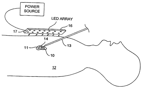
FIGS. 1 and 2 show a photoreactive agent 10 being delivered locally to
target tissue 11 of a patient 12. The target tissue 11 of the patient 12 is a
tumor
located within the chest cavity below a dermal layer of the patient 12. The
photoreactive agent 10 is being locally delivered by a hypodermic needle 13
which is inserted into the patient's chest with the tip 14 of the needle
disposed
within the target tissue 11. Photoreactive agent 10 is being dispensed from
the
tip 14 of the hypodermic needle 13 and is shown permeating the target tissue
11. FIG. 2 also shows an alternative method and device for local delivery of a
photoreactive agent which includes a photoreactive agent depot 15 disposed
within the target tissue 11. The photoreactive agent depot 15 is a device that
contains photoreactive agent 10 and is configured to dispense the
CA 02473924 2004-07-21
WO 03/061696 PCT/US03/02303
14
photoreactive agent 10 at a predetermined rate. For some embodiments, the
photoreactive agent depot 15 can be a polymer material impregnated with a
photoreactive agent 10 that dissolves into the adjacent target tissue 11 over
time. Once an appropriate amount of the photoreactive agent 10 has been
dispensed into and absorbed by the target tissue 11, the photoreactive agent
may then. be photoactivated in order to treat the target tissue 11.
The appropriate amount of photoreactive agent 10 to be absorbed by the
target tissue will be a factor of the desired clinical result and the specific
photoreactive agent 10 used. However, by use of a localized delivery method,
10 as discussed above, less photoreactive agent 10 is used than would be
required for the sartie photoreactive agent,10 delivered intravenously or
otherwise systemically to the patient 12.
Once the target tissue 11 has absorbed an appropriate amount of the
photoreactive agent 10, a source of electromagnetic radiation 16 having a
wavelength within an activation wavelength of the photoreactive agent 10 is
used to activate the photoreactive agent 10. A source of electromagnetic
radiation 16 consisting of one or more light sources can be used. Various
types
of light sources can be used, such as, for example, at least one light-
emitting
diode, laser diode, incandescent light bulb, gas discharge device, polymeric .
electroluminescent device, halogen bulb, chemical luminescence, vacuum
fluorescence, radio frequency excited gas, microwave excited gas, cold cathode
fluorescent tube, or combination thereof.
An exemplary source of electromagnetic radiation 1.6 consisting of an
array of light emitting diodes 17 (LEDs) is seen in FIGS. 1 and 3. The LED
RECTIFIED SHEET (RULE 91)
CA 02473924 2004-07-21
WO 03/061696 PCT/US03/02303
array 16 can have an emission wavelength of about 500 to about 900, or about
600 to about 700, nanometers, depending on the photoreactive agent used,
and is in electrical communication with a power supply unit 18. In some
embodiments, long wavelength LEDs 17 can be used that have an emission
5 wavelength of greater than about 700 nanometers in the infrared band up to
about 900 nanometers. The light produced by such an array of long
wavelength LEDs 17 can easily penetrate tissue and a photoreactive agent 10
having an activation wavelength range corresponding to the long wavelength of
the emitted light. The LED array 16 may include LEDs 17 that are made from
10 either polymeric, organic or metallic materials.
The LED array 16 can emit long wavelength infrared light with an output
power of about 5 mW/cm2 to about 500 mW/cm2.
The LED array 16 is shown activated in FIG. 3 with electromagnetic
energy in the form of photoreactive light, as shown by the arrows 19, being
15 emitted from the LED array 16 through the dermal layer of the patient 12
and
the underlying tissue. The photoactivating light continues to the target
tissue 11
and impinges on the photoreactive agent 10 within the target tissue 11. The
photoreactive agent 10 then undergoes photochemical excitation and induces
formation of a free radical species, such as singlet oxygen, which is toxic to
surrounding target tissue 11. The tumor or target tissue 11 is thereby lysed
with a minimal amount of photoreactive agent 10 used and without the use of
an invasive photoactivation light delivery system such as a fiber optic probe
or
the like. Because the LED array 16 is external to the patient's body 12, the
photoactivating light can be delivered at a rate, which is slower than the
rate
CA 02473924 2004-07-21
WO 03/061696 PCT/US03/02303
16
that would be used if an invasive. source of photoactivating light were being
used. This results in reduced photobleaching and oxygen consumption, which
enhances the efficacy of PDT. In addition, the total dose of light that can be
delivered is much greater with an external non-invasive source of
photoactivating light 16 because the dose can be administered over a longer
period of time as compared with an invasive light source without the risks
that
are present with an invasive photoactivating light source, such as infection,
bleeding and the risks associated with the administration of anesthetics.
Referring to FIG. 4, a patient 21. is shown with coronary artery disease
being treated with PDT. A distal end 22 of a coronary delivery catheter 23 is
disposed within a coronary artery 24 of the patient 21 as seen in more detail
in
FIG. 5. The coronary delivery catheter 23 is a multi-lumen catheter having an
optional expandable balloon 25 secured to a distal portion 26 of the catheter
23.
and a guidewire lumen (not shown). A plurality of outlet ports 27 are disposed
on the expandable balloon 25 as seen more clearly in FIG. 6. The outlet ports
27 are in fluid communication with an interior chamber 28 of the balloon 25,
which is in fluid communication with an injection lumen 31 (not shown)
disposed
within a shaft 32 of the coronary delivery catheter 23. A proximal end of the
injection lumen 31 is connected to a Luer adapter at a proximal end (not
shown)
of the coronary delivery catheter 23 to facilitate injection of a
photoreactive
agent 34 into the injection lumen 31.
In use, the distal end 22 of the. coronary delivery catheter 23 is advanced
into the patient's vasculature 35 using a standard percutaneous technique,
such as the Seldinger technique. In one embodiment, the coronary delivery
RECTIFIED SHEET (RULE 91)
CA 02473924 2004-07-21
WO 03/061696 PCT/US03/02303
17
catheter 23 is advanced over a coronary guidewire 36 previously placed across
the target lesion 37 in the coronary artery 24. The coronary delivery catheter
23
is advanced distally until the expandable balloon 25 is disposed adjacent the
target lesion 37. A photoreactive agent 34 is then injected into the injection
lumen 31 of the catheter 23 and travels distally in the injection lumen 31 to
the
interior chamber 28 of the expandable balloon 25, which expands the
expandable balloon 25 against the target tissue 37. The photoreactive agent
34 is then expelled from the outlet ports 27, as shown by the arrows 38 in
FIG.
6, and into contact with the target tissue 37. This process is continued until
the
target tissue 37 has absorbed an appropriate amount of the photoreactive
agent 34. Thereafter, a source of photoactivating light, such as the LED array
39 shown in FIGS. 4 and 5 can be positioned external to the patient's body 21
adjacent the target tissue 37 and activated.
Upon activation of the LED array 39, photoreactive light having a
wavelength within an activation wavelength range of the photoreactive agent 34
travels from the LEDs 40 of the LED array 39 and into the tissue 41 of the
patient 21. The photoreactive light passes through the dermal layer 44 of the
patient 21 and the underlying tissue 41 until it reaches the target tissue 37
which contains the photoreactive agent 34. The photoreactive agent 34 then
undergoes photochemical excitation and induces formation of a free radical
species, such as singlet oxygen, which is toxic to surrounding target tissue
37
and the target tissue 37 is destroyed. The coronary catheter delivery catheter
23 can be withdrawn either before or after the administration of
photoactivating
light, however, it may be desirable to withdraw the catheter 23 prior to
CA 02473924 2004-07-21
WO 03/061696 PCT/US03/02303
18
administration of the photoactivating light so that the catheter 23 does not
prevent any of the photoactivating light from penetrating the target tissue
37.
The total dose of photoactivating light that can be delivered is much greater
with an external non-invasive source of photoactivating light because the dose
can be administered over a long period of time without the risks that would be
present with an invasive photoactivating light source, such as infection,
bleeding and the risks associated with the administration of anesthetics. In
addition, the insertion of an invasive fiber optic photoactivating light
source into
the patient's vasculature 35 can lead to thrombosis and vessel wall injury
including the creation of an intimal flap. These risks are also avoided by use
of
an external source of photoactivating light.
Referring to FIG. 7, a patient 47 is shown with benign prostatic
hypertrophy disease being treated with PDT. A distal end 48 of a urinary
delivery catheter 49 is disposed within a bladder 50 of the patient 47. The
urinary delivery catheter 49 is a multi-lumen catheter having an optional
expandable balloon 51 secured to the distal end 48 of the catheter 49. A
plurality of outlet ports 52 are disposed in a distal portion 53 of a shaft 54
of the
urinary delivery catheter 49 as seen more clearly in FIG. 8. The outlet ports
52
are in fluid communication with a photoreactive agent injection lumen 55
disposed within the shaft 54 of the urinary delivery catheter 49. A proximal
end
of the photoreactive agent injection lumen is connected to a Luer adapter at a
proximal end (not shown) of the urinary delivery catheter 49 to facilitate
injection of a photoreactive agent 57 into the injection lumen 55.
In use, the distal end 48 of the urinary delivery 49 catheter is advanced
CA 02473924 2004-07-21
WO 03/061696 PCT/US03/02303
19
into the patient's urethra 58 using standard techniques. In one embodiment,
the urinary delivery catheter 49 will be advanced distally until the
expandable
balloon 51, in a collapsed state, is disposed within the patient's bladder 50.
The expandable balloon 50 can then be expanded by injection of a suitable
material, such as saline, into a balloon injection lumen 59 and into an
interior
chamber 60 of the balloon 51. A photoreactive agent 57 is then injected into
the photoreactive agent injection lumen 55 of the catheter 49 and travels
distally in the injection lumen 55 to the outlet ports 52 and is then expelled
from
the outlet ports 52, as shown by the arrows 61 in FIG. 8, and into contact
with
the target tissue 62. This process is continued until the target tissue has
absorbed an appropriate amount of the photoreactive agent 57. Thereafter, a
source of photoactivating light 63, such as the LED array 39 shown in FIGS. 4
and 5 can be positioned external to the patient's body 47 adjacent the target
tissue 62 and activated.
Upon activation of the LED array 63, photoreactive light having a
wavelength within an activation wavelength range of the photoreactive agent
travels from the LEDs 64 of the LED array 63 and into the tissue of the
patient
47. The photoreactive light passes through the dermal layer of the patient 47
and the underlying tissue until it reaches the target tissue 62 which contains
the
photoreactive agent 57. The photoreactive agent 57 then undergoes
photochemical excitation and induces formation of a free radical species, such
as singlet oxygen, which is toxic to surrounding target tissue 62 and the
target
tissue 62 is destroyed. The urinary catheter 49 delivery catheter can be
withdrawn either before or after the administration of photoactivating light,
CA 02473924 2004-07-21
WO 03/061696 PCT/US03/02303
however, it may be desirable to withdraw the catheter 49 prior to
administration
of the photoactivating light so that the catheter 49 does not prevent any of
the
photoactivating light from penetrating the target tissue 62.
Referring generally to FIGS. 9-18, vascular closure has been observed
5 as one of the consequences of therapeutic PDT which has recently led to the
use of PDT in opthalmological disease. The exudative stage of age-related
macular degeneration (AMD) with choroidal neovascularization (CNV)
commonly leads to rapidly progressive loss of sight. PDT can induce a
selective occlusion of CNV via light-induced chemical thrombosis and this
effect
10 can be used to effectively treat AMD. Diabetic retinopathy (DR) can be
similarly
treated. However, destruction of CNV that is not properly limited or targeted
to
the area requiring treatment can result in undesirable collateral damage to
retinal tissue. This, in turn, can lead to reduction in visual acuity. These
complications are addressed by a system, such as that shown in FIG. 16, that
15 targets photoactivation energy or light to a desired area of treatment.
FIG. 9 is a sectional view of a patient's eye 68 being prepared for PDT.
A thin hypodermic needle 69 is shown disposed within the vitreous humor 70 of
the patient's eye 68 adjacent the retina 71. A photoreactive agent 72 is being
dispensed from a distal end 73 of the hypodermic needle 69 adjacent target
20 tissue 74 within the patient's retina 71. In one embodiment of a method of
treatment, prior to insertion of the hypodermic needle 69, a corneal surface
75
of the patient's eye 68 is first anesthetized with a topical anesthetic such
as
Tetracaine~ or the like. The hypodermic needle 69 is then advanced into the
vitreous 70 and the photoreactive agent 72 injected as a single bolus
infusion.
CA 02473924 2004-07-21
WO 03/061696 PCT/US03/02303
21
It may be necessary is some instances to depress the globe of the eye 68 in
order to facilitate posterior placement of the distal end 73 of the hypodermic
needle 69 prior to injection of the photoreactive agent 72. The photoreactive
agent 72 can be an aqueous formulation that facilitates transport of the drug
through the retina 71 and into the target tissue 74, i.e., the neovasculature,
shown in FIGS. 14 and 15, beneath the retina 71. The photoreactive agent 72
is thereafter allowed to absorb into the target tissue 74 for a predetermined
amount of time. Optionally, the eye 68 may be examined using standard
ophthalmic imaging and pressure measurement during this period. Once an
appropriate amount of the photoreactive agent 72 has been absorbed by the
target tissue 74 in order to achieve the desired clinical result, the
photoreactive
agent 72 in the target tissue 74 can be photoactivated as discussed below.
Note that in the case of "wet" AMD, certain photoreactive agents will be
dissipated from the normal retinal tissue after the absorption period and will
be
localized to the neovessels of the target tissue. For example, a conjugate of
a
photosensitive agent and an antibody may be used for specific binding to
neovessels.
Also shown in FIG. 9 are two photoreactive agent depots 78 which have
been placed behind the patient's eye 68 in order to deliver photoreactive
agent
72 into the interior structure of the eye 68. The photoreactive agent depots
78
may be made of a polymer material which is impregnated with a suitable
photoreactive agent. The polymer can be chosen to allow the photoreactive
agent to emanate from the photoreactive agent depots 78 at a predetermined
rate. The photoreactive agent 72 then absorbs into the sclera of the patient's
CA 02473924 2004-07-21
WO 03/061696 PCT/US03/02303
22
eye 68 and eventually perfuses into the target tissue 74 beneath the retina
71.
FIG. 10 illustrates an alternative method of localized delivery of a
photoreactive agent 72 which includes a contact disk 79 disposed on a corneal
surface 75 of the eye 68. The contact disk 79 can have properties similar to
those of the photoreactive agent depots 78 discussed above, however, an
optional first electrical conductor 81 in electrical communication with the
contact
disk 79 extends from the contact disk 79 to a first pole 82 of a voltage
source
83. A second pole 84 of the voltage source 83 is in electrical communication
with a second electrical conductor 85 which is connected to an electrical
contact pad 86 in electrical communication with the patient's body,
specifically,
the sclera 87 of the patient's eye 68. In this way, an electrical voltage
potential
can be imposed by the voltage source 83 between the contact disk 79 and the
corneal surface 75, or any other surface, of the patient's eye 68. The
application of such an electrical potential can facilitate perfusion of the
photoreactive agent 72 from the contact disk 79 into the patient's eye 68.
FIG. 11 illustrates another alternative method for localized delivery of a
photoreactive agent 72 to target tissue 74 within a patient's eye 68. FIG. 11
is
a sectional view of a patient's eye 68 with a distal end 89 of a gas jet
injector 90
or drug aerosol device disposed between the eye 68 and eye socket of the
patient. Photoreactive agent 72 is delivered by gas jet injection, as shown by
the arrows 91 in FIG. 11, to the tissue behind the eye 92 adjacent the target
tissue 74 below the patient's retina 71. A controller 93 is shown in
electrical
communication with the gas jet injector 90 for controlling the duration,
pressure, and volume of gas jet injection. By using gas jet injection of the
CA 02473924 2004-07-21
WO 03/061696 PCT/US03/02303
23
photoreactive agent 72, the photoreactive agent 72 can be distributed to a
wide
surtace area behind the patient's eye 68 which may aid in more rapid transport
of the agent 72 to the target tissue 74 within the eye 68. The photoreactive
agent 72 can be delivered more posterior in the eye 68 by penetrating the
conjunctiva) membrane 94 with air or another gas during injection which may
increase proximity of the photoreactive agent 72 to the macula 95 and
posterior
retina 71.
FIG. 12 illustrates yet another embodiment of a device and method for
localized delivery of a photoreactive agent 72. FIG. 12 is a sectional view of
a
patient's eye 68 with a distal end 97 of an ultrasonic probe 98 for delivery
of a
photoreactive agent 72 disposed on the sclera 87 of the patient's eye 68. The
ultrasonic probe 98 includes an ultrasonic emitter 99 disposed in a distal
portion
100 of an elongate shaft 101. The ultrasonic emitter 99 generates ultrasonic
energy which is transmitted to an outer surface 87 of the patient's eye 68
through a contact ring 102 disposed on a distal end 97 of the elongate shaft
101. The contact ring 102 is in contact with the outer surface 87 of the eye
68
and can form an annular seal between the distal end 97 of the elongate shaft
and the outer surface 87 of the eye 68. A distal cavity 103 is disposed within
the contact ring 102 which allows for dispersion of a photoreactive agent 72
which is delivered to the distal cavity 103 as shown by the arrows 104 in FIG.
12.
The photoreactive agent 72 is delivered through an injection lumen 105
which is in fluid communication with the distal cavity 103 and a photoreactive
agent reservoir 106 disposed in a proximal portion 107 of the elongate probe
CA 02473924 2004-07-21
WO 03/061696 PCT/US03/02303
24
98. A controller 108 is in electrical communication with the ultrasonic
emitter 99
and a pump 109 disposed within the photoreactive agent reservoir 106. The
controller 108 determines the frequency, amplitude and duration of ultrasonic
energy produced by the ultrasonic emitter 99. The controller 108 is also
configured to control the rate and amount of injection of the photoreactive
agent
72 from the photoreactive agent reservoir 106 to the distal cavity 103.
Ultrasonic energy is emitted from the ultrasonic emitter 99 once photoreactive
agent 72 is disposed within the distal cavity 103 which facilitates permeation
of
the photoreactive agent 72 into the patient's eye 68 and reduces the time
required to deliver an appropriate amount of photoreactive agent 72 to the
target tissue 74 within the patient's eye 68. The frequency of the emitted
ultrasonic energy can be from about 1 to about 50 MHz, specifically, about 10
to about 40 MHz.
FIG. 13 illustrates a sectional view of a patient's eye 68 that has been
dosed with an appropriate amount of photoreactive agent. FIG. 14 is a cross
sectional view of the eye 68 of FIG. 13 taken along lines 14-14 in FIG. 13 and
illustrates the fundus 111 of the patient's eye 68. In FIG. 14, an area or
target
tissue 112 is indicated by a hatched area. The target tissue 112 is disposed
in
an area of the patient's retina 71 that would be consistent with an area of
deterioration due to age-related macular degeneration. The target tissue 112
would likely contain neovascularization with the potential for visual loss for
the
patient. FIG. 15 illustrates a view similar to that of FIG. 14 and shows a
first
target tissue area 113 and a second target tissue area 114 that would be
consistent with areas of deterioration due to diabetic retinopathy. The target
CA 02473924 2004-07-21
WO 03/061696 PCT/US03/02303
tissue areas 112, 113 and 114 of FIGS. 14 and 15 can be dosed with an
appropriate amount of photoreactive agent 72 by any of the methods discussed
above, as well as other suitable methods. Once the target tissue areas 112,
113 and 114 have been appropriately dosed with a photoreactive agent 72, the
5 photoreactive agent 72 must be photoactivated. Indiscriminate
photoactivation
of the photoreactive agent 72 in the tissue of the patient's eye can be
undesirable because of the possible risk of damage to healthy collateral
tissue
115 adjacent the target tissue areas 112, 113 and 114. A system for
performing PDT 117 such as shown in FIG. 16 can be useful for avoiding such
10 risks.
The PDT system 117 shown in FIG. 16 includes a source of
fluorescence generating light 118 which is configured to illuminate the fundus
111 of a patient's eye 68 as indicated by the ray trace arrows 119 in FIG. 16.
The fluorescence generating light is emitted by the source of fluorescence
15 generating light 118 and travels through a beam splitting member 120, a
focusing member 121 and the cornea 75 and lens 122 of the patient's eye 68.
The fluorescence generating light then impinges on the retina 71 of the
patient's eye 68 and the tissue underlying the retina 71 and has sufficient
intensity and wavelength to cause fluorescence of a photoreactive agent 72
20 without causing photoactivation of the photoreactive agent 72. The target
tissue areas 112, 113 and 114, and any other tissue that contain a
concentration of photoreactive agent 72 will then fluoresce.
Initiation of emission of the fluorescence generating light from the source
of fluorescence generating light 118 is carried out by a processor 123 which
is
CA 02473924 2004-07-21
WO 03/061696 PCT/US03/02303
26
in electrical communication with the source of fluorescence generating light
118
with a bundle of electrical conductors 124. In one embodiment, the source of
fluorescence generating light 118 includes a laser 125 having an operating
wavelength of about 600 to about 700 nanometers, specifically, about 660 to
about 670 nanometers. The beam splitting member 120 can be any of a
suitable variety of commercially available beam splitters which is relatively
transmissive in the direction of the fluorescence generating light shown in
FIG.
16 and relatively reflective for light traveling in the opposite direction, as
shown
in FIG. 17. The focusing member 121 can be a commercially available lens
made from any suitable material which is transmissive for the wavelength of
the
fluorescence generating light.
Once the fluorescence generating light hits the target tissue 112, 113
and 114 and surrounding tissue 115, and the photoreactive agent 72 therein
fluoresces, the fluorescent light then travels from the target tissue 112, 113
and
114 and surrounding tissue 115 out of the patient's eye 68 and back into the
focusing member 121 as shown in FIG. 17 by the arrows 126. After passing
through the focusing member 121, the fluorescent light hits the beam splitter
member 120 and substantially reflects up to a fluorescence detector 127 which
is configured to measure the intensity of fluorescent light emanating from
each
coordinate point of the fundus 111 of the patient's eye 68. The fluorescence
detector 127 can be a charged couple chip or device, but could also use slit
lamp photography in order to plot the fluorescence distribution. The time
course of the photography will be determined by the initial fluorescence
appearance and distribution in the choroid and later in the retina.
CA 02473924 2004-07-21
WO 03/061696 PCT/US03/02303
27
This fluorescence response data is then captured by the processor 123
which is in electrical communication with the fluorescence detector 127 with a
bundle of electrical conductors 128. The processor 123 then analyzes the
fluorescence response data and generates a virtual map that indicates the
coordinates of the target tissue 112, 113 and 114 relative to the surrounding
normal tissue 115 as indicated by the ray trace arrows in FIG. 17. In some
embodiments, the target tissue 112, 113 and 114 is distinguished from the
surrounding tissue 115 by the presence of supra-threshold photoreactive agent
72 concentrations in the tissue. The processor 123 may also display the
virtual
map, or any other fluorescence response data visually on an optional monitor
display 130 which is in electrical communication with the processor 123 with a
bundle of electrical conductors 131.
Once the processor 123 has generated a virtual map which distinguishes
the coordinates of the target tissue 112, 113 and 114 from the surrounding
normal or non-target tissue 115, the processor 123 can then be used to control
the output beam of a source of photoactivating light 118 so that the
photoactivating light is directed only to the target tissue area 112, 113 and
114
of the patient's eye 68 as shown in FIG. 18 by the ray trace arrows 132. The
source of photoactivating light 118 can be the same laser 125 as that used for
the source of fluorescence generating light 118, or another device can be
used.
The controller 123 can control the delivery of the photoactivating light by
any
suitable method including aiming and scanning a thin beam of photoactivating
light across the entire region of target tissue 112, 113 and 114 while
avoiding
the collateral areas 115 of healthy tissue. In this way, only the
photoreactive
CA 02473924 2004-07-21
WO 03/061696 PCT/US03/02303
28
agent 72 within the target tissue areas 112, 113 and 114 are photoactivated
with the production and lysing effect of singlet oxygen or the like.
FIG. 19 shows yet another device that can be used for localized delivery
of a photoreactive agent. The device is an injection device 2500 that can be
used for localized delivery of a photoreactive agent to a patient's eye. The
injection device 2500 includes a syringe 2510 in which is mounted a plunger
2515 that is movably mounted in the syringe 2510. A hypodermic needle 2520
is coupled to the syringe by a flexible coupling 2525. The needle has a
sharpened distal tip 2530 that can be used to penetrate eye tissue. A cannula
or sheath 2535 covers at least a portion of the needle 2520. The needle 2520
and the sheath 2535 both have a curved shape that can conform to the
curvature of the outer surface of a patient's eye. Thus, the needle and sheath
define a substantially circular curvature, although the curvature can vary.
The
curvature of the needle/sheath can vary based upon the curvature of the eye
with which the device will be used. In one embodiment, the needle and sheath
conform to a radius of approximately 12 mm. As described below, the curved
shape of the needle/sheath facilitate placement of the distal tip 2530 of the
needle 2520 to posterior regions of the eye. The needle and sheath can be
manufactured of a variety of materials, including stainless steel and plastic.
The needle 2520 can be retractable with respect to the sheath 2535
such that the distal tip 2530 can be retracted so that it is positioned within
the
sheath 2535. The needle can also be advanced in a distal direction
(represented by the arrow 2540 in FIG. 19) such that the distal tip 2530
protrudes outwardly from the sheath 2535, such as is shown in FIG. 19. In one
CA 02473924 2004-07-21
WO 03/061696 PCT/US03/02303
29
embodiment, the needle 2520 can only be advanced a limited distance so that
the distal tip 2530 can only extend a distance D outward from the edge of the
sheath 2535. This feature can prevent inadvertent over-penetration of the
needle into the eye tissue.
As mentioned, a flexible coupling 2525 attaches the needle 2520 to the
syringe. The flexible coupling 2525 permits the curved needle 2520 to be
moved to various orientations relative to the syringe 2510 in order to
facilitate
positioning of the needle relative to the eye upon delivery of the
photoreactive
agent. The syringe can be filled with a desired photoreactive agent, which can
be dispensed out of the distal tip 2530 of the needle 2520 by pressing the
plunger 2515 in a well-known manner.
A method of using the injection device is now described with reference to
FIG. 20, which shows a sectional view of a patient's eye 68. Various
anatomical details of the eye 68 are omitted from FIG. 20 for clarity of
illustration. In use, the needle 2520 and sheath 2535 are inserted between the
eye and the eye socket (not shown) such that the needle and sheath are
positioned substantially adjacent the outer surface of the eye 68. The curved
shape of the needle and sheath facilitate such insertion. In one embodiment,
the needle 2520 is retracted into the sheath 2535 prior to placement of the
needle around the eye. Thus, the sharpened, distal tip 2530 of the needle
2520 is positioned within the sheath 2535 while the needle and sheath are
inserted around the eye. In this manner, the sheath 2535 will shield the
sharpened, distal tip 2530 of the needle 2520 from contact with the eye and
thereby eliminate the risk of the sharp needle injuring the eye while the
needle
CA 02473924 2004-07-21
WO 03/061696 PCT/US03/02303
is being positioned. The distal edge of the sheath 2535 can have an atraumatic
shape in order to reduce the risk of the sheath damaging the eye.
. When the distal tip of the needle 2520 is at a desired location relative to
the eye 26, the needle is then advanced so that the distal tip 2530 protrudes
5 from the sheath 2535. The needle 2520 is of sufficient length so that the
distal
tip can reach any desired location of the eye, such as diseased tissue
comprised of the neovascular membrane (not shown). The distal tip can then
be advanced so that it penetrates the eye to a desired depth. In one
embodiment, the needle penetrates only the sclera 2550 of the eye 68 without
10 penetrating any deeper. It should be appreciated, however, that the needle
can
optionally penetrate the eye to any desired depth. When the needle has
penetrated the eye 68 to the desired depth, the photoreactive agent is
delivered
to target region of the eye by dispensing the photoreactive agent through the
distal tip of the needle 2520. As mentioned, this is accomplished by pressing
15 the plunger 2515 so that the agent is forced out of the distal tip of the
needle
2530 and into the eye 68.
After the photoreactive agent has been delivered to the target region of
the eye, the target region can be exposed to photoreactive fight to thereby
photoactivate the agent. FIG. 21 shows a PDT device 2710 that can be used to
20 expose a treated eye region to light. The PDT device 2710 includes an
elongated arm 2715 that has a curved shape. The curvature of the arm 2715
conforms to the curvature of the outer surface of a patient's eye. This
facilitates
positioning of the arm 2715 around the outer surface of the eye. The curvature
of the arm 2715 can vary based upon the curvature of the eye with which the
RECTIFIED SHEET (RULE 91)
CA 02473924 2004-07-21
WO 03/061696 PCT/US03/02303
31
device will be used. In one embodiment, the arm 2715 follows a curve with a
radius of approximately 12 mm.
The arm 2715 has a distal end 2720 upon which is mounted a source of
photoreactive light. The source of light can be, for example, an LED 2730. The
LED 2730 is positioned such that it can emit light in a predetermined
direction,
such as toward a target region of the eye. The LED 2730 is electrically
coupled
to a source of power (not shown) and a controller 2735 that can be used to
control power to the LED 2730. A lens 2740 can be positioned over the LED
2730 in order to focus the light from the LED 2730. The lens 2740 can be
manufactured of any suitable material, such as, for example, Polymethyl
methacrylate (PMMA)
In use, the PDT device 2710 is deployed such that the distal tip 2720 is
positioned adjacent the region of the eye to be treated. The device 2710 is
oriented so that the LED 2730 is positioned to emit light toward the target
region of the eye. As mentioned, the curvature of the elongated arm 2715
facilitates positioning of the arm 2715 around the outer surface of the eye.
Once the LED is properly positioned, the LED is activated so that it emits
light
toward the region of the eye that has been treated with the photoreactive
agent.
C. Photoreactive Agents
Any chemical compound that absorbs light may be used in the methods
provided herein (see, e.g., Kreimer-Birnbaum (1989) Sem. Hematol. 26:157-
173). Photoreactive agents for use in the methods provided herein include, but
are not limited to, indocyanine green, toluidine blue, prodrugs such as
aminolevulinic acid, texaphyrins, benzoporphyrins, phenothiazines,
CA 02473924 2004-07-21
WO 03/061696 PCT/US03/02303
32
phthalocyanines, porphyrins, merocyanines, psoralens, protoporphyrin,
methylene blue, Rose Bengal (see, e.g., Picaud et al. (1990) Brain Res.
531:117-126 and Picaud et al. (1993) J. Neurosci. Res. 35:629-642), chlorins
such as mono-L-aspartyl chlorin e6, alkyl ether analogs of chlorins,
purpurins,
bacteriochlorins, pheophorbides, pyropheophorbides, cationic dyes and any
other agent that absorbs light in a range of about 500 to about 1100
nanometers. Photoreactive agents for use in the methods provided herein are
also disclosed in commonly assigned U.S. Patent Applications, Ser. No.
09/078,329, filed May 13, 1998, entitled "Controlled Activation of Targeted
Radionuclides", Ser. No. 60/116,234, filed January 15, 1999, entitled
"Targeted
Transcutaneous Cancer Therapy", Ser. No. 09/271,575, filed March 18, 1999,
entitled "Targeted Transcutaneous Cancer Therapy", Ser. No. 09/905,501, filed
July 13, 2001, entitled "Targeted Transcutaneous Cancer Therapy", Ser. No.
09/905,777, filed July 13, 2001, entitled "Non-invasive Vascular Therapy",
Ser.
No. 60/175,689, filed on January 12, 2000, entitled "Novel Treatment for Eye
Disease", Ser. No. 09/760,362, filed on January 12, 2001, entitled "Novel
Treatment for Eye Disease", and Ser. No. 60/116,235, filed on January 15,
1999, entitled "Non-invasive Vascular Therapy", the disclosure of each of
which
is hereby incorporated by reference in its entirety. Photoreactive agents for
use
in the methods provided herein are also disclosed in U.S. Patent Nos.
6,319,273, RE37,180, 4,675,338, 4,693,885, 4,656,186, 5,066,274, 6,042,603,
5,913,884, 4,997,639, 5,298,018, 5,308,861, 5,368,841, 5,952,366, 5,430,051,
5,567,409, 5,942,534, and U.S. patent application Publication No.
2001/0022970. In one embodiment, the photoreactive agent for use in the
CA 02473924 2004-07-21
WO 03/061696 PCT/US03/02303
33
methods provided herein is taporfin sodium, also referred to as mono-L-
aspartyl
chlorin e6, (+)-tetrasodium (2S,3S)-18-carboxylato-20-[N-(S)-1,2-
dicarboxylatoethyl]carbamoylmethyl-13-ethyl-3,7,12,17-tetramethyl-8-
vinylchlorin-2-propanoate, NPe6 or ME2906.
In another embodiment, the photoreactive reagents for use in the
methods provided herein include but are not limited to porphyries such as
PHOTOPHRINT"" (a QLT, Ltd. brand of sodium porfimer), and FOSCANT"",
which is a brand of chlorin.
(n another embodiment, the photoreactive reagents for use in the
methods provided herein include but are not limited to indocyanine green
(ICG),
methylene blue; toluidine blue, aminolevulinic acid (ALA), chlorins, .
phthalocyanines, porphyries, pupurins, texaphyrins, and other photosensitizer
agents that have characteristic light absorption peaks in a range of from
about
500 nm to about 1100 nm.
In another embodiment, the photoreactive reagents for use in the
methods provided herein include but are not limited to chlorins,
bacteriochlorins, phthalocyanines, porphyries, purpurins, merocyanines,
psoralens, benzoporphyrin derivatives (BPD), and porfimer sodium and pro-
drugs such as delta-aminolevulinic acid, which can produce photosensitive
agents such as protoporlphyrin IX, and. other suitable photosensitive
compounds including ICG, methylene blue, toluidine blue, texaphyrins, and any
other agent that absorbs light in a range of 500 nm to 1100 nm.
In another embodiment, the photoreactive reagents for use in the
methods provided herein include but are not limited to LUTRINTM(lutetium
RECTIFIED SHEET (RULE 91)
CA 02473924 2004-07-21
WO 03/061696 PCT/US03/02303
34
texaphyrin, brand; Pharmacyclics, Inc. Sunnyvale, CA), and
bacteriochlorphylls.
In another embodiment, the photoreactive reagents for use in the
methods provided herein include but are not limited to clorins,
bacteriochlorophylls, phthalocyanines, porphyrins, purpurins, merocyanines,
psoralens, benzoporphyrin derivatives (BPD) and porfimer sodium and pro-
drugs such as delta-artiinolemulinic acid, which can produce drugs such as
protoporphyrin; and others such as indocyanince green (ICG); methylene blue;
toluidine blue; texaphyrins; pyropheophorbide compounds; bacteriochlorophyll
derivatives; alkyl ether analogs of chlorins, and an other agent that absorbs
light in a range of 500 nm to 1100 nm.
In another embodiment, the photoreactive reagents for use in the
methods provided herein include but are not limited to PURYLITINT""(tin ethyl
etiopurpurin) or VERTEPORFINT"" (a liposomal benzoporphyrin derivative).
In another embodiment, the photoreactive reagents for use in the
methods provided herein include but are not limited to photosensitizers
selected
from:
1. Photofrin~. .
2. Synthetic diporphyrins and dichlorins
3. Hydroporphyrins, e.g., chlorins and.bacteriochlorins of the
tetra(hydroxyphenyl) porphyrin series
4. phthalocyanines
5. O-substituted tetraphenyl porphyrins (picket fence porphyrins)
6. 3,1-meso tetrakis (o-propionamido phenyl) porphyrin
7. Verdins
RECTIFIED SHEET (RULE 91)
CA 02473924 2004-07-21
WO 03/061696 PCT/US03/02303
8. Purpurins, e.g., tin and zinc derivatives of octaethylpurpurin (NT2),
and etiopurpurin (ET2)
9. Chlorins, e.g., chlorin e6, and mono-I-aspartyl derivative of chlorin e6
10. Benzoporphyrin derivatives (BPD), e.g., benzoporphyrin monoacid
5 derivatives, tetracyanoethylene adducts of benzoporphyrin, dimethyl
acetylenedicarboxylate adducts of benzoporphyrin, Diels-Adler adducts,
and monoacid ring "a" derivative of benzoporphyrin
11. Low density lipoprotein mediated localization parameters similar to
those observed with hematoporphyrin derivative (HPD)
10 12. sulfonated aluminum phthalocyanine (Pc) sulfonated AIPc
disulfonated (AIPcS2) tetrasulfonated derivative sulfonated
aluminum naphthalocyanines chloroaluminum sulfonated phthalocyanine
(CASP)
13. zinc naphthalocyanines
15 14. anthracenediones
15. anthrapyrazoles
16. aminoanthraquinone
17. phenoxazine dyes
18. phenothiazine derivatives
20 19. chalcogenapyrylium dyes cationic selena and tellurapyrylium
derivatives
20. ring-substituted cationic PC
21. pheophorbide .alpha.
22. hematoporphyrin (HP)
CA 02473924 2004-07-21
WO 03/061696 PCT/US03/02303
36
23. protoporphyrin
24. 5-amino levulinic acid
In another embodiment, the photoreactive reagents for use in the
methods provided herein include but are not limited to photosensitizers
selected
from members of the following classes of compounds: porphyrins, chlorins,
bacteriochlorins, purpurins, phthalocyanines, naphthalocyanines, texaphyrines,
and non-tetrapyrrole photosensitizers. Specific examples are PhotofrinT"~,
benzoporphyrin derivative, tin etiopurpurin, sulfonated chloroaluminum
phthalocyanine and methylene blue.
In another embodiment, the photoreactive reagents for use in the
methods provided herein include but are not limited to BPD which is a second
generation porphyrin photosensitizer that diffuses rapidly from
microvasculature
and disseminates throughout a joint. In addition, BPD has a low affinity for
chondrocytes and articular cartilage following systemic or intra-articular
injection. CASPc, a phthalocyanine inactivates growth factors TGF-.beta. and
bFGF.
In another embodiment, the photoreactive reagents for use in the
methods provided herein include but are not limited to photosensitizers
selected
from:
1. Photofrin~.
2. Synthetic diporphyrins and dichlorins
3. Hydroporphyrins such as chlorins and bacteriochlorins of the
tetra(hydroxyphenyl) porphyrin series
4. phthalocyanines (PC) with or without metal substituents, e.g.,
CA 02473924 2004-07-21
WO 03/061696 PCT/US03/02303
37
chloroaluminum phthalocyanine (CASP) with or without varying
substituents
5. O-substituted tetraphenyl porphyrins (picket fence porphyrins)
6. 3,1-meso tetrakis (o-propionamido phenyl) porphyrin
7. Verdins
8. Purpurins tin and zinc derivatives of octaethylpurpurin (NT2)
etiopurpurin (ET2)
9. Chlorins chlorin e6 mono-I-aspartyl derivative of chlorin e6 di-I-aspartyl
derivative of chlorin e6
10. Benzoporphyrin derivatives (BPD) benzoporphyrin monoacid
derivatives tetracyanoethylene adducts of benzoporphyrin dimethyl
acetylenedicarboxylate adducts of benzoporphyrin Diels-Adler adducts
monoacid ring "a" derivative of benzoporphyrin
11. sulfonated aluminum PC sulfonated AIPc disulfonated (AIPcS2)
tetrasulfonated derivative sulfonated aluminum naphthalocyanines
12. naphthalocyanines with or without metal substituents with or without
varying substituents
13. anthracenediones
14. anthrapyrazoles
15. aminoanthraquinone
16. phenoxazine dyes
17. phenothiazine derivatives
18. chalcogenapyrylium dyes cationic selena and tellurapyrylium
derivatives
CA 02473924 2004-07-21
WO 03/061696 PCT/US03/02303
38
19. ring-substituted cationic PC
20. pheophorbide derivative
21. hematoporphyrin (HP)
22. other naturally occurring porphyrins
23. 5-aminolevulinic acid and other endogenous metabolic precursors
24. benzonaphthoporphyrazines
25. cationic imminium salts
26. tetracyclines
In another embodiment, the photoreactive reagents for use in the
methods provided herein include but are not limited to compounds of the
formula (I):
CH2CH3
12 13
10 11 j 14 15
H ~N
iC 9 I 16 CHs
H2C
$ ~~ 17
A NH HN
7 _ 18
H3C 6 ~ 19 ~COOH
N\ 20
4 D 1 ~ H
3 2 CH2C0-N ~ ~ (CH2)"COOH
H3C CH2CH2COOH H COOH
where n stands for an integer of 1 or 2, or a pharmaceutically acceptable salt
thereof; and a pharmaceutically acceptable carrier for the effective
ingredient.
CA 02473924 2004-07-21
WO 03/061696 PCT/US03/02303
39
In another embodiment, the photoreactive agent has the general formula
H3C CH2CH3
12 13
11 B/ 14 15
H ~N
16 CHs
H2C
8 ~\ 17
A NH HN
7 _ 18
H3C 6 ~ 19 ~COOH
4 N\ 1 20
D \ H
CH~CO-N C-(CH2)~COOH
3 2 \-;
H3C CH2CH2COOH H CH200H
5
Among the compounds of the general formula shown above, the
compound where n is 1 is such compound wherein L-aspartic acid is combined
via an amido linkage with the side chain group CHZCOOH at the 20-position.
This particular compound is mono-L-aspartyl-chlorin e6. This mono-L-aspartyl-
10 chlorin e6 may be in the form of its tetra-sodium salt at the four carboxyl
groups
of the compound.
Among the compounds of the general formula shown above, the
compound where n is 2 is such compound wherein L-glutamic acid, in stead of
said L-aspartic acid, is combined via the amido linkage of the side chain
group
CH2COOH at the 20-position of the tetrapyrrole ring. This compound is mono-
L-glutamyl-chlorin e6.
CA 02473924 2004-07-21
WO 03/061696 PCT/US03/02303
In another embodiment, the photoreactive reagents for use in the
methods provided herein include but are not limited to compounds of the
formula:
C02R3
5 where R', R2 and R3 are independently alkyl of 3 through about 10 carbon
atoms; provided that, R'.and R2 together contain at least six carbon atoms. R3
is preferably methyl or ethyl and R2 and R3 are preferably alkyl of 3 through
8
carbon atoms.
In another embodiment, the photoreactive reagents for use in the
10 methods provided herein include but are not limited to the following
classes:
purpurins, verdins, chlorins, phthalocyanines, phorbides,
bacteriochlorophylls,
porphyrins, chalcogenapyryliums, texaphyrins, xanthenes, benzophenoxazines,
phenothiazines, di- and triayl urethanes, and kryptocyanines. Exemplary
members of the above classes are listed in the following Table.
RECTIFIED SHEET (RULE 91)
CA 02473924 2004-07-21
WO 03/061696 PCT/US03/02303
41
Class Exemplary Compound
Purpurins Tin Ethyl Etiopurpurin
Verdins Coproverdin-II-tripotassium Salt
Chlorins Octaethyl Chlorin
Phthalocyanines Chloaluminum Sulfonated Phthalocyanine
Phorbides Mono-L-Aspartyl Chlorin e6
Bacteriochlorophylls Bacteriochlorophyll-a
Porphyrins Protoporphyrin-IX
Chalcogenapyryliums Chalcogenapyrylium 8b
Texaphyrins Texaphyrin
Xanthenes Rhodamine 123
Benzophenoxazines Nile Blue
Phenothiazines Methylene Blue
Di- and Triayl Methanes Victoria Blue-BO
Kryptocyanines EDKC
*EDKC = N,Nbis[2 ethyl1,3-dioxolane] kryptocyanine
In another embodiment, the photoreactive reagents for use in the
methods provided herein include but are not limited to the halogenated
xanthanes below:
Fluorescein
4',5'-Dichlorofluorescein
2',T-Dichlorofluorescein
4,5,6,7-Tetrachlorofluorescein
RECTIFIED SHEET (RULE 91)
CA 02473924 2004-07-21
WO 03/061696 PCT/US03/02303
42
2',4',5',7'-Tetrachlorofluorescein
Dibromofluorescein
Solvent Red 72
Diiodofluorescein
Eosin B
Eosin Y
Ethyl Eosin
Erythrosin B
Phloxine B
Rose Bengal
Rose Bengal Lithium Salt
Rose Bengal Derivative I
Rose Bengal Derivative II
4,5,6,7-Tetrabromoerythrosin
In another embodiment, the photoreactive reagents for use in the
methods provided herein include but are not limited to psoralen and its
derivatives (including 5-methoxypsoralen [or 5-MOP]; 8-methoxypsoralen [8-
MOP]; 4,5',8-trimethylpsoralen [TMP]; 4'-aminomethyl-4,5',8-trimethylpsoralen
[AMT]; 4'-hydroxymethyl-4,5',8-trimethylpsoralen [HMT]; 5-chloromethyl-8-
methoxypsoralen, Angelicin [isopsoralen]; 5-methlyangelicin [5-MIP]; and 3-
carbethoxypsoralen); various porphyrin and hematoporphyrin derivatives
(including haematoporphyrin derivative [HPD]; Photofrin II; benzoporphyrin
derivative [BPD]; protoporphyrin IX [Pp IX]; dye hematoporphyrin ether [DHE];
polyhematoporphyrin esters [PHE]; 13,17-N,N,N-dimethylethylethanolamine
CA 02473924 2004-07-21
WO 03/061696 PCT/US03/02303
43
ester of protoporphyrin [PH1008]; tetra(3-hydroxyphenyl)porphyrin [3-THPP];
tetraphenylporphyrin monosulfonate [TPPS1]; tetraphenylporphyrin disulfonate
[TPPS2a]; dihematoporphyrin ether; meso-tetraphenyl-porphyrin; and
mesotetra(4N-methylpyridyl)porphyrin [T4MPyP]) along with various
tetraazaporphyrins (including octa-(4-tert-butylphenyl)-
tetrapyrazinoporphyrazine [OPTP]; tetra-(4-ten-butyl)phthalocyanine [t4 -
PcH2 ]; and tetra(4-tert-butyl) phthalocyanatomagnesium [t4 -PcMg]);
various phthalocyanine derivatives (including chloroaluminum-sulfonated
phthalocyanine [CASPc]; chloroaluminum phthalocyanine tetrasulfate [AIPcTS];
mono-, di-, tri- and tetra-sulphonated aluminum phthalocyanines [including
AISPc, AIS2Pc, AIS3Pc and AIS4Pc]; silicon phthalocyanine [SiPc IV]; zinc(II)
phthalocyanine [ZnPc]; bis(di-isobutyl octadecylsiloxy)silicon 2,3-
naphthalocyanine [isoBOSINC]); and Ge(IV)-octabutoxy-phthalocyanine various
rhodamine derivatives (including rhodamine-101 [Rh-101]; rhodamine-110 [Rh-
110]; rhodamine-123 [Rh-123]; rhodamine-19 [Rh-19]; rhodamine-560 [Rh-
560]; rhodaine-575 [Rh-575]; rhodarnine-590 [Rh-590]; rhodamine-610 [Rh-
610]; rhodamine-640 [Rh-640]; rhodamine-6G [Rh-6G]; rhodamine-700 [Rh-
700]; rhodamine-800 [Rh-800]; rhodarnine-B [Rh-B]; sulforhodamine 640 or
101; and sulforhodamine B); various coumarin derivatives (including coumarin
1, 2, 4, 6, 6H, 7, 30, 47, 102, 106, 120, 151, 152, 152A, 153, 311, 307, 314,
334, 337, 343, 440, 450, 456, 460, 461, 466, 478, 480, 481, 485, 490, 500,
503, 504, 510, 515, 519, 521, 522, 523, 535, 540, 540A, 548); various
benzophenoxazine derivatives (including 5-ethylamino-9-diethylamimobenzo[a]-
phenoxazinium [EtNBA]; 5-ethylamino-9-diethylaminobenzo[a]phenothiaziniuna
CA 02473924 2004-07-21
WO 03/061696 PCT/US03/02303
44
[NBS]; and 5-ethylamino-9-iethylaminobenzo[a]phenoselenazinium [EtNBSe]);
chlorpromazine and its derivatives; various chlorophyll and
bacteriochlorophyll
derivatives (including bacteriochlorin a [BCA]); various metal-ligand
complexes,
such as tris(2,2'-bipyridine)ruthenium (II) dichloride (RuBPY); pheophorbide a
[Pheo a]; merocyanine 540 [MC 540]; Vitamin D; 5-amino-laevulinic acid [ALA];
photosan; chlorin e6, chlorin e6 ethylenediamide, and mono-L-aspartyl chlorin
e6; pheophorbide-a [Ph-a]; phenoxazine Nile blue derivatives (including
various
phenoxazine dyes); various charge transfer and rediative transfer agents, such
as stilbene, stilbene derivatives and 4-(N-(2-hydroxyethyl)-N-methyl)-
aminophenyl)-4'-(6-hydroxyhexyisulfonyl)stiibene (APSS).
In certain embodiments, the photoreactive agents for use in the methods
provided herein are aminocarboxylic acid adducts of a tetrapyrrole containing
atleast three carboxyl groups. In other embodiment, the compounds are di or
tetrahydrotetrapyrrole carboxylic acids. In other embodiment, the compounds
are pharmaceutically acceptable salts of the of the carboxylic acids such as
salts of alkali metals, alkaline earth metals, ammonium and amines.
In another embodiment, the aminocarboxylic acids are amino
monocarboxylic acids selected from serine, glycine, a-aminoalanine, ~-
aminoalanine, E-amino-n-caproic acid, piperidine-2-carboxylic acid, piperidine-
6-
carboxylic acid, pyrrole-2-carboxylic acid, piperidine-2-propionic acid,
pyrrole-5-
acetic acid, and similar such acids. In other embodiment, the amino acids are
the naturally occurring .alpha.-amino monocarboxylic acids such as serine,
alanine or glycine.
In another embodiment, the amino carboxylic acids are dicarboxylic
RECTIFIED SHEET (RULE 91)
CA 02473924 2004-07-21
WO 03/061696 PCT/US03/02303
acids selected from a-aminosuccinic acid (aspartic acid), a-aminoglutaric acid
(glutamic acid), (3-aminoglutaric acid, ~3-aminosebacic acid, 2,6-piperidine
dicarboxylic acid, 2,5-pyrrole dicarboxylic acid, 2-carboxypyrrole-5-acetic
acid,
2-carboxypiperidine 6-propionic acid, a-aminoadipic acid, and a-aminoazelaic
5 acid. In other embodiment, the amino dicarboxylic acids are the naturally
occurring a-amino dicarboxylic acids such as aspartic acid and glutamic acid.
In another embodiment, the compounds are mono-, di- or polyamides of
amino monocarboxylic acid and a tetrapyrrole containing atleast three carboxyl
groups of the formula:
0
Z N C X
H n
10
wherein Z is the aminomonocarboxylic acid residue less the amino group end X
is the tetrapyrrole residue less the carboxy group and "n" is an integer from
1 to
4.
In another embodiment, the compounds are fluorescent mono- or
15 polyamides of an aminocarboxylic acid and tetrapyrrole compound of the
formula:
CA 02473924 2004-07-21
WO 03/061696 PCT/US03/02303
46
R~
~R2
'R3
'R5
R6
~R~
R$
Rg
or the corresponding di- or tetrahydrotetrapyrroles, wherein
R~ is methyl;
H or -OH ;
-CH3 -CH3
R2 is H, vinyl, ethyl, -CH(OH)CH3, acetyl, -C(H)=O, -CH2CH2C02H, =CHCHO or
-H
-ethyl '
R3 is methyl,
H Or -CHg
-CH3 -OH
Ra is H, vinyl, ethyl, -CH(OH)CHs, -CH2CH2C02H, =CHCHO or
-H
-ethyl '
R5 is methyl;
Rs is H, -CH2CH2C02H, -CH2CH2C02R, or -COOH;
R7 is -CH2CH2C02H, -CH2CH2C02R, or
CA 02473924 2004-07-21
WO 03/061696 PCT/US03/02303
47
-CH2CH2C02H
-H '
R8 is methyl,
or -CH3
-H
Rs is H, -COOH, -CH2COOH or methyl; provided that when R~, R2, Rs, Ra, R7
and Rs represent two substituents or are divalent and attached to the same
carbon, the respective pyrrole ring to which they are attached, is a
dihydropyrrole;
R is lower alkyl or benzyl;
Rs and Rs taken together are
~ or
CH2 CHC02CH3
with the proviso that at least one of R~-R9 includes a free carboxyl group;
and
salts thereof.
In another embodiment the compounds are derived from tetrapyrroles of
the formula:
CA 02473924 2004-07-21
WO 03/061696 PCT/US03/02303
48
R
R7
or the corresponding di- or tetrahydrotetrapyrroles and salts thereof, wherein
R~-R9 are as previously defined.
In another embodiment the photoreactive agents are compounds of the
formula:
Y E
B
\ N ~
X ~ ~ ~M
'4 'N H HN C
M ~, ~-COOH
/ N
D
COOH
H C ~ H2)2
COOH
wherein,
X = H, vinyl, ethyl, acetyl or formyl;
R, R
CA 02473924 2004-07-21
WO 03/061696 PCT/US03/02303
49
Y = methyl, formyl or
-H
-CH3
M = methyl; and
E = ethyl or
-H
-ethyl '
and pharmaceutically-acceptable salts thereof.
In another embodiment, X, Y, M and E are as defined above with the
proviso that the compound is not chlorin es.
In another embodiment X is H, vinyl, ethyl, acetyl or formyl; Y is methyl
or formyl; M is methyl; and E is ethyl.
In another embodiment, the photoreactive agents are selected from
coproporphyrin III, deuteroporphyrin IX, hematoporphyrin IX, protoporphyrin
IX,
photoprotoporphyrin IX, mesoporphyrin IX, pyropheophorbide a,
transmesochlorin IX, pheophorbide a, chlorine ea, chlorine es, mesochlorin ea,
isochlorin ea, mesoisochlorin ea, mesochlorin es, bacteriopheophorbide a,
~bacteriopheophorbide a,_bacteriochlorin es, bacteriochlorin e4,
bacterioisochlorin ea, bacteriochlorin es, 2-desvinylchlorin es (or
deuterochlorin
es), 2-acetylchlorin es, 2-formylchlorin es and rhodin g~.
In another embodiment, the photoreactive agents are selected from
coproporphyrin III, deuteroporphyrin IX, hematoporphyrin IX, protoporphyrin
IX,
photoprotoporphyrin IX, mesoporphyrin IX, pyropheophorbide a,
transmesochlorin IX, pheophorbide a, chlorine ea, chlorine es, mesochlorin ea,
CA 02473924 2004-07-21
WO 03/061696 PCT/US03/02303
isochlorin ea, mesoisochlorin ea, mesochlorin es, bacteriopheophorbide a.,
.~~ rLobacteriopheophorbide ~bacteriochlorin es, bacteriochlorin ea and
bacterioisochlorin ea.
In another embodiment, the photoreactive agents are selected from
5 chlorine e6, mesochlorin es, bacteriochlorin es, 2-desvinylchlorin es (or
deuterochlorin es), 2-acetylchlorin es, 2-formylchlorin es and rhodin g~.
In another embodiment, the photoreactive agents are chlorin derivatives
selected from mono, di and triserinyl chlorin es; mono, di and triserinyl
mesochlorin es; mono, di and trithreoninyl chlorin es; mono, di and
trithreoninyl
10 chlorin es; mono, di and triglycyl acetylchlorin es; mono, di and
triserinyl rhodin
g~; mono, di and trimethionyl formylchlorin es; mono, di and trithreoninyl
rhodin
g~; mono, di and tricysteinyl chlorin es; and mono, di and tricysteinyl rhodin
g~.
In another embodiment, the compounds are chlorine derivatives selected
from mono and diaspartyl trans-mesochlorin IX; mono and diglutamyl trans-
15 mesochlorin IX; mono, di and triaspartyl chlorin es; mono, di and
triaspartyl
mesochlorin es; mono, di and triglutamyl chlorin es; mono, di and triglutamyl
mesochlorin es; mono and diaspartyl chlorin ea; mono and diaspartyl
mesochlorin ea; mono and diaspartyl isochlorin ea; mono and diaspartyl
mesochlorin ea; mono and diglutamyl chlorin ea; mono and diglutamyl
20 mesochlorin ea; mono and diglutamyl isochlorin ea; mono and diglutamyl
mesoisochlorin ea; monoaspartylpyropheophorbide a;
monoglutamylpyropheophorbide a; monoaspartylpheophorbide a;
monoglutamylpheophorbide a; mono and diaspartylphotoprotoporphyrin IX;
CA 02473924 2004-07-21
WO 03/061696 PCT/US03/02303
51
mono and diglutamylphotoprotoporphyrin IX and mono and di-L-alpha-
aminoadipyl trans-mesochlorin IX.
In another embodiment, the compounds are chlorine derivatives selected
from mono, di and triaspartyl chlorin es; mono, di and triaspartyl mesochlorin
es;
mono, di and triglutamyl chlorin es; mono, di and triglutamyl mesochlorin es;
moni, di and triaspartyl acetylchlorin es; mono, di and triaspartyl rhodin g~;
mono, di and triaspartyl formylchlorin es; mono, di and triglutamyl rhodin g~;
mono, di and triglutamyl acetylchlorin es; mono, di and triglutamyl
acetylchlorin
es; mono, di and triglutamyl formylchlorin es; mono, di and triaspartyl
deuterochlorin es; and mono, di and triglutamyl deuterchlorin es.
In another embodiment, the photoreactive agents are bacteriochlorine
derivatives selected from mono, di and triserinyl bacteriochlorin es; mono, di
and trithreoninyl bacteriochlorin es; and mono, di and tricysteinyl
bacteriochlorin
es.
In another embodiment, the compounds are bacteriochlorin derivatives
selected from mono and diaspartylbacteriochlorin ea; mono and
diglutamylbacteriochlorin ea; mono and diaspartylbacterioisochlorin ea; mono
and diglutamylbacterioisochlorin ea; mono, di and triaspartylbacteriochlorin
es;
mono, di and triglutamylbacteriochlorin es;
monoaspartylpyrobacteriopheophorbide a;
monoglutamylpyrobacteriopheophorbide a; monoaspartylbacteriopheophorbide
a; and monoglutamylbacteriopheophorbide a.
In another embodiment, the compounds are bacteriochlorin derivatives
selected from mono, di and triaspartyl bacteriochlorin es and mono, di and
CA 02473924 2004-07-21
WO 03/061696 PCT/US03/02303
52
triglutamyl bacteriochlorin es.
In another embodiment, the compounds are porphyrin derivatives
selected from mono and diaspartylmesoporphyrin IX; mono and
diglutamylmesoporphyrin IX; mono and diaspartylprotoporphyrin IX; mono and
diglutamyl protoporphyrin IX; mono and diaspartyldeuteroporphyrin IX; mono
and diglutamyldeuteroporphyrin IX; mono, di, tri and
tetraaspartylcoproporphyrin III (isomer mixture); mono, di, tri and
tetraglutamylcoporphyrin III; mono and diaspartylhematoporphyrin IX and mono
and diglutamylhematoporphyin IX.
D. Preparation of the Photoreactive Agents
The photoreactive agents for use in the methods provided herein may be
prepared from readily available starting materials by methods well known to
those of skill in the art, or routine modification thereof, or are
commercially
available (e.g., from Sigma-Aldrich Chemical Co., Milwaukee, WI). Methods for
preparation of the photoreactive agents are disclosed in commonly assigned
U.S. Patent Applications, Ser. No. 09/078,329, filed May 13, 1998, entitled
"Controlled Activation of Targeted Radionuclides", Ser. No. 60/116,234, filed
January 15, 1999, entitled "Targeted Transcutaneous Cancer Therapy", Ser.
No. 09/271,575, filed March 18, 1999, entitled "Targeted Transcutaneous
Cancer Therapy", Ser. No. 09/905,501, filed July 13, 2001, entitled "Targeted
Transcutaneous Cancer Therapy", Ser. No. 09/905,777, filed July 13, 2001,
entitled "Non-invasive Vascular Therapy", Ser. No. 60/175,689, filed on
January
12, 2000, entitled "Novel Treatment for Eye Disease", Ser. No. 09/760,362,
filed
on January 12, 2001, entitled "Novel Treatment for Eye Disease", and Ser. No.
CA 02473924 2004-07-21
WO 03/061696 PCT/US03/02303
53
60/116,235, filed on January 15, 1999, entitled "Non-invasive Vascular
Therapy", the disclosure of each of which is hereby incorporated by reference
in
its entirety. Methods for preparation of the photoreactive agents for use in
the
methods provided herein are also disclosed in, e.g., U.S. Patent Nos.
6,319,273, RE37,180, 4,675,338, 4,693,885, 4,656,186, 5,066,274, 6,042,603,
5,913,884, 4,997,639, 5,298,018, 5,308,861, 5,368,841, 5,952,366, 5,430,051,
5,567,409, 5,942,534, and U.S. patent application Publication No.
2001/0022970. Methods for the preparation of taporfin sodium, also known as
mono-L-aspartyl chlorin e6 are disclosed in, e.g., U.S. Patent Nos. RE37,180,
4,675,338 and 4,693,885.
E. Formulation of pharmaceutical compositions
The photoreactive agents for use in the methods provided herein may be
formulated as pharmaceutical compositions prior to local administration. The
pharmaceutical compositions contain a therapeutically or diagnostically
effective amount of a photoreactive agent that is useful in photodynamic
therapy. The compositions contain one or more photoreactive agents, in one
embodiment one photoreactive agent. Typically the photoreactive agents
described above are formulated into pharmaceutical compositions using
techniques and procedures well known in the art (see, e.g., Ansel Introduction
to Pharmaceutical Dosage Forms, Fourth Edition 1985, 126).
In the compositions, effective concentrations of one or more
photoreactive agents or pharmaceutically acceptable derivatives is (are) mixed
with a suitable pharmaceutical carrier or vehicle. The photoreactive agents
may
be derivatized as the corresponding salts, esters, enol ethers or esters,
acids,
CA 02473924 2004-07-21
WO 03/061696 PCT/US03/02303
54
bases, solvates, hydrates or prodrugs prior to formulation, as described
above.
The concentrations of the photoreactive agents in the compositions are
effective for delivery of an amount, upon administration, that is useful for
photodynamic therapy, such as in the methods provided herein.
Typically, the compositions are formulated for single dosage
administration. To formulate a composition, the weight fraction of
photoreactive
agent is dissolved, suspended, dispersed or otherwise mixed in a selected
vehicle at an effective concentration such that the treated condition is
relieved
or ameliorated. Pharmaceutical carriers or vehicles suitable for
administration
of the photoreactive compounds provided herein include any such carriers
known to those skilled in the art to be suitable for the particular mode of
administration.
In addition, the photoreactive agents may be formulated as the sole
pharmaceutically active ingredient in the composition or may be combined with
other active ingredients. Liposomal suspensions, including tissue-targeted
liposomes, such as tumor-targeted liposomes, may also be suitable as
pharmaceutically acceptable carriers. These may be prepared according to
methods known to those skilled in the art. For example, liposome formulations
may be prepared as described in U.S. Patent No. 4,522,811. Briefly, liposomes
such as multilamellar vesicles (MLV's) may be formed by drying down egg
phosphatidyl choline and brain phosphatidyl serine (7:3 molar ratio) on the
inside of a flask. A solution of a compound provided herein in phosphate
buffered saline lacking divalent cations (PBS) is added and the flask shaken
until the lipid film is dispersed. The resulting vesicles are washed to remove
CA 02473924 2004-07-21
WO 03/061696 PCT/US03/02303
unencapsulted compound, pelleted by centrifugation, and then resuspended in
PBS.
The photoreactive agent is included in the pharmaceutically acceptable
carrier in an amount sufficient to exert a therapeutically or diagnostically
useful
5 effect in the absence of undesirable side effects on the patient treated.
The
therapeutically or diagnostically effective concentration may be determined
empirically by testing the compounds in vifro and in vivo systems well known
to
those of skill in the art and then extrapolated therefrom for dosages for
humans.
The concentration of photoreactive agent in the pharmaceutical
10 composition will depend on absorption, inactivation and excretion rates of
the
photoreactive agent, the physicochemical characteristics of the agent, the
dosage schedule, and amount administered as well as other factors known to
those of skill in the art. For example, the amount that is delivered is
sufficient to
exert a photodynamic therapeutic or diagnostic effect, as described herein.
15 Typically a therapeutically effective dosage should produce a tissue
concentration of photoreactive agent of from about 0.1 ng/cm3 to about 50-100
Ng/cm3. The pharmaceutical compositions typically should provide a dosage of
from about 0.001 mg to about 2000 mg of photoreactive agent. Pharmaceutical
dosage unit forms are prepared to provide from about 1 mg to about 1000 mg
20 and preferably from about 10 to about 500 mg of the photoreactive agent or
a
combination of photoreactive agents per dosage unit form.
The photoreactive agent may be administered at once, or may be
divided into a number of smaller doses to be administered at intervals of
time.
It is understood that the precise dosage and duration of treatment is a
function
CA 02473924 2004-07-21
WO 03/061696 PCT/US03/02303
56
of the disease being treated and may be determined empirically using known
testing protocols or by extrapolation from in vivo or in vitro test data. It
is to be
noted that concentrations and dosage values may also vary with the severity of
the condition to be alleviated. It is to be further understood that for any
particular subject, specific dosage regimens should be adjusted over time
according to the individual need and the professional judgment of the person
administering or supervising the administration of the compositions, and that
the concentration ranges set forth herein are exemplary only and are not
intended to limit the scope or practice of the compositions.
Pharmaceutically acceptable derivatives include acids, bases, enol
ethers and esters, salts, esters, hydrates, solvates and prodrug forms. The
derivative is selected such that its pharmacokinetic properties are superior
to
the corresponding neutral compound.
Thus, effective concentrations or amounts of one or more of the
photoreactive agents described herein or pharmaceutically acceptable
derivatives thereof are mixed with a suitable pharmaceutical carrier or
vehicle
for local administration to form pharmaceutical compositions. Photoreactive
agents are included in an amount effective for ameliorating one or more
symptoms of, or for treating or preventing diseases or disorders via
photodynamic therapy or diagnosis, as described herein.
The compositions are intended to be administered locally. Solutions or
suspensions used for parenteral, intradermal or subcutaneous application can
include any of the following components: a sterile diluent, such as water for
injection, saline solution, fixed oil, polyethylene glycol, glycerine,
propylene
CA 02473924 2004-07-21
WO 03/061696 PCT/US03/02303
57
glycol or other synthetic solvent; antimicrobial agents, such as benzyl
alcohol
and methyl parabens; antioxidants, such as ascorbic acid and sodium bisulfite;
chelating agents, such as ethylenediaminetetraacetic acid (EDTA); buffers,
such as acetates, citrates and phosphates; and agents for the adjustment of
tonicity such as sodium chloride or dextrose. Parenteral preparations can be
enclosed in ampules, disposable syringes or single or multiple dose vials made
of glass, plastic or other suitable material.
In instances in which the photoreactive agents exhibit insufficient
solubility, methods for solubilizing compounds may be used. Such methods are
known to those of skill in this art, and include, but are not limited to,
using
cosolvents, such as dimethylsulfoxide (DMSO), using surfactants, such as
TWEEN~, or dissolution in aqueous sodium bicarbonate. Derivatives of the
photoreactive agents, such as prodrugs of the compounds may also be used in
formulating effective pharmaceutical compositions.
Upon mixing or addition of the photoreactive agent(s), the resulting
mixture may be a solution, suspension, emulsion or the like. The form of the
resulting mixture depends upon a number of factors, including the intended
mode of administration and the solubility of the photoreactive agent in the
selected carrier or vehicle. The effective concentration is sufficient for
ameliorating the symptoms of the disease, disorder or condition treated or is
sufficient for diagnostic applications, and may be empirically determined.
The pharmaceutical compositions are provided for administration to
humans and animals in unit dosage forms, such as sterile parenteral solutions
or suspensions, containing suitable quantities of the photoreactive agents or
CA 02473924 2004-07-21
WO 03/061696 PCT/US03/02303
58
pharmaceutically acceptable derivatives thereof. The pharmaceutically
therapeutically or diagnostically active photoreactive agents and derivatives
thereof are typically formulated and administered in unit-dosage forms or
multiple-dosage forms. Unit-dose forms as used herein refers to physically
discrete units suitable for human and animal subjects and packaged
individually
as is known in the art. Each unit-dose contains a predetermined quantity of
the
therapeutically or diagnostically active compound sufficient to produce the
desired therapeutic or diagnostic effect, in association with the required
pharmaceutical carrier, vehicle or diluent. Examples of unit-dose forms
include
ampoules and syringes and individually packaged tablets or capsules.
Unit-dose forms may be administered in fractions or multiples thereof. A
multiple-dose form is a plurality of identical unit-dosage forms packaged in a
single container to be administered in segregated unit-dose form. Examples of
multiple-dose forms include vials, bottles of tablets or capsules or bottles
of
pints or gallons. Hence, multiple dose form is a multiple of unit-doses which
are
not segregated in packaging.
The composition can contain along with the active ingredient: a diluent
such as lactose, sucrose, dicalcium phosphate, or carboxymethylcellulose; a
lubricant, such as magnesium stearate, calcium stearate and talc; and a binder
such as starch, natural gums, such as gum acaciagelatin, glucose, molasses,
polvinylpyrrolidine, celluloses and derivatives thereof, povidone,
crospovidones
and other such binders known to those of skill in the art. Liquid
pharmaceutically administrable compositions can, for example, be prepared by
dissolving, dispersing, or otherwise mixing an active compound as defined
CA 02473924 2004-07-21
WO 03/061696 PCT/US03/02303
59
above and optional pharmaceutical adjuvants in a carrier, such as, for
example,
water, saline, aqueous dextrose, glycerol, glycols, ethanol, and the like, to
thereby form a solution or suspension. If desired, the pharmaceutical
composition to be administered may also contain minor amounts of nontoxic
auxiliary substances such as wetting agents, emulsifying agents, or
solubilizing
agents, pH buffering agents and the like, for example, acetate, sodium
citrate,
cyclodextrine derivatives, sorbitan monolaurate, triethanolamine sodium
acetate, triethanolamine oleate, and other such agents. Actual methods of
preparing such dosage forms are known, or will be apparent, to those skilled
in
this art; for example, see Remington's Pharmaceutical Sciences, Mack
Publishing Company, Easton, Pa., 15th Edition, 1975. The composition or
formulation to be administered will, in any event, contain a quantity of the
active
compound in an amount sufficient to alleviate the symptoms of the treated
subject or to be useful is diagnostic applications.
Dosage forms or compositions containing photoreactive agent in the
range of 0.005% to 100% with the balance made up from non-toxic carrier may
be prepared. The contemplated compositions may contain .001 %-100% active
ingredient, preferably 0.1-85%, typically 75-95%.
The photoreactive agents or pharmaceutically acceptable derivatives
may be prepared with carriers that protect the compound against rapid
elimination from the body, such as time release formulations or coatings. The
compositions may include other active compounds to obtain desired
combinations of properties. The photoreactive agents, or pharriiaceutically
acceptable derivatives thereof as described herein, may also be
CA 02473924 2004-07-21
WO 03/061696 PCT/US03/02303
advantageously administered for therapeutic or prophylactic purposes together
with another pharmacological agent known in the general art to be of value in
treating one or more of the diseases or medical conditions referred to herein.
It
is to be understood that such combination therapy constitutes a further aspect
5 of the methods of treatment and diagnosis provided herein.
1. Injectables, solutions and emulsions
Local parenteral administration, generally characterized by injection,
either subcutaneously, intramuscularly or intravenously is contemplated
herein.
Injectables can be prepared in conventional forms, either as liquid solutions
or
10 suspensions, solid forms suitable for solution or suspension in liquid
prior to
injection, or as emulsions. Suitable excipients are, for example, water,
saline,
dextrose, glycerol or ethanol. In addition, if desired, the pharmaceutical
compositions to be administered may also contain minor amounts of non-toxic
auxiliary substances such as wetting or emulsifying agents, pH buffering
15 agents, stabilizers, solubility enhancers, and other such agents, such as
for
example, sodium acetate, sorbitan monolaurate, triethanolamine oleate and
cyclodextrins. Implantation of a slow-release or sustained-release system,
such
that a constant level of dosage is maintained (see, e.g., U.S. Patent No.
3,710,795) is also contemplated herein. Briefly, a photoreactive agent is
20 dispersed in a solid inner matrix, e.g., polymethylmethacrylate,
polybutylmethacrylate, plasticized or unplasticized polyvinylchloride,
plasticized
nylon, plasticized polyethyleneterephthalate, natural rubber, polyisoprene,
polyisobutylene, polybutadiene, polyethylene, ethylene-vinylacetate
copolymers, silicone rubbers, polydimethylsiloxanes, silicone carbonate
CA 02473924 2004-07-21
WO 03/061696 PCT/US03/02303
61
copolymers, hydrophilic polymers such as hydrogels of esters of acrylic and
methacrylic acid, collagen, cross-linked polyvinylalcohol and cross-linked
partially hydrolyzed polyvinyl acetate, that is surrounded by an outer
polymeric
membrane, e.g., polyethylene, polypropylene, ethylene/propylene copolymers,
ethylene/ethyl acrylate copolymers, ethylene/vinylacetate copolymers, silicone
rubbers, polydimethyl siloxanes, neoprene rubber, chlorinated polyethylene,
polyvinylchloride, vinylchloride copolymers with vinyl acetate, vinylidene
chloride, ethylene and propylene, ionomer polyethylene terephthalate, butyl
rubber epichlorohydrin rubbers, ethylene/vinyl alcohol copolymer,
ethylene/vinyl
acetate/vinyl alcohol terpolymer, and ethylene/vinyloxyethanol copolymer, that
is insoluble in body fluids. The photoreactive agent diffuses through the
outer
polymeric membrane in a release rate controlling step. The percentage of
photoreactive agent contained in such parenteral compositions is highly
dependent on the specific nature thereof, as well as the activity of the
compound and the needs of the subject.
Parenteral administration of the compositions includes local
subcutaneous and intramuscular administrations. Preparations for parenteral
administration include sterile solutions ready for injection, sterile dry
soluble
products, such as lyophilized powders, ready to be combined with a solvent
just
prior to use, including hypodermic tablets, sterile suspensions ready for
injection, sterile dry insoluble products ready to be combined with a vehicle
just
prior to use and sterile emulsions. The solutions may be either aqueous or
nonaqueous.
CA 02473924 2004-07-21
WO 03/061696 PCT/US03/02303
62
Pharmaceutically acceptable carriers used in parenteral preparations
include aqueous vehicles, nonaqueous vehicles, antimicrobial agents,
isotonic agents, buffers, antioxidants, local anesthetics, suspending and
dispersing agents, emulsifying agents, sequestering or chelating agents
and other pharmaceutically acceptable substances.
Examples of aqueous vehicles include Sodium Chloride Injection,
Ringers Injection, Isotonic Dextrose Injection, Sterile Water Injection,
Dextrose
and Lactated Ringers Injection. Nonaqueous parenteral vehicles include fixed
oils of vegetable origin, cottonseed oil, corn oil, sesame oil and peanut oil.
Antimicrobial agents in bacteriostatic or fungistatic concentrations must be
added to parenteral preparations packaged in multiple-dose containers which
include phenols or cresols, mercurials, benzyl alcohol, chlorobutanol, methyl
and propyl p-hydroxybenzoic acid esters, thimerosal, benzalkonium chloride
and benzethonium chloride. Isotonic agents include sodium chloride and
dextrose. Buffers include phosphate and citrate. Antioxidants include sodium
bisulfate. Local anesthetics include procaine hydrochloride. Suspending and
dispersing agents include sodium carboxymethylcelluose, hydroxypropyl
methylcellulose and polyvinylpyrrolidone. Emulsifying agents include
Polysorbate 80 (TWEEN~ 80). A sequestering or chelating agent of metal ions
include EDTA. Pharmaceutical carriers also include ethyl alcohol, polyethylene
glycol and propylene glycol for water miscible vehicles and sodium hydroxide,
hydrochloric acid, citric acid or lactic acid for pH adjustment.
The concentration of the photoreactive agent is adjusted so that an
injection provides an effective amount to produce the desired pharmacological
CA 02473924 2004-07-21
WO 03/061696 PCT/US03/02303
63
effect. The exact dose depends on the age, weight and condition of the patient
or animal as is known in the art.
The unit-dose parenteral preparations are packaged in an ampoule, a
vial or a syringe with a needle. All preparations for parenteral
administration
must be sterile, as is known and practiced in the art.
Injectables are designed for local administration. Typically a
therapeutically effective dosage is formulated to contain a concentration of
at
least about 0.1 % w/w up to about 90% w/w or more, preferably more than 1
w/w of the photoreactive agent to the treated tissue(s). The active ingredient
may be administered at once, or may be divided into a number of smaller doses
to be administered at intervals of time. It is understood that the precise
dosage
and duration of treatment is a function of the tissue being treated and may be
determined empirically using known testing protocols or by extrapolation from
in
vivo or in vitro test data. It is to be noted that concentrations and dosage
values may also vary with the age of the individual treated. It is to be
further
understood that for any particular subject, specific dosage regimens should be
adjusted over time according to the individual need and the professional
judgment of the person administering or supervising the administration of the
formulations, and that the concentration ranges set forth herein are exemplary
only and are not intended to limit the scope or practice of the claimed
formulations.
The compound may be suspended in micronized or other suitable form
or may be derivatized to produce a more soluble active product or to produce a
prodrug. The form of the resulting mixture depends upon a number of factors,
CA 02473924 2004-07-21
WO 03/061696 PCT/US03/02303
64
including the intended mode of administration and the solubility of the
compound in the selected carrier or vehicle. The effective concentration is
sufficient for ameliorating the symptoms of the condition and may be
empirically
determined.
2. Articles of manufacture
The photoreactive agents or pharmaceutically acceptable derivatives
may be packaged as articles of manufacture containing packaging material, a
photoreactive agent or pharmaceutically acceptable derivative thereof, which
is
effective for photodynamic therapy or diagnosis, within the packaging
material,
and a label that indicates that the photoreactive agent, or pharmaceutically
acceptable derivative thereof, is used for photodynamic therapy or diagnosis.
The articles of manufacture provided herein contain packaging materials.
Packaging materials for use in packaging pharmaceutical products are well
known to those of skill in the art. See, e.g., U.S. Patent Nos. 5,323,907,
5,052,558 and 5,033,252. Examples of pharmaceutical packaging materials
include, but are not limited to, blister packs, bottles, tubes, inhalers,
pumps,
bags, vials, containers, syringes, bottles, and any packaging material
suitable
for a selected formulation and intended mode of administration and treatment.
A wide array of formulations of the photoreactive agents provided herein are
contemplated as are a variety of treatments for any disease or disorder in
which
photodynamic therapy or diagnosis is indicated.
Since modifications will be apparent to those of skill in this art, it is
intended that this invention be limited only by the scope of the appended
claims.