Note: Descriptions are shown in the official language in which they were submitted.
CA 02474130 2004-07-22
WO 03/065004 PCT/US03/02520
1
ENZYME ACTIVATION PROTEASE ASSAY
BACKGROUND OF THE INVENTION
FIELD OF THE INVENTION
The invention is in the field of protease assays.
BACKGROUND INFORMATION
Proteases play a vital role in the viability and
regulation of cellular activity. Proteases act by inter-
and intramolecular mechanisms, to activate and inactivate
proteins, and regulate expression of proteins by their
action with transcription factors and transcription
factor regulating proteins. Proteases are active in
blood clotting and embolism dissolution, apoptosis,
inflammatory activity, processing of proteins,
metabolism, degradation of proteins, etc. The processes
are greatly varied as to their action, mechanism and
function. Proteases come within the class of hydrolases,
hydrolyzing amide bonds. For this purpose, there are
numerous classes of proteins, such as the
serine/threonine hydrolases, metalloproteinases, cysteine
proteases, etc. While many proteases are promiscuous in
their recognition sequences, such as trypsin,
chymotrypsin, bromelain, papain, etc., having fairly
common recognition sites, many other proteases have
recognition sequences that are rare except for the
particular protease substrate. In addition, there are
many microorganisms that depend upon specific protease
activity for their infectivity. Being able to inhibit
proteases essential to the viability of the organism
would diminish its infectivity. Viruses depend, to a
great degree, on express proproteins that are cleaved to
active products. Inhibiting such selective cleavage
would inhibit the viability of the virus. There is,
therefore, an interest in providing methods that can
detect the presence of a specific protease in a sample,
CA 02474130 2004-07-22
WO 03/065004 PCT/US03/02520
2
be capable of being used for rapid screening, be
sensitive to the particular protease at low
concentrations of the protease, while being reasonably
stable to other proteases, and provide for a ready
reliable readout.
Recently, in WO 00/39348 and references cited
therein, a system is described that employs a-
complementation between a small fragment of 13-
galactosidase called the enzyme donor fragment ("ED") and
a larger fragment referred to as the enzyme acceptor
("EA") , where the two fragments complex to form an active
(3-galactosidase. The method described in the
aforementioned application fuses the ED to a protein of
interest, where there is a recognition sequence in the
protein of interest. The fusion protein is reported to
have substantially less activity than the protease
catalyzed product. This method has numerous
deficiencies. One of the advantages of the ED use is
that it is readily degraded intracellularly, so that ED,
by itself, does not provide a background. Where the ED
is cleaved from the protein of interest, it may be
rapidly degraded, so as to confuse the result.
Furthermore, the inhibition of complexing of ED to EA is
difficulty achieved, so that the fusion protein will have
significant activity. Since initially the fusion protein
will be present in much greater amount than the cleavage
product, one will be dealing with small differences in
observed signal, substantially reducing the sensitivity
of the assay.
RELEVANT LITERATURE
W000/039348, as indicated above, describes a
protease assay where the marker is a (3-galactosidase
fragment fused to a protein having a specific protease
cleavage site. There are numerous other references
concerned with the use of 3-galactosidase fragments in
CA 02474130 2004-07-22
WO 03/065004 PCT/US03/02520
3
assay systems. The following are illustrative. Douglas,
et al., Proc. Natl. Acad. Sci. USA 1984, 81:3983-7
describes the fusion protein of ATP-2 and lacZ.
W092/03559 describes a fusion protein employing a-
complementation of (3-galactosidase for measuring
proteinases. W001/0214 describes protein folding and/or
solubility assessed by structural complementation using
the a-peptide of 3-galactosidase as a fusion protein.
W001/60840 describes fusion proteins including a fusion
protein comprising an enzyme donor 3-galactosidase for
measuring protein folding and solubility. Homma, et al.,
Biochem. Biophys. Res. Commun., 1995, 215, 452-8
describes the effect of a-fragments of ~-galactosidase
on the stability of fusion proteins. Abbas-Terki, et al.,
Eur. J. Biochem. 1999, 266, 517-23 describes a-
complemented (3-galactosidase as an in vivo model
substrate for the molecular chaperone heat-shock protein
in yeast. Miller, et al., Gene, 1984, 29, 247-50
describes a quantitative (3-galactosidase a-
complementation assay for fusion proteins containing
human insulin 3-chain peptides. Thomas and Kunkel, Proc.
Natl. Acad. Sci. USA, 1993, 90, 7744-8 describe an ED
containing plasmid to measure mutation rate. W098/42854
discloses non-independently complexing 3-galactosidase
fragments forming an active enzyme upon complexing of
fused auxiliary proteins.
SUMMARY OF THE INVENTION
Target protease assays are provided comprising a
protein reagent comprising first and second moieties
linked by an enzyme donor fragment ("ED") and proximal to
the ED is a protease recognition site, with the proviso
that when one of the moieties is a surface, optionally
only the surface hindering moiety need be present for
hindrance. The protein reagent has low affinity for the
enzyme acceptor fragment to form an active enzyme, while
CA 02474130 2004-07-22
WO 03/065004 PCT/US03/02520
4
the proteolytic cleavage product has substantially
enhanced activity and the cleavage product comprising the
ED retains substantial stability in a cytosolic medium.
By bringing together the protein reagent and a sample
suspected of or comprising the protease(s) of interest,
in the presence of the enzyme acceptor and enzyme
substrate, the turnover rate of the substrate indicates
the amount of protein reagent cleaved. The subject
assays can be used to identify organisms or tissues, to
screen for candidate compounds that serve as protease
agonists or antagonists, and as bioassays for biological
samples.
BRIEF DESCRIPTION OF THE FIGURES
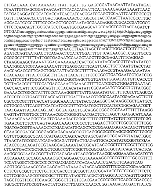
Figure 1 is a nucleotide sequence of the fragment
600 bp Bam HI/Bam fragment to ED-IL4;
Figure 2 shows the Bam HI ED-IL4 fragment ligated
into pGEX 6p-1 plasmid to generate translated sequence
(Seq. ID: No. 4) and DNA sequence (Seq. ID: No. 5);
Figure 3 shows expression of the fusion protein
product after induction with 0.1 mM IPTG displayed by
Coomasie blue staining. Figure 3A shows pGEX-ED-IL4
clones in MC 1061 cells. Figure 3B shows pGEX-ED-IL4
clones in BW 26444 cells;
Figure 4 shows schematic representation of the
protocol followed to purify the inactive fusion construct
and the cleaning and removing of the GST moiety away from
the active ED-IL4 product;
Figure 5 demonstrates EFC activity of the GST-ED-IL4
construct after addition of increasing amounts of a
specific protease at 15 min (Figure 5A) and at 30 min
(Figure 5B) read times;
Figure 6 shows bar graphs showing the results of
assays employing a supernatant fraction (Figure 6A) and
an adherent fraction (Figure 6B) , where the former is not
lysed and the latter is lysed;
CA 02474130 2004-07-22
WO 03/065004 PCT/US03/02520
Figure 7 is a plot of the effect of FXa
concentration on the observed signal in two different
buffers with an expression construct comprising ProLabel
(ED) ;
5 Figure 8 shows plasmid sequence (Seq. ID: No. 6);
Figure 9 is a plot of the effect of FXa on lysis of
cells; and
Figure 10 is a plot of the effect of adding a signal
sequence to a genetic construct on the available surface
concentration of the expression product of the genetic
construct.
DETAILED DESCRIPTION OF THE INVENTION
Target protease assays are provided using a protein
reagent that is specifically designed to be responsive to
cleavage by one or a related family of target proteases
and, upon cleavage, provide an entity that can be readily
detected. The protein reagent comprises a fragment of an
indicator enzyme referred to as the enzyme donor ("ED"),
where the fragment may be derived from the N- or C-
proximal portion of the indicator enzyme and will
generally be an oligopeptide of less than 100kDa. The ED
will link two hindering entities, usually sterically
hindering, wherein the linkage to at least one of the
hindering entities comprises a protease recognition site,
with the provison that when one of the hindering entities
is a surface, only the surface hindering entity need be
present to provide hindrance. Preferably, there will be
two hindering entities. By hindering is intended that
there is at least a 5-fold reduction in the activity of
the ED in the presence of the EA when bound to the
hindering entities. The protein reagent is substantially
inhibited from binding to a complementary fragment of the
cognate enzyme, referred to as the enzyme acceptor
("EA"), where the complex of the ED and EA results in a
functional indicator enzyme.
CA 02474130 2004-07-22
WO 03/065004 PCT/US03/02520
6
The indicator enzymes and their fragments are
required to have a number of characteristics. The
fragments should be substantially inactive, in that there
should be little, if any, background with only one
fragment present in the presence of substrate. Secondly,
the fragments should have sufficient affinity for each
other, so that upon scission of one of the hindering
entities from a fragment of the protein reagent, the
fragments will combine to provide an active enzyme. The
ED fragment of the protein reagent will complex with the
EA fragment as a result of the affinity of the fragments
of the enzyme for each other or as a result of being
fused to auxiliary binding entities that will bring the
enzyme fragments together resulting in an active enzyme.
That is, in the former case, the enzyme fragments are
capable of complexing without having an auxiliary binding
entity to bring the fragments together to form a complex.
In the latter case, the enzyme fragments will not
independently form a complex, but when the auxiliary
proteins form a complex, the enzyme fragments are then
able to form an active enzyme.
Various indicator enzymes are known that fulfill
these criteria and additional enzymes may be developed in
accordance with known technologies. Indicator enzymes
that fit these criteria include f3-galactosidase (see U.S.
Patent no.4,708,929), ribonuclease A (see U.S. Patent no.
4,378,428), where the smaller fragment may come from the
amino or carboxy terminus, (3-lactamase (see WO 00/71702
AND 01/94617 and Wehrman, et al., Proc. Natl. Acad. Sci.
2002, 99, 3469-74), or enzymes that have small peptide
cofactors, such as adenovirus proteases (see U.S. Patent
no. 5, 935, 840) . To identify other indicator enzymes that
can serve in place of the above indicator enzymes, enzyme
genes may be cleaved asymmetrically to define a small and
a large fragment, and expressed in the same and different
CA 02474130 2004-07-22
WO 03/065004 PCT/US03/02520
7
cells. In the presence of the substrate, the cells
producing both fragments would catalyze the reaction of
the substrate, while there should be little, if any,
turnover with the individual fragments. Alternatively,
one may express the fragments individually and, if there
is no reaction, combine the mixtures to see whether an
enzyme-catalyzed reaction occurs. As for the enzyme
fragments comprising auxiliary fragments, a number of
enzymes are known whose fragments will complex to form an
active enzyme, such as DHFR, and others may be determined
as described above.
Indicator enzymes of interest are those that are
below about 300kDa, generally below about 150kDa. The
independently complexing small fragment will be under 15
kDa, more usually under about 10kDa, frequently under
about 125 amino acids, generally under about 100 amino
acids and preferably not more than about 75 amino acids.
Depending on the enzyme, the independently complexing ED
may be as small as 10 amino acids, usually being at least
about 25, more usually at least about 35 amino acids.
With this criterion in mind, the fragments that are
screened can be selected to provide the appropriately-
sized small fragment.
The enzymes having fragments that complex in
conjunction with a fused auxiliary protein will generally
have fragments having from 20 - 80, more usually 25-75%
of the amino acids of the enzyme. The fragments may be
modified by the addition of from about 1 to 20, usually
2 to 10, amino acids to enhance the affinity of the
fragments during complexation. Enzymes that provide for
low affinity complexation to an active enzyme include ~-
galactosidase, (3-glucuronidase, Staphylococcal nuclease,
3-lactamase, as exemplary. The binding proteins may have
as few as 8, more usually at least 10 amino acids and may
be 150, usually not more than about 100kDa. Binding
CA 02474130 2004-07-22
WO 03/065004 PCT/US03/02520
8
proteins may include homo- and heterodimers, epitopes and
immunoglobulins or fragments thereof, e.g., Fab, ligands
and receptors, etc. In some instances, complexation may
require the addition of an additional reagent, so that
complexation with formation of an active enzyme does not
occur to any significant degree in the absence of the
additional reagent, e.g., FK1012, rapamycin and
cyclosporin.
Each of the indicator enzymes will have an
appropriate substrate. (3-galactosidase uses effectively
fluorescers having phenolic groups that are etherified
with a (3-galactosyl group. Ribonuclease A, fluorescer
modified nucleotides, exemplified by 5'-O-acetyl 2'-O-
(tetrahhydropyran-2-yl)uridine 3'-(4-methylumbelliferon-
7-yl) ammonium phosphate, adenovirus proteinase, -(L, I,
M)-X-G-G/X- or -(L, I, M)-X-G-X/G-, where the vertical
line denotes the position of cleavage; the P3 (X)
position appears to be unimportant for cleavage
(Anderson, C. W., Virology, 177;259 (1990); Webster, et
al., J. Gen. Virol., 70;3225 (1989)) and the peptide
substrate can be designed to provide a detectable
signal, e.g. using fluorescence resonance energy
transfer, by having a fluorescer and a quencher on
opposite sides of the cleavage site. (3-Glucuronidase
substrates are exemplified by 5-Br-4-Cl-3-indolyl (3-D-
glucuronidase.
Since 3-galactosidase is paradigmatic of the
peptides used in the subject invention, demonstrating the
criteria for having two peptides that when combined
complex non-covalently to form an active enzyme, this
enzyme will be frequently referred to hereafter as
illustrative of the class, except for those situations
where the different enzymes must be considered
independently. The ED for 3-galactosidase is extensively
described in the patent literature. U.S. Patent nos.
CA 02474130 2004-07-22
WO 03/065004 PCT/US03/02520
9
4,378,428; 4,708,929; 5,037,735; 5,106,950; 5,362,625;
5,464,747; 5,604,091; 5,643,734; and PCT application nos.
W096/19732 and W098/06648 describe assays using
complementation of enzyme fragments. The 3-galactosidase
ED will generally be of at least about 35 amino acids,
usually at least about 37 amino acids, frequently at
least about 40 amino acids, and usually not exceed 100
amino acids, more usually not exceed 75 amino acids. The
upper limit is defined by the effect of the size of the
ED on the performance and purpose of the determination,
the inconvenience of a larger construct, and the like.
While the subject methodology has particular application
for target proteases, the method may be used for any
enzyme that results in the cleavage of a covalent bond,
e.g., hydrolases, so as to make the ED accessible to the
EA. By providing for ester linkages, either organic or
inorganic, phosphate anhydrides, etc., for example,
cleavage of such linkages would make the ED accessible.
However, these protein reagents could not be directly
synthesized by recombinant techniques and to that extent
these types of assays are less attractive commercially.
The protein reagent may have the recognition
sequence proximal to the N- or C-terminus of the ED.
Generally, fewer than 50 amino acids, more usually fewer
than 25 amino acids and preferably fewer than about 15
amino acids will remain joined to the ED after scission
of the recognition sequence. However, the fragment that
is released should comprise at least about 125 amino
acids, more usually at least about 150 amino acids and
not more than about 300 amino acids for the independently
complexing fragment and may be 500 or more amino acids
for the non-independently fusion protein fragments.
Stability of the small fragments is greatly enhanced by
having a protein that is stable to degradation, which is
achieved by having amino acids additional to the ED,
CA 02474130 2004-07-22
WO 03/065004 PCT/US03/02520
particularly the smaller independently complexing EDs.
The additional amino acids will usually be added to the
terminus of the ED distal to the protease recognition
sequence. When there are two EDs on either side of the
5 recognition sequence, long linkers can be employed to
provide the stability for the ED, where the linkers may
be greater than 50 amino acids, but usually fewer than
100 amino acids. The complex formation inhibiting
entities, usually sterically inhibiting entities, may be
10 any moiety that substantially reduces the ability of EA
to complex with the ED. Various entities may serve this
purpose, including surfaces, liposomes, which includes
cells or cell ghosts, and large molecules, that are able
to impede the complex formation of ED and EA.
The inhibiting entities will frequently be
poly (amino acids) although other chemical moieties may be
employed such as polysaccharides or surfaces involving
glass, plastic, lipid membranes, etc. The poly(amino
acids) may be glycosylated to enhance the steric effect
of a sterically hindering entity. The poly(amino acids)
may be naturally occurring proteins, mutants of naturally
occurring proteins or synthetic proteins, where synthetic
proteins means that there is no known naturally occurring
analog. Generally the poly(amino acids) will be at least
10 kDa, usually at least about 20 kDa, more usually at
least about 25 kDa, and preferably at least about 30 kDa.
Since beyond a particular size, there will be no
advantage to further increasing the size, where the
poly(amino acids) are selected arbitrarily, that is, the
poly(amino acid) does not serve a specific protein
function, the poly(amino acid) will generally be less
than about 125 kDa, usually less than about 100 kDa, and
preferably less than about 75 kDa. The molecular weight
of a poly(amino acid) hindering entity is not the only
consideration, as conformation also has an effect. An
CA 02474130 2004-07-22
WO 03/065004 PCT/US03/02520
11
extended chain will have less hindering effect than a
poly(amino acid) having a globulin formation. At least
one of the poly(amino acid) entities will be a naturally
occurring protein or fragment thereof, when the
recognition sequence is part of the entity.
In many instances, one of the complex inhibiting
entities will play a functional role. For example, the
poly(amino acid) may comprise the recognition sequence,
so that the recognition sequence will be in the proper
conformation for cleavage. Alternatively, the poly(amino
acid) may undergo self-cleavage when modified, so that
the protease assay will detect the modification of the
poly (amino acid) . Other protease related events that can
be measured include complexing to a second protein that
makes the recognition sequence available for
intermolecular cleavage, activation of a pathway that
results in cleavage of the recognition sequence, the
presence of a cofactor necessary, directly or indirectly,
for cleavage of the recognition sequence, and the like.
In effect, any event in the cell where there exists the
capability for cleavage of the recognition sequence or
the outcome of the event, the capability of cleavage of
the recognition sequence can be monitored.
The protein reagent may be primarily comprised of
amino acids, amino acids and other common modifiers, such
as saccharides, phosphates, lipids, acyl groups, alkyl
groups, etc. The protein reagent may also be linked to
macromolecules, such as surfaces, e.g. wells, slides,
chips, etc., or liposomes, or cells. Generally, only one
terminus of the poly(amino acid) will be linked to a
surface, except where cells are involved, where a single
linkage may be involved or the poly(amino acid) may be
threaded repeatedly through the membrane.
The protein reagent, when other than linked to a
surface, will generally be at least about 10 kDa and not
CA 02474130 2004-07-22
WO 03/065004 PCT/US03/02520
12
more than about 500 kDa, usually not more than about 200
kDa, frequently not more than about 150 kDa, usually in
the range of about 15 kDa to 100 kDa, more usually in the
range of about 15 kDa to 75 kDa for independently
complexing enzyme fragments and usually in the range of
about 25 kDa to 400 kDa, more usually in the range of
about 25 kDa to 250 kDa when comprising non-independently
complexing enzyme fragments. When linked to a surface,
the protein reagent will be at least about 10 kDa, but
usually not more than about 150kDa for independently
complexing enzyme fragments and about 25 to 300 kDa for
non-independently complexing enzyme fragments. As
previously indicated, the complex inhibiting entities
will be chosen so as to substantially interfere with the
binding of the EA to the ED, when both hindering entities
are present, while there will be significantly less
interference when one of the entities has been removed.
While for the most part, the protein reagent can be
a single molecule comprised solely of covalent bonds,
this is not necessary. For example, one may have a
sequence that has an entity that will complex with
another protein to provide steric hindrance. One may
have biotin or an amino acid equivalent at one terminus
of the linker, whereby strept/avidin will serve as a
sterically hindering protein. Alternatively, other
oligopeptides can serve to bind to antibodies or Fab to
provide the sterically hindering protein.
The poly(amino acid) portion of the protein reagent
may be linked to a surface by any convenient means. In
many cases, the method of linking will depend upon the
composition of the poly(amino acid) . For example, where
there are no available cysteines in the poly(amino acid)
portion, then a terminal cysteine can be used to link to
an activated olefin, e.g., maleimide, bonded to the
surface. Alternatively, one may have a terminal
CA 02474130 2004-07-22
WO 03/065004 PCT/US03/02520
13
poly(histidine) to complex with complex metal ions, such
as nickel, so that a nickel complex on the surface will
bind to the poly(histidine). Another technique is to
have an amino acid sequence defining an epitope and have
antibodies or Fab fragments or their equivalents bound to
the surface or analogously, ligand and receptor. In
similar vein, amino acid sequences can serve as
surrogates for small ligands, such as biotin, so
strept/avidin may be bound to a surface for binding such
amino acid biotin surrogate, or the like. The particular
choice of linkage, whether covalent or non-covalent, will
generally not be crucial and will be dictated by the
poly(amino acid) portion of the protein reagent,
convenience and stability under the conditions of the
assay.
For liposomes, one can use various recognition
sequences that encode for lipid modification. References
illustrative of the different recognition sequences
include Magee, et al., Biol Res 2002, 35, 127-31; Kohl,
et al., J Biol Chem 2002, 277, 36760-7; Ikezawa, Biol
Pharm Bull 2002, 25, 409-17 and Smialowski-Fetter, et
al., Eur J Biochem 2002, 269, 1109017. By providing the
lipid recognition sequences on one side of the protease
recognition sequence distal from the ED, cleavage of the
protease recognition sequence will release the ED from
the complex inhibiting effect of the liposome.
The preparation of liposomes comprising proteins
bound to the outer leaf of the membrane is well
established in the literature. See, for example, Willis,
et al., Bioconjug Chem 1998, 9, 573-82; Sankaram, Biophys
J 1994, 67, 105-12; Radford, et al., Biochem Pharmacol
1991, 41, 307-9; and Claassen and van Rooijen, Prep
Biochem 1983, 13, 167-74.
As indicated, when a hindering entity is a surface,
one can obtain sufficient differentiation between ED
CA 02474130 2004-07-22
WO 03/065004 PCT/US03/02520
14
bound to the surface and ED released from the surface in
the affinity for the EA. Therefore, it is possible to
have a single hindering entity, where the ED is linked to
the hindering surface through the recognition sequence.
Further hindrance is achieved with the second hindering
entity.
The recognition sequence will normally be linked to
the surface or liposome by a poly (amino acid) sequence of
at least about 10 amino acids, more usually at least
about 25 amino acids and not more than about 150 amino
acids, usually not more than about 100 amino acids. The
length of the chain will be governed by the inhibition of
the surface or liposome to inhibit protease cleavage,
where the shortest chain providing the desired
characteristic will generally be the most convenient.
When the linker is a protein that passes through the
membrane repeatedly, only the linking group to the first
membrane bound sequence is counted. Also, depending upon
the nature of the surface, the surface may have a
functionality for linking which is tethered by other than
a poly(amino acid) chain, so that the length of this
chain will affect what minimum length of chain is
required for the protein reagent.
The protein reagent may be present intracellularly,
in a lysate or in a sample composition.. The protein
reagent, when a poly(amino acid) backbone, can be
prepared by the expression of a gene encoding the protein
reagent. By introducing an expression construct
containing the gene encoding the protein reagent into an
appropriate host, the protein reagent will be expressed
and available for the protease measurement. The protease
measurement may be in relation to an event in the cell
that activates the protease of interest, the expression
of the protease, a change in the environment that results
in the expression, activation or deactivation of the
CA 02474130 2004-07-22
WO 03/065004 PCT/US03/02520
protease, the presence of a gene in the host that is
activated to express a protein that results in a change
in the activity of the protease of interest, the
production or destruction of a cofactor that affects the
5 activity of the protease of interest, or the like.
Therefore, many different events may be of interest that
affect the protease of interest, allowing for an indirect
measurement of the event. Thus, candidate compounds may
be studied for their activity where the readout is a
10 change in activity of the protease of interest. While in
some instances, the result may be ambiguous as to the
manner in which the compound is acting, the observed
change in the activity of the protease of interest
indicates that the candidate compound directly or
15 indirectly affects the protease activity in the cell.
The subject method may be performed intracellularly
or extracellularly, in the latter case with a lysate, a
lysate enriched for one or more components, a lysate
fraction, substantially pure protein reagent or a mixture
of protein reagents, or the like. In the case of the
intracellular assay, the expression construct comprising
the gene encoding the protein reagent will be present or
introduced into a host cell. The expression may be
constitutive or inducible, depending upon the promoter
employed for initiating transcription. The expression
construct may be integrated into the genome of the host
or be present on an extrachromosomal element, either
stably or transiently present in the host. Numerous
vectors based on origins of replication or viral origins
are commercially available and may be used in the present
invention. Once expression occurs, the assay may then be
performed by providing for the expression of EA in the
host cell and introducing a non-limiting amount of
substrate or lysing the cells at a single time point or
a plurality of time points and adding EA and substrate to
CA 02474130 2004-07-22
WO 03/065004 PCT/US03/02520
16
the lysate. In some instances, it may be desirable to
enrich the lysate for the protease, the protein reagent
or other component, where such enrichment may involve the
use of antibodies to remove undesirable components in the
lysate, chromatography, or other separation that does not
inactivate the protease and desirably not denature the
protein reagent.
Alternatively, one may wish to perform a protease
assay with pure or impure samples of the protein reagent
and the protease, where the individual reactive component
of the assay medium is at least about 0.1 weight % of
total' protein and may be at least about 1 weight % or
higher. In some instances, one may be interested in a
mixture of protein reagents, where one is interested in
the effect of the presence of different proteins on the
protease action with the protein reagent. Where a number
of different proteins share the same recognition
sequence, one can determine the susceptibility of the
different proteins to the protease, by measuring the
total reaction and then deconvoluting using antibodies to
the two proteins of the protein reagent. By having a
capturing antibody that is retained with the ED and a
labeled detection antibody, one can determine for each
protein reagent, the amount of the protein reagent that
has been cleaved. In this way, one may determine a
hierarchy as to the susceptibility for the different
proteins to protease cleavage. In the presence of a
limiting amount of protease, by taking aliquots of the
sample and analyzing for total cleavage and cleavage as
to one or more of the protein reagents, the rate of
cleavage of each of the protein reagents in the presence
of the other protein reagents can be determined. Those
protein reagents bound to surfaces or liposomes find
particular application in these assays.
The target proteases of interest are for the most
CA 02474130 2004-07-22
WO 03/065004 PCT/US03/02520
17
part those that have specific recognition sequences,
preferably having at least about 3 amino acids as their
recognition sequence and usually not more than about 12
amino acids, although additional amino acids may be
involved in enhancing the recognition by the protease.
As indicated, the protease may be an intra- or
intermolecular protease, where in the former case, the
protease will require activation before self-cleavage.
Enzymes of interest include serine/threonine hydrolases,
cysteine hydrolases, metalloproteases, BACEs (e.g., a-,
3- and y-secretases). Included within these classes are
such protein groups as caspases, the individual MMPs,
elastases, collagenases, ACEs, carboxypeptidases, blood
clotting related enzymes, complement components,
cathepsins, dipeptidyl peptidases, granzymes, etc. For
other enzyme groups, see Handbook of Proteolytic enzymes,
ed. AJ Barnet, ND Rowland, and JF Woessner. Other types
of enzymes include abzymes.
Specific serine proteases include neutrophil
elastase, involved in 'pulmonary emphysema, leukocyte
elastase, tyrosine carboxypeptidase, lysosomal
carboxypeptidase C, thrombin, plasmin, dipeptidyl
peptidase IV; metalloproteinases include
carboxypeptidases A and B, angiotensin converting enzyme,
involved in hypertension, stromelysin, involved with
inflammatory disorders, e.g., rheumatoid arthritis, P.
aeruginosa elastase, involved in lung infections;
aspartic proteases include renin, involved in
hypertension, cathepsin D, HIV protease; cysteine
proteases include lysosomal carboxypeptidase, cathepsin
B, involved in cell proliferative disorders, cathepsin G,
cathepsin L, calpain, involved with brain cell
destruction during stroke; etc.
The proteases may be involved with various
processes, such as infections and replication of the
CA 02474130 2004-07-22
WO 03/065004 PCT/US03/02520
18
infectious agent, viral, bacterial, fungal, and protista;
phagocytosis, fibrinolysis, blood clotting cascases,
complement cascades, caspase cascades, activation of
proforms of proteins, protein degradation, e.g.,
ubiquitinated proteins, apoptosis,'etc. , cell growth,
attachment, synaptic processes, etc. The proteases may
come from a variety of sources, either prokaryotes,
eukaryotes or viruses, depending on the nature of the
assay. For detection of infectious diseases, the source
of the protease may be a virus, a bacterium, protista,
fungus or other unicellular organism. For higher orders
of species, the enzyme may be derived from plants, non-
vertebrates, vertebrates, particularly mammals, such as
domestic animals, e.g., bovine, porcine, canine, feline,
lagomorpha, murine, etc., primates, e.g., humans. The
purpose of measuring the protease will be widely varied.
In some instances, one will be concerned with identifying
the source, such as a virus, where the protein reagent
will comprise a viral protein specifically cleaved by the
protease. In other cases, one may be interested in the
presence of the protease in a biological sample,
determining whether the protease is present and in what
concentration. One will also be interested in
determining the amount or change in amount of protease in
response to changes in the nature of the cell, e.g.,
normal and cancerous, or in response to a change in
environment, e.g., physical or chemical environment,
native or diseased state, e.g., infection, or the like.
The subject system is particularly useful for high
throughput screening of drug candidates, as to their
effect on the target protease or non-target proteases.
As already indicated, the organisms from which the
proteases are naturally derived are varied. Among
viruses, the proteases may be derived from HIV-1, and -2,
adenoviruses, hepatitis viruses, A, B, C, D and E,
CA 02474130 2004-07-22
WO 03/065004 PCT/US03/02520
19
rhinoviruses, herpes viruses, e.g., cytomegalovirus,
picornaviruses, etc. Among unicellular microorganisms
are Listeria, Clostridium, Escherichia, Micrococcus,
Chlamydia, Giardia, Streptococcus, Pseudomonas, etc. Of
course, as indicated above, there are numerous mammalian
proteases of interest, particularly human proteases.
Depending on the target protease to be measured, one
of the proteins linked to the ED may be defined. Where
the recognition sequence is dependent on the conformation
of the protein, it will usually be necessary to use at
least a portion of the natural protein to obtain the
desired conformation. Where one is interested in a
modification of the protein that permits intra- or
intercellular proteolysis, the protein will also be
defined. Where the recognition sequence is not dependent
on the natural conformation, one may then use the
recognition sequence linked to the ED and joined at the
other terminus to an arbitrary protein that does not
interfere with the protease hydrolysis of the recognition
sequence. Therefore, the protein associated with the
recognition sequence will be widely varied, being either
specific for the protease being measured or being
arbitrary and joined to the recognition sequence to
provide the inhibition of binding of the EA to the ED.
There are numerous scientific articles describing
proteases and their substrates. Illustrative articles
are as follows, whose relevant content is specifically
incorporated herein by reference. Among the
metalloproteinases are MMP-2, having target sequences
L/IXXXHy; XHySXL; and HXXXHy (where Hy intends a
hydrophobic residue), Chen, et al., J. Biol. Chem., 2001.
Other enzymes include mitochondrial processing peptidase,
having the target sequence RXXAr (where Ar is an aromatic
amino acid), Taylor, et al., Structure 2001, 9, 615-25;
caspases, VAD, DEVD and DXXD, as well as the RB protein,
CA 02474130 2010-03-03
WO 03/065004 PCT/US03/025211
Fattman, et al., Oncogene 2001, 20, 2918-26, DDVD of HPK-
1, Chen, et al., Oncogene 1999, 18, 7370-7; VEMD/A and
EVQD/G of Keratins 15 and 17, Badock, et al., Cell Death
Differ. 2001, 8, 308-15; WEHD of pro-interleukin-113,
5 Rano, et al., Chem. Biol. 1997, 4,149-55; furin, KKRKRR
of RSV fusion protein, Zimmer, et al., J. Biol.
Chem.2001, 20, 2918-26; HIV-1 protease, GSGIF*LETSL,
Beck, et al., Virology 2000, 274, 391-401. Other enzymes
include thrombin, VPRGS, Factor Xa protease, IEGR,
10 enterokinase, DDDDK, 3C human rhinovirus protease,
LEVLFQ/GP.
Other references describing proteases include:
Rabay, G. ed., "Proteinases and Their Inhibitors in Cells
and Tissues, 1989, Gustav Fischer Verlag, Stuttgart;
15 Powers, et al., in "Proteases-Structures, Mechanism and
Inhibitors," 1993, Birkhauser Verlag, Basel, pp.3-17;
Patick and Potts, Clin. Microbiol. Rev. 1998, 11, 614-27;
Dery, et al., Am. J. Physiol. 1998, 274, C1429-52;
Kyozuka, et al., Cell Calcium 1998, 23, 123-30; Howells,
20 et al., Br. J. Haematol. 1998, 101, 1-9; Hill and Phylip,
Adv. Exp. Med. Biol. 1998, 436, 441-4; Kidd, Ann. Rev.
Physiol. 1998, 60, 533-73; Matsushita, et al., Curr.
Opin. Immunol. 1998, 10, 29-35; Pallen and Wren, Mol.
Microbiol.1997, 26, 209-21; DeClerk, et al., Adv. Exp.
Med. Biol. 1998, 425, 89-97; Thornberry, Br. Med. Bull.
1997, 53, 478-90.
Besides the naturally occurring recognition
sequences, using combinatorial approaches, one can design
recognition sequences that will be specific for one or a
family of enzymes. By preparing a library of
oligopeptides that are labeled and having an array of the
labeled oligopeptides where the location identifies the
sequence, one need only add the protease of interest to
the array and detect the release of the label. Having
CA 02474130 2004-07-22
WO 03/065004 PCT/US03/02520
21
microwell plates, with the oligopeptides bound to the
surface and labeled with a fluorescer, allows one to
follow cleavage by internal reflection of activating
irradiation. Numerous other approaches can also be used.
By using synthetic sequences, one can optimize the
cleavage for a particular protease. By using a plurality
of protein reagents, one can obtain profiles that will be
specific for specific enzymes.
Proteins can find use as part of the protein reagent
that are not specific for the protease, but have the
desired stability in a cell in that they are not readily
degraded, provide solubility, are substantially free from
adverse interactions with other proteins in the cell.
The protein reagent will usually be prepared by
expression of a gene encoding the protein reagent. An
expression construct is prepared having a transcriptional
and translational regulatory region, which may include an
enhancer that will be functional in the host cell. Where
one is interested in the protein reagent for use in
vitro, the host will be selected primarily for
convenience as to expression and purification. For the
most part, unicellular hosts, such as bacteria and yeast,
will be employed. If glycosylation is desired, one will
usually use a mammalian host cell that provides for
glycosylation, particularly the natural glycosylation
associated with the protein undergoing cleavage of the
protein reagent. The expression construct is produced in
accordance with conventional ways, as described in
various laboratory manuals and by suppliers of vectors
that are functional in numerous hosts. See, for example,
Sambrook, Fritsch & Maniatis, "Molecular Cloning: A
Laboratory Manual," Second Edition (1989) Cold Spring
Harbor Laboratory Press, Cold Spring Harbor, N.Y. (herein
"Sambrook et al., 1989"); "DNA Cloning: A Practical
Approach," Volumes I and II (D. N. Glover ed. 1985);
CA 02474130 2004-07-22
WO 03/065004 PCT/US03/02520
22
"Oligonucleotide Synthesis" (M. J. Gait ed. 1984);
"Nucleic Acid Hybridization" [B. D. Hames & S. J. Higgins
eds. (1985)]; "Transcription And Translation" [B. D.
Hames & S. J. Higgins, eds. (1984)]; "Animal Cell
Culture" [R. I. Freshney, ed. (1986)]; "Immobilized Cells
And Enzymes" [IRL Press, (1986) ] ; B. Perbal, "A Practical
Guide To Molecular Cloning" (1984).
Vectors that may be used include viruses, plasmids,
cosmids, phagemids, YAC, BAC and HAC. Other components
of the vector may include origins of replication for one
or more hosts, expression constructs for selection,
including antibiotic resistance, proteins providing for
a signal, etc., integration sequences and enzymes
providing for the integration, multiple cloning sites,
expression regulatory sequences, expression construct for
a protein of interest, particularly where the protein is
coordinately or differentially expressed in relation to
the protein reagent, sequences allowing for ready
isolation of the vector, etc. Commercially available
vectors have many or all of these capabilities and may be
used to advantage.
The DNA or RNA vectors may be introduced into a
cellular host, whereby the expression of the protein
reagent can occur. The host may be a primary cell, a
cell line, a unicellular microorganism, or the like,
where the cell may be modified having an expression
construct integrated or transiently present in the cell
expressing EA, expressing or over- expressing a protein
that the cell does not normally express under the
conditions of the assay, not expressing a protein that
the cell normally expresses as a result of a knockout,
transcription or translation inhibitor, or the like.
The gene encoding the protein reagent will be part
of an expression construct. The gene is positioned to be
under transcriptional and translational regulatory
CA 02474130 2004-07-22
WO 03/065004 PCT/US03/02520
23
regions functional in the cellular host. In many
instances, the regulatory regions may be the native
regulatory regions of the gene encoding the protein of
interest, where the protein reagent may replace the
native gene, particularly where the protein reagent is
functional as the native protein, may be in addition to
the native protein, either integrated in the host cell
genome or non-integrated, e.g., on an extrachromosomal
element. In those cells in which the native protein is
present and expressed, the protein reagent will be
competing with the native protein for transcription
factors for expression. The site of the gene in an
extrachromosomal element or in the chromosome may vary as
to transcription level. Therefore, in many instances,
the transcriptional initiation region will be selected to
be operative in the cellular host, but may be from a
virus or other source that will not significantly compete
with the native transcriptional regulatory regions or may
be associated with a different gene from the gene for the
protein of interest, which gene will not interfere
significantly with the transcription of the protein
reagent. However, where one is interested in the
transcription of the gene of interest, that is, proteins
involved in controlling the induction and transcription
of the protein of interest, it will usually be desirable
to use the native transcriptional regulatory region.
It should be understood that the site of integration
of the expression construct, if integrated into a host
chromosome, would affect the efficiency of transcription
and, therefore, expression of the protein reagent. One
may optimize the efficiency of expression by selecting
for cells having a high rate of transcription, one can
modify the expression construct by having the expression
construct joined to a gene that can be amplified and
coamplifies the expression construct, e.g., DHFR in the
CA 02474130 2004-07-22
WO 03/065004 PCT/US03/02520
24
presence of methotrexate, or one may use homologous
recombination to ensure that the site of integration
provides for efficient transcription. By inserting an
insertion element into the genome, such as Cre-Lox at a
site of efficient transcription, one can direct the
expression construct to the same site. In any event, one
will usually compare the enzyme activity from cells in a
predetermined environment to cells in the environment
being evaluated.
The vector may be introduced into the host cells by
any convenient and efficient means, such as transfection,
electroporation, lipofection, fusion, transformation,
calcium precipitated DNA, etc. The manner in which the
vector is introduced into the host cells will be one of
efficiency and convenience in light of the nature of the
host cell and the vector and the literature has numerous
directions for the introduction of a vector into a host
cell and the selection of the host cells that have
effectively received the vector. By employing expression
constructs that allow for selection, e.g., antibiotics,
the cells may be grown in a selective medium, where only
the cells comprising the vector will survive.
Once the host cells have been transformed and
comprise the vector and are expressing the protein
reagent, the cells may be used in a variety of ways.
Where the protein of interest is an endogenous protein,
when the cell has EA and a substrate that produces a
detectable signal, one may measure the signal from the
culture medium. Alternatively, one can use such devices
as a fluorescence activated cell sorter, where the signal
is fluorescence, or other method for measurement. Where
one needs to add the necessary (3-galactosidase reagents
for the (3-galactosidase reaction, the cells are lysed and
the necessary reagents added and the signal determined.
The cells may be grown under conditions that affect the
CA 02474130 2004-07-22
WO 03/065004 PCT/US03/02520
protease of interest, for example, inhibiting
transcription, translation or the protease activity. By
introducing compounds that may serve as agonists or
antagonists of the protease of interest, one can measure
5 the rate at which the protein reagent is cleaved by the
increase in activity of the (3-galactosidase. By taking
a determination at a specific time or at two or more
different times, one can measure the rate of the (3-
galactosidase reaction. By comparing cells in the
10 presence and absence of the candidate compound, one can
determine the effect of the candidate compound on the
protease activity.
Expression vectors containing the protein reagent
gene inserts can be identified by four general
15 approaches: (a) PCR amplification of the desired plasmid
DNA or specific mRNA, (b) nucleic acid hybridization, (c)
presence or absence of "marker" gene functions, and (d)
expression of inserted sequences. In the first approach,
the nucleic acids can be amplified by PCR with
20 incorporation of radionucleotides or stained with
ethidium bromide to provide for detection of the
amplified product. In the second approach, the presence
of the protein reagent gene inserted in an expression
vector can be detected by nucleic acid hybridization
25 using probes comprising sequences that are homologous to
the protein reagent gene. In the third approach, the
recombinant vector/host system can be identified and
selected based upon the presence or absence of certain
"marker" gene functions (e.g., 3-galactosidase activity,
thymidine kinase activity, resistance to antibiotics,
transformation phenotype, occlusion body formation in
baculovirus, etc.) caused by the insertion of foreign
genes in the vector. In the fourth approach, recombinant
expression vectors can be identified by assaying for the
activity of the protein reagent gene product expressed by
CA 02474130 2004-07-22
WO 03/065004 PCT/US03/02520
26
the recombinant expression vector.
One may use promoters that are active for a short
time, such as viral promoters for early genes, for
example, the human cytomegalovirus (CMV),immediate early
promoter. Other viral promoters include, but are not
limited to, strong promoters, such as cytomegaloviral
promoters (CMV), SR.alpha. (Takebe et al., Mole. Cell.
Biol. 8:466 (1988)), SV40 promoters, respiratory
syncytial viral promoters (RSV), thymidine kinase (TK),
beta-globin, etc. Alternatively, an inducible promoter
can be used.
A large number of promoters have found use in
various situations, for various purposes and for various
hosts. Many promoters are commercially available today.
Expression of the protein reagent may be controlled by
any promoter/ enhancer element known in the art, but these
regulatory elements must be functional in the host or
host cell selected for expression. Promoters which may be
used to control fusion gene expression include, but are
not limited to, the SV40 early promoter region (Benoist
and Chambon, 1981, Nature 290:304-310), the promoter
contained in the 3' long terminal repeat of Rous sarcoma
virus (Yamamoto, et al., 1980, Cell 22:787-797), the
herpes thymidine kinase promoter (Wagner et al., 1981,
Proc. Natl. Acad.. Sci. U.S.A. 78:1441-1445), the
regulatory sequences of the metallothionein gene
(Brinster et al., 1982, Nature 296:39-42); and the
following animal transcriptional control regions, which
exhibit tissue specificity and have been utilized in
transgenic animals: elastase I gene control region which
is active in pancreatic acinar cells (Swift et al., 1984,
Cell 38:639-646; Ornitz et al., 1986, Cold Spring Harbor
Symp. Quant. Biol. 50:399-409; MacDonald, 1987,
Hepatology 7:425-515); insulin gene control region which
is active in pancreatic beta cells (Hanahan, 1985, Nature
CA 02474130 2004-07-22
WO 03/065004 PCT/US03/02520
27
315:115-122); immunoglobulin gene control region which is
active in lymphoid cells (Grosschedl et al., 1984, Cell
38:647-658; Adames et al., 1985, Nature 318:533-538;
Alexander et al., 1987, Mol. Cell. Biol. 7:1436-1444);
mouse mammary tumor virus control region which is active
in testicular, breast, lymphoid and mast cells (Leder et
al., 1986, Cell 45:485-495): albumin gene control region
which is active in liver (Pinkert et al., 1987, Genes and
Devel. 1:268-276); alpha-fetoprotein gene control region
which is active in liver (Krumlauf et al., 1985, Mol.
Cell. Biol. 5:1639-1648; Hammer et al., 1987, Science
235:53-58); alpha 1-antitrypsin gene control region which
is active in the liver (Kelsey et al., 1987, Genes and
Devel. 1:161-171); beta-globin gene control region which
is active in myeloid cells (Mogram et al., 1985, Nature
315:338-340; Kollias et al., 1986, Cell 46:89-94); myelin
basic protein gene control region which is active in
oligodendrocyte cells in the brain (Readhead et al.,
1987; Cell 48:703-712); myosin light chain-2 gene control
region which is active in skeletal muscle (Sani, 1985,
Nature 314:283-286) ; prostate specific antigen control
region, which is active in prostate cells (U.S. Patent
nos. 6,197,293 and 6,136,792); and gonadotropic releasing
hormone gene control region which is active in the
hypothalamus (Mason et al., 1986, Science 234:1372-1378).
Alternatively, expression of the protein reagent gene can
be under control of an inducible promoter, such as
metallothionein promoter, which is induced by exposure to
heavy metals. For control of the gene transfected into
certain brain cells, a glucocorticoid inducible promoter
can be used, since glucocorticoids can cross the blood-
brain barrier. Alternatively, an estrogen-inducible
promoter, which would be active in the hypothalamus and
other areas responsive to estrogen, can be used. The
present invention contemplates the use of any promoter
CA 02474130 2004-07-22
WO 03/065004 PCT/US03/02520
28
inducible by a pharmacologic agent that can cross or
transmit a signal across the membrane and for neuronal
cells, the blood-brain barrier and influence
transcription.
Vectors containing DNA encoding the following
proteins, for example, have been deposited with the
American Type Culture Collection (ATCC) of Rockville, MD:
Factor VIII (pSP64-VIII, ATCC No. 39812); a Factor VIII
analog, "LA", lacking 581 amino acids (pDGR-2, ATCC No.
53100; VWF (pMT2-VWF, ATCC No. 67122); EPO (pRK1-4, ATCC
No. 39940; pdBPVMMTneo 342-12 (BPV-type vector) ATCC No.
37224); and GM-CSF (pCSF-l, ATCC No. 39754).
The vector will include the fusion gene under the
transcriptional and translational control of a promoter,
usually a promoter/enhancer region, optionally a
replication initiation region to be replication
competent, a marker for selection, as described above,
and may include additional features, such as restriction
sites, PCR initiation sites, an expression construct
providing constitutive or inducible expression of EA, or
the like. As described above, there are numerous vectors
available providing for numerous different approaches for
the expression of the protein reagent in a host.
The host cells will be selected to provide the
necessary transcription factors for expression of the
protein reagent and the other components for the purposes
of determination. The host cells will also be selected
toward providing an environment resembling the
environment being simulated. In many cases, primary cells
may be employed, both those maintained in culture and
obtained directly from,a patient. However, in many other
cases, established cell lines will be used, since the
cell lines can provide the desired environment and allow
for direct comparisons between studies, which comparisons
may not be available where using primary cell lines from
CA 02474130 2004-07-22
WO 03/065004 PCT/US03/02520
29
patients.
The efficiency of transcription can also be
determined by using a protein reagent that is stable,
i.e., it is not subject to significant modification
during the period of the assay. By using a stable
protein, such as a prion, (3-amyloid, synthetic
polypeptides, such as using collagen, keratin or elastin
motifs, or providing for secretion into a non-proteolytic
environment, one can determine the rate of expression
from a regulatory region of interest. By using
homologous recombination, one can insert the protein
reagent to be under the regulatory control of the
regulatory region of interest, including promoters.
enhancers, etc. Alternatively, one may introduce a
construct with the appropriate regulatory region, where
the native and constructed expression systems would both
be active, while the protein reagent would indicate the
effectiveness of the expression system. In this
instance, one would usually be interested in the effect
of a change, e.g., environment, genome, etc., on the
transcriptional activity of the regulatory region. One
could then evaluate the effect of an agent on the
transduction of a signal as a result of a binding event
at the cell surface, the effect of an intracellular
inhibitor, or the effect of a second pathway that
involves a first pathway. Desirably, the protein reagent
would replace one of the copies of the natural gene, so
as to have the same environment for transcription.
When using R-galactosidase as the enzyme, a number
of substrates for (3-galactosidase are known, where the
product is fluorescent or emits light. The common
substrates are (3-D-galactopyranosyl phenols, such as
fluorescein, mono- and di-susbtituted, o-nitrophenyl-(3-D-
galactoside, (3-methylumbelliferyl-(3-D-galactoside, X-gal,
resorufin-(3-D-galactoside, commercially available
CA 02474130 2004-07-22
WO 03/065004 PCT/US03/02520
oxetanes, e.g.,Galacto-Light Plus kits
(chemiluminescence) and chlorophenol red. The di-(3-D-
galactopyranosylfluorescein, and chlorophenol red- (3-D-
galactopyranoside may be used as intracellular markers.
5 The simplest procedure to describe is the use of
cells in culture and analysis of the lysate. In this
case, the cells are grown in culture. The protein
reagent and other constructs, as appropriate, may be
present in the cell integrated into the genome or may be
10 added transiently by the various methods for introducing
DNA into a cell for functional translation. The cells
may be in culture or in vivo. These methods are amply
exemplified in the literature, as previously described.
By employing a marker with the protein reagent for
15 selection of cells comprising the construct, such as
antibiotic resistance, development of a detectable
signal, etc., cells in culture comprising the protein
reagent can be separated from cells in which the
construct is absent. Once the protein reagent is being
20 expressed, the environment of the cells may be modified,
if desired. Candidate compounds may be added, ligand for
receptors, surface membrane or nuclear, or the two of
these may be added in combination, changes in the culture
medium may be created, other cells may be added for
25 secretion of factors or binding to the transformed cells,
viruses may be added, or the like. Given sufficient time
for the environment to take effect and/or taking aliquots
of the culture at different time intervals, the cells may
be lysed with a lysis cocktail comprising EA and enzyme
30 substrate and the signal from the product read. One can
then relate this result to the amount of protein reagent
present, particularly by using standards where the lysate
is spiked with different amounts of the protein reagent
and the amount of active protein reagent determined. One
would then have a graph relating signal to amount of
CA 02474130 2004-07-22
WO 03/065004 PCT/US03/02520
31
active protein reagent in the lysate.
For convenience, kits can be provided that may
include all or some of the major components of the
assays. For example, a kit may include an expression
construct, by itself or as part of a vector, e.g.,
plasmid, virus, usually attenuated, where the expression
construct may include a marker, a gene encoding a protein
for integration, a replication initiation site, and the
like. In addition to the expression construct, the kit
may include EA, substrate for (3-galactosidase, one or
more cell lines or primary cells, a graph of response in
relation to the amount of ED present, buffer, etc. In
some instances, cells may be engineered to provide a
desired environment, such as high levels of expression of
a protein involved in a pathway of interest, such as
surface membrane receptors, GPCRs, nuclear receptors,
e.g., steroid receptors, transcription factors, etc., or
may have been mutated, so as to have reduced levels of
expression affecting the expression of the protein
reagent and one is interested in enhancing the level of
expression.
As indicated, the subject method can be used in a
variety of situations to great effect, since the ED is
small enough to allow for functioning of the protein of
interest as a protein reagent with ED, while allowing for
ED to complex with EA to provide a functional f3-
galactosidase.
The following examples are intended to illustrate
but not limit the invention.
EXPERIMENTAL
In order to demonstrate the subject invention, the
DNA sequences of ED (also referred to as ProLabel) and
Interleukin-4 (IL4) are molecularly cloned into a
glutathione-S-Transferase (GST) fusion protein expression
vector (pGEX6P-1) where the fusion: GST-ED-IL4 protein is
CA 02474130 2004-07-22
WO 03/065004 PCT/US03/02520
32
expressed in bacterial cells. By expressing proteins
flanking both the NH2 and COOH-terminal ends of ED, the
ability of ED to complement with EA is substantially
reduced. Cleavage of the GST moiety from the purified
fusion protein results in substantially increased
complementation activity.
1. Generated by PCR, a 600 base pair Dam HI/Bam HI
fragment to ED-IL4, using DNA from plasmid pQE30-ED-
IL4 generated by DiscoveRx, Corp as the template.
The sequence of the fragment (Seq. ID: No. 1) is
shown in Figure 1.
2. The following primers were used:
a) B-ED-f:5'-CACGGATCCAGCTCCAATTCACTGGCCGTCG-3'
(Seq. ID: No. 2)
b) BH-IL4-R:
5'- CGCGGATCCAAGCTTTCAGCTCGAA.CACTTTGAATA-3'
(Seq. ID: No. 3)
3. The generated PCR fragment was digested with Bam HI
restriction enzyme following standard protocols and
then gel-purified.
4. The DNA was recovered from the gel slice by using a
Qiagen Gel extraction kit.
5. The Dam HI fragment was then ligated with gel
purified pGEX 6P-1 plasmid (Amersham Pharmacia
Biotechnology) DNA that had also been digested with
Dam HI and gel purified following the same methods.
6. Ligation was performed using New England Biolabs
Quick Ligation kit following the protocol. Half of
the ligation mix was used to transform DH5a cells.
CA 02474130 2004-07-22
WO 03/065004 PCT/US03/02520
33
The transformation mix was plated on LB ampicillin
plates and potential transformants were selected and
plasmid DNA was isolated and digested with a series
of restriction enzymes to confirm the cloning
reaction (see Figure 2; Protein Seq. ID: No. 4 and
DNA Seq. ID: No. 5).
7. Transform MC1061 cells (see Figure 3A) and BW26444
cells (see Figure 3B) with the GST-ED-IL4 plasmid
clone. Examine expression of the fusion protein
product after induction with 0.1 mM IPTG. Total
cellular protein displayed by Coomassie blue
staining.
8. Confirmed by western blot analysis using both anti-
GST polyclonal antibody and an anti-hIL4 antibody
that clones #2 and #40A and #40B are expressing the
GST-ED-IL4 fusion protein of predicted size.
9. Testing for complementation activity of the purified
GST-ED-IL4 fusion protein after treatment with a
sequence specific protease (PRE-SCISSIONTMprotease)
that cleaves the GST moiety from the fusion protein.
-Control (pGEX plasmid vector) and test (ED-IL4
clones) were grown overnight in L-broth with 50
mg/mL ampicillin. The next day, the cultures are
used to inoculate duplicate 3 mL tubes with fresh L-
Broth with ampicillin. The cultures are allowed to
go for 3 hours (-OD600 reading of 0 .2 - 0 .3) .
Induction of the fusion is done by adding 100 mM
IPTG to 0.1mM for one sample set of each culture
being tested. The cultures are allowed to go for
another 2 hours at 37 C.
After this time, the cultures are collected by
CA 02474130 2004-07-22
WO 03/065004 PCT/US03/02520
34
centrifugation, the cell pellet is resuspended in 1
mL PBS + a protease inhibitor (PI) cocktail. The
resuspended cells are then sonicated two times for
30 seconds with a one minute interval on ice between
sonications. The lysate is clarified with a low
speed spin. The supernatant is removed and to this,
300 ja.L of GST-agarose resin is added. The mixture
is incubated at 4 C, rocking for 2 hours. After
this time, the resin is pelleted, washed four times
with PBS +PI. Next, the fusion protein is eluted
from the resin with the addition of 250 ja.L of 20 mM
reduced glutathione. This mixture is allowed to
rock at 4 C for 1 hour. The resin is again pelleted
and the supernant is retained for analysis. 0, 2 or
10 units of protease is add to 50 pL of the eluted
fusion protein and incubated for 4 hours at 4 C.
15 aL of the treated sample is then transferred in
triplicate to a well on a 384-well plate. To this,
15 pL of 1X EA is added and 20 }1L of
chemiluminescent substrate is added. The samples
are read on a Packard lumicount reader immediately
and for 15 minute intervals thereafter for the next
hour. See Figure 4.
10. To determine specificity of the cleavage event, the
eluted GST-ED-IL4 material is treated again with 0,
1 or 3 pL of the pre-scission protease (2 mg/mL) or
1 or 3 p1 of Caspase 3 enzyme (7.3 pg/mL). The
samples are treated for 4.5 hours at 4 C . 15 pa.L of
1X EA is added and 20 'pL of chemiluminescent
substrate is added. The samples are read on a
Packard lumicount reader immediately and for 15
minute intervals for the next hour. See Figures 5A
and 5B.
CA 02474130 2010-03-03
WO 03/065004 PCT/US03/02520
Materials:
HEK293 parental cell line
HEK293 IKB - (3-galactosidase ED (55mer) stable
transfectant.
5 Growth media (DMEM/10% FES).
Factor Xa, lug/uL stock in water, kept as a -80 C
stock (Roche, Cat. no. 1 585 924)
Factor Xa cleavage buffer, made by adding 10mg/mL
BSA to 0.lmg/ml final concentration (New England Biolabs)
10 and 0.5 parts 0.2M CaC12 to 98.5 parts Dulbecco's PBS
(Sigma Cat. no. D8537) BSA is added just prior to use,
EA core buffer (PIPES, 30.24 g/L; NaCl, 23.38 g/L; EGTA,
3.80 g/L; Mg acetate, 2.15 g/L; Tween* 0.5 mL; NaOH,
6.9g/l; NaN3, 0.95 g/L, pH 6.9
15 EA reagent (1.8)M in EA core buffer)
Cell lysis buffer (KH2PO41 0.6805g/L; K;HPO4,
0.8709g/l; NaCl, 0.5844g/L; CHAPS, 10g/L; pH 6.9 with
NaOH)
Chemiluminescent substrate (Tropix, Applied
20 Biosystems Inc.) + Gal-Star + Emerald Enhancer Plasmid
pPL-FXa-(32-AR (or pPL (ED)-FXa (FXa cleavage consensus
sequence)- R2-adrenergic receptor (b2-AR))
FuGene 6 transfection reagent (Roche Cat. no. 1 815
091)
25 A construct, named pPL-FX-b2AR, was prepared as
follows. First, pCMV-PL-N1, a mammalian expression vector
for creating N-terminal ED fusion proteins, was created
by precise replacement of the EGFP coding sequences in
pEGFP-C1 (Clontech) with sequences encoding ED. Next, a
30 XhoI/BamHI DNA fragment encoding the FXa cleavage site
followed by the b2AR was subcloned into the XhoI/BamHI
sites of pCMV-PL-N1, creating a fusion of ED-FXa-b2AR.
The FXa-b2AR DNA fragment was obtained by PCR
amplification from a b2AR DNA template using PCR primers
35 that introduced a XhoI site and FXa cleavage site
* Trade-mark
CA 02474130 2004-07-22
WO 03/065004 PCT/US03/02520
36
encoding sequences at the 5' end, and a BamHI site at the
3' end. The sequence is described subsequently as SEQ
ID:NO. 6, except that GST is present at one terminus.
Procedure:
In this study, the assay procedure was as follows:
HEK293 transfectant cells expressing either the
cytoplasmic protein IkB-PL (stable transfectant) or PL-
FXa-b2AR (transient transfectant) were seeded into two
wells each of a 6-well culture dish at a density that
gave -.80% confluency following 2 days growth. For
transfection, transfection mix was prepared according to
the supplier FUGENE, using 0.15 p.L FUGENE reagent, 0.05
fag DNA and 5 j.iL serum free media. The transfected cells
were grown in DMEM/10 % FBS media for 48 to 72 hours prior
to assay.
At this time, culture media above the cells was
removed by gentle aspiration. To one set of wells was
added 1.0 mL of FXa buffer composed of PBSC/BSA
containing 2 j.1g/mL FXa. To the other was added the same
buffer lacking FXa. Reactions were incubated at room
temperature for 1 hour. Liquid above the cells (the
supernatant fraction) was carefully collected by
pipetting and then transferred to individual microfuge
tubes. The supernatants were cleared of any cells that
might have been carried over in the transfer by two
sequential, gentle centrifugations. Fifty microliters of
each supernatant fraction was aliquoted in quadruplicate
to individual wells of a 96-well assay plate. To these
wells was added 80 pL of EA Core Buffer/EA Reagent (3 : 1) .
To the cells remaining in the culture wells (the adherent
cell fraction) was added 1 mL of PBSC/BSA followed by 1.6
mL of Cell Lysis Buffer/EA Reagent (3:1). The cells were
lysed in this solution by pipetting up and down and then
130 J1L of each sample was aliquoted in quadruplicate to
individual wells of the 96-well assay plate. The assay
CA 02474130 2004-07-22
WO 03/065004 PCT/US03/02520
37
plate was incubated at 37 C for 1 hour, after which 30 pL
of CL substrate was added per well. The plate was
incubated at room temperature protected from light and
readings were taken periodically on the Northstar plate
reader from 15 minutes to 1 hour following substrate
addition. See Figures 6A and 6B for results.
That Factor Xa does not cause cell lysis was
established by an experiment in which a 96-well
microtiter plate is seeded 100 p.1L/well with two cell
lines: a HEK293 IKB - (3-galactosidase ED (55mer) stable
transfectant and the HEK293 parental cell line
transiently transfected with plasmid pPL-FXa-(32-AR.
HEK293 IKB - (3-galactosidase ED (55mer) stable
transfectant cells were obtained from a stock plate (-50%
confluent) treated with trypsin, quenched with media,
centrifuged and resuspended in 6ml fresh media. The
suspension for seeding the microtiter plate was 0.764 mL
washed cell suspension and 3.23 mL of fresh media. 100 pL
aliquots were transferred to the microtiter plate wells,
4 columns by 8 rows. The HEK293 parental cell line
transfected with pPL-FXa-(32-AR was treated as above,
except that the parental cell line was -90% confluent and
4.2mls of the washed cell suspension was diluted with
17.8 mL of media. For transfection, transfection mix was
prepared according to the supplier FuGENE, using 0.15 p.L
FuGENE reagent, 0.05 pg DNA and 5 p.L serum free media.
After seeding the wells, the cells were allowed to grow
-2 days to -80% confluence. Serial dilutions of the FXa
solution were made and the dilutions added as 50 p.L to
separate wells of the different HEK293 cells that had
their growth media removed by aspiration.
The mixtures were then incubated for 1hl0min at room
temperature. To the treated mixtures were added 80 pL of
EA Core Buffer/EA Reagent (3:1) to each well (whole-cell
assay). To a replica set of mixtures that were not
CA 02474130 2004-07-22
WO 03/065004 PCT/US03/02520
38
treated with Factor Xa was added 80 pL Cell Lysis
Buffer/EA Reagent (3:1) per well (lysed-cell assay). The
plate was gently agitated to facilitate mixing of the
reagents and then incubated at 37 C/5o CO2 for lh.
Chemiluminescent substrate (30 pL) was added to each
well, followed by gentle agitation and incubation in the
dark at room temperature for 15 min prior to a 90sec
reading taken with a Northstar plate reader. After
adjusting the results for whole-cell values as compared
to lysed-cell values, the data were graphed showing that
for the IKB - (3-galactosidase ED stable transfectant
cells there was substantially no change in the readings
with variation in the concentration of Factor Xa, while
the pPL-FXa-32-AR transient transfectant cells showed an
increase in the readings from 0.01 ng/well to 1000
ng/well of Factor Xa. The results are shown in Fig. 7.
The assay compared the effect of FXa enzyme
concentration on the EFC (enzyme fragment complementation
with formation of (3-galactosidase) activity as observed
with relative luminescent units (RLU). Using either PBSC
or PBSC/BSA (0.1%) buffer, a difference in about 30,000
RLUs was observed over a range in concentration of FXa of
about 10-3 to 101 }ig/mL.
These results show that one can obtain differential
activity of the ED with only one hindering entity, when
that one hindering entity is a surface. Enhanced
hindrance would be obtained with a second hindering
entity.
In the next study, a genetic construct was prepared
where the steric hindrance to formation of an active (3-
galactosidase enzyme by complexing with the EA came from
a cellular membrane and a protein at the other terminus.
By employing a construct that expresses a protein that is
directed to the cell membrane, where the protease
cleavage site and ED are extracellular and having a
CA 02474130 2004-07-22
WO 03/065004 PCT/US03/02520
39
protein as the extracellular terminus, the formation of
(3-galactosidase is substantially suppressed. The
strategy for preparation of the DNA construct having the
formula GST-ED-Factor Xa Cleavage Site-(32 Adrenergic
Receptor ( (32AR) is as follows:
Plasmid pGST-PL-FX-b2AR was constructed in two
steps. First, a DNA segment encoding GST and flanked by
Agel restriction sites was amplified by PCR using pGEX-
6P-1 (Amersham) as template DNA. The PCR primers GST
Forward (5' -AAAACCGGTATGTCCCCTATACTAGGTTA- 3 ' ) (Seq ID: No. 4)
and GST Reverse (5'-AAAACCGGTTTATCCGATTTTGGAGGATGGT-3')
(Seq ID: No. 5)both introduce an Agel restriction site
(underlined). In the second step, the PCR-amplified DNA
was digested with Agel and ligated to pPL-FX-b2AR DNA
that had been prepared by digestion with Agel followed by
treatment with alkaline phosphatase. The unique Agel site
in pPL-FX-b2AR is immediately 5' to the sequences
encoding ProLabel (PL) . The final construct, pGST-PL-FX-
b2AR, was confirmed by restriction analysis and DNA
sequencing.
The complete plasmid sequence (SEQ ID:NO 6) is as
shown in Figure 8.
Using the above construct, the construct is
transfected into HEK293 cells. The resulting
transfectants are selected for expression as described
above. Assays are performed with and without FXa and the
construct is shown to be substantially inhibited from
forming (3-galactosidase when the ED is part of the fusion
protein and bound to the cell membrane and is active when
released from the fusion protein upon cleavage with FXa.
The results are further substantiated by the above and
following observations.
That FXa does not cause cell lysis was established
by using cells transfected with an IKB-ED construct,
where only background activity was observed over a
CA 02474130 2004-07-22
WO 03/065004 PCT/US03/02520
concentration range of FXa of about 10-4 to 2x10' ~1g/mL.
See Figure 9.
It was found that, adding the cleavable
hemagglutinin signal sequence (MKTIIALSYIFCLVFA), N-
5 terminal to ED further enhanced the transport of the
fusion protein to the surface to increase its
concentration at the surface. This resulted in a
significant increase in signal. See Figure 10. The
signal sequence (SS) expressing DNA construct, pSS-PL-FX-
10 b2AR, was made by replacing the AgeI/XhoI PL fragment of
pPL-FX-b2AR with a AgeI/XhoI fragment encoding the
cleavable signal sequence followed by PL. The SS-PL
fragment was obtained by PCR amplification of PL encoding
DNA using pPL-FX-b2AR as a template and PCR primers that
15 introduced an Agel site and SS sequences 5' of the PL
sequences.
The above result demonstrates that a single protein
bound to the ED through a protease cleavage site is
insufficient for providing a significant difference in
20 enzyme activity. As observed, the construct is as active
as the cleaved ED, indicating that the (32-AR has
substantially no effect on the activity of the ED in
forming active (3-galactosidase.
In the final study, the effect of an FXa inhibitor
25 was evaluated. The assay was as follows: HEK293
transient transfectant cells expressing Signal Sequence-
PL-FXa-b2AR were seeded into individual wells of a 96-
well plate at a density that gave -80% confluency
following 2 days growth. Two sets of assay buffers were
30 prepared in PBSC/BSA, one without and one with FXa at 4
p.1g/mL. Each set represented a 5-fold serial dilution
series of the protease inhibitor cocktail Complete Mini,
EDTA-free (Roche Cat. No. 1 873 580), with the highest
system concentration being 1X according to the
35 manufacturer's instructions. The assay solutions were
CA 02474130 2004-07-22
WO 03/065004 PCT/US03/02520
41
held at room temperature for 30 minutes after preparation
to provide time for inhibition to occur. Culture fluid
above the cells was removed by aspiration and replaced by
50 p.a.L of the assay solutions described above, and the
plate was incubated at room temperature for 1 hour.
Eighty microliters of EA Core Buffer/EA Reagent (3:1) was
added to each well and the plate was maintained at 37 C
for 1 hour. Finally 30 piL of CL substrate was added per
well and the plate was incubated at room temperature
protected from light. Readings were taken periodically on
the Northstar plate reader from 15 minutes to 1 hour
following substrate addition. It was shown that over the
range of inhibitor concentration of lx to 1/25x the RLUs
varied from about 8,000 to about 11,000, demonstrating
that the subject methodology can be used for screening
the effect of protease inhibitors in a rapid and
convenient assay. The background was shown to be
substantially constant in the absence of FXa.
It is evident from the above results that a novel
sensitive specific assay is provided for determining
enzyme activity by employing a protein reagent having two
sterically hindering proteins joined by a linker
comprising a protease susceptible linkage and an enzyme
donor fragment. The sterically hindering proteins serve
to substantially reduce background, so that an accurate
amplified signal may be obtained for each cleavage event.
Since the enzyme activity may cleave a plurality of
protein reagents and each cleavage is further amplified
by the reporter enzyme, one obtains a robust signal even
in the presence of small amounts of the target enzyme.
The protein reagent is readily synthesized using
recombinant techniques. Small assay volumes and
automated procedures can be employed.
Although the invention has been described with
reference to the above examples, it will be understood
CA 02474130 2004-07-22
WO 03/065004 PCT/US03/02520
42
that modifications and variations are encompassed within
the spirit and scope of the invention. Accordingly, the
invention is limited only by the following claims.
CA 02474130 2005-04-26
42-1
SEQUENCE LISTING
<110> KHANNA, PYARE
FUNG, PETER
HORECKA, JOSEPH L.
<120> ENZYME ACTIVATION PROTEASE ASSAY
<130> 3817.0901
<140> 10/353,908
<141> 2003001028
<150> 60/352,780
<151> 2002001029
<160> 18
<170> Patentln Ver. 2.1
<210> 1
<211> 4010
<212> DNA
<213> Artificial sequence
<220>
<223> Description of Artificial sequence: Synthetic
PQE300EDOIL4 sequence
<400> 1
ctcgagaaat cataaaaaat ttatttgctt tgtgagcgga taacaattat aatagattca 60
attgtgagcg gataacaatt tcacacagaa ttcattaaag aggagaaatt aactatgaga 120
ggatcgcatc accatcacca tcacggatcc agctccaatt cactggccgt cgttttacaa 180
cgtcgtgact gggaaaaccc tggcgttacc caacttaatc gccttgcagc acatccccct 240
ttcgccagct ggcgtaatag cgaagaggcc cgcaccgatc gcccttccca acagttgcgc 300
agcctgaatg gcgaagcatg cgagctcggt accagatctg tcgaccacaa gtgcgatatc 360
accttacagg agatcatcaa aactttgaac agcctcacag agcagaagac tctgtgcacc 420
gagttgaccg taacagacat ctttgctgcc tccaagaaca caactgagaa ggaaaccttc 480
tgcagggctg cgactgtgct ccggcagttc tacagccacc atgagaagga cactcgctgc 540
ctgggtgcga ctgcacagca gttccacagg cacaagcagc tgatccgatt cctgaaacgg 600
ctcgacagga acctctgggg cctggcgggc ttgaattcct gtcctgtgaa ggaagccaac 660
cagagtacgt tggaaaactt cttggaaagg ctaaagacga tcatgagaga gaaatattca 720
aagtgttcga gctgaaagct taattagctg agcttggact cctgttgata gatccagtaa 780
tgacctcaga actccatctg gatttgttca gaacgctcgg ttgccgccgg gcgtttttta 840
ttggtgagaa tccaagctag cttggcgaga ttttcaggag ctaaggaagc taaaatggag 900
aaaaaaatca ctggatatac caccgttgat atatcccaat ggcatcgtaa agaacatttt 960
gaggcatttc agtcagttgc tcaatgtacc tataaccaga ccgttcagct ggatattacg 1020
gcctttttaa agaccgtaaa gaaaaataag cacaagtttt atccggcctt tattcacatt 1080
cttgcccgcc tgatgaatgc tcatccggaa tttcgtatgg caatgaaaga cggtgagctg 1140
gtgatatggg atagtgttca cccttgttac accgttttcc atgagcaaac tgaaacgttt 1200
tcatcgctct ggagtgaata ccacgacgat ttccggcagt ttctacacat atattcgcaa 1260
gatgtggcgt gttacggtga aaacctggcc tatttcccta aagggtttat tgagaatatg 1320
tttttcgtct cagccaatcc ctgggtgagt ttcaccagtt ttgatttaaa cgtggccaat 1380
atggacaact tcttcgcccc cgttttcacc atgggcaaat attatacgca aggcgacaag 1440
gtgctgatgc cgctggcgat tcaggttcat catgccgttt gtgatggctt ccatgtcggc 1500
agaatgctta atgaattaca acagtactgc gatgagtggc agggcggggc gtaatttttt 1560
taaggcagtt attggtgccc ttaaacgcct ggggtaatga ctctctagct tgaggcatca 1620
aataaaacga aaggctcagt cgaaagactg ggcctttcgt tttatctgtt gtttgtcggt 1680
gaacgctctc ctgagtagga caaatccgcc ctctagagct gcctcgcgcg tttcggtgat 1740
gacggtgaaa acctctgaca catgcagctc ccggagacgg tcacagcttg tctgtaagcg 1800
gatgccggga gcagacaagc ccgtcagggc gcgtcagcgg gtgttggcgg gtgtcggggc 1860
gcagccatga cccagtcacg tagcgatagc ggagtgtata ctggcttaac tatgcggcat 1920
cagagcagat tgtactgaga gtgcaccata tgcggtgtga aataccgcac agatgcgtaa 1980
ggagaaaata ccgcatcagg cgctcttccg cttcctcgct cactgactcg ctgcgctcgg 2040
tcgttcggct gcggcgagcg gtatcagctc actcaaaggc ggtaatacgg ttatccacag 2100
aatcagggga taacgcagga aagaacatgt gagcaaaagg ccagcaaaag gccaggaacc 2160
gtaaaaaggc cgcgttgctg gcgtttttcc ataggctccg cccccctgac gagcatcaca 2220
aaaatcgacg ctcaagtcag aggtggcgaa acccgacagg actataaaga taccaggcgt 2280
ttccccctgg aagctccctc gtgcgctctc ctgttccgac cctgccgctt accggatacc 2340
tgtccgcctt tctcccttcg ggaagcgtgg cgctttctca tagctcacgc tgtaggtatc 2400
CA 02474130 2005-04-26
42-2
tcagttcggt gtaggtcgtt cgctccaagc tgggctgtgt gcacgaaccc cccgttcagc 2460
ccgaccgctg cgccttatcc ggtaactatc gtcttgagtc caacccggta agacacgact 2520
tatcgccact ggcagcagcc actggtaaca ggattagcag agcgaggtat gtaggcggtg 2580
ctacagagtt cttgaagtgg tggcctaact acggctacac tagaaggaca gtatttggta 2640
tctgcgctct gctgaagcca gttaccttcg gaaaaagagt tggtagctct tgatccggca 2700
aacaaaccac cgctggtagc ggtggttttt ttgtttgcaa gcagcagatt acgcgcagaa 2760
aaaaaggatc tcaagaagat cctttgatct tttctacggg gtctgacgct cagtggaacg 2820
aaaactcacg ttaagggatt ttggtcatga gattatcaaa aaggatcttc acctagatcc 2880
ttttaaatta aaaatgaagt tttaaatcaa tctaaagtat atatgagtaa acttggtctg 2940
acagttacca atgcttaatc agtgaggcac ctatctcagc gatctgtcta tttcgttcat 3000
ccatagttgc ctgactcccc gtcgtgtaga taactacgat acgggagggc ttaccatctg 3060
gccccagtgc tgcaatgata ccgcgagacc cacgctcacc ggctccagat ttatcagcaa 3120
taaaccagcc agccggaagg gccgagcgca gaagtggtcc tgcaacttta tccgcctcca 3180
tccagtctat taattgttgc cgggaagcta gagtaagtag ttcgccagtt aatagtttgc 3240
gcaacgttgt tgccattgct acaggcatcg tggtgtcacg ctcgtcgttt ggtatggctt 3300
cattcagctc cggttcccaa cgatcaaggc gagttacatg atcccccatg ttgtgcaaaa 3360
aagcggttag ctccttcggt cctccgatcg ttgtcagaag taagttggcc gcagtgttat 3420
cactcatggt tatggcagca ctgcataatt ctcttactgt catgccatcc gtaagatgct 3480
tttctgtgac tggtgagtac tcaaccaagt cattctgaga atagtgtatg cggcgaccga 3540
gttgctcttg cccggcgtca atacgggata ataccgcgcc acatagcaga actttaaaag 3600
tgctcatcat tggaaaacgt tcttcggggc gaaaactctc aaggatctta ccgctgttga 3660
gatccagttc gatgtaaccc actcgtgcac ccaactgatc ttcagcatct tttactttca 3720
ccagcgtttc tgggtgagca aaaacaggaa ggcaaaatgc cgcaaaaaag ggaataaggg 3780
cgacacggaa atgttgaata ctcatactct tcctttttca atattattga agcatttatc 3840
agggttattg tctcatgagc ggatacatat ttgaatgtat ttagaaaaat aaacaaatag 3900
gggttccgcg cacatttccc cgaaaagtgc cacctgacgt ctaagaaacc attattatca 3960
tgacattaac ctataaaaat aggcgtatca cgaggccctt tcgtcttcac 4010
<210> 2
<211> 31
<212> DNA
<213> Artificial Sequence
<220>
<223> Description of Artificial sequence: Primer
<400> 2
cacggatcca gctccaattc actggccgtc g 31
<210> 3
<211> 36
<212> DNA
<213> Artificial sequence
<220>
<223> Description of Artificial sequence: Primer
<400> 3
cgcggatcca agctttcagc tcgaacactt tgaata 36
<210> 4
<211> 22
<212> PRT
<213> Artificial Sequence
<220>
<223> Description of Artificial Sequence: synthetic Bam
HI EDOIL4 fragment ligated into pGEX 6po1
<400> 4
Leu Glu Val Leu Phe Gln Gly Pro Leu Gly Ser Pro Glu Phe Pro Gly
1 5 10 15
Arg Leu Glu Arg Pro His
CA 02474130 2005-04-26
42-3
<210> 5
<211> 66
<212> DNA
<213> Artificial Sequence
<220>
<223> Description of Artificial sequence: synthetic Bam
HI EDOIL4 fragment ligated into pGEx 6p91
<220>
<221> CDS
<222> (1)..(66)
<400> 1
ctg gaa gtt ctg ttc cag ggg ccc ctg gga tcc ccg gaa ttc ccg ggt 48
Leu Glu Val Leu Phe Gin Gly Pro Leu Gly Ser Pro Glu Phe Pro Gly
1 5 10 15
cga ctc gag cgg ccg cat 66
Arg Leu Glu Arg Pro His
<210> 6
<211> 6062
<212> DNA
<213> Artificial Sequence
<220>
<223> Description of Artificial sequence: synthetic
pGSTOPLOFxOb2AR plasmid sequence
<400> 6
tagttattaa tagtaatcaa ttacggggtc attagttcat agcccatata tggagttccg 60
cgttacataa cttacggtaa atggcccgcc tggctgaccg cccaacgacc cccgcccatt 120
gacgtcaata atgacgtatg ttcccatagt aacgccaata gggactttcc attgacgtca 180
atgggtggag tatttacggt aaactgccca cttggcagta catcaagtgt atcatatgcc 240
aagtacgccc cctattgacg tcaatgacgg taaatggccc gcctggcatt atgcccagta 300
catgacctta tgggactttc ctacttggca gtacatctac gtattagtca tcgctattac 360
catggtgatg cggttttggc agtacatcaa tgggcgtgga tagcggtttg actcacgggg 420
atttccaagt ctccacccca ttgacgtcaa tgggagtttg ttttggcacc aaaatcaacg 480
ggactttcca aaatgtcgta acaactccgc cccattgacg caaatgggcg gtaggcgtgt 540
acggtgggag gtctatataa gcagagctgg tttagtgaac cgtcagatcc gctagcgcta 600
ccggtatgtc ccctatacta ggttattgga aaattaaggg ccttgtgcaa cccactcgac 660
ttcttttgga atatcttgaa gaaaaatatg aagagcattt gtatgagcgc gatgaaggtg 720
ataaatggcg aaacaaaaag tttgaattgg gtttggagtt tcccaatctt ccttattata 780
ttgatggtga tgttaaatta acacagtcta tggccatcat acgttatata gctgacaagc 840
acaacatgtt gggtggttgt ccaaaagagc gtgcagagat ttcaatgctt gaaggagcgg 900
ttttggatat tagatacggt gtttcgagaa ttgcatatag taaagacttt gaaactctca 960
aagttgattt tcttagcaag ctacctgaaa tgctgaaaat gttcgaagat cgtttatgtc 1020
ataaaacata tttaaatggt gatcatgtaa cccatcctga cttcatgttg tatgacgctc 1080
ttgatgttgt tttatacatg gacccaatgt gcctggatgc gttcccaaaa ttagtttgtt 1140
ttaaaaaacg tattgaagct atcccacaaa ttgataagta cttgaaatcc agcaagtata 1200
tagcatggcc tttgcagggc tggcaagcca cgtttggtgg tggcgaccat cctccaaaat 1260
cggataaacc ggtcgccacc atgagctcca attcactggc cgtcgtttta caacgtcgtg 1320
actgggaaaa ccctggcgtt acccaactta atcgccttgc agcacatccc cctttcgcca 1380
gctggcgtaa tagcgaagag gcccgcaccg atcgcccttc ccaacagttg cgcagcctga 1440
atggcgaacc ggactcagat ctcgagatcg aaggtcgtat ggggcaaccc gggaacggca 1500
gcgccttctt gctggcaccc aatagaagcc atgcgccgga ccacgacgtc acgcagcaaa 1560
gggacgaggt gtgggtggtg ggcatgggca tcgtcatgtc tctcatcgtc ctggccatcg 1620
tgtttggcaa tgtgctggtc atcacagcca ttgccaagtt cgagcgtctg cagacggtca 1680
ccaactactt catcacttca ctggcctgtg ctgatctggt catgggcctg gcagtggtgc 1740
cctttggggc cgcccatatt cttatgaaaa tgtggacttt tggcaacttc tggtgcgagt 1800
tttggacttc cattgatgtg ctgtgcgtca cggccagcat tgagaccctg tgcgtgatcg 1860
cagtggatcg ctactttgcc attacttcac ctttcaagta ccagagcctg ctgaccaaga 1920
ataaggcccg ggtgatcatt ctgatggtgt ggattgtgtc aggccttacc tccttcttgc 1980
ccattcagat gcactggtac cgggccaccc accaggaagc catcaactgc tatgccaatg 2040
agacctgctg tgacttcttc acgaaccaag cctatgccat tgcctcttcc atcgtgtcct 2100
tctacgttcc cctggtgatc atggtcttcg tctactccag ggtctttcag gaggccaaaa 2160
ggcagctcca gaagattgac aaatctgagg gccgcttcca tgtccagaac cttagccagg 2220
CA 02474130 2005-04-26
42-4
tggagcagga tgggcggacg gggcatggac tccgcagatc ttccaagttc tgcttgaagg 2280
agcacaaagc cctcaagacg ttaggcatca tcatgggcac tttcaccctc tgctggctgc 2340
ccttcttcat cgttaacatt gtgcatgtga tccaggataa cctcatccgt aaggaagttt 2400
acatcctcct aaattggata ggctatgtca attctggttt caatcccctt atctactgcc 2460
ggagcccaga tttcaggatt gccttccagg agcttctgtg cctgcgcagg tcttctttga 2520
aggcctatgg gaatggctac tccagcaacg gcaacacagg ggagcagagt ggatatcacg 2580
tggaacagga gaaagaaaat aaactgctgt gtgaagacct cccaggcacg gaagactttg 2640
tgggccatca aggtactgtg cctagcgata acattgattc acaagggagg aattgtagta 2700
caaatgactc actgctgtaa ggatccaccg gatctagata actgatcata atcagccata 2760
ccacatttgt agaggtttta cttgctttaa aaaacctccc acacctcccc ctgaacctga 2820
aacataaaat gaatgcaatt gttgttgtta acttgtttat tgcagcttat aatggttaca 2880
aataaagcaa tagcatcaca aatttcacaa ataaagcatt tttttcactg cattctagtt 2940
gtggtttgtc caaactcatc aatgtatctt aacgcgtaaa ttgtaagcgt taatattttg 3000
ttaaaattcg cgttaaattt ttgttaaatc agctcatttt ttaaccaata ggccgaaatc 3060
ggcaaaatcc cttataaatc aaaagaatag accgagatag ggttgagtgt tgttccagtt 3120
tggaacaaga gtccactatt aaagaacgtg gactccaacg tcaaagggcg aaaaaccgtc 3180
tatcagggcg atggcccact acgtgaacca tcaccctaat caagtttttt ggggtcgagg 3240
tgccgtaaag cactaaatcg gaaccctaaa gggagccccc gatttagagc ttgacgggga 3300
aagccggcga acgtggcgag aaaggaaggg aagaaagcga aaggagcggg cgctagggcg 3360
ctggcaagtg tagcggtcac gctgcgcgta accaccacac ccgccgcgct taatgcgccg 3420
ctacagggcg cgtcaggtgg cacttttcgg ggaaatgtgc gcggaacccc tatttgttta 3480
tttttctaaa tacattcaaa tatgtatccg ctcatgagac aataaccctg ataaatgctt 3540
caataatatt gaaaaaggaa gagtcctgag gcggaaagaa ccagctgtgg aatgtgtgtc 3600
agttagggtg tggaaagtcc ccaggctccc cagcaggcag aagtatgcaa agcatgcatc 3660
tcaattagtc agcaaccagg tgtggaaagt ccccaggctc cccagcaggc agaagtatgc 3720
aaagcatgca tctcaattag tcagcaacca tagtcccgcc cctaactccg cccatcccgc 3780
ccctaactcc gcccagttcc gcccattctc cgccccatgg ctgactaatt ttttttattt 3840
atgcagaggc cgaggccgcc tcggcctctg agctattcca gaagtagtga ggaggctttt 3900
ttggaggcct aggcttttgc aaagatcgat caagagacag gatgaggatc gtttcgcatg 3960
attgaacaag atggattgca cgcaggttct ccggccgctt gggtggagag gctattcggc 4020
tatgactggg cacaacagac aatcggctgc tctgatgccg ccgtgttccg gctgtcagcg 4080
caggggcgcc cggttctttt tgtcaagacc gacctgtccg gtgccctgaa tgaactgcaa 4140
gacgaggcag cgcggctatc gtggctggcc acgacgggcg ttccttgcgc agctgtgctc 4200
gacgttgtca ctgaagcggg aagggactgg ctgctattgg gcgaagtgcc ggggcaggat 4260
ctcctgtcat ctcaccttgc tcctgccgag aaagtatcca tcatggctga tgcaatgcgg 4320
cggctgcata cgcttgatcc ggctacctgc ccattcgacc accaagcgaa acatcgcatc 4380
gagcgagcac gtactcggat ggaagccggt cttgtcgatc aggatgatct ggacgaagag 4440
catcaggggc tcgcgccagc cgaactgttc gccaggctca aggcgagcat gcccgacggc 4500
gaggatctcg tcgtgaccca tggcgatgcc tgcttgccga atatcatggt ggaaaatggc 4560
cgcttttctg gattcatcga ctgtggccgg ctgggtgtgg cggaccgcta tcaggacata 4620
gcgttggcta cccgtgatat tgctgaagag cttggcggcg aatgggctga ccgcttcctc 4680
gtgctttacg gtatcgccgc tcccgattcg cagcgcatcg ccttctatcg ccttcttgac 4740
gagttcttct gagcgggact ctggggttcg aaatgaccga ccaagcgacg cccaacctgc 4800
catcacgaga tttcgattcc accgccgcct tctatgaaag gttgggcttc ggaatcgttt 4860
tccgggacgc cggctggatg atcctccagc gcggggatct catgctggag ttcttcgccc 4920
accctagggg gaggctaact gaaacacgga aggagacaat accggaagga acccgcgcta 4980
tgacggcaat aaaaagacag aataaaacgc acggtgttgg gtcgtttgtt cataaacgcg 5040
gggttcggtc ccagggctgg cactctgtcg ataccccacc gagaccccat tggggccaat 5100
acgcccgcgt ttcttccttt tccccacccc accccccaag ttcgggtgaa ggcccagggc 5160
tcgcagccaa cgtcggggcg gcaggccctg ccatagcctc aggttactca tatatacttt 5220
agattgattt aaaacttcat ttttaattta aaaggatcta ggtgaagatc ctttttgata 5280
atctcatgac caaaatccct taacgtgagt tttcgttcca ctgagcgtca gaccccgtag 5340
aaaagatcaa aggatcttct tgagatcctt tttttctgcg cgtaatctgc tgcttgcaaa 5400
caaaaaaacc accgctacca gcggtggttt gtttgccgga tcaagagcta ccaactcttt 5460
ttccgaaggt aactggcttc agcagagcgc agataccaaa tactgtcctt ctagtgtagc 5520
cgtagttagg ccaccacttc aagaactctg tagcaccgcc tacatacctc gctctgctaa 5580
tcctgttacc agtggctgct gccagtggcg ataagtcgtg tcttaccggg ttggactcaa 5640
gacgatagtt accggataag gcgcagcggt cgggctgaac ggggggttcg tgcacacagc 5700
ccagcttgga gcgaacgacc tacaccgaac tgagatacct acagcgtgag ctatgagaaa 5760
gcgccacgct tcccgaaggg agaaaggcgg acaggtatcc ggtaagcggc agggtcggaa 5820
caggagagcg cacgagggag cttccagggg gaaacgcctg gtatctttat agtcctgtcg 5880
ggtttcgcca cctctgactt gagcgtcgat ttttgtgatg ctcgtcaggg gggcggagcc 5940
tatggaaaaa cgccagcaac gcggcctttt tacggttcct ggccttttgc tggccttttg 6000
ctcacatgtt ctttcctgcg ttatcccctg attctgtgga taaccgtatt accgccatgc 6060
at 6062
<210> 7
<211> 4
CA 02474130 2005-04-26
42-5
<212> PRT
<213> Artificial sequence
<220>
<223> Description of Artificial sequence: Illustrative
recognition peptide
<400> 7
Asp Glu val Asp
1
<210> 8
<211> 4
<212> PRT
<213> Artificial sequence
<220>
<223> Description of Artificial sequence: Illustrative
recognition peptide
<400> 8
Asp Asp Val Asp
1
<210> 9
<211> 4
<212> PRT
<213> Artificial sequence
<220>
<223> Description of Artificial sequence: Illustrative
recognition peptide
<400> 9
Trp Glu His Asp
1
<210> 10
<211> 6
<212> PRT
<213> Artificial sequence
<220>
<223> Description of Artificial sequence: Illustrative
recognition peptide
<400> 10
Lys Lys Arg Lys Arg Arg
1 5
<210> 11
<211> 10
<212> PRT
<213> Artificial sequence
<220>
<223> Description of Artificial sequence: Illustrative
recognition peptide
<400> 11
Gly ser Gly Ile Phe Leu Glu Thr Ser Leu
1 5 10
<210> 12
<211> 5
CA 02474130 2005-04-26
42-6
<212> PRT
<213> Artificial sequence
<220>
<223> Description of Artificial sequence: Illustrative
recognition peptide
<400> 12
Val Pro Arg Gly Ser
1 5
<210> 13
<211> 4
<212> PRT
<213> Artificial sequence
<220>
<223> Description of Artificial sequence: Illustrative
recognition peptide
<400> 13
Ile Glu Gly Arg
1
<210> 14
<211> 5
<212> PRT
<213> Artificial Sequence
<220>
<223> Description of Artificial sequence: Illustrative
recognition peptide
<400> 14
Asp Asp Asp ASP Lys
1 5
<210> 15
<211> 7
<212> PRT
<213> Artificial sequence
<220>
<223> Description of Artificial sequence: Illustrative
recognition peptide
<220>
<221> MOD_RES
<222> (6)
<223> Gln or Gly
<400> 15
Leu Glu Val Leu Phe Xaa Pro
1 5
<210> 16
<211> 29
<212> DNA
<213> Artificial sequence
<220>
<223> Description of Artificial sequence: Primer
<400> 16
aaaaccggta tgtcccctat actaggtta 29
CA 02474130 2005-04-26
42-7
<210> 17
<211> 31
<212> DNA
<213> Artificial sequence
<220>
<223> Description of Artificial sequence: Primer
<400> 17
aaaaccggtt tatccgattt tggaggatgg t 31
<210> 18
<211> 16
<212> PRT
<213> Artificial sequence
<220>
<223> Description of Artificial sequence: synthetic
signal sequence
<400> 18
Met Lys Thr Ile Ile Ala Leu Ser Tyr Ile Phe Cys Leu Val Phe Ala
1 5 10 15