Note: Descriptions are shown in the official language in which they were submitted.
CA 02474216 2004-04-08
WO 03/033701 PCT/GB02/04633
CONTROL OF GENE EXPRESSION USING A COMPLEX OF
AN OLIGONUCLEOTIDE AND A REGULATO RY PEPTIDE
The present invention relates to the control of gene expression and, in
particular, it relates to methods of, and means for, modulating, preferably
suppressing, the expression of a particular, selected gene.
The ability to selectively suppress the expression of a gene is useful in many
areas of biology, for example in methods of treatment where the expression
of the gene may be undesirable; in preparing models of disease where lack
of expression of a particular gene is associated with the disease; in
modifying the phenotype in order to produce desirable properties. Thus, the
ability to selectively suppress the expression of a gene may allow the
"knockout" of human genes in human cells (whether wild type or mutant)
and the knockout of eukaryotic genes in studies of development and
differentiation.
Present methods of attempting to suppress the expression of a particular
gene fall into three main categories, namely antisense technology, ribozyme
technology and targeted gene deletion brought about, by homologous
recombination.
Antisense techniques rely on the introduction of a nucleic acid molecule into
a cell which typically is complementary to a mRNA expressed by the
selected gene. The antisense molecule typically suppresses translation of
the mRNA molecule and prevents the expression of the polypeptide encoded
by the gene, whilst the antisense molecule remains bound to the mRNA
molecule. Modifications of the antisense technique may prevent the
transcription of the selected gene by the antisense molecule (triplex forming
CA 02474216 2004-04-08
WO 03/033701 PCT/GB02/04633
2
oligonucleotide; TFO) binding to the gene's DNA to form a triple helix. In
this method, the presence of the third strand blocks DNA transcription
whilst it remains bound.
Chemical modifying groups, for example psoralen cross-linking groups,
have been included in TFOs, but these can lead to irreversible DNA damage
and mutation. Controlling such chemical modifying groups in cells is also
difficult. They may also have disadvantages in relation to cellular delivery
of the molecules.
Ribozyme techniques rely on the introduction of a nucleic acid molecule
into a cell which expresses a RNA molecule which binds to, and catalyses
the selective cleavage of, a target RNA molecule. The target RNA molecule
is typically a mRNA molecule, but it may be, for example, a retroviral RNA
molecule.
Antisense- and ribozyme-based techniques have proven difficult to
implement and they show varying degrees of success in target gene
suppression or inactivation. Furthermore, these two techniques require
persistent expression or administration of the gene-inactivating agent.
Linkage of a TFO to a VP 16 viral activation domain (Kusnetsova et al
(1999) Nucleic Acids Res 20, 3995-4000) has been used to broaden the
application of TFOs to include gene activation (as opposed to previous uses
in gene suppression or inactivation).
Targeted gene deletion by homologous recombination requires two gene-
inactivating events (one for each allele) and is not easily applicable to
CA 02474216 2004-04-08
WO 03/033701 PCT/GB02/04633
3
primary cells, particularly for example primary human mammary cells
which can only be maintained in culture for a few passages. Targeted gene
deletion has remained difficult to perform in plants. The ere-lox mediated
site-specific integration has been the method of choice although the
efficiency of specific integrative events is low (Alberts et al (1995) Plant
J.
7, 649-659; Vergunst & Hooykass (1998) Plant Mol. Biol. 38, 393-406;
Vergunst et al (1998) Nucl. Acids Res. 26, 2729-2734). .
These major shortcomings in existing technology have led us to seek an
1o alternative strategy.
A first aspect of the invention provides a method for suppressing the
expression of a selected gene in a cell the method comprising introducing
into the cell a molecule comprising (1) a nucleic acid binding portion which
binds to a site at or associated with the selected gene which site is present
in
a genome and (2) an expression repressor portion, wherein the nucleic acid
binding portion comprises an oligonucleotide or oligonucleotide mimic or
analogue, and wherein the repressor portion comprises a polypeptide or
peptidomimetic.
A second aspect of the invention provides a method for modulating the
expression of a selected gene in a cell the method comprising introducing
into the cell a molecule comprising (1) a nucleic acid binding portion which
binds to a site at or associated with the selected gene which site is present
in
a genome and (2) a modifying portion, wherein the nucleic acid binding
portion comprises an oligonucleotide or oligonucleotide mimic or analogue,
and wherein the modifying portion comprises a polypeptide or
CA 02474216 2004-04-08
WO 03/033701 PCT/GB02/04633
4
peptidomimetic which is capable of modulating covalent modification of
nucleic acid or chromatin.
A third aspect of the invention provides a molecule comprising (1) a nucleic
acid binding portion which binds to a site at or associated with a selected
gene which site is present in a genome and (2) an expression repressor
portion, wherein the nucleic acid binding portion comprises an
oligonucleotide or oligonucleotide mimic or analogue, and wherein the
repressor portion comprises a polypeptide or peptidomimetic.
A fourth aspect of the invention provides a molecule comprising (1) a
nucleic acid binding portion which binds to a site at or associated with a
selected gene which site is present in a genome and (2) a modifying portion,
wherein the nucleic acid binding portion comprises an oligonucleotide or
oligonucleotide mimic or analogue, and wherein the modifying portion
comprises a polypeptide or peptidomimetic which is capable of modulating
covalent modification of nucleic acid or chromatin.
It is preferred that the cell or genome is a eukaryotic cell or genome, for
example a fungal, animal or plant cell.
It is preferred that the repressor portion is a modifying portion. It is
preferred that the repressor or modifying portion is a chromatin inactivation
portion. The chromatin inactivation portion may be any polypeptide or part
thereof which directly or indirectly leads to chromatin inactivation. By
"directly" leading to chromatin inactivation we mean that the polypeptide or
part thereof itself acts on the chromatin to inactivate it. By. "indirectly"
leading to chromatin inactivation we mean that the polypeptide or part
CA 02474216 2004-04-08
WO 03/033701 PCT/GB02/04633
thereof does not itself act on the chromatin but rather it is able to recruit
or
promote a cellular component to do so.
Chromatin inactivation generally results in the suppression or inactivation of
5 gene expression. Chromatin inactivation is typically a localised event such
that suppression or inactivation of gene expression is restricted to,
typically,
one or a few genes. Thus, the chromatin inactivation portion is any suitable
polypeptide which, when part of the polypeptide of the invention and when
targeted to a selected gene by the nucleic acid binding portion, locally
inactivates the chromatin associated with the selected gene so that
expression of the gene is inactivated or suppressed. Histone deacetylation is
associated with chromatin inactivation and so it is particularly preferred if
the chromatin inactivation portion facilitates histone deacetylation.
Targeted deacetylation of histones associated with a given gene leads to
gene inactivation in an, essentially, irreversible manner. By "suppression"
or "inactivation" of gene expression we mean that in the presence of the
polypeptide of the invention the expression of the selected, targeted gene is
1.2-fold, 1.4-fold, 1.6-fold, two-fold, three-fold, five-fold, ten-fold,
twenty-
fold, 50-fold, 100-fold, or 1000-fold lower than in the absence of the
polypeptide of the invention under equivalent conditions. Gene expression
can be measured using any suitable method including using reverse
transcriptase-polymerase chain reaction (RT-PCR), RNA hybridisation,
RNAse protection assays, nuclear run-off assays and alteration of chromatin
as judged by DNAse 1 hypersensitivity.
In animal and plant cells histone deacetylation is brought about by the so-
called histone deacetylase complex (HDAC) which contains, in addition to
one or more histone deacetylase enzymes, ancillary proteins which are
CA 02474216 2010-08-06
6
involved in the formation and function of the complex. In addition, there
are other protein components which although they may not be part of
HDAC they bind to or otherwise interact with HDAC and help facilitate
histone deacetylation.
Deacetylation and acetylation of histones is a well-known phenomenon
which is reviewed in the following: Chen & Li (1998) Crit. Rev. Eukaryotic
Gene Expression 8, 169-190; Workman & Kingston (1998) Ann. Rev.
Biochem. 67, 545-579; Perlmann & Vennstrom (1995) Nature 377, 387- ;
Wolfe (1997) Nature 387, 16-17; Grunstein (1997) Nature 389, 349-352;
Pazin & Kadonaga (1997) Cell 89, 325-328; DePinho (1998) Nature 391,
533-536; Bestor (1998) Nature 393, 311-312; and Grunstein (1998) Cell 93,
325-328.
The polypeptide composition of the HDAC complex is currently under
investigation. Polypeptides which may form part of, or are associated with,
certain HDAC complexes include histone deacetylase 1 (HDAC 1) Taunton
et al (1996) Nature 272, 408-441); histone deacetylase 2 (HDAC2) (Yang et
al (1996) Proc. Natl. Acad. Sci. USA 93, 12845-12850); histone deacetylase
3 (HDAC3) (Dangond et al (1998) Biochem. Biophys. Res. Coinm. 242,
648-652); N-CoR (Horlein et al (1995) Nature 377, 397-404); SMRT (Chen
& Evans (1995) Nature 377, 454-457); SAP30 (Zhang et al (1998)
Molecular Cell 1, 1021-1031). Sin3 (Ayer et al (1995) Cell 80, 767-776;
Schreiber-Agus et al (1995) Cell 80, 777-786) SAP18 (Zhang et al (1997)
Cell 89, 357-364); and RbAp48 (Qian et al (1993) Nature 364, 648-652).
It is believed that there may be further components of the HDAC complex
or which interact
CA 02474216 2010-08-06
7
with the HDAC complex which are, as yet, undiscovered. It is envisaged
that these too will be useful in the practice of the invention.
PLZF has been shown to interact with N-CoR and SMRT, which in turn
recruit a HDAC complex. PLZF will also directly interact with HDAC (Lin
et al (1998) Nature 391, 811-814; Grignani et al (1998) Nature 391, 815-
818; David et al (1998) Oncogene 16, 2549-2556).
Madl is a member of the Mad family and has an ability to act as a
transcriptional repressor. It has been shown that Madl is able to interact
with Sin3, which in turn interacts with class I histone deacetylases (HDAC1
and HDAC2). Mad!Sin3 functions as a large protein scaffold capable of
multiple protein - protein interactions (Hassig et al (1997) Cell 89, 341-347;
Laherty et al (1997) Cell 89, 349-356; Zhang et al (1997) Cell 89, 357-
364)).
Complexes formed which contain any of N-CoR, SMRT, Sin3, SAP 18,
SAP30 and histone deacetylase are described in Heinzel et al (1997) Nature
387, 43-48; Alland et al (1997) Nature 387, 49-55; Hassig et al (1997) Cell
89, 341-347; Laherty et al (1997) Cell 89, 349-356; Zhang et al (1997) Cell
89, 357-364; Kadosh & Struhl (1997) Cell 89, 365-371; Nagy et al (1997)
Cell 89, 373-380; and Laherty et al (1998) Molecular Cell 2, 33-42.
Thus, it is particularly preferred if the component of a HDAC complex or
the polypeptide which binds to or facilitates recruitment ' of a HDAC
complex is any one of MAD 1, E7, PLZF, SMRT, Sin3, SAP 18, SAP30 or
N-CoR, or HDACs including HDAC 1, HDAC2 or HDAC3, or NuRD,
CA 02474216 2004-04-08
WO 03/033701 PCT/GB02/04633
8
MAD2, MAD3, MAD4 or Rb. It will be appreciated that it may not be
necessary for all of the polypeptides to be present so long as a functional
portion thereof is present. For example, with respect to histone deacetylase
enzymes (for example, HDAC 1, HDAC2 or HDAC3) the functional portion
may be a portion that retains histone deacetylase activity or it may be a
portion which contains a binding site for other components of a HDAC
complex or a portion which otherwise recruits the HDAC complex and
promotes histone deacetylation. Similarly, with respect to other components
of the HDAC complex or polypeptides which bind to the HDAC complex
the functional portion may be a portion which contains a binding site for
other components of the HDAC complex. To date six mammalian HDAC
genes have been described (Grozinger et al (1999) Proc. Natl. Acad. Sci.
USA 96, 4868-4873), it is believed that any one or more of these may be
useful in the practise of the present invention.
For the avoidance of doubt, VP 16 or KRAB are not included within the
meaning of the term "modifying portion" or "chromatin inactivation
portion". VP 16 is a transcriptional activator whose mode of action is not
considered to involve covalent modification of DNA or chromatin. KRAB
is a transcriptional repressor whose mode of action is considered to involve
mechanisms other than chromatin inactivation. Although not preferred, any
fragment of KRAB that, when part of the molecule/polypeptide as defined
above and when targeted to a selected gene by the nucleic acid binding
portion, locally inactivates the chromatin associated with the selected gene
so that expression of the gene is inactivated or suppressed, is included
within the term "chromatin inactivation portion". For example, any
fragment of KRAB that is capable of binding to or facilitating recruitment of
CA 02474216 2004-04-08
WO 03/033701 PCT/GB02/04633
9
a HDAC complex is included within the term "chromatin inactivation
portion". However, any such fragments are not preferred.
It is believed that binding motifs are present within the components of the
HDAC complex or within polypeptides which bind the HDAC complex and
these motifs may be sufficient to act as chromatin inactivation portions in
the polypeptide of the invention since they may facilitate histone
deacetylation by recruiting a HDAC complex.
Furthermore, it will be appreciated that variants of a component of the
HDAC complex or variants of a polypeptide which binds to the HDAC
complex may be used. Suitable variants include not only functional portions
as described above, but also variants in which amino acid residues have
been deleted or replaced or inserted provided that the variant is still able
to
facilitate histone deacetylation. Thus, suitable variants include variants of
histone deacetylase in which the amino acid sequence has been modified
compared to wild-type but which retain their ability to deacetylate histones.
Similarly, suitable variants include variants of, for example, Sin3 or PLZF
in which the amino acid sequence has been modified compared to wild-type
but which retain their ability to interact with or in the HDAC complex.
Similarly, variants of other proteins interacting with components of the
HDAC complex and other transcription factors that can bring about gene
inactivation through HDAC activity may be used.
All or parts of the Rb, MAD and MeCpG2 proteins may interact with the
HDAC complex.
CA 02474216 2004-04-08
WO 03/033701 PCT/GB02/04633
While most work has been done on HDAC complexes and polypeptides
involved in recruiting HDAC complexes in mammalian systems, the
fundamental nature of the system is such that functionally equivalent
polypeptides are expected to be found in other eukaryotic cells, in particular
5 in other animal cells and in plant cells. For example, Figure 5 shows that
polypeptides very closely related to human HDAC1 are present in
arabidopsis and in yeast. A plant HDAC complex has been isolated from
maize (Lussen et al (1997) Science 277, 88-91) and a comparative study of
histone deacetylases from plant, fungal and vertebrate cells has been
10 undertaken (Lechner et al (1996) Biochim. Biophys. Acta 1296, 181-188).
Histone deacetylase inhibitors have been shown to derepress silent rRNA
genes in Brassica (Chen & Pickard (1997) Genes Dev. 11, 2124-2136) and
a naturally occurring host selective toxin (HC toxin) from Cochliobolus
carbonum inhibits plant, fungal and mammalian histone deacetylases
(Brosch et al (1995) Plant Cell 7, 1941-1950).
It is not necessary that the chromatin inactivation portion is from the same
cell type or species as the cell into which the polypeptide (or polynucleotide
encoding the polypeptide) is introduced but it is desirable if it is since
such a
chromatin inactivation portion may be able to inactivate chromatin more
effectively in that cell.
It is particularly preferred if the chromatin inactivation portion of the
polypeptide is PLZF, E7, MAD 1, Rb or SAP 18, or a portion of PLZF or E7
or MAD 1 or Rb or SAP 18 that can facilitate histone deacetylation, or a
polypeptide, or portion of a polypeptide, known to cause gene activation via
histone deacetylation. For example, the portion of PLZF in PLZF-RARa
which is involved in APL is believed to interact with N-CoR and SMRT.
CA 02474216 2004-04-08
WO 03/033701 PCT/GB02/04633
11
Preferred chromatin inactivation portions are described in the Examples, and
include a polypeptide/polypeptide mimic or analogue derivable from SAP 18
with the amino acid sequence
.XXXMAVESRVTQEEIKKEPEKPIDREKTCPLLLRVF (where XXX is,
for example, a AAA or DDD linker) and a polypeptide derivable from
MAD 1 with the amino acid sequence
XXXMNIQMLLEAADYLERREREAEHGYASMLP (where XXX is, for
example, a AAA or DDD linker).
It is also particularly preferred if the chromatin inactivation portion is a
polypeptide with histone deacetylase enzyme activity such as contained in
HDAC 1, HDAC2 or HDAC3.
Alternatively, the modifying portion may be a portion that is capable of
modulating covalent modification, for example methylation, of nucleic acid,
preferably DNA. Thus, the modifying portion may be or comprise a DNA
modifying enzyme, or may be capable of recruiting such an enzyme. The
modulation preferably has the effect of suppressing the selected gene.
It is preferred that the modifying portion does not change the sequence of
the nucleic acid. It is preferred that the modifying portion does not cleave
the nucleic acid backbone. The modifying portion is preferably not a
recombinase or a restriction endonuclease.
For example, the modifying portion may comprise (or be capable of
recruiting) all or a portion of a methyl transferase or a component of a
methyltransferase complex, for example as discussed in Okano M, Xie S, Li
CA 02474216 2004-04-08
WO 03/033701 PCT/GB02/04633
12
E. (1998) Cloning and characterization of a family of novel mammalian
DNA (cytosine-5) methyltransferases. Nat Genet 19:219-220; Adrian P.
Bird and Alan P. Wolffe (1999) Methylation-Induced Repression: Belts,
Braces, and Chromatin. Cell 99, 451-454.
It is preferred that the repressor or modifying portion is not an endonuclease
or other molecule that produces a persistent break in the DNA strand.
It is preferred that a polypeptide/polypeptide mimic or analogue portion of
1o the molecule (for example the modifying portion) has a molecular mass of
less than 11 kDa, preferably less than 8 kDa, still more preferably less than
6 kDa. For example, it is preferred that the polypeptide/polypeptide mimic
or analogue portion has less than 100, still more preferably less than 90, 80,
70, 60, 50, 45, 40, 35, 30, 25 or 20 amino acids (or mimics or analogues
thereof), most preferably between about 60 and 25 amino acids (or mimics
or analogues thereof).
It is particularly preferred that the modifying portion consists of peptides
derivable from SAP 18 or MAD 1 or Rb and appropriate linkers, for example
the peptides derivable from SAP 18 or MAD 1 and linkers as described above
and in Example 1.
The molecule may further comprise a portion which facilitates cellular entry
and/or nuclear localisation (locating portion). This portion may also be a
polypeptide or polypeptide mimic/analogue. For example, the locating
portion may comprise or consist of a peptide with membranotropic activity
as discussed, for example, in Soukchareun et al (1998) Bioconjugate Chem
9, 466-475 and references cited therein, for example Soukchareun et al
CA 02474216 2004-04-08
WO 03/033701 PCT/GB02/04633
13
(1995) Bioconjugate Chem 6, 43-53 (viral fusion peptides) or Eritja et al
(1991) Tetrahedron 47, 4113-4120 (nuclear transport signal sequences). It
may be a nuclear localisation signal peptide or endosomal lytic peptide
(which may facilitate release of the molecule from the endosomal
compartment) mentioned in WO 99/13719. It is preferred that this portion
is of less than 3 kDa, preferably of less than 2.5 kDa. It is preferred that
the
total polypeptide/mimic/analogue content of the molecule is less than 11
kDa. Typically, a localisation portion may have between about 7 and 16
amino acids.
Further examples of localisation portions include modified Antenriapedia
homeodomain based Penetratins (for example RQIKIWFQNRRMKWKK),
or TAT (for example C(Acm)GRKKRRQRRRPPQC, where C(Acm) is a
Cys-acetamidomethyl) or VP22 based molecules (Prochiantz (2000) Curr
Opin Cell Biol 9, 420-429).) or basic HIV TAT internalisation peptide.
The molecules of the invention may be useful in methods and uses provided
by aspects of the invention, for example as discussed in more detail below.
In particular, the polypeptides of the invention may be useful in a method of
the first or second aspect of the invention.
It is preferred if the molecules of the invention are hybrid molecules which
do not occur in nature. For example, it is preferred if the nucleic acid
binding portion and the modifying portion are not derivable from a naturally
occurring complex or molecule. The molecules (if any) from which the
nucleic acid binding portion and the chromatin inactivation portion are
derived may be from the same species (for example, as is described in more
detail below, the nucleic acid binding portion may be an oligonucleotide
CA 02474216 2010-08-06
14
having a sequence found in human nucleic acid and the chromatin
inactivation portion may be a portion of human PLZF) or they may be from
different species (for example an oligonucleotide having a sequence not
found in human nucleic acid, for example capable of binding to a bacterial
DNA sequence, may be fused to a portion of human PLZF).
Thus, in a particular preferred embodiment the molecule of the invention is
one which is produced by chemical synthesis methods wherein the nucleic
acid binding portion and the modifying or chromatin inactivation portion are
to selected as is described in more detail below.
Synthesis and joining techniques are discussed in WO 01/14737 and
references therein. The methods of WO 01/147373 are preferred.
Alternatively, techniques described in Kusnetsova et al (1999) Nucleic
Acids Res 27, 3995-4000 may also be used.
The site present in a eukaryotic genome is a site which is at or associated
with a selected gene or genes whose expression it is desirable to modulate,
preferably suppress or inactivate. It is preferred if the site is a site which
is
naturally present in a eukaryotic genome. However, as is discussed in more
detail below, the site may be one which has been engineered into the
genome, or it may be a site associated with an inserted viral sequence. The
site engineered into the genome to be in the vicinity of the gene whose
expression is to be suppressed may be a site from the same species (but
present elsewhere in the genome) or it may be a site present in a different
species. By "genome" we include not only chromosomal DNA but other
DNA present in the eukaryotic cell, such as DNA which has been
introduced into the cell, for example plasmid or viral DNA. It is preferred if
CA 02474216 2004-04-08
WO 03/033701 PCT/GB02/04633
the nucleic acid binding portion can bind to chromosomal DNA or, as is
described in more detail below, to RNA transcribed from chromosomal
DNA.
5 In an embodiment, it is preferred that the gene is an endogenous gene. The
term "endogenous gene" refers to a gene that is native to the cell ie which is
not heterologous to the cell and is in its natural genomic context. In this
context the site present in a eukaryotic genome is a site which is at or
associated with the selected endogenous gene or genes whose expression it
10 is desirable to suppress or inactivate. The site is a site which is
naturally
present in a eukaryotic genome and is in its natural genomic context.
It may be desirable for the site to have particular sequence characteristics
that promote binding to an oligonucleotide to form a triple helix, as known
15 to those skilled in the art. However, it is considered in relation to the
present invention that such sequence characteristics may be less important
than for oligonucleotides in the absence of a polypeptide portion, because
the suppressing or modulating effect of the molecule of the invention may
persist even when the molecule is no longer bound to the target site; thus the
affinity of binding may be less critical. The sequence of the oligonucleotide
is still important so that specific recognition is obtained; however the bonds
that are formed between oligo and target sequence may not need to be as
strong when the polypeptide/peptidomimetic portion is present.
Positioning of the oligonucleotide binding site relative to the gene whose
transcription is to be suppressed or modulated may also be less critical than
for oligonucleotides, for example TFOs, without a modifying portion as the
modulating or suppressing effect of the molecule of the invention (for
CA 02474216 2004-04-08
WO 03/033701 PCT/GB02/04633
16
example when the modifying domain is or is capable of recruiting a
methyltransferase or histone deacetylase) may extend to either side of the
oligonucleotide binding site. The nucleic acid binding portion may bind to
the gene promoter, but may alternatively bind to another sequence within or
in proximity to the gene of interest.
WO 90/06934/EP 0 375 408 and WO 91/06626 discuss sequence
requirements for TFOs. Two motifs for the formation of a triple helix are
termed the "CT" motif and the "GT" motif. The first of these involves the
use of a polypyrimidine oligonucleotide as the TFO. For every GC base
pair, a C is present in the TFO and for every AT base pair, a T (or xanthine
or inosine or a halogenated derivative) is present in the TFO. The TFO is
considered to be oriented in a parallel direction to the purine-rich strand of
the duplex. Alternatively, using the "GT" motif, a G (or halogenated
derivative) is present for every GC base pair and a T (or xanthine or inosine
or a halogenated derivative) for each AT base pair, and the TFO is
considered to be oriented in an anti-parallel direction to the purine-rich
strand of the duplex. The target sequence should have at least about 65%
purine bases or at least about 65% pyrimidine bases. EP 0 266 099 also
discusses how suitable target sequences may be selected.
W094/17086 discusses oligonucleotides that are intended to bind to DNA
sequences that are considered to be capable of adopting a single-stranded
conformation. Such sequences may be purine-rich and have substantial
mirror symmetry. The oligonucleotides may be substantially
complementary to the purine strand, or may have a circular or stem-loop
functioning structure that may form both Watson-Crick and Hoogensteen
bonds with the single-stranded target DNA.
CA 02474216 2004-04-08
WO 03/033701 PCT/GB02/04633
17
W096/35706 describes oligonucleotides with structures and sequence
characteristics that are considered to promote specific and stable complex
formation with target nucleic acid (pyrimidine single-stranded nucleic acids)
and which may have greater stability due to formation of a parallel-stranded
hairpin structure in the absence of target nucleic acid.
Debin et al (1999) Nucl Acids Res 27(13), 2699-2707 comments on factors
affecting the stability of G,A triple helices and the consequences for TFO
design. Xodo et al (2001) Eur J Biochem 268, 656-664 also investigates
factors affecting TFO binding to target sites, for example binding of short
oligonucleotides to neighbouring sites.
Blume et al (1999) Nucl Acids Res 27, 695-702 investigates the involvement
of a divalent cation in triple helix formation and how formation may be
positively or negatively modulated. Faria et al (2001) JMo1 Biol 306, 15-24
describes an assay for evaluating TFOs in cells and results with various
oligonucleotides. Cheng et al (2000) Biotech and Bioeng 70, 467-472
presents the results of mathematical modelling of TFO bindings and the
consequences for choosing binding sites and TFO sequences.
Demidov & Frank-Kamenetskii review binding of peptide nucleic acids
(PNAs), particularly cationic pyrimidine PNAs (cpyPNAs) to duplex DNA.
Rules for designing potential TFOs are reviewed in Vasquez & Wilson
(1998) Trends Biochem Sci 1, 4-9. Three types of TFOs are indicated to be
effective: pyrimidine rich (CT); purine rich (GA) and mixed (GT or GAT).
CT TFOs bind in a parallel motif, in which the third strand has the same 5'
CA 02474216 2004-04-08
WO 03/033701 PCT/GB02/04633
18
to 3' orientation as the purine strand of the duplex. GA TFOs bind in an
antiparallel motif. Mixed TFOs may bind in either manner, depending on
the target sequence. Other properties also differ between the types of TFOs;
for examkple CT TFOs are pH dependent. Each type of TFO may be
suitable in relation to the present invention.
It is preferred that the oligonucleotide or mimic portion is about 10 to 80,
preferably 15 to 40 bases long, still more preferably about 20 to 40 bases
long. Oligonucleotides of less than 20 bases may display weaker and/or less
to specific binding but may nevertheless be useful in the practice of the
invention, for example because only transient binding is required, as noted
above.
By "DNA" or "oligo(deoxy)nucleotide" we mean a molecule with a sugar-
linkage-sugar backbone wherein the sugar residue comprises a 2'-
deoxyribose (and therefore includes a DNA chain terminated with a
nucleoside comprising a 2,3' dideoxyribose moiety) and wherein, attached
to the sugar residue at the 1 position is a base such as adenine (A), cytosine
(C), guanine (G), thymidine (T), inosine (I), uridine (U) and the like. In
normal DNA the linkage between sugar residues (the "sugar-sugar linkage")
is a phosphate moiety which forms a diester with the said sugar residues.
However, we include in the term "nucleic acid" (and more particularly in
the term DNA) molecules with non-phosphate linkages.
Thus, we include a phosphorothioate linkage and a phosphoroselenoate
linkage. It may be preferred that the linkages are more resistant to attack by
cellular nucleases than normal DNA. Such linkages may also include
CA 02474216 2004-04-08
WO 03/033701 PCT/GB02/04633
19
methyl phosphate, phosphotriester and the a enantiomer of naturally
occurring phosphodiester.
By the terms "nucleic acid" or "oligonucleotide" we also include molecules
with non-natural base analogues; molecules in which the 2' and 3' positions
of the pentose sugar are independently any of -H, -OH or -NH2; and
molecules in which an oxygen attached to the phosphorus atom but not in
phosphodiester linkage is replaced by -SH, SeH, -BH2, -NH2, -PH3, -F, -Cl,
-CH3, -OCH3, -CN and -H.
The oligonucleotide may be a oligoribonucleotide or a
oligodeoxyribonucleotide. Oligodeoxyribonucleotides are preferred as
oligoribonucleotides may be more susceptible to enyzymatic attack than
oligodeoxyribonucleotides.
The oligonucleotide or analogue or mimic may be a peptide nucleic acid, as
known to those skilled in the art and described in, for example WO
99/13719 and references therein, and in Demidov & Frank-Kamenetskii
(2001) supra. PNAs are nucleic acid analogs with a polyamide (peptide)
backbone containing 2-aminoethyl glycine units in place of the deoxyribose-
phosphate backbone of DNA. The PNA backbone is neutral (unlike the
DNA backbone, which is negatively charged) and may therefore bind more
stably to a charged nucleic acid molecule than would the corresponding
DNA molecule.
It is preferred that the oligonucleotide or analogue or mimic is a DNA
oligonucleotide.
CA 02474216 2004-04-08
WO 03/033701 PCT/GB02/04633
References to an oligonucleotide include (where appropriate) reference to an
oligonucleotide mimic or analogue, for example a PNA.
The oligonucleotide may comprise a linker, which may be attached to the 5'
5 or 3' terminus of the oligonucleotide. Examples of suitable 'linkers are
described in, for example, WO 90/06934.
It may be preferred that the nucleic acid binding portion is or comprises a
peptide nucleic acid (PNA).
The nucleic acid binding portion may be any suitable binding portion as
defined which binds to a site present in a eukaryote, such as a plant or
animal, genome. It is particularly preferred that the nucleic acid binding
portion is able to bind to a site which is at or associated with a selected
gene
whose expression is to be suppressed by the presence of the chromatin
inactivating portion of the polypeptide of the invention. It is preferred that
the nucleic acid binding portion binds selectively to the desired site. There
may be one or more desired sites to which the nucleic acid binding portion
may bind. For example, the polypeptide of the invention may be used to
suppress the expression of a group of genes which each have a binding site
for a common DNA binding portion (for example, are under the controls of
a steroid hormone receptor such as the oestrogen receptor (ER)). For the
avoidance of doubt, the site present in the eukaryote may be a naturally
occurring site, or it may be a site which has been engineered to be there.
The site need not be originally from the same or any other eukaryote. For
example, it may be a bacterial or viral sequence or artificial sequence for
which TFOs have previously been characterised, which has been engineered
to be present in the DNA of the eukaryotic cell, for example a plant cell.
CA 02474216 2004-04-08
WO 03/033701 PCT/GB02/04633
21
Examples may include response elements, such as ERE and IRE as
described in examples here, or other characterised binding sites. It may be
desirable to use such a site in a cell which does not contain an endogenous
regulator of the site. Alternatively, the site may be a modified version of a
naturally occuring response element, which modified version may serve as a
binding sites for TFOs, but may not be regulated by a naturally occuring
regulator of the naturally occuring response element. However, it is
preferred if the site to which the nucleic acid binding portion binds is
naturally present in the eukaryotic cell and is present in its natural
position
in the genome.
The nucleic acid binding portion may be a DNA binding portion or an RNA
binding portion. Thus, the nucleic acid binding portion may bind to double-
stranded nucleic acid (for example DNA) or to single-stranded nucleic acid
(for example RNA or single-stranded DNA). In the case of the RNA
binding portion, the site present in the eukaryotic genome which binds the
RNA binding portion is, typically, nascent RNA being transcribed from
DNA at the selected site for inactivation. The RNA may be that which is
being transcribed by the gene whose expression is to be suppressed, or it
may be that which is being transcribed by a gene adjacent to, or at least
close to, the gene whose expression is to be suppressed. It is preferred that
the RNA binding portion binds to an RNA sequence which is at or close to
the 5' end of the transcript. It will be appreciated that whilst being
transcribed, nascent RNA remains at or close to its site of transcription and
that if the site of transcription is at or close to the gene whose expression
is
to be suppressed, using an RNA binding portion in the molecule of the
invention facilitates the localisation of the chromatin inactivation portion
to
the desired site.
CA 02474216 2010-08-06
22
It may be useful if the DNA binding portion binds to a transcription factor
binding site, for example so that expression of more than one gene to which
the transcription factor binds may be modulated.
It may be useful if the DNA binding portion binds to a promoter region or
other regulatory regions or sequences just upstream of the transcription start
site. In some applications it may be preferred to target sequences within the
1o gene in order to differentiate amongst splice variants.
As noted, oligonucleotides may be designed/engineered so as to bind to a
particular, selected target DNA sequence which is at or associated with a
selected gene. In one embodiment of the invention the oligonucleotide is
one which has been engineered to bind to a site which is present in a mutant
CA 02474216 2004-04-08
WO 03/033701 PCT/GB02/04633
23
gene sequence within the plant or animal cell but is not present in the
equivalent wild type sequence. For example, and as is discussed in more
detail below, the oligonucleotide may bind selectively to a dominant
negative, mutated gene, such as a mutant oncogene and, upon binding, DNA
methylation or chromatin inactivation occurs and suppresses the expression
of the mutant oncogene. Examples of oncogenes mutated in human cancer
include RAS (H-ras) and Bcl-10.
Typically, the nucleic acid binding portion and the modifying or chromatin
1o inactivation portion are fused. The nucleic acid binding portion and
modifying or chromatin inactivation portion may be synthesised as a single
molecule (total synthesis approach), for example by consecutive assembly
of the peptide and then the oligonucleotide on a solid support, for example
as described in Soukchareun et al (1998) supra and references cited therein,
or in Basu et al (1995) Tetrahedron Lett 36, 4943. Preferably, an automated
procedure is used.
Alternatively, the nucleic acid binding portion and modifying or chromatin
inactivation portion are synthesised separately, using techniques well known
to those skilled in the art, and then joined. Techniques suitable for the
coupling of peptide nucleic acids to peptides include the use of
heterobifunctional conjugation reagents such as SPDP (N-succinimidyl 3-
(2-pyridyldithio)propionate) and SMCC (succinimidyl 4-(N-
maleimidomethyl) cylohexene-carboxylate) and are described, for example,
in WO 99/13719, particularly in Examples 12 to 15. Techniques suitable for
coupling oligodeoxynucleotides to peptides include the use of N `-Fmoc-
cysteine(S-thiobutyl) derivatised oligodeoxynucleotides, as described in
Soukchareun et al (1998) supra. Other techniques include the use of N-
CA 02474216 2004-04-08
WO 03/033701 PCT/GB02/04633
24
hydroxybenzotriazole (HOBT) ester activation of the 3' or 5' ends of
oligonucleotide phosphates prior to coupling of an unprotected peptide via a
nucleophilic group (such as an a-NH2 group) in the peptide (see
Ivanovskaya et al (1995) Nucl Nucl 6, 931-934; Ivanovskaya et al (1987)
Dokl Acad Nauk SSSR 293, 477-481; Kuznetsova et al (1999) Nuc Acids
Res 27, 3995-4000). Peptide-olignucleotide conjugation techniques are
reviewed in, for example, Tung & Stein (2000) Bioconjugate Chem 11(5),
605-618.
1o Preferably, a "native ligation" technique is used, as described in WO
01/15737 and Stetsenko & Gait (2000) Organic Chem 65(16), 4900-4908..
A N-terminal thioester-functionalised peptide is ligated to a 5'-cysteinyl
oligonucleotide derivative.
Suitably, the nucleic acid binding portion and the repressor, modifying or
chromatin inactivation portion are joined so that both portions retain their
respective activities such that, for example, the nucleic acid binding portion
may bind to a site present in a plant or animal genome and, upon binding,
the modifying portion is still able to modulate covalent modification of
nucleic acid or chromatin, for example a chromatin inactivation portion is
still able to inactivate chromatin. The two portions may be joined directly,
but they may be joined by a linker peptide or oligonucleotide. Suitable
linker peptides are those that typically adopt a random coil conformation,
for example the polypeptide may contain alanine or proline or a mixture of
alanine plus proline residues. Preferably the amino acids promote
solubility; thus, the linker may contain, for example, charged or hydrophilic
amino acids such as aspartic acid residues. Preferably the linker may
contain or consist of aspartic acid residues. It is preferred that the amino
CA 02474216 2004-04-08
WO 03/033701 PCT/GB02/04633
acids are not hydrophobic amino acids, such as phenylalanine or tryptophan.
Preferably, the linker contains between 10 and 100 amino acid residues,
more preferably between 10 and 50 and still more preferably between 10
and 20. A shorter linker, for example of between 3 and 9 amino acids, may
5 also be useful. In any event, whether or not there is a linker between the
portions of the molecule the molecule is able to bind its target nucleic acid
and is able to repress expression or modulate covalent modification of
nucleic acid or chromatin, for example inactivate chromatin thereby
selectively suppressing or inactivating gene expression.
Polynucleotides which encode suitable repressor, modifying or chromatin
inactivation portions are known in the art or can readily be designed from
known sequences and made. Polynucleotide sequences encoding various
suitable chromatin inactivation portions are given above in the references
which refer to the polypeptides or are available from GenBank or EMBL or
dbEST. A reference for PLZF is Chen et al (1993) EMBO J. 12, 1161-1167.
A reference for E7 is Tommasino et al (1995) Bioessays 17, 509-518.
References for SAP18, MAD1 and Rb are respectively Zhang et al (1997)
Cell 89, 357-364; Ayer et al (1993) Cell 72, 211-222 and Weinberg (1995)
Cell 81, 323-330.
Polynucleotides which encode suitable linker peptides can readily be
designed from linker peptide sequences and made.
Thus, polynucleotides which encode the repressor or modifying portions of
the molecules of the invention can readily be constructed using well known
genetic engineering techniques. The repressor or modifying portions may
therefore be synthesised by expression, using techniques of molecular
CA 02474216 2004-04-08
WO 03/033701 PCT/GB02/04633
26
biology well known to those skilled in the art. However, it may be preferred
to synthesise the polypeptide/analogue/mimic portion(s) of the molecules of
the invention by techniques of organic chemistry, as known to those skilled
in the art and discussed herein.
The present invention also relates to a host cell transformed with a molecule
of the present invention. The host cell can be either prokaryotic or
eukaryotic. Bacterial cells are preferred prokaryotic host cells and typically
are a strain of E. coli such as, for example, the E. coli strains DH5
available
from Bethesda Research Laboratories Inc., Bethesda, MD, USA, and RR1
available from the American Type Culture Collection (ATCC) of Rockville,
MD, USA (No ATCC 31343). Preferred eukaryotic host cells include plant,
yeast, insect and mammalian cells, preferably vertebrate cells such as those
from a mouse, rat, monkey or human fibroblastic and kidney cell lines.
Yeast host cells include YPH499, YPH500 and YPH501 which are
generally available from Stratagene Cloning Systems, La Jolla, CA 92037,
USA. Preferred mammalian host cells include Chinese hamster ovary
(CHO) cells available from the ATCC as CCL61, NIH Swiss mouse embryo
cells NIH/3T3 available from the ATCC as CRL 1658, monkey kidney-
derived COS-1 cells available from the ATCC as CRL 1650; 293 cells
which are human embryonic kidney cells, and HT1080 human fibrosarcoma
cells.
Protoplasts for transformation are typically generated as required by
methods known in the art. Plant cell lines are not generally available.
However, one cell line which is commonly used is the Bright Yellow 2 cell
line from tobacco (BY2; Mu et al (1997) Plant Mol. Biol. 34, 357-362).
CA 02474216 2004-04-08
WO 03/033701 PCT/GB02/04633
27
Transformation of appropriate cell hosts with a molecule of the present
invention is accomplished by well known methods that typically depend on
the type of molecule used. With regard to transformation of prokaryotic
host cells, see, for example, Cohen et al (1972) Proc. Natl. Acad. Sci. USA
69, 2110 and Sambrook et al (1989) Molecular Cloning, A Laboratory
Manual, Cold Spring Harbor Laboratory, Cold Spring Harbor, NY.
Transformation of yeast cells is described in Sherman et al (1986) Methods
In Yeast Genetics, A Laboratory Manual, Cold Spring Harbor, NY. The
method of Beggs (1978) Nature 275, 104-109 is also useful. With regard to
vertebrate cells, reagents useful in transfecting such cells, for example
calcium phosphate and DEAE-dextran or liposome formulations, are
available from Stratagene Cloning Systems, or Life Technologies Inc.,
Gaithersburg, MD 20877, USA. With regard to plant cells and whole plants
the following plant transformation approaches (J. Draper and R. Scott in D.
Grierson (ed.), "Plant Genetic Engineering", Blackie, Glasgow and London,
1991, vol. 1, pp 38-81) may be used:
i) DNA-mediated gene transfer, by polyethylene glycol-stimulated
DNA uptake into protoplasts, by electroporation, or by microinjection of
protoplasts or plant cells (J. Draper, R. Scott, A. Kumar and G. Dury, ibid.,
pp 161-198). Direct gene transfer into protoplasts is also described in
Neuhaus & Spangenberg (1990) Physiol. Plant 79, 213-217; Gad et al
(1990) Physiol. Plant 79, 177-183; and Mathur & Koncz (1998) Method
Mol. Biol. 82, 267-276;
ii) transformation using particle bombardment (D. McCabe and P.
Christou, Plant Cell Tiss. Org. Cult., 3, 227-236 (1993); P. Christou, Plant
J., 3, 275-281 (1992)).
CA 02474216 2010-08-06
28
Preferred techniques include electroporation, microinjection and liposome
formulation.
Some species are amenable to direct transformation, avoiding a requirement
for tissue or cell culture (Bechtold et al (1993) Life Sciences, C.R. Acad.
Sci. Paris 316, 1194-1199).
In all approaches a suitable selection marker, such as kanamycin- or
1o herbicide-resistance, is preferred or alternatively a screenable marker
("reporter") gene, such as P-glucuronidase or luciferase (see J. Draper and
R. Scott in D. Grierson (ed.), "Plant Genetic Engineering", Blackie,
Glasgow and London, 1991, vol. 1 pp 38-81).
Electroporation is also useful for transforming and/or transfecting cells and
is well known in the art for transforming yeast cell, bacterial cells, insect
cells, vertebrate cells and some plant cells (eg barley cells, see Lazzeri
(1995) Methods Mol. Biol. 49, 95-106).
For example, many bacterial species may be transformed by the methods
described in Luchansky et al (1988) Mol. Microbiol. 2, 637-646.
The greatest number of transformants is consistently recovered following
electroporation of the DNA-cell mixture suspended in 2.5X PEB using
6250V per cm at 25iFD.
Methods for transformation of yeast by electroporation are disclosed in
Becker & Guarente (1990) Methods Enzymol. 194, 182.
CA 02474216 2004-04-08
WO 03/033701 PCT/GB02/04633
29
Successfully transformed cells, ie cells that contain a molecule of the
present invention, can be identified by well known techniques. For
example, labelled oligos and/or GFP markers may be used.
Thus, in addition to the transformed host cells themselves, the present
invention also contemplates a culture of those cells, preferably a monoclonal
(clonally homogeneous) culture, or a culture derived from a monoclonal
culture, in a nutrient medium.
In relation to plants, it is envisaged that the invention includes single cell
derived cell suspension cultures, isolated protoplasts or stable transformed
plants.
Although the molecules of the invention may be introduced into any suitable
host cell, it will be appreciated that they are primarily designed to be
effective in appropriate animal or plant cells, particularly those that have
one or more sites within their DNA to which the molecule of the invention
may bind.
Thus, the animal or plant cells which contain a molecule of the invention
whose presence suppresses the expression of a particular gene, or the
animals or plants containing these cells, may be considered to have the gene
"knocked out" in the sense that it can no longer be expressed. The
chromatin inactivation by histone deacetylation may be essentially
irreversible without further intervention. Repression by histone
deacetylation may be reversed by using an inhibitor of histone deacetylase,
for example Trichostatin A (TSA), Trapoxin or sodium butyrate (NaB), as
known to those skilled in the art. Similarly, methylation may be essentially
CA 02474216 2004-04-08
WO 03/033701 PCT/GB02/04633
irreversible without further intervention, for example administration of
methylation inhibitors/reversers, which are known in the art and include the
compound azacytidine. Other methylation inhibitors include 5 deoxy-
azacytidine, or, for example, antisense oligos (or gene expression
5 suppressors as described herein) directed to a DNA methyltransferase.
It will be readily appreciated that introduction of a molecule of the
invention
into an animal or plant cell will allow targeting of the molecule to an
appropriate binding site within the nucleic acid, for example DNA (and
1o which is bound by the DNA-binding portion of the polypeptide) and allow
for suppression or inactivation or other modulation of gene expression, for
example by allowing the chromatin at or associated with the target binding
site to be inactivated. Typically, the molecule of the invention is selected
so
that it targets a selected gene. Thus, suitably, the targeted gene has a site
15 which is bound by the DNA binding portion of the molecule associated with
it. The site which is so bound may be within the gene itself, for example
within an intron or within an exon of the gene; or it may be in a region 5' of
the transcribed portion of the gene, for example within or adjacent to a
promoter or enhancer region; or it may be in a region 3' of the transcribed
20 portion of the gene.
Genes regulated by oestrogen receptor (ER) include the progesterone
receptor (PR) gene and the PS2 (trefoil related protein) gene. Thus, the
method of the invention may be used to inactivate the PR gene or the PS2
25 gene when the DNA binding portion of the compound of the invention is
able to bind to the ER binding site. Anti-oestrogen therapy is used in the
treatment of breast cancer. The full repertoire of oestrogen regulated genes
involved in breast cancer is presently unknown. It is generally considered
CA 02474216 2004-04-08
WO 03/033701 PCT/GB02/04633
31
that anti-oestrogen therapy results in the altered expression of key oestrogen
regulated genes involved in breast cancer cell growth and transformation.
The methods of the invention described below may provide an alternative,
potentially more effective, way of regulating the expression (particularly
inhibiting) of oestrogen-responsive genes. It may be that for certain DNA
binding portions, in a given plant or animal cell there is only one target
site
and the expression of only one gene is suppressed or modulated by the
repressor, modifying or chromatin inactivation portion. However, there may
be more than one target site and introduction of a molecule of the invention
1o may lead to suppression of expression of a number of genes.
The ability to suppress the expression of a selected gene is useful in many
areas of biology.
Typically, when the gene whose expression is suppressed is in an animal
cell, the animal cell is a cell within an animal and the method of the
invention is used to suppress the expression of a selected gene in an animal
(which may be a human or a non-human animal). Examples of particular
uses in animal cells include allele-specific inactivation of oncogenic
proteins such as mutant Ras and mutant Bel-10; inhibition of oestrogen
receptor regulated gene expression in breast cancer; inhibition of androgen
receptor; inhibition of genes of interest for developmental studies;
inhibition
of genes for developing transgenic models of human diseases, for example
cancer; elucidation of biochemical pathways, for example signalling
pathways; drug target validation studies/cell models for diseases; and
inhibition of genes involved in tissue modelling, as found in cancer and
wound healing.
CA 02474216 2004-04-08
WO 03/033701 PCT/GB02/04633
32
Also typically, the plant cell is a cell within a plant and the method of the
invention is used to suppress the expression of a selected gene in a plant.
In one embodiment, the method of the invention is used to suppress the
expression of socially or environmentally unacceptable or undesirable genes
in commercially engineered transgenic plants. Such genes may include, for
example, antibiotic or herbicide selectable marker genes. In this
embodiment, the gene in the transgenic plant is targeted for silencing.
1o In a further embodiment of the invention novel plant architecture or floral
morphology may be achieved by targeting some known homeotic genes
involved in these developmental pathways.
Suitably, the method of the invention is used to suppress or inactivate the
expression of a gene whose expression it is desirable to suppress or
inactivate. Such genes include oncogenes, viral genes including genes
present in proviral genomes and so the method in relation to animals may
constitute a method of medical treatment. Oncogenes may be overexpressed
in certain cancers and it may be desirable to suppress their expression.
Some oncogenes are oncogenic by virtue of having an activating mutation.
Using the method of the invention the selective suppression of expression of
a mutant oncogene may be achieved using a DNA binding portion that
selectively binds to the mutant oncogene sequence and wherein the
repressor or modifying portion, for example chromatin inactivation portion
suppresses expression of the mutant oncogene, for example by inactivating
the chromatin in which the oncogene resides or with which it is associated.
Suppression of oncogene overexpression or of mutant (especially activated)
oncogene expression is generally desirable in treating cancers in which the
CA 02474216 2004-04-08
WO 03/033701 PCT/GB02/04633
33
oncogenes play a role. Mutant oncogenes which may be targeted by the
method of the invention include Ras and Bcl-10. These may be targeted by
DNA binding portions capable of recognising the mutated genes in a
sequence specific manner.
The expression of viral genes in an animal or plant cell is generally
undesirable since this expression is often associated with pathogenesis. The
nucleic acid of certain viruses may be formed into chromatin and the
expression of such viral genes may be controlled by modification of this
1o chromatin. For example, retroviral proviruses (ie DNA copies of retroviral
RNAs) are often incorporated into animal and plant genomes where they
become part of the chromatin, for example, integrated HIV provirus and
integrated human papillomavirus. Gypsy and Copia-like retrotransposons
appear to be widely distributed in the plant kingdom. Copia-like
retrotransposons, or at least their reverse transcriptase domains, appear
broadly distributed in higher plants while the Gypsy-like elements (which
share their organisation with the retroviruses but lack retroviral envelope
domains) are less abundant (Suoniemi et al (1998) Plant J. 13, 699-705).
Integration of viral DNA into the plant genome has been demonstrated for
geminiviral DNA into the tobacco nuclear genome (Bejarano et al (1996)
Proc. Natl. Acad. Sci. USA 93, 759-764). Potential retroviruses have also
recently been described in plants (Wright & Voytus (1998) Genetics 149,
703-715). Using the method of the invention the selective suppression of
expression of a viral gene may be achieved. The
oligonucleotide/mimic/analogue nucleic acid binding domain may be used
to target a repressor or modifying, for example chromatin inactivation,
portion and lead to proviral genome inactivation by binding to nascent
genomic RNA transcripts, achieving (for example) histone deacetylation by
CA 02474216 2004-04-08
WO 03/033701 PCT/GB02/04633
34
proximity.
Certain genetic diseases are caused by dominant mutations, such as
achondroplasia. Suppression of expression of the mutant allele may be
useful in treating these diseases. Using the method of the invention the
selective suppression of expression of the mutant allele may be achieved
using a DNA binding portion that selectively binds to the mutant allele
sequence and wherein the (for example) chromatin inactivation portion
inactivates the chromatin in which the mutant allele resides or with which it
1o is associated so that expression of the mutant allele is suppressed.
These methods of the invention typically involve the transfer of the
molecule of the invention into an animal or plant cell.
Tranfer systems useful with oligonucleotides or oligonucleotide-peptide
fusions will be known to those skilled in the art and may be useful in the
practice of the methods of the present invention in which the molecule of
the invention is introduced into a cell either within or outwith an animal
body.. For example, liposome or virus-based methods may be used.
Electroporation (see, for example, Kuznetsova et al (1999) Nucl Acids Res
27(20), 3995-4000), ballistic methods, cationic lipids (for example as
described in Feigner et al (1997) Hum Gene Ther 8, 511-512 or WO
99/13719) or specific ligands attached to the oligonucleotide or polypeptide
portion of the molecule, or to the carrier may be used, for example as
described in WO 99/13719.
Viral or nonviral transfer methods may be used. A number of viruses have
been used as gene transfer vectors, including papovaviruses, eg SV40
CA 02474216 2004-04-08
WO 03/033701 PCT/GB02/04633
(Madzak et al (1992) J. Gen. Virol. 73, 1533-1536), adenovirus (Berkner
(1992) Curr. Top. Microbiol. Immunol. 158, 39-61; Berkner et al (1988)
BioTechniques 6, 616-629; Gorziglia and Kapikian (1992) J. Virol. 66,
4407-4412; Quantin et al (1992) Proc. Natl. Acad. Sci. USA 89, 2581-2584;
5 Rosenfeld et al (1992) Cell 68, 143-155; Wilkinson et al (1992) Nucleic
Acids Res. 20, 2233-2239; Stratford-Perricaudet et al (1990) Hum. Gene
Ther. 1, 241-256), vaccinia virus (Moss (1992) Curr. Top. Microbiol.
Immunol. 158, 25-38), adeno-associated virus (Muzyczka (1992) Curr. Top.
Microbiol. Immunol. 158, 97-123; Ohi et al (1990) Gene 89, 279-282),
1o herpes viruses including HSV and EBV (Margolskee (1992) Curr. Top.
Microbiol. Immunol. 158, 67-90; Johnson et al (1992) J. Virol. 66, 2952-
2965; Fink et al (1992) Hum. Gene Ther. 3, 11-19; Breakfield and Geller
(1987) Mol. Neurobiol. 1, 337-371; Freese et al (1990) Biochem.
Pharmacol. 40, 2189-2199), and retroviruses of avian (Brandyopadhyay and
15 Temin (1984) Mol. Cell. Biol. 4, 749-754; Petropoulos et al (1992) J.
Virol.
66, 3391-3397), murine (Miller (1992) Curr. Top. Microbiol. Immunol. 158,
1-24; Miller et al (1985) Mol. Cell. Biol. 5, 431-437; Sorge et al (1984) Mol.
Cell. Biol. 4, 1730-1737; Mann and Baltimore (1985) J. Virol. 54, 401-407;
Miller et al (1988) J. Virol. 62, 4337-4345), and human origin (Shimada et
20 al (1991) J. Clin. Invest. 88, 1043-1047; Helseth et al (1990) J. Virol.
64,
2416-2420; Page et al (1990) J. Virol. 64, 5370-5276; Buchschacher and
Panganiban (1992) J. Virol. 66, 2731-2739). To date most human gene
therapy protocols have been based on disabled murine retroviruses.
25 Nonviral gene transfer methods known in the art include chemical
techniques such as calcium phosphate coprecipitation (Graham and van der
Eb (1973) Virology 52, 456-467; Pellicer et al (1980) Science 209, 1414-
1422); mechanical techniques, for example microinjection (Anderson et al
CA 02474216 2004-04-08
WO 03/033701 PCT/GB02/04633
36
(1980) Proc. Natl. Acad. Sci. USA 77, 5399-5403; Gordon et al, 1980;
Brinster et al (1981) Cell 27, 223-231; Constantini and Lacy (1981) Nature
294, 92-94); membrane fusion-mediated transfer via liposomes (Feigner et
al (1987) Proc. Natl. Acad. Sci. USA 84, 7413-7417; Wang and Huang
(1989) Biochemistry 28, 9508-9514; Kaneda et al (1989) J. Biol. Chem. 264,
12126-12129; Stewart et al (1992) Hum. Gene Ther. 3, 267-275; Nabel et
al, 1990; Lim et al (1992) Circulation 83, 2007-2011); and direct DNA
uptake and receptor-mediated DNA transfer (Wolff et al (1990) Science
247) 1465-1468; Wu et al (1991) J. Biol. Chem. 266, 14338-14342; Zenke
et al (1990) Proc. Natl. Acad. Sci. USA 87, 3655-3659; Wu et al, 1989b;
Wolff et al (1991) BioTechniques 11, 474-485; Wagner et al, 1990; Wagner
et al (1991) Proc. Natl. Acad. Sci. USA 88, 4255-4259; Cotten et al (1990)
Proc. Natl. Acad. Sci. USA 87, 4033-4037; Curiel et al (1991a) Proc. Natl.
Acad. Sci. USA 88, 8850-8854; Curiel et al (1991b) Hum. Gene Ther. 3,
147-154). Viral-mediated gene transfer can be combined with direct in vivo
gene transfer using liposome delivery, allowing one to direct the viral
vectors to the tumour cells and not into the surrounding nondividing cells.
Other suitable systems include the retroviral-adenoviral hybrid system
described by Feng et al (1997) Nature Biotechnology 15, 866-870, or viral
systems with targeting ligands such as suitable single chain Fv fragments.
In an approach which combines biological and physical gene transfer
methods, plasmid DNA (or, for example, oligonucleotide/peptide fusion) of
any size is combined with a polylysine-conjugated antibody specific to the
adenovirus hexon protein, and the resulting complex is bound to an
adenovirus vector. The trimolecular complex is then used to infect cells.
CA 02474216 2004-04-08
WO 03/033701 PCT/GB02/04633
37
The adenovirus vector permits efficient binding, internalization, and
degradation of the endosome before the coupled DNA is damaged.
Ebbinghaus et al (1996) Gene Then 3(4), 287-297 describes methods by
which TFOs may be delivered to cells using adenovirus-polylysine
complexes. Pichon et al (2000) Nucl Acids Res 28(2), describes methods by
which the uptake, cytosolic delivery and nuclear accumulation of
oligonucleotides may be improved, using histidylated oligolysines.
to Liposome/DNA complexes have been shown to be capable of mediating
direct in vivo gene transfer. While in standard liposome preparations the
gene transfer process is nonspecific, localized in vivo uptake and expression
have been reported in tumour deposits, for example, following direct in situ
administration (Mabel (1992) Hum. Gene Then. 3, 399-410).
Gene transfer techniques which target the molecule directly to a target cell
or tissue, is preferred. Receptor-mediated gene transfer, for example, is
accomplished by the conjugation of DNA to a protein ligand via polylysine.
Ligands are chosen on the basis of the presence of the corresponding ligand
receptors on the cell surface of the target cell/tissue type. These ligand-
DNA conjugates can be injected directly into the blood if desired and are
directed to the target tissue where receptor binding and internalization of
the
DNA-protein complex occurs. To overcome the problem of intracellular
destruction of DNA, coinfection with adenovirus can be included to disrupt
endosome function.
Preferably, the method of suppressing or modulating the expression of a
selected gene is used to suppress (or modulate) expression of a gene in a
CA 02474216 2004-04-08
WO 03/033701 PCT/GB02/04633
38
human cell; in one particularly preferred embodiment the human cell is
within a human body.
However, the method of the invention may involve the modification of
animal cells (including human cells) outside of the body of an animal (ie an
ex vivo treatment of the cells) and the so modified cells may be reintroduced
into the animal body.
The method of the invention may also involve the in vitro investigation or
characterisation of modified cells, for example in identifying potential drug
targets or screening candidate compounds for potentially pharmaceutically
useful activities or properties.
From the foregoing, it will be appreciated that the method of the invention
may be useful to suppress the activity of a plurality of selected genes. In
particular, the method of the invention may be used to suppress the. activity
of a group of genes whose expression is controlled, at least to a large
extent,
by a single transcription factor. For example, the method may be used to
suppress oestrogen-regulated genes.
The molecules of the invention, and the methods of the invention, may be
used to analyse the role of genes in, amongst other things, floral
development, cold regulation/adaptation, and plant responses to ethylene or
pathogens these developmental and other processes.
A further aspect of the invention provides the use of a molecule of the
invention in the manufacture of an agent for suppressing (or modulating) the
expression of the selected gene in a (preferably eukaryotic) cell. It is
CA 02474216 2004-04-08
WO 03/033701 PCT/GB02/04633
39
preferred that the selected gene is an endogenous gene. Other preferences
indicated above in relation to earlier aspects of the invention also apply.
It will be appreciated that it is particularly preferred if the molecule is
used
in the preparation of a medicament for suppressing (or modulating) the
expression of a selected gene in an animal. For the avoidance of doubt, by
"animal" we include human and non-human animals.
A further aspect of the invention provides a method of treating a patient in
need of suppression (or modulation) of the expression of a selected gene, the
method comprising administering to the patient an effective amount of a
molecule of the invention.
It will be appreciated that suppression of the expression of a selected gene
is
useful where the expression or overexpression of the selected gene is
undesirable and contributes to a disease state in the patient. Examples of
undesirable expression of a gene include the expression of certain activated
oncogenes in cancer. An increase in expression of the selected gene may be
useful where the lack of or insufficient expression of the selected gene is
undesirable and contributes to a disease state in the patient. For example,
the
insufficient expression of a tumour suppressor gene may contribute to
cancer; accordingly, it may be useful to increase expression of the tumour
suppressor gene.
Suppression of the expression of the ER upregulated genes is desirable in
the treatment of breast cancer. Similarly, suppression of the expression of
the androgen receptor (AR)-regulated genes is desirable in the treatment of
prostate cancer.
CA 02474216 2004-04-08
WO 03/033701 PCT/GB02/04633
Further aspects of the invention provides use of a molecule of the invention
in the manufacture of a medicament for suppressing (or modulating) the
expression of a selected gene in a patient in need of such suppression (or
5 modulation).
Still further aspects of the invention provides a molecule of the invention
for
use in medicine. Thus, the molecule of the invention is packaged and
presented for use in medicine.
Yet still further aspects of the invention provide a pharmaceutical
composition comprising a molecule of the invention and a pharmaceutically
acceptable carrier.
By "pharmaceutically acceptable" is included that the formulation is sterile
and pyrogen free. Suitable pharmaceutical carriers are well known in the art
of pharmacy.
The invention will now be described in more detail with reference to the
following Figures and Examples wherein:
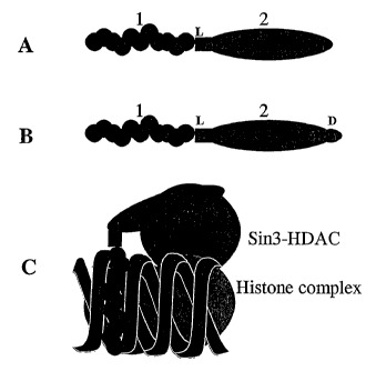
Figure 1: Schematic representation of fusion molecules and binding to a
target site
In A, "1" represents the oligonucleotide module 1 of Example 1; "2"
represents the polypeptide module 2. "L" indicates a linker region. In B, D
represents an additional delivery peptide. Peptides may be linked to either
or both ends of the oligonucleotide; for example, the repressor/modifying
polypeptide may be linked to one end and the delivery peptide to the other.
CA 02474216 2004-04-08
WO 03/033701 PCT/GB02/04633
41
C represents the situation where the oligonucleotide portion has formed a
triple helix with double-stranded DNA and the repressor peptide has
recruited Sin3-HDAC complex to the site.
Figure 2: Quantitation of androgen receptor mRNA
Columns represent the expression of mRNA as a percentage from the
control value. Each column is a representative of the mean OD and standard
error of the mean of four independent polymerase chain reaction. Mean
optical densities were determined.
Figure 3: Effect of ARP-L218 and R1881 on the quantity of androgen
receptor mRNA.
RT-PCR analysis of androgen receptor mRNA. After ethidium bromide
staining the quantity of electrophoresed amplicons was determined in
arbitrary units using a Labworks image reader. The columns represent an
N=1 experiment.
Figure 4: Effect of ARP-L218 and cyproterone acetate on the quantity
of androgen receptor mRNA.
RT-PCR analysis of androgen receptor mRNA. After ethidium bromide
staining the quantity. of electrophoresed amplicons was determined in
arbitrary units using a Labworks image reader. The columns are one
representative of an N=1 experiment respectively.
Figure 5: Down regulation of interferon stimulated, genome
incorporated gene.
CA 02474216 2004-04-08
WO 03/033701 PCT/GB02/04633
42
Cells habouring EGFP under the control of the interferon stimulated
response element (ISRE) of the human 6-16 gene were transiently
transfected with different compounds. Cells were then treated with 300
units/ml of IFN for one day and analysed by FACS for the GFP expression.
a) Parental cell line, no treatment
b) Cells expressing the stably transfected construct (C8), no treatment
c) C8 treated with interferon (+INF)
d) C8 transfected with 500 pm TFO, +INF
e) C8 specific TFO 1000pM, +INF
f) C8, GeneICEl 500pM, +INF
g) C8, GeneICE 2 500pM, +INF
h) C8, unspecific control TFO 500pM, +INF
i) C8, unspecific control TFO 1000pM, +INF
Figure 6: Chromatin Immunopreciptation of androgen receptor DNA.
An example of the type of data that may be produced by Example 10. The
data shows Chromatin Immunopreciptation of Parental MCF-7 Tet-OFF
cells and PLZF-ER stably transfected cell lines. Cell lines MCF7-TO or
MCF7 JP 13 (with PLZF-ER, a tetracycline-inducible chromatin remodelling
gene linked to a progesterone receptor DNA binding peptide) were grown to
80% confluence in phenol red free DMEM, 5% DSS, P/S/G plus selection
reagents and Dox as required. Cells were treated with E2 (10-8M) or an
ethanol control for 30 minutes at 37 C prior to formaldehyde cross-linking
and sonication. Upper panel shows PR PCR product using DNA associated
with immunoprecipitated chromatin (P) or total DNA extracted from the
cells (S), using acetylated histone H4 antibody. Cells without (MCF7-TO)
or with (MCF7 JP13) the PLZF-ER construct were grown in the presence
(+) or absence (-) of TET. Lower panel displays PR PCR product expressed
CA 02474216 2004-04-08
WO 03/033701 PCT/GB02/04633
43
relative to the quantity of PR PCR product using DNA immunoprecipitated
with an acetylated histone H4.
Figure 7: Down-regulation of PSA protein secretion in LNCap cells by
ARP-L218.
Cells were incubated in the presence of R1881 for 3 days. PSA levels in the
supernatants were measure by ELISA. Each column represent total prostate
specific antigen (PSA).
Example 1: Construction and use of oligo-regulator peptide fusion
molecules.
A series of oligopeptide conjugates useful as gene regulatory molecules has
been produced. These consist of at least two specific portions or modules,
namely an oligonucleotide capable of forming a DNA triple helix with a
selected double-stranded target sequence (Triplex Forming Oligonucleotide,
or TFO; Module 1); and a discrete peptide sequence derived from either a
gene repressor or activator ( Module 2). The TFO is fused to the repressor
or activator peptide.
As an example, the TFO is designed to form a triplex with the Interferon
Stimulatable Response Element (ISRE) of the human Interferon Stimulated
Gene (ISG) 6-16. (Porter A., et al., EMBO J. 7: 85-92, 1988). The ISRE is
very purine rich on one DNA strand and is, therefore, a candidate sequence
for forming a DNA triplex by Hoogsteen base pairing. The rules for
designing potential TFO are summarised in: Vasquez KM and Wilson JH,
Trends Biochem Sci, 1: 4-9, 1998. The sequence 5'-
CA 02474216 2004-04-08
WO 03/033701 PCT/GB02/04633
44
AAAGTAAAAGGGGAGAGAGGG-3' was produced as an oligonucleotide
(Module 1) with an activated 5' end for chemical coupling to Module 2
peptides. Module 2 peptides explored in this study include (at least one
copy of the minimal transcriptional activator domain of the Herpes Simplex
Virus VP 16 Transcriptional Activator protein for example the amino acid
sequence GGGPADALDDFDLDMLPADALDDFDLDML or
GGGPADALDDFDLDMLPADALDDFDLDMLPADALDDFDLDMLPA
DALDDFDLDML-CONH2 (including GGG linker)),and the human MAD1
transcriptional repressor domain (for example amino acids
X.XXMNIQMLLEAADYLERREREAEHGYASMLP (where XXX is, for
example, a AAA or DDD linker)). The latter is a region known to interact
with the histone deacetylase complex protein Sin3a. Additionally, we have
explored the use of amino acids
XXXMAVESRVTQEEIKKEPEKPIDREKTCPLLLRVF (where XXX is,
for example, a AAA or DDD linker) of the human Sapl8 protein, also
known to associated with Sin3a protein. This region corresponds to a
sequence of high evolutionary conservation and overlaps with a region that
can mediate gene repression. Module 2 peptides were synthesised in an
activated form to enable subsequent coupling to the activated Module 1
oligonucleotide by "native ligation" chemistry (see WO 01/15737 and
Stetsenko & Gait (2000) Organic Chem 65(16), 4900-4908), in which an N-
terminal thioester-functionalised peptide is coupled to a 5'-cysteinyl
oligonucleotide.
Cell lines for transfection work included COS, cells and HT1080 human
fibrosarcoma cells. Cells were transiently transfected using a standard
liposome based transfection method (or alternatively another delivery
method, for example electroporation or microinjection) with an ISRE
CA 02474216 2004-04-08
WO 03/033701 PCT/GB02/04633
regulated luciferase reporter gene together with a varying amount of
oligopeptide conjugate. This enables a comparison to be made of Interferon
dependent luciferase expression with oligopeptide mediated gene regulatory
effects. As controls for specificity, cells were also treated with either the
5 oligonucleotide, the peptide or both (ie as separate, unlinked molecules).
After suitable times, for example 0.5, 1, 2, 4, 8 and/or 12 hours, the cells
were stimulated with IFN and reporter gene activity was measured using a
luminometer based assay for luciferase enzyme. Correction for transfection
efficiency was determined by the use of a non-interferon dependent GFP
to control gene.
The ability of the various oligopeptide conjugates to activate or repress
transcription of IFN responsive genes was investigated. Delivery of the
increasing concentration of fusion molecule oligo-MAD 1 represses the
15 reporter gene activity in a concentration dependent manner. Similarly, the
delivery of the Sap 18-oligo fusion molecule represses the gene activity. In
addition, delivery of oligoVP 16 into the reporter gene-containing cells
results in reporter gene activity in the absence of IFN. After the TFO (ie
without a peptide domain) was delivered into the cells in addition to an
20 oligo-peptide fusion molecule, the repression of gene activity was less
than
that seen with the fusion molecule alone, ie repression was equivalent to that
seen with a lower concentration of the fusion molecule. This result shows
that the molecules with the repressor peptide are more effective regulators
of gene activity than the TFO without a peptide domain, and the TFO may
25 compete with the oligo-peptide fusion molecule for binding to the target
sequence. The oligo and peptide alone or added together had no repressor
effect, demonstrating the specificity of the oligopeptide conjugates in gene
regulation.
CA 02474216 2004-04-08
WO 03/033701 PCT/GB02/04633
46
This example demonstrates the design and construction of fusion molecules
consisting of DNA binding oligonucleotides and functional peptides, and
their delivery into the cells. The oligopeptide conjugates are able to target
specific genes and repressor peptide sequences mediating Gene ICE can
repress genes in a specific, targeted manner. Thus, oligopeptide conjugates
can be designed to be potent regulators of gene activity.
Example 2: Repression of chromosomal genes by oligo-regulator fusion
molecules
Fusion molecules are able' to regulate gene activity, when such genes are
integrated into the genome. Fusion molecules containing a DNA binding
oligonucleotide (TFO) fused to a MAD 1, Sap 18 or VP 16 peptide were
designed and constructed as described in Example 1.
Cells were transfected as described in Example 1 with ISRE-containing
reporter genes, and cell lines stably expressing these genes were
selected. In these stable cell lines, the gene is integrated in the
genome and therefore may function as an endogenous gene.
The fusion molecules were delivered into the cells and experiments
were. carried out as described in Example 1. Furthermore, the repression
was measured at different times in order to establish a time course for
repressor effects. The fusion molecules were more effective repressors that
TFOs alone. The effect was also specific (for example, the unfused peptide
and oligonucleotide did not have the same effect as the oligo-peptide
fusion). In addition, the repression by fusion molecules was seen at later
CA 02474216 2004-04-08
WO 03/033701 PCT/GB02/04633
47
time points than any repression seen by the TFO alone, suggesting a more
permanent effect. Again, fusion molecule with VP 16 peptide was capable of
activating the gene expression.
Thus, the fusion molecules described in example 1 are able to regulate
chromosomal gene activity. Fusion molecules with a DNA binding
oligonucleotide targeting portion are able to target specific chromosomal
genes. A Gene ICE repressor peptide fused to the DNA binding
oligonucleotide is able to repress predetermined chromosomal gene
activity. Thus, the described fusion molecules are potent regulators of
chromosomal gene activity.
Endogenous gene regulation is measured, for example by assessing
transcription of the gene (for example using PCR) or by assessing the
quantity or activity of the encoded polypeptide. In an example, the
oligonucleotide is directed to the Androgen receptor gene regulatory site. In
particular, the oligonucleotide has the sequence 5' gggaaaggaaaagaggggaggg
3' or 5' gggaggggaaaggaaaagagg 3'.
In an example, the prostate cancer cell line LnCap is treated with the
oligonucleotide-peptide fusion comprising a MAD1 or Sapl8 peptide as
described in Example 1. Transfected cells are optionally identified and/or
isolated (for example using a GFP marker and FACS techniques) and are
assayed for androgen receptor gene expression as well as for the classic
prostate cancer marker PSA. For example, GFP-positive, FACS sorted
cells were cultured in the presence or absence of the AR agonist R1881.
After 72 hours, culture media were collected and the amount of the
androgen-regulated protein PSA determined by immunoassay. Addition of
CA 02474216 2004-04-08
WO 03/033701 PCT/GB02/04633
48
R1881 results in an approximately 25-fold increase in levels of secreted
PSA in control cells (with no oligonucleotide-peptide fusion, or an
irrelevant oligonucleotide-peptide fusion). By contrast PSA levels in test
oligonucleotide-peptide transfected cells were raised only about 5-fold,
demonstrating an about 5-fold decrease in PSA levels, relative to the control
cells. This demonstrates repression of an endogenous gene.
Alternatively, PCR is used to detect and quantify expression and show that
the oligonucleotide-peptide fusions repress endogenous androgen receptor
gene expression as well as Androgen-regulated gene expression. The
Androgen Receptor (AR)-positive human T47D cell line is infected with the
test oligonucleotide-peptide fusion (for example labelled with green
fluorescent protein (GFP)) or an irrelevant olignucleotide-peptide fusion
(which may also be GFP labelled). Infected cells may be purified by FACS
and GFP-positive cells cultured in the presence of the AR ligand R1881
prior to harvesting and preparation of RNA. Expression of the androgen
receptor gene as well as androgen-regulated genes PSA and DRG-1 and a
non-androgen-regulated gene GAPDH, was determined by PCR. This
demonstrates repression of an endogenous gene.
Example 3: Fusion molecule binding to a target sequence and histone
deacetylase complex.
This Example demonstrates that the fusion molecule binds to
a specific target sequence as well as to a component of histone
deacetylase complex. The fusion molecules containing an oligonucleotide
(TFO) and a repressor peptide were produced as described in Example 1.
CA 02474216 2004-04-08
WO 03/033701 PCT/GB02/04633
49
The different fusion molecules were incubated with labelled
oligonucleotide, which was made complementary to the oligo part of a
fusion. The same fusion molecules were also incubated with Sin3, which is
a component of a histone deacetylation complex. Furthermore, the
fusions were also incubated with both the complementary oligonucleotide
and Sin3 protein.
The complexes were then analysed by standard band shift analysis methods.
The fusion molecules were able to bind to both the labelled complementary
oligonucleotide as well as the Sin3 protein, both separately and
simultaneously. The unspecific fusions were not able bind the labelled oligo
or Sin3, thus demonstrating the specificity of the effect with repressor
fusions.
It can be concluded that the repressor fusions can specifically bind their
target sequences. The repressor fusions are able to recruit histone
deacetylase complexes by binding proteins that are part of this complex, and
by binding their target sequences and recruiting the histone deacetylase
complexes simultaneously, the described fusion molecules are very potent
and specific repressors of gene activity.
Example 4: Target validation protocol
The available DNA sequence for the gene of interest (including flanking
sequence) is analysed in order to select a suitable site for targeting an
oligo/peptide to. The oligo/peptide is synthesised and may be tested prior to
use in the intended cells or animals or humans, for example using a reporter
CA 02474216 2004-04-08
WO 03/033701 PCT/GB02/04633
gene system. The oligo/peptide may be used or tested further in cells in
vitro or in animals or humans.
Once a gene sequence has been provided, the process will involve:
5 = The gene of interest (including flanking sequences if necessary)
will be scanned for unique sequence elements not found elsewhere
in the human genome using bio-informatics data-mining tools (for
example the Genetics Computer Group (GCG) program as used in
Perkins et al (1998) Biochemistfy 37, 11315-11322). A nucleic acid
10 based DNA binding molecule predicted to bind to the identified
unique sequence (for example as a TFO) is designed and
synthesised.
The DNA binding molecule is likely to be an oligonucleotide, preferably
15 with the following features:
(a) at least 16 nucleotides in length
(b) targeted to a gene promoter or at or near to the transcription initiation
site of the gene.
20 It is preferred that the target site for the binding to a TFO is purine-
rich in
one strand.
The TFO may be pyrimidine rich (predominantly C or T); purine rich
(predominantly G or A) or mixed (predominantly G or T, or G, A or T). CT
25 TFOs are considered to bind in a parallel motif, in which the third strand
(TFO) has the same 5' to 3' orientation as the purine strand of the duplex.
GA TFOs are considered to bind in an antiparallel motif, in which the TFO
is oriented oppositely to the purine strand. Mixed TFOs may bind in a
CA 02474216 2004-04-08
WO 03/033701 PCT/GB02/04633
51
parallel or antiparallel motif, depending on the target sequence. Base
pairing arises from formation of Hoogsteen hydrogen bonds in parallel
triplexes (T:AT, C+:GC and G:GC) and reverse Hoogsteen hydrogen bonds
in antiparallel triplexes (G:GC, A:AT and T:AT).
It is intended that the oligonucleotide is a DNA oligonucleotide, possibly
with stabilising chemical modifications. Alternative bases, for example N6-
methyl-8-oxo-2-deoxyadenine may be used in place of cytosine, 2-deoxy-6-
thioguanine in place of guanine or 7-deaza-2-deoxyxanthine in place of
thymine.
= The repressor peptide or peptides may be produced in bulk using a
peptide synthesiser and stored frozen until used.
= The repressor peptide-DNA binding molecule construct is prepared
and purified. The chemistry used may be that described in WO
01/15737. Kits are available from Link Technologies.
= The construct may be quality controlled by mass spectroscopy
and/or by use of labelled complementary oligonucleotide or
labelled antibody moieties (using for example fluorescent,
chemiluminescent or enzyme labels). Typically in such a method
the construct is added to a solid support on which an antibody that
binds to the peptide portion of the construct is immobilised and a
labelled oligonucleotide that binds to the oligonucleotide portion of
the construct is added. In this method detection of the label bound
to the solid support demonstrates that the construct is intact. In
another typical format the oligonucleotide may be attached to a
solid support and the antibody labelled.
CA 02474216 2004-04-08
WO 03/033701 PCT/GB02/04633
52
= A reporter gene construct may be prepared for the gene of interest
(though this is not generally necessary).
= The candidate DNA binding oligonucleotide or oligo/peptide may
be tested for the following:
o Affinity of binding to the target sequence;
o Specificity of binding by exposure to a whole genome
DNA chip.
= The oligo/peptide may be tested for effectiveness using the
reporter gene system.
The oligo/peptide may then be used for modulating or suppressing
expression of the gene of interest in the cell or animal of interest.
Example 5: Target validation
The oligo/peptide fusion molecules will be used to validate drug targets.
This will involve:
= Carrying out the protocol set out in Example 1.
= Delivery of the construct into cells or tissues. These may be normal or
disease tissues, cell lines or primary cells appropriate to the study of the
molecule of interest.
= Analysis of the phenotype by any expression analysis methods; or any
functional analysis such as assessment of cell motility, growth or
apoptosis analysis.
= Comparison with any available data for a particular disease and analysis
of desired effects such as cell death or motility.
CA 02474216 2004-04-08
WO 03/033701 PCT/GB02/04633
53
The obtained data will be used to validate the pre-determined drug targets
for drug development programmes.
Example 6: Patient treatment example
A oligo/peptide fusion is produced as described which targets the androgen
or estrogen regulated genes. The fusion molecules are prepared in a sterile
environment and formulated into liposomes. The fusion-containing
liposomes are targeted into the vicinity of breast or prostate. The liposomes
to are taken up by cancer cells and androgen or estrogen receptor mediated
transcription is suppressed selectively in respective cells.
Example 7: Target identification screen
The oligo/peptide fusion molecules will be used to identify drug targets.
This will involve:
= Preparation of a fusion molecule as set out in Example 1
= Delivery of the fusion molecule into the cells or tissues. These may be
normal or disease tissues, cell lines or primary cells.
= Analysis of gene expression profile resulting from gene silencing, using
DNA arrays. This indicates the effect of the construct on overall gene
expression in the model.
= Analysis of the phenotype by any expression analysis methods, or any
functional analysis such as cell motility, growth or apoptosis analysis.
The obtained data will be used to find potential drug targets for diseases
such as breast or prostate cancer. These targets can be further validated by
appropriate methods including any further similar screens, in vitro methods
and cell and animal models.
CA 02474216 2004-04-08
WO 03/033701 PCT/GB02/04633
54
Example 8: Oligo/peptide fusion molecules targeted to the androgen
receptor.
As discussed in Example 2, oligo/peptide fusion molecules are able to
regulate gene activity when the genes are integrated into the genome. In this
example we provide further experimental data supporting this.
We constructed fusions between a DNA binding oligonucleotide (TFO)
targeted at the promoter sequence of the androgen receptor gene (ARP), and
a peptide containing a 14 or 29 amino acid MAD 1 repressor sequence linked
to a 16 amino acid penetratin sequence.
The sequence of the ARP TFO used in this example was:
ARP TFO: (5') GFGUGGTGFGGTTGTGTT (3')
U=5-fluro-deoxyuracil
F= 2'-deoxy-6-thioguanine
TFO's are modified at 5' end for conjugation as described by Gait et al
(2000) J.Org. Chem, 65, 4900-4908, and at 3' end with standard amino-
link to protect from degradation.
The oligonucleotide sequence is designed to form a triplex with the
promoter sequence of the androgen receptor.
The TFO was fused to two different peptide fragments. The peptides
fragments used in this example were:
CA 02474216 2004-04-08
WO 03/033701 PCT/GB02/04633
L217: HHHHHH-Penetratin-DDD-14aaMAD
(Link)HHHHHHRQIKIWFQNRRMKWKKDDDMNIAMLLEAADYL
E(amide)
5 L218: HHHHHH-29aaMAD-DDD-Penetratin
(Link)HHHHHHMNIAMLLEAADYLERREREAEHGYASMLPDDDR
QIKIWFQNRRMKWKK(amide)
The 14 and 29 amino acid peptide sequences are from the transcriptional
1o repressor domain of the MAD1 protein, a region known to interact with the
histone deacetylase complex protein Sin3a.
The three Aspartic acid residues (DDD) are a linker sequence, while the
Penetratin peptide sequence mediates efficient plasma membrane
translocation of the oligo/peptide fusion molecule.
The ISIS residues were added in order to provide further purification
options.
The experiments were conducted in LNCap (Lymph Node Prostate
Carcinoma) human prostate tumour cell line. LNCap cells were obtained
from the European Collection of Cell Cultures, accession number
B9110211. Cultures were grown and propagated in-house and used as
monolayers in disposable tissue culture labware. On the day of testing, cells
were observed as having proper cell integrity and therefore, were acceptable
for use in this study.
Intra-cellular and intra-nuclear delivery of oligo/peptide fusion molecules to
the LnCAP cells was demonstrated using a fluorescent (Cy3) labelled oligo-
peptide. The labelled construct (5 pmol in lml) and Lipofectamine 2000
(used as per manufacturer's instructions) were added to LnCAP cells and left
CA 02474216 2004-04-08
WO 03/033701 PCT/GB02/04633
56
to incubate for 24 hours. The cells were then examined by fluorescent
microscopy. Intra-nuclear localisation of the construct was demonstrated by
co-location with a nuclear (DAPI) stain.
The effect of the oligo/peptide fusion molecules ARP-L217 and ARP-L218
on androgen receptor gene expression was measured in the following
experiments.
1) Treatment of LNCap cells with ARP-L217
To measure the effect of ARP-L217 on the androgen receptor gene
expression the following experiments were conducted.
ARP-L217 was prepared in phosphate buffer saline (PBS), 0.2 m syringed
filtered and used on day of test set-up.
LNCap cells were transfected with ARP-L217 using lipofectamine2000
(cationic activated dendrimer, InVitrogen life technologies). Various
amounts of ARP-L217 were mixed with a fixed amount of
lipofectamine2000 (3 i) in a total volume of 100 l serum (10% FBS)
supplemented medium without antibiotics. After 20mins incubation at room
temperature, the transfection mixture was added to cells at 70% confluency
and cells were incubated for 3 days.
RT-PCR conditions were established to give optimal sensitivity within the
exponential phase of the amplification process. Total cellular RNA was
isolated from the treated LNCap cells using the RNeasy Kit (Qiagen). RNA
CA 02474216 2004-04-08
WO 03/033701 PCT/GB02/04633
57
(1 g of each sample) was reverse transcribed into cDNA using the
Omniscript RT kit (Qiagen). The synthesized cDNA (21il of each sample)
was subjected to PCR amplification (Qiagen Taq Kit) with human androgen
receptor primers sense 5'-TCCAGAATCTGTTCCAGAGCG-3' and
antisense 5 `-TTCGGATACTGCTTCCTGC-3 ` to yield a 281bp product. To
verify the quality of RNA/cDNA preparation, PCR amplification was
carried out with human (3-actin primers (Promega, UK). To verify that PCR
product were not amplified from residual DNA left in RNA samples, an RT
negative control was subjected to R-actin PCR amplification.
The AR primer sequences used in this study were synthesized by Qiagen-
Operon and designed to avoid 5'-3'complementarity and included at least
one intron in the product to detect contamination with genomic DNA.
Electrophoresis of PCR products was performed on 2% agarose gels and the
gels were pre-casted with ethidium bromide (1:10 dilution). The gels were
photographed using the GelDoc system
The comparison and analysis of mRNA expression was done by gel band
quantitation using Labworks software system. Sample/R-actin OD ratio was
calculated for each sample and then used for comparison. Thus, increased
expression of mRNA is presented as a percentage from the control
(untreated cells) value.
The results from this experiment as shown in Table 1 below and also as a
bar graph in Figure 2.
CA 02474216 2004-04-08
WO 03/033701 PCT/GB02/04633
58
Table la Data illustrating band quantitation after exposure of ARP-L217 to
LNCap cells for 3days.
ARP n Q'U t7FILifiotl ([7' y 3)
iple ID IODa IODa IODb IODb Mean
Untreated 2280 2406 2105 2162 2238.25
PBS 25u1 2283 1908 2106 2544 2210.25
PBS 50u1 1799 1799 2677 2268 2135.75
25 M ARP-L217+Lipofectamine 1428 1749 1419 1325 1480.25
0.5uM ARP-L217+Lipofectamine 783 971 1037 1191 995.5
;aut n PUR Band Quaritita(Tay3)`
IODa IODa IODb IODb Mean
Untreated 8957 9589 8996 9663 9301.25
PBS 25u1 8228 7980 9098 8544 8462.5
PBS 50ul 7413 8762 8920 9005 8525
0.25 M ARP-L217+Lipofectamine 6632 6322 6605 7924 6870.75
0.5uM ARP-L217+Lipofectamine 6323 6180 5834 6695 6258
Table lb Determination of ratio per beta-actin and percentage inhibition
from control.
ample ID Ratio/R-actin Percentage from untreated
control
Untreated 0.2406 100
PBS 25uI 0.2611 108.5364073
PBS 50u1 0.2505 104.1091143
0.25 M ARP-L217+Lipofectamine 0.2154 89.52898226
.5uM ARP-L217+Lipofectamine 0.1590 66.10562717
From this data it can be seen that the cell samples which were incubated
with ARP-L217 have greatly reduced androgen receptor gene expression
levels compared with those treated with control PBS solutions.
2) Treatment of LNCap cells with ARP-L218 and R1881
CA 02474216 2004-04-08
WO 03/033701 PCT/GB02/04633
59
The effect of ARP-L218 on androgen receptor gene expression with and
without a synthetic androgen (R1881) was measured using the protocol
outlined in the above section.
ARP-L218 was prepared in phosphate buffer saline (PBS), 0.2 m syringed
filtered and used on day of test set-up. R1881 (synthetic androgen) was
purchased from Perkin Elmer, Catalogue number NLP005005MG.
LNCap cells were transfected with ARP-L218 using lipofectamine2000
(cationic activated dendrimer, InVitrogen life technologies) with/without
agonist. Various amounts of ARP-L218 were mixed with a fixed amount of
lipofectamine2000 (3 J) in a total volume of l00 1 serum free medium
without antibiotics (OptiMEM). After 20mins incubation at room
temperature, the transfection mixture was added to cells at 70% confluency
and cells were incubated for 3 days.
Intra-cellular and infra-nuclear delivery of oligo/peptide fusion molecules to
the LnCAP cells was demonstrated using a fluorescent (Cy3) labelled oligo-
peptide. The labelled construct (5 pmol in lml) and Lipofectamine 2000
(used as per manufacturer's instructions) were added to LnCAP cells and left
to incubate for 24 hours. The cells were then examined by fluorescent
microscopy. Intra-nuclear localisation of the construct was demonstrated by
co-location with a nuclear (DAPI) stain.
RNA was extracted and RT-PCR analysis of AR mRNA levels conducted as
described above.
CA 02474216 2004-04-08
WO 03/033701 PCT/GB02/04633
The results from this experiment as shown in Table 2 below and also as a
bar graph in Figure 3
Table 2: Data illustrating band quantitation after exposure of ARP-L218 in
5 presence/absence of synthetic androgen (R1881) to LNCap cells for 3days.
Determination of ratio per beta-actin and percentage inhibition from control.
Sample Id AR AR Average beta-actin beta-actin Average Ratio/beta- Percentage
_ actin of control
PBS treated control 3142 - 3142 9521 - 9521 0.330 100
ARP-L218 (0.125uM) 2327 2647 2487 10592 14178 12385 0.201 60.84
ARP-L218 (0.25uM) 2728 2212 2470 17103 15691 16397 0.151 45.64
R1881 1 uM + ARP-1_218 1291 - 1291 14949 - 14949 0.086 26.16
R1881 100nM + ARP-1_218 1068 - 1068 16258 - 16258 0.066 19.90
R1881 1 nM + ARP-L218 1393 - 1393 16950 - 16950 0.082 24.90
R1881 0.01 nM + ARP-L218 2532 - 2532 16607 - 16607 0.152 46.20
R1881 1 uM 1388 - 1388 8424 - 8424 0.165 49.92
R1881100nM 1455 - 1455 12536 - 12536 0.116 35.17
R18811nM 1471 - 1471 14242 - 14242 0.103 31.29
R18810.01nM 2083 - 2083 8799 - 8799 0.237 71.73
10 From this data it can be seen that the cell samples which were incubated
with ARP-L218 and R1881 have greatly reduced androgen receptor gene
expression levels compared with those treated with R1881-containing
solutions. Both types of samples had reduced AR mRNA levels than the
control cell sample treated with PBS.
Therefore while R1881 can act to reduce AR gene expression this reduction
can be enhanced by coincubating the cells with the ARP-L218 oligo/peptide
fusion molecule.
3) Treatment of LNCap cells with ARP-L218 and cyproterone acetate
CA 02474216 2004-04-08
WO 03/033701 PCT/GB02/04633
61
The effect of ARP-L218 with and without cyproterone acetate, a receptor
bound androgen receptor antagonist, on androgen receptor gene expression
was measured using the protocol outlined in the above section.
ARP-L218 was prepared in phosphate buffer saline (PBS), 0.2 m syringed
filtered and used on day of test set-up. Cyproterone acetate was purchased
from Sigma, lot number 41K1195.
LNCap cells were transfected with ARP-L218 using lipofectamine2000
(cationic activated dendrimer, InVitrogen life technologies) with/without
antagonist. Various amounts of ARP-L218 were mixed with a fixed amount
of lipofectamine2000 (3 l) in a total volume of 100 l serum free medium
without antibiotics (OptiMEM). After 20mins incubation at room
temperature, the transfection mixture was added to cells at 70% confluency
and cells were incubated for 3 days.
Intra-cellular and intra-nuclear delivery of oligo/peptide fusion molecules to
the LnCAP cells was demonstrated using a fluorescent (Cy3) labelled oligo-
peptide. The labelled construct (5 pmol in lml) and Lipofectamine 2000
(used as per manufacturer's instructions) were added to LnCAP cells and left
to incubate for 24 hours. The cells were then examined by fluorescent
microscopy. Intra-nuclear localisation of the construct was demonstrated by
co-location with a nuclear (DA-PI) stain.
RNA was extracted and RT-PCR analysis of AR mRNA levels conducted as
described above.
CA 02474216 2004-04-08
WO 03/033701 PCT/GB02/04633
62
The results from this experiment as shown in Table 3 below and also as a
bar graph in Figure 4
Table 3: Data illustrating band quantitation after exposure of ARP-L218 in
presence/absence of androgen receptor antagonist (cyproterone acetate) to
LNCap cells for 3days. Determination of ratio per beta-actin and percentage
inhibition from control.
ample ID AR beta-actin Ratio % of control
PBS treated control 3142 9521 0.330 100
Cyproterone Acetate 10uM 1568 14691 0.107 32.4
Cyproterone Acetate 1uM 1868 11856 0.158 47.8
yproterone Acetate 1uM + ARP-L218(0.0625uM) 1583 14861 0.107 32.4
yproterone Acetate 10nM 2401 14112 0.170 51.5
Cyproterone Acetate 1 OnM + ARP-L218 (0.03125) 1241 16885 0.074 22.4
yproterone Acetate 0.1nM '2357 13278 0.178 54.0
From this data it can be seen that the cell samples which were incubated
with ARP-L218 and cyproterone acetate have reduced androgen receptor
gene expression levels compared with those treated with a cyproterone
acetate only. Both types of samples had reduced AR mRNA levels
compared with the control cell sample treated with PBS.
Therefore while cyproterone acetate can act to reduce AR gene expression
this reduction can be enhanced by coincubating the cells with the ARP-L218
oligo/peptide fusion molecule.
Example 9: Down regulation of an interferon stimulated, genome
incorporated gene.
CA 02474216 2004-04-08
WO 03/033701 PCT/GB02/04633
63
As discussed in Example 2, oligo/peptide fusion molecules are' able to
regulate gene activity when the genes are integrated into the genome. In this
example we provide further experimental data supporting this.
We constructed fusions between a DNA binding oligonucleotide (TFO)
targeted to an interferon stimulated response element (IRE), and a peptide
containing a 29 amino acid MAD 1 repressor sequence linked to a 16 amino
acid penetratin sequence.
1o The sequence of the IRE TFO used in this example was:
IRE TFO: (5') GGGUGGTGGGGTTGTGTT (3')
U=5-fluro-deoxyuracil
TFO's are modified at 5' end for conjugation as described by Gait et
al (2000) J.Org. Chem, 65, 4900-4908, and at 3' end with standard
amino-link to protect from degradation.
The oligonucleotide sequence is designed to form a triplex with the
Interferon Stimulatable Response Element (ISRE) of the human Interferon
Stimulated Gene (ISG) 6-16 (Porter et al., (1988) EMBO J, 7, 85-92).
The TFO was fused to two different peptide fragments. The peptides
fragments used in this example were:
L218: HHHHHH-29aaMAD-DDD-Penetratin
(Link)HHHHHHMNIAMLLEAADYLERREREAEHGYASMLPDDDR
QIKIWFQNRRMKWKK(carboxamide)
CA 02474216 2004-04-08
WO 03/033701 PCT/GB02/04633
64
L219: DDD-29aaMAD-HHHHHH-Penetratin
(Link)DDDMNIAMLLEAADYLERREREAEHGYASMLPHHHHHHR
QIKIWFQNRRMKWKK(carboxamide)
The 29 amino acid peptide sequence is from the transcriptional repressor
domain of the MAD I protein, a region known to interact with the histone
deacetylase complex protein Sin3a.
The three Aspartic acid residues (DDD) are a linker sequence, while the
Penetratin peptide sequence mediates efficient plasma membrane
translocation of the oligo/peptide fusion molecule.
The HIS residues were added in order to provide further purification
options.
Modified HT1080 human fibrosarcoma cells were used to demonstrate the
oligo/peptide fusion molecules capacity to downregulate an interferon
stimulated reporter gene expression. These cells have a stably incorporated
gene expressing EGFP under the control of the interferon stimulated
response element (IRE) of the human 6-16 gene.
For the following data, cells were transiently transfected with different
compounds using standard lipofectamine protocols. Cells were then treated
with 300 units/ml of interferon (INF) for one day and analysed by FACS for
the GFP expression.
Figure 5 shows the FACS analysis of a number of different cell populations,
in which the following experimental conditions were applied:
CA 02474216 2004-04-08
WO 03/033701 PCT/GB02/04633
a) Parental cell line, no treatment
b) Cells expressing the stably transfected construct (C8), no treatment
c) C8 treated with interferon (+INF)
5 d) C8 transfected with 500 pm TFO, +INF
e) C8 specific TFO 1000pM, +INF
f) C8, IRE-L218 500pM, +INF
g) C8, IRE-L219 500pM, +INF
h) C8, unspecific control TFO 500pM, +INF
10 i) C8, unspecific control TFO 1000pM, +INF
Table 4 shows the interferon-induced GFP expression of Figure 5 presented
as percentage.
15 Table 4: Interferon-induced GFP expression.
Condition applied GFP expression
(a) 0%
(b) <1%
(c) 90%
(d) 53%
(e) 63%
(f) 40%
(g) 42%
(h) 87%
(i) 85%
The IRE - TFO system, which was used as a model system, has been
20 previously described (Roy, 1994, Eur. J. Biochem. 220, 493-503). The
results from FACS analysis show that IRE-L218 and IRE-L219 were able to
repress the expression by an average of 59%, whereas the specific TFO of
CA 02474216 2004-04-08
WO 03/033701 PCT/GB02/04633
66
the same sequence performed an average 42% repression. The control TFOs
had only marginal effect over the positive control.
These results clearly illustrate that the oligo/peptide fusion molecules are
most effective in repressing the INF induced GFP gene expression.
Example 10: Down-regulation of gene expression by oligo/peptide
fusion molecules is associated with chromatin histone deacetylation
As seen in Examples 8 and 9 the oligo/peptide fusion molecules are
effective in repressing gene expression. We propose that this is due to a
MAD1-mediated change in the histone acetylation state of DNA at or close
to where the ARP or IRE oligo binds.
To show that reduced gene expression is associated with chromatin histone
deacetylation, the Chromatin immunoprecipitation (ChIP) method may be
used. Chromatin immunoprecipitation is carried out using a ChIP Assay kit
according to the manufacturer's instructions (Upstate Biotechnology, Bucks,
UK).
LNCap human prostate tumour cells are grown and propagated and
incubated with oligo/peptide fusion molecules which are effective in
repressing androgen receptor gene expression. Untreated LNCap cells are
used as a control.
Cells are grown to 95% confluence on 35cm tissue culture plates in DMEM,
lacking phenol red, supplemented with 5% DSS, P/S/G and G418
CA 02474216 2010-08-06
67
(100 g/ml). Hygromycin B (80 g/ml) and doxycycline (1 g/ml) are added
as appropriate. 30 minutes prior to fixation, E2 (10-8M) or ethanol (as a
control) is added to the cells. 37% formaldehyde is added dropwise directly
to the medium to a final concentration of 1%. Cells are incubated for 10
minutes at 37 C.
On ice, the medium is aspirated from the plates, cells are washed twice with
ice cold PBS containing lx protease inhibitors (PI) (Sigma, Dorset, UK).
For harvesting, lml of ice cold PBS containing lx PI is added to the plate
1o and the cells scraped into pre-cooled microfuge tubes, using a rubber
policeman. Cells are pelleted by centrifugation at 2000rpm for 4 minutes, at
4 C. The pellets are resuspended in 400 l of warmed ChIP SDS-lysis
buffer (1% SDS; 10mM Na EDTA pH 8.0; 50MM Tris-HC1 pH 8.1)
containing PI, and incubated on ice for 10 minutes.
The lysates are sonicated to shear the DNA into 200 - 1000bp lengths.
During sonication, the samples are placed in an ice-water beaker, to keep
them cold in order to prevent sample degradation. Sonication is carried out
using a Soniprep 150 sonicator with attached Soniprep 150 exponential
titanium probe (Sanyo-Gallenkamp, Leics, UK) with four 10 second bursts,
separated by 30 second intervals.
Samples are centrifuged at 13,000 rpm for 10 minutes at 4 C. The
supernatant is collected into 15m1 sterile falcon tubes and diluted 10 fold
with 3600 1 ChIP dilution buffer (0.01% SDS; 1.1% Triton-X-100; 1.2mM
Na EDTA pH 8.0; 16.7mM Tris-HCI, pH 8.1; 167mM NaCI). The samples
are then divided into two 2m1 aliquots in 2.5rnl tubes, one for incubation
*Trade-mark
CA 02474216 2004-04-08
WO 03/033701 PCT/GB02/04633
68
with an anti-acetylated histone H4 antibody, ChIPs grade (Upstate
Biotechnology, Bucks, UK), and the other for use as a no antibody control.
To reduce non-specific background, each 2m1 aliquot is pre-cleared by
adding 80 l of salmon sperm DNA/protein A agarose-50% slurry
(suspended in 10mM Tris-HC1 pH 8.0; 1mM Na EDTA pH 8.0) for 30
minutes at 4 C on a vertical rotating platform (Stuart Scientific, Staffs,
UK).
The agarose beads are then pelleted by a 30-second centrifugation at 1000
rpm and the supernatant fractions collected into fresh 2.5m1 tubes. The
immunoprecipitating antibody is added at a dilution of 1:500 to the first
sample but not to the no antibody control sample. Both tubes are incubated
overnight at 4 C on a vertical rotating platform (Stuart Scientific, Staffs,
UK).
60 l of salmon sperm DNA/protein A Agarose-50% slurry are incubated
with each tube for 1 hour at 4 C, with rotation, to collect the
antibody/histone complexes or non-specifically bound proteins in the case
of the no antibody control. The agarose beads are pelleted by brief
centrifugation at 800 rpm for 1 minute. The supernatants are carefully
transferred into fresh 2.5m1 tubes and stored at -20 C.
The protein A Agarose beads/antibody/histone complex is washed for 5
minutes on a vertical rotating platform at 4 C with lml of each of the
following buffers in the order listed below:
(a) Low Salt Immune Complex Wash Buffer - one wash
(0.1% SDS; 1% Triton-X-100; 2mM Na EDTA pH 8.0; 20mM Tris-
HCl pH 8.1; 150mM NaCl)
CA 02474216 2004-04-08
WO 03/033701 PCT/GB02/04633
69
(b) High Salt Immune Complex Wash Buffer - one wash
(0.1% SD S; 1% Triton-X-100; 2mM Na EDTA pH 8.0; 20mM Tris-
HCl pH 8.1; 500mM NaCl)
(c) LiCl Immune Complex Wash Buffer - one wash
(0.25M LiCI; 1% NP40 (nonidet); 1% deoxycholate; 1mM Na EDTA
pH 8.0; 10mM Tris-HC1 pH8.1)
(d) 1x TE - two washes
(IOmM Tris-HC1, 1mM Na EDTA pH 8.0)
The histone/immune complex is eluted from the agarose beads by addition
of 250 1 freshly prepared Elution buffer (1% SDS; O.1M NaHCO3). The
samples are vortexed briefly to mix and incubated at room temperature for
minutes on a vertical rotating platform. The beads are centrifuged at
1000 rpm for 2 minutes at room temperature and the eluate transferred to
15 fresh microfuge tubes. The elution step is repeated with a further 250 l of
Elution buffer and the eluates combined.
l of 5M NaCl are added to the eluates and histone-DNA crosslinks
reversed by heating to 65 C for at least 4 hours. 10 l of 0.5M Na EDTA
20 pH 8.0, 20 l of 1M Tris-HCI, pH 6.5 and 2 l of 10mg/ml Proteinase K are
added to the eluted samples. The crosslinks are also reversed on the
supernatant fraction from the IP. 40gl of 0.5M Na EDTA pH 8.0, 80 l of
IM Tris-HC1, pH 6.5 and 8p1 of 10mg/ml Proteinase K are added to
supernatant samples. Samples are incubated for 1 hour at 45 C to degrade
protein in the samples.
CA 02474216 2004-04-08
WO 03/033701 PCT/GB02/04633
20 g of glycogen are added to the samples as an inert carrier and then the
sample DNAs are recovered by phenol/chloroform/isoamyl alcohol
extraction and ethanol precipitation. DNA pellets are resuspended in 50 l
sterile water for PCR reactions. 1 l of sample and 35 - 40 cycles are used
5 for each PCR-amplification. Computer-based image analysis (NIH image
analysis program) is employed to evaluate the relative levels of the
estrogen-regulated androgen receptor gene PCR product, compared with the
B-actin and no antibody controls. This allowed a calculation of the relative
amount of the gene transcript contained within a sample to be compared
10 with that in other samples.
Oligonucleotides used for ChIP analysis:
Progesterone Receptor Forward primer:
5'- TCCAGAATCTGTTCCAGAGCG -3'
15 Progesterone Receptor Reverse primer:
5'- TTCGGATACTGCTTCCTGC -3'
13-actin Forward primer: 5'-TTTTCGCAAAAGGAGGGGAG-3'
13-actin Reverse primer: 5'-AAAGGCAACTTTCGGAACGG-3'
An example of the type of result that may be achieved is shown in Figure 6.
The amount of precipitated androgen receptor DNA, and hence the degree
of acetylation of the chromatin histone proteins associated with the
androgen receptor, may be decreased by approximately 75% in the cells in
which androgen receptor expression is inhibited by the described method
compared to untreated cells.
CA 02474216 2004-04-08
WO 03/033701 PCT/GB02/04633
71
Example 11: Gel shift assay
The previous examples have demonstrated that the ARP-L217 and ARP-
L218 oligo/peptide fusion molecules are able to regulate targeted gene
activity. This is considered to be due to a MAD1-mediated change in the
histone acetylation state of DNA at or close to where the ARP or IRE oligo
binds.
A gel shift assay is used to demonstrate that the oligo/peptide fusion
molecules can bind to DNA fragment containing the target promoter.
A 281bp androgen receptor DNA fragment containing the target promoter
sequence is incubated with ARP-L217. Samples are subjected to non-
denaturing gel electrophoresis . in order to characterize triplex-mediated
photoadduct. Adducts are detected in samples containing the ARP-L217
product which shifted with increasing doses.
In this way it is possible to show that oligo/peptide fusion molecules can
bind to DNA fragment containing the target promoter.
Example 12: Oligo/peptide fusion molecules with a nuclear localisation
signal.
The peptide portion of the molecule may have a nuclear localisation signal
(NLS) to target the molecule to the nucleus. The peptides used in this
example are:
a) DDD-MAD 1-DDD-NLS,
CA 02474216 2004-04-08
WO 03/033701 PCT/GB02/04633
72
which has the amino acid sequence:
(Link)DDDMNIQMLLEAADYLERREREAEHGYASMLPDDDPKKKRK
V (carboxamide)
and,
b) DDD-NLS-DDD-MAD 1,
which has the amino acid sequence:
(Link)DDDPKKKRK VDDDMNIQMLLEAADYLERREREAEHGYASML
P (carboxamide)
The NLS is a 7 amino acid (sequence PKKKRKV) functional nuclear
localisation signal derived from the SV40 T-antigen.
The DDD linker sequence and 29 amino acid MAD 1 amino acid sequence
are the same as those discussed in the earlier examples.
To demonstrate that the DDD-MADI-DDD-NLS, and DDD-NLS-DDD-
MAD 1 peptide sequences are targeted to the nucleus, A459 human lung
carcinoma cells were transfected using with GeneICE oligopeptides
consisting of both the DDD-MADl-DDD-NLS, and DDD-NLS-DDD-
MAD 1 peptide sequences linked to a Cy3 labelled oligonucleotide by
standard Lipofectamine 2000 protocols. The cells were then fixed in
formaldehyde and stained with DAPI nuclear stain using the manufacturer's
recommended procedure. Examination of the cells by fluorescence
CA 02474216 2004-04-08
WO 03/033701 PCT/GB02/04633
73
microscopy showed that the Cy3 labelled GeneICE oligopeptide molecule
was co-localised with the nuclear DA-PI stain. From the resulting
experimental data it was concluded that the NLS is very effective at
targeting the peptide to the nucleus.
The DDD-MADI-DDD-NLS, and DDD-NLS-DDD-MAD1 peptide
sequences may mediate target gene expression when incorporated into an
oligo/peptide molecules of the invention. This can be demonstrated using
the experimental approach outlined in Examples 8 and 9.
For example, DDD-MAD 1-DDD-NLS, and DDD-NLS-DDD-MAD1
peptide sequences can be incorporated into the oligo/peptide molecules:
ARP:-DDD-MADl-DDD-NLS, and
ARP:-DDD-NLS-DDD-MAD1.
ARP is the TFO oligo sequence shown in Example 8.
The ARP:-DDD-MADI-DDD-NLS and ARP:-DDD-NLS-DDD-MAD1
molecules are then transfected into LNCap cells, as in Example 8, or
modified HT1080 cells, as in Example 9. Using the experimental procedures
outlined in those sections it is possible to show the effect of the AR.P:-DDD-
MAD1-DDD-NLS and ARP:-DDD-NLS-DDD-MAD1 molecules on target
gene expression.
CA 02474216 2004-04-08
WO 03/033701 PCT/GB02/04633
74
Example 13: Regulation of prostate specific antigens levels using
oligo/peptide fusion molecules.
As discussed in Examples 2,8 and 9 oligo/peptide fusion molecules are able
to regulate gene activity when the genes are integrated into the genome. In
this example we provide further experimental data supporting this.
Prostate specific antigens (PSA) protein levels are regulated by the androgen
receptor protein (AR). Since we have shown in Example 8 that AR mRNA
to levels are reduced in cells transfected with the ARP-L218 oligo/peptide
fusion molecules, we reasoned that there should be a decrease in PSA
protein levels in cells transfected with ARP-L218.
Therefore, using the protocols described in Example 8 ARP-L218 was
transfected into LNCap cells (GET/LNC/W39/P6) with Lipofectamine 2000
(InVitrogen) with or without R1881 (Perkin Elmer), a synthetic androgen,
for 3 days, after which the supernatant were harvested for quantitation of
total PSA protein at the Pathology Centre, Hammersmith Hospital, London,
U.K.
PSA measurements were carried out by a chemiluininescent labelled
immunometric assay using an automated immunoassay analyser (Abbott
Architect Immunoassay Analyser).
The results from this experiment are shown in Table 5 below and also as a
bar graph in Figure 7.
CA 02474216 2004-04-08
WO 03/033701 PCT/GB02/04633
Table 5: Total PSA quantified from cell supernatants
Total
Sample identification PSA(ng/ml)
PBS treated control 279
ARP-L218 (0.125uM) 279
ARP-L218 (0.125uM) 270
ARP-L218 (0.25uM) 287
ARP-L218 (0.25uM) 282
R1881 100nM + ARP-L218 (0.25uM) 757
R1881100nM 1980
R1881 0.01 nM + ARP-L218 (0.25uM) 330
R1881 0.01 nM 372
From this data it can be seen that cells which were incubated with the
5 synthetic androgen R1881 show an increase in PSA levels, as would be
expected. However, when the cells are transfected with R1881 and ARP-
L218, the increase in PSA levels is greatly reduced. As can be seen from the
samples transfected with ARP-L218 but not R1881, ARP-L218 has no
direct effect on PSA levels.
Therefore, we propose that R1881 acts to increase AR activity which, in
turn, leads to an increase in PSA levels. However, ARP-L218 acts to block
AR gene expression (as is shown in Example 8), and so PSA levels are
subsequently reduced. The data suggests that the oligo/peptide fusion
molecules of the invention can be used to directly or indirectly regulate a
target gene.
CA 02474216 2004-12-09
P27203CA.ST25.txt
SEQUENCE LISTING
<110> Imperial college innovations Limited
<120> Control of gene expression using a complex of an oligonucleotide and a
regulatory peptide
<130> ICOY/P27203CA
<160> 23
<170> Patentln version 3.1
<210> 1
<211> 36
<212> PRT
<213> Artificial
<220>
<223> Mimic or analogue of SAP18
<220>
<221> MISC_FEATURE
<222> (1)..(3)
<223> linking amino acids, for example AAA or DDD
<400> 1
Xaa Xaa Xaa Met Ala Val Glu Ser Arg Val Thr Gln Glu Glu Ile Lys
1 5 10 15
Lys Glu Pro Glu Lys Pro Ile Asp Arg Glu Lys Thr Cys Pro Leu Leu
20 25 30
Leu Arg Val Phe
<210> 2
<211> 32
<212> PRT
<213> Artificial
<220>
<223> Mimic or analogue of MAD1
<220>
<221> MISC_FEATURE
<222> (1)..(3)
<223> linking amino acids, for example AAA or DDD
<400> 2
Xaa Xaa Xaa Met Asn Ile Gln Met Leu Leu Glu Ala Ala Asp Tyr Leu
1 5 10 15
Glu Arg Arg Glu Arg Glu Ala Glu His Gly Tyr Ala Ser Met Leu Pro
20 25 30
<210> 3
<211> 16
<212> PRT
Page 1
CA 02474216 2004-12-09
P27203CA.ST25.txt
<213> Artificial
<220>
<223> Antennapedia homeodomain based penetratin
<400> 3
Arg Gln Ile Lys Ile Trp Phe Gln Asn Arg Arg Met Lys Trp Lys Lys
1 5 10 15
<210> 4
<211> 16
<212> PRT
<213> Artificial
<220>
<223> Penetratin from TAT
<220>
<221> MISC_FEATURE
<222> (2)..(2)
<223> x is C(Acm), Cys-acetamidomethyl
<400> 4
Cys xaa Gly Arg Lys Lys Arg Arg Gln Arg Arg Arg Pro Pro Gln Cys
1 5 10 15
<210> 5
<211> 21
<212> DNA
<213> Artificial
<220>
<223> oligonucleotide sequence used in module 1
<400> 5
aaagtaaaag gggagagagg g 21
<210> 6
<211> 29
<212> PRT
<213> Artificial
<220>
<223> Peptide sequence used in module 2
<400> 6
Gly Gly Gly Pro Ala Asp Ala Leu Asp Asp Phe Asp Leu Asp Met Leu
1 5 10 15
Pro Ala Asp Ala Leu Asp Asp Phe Asp Leu Asp Met Leu
20 25
<210> 7
<211> 55
<212> PRT
<213> Artificial
<220>
<223> Peptide sequence used in module 2
Page 2
CA 02474216 2004-12-09
P27203CA.ST25.txt
<400> 7
Gly Gly Gly Pro Ala Asp Ala Leu Asp Asp Phe Asp Leu Asp Met Leu
1 5 10 15
Pro Ala Asp Ala Leu Asp Asp Phe Asp Leu Asp Met Leu Pro Ala Asp
20 25 30
Ala Leu Asp Asp Phe Asp Leu Asp Met Leu Pro Ala Asp Ala Leu Asp
35 40 45
Asp Phe Asp Leu Asp Met Leu
50 55
<210> 8
<211> 22
<212> DNA
<213> Artificial
<220>
<223> Oligonucleotide to androgen receptor gene regulatory site
<400> 8
gggaaaggaa aagaggggag gg 22
<210> 9
<211> 21
<212> DNA
<213> Artificial
<220>
<223> Oligonucleotide to androgen receptor gene regulatory site
<400> 9
gggaggggaa aggaaaagag g 21
<210> 10
<211> 18
<212> DNA
<213> Artificial
<220>
<223> Oligo sequence of ARP-TFO
<220>
<221> misc_feature
<222> (2)..(2)
<223> 2'-deoxy-6-thioguanine
<220>
<221> misc_feature
<222> (9)..(9)
<223> 2'-deoxy-6-thioguanine
<220>
<221> misc_feature
<222> (4)..(4)
<223> 5-fluro-deoxyuracil
Page 3
CA 02474216 2004-12-09
P27203CA.ST25.txt
<400> 10
gngnggtgng gttgtgtt 18
<210> 11
<211> 39
<212> PRT
<213> Artificial
<220>
<223> peptide sequence used in ARP TFO
<400> 11
His His His His His His Arg Gln Ile Lys Ile Trp Phe Gln Asn Arg
1 5 10 15
Arg Met Lys Trp Lys Lys Asp Asp Asp Met Asn Ile Ala Met Leu Leu
20 25 30
Glu Ala Ala Asp Tyr Leu Glu
<210> 12
<211> 54
<212> PRT
<213> Artificial
<220>
<223> peptide sequence used in ARP TFO
<400> 12
His His His His His His Met Asn Ile Ala Met Leu Leu Glu Ala Ala
1 5 10 15
Asp Tyr Leu Glu Arg Arg Glu Arg Glu Ala Glu His Gly Tyr Ala Ser
20 25 30
Met Leu Pro Asp Asp ASP Arg Gln Ile Lys Ile Trp Phe Gln Asn Arg
35 40 45
Arg Met Lys Trp Lys Lys
<210> 13
<211> 21
<212> DNA
<213> Artificial
<220>
<223> sense PCR primer
<400> 13
tccagaatct gttccagagc g 21
<210> 14
<211> 19
<212> DNA
Page 4
CA 02474216 2004-12-09
P27203CA.ST25.txt
<213> Artificial
<220>
<223> antisense PCR primer
<400> 14
ttcggatact gcttcctgc 19
<210> 15
<211> 18
<212> DNA
<213> Artificial
<220>
<223> Oligonucleotide sequence of IRE TFO
<220>
<221> misc_feature
<222> (4)..(4)
<223> 5-fluro-deoxyuraci1
<400> 15
gggnggtggg gttgtgtt 18
<210> 16
<211> 54
<212> PRT
<213> Artificial
<220>
<223> peptide sequence used in IRE TFO
<400> 16
Asp Asp Asp Met Asn Ile Ala Met Leu Leu Glu Ala Ala Asp Tyr Leu
1 5 10 15
Glu Arg Arg Glu Arg Glu Ala Glu His Gly Tyr Ala Ser Met Leu Pro
20 25 30
His His His His His His Arg Gln Ile Lys Ile Trp Phe Gln Asn Arg
35 40 45
Arg Met Lys Trp Lys Lys
<210> 17
<211> 21
<212> DNA
<213> Artificial
<220>
<223> forward primer
<400> 17
tccagaatct gttccagagc g 21
<210> 18
<211> 19
<212> DNA
Page 5
CA 02474216 2004-12-09
P27203CA.ST25.txt
<213> Artificial
<220>
<223> reverse primer
<400> 18
ttcggatact gcttcctgc 19
<210> 19
<211> 20
<212> DNA
<213> Artificial
<220>
<223> forward primer
<400> 19 20
ttttcgcaaa aggaggggag
<210> 20
<211> 20
<212> DNA
<213> Artificial
<220>
<223> reverse primer
<400> 20
aaaggcaact ttcggaacgg 20
<210> 21
<211> 42
<212> PRT
<213> Artificial
<220>
<223> nuclear localised oligo/peptide fusion
<400> 21
Asp Asp Asp Met Asn Ile Gln Met Leu Leu Glu Ala Ala Asp Tyr Leu
1 5 10 15
Glu Arg Arg Glu Arg Glu Ala Glu His Gly Tyr Ala Ser Met Leu Pro
20 25 30
Asp Asp Asp Pro Lys Lys Lys Arg Lys val
35 40
<210> 22
<211> 42
<212> PRT
<213> Artificial
<220>
<223> nuclear localised oligo/peptide fusion
<400> 22
Asp Asp Asp Pro Lys Lys Lys Arg Lys Val Asp Asp Asp Met Asn Ile
1 5 10 15
Page 6
CA 02474216 2004-12-09
P27203CA.ST25.txt
Gln Met Leu Leu Glu Ala Ala Asp Tyr Leu Glu Arg Arg Glu Arg Glu
20 25 30
Ala Glu His Gly Tyr Ala Ser Met Leu Pro
35 40
<210> 23
<211> 7
<212> PRT
<213> Artificial
<220>
<223> nuclear localisation sequence
<400> 23
Pro Lys Lys Lys Arg Lys Val
1 5
Page 7