Note: Descriptions are shown in the official language in which they were submitted.
CA 02474316 2004-07-23
WO 03/063782 PCT/US03/02384
TITLE OF THE INVENTION
KAPPA-PVIIA-RELATED CONOTOXINS AS ORGAN PROTECTANTS
BACKGROUND OF THE TNVENTION
The invention relates to o-PVIIA-related conotoxins and pharmaceutically
acceptable salts
thereof and their use as organ protecting agents, i.e., organ protectants.
These conotoxins can be
used for arresting, protecting or preserving an organ, such as a circulatory
orgm, a respiratory organ,
a urinary organ, a digestive organ, a reproductive organ, an endocrine organ
or a neurological organ.
These conotoxins can also be used for arresting, protecting or preserving
somatic cells.
The publications and other materials used herein to illuminate the bacl~ground
of the
invention, and in particular, cases to provide additional details respecting
the practice, are
incorporated by reference, and for convenience are referenced in the following
text by author and
date and are listed alphabetically by author in the appended bibliography.
o-PVIIA, a 27 amino acid peptide that was originally purified from the venom
of the purple
cone snail Conus pmpu~aseens (Terlau et al., 1996; US Patent No. 5,672,682),
has been previously
identified as a potent antagonist of the, Shc~lzef- H4 potassium channel (ICSO
~60nM). In the same
study, no detectable activity on the voltage-gated potassium chamlels Kvl.l or
Kvl.4 (Terlau et al.,
1996) was noted. Chimeras constricted from the Slaahei° and the Kvl.l
K+ channels have identified
the putative pore-forming region between the fifth and sixth transmembrane
region as the site of the
toxin sensitivity (Shon et ah., 1998). It appears that ic-PVIIA interacts with
the external tetraethyl-
ammonium binding site on the S7Za7zer channel. Although both o-PVIIA and
charybdotoxin inlubit
the Sha7ze~ chamlel, they must interact differently. The F425G Shaper mutation
increases
charybdotoxin affinity by three orders of magtutude bltt abolishes ~c-PVIIA
sensitivity (Shon et al.,
1998). o-PVIIA appears to block the ion pore with a 1:1 stoichiometry, and its
binding to open or
closed chamlels is very different (Terlau et al., 1999). Chronically applied
to whole oocytes or
outside-out patches, ~c-PVIIA itW ibition appears as a voltage-dependent
relaxation in response to
the depolarizing pulse used to activate the chamlels (Garcia et al., 1999).
Potassium channels are vital in controlling the resting membrane potential in
excitable cells
and can be broadly subdivided into tluee classes, voltage-gated K+ channels,
Ca'+ activated K+
channels and ATP-sensitive K+ charnels (KATP chamlels). ATP-sensitive
potassium channels were
originally described in cardiac tissue (Noma, 1983). In subsequent years they
have also been
identified in pancreatic cells, slceletal, vascular and neuronal tissue. This
group of K+ channels is
modulated by intracellular ATP levels and as such, couples cellular metabolism
to electrical activity.
CA 02474316 2004-07-23
WO 03/063782 PCT/US03/02384
2
Enhanced levels of ATP result in closure of the KATp charnels. The I~ATp
channel is thought to be
an octomeric complex comprised of two different subunts in a 1:1
stoichiometry; a weal~ly inward
rectifying I~+ channel I~ir6.x (6.1 or 6.2), which is thought to form the
chamzel pore, and a
sulphonylurea (SUR) subunit. So far, tluee variants of the SUR have been
identified: SURl,
SUR2A and SUR2B. While the Kir6.2 subunit is cormnon to KATP charnels in
cardiac, pancreatic
and neuronal tissue (I~ir6.1 is preferentially expressed in vascular smooth
muscle tissue), the SUR
is differentially expressed. Kir6.2/SURl reconstitute the neuronal/pancreatic
beta-cell I~ATP channel,
whereas Kir6.2/SUR2A are proposed to reconstitute the cardiac KaTP chamlels.
Potassium chamzels comprise a large and diverse group of proteins that,
through
maintenance of the cellular membrme potential, are fundamental in normal
biological function. The
potential therapeutic applications for compounds that open I~+ chamlels are
far-reaching and iilclude
treatments of a wide range of disease and injury states, including cerebral
aild cardiac ischemia and
asthma. Recently, considerable interest has focused around the ability of K+
channel openers to
produce relaxation of airway smooth muscle, and as such, these compounds may
offer a novel
approach to the treatment of bronchial astluna (Lin et al., 1998; Muller-
Schweinitzer and Fozard,
1997; Morley, 1994; Barnes, 1992). Furthermore, the cardioprotective effects
of I~+ channel openers
are now well established in experimental animal models of cardiac ischemia
(Grower, 1996; Jung
et al., 1998; Kouchi et al., 1998). Less is l~nown about the ability of these
compounds to limit
neuronal damage caused from cerebral ischemia. Most progress in the treatment
of cerebral
ischemia has focused around the development of compounds to reduce the influx
of sodium and
calcium ions. K+ chaxmel openers, which restore the resting membrane
potential, could also be
employed to reduce acute damage associated with an ischemic episode in
neuronal tissue (Reshef
et al., 1998; Wind et al., 1997), as well as reducing glutamate-induced
excitotoxicity (Lauritzen et
al., 1997). However, clinical use of I~ATP openers has been somewhat limited
due to their
cardiovascular side effects (i.e., drop in blood pressure).
Thus, it is desired to develop new agents for opening ATP-sensitive potassium
channels
which can be used as organ protecting agents.
SUMMARY OF THE INVENTION
The invention relates to ~c-PVIIA-related conotoxins and pharmaceutically
acceptable salts
thereof and their use as organ protecting agents, i.e., organ protectants.
These conotoxins can be
used for arresting, protecting or preserving an organ, such as a circulatory
organ, a respiratory organ,
CA 02474316 2004-07-23
WO 03/063782 PCT/US03/02384
3
a urinary organ, a digestive organ, a reproductive organ, an endocrine organ
or a neurological organ.
These conotoxins can also be used for arresting, protecting or preserving
somatic cells.
In accordance with the present invention, o-PVIIA-related conotoxins refer to
the conotoxins
o-PVIIA, E6.2, P6.1, P6.3, congeners thereof, analogs thereof or derivatives
thereof. These peptides
have been found to have organ protecting activity.
In one embodiment, the present invention provides a method for arresting,
preserving or
protecting an organ by administering a therapeutically effective amount of a o-
PVIIA-related
conotoxin or pharmaceutically acceptable salt thereof. As used herein, the
term "arresting" shall
mean the act of stopping as in the act of stopping the pathological process
resulting from myocardial
ischemia. The term "preserving" shall mean the act of beeping alive or beeping
safe from harm or
injury The term "protecting" shall mean the act of affording defense against a
deleterious influence
such as the pathological process resulting from myocardial ischemia.
In a second embodiment, the present provides a method for arresting,
preserving or
protecting an organ by administering a therapeutically effective amount of a o-
PVIIA-related
conotoxin or pharmaceutically acceptable salt thereof in combination with an
adenosine receptor
agonist (Al, A2a or A3).
In a third embodiment, the present provides a method for arresting, preserving
or protecting
ail organ by administering a therapeutically effective amount of a o-PVIIA-
related conotoxin or
pharmaceutically acceptable salt thereof in combination with an adenosine
receptor agonist and a
local anesthetic.
W a fourth embodiment, the present provides a method for arresting, preserving
or protecting
am organ by administering a therapeutically effective amount of a ~c-PVIIA-
related conotoxin or
pharmaceutically acceptable salt thereof in combination with a potassium
channel opener or agonist
and optionally an atrioventricular (AV) blocl~er.
In a fifth embodiment, a hemostatic agent is also administered to an W
dividual receiving any
of the above treatments. Such a hemostatic agent may be a "clot buster" agent,
a thrombolytic agent,
an anti-coagulant agent or an anti-platelet aggregation agent.
In accordance with the present invention, suitable organs which can be
protected include a
circulatory organ, a respiratory organ, a urinary organ, a digestive organ, a
reproductive organ, an
endocrine organ or a neurological organ. Somatic cells can also be protected
by the present method.
CA 02474316 2004-07-23
WO 03/063782 PCT/US03/02384
4
Unless dictated otherwise by the context of its usage, the teen "protect" is
intended to include
"anest" and "preserve" as used herein.
W a particularly preferred embodiment, the organ is the heart. The method can
be used to
arrest, protect or preserve the heart during open heart surgery, angioplasty,
valve surgery,
transplantation or cardiovascular disease so as to reduce heart damage before,
during or following
cardiovascular intervention or to protect from damage those portions of the
heart that have been
starved of normal flow of blood, nutrients or oxygen, such as in reperfusion
injury.
BRIEF DESCRIPTION OF THE FIGURES
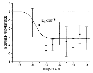
Figure 1 shows fluorimetry measurements of intracellular K+ (determined with
PBFI dye)
following exposure to increasing concentrations of ~c-PVIIA in primary
cultures of ventricular
myocytes. The data shown is from one trial and is represented as mean change
in fluorescence ~
S.E.M. (a'p<0.05, unpaired t-test).
Figures 2A-2B show fluorimetty measurements of membrane potential (determined
with Di-
~-ANEPPs dye) following exposure to increasing concentrations of o-PVIIA in
primary cultures of
ventricular myocytes (Fig. 2A) or cortex (Fig. 2B). Cells were loaded into 96
well plates at least
six days before the experiment. Results are expressed as Mean ~ SEM and
represent average data
from between two and five individual trials.
Figures 3A-3B are bar graphs showing the inhibition of the o-PVIIA (100y
response with
l OnM Glibenclamide (Glib) in primary cultures of myocytes (Fig. 3A) or with
SOuM Tolbutamide
(Tolb) in primary cultures of cortex (Fig. 3B). Data represents mea~1 ~ S.E.M.
Figures 4A-4C are whole cell recordings showing currents elicited by o-PVIIA
in (Fig. 4A)
cortical cells and (Fig. 4B) myocytes. Fig 4C shows I-V relationship of o-
PVIIA-induced current
from a cardiac myocyte.
Figure 5 is a bar graph showing the protective effect of 10 mM ~c-PVIIA
against hypoxia
induced depolarization. Bars represent Mean ~ S.E.M.
Figure 6 shows the effect of increasing concentrations of o-PVIIA on glutamate-
induced
(100uM) excitotoxicity measured six hours following glutamate washout (three
to six trials).
Figure 7 shows the infarct size as a % of the risl~ region (ischemic zone) as
plotted for the
six groups studied. Open symbols indicate individual experiments and solid
synbols indicated
CA 02474316 2004-07-23
WO 03/063782 PCT/US03/02384
group means. Triangles indicate animals receiving drug 5 min prior to
reperfusion and indicate
animals receiving drug 10 min after reperfiision.
Figure 8 is a plot of infarct size vs. risl~ zone size. Solid circles indicate
the plot for the
untreated controls and the line is the regression for that group. Protected
groups all lie below the
5 line as indicated by the open symbols.
Figure 9 is a graph showing ~c-PVIIA (CGX-1051) induced reduction in infarct
size
expressed as a percentage of the area at risl~ ill the canine AMI model. Data
r epresents mean+/- SEM
from 6 dogs per dose. CON-Control, OCC(30')- 30 min after occlusion, DRUG-
hnmediately
following dnlg administration, REPl, REP2, REP3- 1, 2 and 3 hours following
reperfusion.
Figtues l0A and l OB are graphs showing the lacy of effect of any of the
examined doses of
o-PVIIA (CGX-1051) on blood pressure (Fig. l0A) and heart rate (Fig. lOB).
Figure 11 is a graph showing reduction in incidence of ventricular
fibrillation following
administration of ~c-PVIIA (CGX-1051).
DETAILED DESCRIPTION OF THE PREFERRED EMBODIMENT
The invention relates to ~c-PVIIA-related conotoxins and pharmaceutically
acceptable salts
thereof and their use as organ protecting agents, i.e., organ protectants.
These conotoxins can be
used for arresting, protecting or preserving an organ, such as a circulatory
organ, a respiratory organ,
a urinary organ, a digestive organ, a reproductive organ, an endocrine organ
or a neurological organ.
These conotoxins can also be used for arresting, protecting or preserving
somatic,cells.
For purposes of the present invention, ~c-PVIIA refers to a peptide having the
following
general formula:
Cys-Xaa,-Ire-Xaa,,-Asn-Gln-Xaa3-Cys-Xaa4 Gln-XaaS-Leu-Asp-Asp-Cys-Cys-Ser-Xaa,-
Xaa3-Cys-Asn-Xaa,-Xaa4 Asn-Xaa3-Cys-Val (SEQ ID NO:1), wherein Xaa, and Xaa3
are
independently Arg, homoarginine, ornithine, Lys, N-methyl-Lys, N,N-dimethyl-
Lys, N,N,N-
trimethyl-Lys, any synthetic basic amino acid, His or halo-His; Xaaz is Pro or
hydroxy-Pro (Hyp);
Xaa4 is Phe, Tyr, meta-Tyr, ortho-Tyr, nor-Tyr, mono-halo-Tyr, di-halo-Tyr, O-
sulpho-Tyr, O-
phospho-Tyr, nitro-Tyr, Trp (D or L), neo-Tlp, halo-Trp (D or L) or any
synthetic aromatic arxlino
acid; and Xaas is His or halo-His. The C-terninus may contain a free carboxyl
group or an amide
group. The halo is preferably bromine, chlorine or iodine. It is preferred
that Xaal is Arg and XaaS
CA 02474316 2004-07-23
WO 03/063782 PCT/US03/02384
6
is His. It is more preferred that Xaa, is Arg, Xaa3 is Lys, Xaa4 is Phe and
Xaas is His. It is further
preferred that the C-terminus contains a free carboxyl group.
For proposes of the present amention, E6.2 refers to a peptide having the
following general
formula:
Xaa,,-Cys-Xaa3-Xaa,,-Xaa3-Gly-Xaa,-Xaa3-Cys-Xaa4 Xaa,-Xaas-Gln-Xaa3-Asp-Cys-
Cys-
Asn-Xaa3-Thr-Cys-Thr-Xaa,-Ser-Xaa3-Cys-Xaa, (SEQ ID N0:26), wherein Xaa,,
Xaa2, Xaa3, Xaad
and XaaS is as defined above. The C-terminus may contain a free carboxyl group
or an amide group,
preferably a free carboxyl. It is preferred that Xaa, is Arg, Xaa3 is Lys,
Xaa4 is Phe and Xaas is His.
It is more preferred that Xaa, is Arg, Xaa, is Pro, Xaa3 is Lys, Xaa4 is Phe
and XaaS is His.
For pui~oses of the present invention, P6.1 refers to a peptide having the
following general
formula:
Xaa2-Cys-Xaa3-Thr-Xaa2-Gly-Xaa,-Xaa3-Cys-Xaa4 Xaaz-Xaas-Gln-Xaa3-Asp-Cys-Cys-
Gly-
Xaa,-Ala-Cys-Ile-Ile-Thr-Ile-Cys-XaaZ (SEQ ~ N0:27), wherein Xaa,, Xaa2, Xaa3,
Xaa4 and XaaS
is as defined above. The C-terminus may contain a free carboxyl group or an
amide group,
preferably a free carboxyl. It is preferred that Xaa, is Arg, Xaa3 is Lys,
Xaa4 is Phe and Xaas is His.
It is more preferred that Xaa, is Arg, Xaa, is Hyp except at the C-terminus
which is Pro, Xaa3 is
Lys, Xaaø is Phe and Xaas is His.
For purposes of the present invention, P6.3 refers to a peptide having the
following general
formula:
Xaa2-Cys-Xaa3-Xaa3-Thr-Gly-Xaa,-Xaa~-Cys-Xaa4 Xaa,,-Xaas-Gln-Xaa3-Asp-Cys-Cys-
Gly-
Xaa,-Ala-Cys-Ile-Ile-Thr-Ile-Cys-Xaa,, (SEQ ID NO:28), wherein Xaa,, Xaa,"
Xaa3, Xaa4 and Xaas
is as defined above. The C-temninus may contain a free carboxyl group or an
amide group,
preferably a free carboxyl. It is preferred that Xaa, is Arg, Xaa~ is Lys,
Xaa4 is Phe and Xaas is His.
It is more preferred that Xaa, is Arg, Xaa2 is Pro, Xaa3 is Lys, Xaa4 is Phe
and Xaas is His.
The ~c-PVIIA analogs refer to peptides having the following formulas:
o-PVIIA[R18A]: Cys-Arg-Ile-Hyp-Asn-Gln-Lys-Cys-Phe-Gln-His-Leu-Asp-Asp-Cys-
Cys-Ser-Ala-Lys-Cys-Asn-Arg-Phe-Asn-Lys-Cys-Val (SEQ ID N0:2);
ic-PVIIA[R22A]: Cys-Arg-Ile-Hyp-Asn-Gh1-Lys-Cys-Phe-Gln-His-Leu-Asp-Asp-Cys-
Cys-Ser-Arg-Lys-Cys-Asn-Ala-Phe-Asn-Lys-Cys-Val (SEQ ID NO:3);
~c-PVIIA[I3A]: Cys-Arg-Ala-Hyp-Asn-Ghz-Lys-Cys-Phe-Ghz-His-Leu-Asp-Asp-Cys-
Cys-Ser-Arg-Lys-Cys-Asn-Arg-Phe-Asn-Lys-Cys-Val (SEQ ID N0:4);
CA 02474316 2004-07-23
WO 03/063782 PCT/US03/02384
7
~c-PVIIA[K19A]: Cys-Arg-Ile-Hyp-Asn-Gh1-Lys-Cys-Phe-Gln-His-Leu-Asp-Asp-Cys-
Cys-Ser-Arg-Ala-Cys-Asn-Arg-Phe-Asn-Lys-Cys-Val (SEQ ID NO:S);
~c-PVIIA[R2A]: Cys-Ala-Ile-Hyp-Asn-Gln-Lys-Cys-Phe-Gln-His-Leu-Asp-Asp-Cys-
Cys-Ser-Arg-Lys-Cys-Asn-Arg-Phe-Asn-Lys-Cys-Val (SEQ ID N0:6);
o-PVIIA[F9A]: Cys-Arg-Ile-Hyp-Asn-Ghl-Lys-Cys-Ala-Gln-His-Leu-Asp-Asp-Cys-
Cys-Ser-Arg-Lys-Cys-Asn-Arg-Phe-Asn-Lys-Cys-Val (SEQ ID N0:7);
o-PVIIA[K25A]: Cys-Arg-Ile-Hyp-Asn-Gln-Lys-Cys-Phe-Gln-His-Leu-Asp-Asp-Cys-
Cys-Ser-Arg-Lys-Cys-Asn-Arg-Phe-Asn-Ala-Cys-Val (SEQ ID N0:8);
~c-PVIIA[R2K]: Cys-Lys-Ile-Hyp-Asn-Gh1-Lys-Cys-Phe-Gln-His-Leu-Asp-Asp-Cys-
Cys-Ser-Arg-Lys-Cys-Asn-Arg-Phe-Asn-Lys-Cys-Val (SEQ ID N0:9);
~c-PVIIA[K7A]: Cys-Arg-Ile-Hyp-Asn-Gln-Ala-Cys-Phe-Gln-His-Leu-Asp-Asp-Cys-
Cys-Ser-Arg-Lys-Cys-Asn-Arg-Phe-Asn-Lys-Cys-Val (SEQ ID NO:10);
o-PVIIA[F9M]: Cys-Arg-Ile-Hyp-Asn-Ghl-Lys-Cys-Met-Ghz-His-Leu-Asp-Asp-Cys-
Cys-Ser-Arg-Lys-Cys-Asn-Arg-Phe-Asn-Lys-Cys-Val (SEQ ID NO:11);
~c-PVIIA[F9Y]: Cys-Arg-Ile-Hyp-Asn-Gln-Lys-Cys-Tyr-Gln-His-Leu-Asp-Asp-Cys-
Cys-Ser-Arg-Lys-Cys-Asn-Arg-Phe-Asn-Lys-Cys-Val (SEQ ~ N0:12);
o-PVIIA[R2Q]: Cys-Gh1-Ile-Hyp-Asn-Gln-Lys-Cys-Phe-Gln-His-Leu-Asp-Asp-Cys-
Cys-Ser-Arg-Lys-Cys-Asn-Arg-Phe-Asn-Lys-Cys-Val (SEQ ID N0:13);
~c-PVIIA[H11A]: Cys-Arg-Ile-Hyp-Asn-Gln-Lys-Cys-Phe-Ghl-Ala-Leu-Asp-Asp-Cys-
Cys-Ser-Arg-Lys-Cys-Asn-Arg-Phe-Asn-Lys-Cys-Val (SEQ ID N0:14);
o-PVIIA[D14A]: Cys-Arg-Ile-Hyp-Asn-Gln-Lys-Cys-Phe-Ghi-His-Leu-Asp-Ala-Cys-
Cys-Ser-Arg-Lys-Cys-Asn-Arg-Phe-Asn-Lys-Cys-Val (SEQ ID NO:15);
o-PVIIA[Q6A]: Cys-Arg-Ile-Hyp-Asn-Ala-Lys-Cys-Phe-Gln-His-Leu-Asp-Asp-Cys-
Cys-Ser-Arg-Lys-Cys-Asn-Arg-Phe-Asn-Lys-Cys-Val (SEQ ID N0:16);
~c-PVIIA[N21A]: Cys-Arg-Ile-Hyp-Asn-Ghi-Lys-Cys-Phe-Gln-His-Leu-Asp-Asp-Cys-
Cys-Ser-Arg-Lys-Cys-Ala-Arg-Phe-Asn-Lys-Cys-Val (SEQ ID N0:17);
~c-PVIIA[S17A]: Cys-Arg-Ile-Hyp-Asn-Gh1-Lys-Cys-Phe-Gln-His-Leu-Asp-Asp-Cys-
Cys-Ala-Arg-Lys-Cys-Asn-Arg-Phe-Asn-Lys-Cys-Val (SEQ ID N0:18);
~c-PVIIA[N24A]: Cys-Arg-Ile-Hyp-Asn-Gln-Lys-Cys-Phe-Gln-His-Leu-Asp-Asp-Cys-
Cys-Ser-Arg-Lys-Cys-Asn-Arg-Phe-Ala-Lys-Cys-Val (SEQ ID N0:19);
CA 02474316 2004-07-23
WO 03/063782 PCT/US03/02384
8
~e-PVIIA[L12A]: Cys-Arg-Ile-Hyp-Asn-Gln-Lys-Cys-Phe-Ghl-His-Ala-Asp-Asp-Cys-
Cys-Ser-Arg-Lys-Cys-Asn-Arg-Phe-Asn-Lys-Cys-Val (SEQ ID NO:20);
~c-PVIIA[D13A]: Cys-Arg-Ile-Hyp-Asn-Gln-Lys-Cys-Phe-Ghl-His-Leu-Ala-Asp-Cys-
Cys-Ser-Arg-Lys-Cys-Asn-Arg-Phe-Asn-Lys-Cys-Val (SEQ ID N0:21);
~c-PVIIA[Q10A]: Cys-Arg-Ile-Hyp-Asn-Gln-Lys-Cys-Phe-Ala-His-Leu-Asp-Asp-Cys-
Cys-Ser-Arg-Lys-Cys-Asn-Arg-Phe-Asn-Lys-Cys-Val (SEQ ID NO:22);
~c-PVIIA[V27A]: Cys-Arg-Ile-Hyp-Asn-Gln-Lys-Cys-Phe-Gh1-His-Leu-Asp-Asp-Cys-
Cys-Ser-Arg-Lys-Cys-Asn-Arg-Phe-Asn-Lys-Cys-Ala (SEQ ID N0:23);
~c-PVIIA[04A]: Cys-Arg-Ile-Ala-Asn-Ghl-Lys-Cys-Phe-Gln-His-Leu-Asp-Asp-Cys-
Cys-Ser-Arg-Lys-Cys-Asn-Arg-Phe-Asn-Lys-Cys-Val (SEQ ID N0:24); and
o-PVIIA[NSA]: Cys-Arg-Ile-Hyp-Ala-Gh1-Lys-Cys-Phe-Gln-His-Leu-Asp-Asp-Cys-
Cys-Ser-Arg-Lys-Cys-Asn-Arg-Phe-Asn-Lys-Cys-Val (SEQ ID N0:25).
It is preferred that the C-terminus contains a free carboxyl group.
The present invention further relates to derivatives of the above peptides or
analogs. In
accordance with the present invention, derivatives include peptides or analogs
in which the Arg
residues may be substituted by Lys, ornithine, homoarginine, nor-Lys, N-methyl-
Lys, N,N-
dimethyl-Lys, N,N,N-trimethyl-Lys or any synthetic basic amino acid; the Xaa,
residues may be
substituted by Arg, omithine, homoarginine, nor-Lys, or any synthetic basic
amino acid; the Tyr
residues may be substituted with any synthetic hydroxy containing amino acid;
the Ser residues may
be substituted with Thr or any s5mthetic hydroxylated amino acid; the Thr
residues may be
substituted with Ser or any synthetic hydroxylated amino acid; the Phe and Trp
residues may be
substituted with axly synthetic aromatic amino acid; and the Asn, Ser, Thr or
Hyp residues may be
glycosylated. The Cys residues may be in D or L configuration and may
optionally be substituted
with homocysteine (D or L). The Tyr residues may also be substituted with''-SI-
Tyr or with the 3-
hydroxyl or 2-hydroxyl isomers (meta-Tyr or ortho-Tyr, respectively) and
corresponding O-sulpho-
and O-phospho-derivatives. The acidic amino acid residues may be substituted
with any synthetic
acidic amino acid, e.g., tetrazolyl derivatives of Gly and Ala. The aliphatic
amino acids may be
substituted by synthetic derivatives bearing non-natural aliphatic branched or
linear side chains
C"H,"+2 up to acid including n=8. The Leu residues may be substituted with Leu
(D). The Gla
residues may be substituted with Glu.
CA 02474316 2004-07-23
WO 03/063782 PCT/US03/02384
9
The present invention is further directed to derivatives of the above peptides
and peptide
derivatives which are cyclic permutations in which the cyclic permutants
retain the native bridging
pattern of native toxin. See Crailc et al. (2001).
Examples of synthetic aromatic amino acid include, but are not limited to,
nitro-Phe, 4-
substituted-Phe wherein the substituent is C,-C3 allcyl, carboxyl,
hydroxymethyl, sulphomethyl,
halo, phenyl, -CHO, -CN, -S03H and -NHAc. Examples of synthetic hydroxy
contaiW ng amino
acid, include, but are not limited to, 4-hydroxymethyl-Phe, 4-hydroxyphenyl-
Gly, 2,6-dimethyl-Tyr
and 5-amino-Tyr. Examples of synthetic basic amino acids include, but are not
limited to, N-1-(2-
pyrazolinyl)-Arg, 2-(4-piperinyl)-Gly, 2-(4-piperinyl)-Ala, 2-[3-
(2S)pyTOlininyl)-Gly and 2-[3-
(2S)pyrrolininyl)-Ala. These and other s~mthetic basic amino acids, synthetic
hydroxy containing
amino acids or synthetic aromatic amino acids are described in Building Block
Index, Version 3.0
(1999 Catalog, pages 4-47 for hydroxy containing amino acids and aromatic
amino acids and pages
66-87 for basic amino acids; see also http://www.amino-acids.com),
incorporated herein by
reference, by and available from RSP Amino Acid Ailalogues, hic., Worcester,
MA. Examples of
synthetic acid amino acids include those derivatives bearing acidic
functionality, including carboxyl,
phosphate, sulfonate and synthetic tetrazolyl derivatives such as described by
Omstein et al. (1993)
and in U.S. Patent No. 5,331,001, each incorporated herein by reference, and
such as shown in the
following schemes 1-3
1 ~ 4
R R
~ 3
2
Fmo FmocHN~COOH
R=COOH, tetazole, CHZCOOH, 4-NHSO2CH3, 4-NHS02Phenyl,
4-CHZS03H, S03H, 4-CH2P03H2, CH2CH2COOH, OCH2Tetrazole,
CH2STetrazole, HNTetrazole, CONHS02R1 where Rl is CH3 or Phenyl
S02-Tetrazole, CH2CHZS03H, 1,2,4-tetrazole, 3-isoxazolone,
amidotetrazole, CH2CH2P03H2
Scheme 1
CA 02474316 2004-07-23
WO 03/063782 PCT/US03/02384
R R
OH OH
R = COOH, tetrazole, CH2COOH, CH2tetrazole
Scheme 2
R R
~~n ]n
FmocHN COOH FmocHN~COOH
R = COOH, tetazole, CH2COOH, 4-NHS02CH3, 4-NHS02Phenyl,
4-CH2S03H, S03H, 4-CH2P03H2, CH2CH2COOH, OCH2Tetrazole,
CH2STetrazole, HNTetrazole, CONHS02R1 where Rl is CH3 or Phenyl
S02-Tetrazole, CHZCH2S03H, 1,2,4-tetrazole, 3-isoxazolone,
amidotetrazole, CH2CH2P03H~ n = 0, 1, 2, or 3
Scheme 3
Optionally, in the peptides and analogs described above, the Asn residues may
be modified
5 to contain an N-glycan and the Ser, Thr and Hyp residues may be modified to
contain an O-glycan
(e.g., g-N, g-S, g-T and g-Hyp). In accordance with the present invention, a
glycan shall mean any
N-, S- or O-liu~ed mono-, di-, tri-, poly- or oligosaccharide that can be
attached to any hydroxy,
amino or thiol group of natural or.modified amino acids by synthetic or
enzymatic methodologies
luiown in the art. The monosaccharides mal~ing up the glycan can include, but
are not limited to,
10 D-allose, D-altrose, D-glucose, D-mannose, D-gulose, D-idose, D-galactose,
D-talose, D-
galactosamine, D-glucosamine, D-N-acetyl-glucosamine (GIcNAc), D-N-acetyl-
galactosamine
(GalNAc), D-fucose or D-arabinose. These saccharides may be stllicturally
modified, e.g., with one
CA 02474316 2004-07-23
WO 03/063782 PCT/US03/02384
11
or more O-sulfate, O-phosphate, O-acetyl or acidic groups, such as sialic
acid, including
combinations thereof. The glycan may also include similar polyhydroxy groups,
such as D-
penicillamine 2,5 and halogenated derivatives thereof or polypropylene glycol
derivatives. The
glycosidic liu~age is beta and 1~4 or 1~3, preferably 1~3. The linl~age
between the glycan and
the amino acid may be alpha or beta, preferably alpha and is 1--~.
Core O-glycans have been described by Van de Steen et al. (1998), incorporated
herein by
reference. Mucin type O-liuced oligosaccharides are attached to Ser or Thr (or
other hydroxylated
residues of the present peptides) by a GaINAc residue. The monosaccharide
building blocl~s and
the linlcage attached to this first GaINAc residue define the core glycans, of
wluch eight have been
identified. The type of glycosidic linl~age (orientation and comzectivities)
are defined for each core
glycan. Suitable glycans and glycan analogs are described further in U.S.
Serial No. 09/420,797,
filed 19 October 1999 and in PCT Application No. PCT/LTS99/24380, filed 19
October 1999 (PCT
Published Application No. WO 00/23092), each incorporated herein by reference.
A preferred
glycanis Gal((31~3)GaINAc(al~).
Optionally, in the above peptides, pairs of Cys residues may be replaced
pairwise with
isosteric lactam or ester-thioether replacements, such as Ser/(Glu or Asp),
Lys/(Glu or Asp) or
Cys/Ala combinations. Sequential coupling by lalown methods (Barnay et al.,
2000; Hruby et al.,
1994; Bitan et al., 1997) allows replacement of native Cys bridges with lactam
bridges. Thioether
analogs may be readily synthesized using halo-Ala residues commercially
available from RSP
Amino Acid Analogues. In addition, individual Cys residues may be replaced
with homoCys,
seleno-Cys or penicillamine, so that disulfide bridges may be formed between
Cys-homoCys or
Cys-penicillamine, or homoCys-penicillamine and the life.
The present invention, in mother aspect, relates to a pharmaceutical
composition comprising
an effective amount of ~c-PVIIA-related conotoxins. Such a pharmaceutical
composition has the
capability of acting as organ protecting agents, i.e., organ protectants.
These conotoxiils can be used
for arresting, protecting or preserving an organ, such as a circulatory organ,
a respiratory organ, a
urinary organ, a digestive organ, a reproductive organ, an endocrine organ or
a neurological organ.
The ~c-PVIIA-related conotoxins can be isolated from Co~2us such as described
in U.S.
Patent No. 5,672,682 for n-PVIIA from Coytus purpuYCZSCe~zs, or it can be
chemically synthesized
by general synthetic methods such as described in U.S. Patent No. 5,672,682.
Alternatively, the
native peptide can be synthesized by conventional recombinant DNA techniques
(Sambrool~ et al.,
CA 02474316 2004-07-23
WO 03/063782 PCT/US03/02384
12
1989) using the DNA encoding the conotoxin, such as DNA encoding ~c-PVIIA
(Shop et al., 1998)
or DNA encoding E6.2, P6.1 or P6.3 as described in U.S. patent application
Serial No. 09/910,082
and international patent application No. PCT/US01/23041, each incorporated
herein by reference.
The peptides are also synthesized using an automated synthesizer. Amino acids
are sequentially
coupled to an MBHA Riu~ resin (typically 100 mg of resin) beginning at the C-
terminus using an
Advanced ChemTech 357 Automatic Peptide Synthesizer. Couplings are carried out
using 1,3-
diisopropylcarbodimide in N-methylpyrrolidhlone (NMP) or by 2-(1H-
benzotriazole-1-yl)-1,1,3,3-
tetramethyluronium hexafluorophosphate (HBTU) and diethylisopropylethylamine
(DIEA). The
FMOC protecting group is removed by treatment with a 20% solution of
piperidine in
dimethylformamide(DMF). Resins are subsequently washed with DMF (twice),
followed by
methanol and NMP.
Muteins, a~lalogs or active fragments, of the foregoing ~c-PVIIA-related
conotoxin peptides
are also contemplated here. See, e.g., Hammerland et al (1992). Derivative
muteins, analogs or
active fragments of the conotoxin peptides may be synthesized according to
lcnown techniques,
including conservative amino acid substitutions, such as outlined in U.S.
Patents No. 5,545,723 (see
particularly col. 2, line 50 to col. 3, line 8); 5,534,615 (see particularly
col. 19, line 45 to col. 22,
line 33); and 5,364,769 (see particularly col. 4, line 55 to col. 7, line 26),
each incorporated herein
by reference.
In accorda~lce with the present invention, o-PVIIA-related conotoxins and
pharmaceutically
acceptable salts thereof are used for arresting, protecting or preserving an
organ. The organ may
be intact in the subject or may have been isolated (such as for
transplantation). The organ may be
a circulatory organ, a respiratory organ, a urinary organ, a digestive organ,
a reproductive organ, an
endocrine organ or a neurological organ. The present invention is particularly
useful for arresting,
protecting or preserving the heart dining open heart surgery, angioplasty,
valve surgery, bypass
surgery, transplantation, or cardiovascular disease so as to reduce heart
damage before, during or
following cardiovascular intervention or to protect those portions of the
heart that have been starved
of normal flow of blood, nutrients and/or oxygen (reperfiision injury). The
present invention is also
particularly useful for cardioplegia, which is a technique of myocardial
preservation during cardiac
surgery, usually employing infusion of a cold, potassium laced solution,
sometimes fixed with
blood, to achieve arrest of the myocardial fibers and to reduce their oxygen
consumption to nearly
nothing. Techniques using warm (body temperaW re) blood can also be used with
the present ~c-
PVIIA-related conotoxins and pharmaceutically acceptable salts thereof.
CA 02474316 2004-07-23
WO 03/063782 PCT/US03/02384
13
The ~c-PVIIA-related conotoxins and pharmaceutically acceptable salts thereof
can be used
in conjunction with other agents for arresting, protecting or preserving
organs in accordance with
the present invention. Thus, ~c-PVIIA-related conotoxins and pharmaceutically
acceptable salts
thereof can be coadministered with an adenosine receptor agonist, a local
anesthetic, a potassium
chan~.le1 opener or agonist, an AV bloclcer, and/or a hemostatic agent.
Examples of adenosine
receptor agonsts include, but are not limited to, Al, A2a and A3 agents. A1
agents include, but are
not limited to, CPA, NECA, CGS-21680, AB-MECA, AMP579, 9APNEA, CHA, ENBA. A2a
agents include, but are not limited to, R-PIA, DPMA, CGS-21680, ATL146e. A3
agents include,
but are not limited to, CCPA, CI-IB-MECA, IB-MECA. Suitable local anesthetics
include, but are
not limited to, mexilitine, diphenylhydantoin, prilocaine, procaine,
mipivicaine, bupivicaine,
lidocaine and class 1B anti-anhytlunic agents, i.e. lignocaine. Suitable
potassium channel openers
or agonists include, but are not limited to, cromalcalin, pinacidil,
nicora~ldil, NS-1619, diazoxide and
minoxidil . Suitable AV bloclcers include, but are not limited to, verapamil.
Hemostatic agents may
be a "clot buster" agent, a thrombolytic agent, an anti-coagulant agent or an
a~iti-platelet aggregation
agent. Suitable "clot buster" agents include, but are not limited to,
streptol~inase, uroleinase and
ACTIVASE. Suitable thrombolytic agents include, but are not limited to,
streptol~inase, urol~inase,
alteplase, reteplase and tenecteplase. Suitable anti-coagulant agents include,
but are not limited to,
heparin, enoxaparin and dalteparin. Suitable anti-platelet aggregation agents
include, but are not
limited to, aspirin, clopidogrel, abciximab, eptifibatide and tirofiban.
The ~c-PVIIA-related conotoxins and pharmaceutically acceptable salts thereof
disclosed
herein can also be used for the treatment of arrhytlunia, urinary
incontinence, angina, reperfusion
injury, diabetes, retinopathy, neuropathy, nephropathy, peripheral circulation
disturbances, acute
heart failure, hypertension, cerebral vasospasm accompanying subarachnoid
hemorrhage, anxiety
disorder, cerebral ischemia, coronary artery bypass graft (CABG) surgery,
ischemic heart disease
and congestive heart failure. The o-PVIIA-related conotoxins a~ld
pharmaceutically acceptable salts
thereof disclosed herein can also be used for open heart surgery, bypass
surgery, heart transplant
surgery and cardioplegia. Cardioplegia is a tech~lique of myocardial
preservation during cardiac
surgery usually employing infusion of a cold, potassimn laced solution,
sometimes fixed with blood,
to achieve arrest of the myocardial fibers and reduce their oxygen consumption
to nearly nothing.
Techniques using warm (body temperature) blood are also used.
Pharmaceutical compositions containng a compound of the present invention or
its
pharmaceutically acceptable salts as the active ingredient can be prepared
according to conventional
CA 02474316 2004-07-23
WO 03/063782 PCT/US03/02384
14
pharmaceutical compounding tecluiques. See, for example, ReTnifzgton's
Pha3°syaaceutical Sciences,
18th Ed. (1990, Mach Publishing Co., Easton, PA). Typically, an ATP-sensitive
potassium channel
opening amount of the active ingredient will be admixed with a
pharmaceutically acceptable Garner.
The carrier may tahce a wide variety of forms depending on the form of
preparation desired for
administration, e.g., intravenous, oral or parenteral. The compositions may
further contain
antioxidizing agents, stabilizing agents, preservatives and the life. For
examples of delivery
methods, see U.S. Patent No. 5,844,077, incorporated herein by reference.
"Pharmaceutical composition" means physically discrete coherent portions
suitable for
medical achninistration. "Pharmaceutical composition in dosage unit forth"
means physically
discrete coherent units suitable for medical administration, each containing a
daily dose or a
multiple (up to four times) or a sub-multiple (down to a fortieth) of a daily
dose of the active
compound in association with a carrier andlor enclosed within an envelope.
Whether the
composition contains a daily dose, or for example, a half, a third or a
quarter of a daily dose, will
depend on whether the pharmaceutical composition is to be administered once
or, for example,
twice, three times or four times a day, respectively.
The term "salt", as used herein, denotes acidic and/or basic salts, formed
with inorganic or
organic acids and/or bases, preferably basic salts. While pharmaceutically
acceptable salts are
preferred, particularly when employing the compounds of the invention as
medicaments, other salts
find utility, for example, in processing these compounds, or where non-
medicament-type uses are
contemplated. Salts of these compounds may be prepared by art-recognized
techniques.
Examples of such pharmaceutically acceptable salts include, but are not
limited to, inorganic
and organic addition salts, such as hydrochloride, sulphates, nitrates or
phosphates and acetates,
trifluoroacetates, propionates, succinates, benzoates, citrates, tartrates,
fumarates, maheates,
methane-sulfonates, isotlionates, theophylline acetates, salicylates,
respectively, or the like. Lower
all~yl quatemaiy ammonium salts and the like are suitable, as well.
As used herein, the tern "pharmaceutically acceptable" carrier means a non-
toxic, inert
solid, semi-solid liquid filler, diluent, encapsulating material, formulation
auxiliary of any type, or
simply a sterile aqueous medium, such as saline. Some examples of the
materials that can serve as
pharmaceutically acceptable carriers are sugars, such as lactose, glucose and
sucrose, starches such
as corn starch and potato starch, cellulose and its derivatives such as sodium
carboxymethyl
cellulose, ethyl cellulose and cellulose acetate; powdered tragacanth; malt,
gelatin, talc; excipients
such as cocoa butter and suppository waxes; oils such as peanut oil,
cottonseed oil, safflower oil,
CA 02474316 2004-07-23
WO 03/063782 PCT/US03/02384
sesame oil, olive oil, corn oil and soybean oil; glycols, such as propylene
glycol, polyols such as
glycerin, sorbitol, mannitol and polyethylene glycol; esters such as ethyl
oleate and ethyl laurate,
agar; buffering agents such as magnesium hydroxide and aluminum hydroxide;
alginic acid;
pyrogen-free water; isotonic saline, Ringer's solution; ethyl alcohol and
phosphate buffer solutions,
5 as well as other non-toxic compatible substances used in pharmaceutical
formulations.
Wetting agents, emulsifiers and lubricants such as sodium lauryl sulfate and
magnesium
stearate, as well as coloring agents, releasing agents, coating agents,
sweetening, flavoring and
perfiuning agents, preservatives and antioxidants can also be present in the
composition, according
to the judgment of the formulator. Examples of pharmaceutically acceptable
antioxidants include,
10 but are not limited to, water soluble antioxidants such as ascorbic acid,
cysteine hydrochloride,
sodium bisulfate, sodium metabisulfite, sodium sulfite, and the like; oil
soluble antioxidants, such
as ascorbyl palmitate, butylated hydroxyanisole (BHA), butylated
hydroxytoluene (BHT), lecithin,
propyl gallate, alpha-tocopherol and the lilce; and the metal chelating agents
such as citric acid,
ethylenediamine tetraacetic acid (EDTA), sorbitol, tartaric acid, phosphoric
acid and the lilce.
15 For oral administration, the compounds can be formulated into solid or
liquid preparations
such as capsules, pills, tablets, lozenges, melts, powders, suspensions or
emulsions. In preparing
the compositions in oral dosage fore, any of the usual pharmaceutical media
may be employed,
such as, for example, water, glycols, oils, alcohols, flavoring agents,
preservatives, coloring agents,
suspending agents and the like in the case of oral liquid preparations (such
as, for example,
suspensions, elixirs and solutions); or can-iers such as starches, sugars,
diluents, granulating agents,
lubricants, binders, disintegrating agents and the like in the case of oral
solid preparations (such as,
for example, powders, capsules and tablets). Because of their ease in
administration, tablets and
capsules represent the most advmtageous oral dosage mut form, in wluch case
solid pharmaceutical
carriers are obviously employed. If desired, tablets may be sugar-coated or
enteric-coated by
standard teclnuques. The active agent can be encapsulated to make it stable
for passage through the
gastrointestinal tract, while at the same time allowhig for passage across the
blood brain barrier. See
for example, WO 96/11698.
For parenteral achninishation, the compound may be dissolved in a
pharmaceutical carrier
and administered as either a solution or a suspension. Illustrative of
suitable carriers are water,
saline, dextrose solutions, fi-uctose solutions, ethanol, or oils of aumal,
vegetative or synthetic
origin. The carrier may also contain other ingredients, for example,
preservatives, suspending
agents, solubilizing agents, stabilizing agents, buffers and the like. One
particularly suitable
CA 02474316 2004-07-23
WO 03/063782 PCT/US03/02384
16
stabilizing agent for the conotoxin peptides contemplated here is
carboxymethyl cellulose. This
agent may be particularly effective due to the excess positive charge of the
contemplated conotoxin
peptides. When the compounds are being administered intrathecally, they may
also be dissolved
in cerebrospinal fluid.
A variety of administration routes are available. The particular mode selected
will depend
of course, upon the particular dnig selected, the severity of the disease
state being treated and the
dosage required for therapeutic efficacy. The methods of this invention,
generally spealting, may
be practiced using any mode of administration that is medically acceptable,
meaning any mode that
produces effective levels of the active compounds without causing clinically
unacceptable adverse
effects. Such modes of admilustration include oral, rectal, sublingual,
topical, nasal, transdennal or
parenteral routes. The term "parenteral" includes subcutaneous, intravenous,
epidural, irrigation,
intramuscular, release pumps, or infusion.
For example, admilustration of the active agent according to this invention
may be achieved
using any suitable delivery means, 111C111dlllg:
(a) pump (see, e.g., Lauer & Hatton (1993), Zilnm et al. (1984), Ettinger et
al. (1978) and
cardioplegia system of Medtronic, Inc.);
(b), microencapsulation (see, e.g., U.S. Patent Nos. 4,352,883; 4,353,888; and
5,084,350);
(c) continuous release polymer implants (see, e.g., U.S. Patent No.
4,883,666);
(d) macroencapsulation (see, e.g., U.S. Patent Nos. 5,284,761, 5,158,881,
4,976,859 and
4,968,733 and published PCT patent applications WO 92/19195, WO 95/05452);
(e) nal~ed or unencapsulated cell grafts to the CNS (see, e.g., U.S. Patent
Nos. 5,082,670 and
5,618,531);
(f) injection, either subcutaneously, intravenously, intra-arterially,
intramuscularly, or to
other suitable site; or
(g) oral administration, in capsule, liquid, tablet, pill, or prolonged
release formulation.
In one embodiment of this invention, an active agent is delivered directly
into the CNS,
preferably to the brain ventricles (e.g. i.c.v.), brain parenchyma, the
intrathecal space or other
suitable CNS location, most preferably intrathecally.
Alternatively, targeting therapies may be used to deliver the active agent
more specifically
to certain types of cells, by the use of targeting systems such as antibodies
or cell-specific ligands.
Targeting may be desirable for a variety of reasons, e.g. if the agent is
unacceptably toxic, if it
would otherwise require too high a dosage, or if it would not otherwise be
able to enter target cells.
CA 02474316 2004-07-23
WO 03/063782 PCT/US03/02384
17
The active agents, which are peptides, can also be administered in a cell
based delivery
system in which a DNA sequence encoding an active agent is introduced into
cells designed for
implantation in the body of the patient, especially in the spinal cord region.
Suitable delivery
systems are described in U.S. Patent No. 5,550,050 and published PCT
Application Nos. WO
92/19195, WO 94/25503, WO 95/01203, WO 95/05452, WO 96/02286, WO 96/02646, WO
96/40871, WO 96/40959 and WO 97/12635. Suitable DNA sequences can be prepared
synthetically
for each active agent on the basis of the developed sequences and the lmown
genetic code.
The active agent is preferably administered in a therapeutically effective
amount. By a
"therapeutically effective amount" or simply "effective amount" of an active
compound is meant a
sufficient amount of the compound to arrest, preserve or protect an organ at a
reasonable benefit/risl~
ratio applicable to any medical treatment. The actual amount administered, and
the rate and time-
course of administration, will depend on the nature and severity of the
condition being treated. The
administration may be continuous or be intermittent. Prescription of
treatment, e.g. decisions on
dosage, timing, etc., is within the responsibility of general practitioners or
specialists, and typically
talces account of the disorder to be treated, the condition of the individual
patient, the site of
delivery, the method of administration and other factors known to
practitioners. Examples of
techniques and protocols can be found in Re~raington's P7~arnaaceutical
Scieyaces.
Dosage may be adjusted appropriately to achieve desired drug levels, locally
or systemically.
Typically, the active agents of the present invention exhibit their effect at
a dosage range of from
about 0.001 mg/l~g to about 250 mg/lcg, preferably from about 0.01 mg/l~g to
about 100 mg/lcg, of
the active ingredient and more preferably, from about 0.05 mg/lcg to about 75
mg/l~g. A suitable
dose can be administered in multiple sub-doses per day. Typically, a dose or
sub-dose may contain
from about 0.1 mg to about 500 mg of the active ingredient per Lmit dosage
form. A more preferred
dosage will contain from about 0.5 mg to about 100 mg of active ingredient per
unit dosage form.
Dosages are generally initiated at lower levels and increased mtil desired
effects are achieved.
Advantageously, the compositions are formulated as dosage Lmits, each unit
being adapted
to supply a fixed dose of active ingredients. Tablets, coated tablets,
capsules, ampoules and
suppositories are examples of dosage forns according to the invention.
It is only necessary that the active ingredient constitute an effective
amount, i.e., such that
a suitable effective dosage will be consistent with the dosage form employed
in single or multiple
unit doses. The exact individual dosages, as well as daily dosages, are
determined according to
CA 02474316 2004-07-23
WO 03/063782 PCT/US03/02384
18
standard medical principles under the direction of a physician or veterinarian
for use humans or
animals.
The pharmaceutical compositions will generally contain from about 0.0001 to 99
wt. %,
preferably about 0.001 to 50 wt. %, more preferably about 0.01 to 10 wt.% of
the active ingredient
by weight of the total composition. In addition to the active agent, the
pharmaceutical compositions
and medicaments can also contain other pharmaceutically active compounds.
Examples of other
pharmaceutically active compounds include, but are not limited to, adenosine
receptor agonists,
local anesthetics, hemostatic agents, potassium chamiel opener or agonist, AV
blocl~ers and
therapeutic agents in all of the major areas of clinical medicine. When used
with other
pharmaceutically active compounds, the conotoxin peptides of the present
invention may be
delivered in the form of drug cocl~tails. A cocl~tail is a mixture of any one
of the compounds useful
with this invention with another drtg or agent. In this embodiment, a common
administration
vehicle (e.g., pill, tablet, implant, pump, injectable solution, etc.) would
contain both the instant
composition in combination supplementary potentiating agent. The individual
dings of the cocl~tail
are each acliniustered in therapeutically effective amounts. A therapeutically
effective atnottnt will
be determined by the parameters described above; but, in any event, is that
amount which
establishes a level of the drugs in the area of body where the drugs are
required for a period of time
which is effective in attaining the desired effects.
The ~c-PVIIA-related conotoxins and pharmaceutically acceptable salts thereof
and their use
as organ protecting agents, i.e., organ protectants, as described herein cm be
used in the treatment
of htunans or animals, i.e., in veterillaiy applications. These conotoxins and
their use can be utilized
for individuals of any age, including pediatric and geriatric patients.
The o-PVIIA-related conotoxins and pharmaceutically acceptable salts thereof
disclosed
herein can also be used for the treatment of arrhythmia, urinary incontinence,
angina, reperfusion
injury, diabetes, retinopathy, neuropathy, nephropathy, peripheral circulation
disturbances, acute
heart failure, hypertension, cerebral vasospasm accompanying subaraclmoid
hemorrhage, anxiety
disorder, cerebral ischemia, CABG surgery, ischemic heart disease and
congestive heart failure. The
o-PVIIA-related conotoxills and pharmaceutically acceptable salts thereof
disclosed herein can also
be used for open heart surgery, bypass surgery, heart transplant surgery and
cardioplegia.
Cardioplegia is a technique of myocardial preservation during caxdiac surgery
usually employing
infusion of a cold, potassitun laced solution, sometimes fixed with blood, to
achieve arrest of the
CA 02474316 2004-07-23
WO 03/063782 PCT/US03/02384
19
myocardial fibers and reduce their oxygen consumption to nearly nothing.
Techniques using warm
(body temperature) blood are also used.
Activators of KATP channels have therapeutic siguficance for the treatment of
asthma,
cardiac ischemia and cerebral ischemia, among others.
Asthfncz: Asthma is a serious and connnon condition that effects approximately
12 million
people in the United States alone. This disorder is particularly serious in
children and it has been
estimated that the greatest nmnber of asthma patients are those under the age
of 18 (National Health
SLUVey, National Center of Health Statistics, 1989). The disease is
characterized by chronic
inflammation and hyper-responsiveness of the airway wluch results in periodic
attacks of wheezing
and difficulty in breathing. An attack occurs when the airway smooth muscle
become inflamed and
swells as a result of exposure to a trigger substance. In severe cases, the
airway may become
blocked or obstructed as a result of the smooth muscle contraction. Further
exacerbating the
problem is the release of large quantities of mucus which also act to block
the airway. Chronic
asthmatics are most colrunony treated prophylactically with inhaled
corticosteroids and acutely with
inhaled bronchodilators, usually [3-2 agonists. However, chronic treatment
with inhaled
corticosteroids has an associated risk of immune system impairment,
hypertension, osteoporosis,
adrenal gland malfunction and an increased susceptibility to fungal infections
(Ralcel, 1997). hl
addition use of (3-2 agonists has been repouted in some cases to cause adverse
reactions including
tremor, tachycardia and palpitations aizd muscle cramps (Rakel, 1997).
Therefore, there is great
potential in developing anti-astlmnatic agents with fewer side-effects.
I~+ chaimel openers have been shown to be effective relaxants of airway smooth
muscle
reducing hyperactivity induced obstuuction of intact airway. h1 cryopreserved
human bronchi
(Muller-Schweinitzer and Fozard, 1997) and iiz the isolated gvvinea pig
tracheal preparation (Lin et
al, 1998; Ando et al., 1997; Nielson-I~udsk, 1996; Nagai et al., 1991), KATP
openers produced
relaxation whether the muscle was contracted spontaneously or induced by a
range of spasmogens.
Under these conditions, the I~''~ chamlel openers are thought to be acting to
produce a I~+ ion efflux
and consequent membrane hyperpolarization. As a result, voltage-sensitive Ca2+
channels would
close and intracellular calcium levels would drop, producing muscular
relaxation. The development
of new and more specific KATP openers may offer a novel approach both to the
prophylactic and
symptomatic treatment of astlnna.
KATP charnels are present in many tissue types beyond just the target tissue,
therefore their
activation may result in unwanted side effects. In particular, as KATP
channels are found in vascular
CA 02474316 2004-07-23
WO 03/063782 PCT/US03/02384
smooth muscle, it is possible that in addition to the beneficial anti-
asthmatic properties of KATP
openers there could be an associated drop in blood pressure. It is possible
that delivering the
compound in inhalant form directly to the airway smooth muscle will allow the
concentration of the
compound to be reduced significantly thereby minimizing adverse reactions.
5 Cay-diac Ischetiaicz: While numerous subtypes of potassium channels in
cardiac tissue have
not yet been fully characterized, openers of KATP channels show great promise
as cardioprotective
agents. The beneficial vasodilatoiy effects afforded by K+ channel openers in
patients with angina
pectoris are now well established (Chen et al., 1997; Goldschmidt et al.,
1996; Yamabe et al., 1995;
Koike et al., 1995). Furthermore, the activation of KnTP charnels appears also
to be involved in the
10 acute preconditioning of the myocardium following brief ischemic periods,
acting to reduce the risk
(Pelf et al., 1998) and size of the reperfusion infarct (Kouchi et al., 1998).
Direct evidence for the cytoprotective properties of KaTP channels was
demonstrated by
Jovanovic et al. (1998a). In these studies, the DNA encoding for the
Kir6.2/SUR2A (caa-diac KATP)
channel were transfected in COS-7 mou~ey cells and the degree of calcium
loading monitored.
15 Untransfected cells were demonstrated to be vulnerable to the increases in
intracellular calcium seen
following hypoxiaJreoxygenation. However, the transfection of the cells with
the KATP channel
conferred resistance to the potentially damaging effects of the hypoxia-
reoxygenation. Thus, the
cardiac KATP channels are likely to play a significant role in protecting the
myocardium against
reperfiision injury.
20 Cercbi°al Ischernicz: Although treatment of cerebral ischemia has
advanced significantly
over the past 30 yeaxs, cerebral ischemia (stroke) still remains the third
leading cause of death in the
Uuted States. More than 500,000 new strolce/ischemia cases are reported each
year. Even though
initial mortality is high (38%), there are close to three million survivors of
stroke in the United
States, and yearly cost for rehabilitation of these patients in the United
States is close to $17 billion
(Rakel, 1997).
The initial cellular effects occl~r very rapidly (a matter of miizutes) after
an ischemic episode,
whereas the actual cellular destntction does not occur until several hours or
days following the
infarction. Initial effects include depolarization due to bioenergetic
failure, and inactivation of Na+
chaimels. Voltage-gated calcium channels are activated resulting in a massive
rise in intracellular
calcium. Further exacerbating the problem is a large transient release of
glutamate which itself
increases both Na and Caz+ influx through ionotropic glutamate receptors.
Glutamate also binds
to metabotropic receptors, which results in activation of the inositol
phosphate pathway. This sets
CA 02474316 2004-07-23
WO 03/063782 PCT/US03/02384
21
off a cascade of intracellular events, including further release of calcium
from intracellular stores.
It is now well accepted that this initial overload of intracellular calcium
ultimately leads to the
delayed cytotoxicity that is seen hours or days later.
Recently it has been reported that dopaminergic neurons exposed to a very
short hypoxic
challenge will hyperpolarize primarily through an opelung Of KnTP channels
(Guatteo et al., 1998).
This stimulatory effect was suggested to be a direct result of the increased
metabolic demand and
the consequent drop in intracellular ATP levels. Ful-thermore Jovanovic et al.
(1998b) recently
reported that cells transfected with DNA encoding for Kir6.2/SUR1 (neuronal
I~A.I.P) channel showed
increased resistance to injury caused through hypoxia-reoxygenation.
Therefore, the opening of
I~ATP channels may serve a vital cytoprotective role during short periods of
reduced oxygen in
neuronal tissue. Thus, there is great therapeutic potential in developing
compounds that not only
will act to prevent this calcium influx prophylactically, but will aid in
reestablishing the normal
resting membrane potential in damaged tissue. Treatment with ~c-PVIIA-related
conotoxins will act
to open KATP channels, 111d11C111g membrane hyperpolarization and indirectly
producing closure of
the voltage-gated Ca2+ chamlels, thereby preventing or reducing deleterious
effects of a massive
calcium influx.
In accordance with the present invention, it has been found that intravenous
(IV) injection
of concentrations of ~c-PVIIA, far higher than those required to produce
maximal hyperpolarization
in tracheal cultures ih vitT°o, had no effect on blood pressure or
heart rate in the anesthetized rat.
Our preliminary data indicates that o-PVIIA induces glibenclamide-sensitive
currents in
primary cultures of myocytes in a highly potent mamler. Ful-thelmore,
incubation of primary
myocyte cultures in the presence of ~c-PVIIA confers protection against
hypoxia-induced
depolarization. Further data demonstrates that ~c-PVIIA reduces the infarct
size, thus providing
protection to an organ from reperfilsion injury.
The present invention also relates to rational drug design for the
identification of additional
drugs which can be used for the purposes described herein. The goal of
rational drug design is to
produce stnlctural analogs of biologically active polypeptides of interest or
of shall molecules with
which they interact (e.g., agonists, antagonists, il~lubitors) in order to
fashion drugs which are, for
example, more active or stable forms of the polypeptide, or which, e.g.,
enhance or interfere with
the function of a polypeptide ifz vivo. Several approaches for use in rational
drug design include
analysis of three-dimensional structure, alanine scans, molecular modeling and
use of anti-id
CA 02474316 2004-07-23
WO 03/063782 PCT/US03/02384
22
antibodies. These techniques are well lmown to those spilled in the art. Such
techniques may
include providing atomic coordinates defining a three-dimensional stmcture of
a protein complex
formed by said first polypeptide and said second polypeptide, and designing or
selecting compounds
capable of interfering with the interaction between a first pohypeptide and a
second polypeptide
based on said atomic coordinates.
Following identification of a substance which modulates or affects polypeptide
activity, the
substance may be fLU-ther hmestigated. Fm-thernore, it may be mmufactL~red
and/or used in preparation,
i.e., manufacture or formulation, or a composition such as a medicament,
pharmaceutical composition
or dnig. These may be administered to individuals.
A substance identified as a modulator of polypeptide function may be peptide
or non-peptide
in nature. Non-peptide "small molecules" are often preferred for many ira vivo
pharmaceutical uses.
Accordingly, a mimetic or mimic of the substance (particularly if a peptide)
may be designed for
pharmaceutical use.
The designing of nineties to a lalown pharmaceutically active compound is a
lmown
approach to the development of pharmaceuticals based on a "lead" compound.
This approach might
be desirable where the active compound is difficult or expensive to synthesize
or where it is
unsuitable for a particular method of administration, e.g., pure peptides are
unsuitable active agents
for oral compositions as they tend to be quiclcly degraded by proteases in the
alimentary canal.
Mimetic design, synthesis and testing is generally used to avoid randomly
screening large numbers
of molecules for a target property.
Once the pharnacophore has been foLmd, its stnlcture is modeled according to
its physical
properties, e.g., stereochemistry, bonding, size and/or charge, using data
from a range of sources,
e.g., spectroscopic techniques, x-ray diffraction data and NMR. Computational
analysis, similarity
mapping (which models the charge and/or volume of a phannacophore, rather than
the bonding
between atoms) and other techniques can be used in this modeling process.
A template molecule is then selected, onto which chemical groups that mimic
the
phannacophore can be grafted. The template molecule and the chemical groups
grafted thereon can
be conveluently selected so that the mimetic is easy to synthesize, is lilcely
to be pharmacologically
acceptable, and does not degrade rya vivo, while retaining the biological
activity of the lead
compound. Alternatively, where the mimetic is peptide-based, ftirther
stability can be achieved by
cyclizing the peptide, increasing its rigidity. The mimetic or nineties found
by this approach can
then be screened to see whether they have the target property, and to what
extent it is exhibited.
CA 02474316 2004-07-23
WO 03/063782 PCT/US03/02384
23 .
Further optimization or modification can then be carned out to arrive at one
or more final mimetics
for ira vivo or clinical testing.
The present invention further relates to the use of a labeled (e.g.,
radiolabel, fluorophore,
chromophore or the life) analog of the o-PVIIA-related conotoxins described
herein as a molecular
tool, both ioa vitro and ih vivo, for discovery of small molecules that exert
their action at or partially
at the same functional site as the native toxin and are capable of eliciting
similar functional
responses as the native toxin. In one embodiment, the displacement of a
labeled o-PVIIA-related
conotoxin from its receptor or other complex by a candidate drug agent is used
to identify suitable
candidate drugs. In a second embodiment, a biological assay on a test compound
to determine the
therapeutic activity is conducted and compared to the results obtained from
the biological assay of
a o-PVIIA-related conotoxin. W a third embodiment, the binding affinity of a
small molecule to the
receptor of a o-PVIIA-related conotoxin is measured and compared to the
binding affinity of a K-
PVIIA-related conotoxin to its receptor.
EXAMPLES
The present invention is described by reference to the following Examples,
which are
offered by way of illustration and are not intended to limit the invention in
any manner. Standard
techniques well l~nown in the art or the techniques specifically described
below were utilized.
EXAMPLE 1
Experimental Methods
1. Cell culture protocol
Primary cultures of rat neonatal cortical cells, ventricular myocytes,
tracheal smooth muscle
cells and hippocampal cells were prepared. Cortical hemispheres were cleaned
of meninges and the
hippocampus removed and dissociated separately using 20 Ulml Papain. Cells
were dissociated
with constant mixing for 45 min at 37°C. Digestion was terminated with
fraction V BSA (1.5
mghnl) and Tiypsin inhibitor (1.5 mg/ml) in 10 ml media (DMEM/F12 ~ 10 % fetal
Bovine serum
~ B27 neuronal supplement; Life Technologies). Cells were gently triturated,
to separate cells from
surrounding connective tissue. Using a fhud-handling robot (Quadra 96, Tomtec)
cells were settled
onto Primaria-treated 96 well plates (Becton-Dicl~inson). Each well was loaded
with approximately
25,000 cells. Plates were placed into a humidified 5% CO~ incubator at
37°C and Dept for at least
CA 02474316 2004-07-23
WO 03/063782 PCT/US03/02384
24
five days before fluorescence screening. Ventricles were diced into 2mtn
square pieces and were
digested in the presence of 20 U/ml Papain and trypsin/EDTA 1X (Life
technologies). Smooth
muscle cells on the surface of the trachea were cultured using the same
digestive enzymes.
Culturing techniques followed the method above.
2. Fluorimetry Assay
The saline solution used for the fluorimetric assay contained [in mM] 137
NaCI, 5 ICI, 10
HEPES, 25 Glucose, 3 CaClz, and 1 MgCI,.
Di-8 ANEPPs: Voltage-se~rsitive dye: The effects of the compounds on membrane-
potential
were examined using the voltage-sensitive dye Di-8-ANEPPs. The Di-8-ANEPPs (2
uM) was
dissolved in DMSO (final bath concentration 0.3%) and loaded into the cells in
the presence of 10%
pluronic acid. The plates were incubated for 40 min and then washed 4 times
with the saline
solution before starting the experiments. Di-8-ANEPPs crosses over the membrme
in the presence
of the pluronic acid creating a cytoplasmic pool of dye. Di-8-ANEPPs inserts
into the plasma
membrane where changes in potential result in molecular rearrangement. During
hyperpolarization,
the dye interchelates into the outer leaflet of the plasma membrane from the
cytoplasmic reservoir
of dye. Hyperpolarizations are represented as a positive shift and
depolarizations as a negative shift
in the fluorescence levels. ANEPPs dyes show a fairly uniform 10% change in
fluorescence
intensity per 100mV change in membrane potential and as such, fluorescence
changes can be
correlated to changes in membrane potential.
PBFL~K+ serasitive dye: A lipid-soluble AM ester of the PBFI dye was used to
examine the
effect of the o-PVIIA on ilitracellular potassium levels. The dye was loaded
into the cytoplasm with
20 % pluronic acid where esterases cleave the dye from the ester effectively
trapping the dye within
the cell. Increases in intracellular potassium (I~+i ) are reflected as a rise
in fluorescence and
decreases in I~+i as a drop in fluorescence. Cells were pre-incubated in SuM
PBFI for three to four
hours prior to screening. As with the Di-8-ANEPPs dye, the plates were rinsed
four times with
saline prior to begimiing the experiments.
Fluo-3- Cezlciuyn-sensitive clye: To examine changes in intracellular calcium
a lipid-soluble
ester of the Fluo-3 dye (2uM in DMSO. Final bath concentration of DMSO 0.3%)
is loaded into
the cells in the presence of 20% pluronc acid. The plates are incubated for 35
minutes and washed
four times with saline solution before begiming the experiments. Increases and
decreases in the
concentration of intracellular calcium are reflected as positive and negative
changes in the percent
fluorescence respectively.
CA 02474316 2004-07-23
WO 03/063782 PCT/US03/02384
Etlzidiuzzz lzoznodizzze>~-l: cellular viability dye: The degl-ee of cellular
damage produced by
a cytotoxic agent was measured using the dye Etludium homodimer-1 (Molecular
probes). This dye
will not cross intact plasma membranes, but is able to readily enter damaged
cells. Upon binding
nucleic acids, the dye undergoes a fluorescent e1W ancement. Thus, the degree
of cellular damage
5 can be correlated to the amount of fluorescence. In preparation for the
excitotoxicity assay, the cells
were rinsed three times and pretreated with the ~c-PVIIA or an equal volume of
saline. The cells
were incubated for 15 minutes and glutamate (5-SOOuM) added to the appropriate
lanes of the plate.
The cells were incubated for a fiu-ther 30 minutes, and washed thoroughly four
times. The
Ethidium Dye (4u1V1) was loaded into all the wells and a reading was tal~en
immediately. Readings
10 were then taken at hourly intervals.
3. Fluorimetry protocol
Fluorolnetric measurements are an averaging of cellular responses from
approximately
25,000 cells per well of a 96 well plate. Cultures of cells from the col-tex
include at least pyramidal
neurons, bipolar neurons, intemeurons and astrocytes. Changes in membrane
potential (Di-8-
15 ANEPPs), cellular damage (Ethldllllll homodimer-1), intracellular K+ (PBFI)
and intracellular Ca~+
(Fluo-3) were used as a measLlre of the response elicited with ~c-PVIIA alone
or with o-PVIIA in the
presence of specific receptor/ion charnel agonists or antagonists.
Concentration-responses were
collected with the ic-PVIIA to determine the effective range. In order to
minimize well-to-well
variability, each well acted as its own control by comparing the degree of
fluorescence in
20 pretreatment to that in post-treatment. This normalization process allows
comparison of relative
responses from plate to plate and culture to culture. Mixed-cell populations
in each well were
measured with the fluorimeter and individual cell siglaling responses were
averaged. Statistics,
including mean and standard error of the mean, from eight wells allowed for
comparison of
significant differences between treatments. Results were expressed as percent
change in
25 fluorescence. An ilutial reading of a plate was tal~en in saline solution.
Measurements using the Di-
8-ANEPPs, Fluo-3 or PBFI dyes were made at time intervals of 15 seconds, two
minutes, five
minutes, 10 minutes, 20 minutes and 30 minutes in the presence of the
compound. Readings with
Ethidium homodimer-1 were made at hourly intervals.
4. Tracheal Smooth Muscle Preparation
Guinea pigs were sacrificed by cervical dislocation and the trachea excised
and cleaned of
connective tissue. Trachea were cut lllt0 four or five sections and opened by
cutting through the ring
of cartilage opposite the tracheal muscle. Each segment was mounted in a organ
bath containing
CA 02474316 2004-07-23
WO 03/063782 PCT/US03/02384
26
(mM) NaCl 118.2; I~Cl 4.7; MgSO~ 1.2; I~H~P041.2; Glucose, 11.7; CaClz 1.9 and
NaHC03 25Ø
The bath was maintained at 37°C and gassed with 95% Oa and 5% CO~. The
preparation was
maintained under lg of tension and equilibrated for 60 minutes before starting
the experiment.
Contractions were measured isometrically using a force-displacement transducer
connected to a
Grass polygraph. Following the 60 minutes equilibration period, the trachea
were exposed to a
submaximal concentration of lustamine. This step was repeated until the
contractile response to the
spasmogen is consistent. The relaxant effects of increasing concentrations of
o-PVIIA was
determined in the absence and presence of the histamine.
5. Patch Clamp Recording
Whole-cell patch clamp recordings were made from cortical neurons on
coverslips coated
with Polyornithine/Poly-D-lysine (5 to 28 days in culture) and from myocytes
on uncoated
coverslips. Patch pipettes were pulled from thin-wall borosilicate glass and
had resistances of 4M
to 6M. Currents were recorded with an EPC 9 amplifier (HEI~A) and controlled
by software (Pulse,
HEI~A) nm on a Macintosh power PC. Whole-cell currents were low-passed
filtered at 10 l~Iiz,
. digitized through a VR-lOb digital data recorder to be stored on videotape
at a sampling rate of 94
l~Iz. The intracellular pipette contained (in mM): 107 ICI, 33 KOH, 10 EGTA, 1
MgClz, 1 CaCIZ
and 10 HEPES. The solution was brought to pH 7.2 with NaOH and 0.1-0.5 mM
Na2ATP and
O.ltnM NaADP were added immediately before the experiment. The extracellular
solution
contained (in mM): 60 ICI, 80 NaCI, 1 MgCh, 0.1 CaCh and 10 HEPES. The pH of
the external
solution was brought to pH 7.4 with NaOH. The high concentration of potassium
results in a
calculated reversal potential for potassium of -20 mV. As a result, if the
holding potential is more
negative than -20mV, opening K+ channels will result in an inward flux of K~
ions and a downward
deflection of the whole cell current. These solutions were chosen as the I~ATP
channel has weal
inward rectifying properties and as such, larger inward currents were
anticipated. Experiments that
are underway will address the effect of o-PVTIA in solutions with low
potassimn levels.
6. Electrophysiology Solutions
Two extracellular solutions were used with different K+ ion and Nay ion
concentrations.
Solution 1 contained 5 mM ICI and has a potassium equilibrium potential (E,~
of -84 mV, and
solution 2 contained 60 mM and has a corresponding E,~ of -20 mV.
Extracellular solution 1
contained (in mM): 5 KCI, 135 NaCI, 1 MgClz, 0.1 CaCh and 10 HEPES. The pH of
the external
solution was corrected to pH 7.4 with NaOH. Extracellular solution 2 contained
(in rnM): 60 I~Cl,
80 NaCl, 1 MgCh, 0.1 CaCh and 10 HEPES. The pH of the exterlal solution was
corrected to pH
CA 02474316 2004-07-23
WO 03/063782 PCT/US03/02384
27
7.4 with NaOH. The intracellular pipette contained (in mM): 107 ICI, 33 I~OH,
10 EGTA, 1
MgCh, 1 CaCh and 10 HEPES. The solution was brought to pH 7.2 with NaOH and
0.1-0.5 mM
Na,,ATP, and O.lmM NaADP was added immediately before the experiment.
7. Interpreting the Electrophysiology Results
In the presence of a low concentration of external I~+ ions (solution 1) and
at holding
potentials more depolarized than -84 mV, the opening of K~ channels will
result in a~.l outward flux
of I~+ ions. W the presence of a lugh concentration of I~+ (solution 2) the
membrane potential would
have to be more negative than -20 mV in order to see an outward movement of
I~+ ions. If the actual
reversal potentials of the cunent evoked by o-PVIIA in two different
extracellular solutions are the
same as the calculated values, it is highly likely that the o-PVIIA-induced
cmTent is a result of the
flux of I~~ ions. The reversal potential of the current was calculated by
holding the cell at the
calculated E,~ and naming SOOms voltage ramps from -100mV to + 80mV both in
the presence and
absence of increasing concentrations of ~c-PVIIA. The average of four control
ramps was subtracted
from the average of four ramps evoked in the presence of ic-PVIIA. The
resultant trace was the
actual cwTent induced by the presence of the compound. Tlus was fitted with a
polynomial function
and the reversal potential calculated.
8. Time-lapse Confocal Ca'+ Imaging
Cortical cell cultures were loaded with the fluorescent Ca2+ indicator Fluo3-
AM (Molecular
Probes, Eugene OR; 2mM final concentration with 0.1% Pluronic acid) 40 minutes
prior to imaging
experiments. Coverslips containing cells were mounted in a laminar flow
perfusion chamber
(Cornell-Bell design; Warner Instruments, Hamden, CT) and rinsed in saline
(137 mM NaCI, 5 mM
I~Cl, 3 mM CaCl2, 1 mM MgCh, 10 mM HEPES, and 20 mM Sorbitol , pH 7.3) for at
least five
minutes to remove excess Fluo-3AM. Time-lapse images were collected on a Nikon
PCM200
(Melville, NY) confocal scarring laser microscope equipped with a Zeiss
Axiovert135 inverted
microscope (Carl Zeiss, Inc., Thornwood, N~ and downloaded with no frame
averaging every 1.8
seconds to an optical memory disk recorder (Panasonic TQ3031F, Secaucus NJ'
(see methods
further described in I~im et al., 1994). Image analysis was performed on a
standardized 5 x 5 pixel
area of cytoplasm in every astrocyte in the field to prevent bias in data
analysis. Time course plots
of intensity measurements (% change in fluorescence) were obtained using
programs written by H.
Sontheimer (Bimningham, AL) and plotted using Origin (MicroCal Northampton,
MA). Routine
analysis consisted of time course plots for up to 200 cells per field with at
least five trials, thus
yielding data analysis often from thousands of cells per experiment.
CA 02474316 2004-07-23
WO 03/063782 PCT/US03/02384
28
EXAMPLE 2
Exposure to o-PVIIA Produces a Dose-Dependent Decrease in Intracellular K
~c-PVIIA was originally isolated from the purple cone snail (CofZUS
pu~pu~ascens) and was
found to bloclc the Df~osop7zila H4 slaa7~ey° K+ channel (Shon et al,
1998). In the same study no
effects of the peptide were noted in oocytes expressing the mammalian slza7~eY-
life voltage-sensitive
K+ channels Kvl .1 and Kvl.3. The potential of the peptide to blocl~ other
voltage-gated K+ channels
present in primary cultures of cortex was tested in this sW dy. A 96-well
fluorimetry assay was used
to loop for changes in potassium levels under depolarized conditions where
voltage-gated potassium
channels (Kv) would be activated. The cells were preloaded with the potassium
iildicator dye PBFI.
If the compound acted to bloclc Kv chamlels in a depolarized enviromnent,
there would be a
resultant increase in intracellular K+. The results, however, suggested that
at concentrations up to
100 nM, there was a reduction in the intracellular K+ concentration in
untreated resting preparations
(Figure 1), as well as those preparations depolarized with 10-100 uM Acoutine.
Wlule the changes
in fluorescence in the PBFI dye evolved with ~c-PVIIA are small, it is
important to stress that they
are significant and repeatable.
EXA1~~IPLE 3
Exposure to o-PVIIA Produces Dose-Dependent Hyperpolarization
The fluorimetly experiments were repeated in the presence of the voltage-
sensitive dye Di-8-
ANEPPs, and the drop in intracellular K+ levels was seen to be accompanied by
a significant
hypeipolarization of the preparation (represented by a positive shift in the
fluorescence, Figures 2A-
2B). ~c-PVIIA is extremely potent in this assay, showing ECsos of 8x10-'~ M in
cortex, 9x10-'~ M
in myocyte cultwes and 9x10-'8 M in primary cultures of tracheal myocytes.
EXAMPLE 4
The o-PVIIA-Induced Hyperpolarization is Blocl~ed by Exposure to KATP
Antagonists
In order to determine the involvement of different K* chaimel subtypes in the
~c-PVIIA
induced hypeipolarization, effects of five well-documented K+ channel
antagonists (4-aminopyridine
(4-AP), Iberiotoxin (IBTX), Apamin, Tolbutamide and Glibenclamide) were
tested. In cortical
preparations, applications of 4-AP, IBTX and Apamin were without any
detectable effect on the
CA 02474316 2004-07-23
WO 03/063782 PCT/US03/02384
29
hyperpolarization seen with 100 nM o-PVIIA. However, both Tolbutamide (1-lOuM)
and
Glibenclamide (1 OmM), antagonists of the I~ATP chaimel, produced significant
reductions in the o-
PVIIA induced hyperpolarization (Figure 3B). Glibenclasnide also produced
signficant reductions
in the o-PVIIA-induced hyperpolarization in cultures of myocytes (Figure 3A).
EXAMPLE 5
~c-PVIIA Induces Tolbutamide or Glibenclamide-Sensitive Currents
The sensitivity of the response to KaTP asltagonists was confirmed using the
whole-cell patch
clamp technique. In these experiments, the extracellular potassium
concentration was increased to
60 mM and the solutions were calculated such that the reversal potential for
potassium (E,~ would
be -20 mV. Thus, the opening of I~+ channels when the membrane potential is
more negative than
-20 mV will result in an influx of I~~ ions. In both primary cultures of
cortex and cardiac myocytes,
the superfusion of 100WVI ~c-PVIIA induced an inward flux of positive ions
that reversed close to
-20 mV, indicating the involvement of I~~ ions. With a holding potential of -
80mV, the currents
evolved by ~c-PVIIA were significantly larger in the myocyte preparation
(87.75.9 pA, n=8)
compared to the cortical preparation (26.2+6.2 pA, n=4). Even when the
currents are corrected for
cell capacitance, responses produced by the myocytes were greater than those
seen in the cortical
preparation (4.6+0.4 pA/pf and 2.4+0.7 pA/pf, respectively).
In both cases, the currents were sensitive either to the I~ATP antagousts
tolbutamide (100uM)
or glibenclamide (lOUVI) (Figures 4A and B). The reversal potential of the o-
PVIIA evolved current
was determined tlSlllg a voltage ramp fiom -100 to +60 mV and fitting the
results with a fourth-order
polynomial fit (Figiue 4C). The experimentally determined E,~ (-23mV) was
close to the calculated
E,~ of -20 mV for these high potassium solutions, indicating the involvement
of K+ cha~.mels.
EXAMPLE 6
o-PVIIA Produces a Slowly Developing Reduction in Intracellular Calcium
The effects of ~c-PVTIA on intracellular calcium levels were determined using
a 96-well
fluorimetry assay plate and loading the cells with the Ca'+ indicator dye Fluo-
3. In primary cultures
of cortical neurons, ~c-PVIIA produced a signficant reduction in intracellular
calcium. Little effect
was noticeable with 1nM ~c-PVIIA at 15 seconds (-2.15 + 0.95%, two trials) but
over time, the drop
in calcium concentration became more profound (30 min, -8.8 + 3.9%).
CA 02474316 2004-07-23
WO 03/063782 PCT/US03/02384
EXAMPLE 7
~c-PVIIA Protects Against Hypoxia-Induced Depolarization
The depolarizilig effects of N~-induced hypoxia have been monitored in cardiac
ventricular
5 myocytes using the voltage sensitive dye Di-8-ANEPPs in a 96 well
fluorimetry assay plate.
Solutions were depleted of oxygen by constant bubbling with NZ gas and were
compared to results
with control tmtreated saline. Under these conditions, hypoxia produced
significant depolarization
of the preparation (reflected as a drop in fluorescence), and incubating the
preparation with 10 mM
o-PVIIA prevented any hypoxia-induced changes in membrane potential (Figure
5).
EXAMPLE 8
~c-PVIIA Protects Against Glutamate-Induced Excitotoxicity
The protective effect of o-PVIIA against glutamate-induced excitotoxicity was
tested, using
the 96-well fluorimetry assay and the EthidiLUn homodimer-1 dead cell dye.
Five lanes of the 96
well plate were pre-exposed to 100pM ~c-PVIIA, and another five to control
saline. Glutamate was
then applied for 30 minutes, at which time the entire plate was washed
thoroughly to remove all o-
PVIIA and glutamate. Ethidiuln dye was loaded, an initial reading tal~en and
the amount of delayed
cytotoxicity monitored for six hours. Increases in fluorescence represent
increased cell destruction.
As can be seen from Figure 6, pre-incubatuig the cortical cells in o-PVIIA
resulted in very effective
protection against the delayed (6 hrs) cytotoxic effects of 100uM glutamate.
This protection was
blocl~ed by 100uM tolbutamide (KATE antagonist).
EXAMPLE 9
Cytotoxicity of ~c-PVIIA
Incubation of primary cortical cultures with 200nM ~c-PVIIA for 20 minutes
induced no
detectable protease activity (three trials). In comparison, a 20 minutes
incubation with 5% Triton
produced an ~14% increase in fluorescence, as detected by the Enzchel~
protease-sensitive dye.
CA 02474316 2004-07-23
WO 03/063782 PCT/US03/02384
31
EXAMPLE 10
Evaluating Protective Ability of ~c-PVIIA 111 aT2 vitro Model of Hypoxia
A combination of the 96-well fluorimetric assay, electrophysiology, and
confocal
microscopy are used to assess the ability of o-PVIIA to protect against the
acute effects of
transiently depleting oxygen in primary cultures. A multi-chamber saline
reservoir has been
constructed that allows the lower half of delivery plate to be filled with
saline that is bubbled with
N~. Individual chambers allow the effects of decreasing oxygen to be monitored
in_the presence and
absence of different concentrations of the ~c-PVIIA. An initial screen in
primary cultures of
ventricular myocytes, using the potentiometric dye Di-8-ANEPPs, shows a strong
protective effect
of the ~c-PVIIA against hypoxia induced depolarization. Similar effects are
seen in the cortex and
trachea. When the calcium-sensitive dye fluo-3 is used to observe changes in
intracellular calcium
levels induced by the hypoxic challenge, it is seen that o-PVIIA is able to
provide protection against
hypoxia in all tluee tissue preparations. A similar result is obtained using
the current-clamp mode
of the whole cell patch clamp technique to monitor changes in membrane
potential induced by
hypoxia electrophysiology. This technique is very sensitive and allows the
examination of the effect
of ~c-PVIIA on single tracheal, neuronal or myocyte cells.
EXAMPLE 11
Evaluating Protective Ability of o-PVIIA in ifa vit~~~ Model of Excitotoxicity
Preliminary fluorimetric experiments monitoring the degree of delayed cellular
death
produced following a challenge to a high concentration of glutamate have been
carried out in
primary cultures of cortex. The results indicate that the presence of the ~c-
PVIIA effectively reduces
the degree of glutamate-induced excitotoxicity in a dose-dependant manner.
Using the current-
clamp mode of the whole-cell patch champ technuque, correlation of the
fluorimetry results to actual
changes in the membrane potential is examined. It is seen that the presence of
the ~c-PVIIA prevents
the initial glutamate-induced depolarization, thereby conferring protection
against the glutamate-
induced calcium influx.
CA 02474316 2004-07-23
WO 03/063782 PCT/US03/02384
32
EXAMPLE 12
Effect of ~c-PVIIA on Infarct Size
Initially, the effect of ~c-PVIIA on infarct size in isolated rabbit hearts
was analyzed. In this
model, an infarct is induced in isolated hearts by a 30 W n occlusion of the
coronary artery followed
by 2 hours of reperfusion. It was found that a 10 min perfusion with 10 nM and
100nM ~c-PVIIA
reduced the infarct size. It was also fond that a 10 min perfusion with 1nM ~c-
PVIIA had no effect
on infarct size. hi view of these results, an rya vivo model was used for
further analysis.
In this study, the ability of o-PVIIA to salvage myocardium when given just
prior to
reperfusion was tested. This study was performed in accordance with Tlae
Gzticle fof° the Cage afZd
Use of Labof°atory Animals (National Academy Press, Washington, DC,
1996).
New Zealand White rabbits of either sex weighing 1.6-2.7 leg were anesthetized
with
pentobarbital (30 mg/lcg iv), intubated through a tracheotomy, and ventilated
with 100% oxygen via
a positive pressure respirator. The ventilation rate and tidal volume were
adjusted to maintain
arterial blood gases in the physiological range. Body temperature was
maintained at 38-39 °C. A
catheter was inserted into the left carotid artery for monitoring blood
pressure. Another catheter was
inserted into the right jugular vein for drug infusion. A left thoracotomy was
performed in the fourth
intercostal space, and the pericardium was opened to expose the heart. A 2-0
silly suture on a curved
taper needle was passed through the myocardium around a prominent branch of
the left coronary
artery. The ends of the suture were passed through a small piece of soft vinyl
tubing to form a snare.
Ischemia was induced by pulling the snare and then fixing it by clamping the
tube with a small
hemostat. Ischemia was confirmed by appearance of cyanosis. All animals
received an ischemic
insult of 30 min (the index ischemia) to create an infarct. Reperfusion was
achieved by releasing the
snare and was confirned by visible hyperemia on the ventricular surface.
After 3 h of reperfusion, the rabbit was given an overdose of pentobarbital
and the heart was
quicl~ly removed from the chest, mounted on a Langendorff apparaW s, and
perfused with saline to
wash out blood. Then the coronary artery was reoccluded, and 5 ml of 0.1%
Fluorescent
microspheres (1-10 ym diameter, Dupe Scientific Corp, Palo Alto, CA) were
infused into the
perfusate to demarcate the rish~ zone as the area of tissue without
fluorescence. The heart was
weighed, frozen, and cut into 2.5-rom-thick slices. The slices were incubated
in 1%
triphenyltetrazolium chloride (TTC) in sodium phosphate buffer at 37 °C
for 20 min. The slices
were irmnersed in 10% fonnalin to enhance the contrast between stained
(viable) and unstained
CA 02474316 2004-07-23
WO 03/063782 PCT/US03/02384
33
(necrotic) tissue and then squeezed between glass plates spaced exactly 2 mm
apart. The
myocardium at rislc was identified by illuminating the slices with ultraviolet
light. The infarcted and
rislc zone areas were traced on a clear acetate sheet by an investigator
blinded to the treatment and
quantified with digital plalumetry. The areas were converted into volumes by
multiplying the areas
by slice thiclcrless. Infarct size is expressed as a percentage of the rislc
zone.
The protocols were as follow. Group I served as a control and after 20 min
stabilization,
underwent the 30 min period of occlusion followed by 3 llr Reperfilsion. Group
2 experienced 5
min of preconditioning (PC) and served as a positive control for a l~nown
protective intervention.
Group 3 received 10 t.~g/lcg o-PVIIA as an intravenous bolus S 111111 prior to
reperfusion. Group 4
received 100 ~.g/lcg ~c-PVIIA 5 min prior to reperfusion. Two other groups
were included. Because
a new investigator was used in this project, he did a small group with 10
~.g/lcg o-PVIIA given as
a bolus 10 min prior to the index ischemia to see if he could duplicate the
data of the previous
investigator. A final group was studied where 100 q.g/lcg ~c-PVIIA was given
10 minutes after
reperftlsion. This would test whether the drug exerted its protection at
reperfusion.
FigLUe 7 shows the resulting infarct sizes when expressed as a % of the rislc
zone. Note that
PC caused a dramatic reduction in infarct size as has been our past
experience. Pretreatment with
~c-PVIIA also caused a robust protective effect almost as potent as PC. Both
10 and 100 ~,g/lcg doses
given just prior to reperfilsion were also equally as protective (p<0.003 vs.
Control, ANOVA).
When the drug was started 10 min after reperfusion protection was lost (p=NS
vs. Control).
Figure 8 shows infarct sizes plotted against the region at rislc. Experience
has shown that
infarct size is not exactly proportional to the rislc zone size in untreated
rabbits but usually has a zero
infarct size with a risk size of about 0.35 cm (Xu et al., 2000). Although the
non- zero intercept is
not apparent in this particular control group it can be shown to occur when
larger groups are
analyzed. The effect of this relationship is that risk zone can independently
influence the infarct size
when infarct size is normalized as a percentage of the rislc zone. In effect,
groups that appear to be
protected may not be protected but simply have smaller rislc sizes. We test
for this artifact by
plotting infarct against the 1-islc zone size. The line shows the regression
for the control hearts (blaclc
circles). Protection is denoted by a shift of the relationship downward.
Notice that all hearts in all
protected groups fall below the control regression indicating tnle protection.
CA 02474316 2004-07-23
WO 03/063782 PCT/US03/02384
34
lc-PVIIA was found to be without any hemodynamic effect at either dose. All
animals tend
to have a fall in blood pressure in the latter stage of reperfusion due to the
stress of the prolonged
surgical procedure.
These results reveal that o-PVIIA is just as protective when administered just
prior to
reperfusion as it is when given as a pretreatment. Many drugs can limit
infarct size when given as
a pretreatment such as sodium hydrogen exchange inhibitors (cariporide) and
the preconditioning
mimetics which include adenosine and other Gi-coupled receptor agonists and
the mitochondrial
KATP openers such as diazoxide. Unfol-tunately, none of these agents are
protective if given once
ischemia has started. Pretreatment is seldom an option in the clilucal
setting, however, since patients
do not present until a coronary thrombosis has already occurred. What is
needed is a drug that will
salvage myocardium when it is administered after ischemia has started. ~c-
PVIIA seems to fulfill that
requirement. We would envision o-PVIIA being used in acute myocardial
111farct1011 patients as an
adjunct to tllrombolysis and direct angioplasty.
There are very few drugs that have been identified that can protect at
reperfusion. In the
1980's it was proposed that free radical scavengers could limit infarct size
if they were in the plasma
during reperfusion. Unfortunately, vil-tually all of those reports have proven
to be irreproducible and
it seems unlikely that this class of agents is effective. We have been
involved with a drug currently
under development by Aventis, AMP579 (Xu et al., 2001a; Xu et al., 2000).
AMP579 is an
adenosine Al/A2 receptor agonist and has similar potency to o-PVIIA.
Pharmacology reveals that
the A2a receptor is involved in AMP579's protection as bloclcers of this
subtype abolish the
protection but interesting A2a agonists or adenosine itself cannot duplicate
AMP579's effect (Xu
et al., 2001b).
Another class of drugs which appear to protect at reperfusion is the growth
factor receptor
agonists. Urocortin is the best studied of this class (Latchrrlan, 2001)
although TGF-(31 has also been
reported to protect (Baxter et al., 2001). The common feature of all of these
drugs that protect at
reperfilsion is that the ERK (Extracellular Receptor Kinase, AKA: p42/p44 MAP
l~inase) inhibitor,
PD 98059, blocks the protection suggesting that ERK activation may be involved
(Baxter et al.,
2001). Why ERK activation would be protective is unlmown nor has it been
proven that PD 98059
blocks protection by blocking ERK as opposed to some non-specific effect.
CA 02474316 2004-07-23
WO 03/063782 PCT/US03/02384
EXAMPLE 13
Effect of o-PVIIA in Carne Model of AMI
To confirm activity in a second species the cardioprotective effect of o-PVIIA
was also
assessed in an open-chest barbital anesthetized canine model of AMI. For a
general reference to this
5 model, see MizLtmura et al. (1995).
For these studies anaesthetized dogs (~-151cg) were subjected to a 60 min
occlusion of the left
anterior descending coronary artery (LAD) followed by a 3 hour reperfusion
period. All dogs were
instrumented for the measurement of hemodynamics. Radioactive microspheres
were used to
measure regional blood flow. Following the reperfusion period the hearts were
removed and stained
10 with TTZ as for the rabbit model to determine the degree of infarct damage.
Four groups of dogs
were treated with either vehicle or o-PVIIA at 30, 100 or 300~g/lcg given as
an IV bolus 5 min prior
to the release of the occluding snare (55 min following,occlusion).
As can be seen from Figure 9 IV administration of o-PVIIA at doses of
100~.gllcg and
300~,g/lcg showed significant protection reducing the infarct size by
approximately 60%. No
15 significant effect was seen at the lower dose of 30ug/lcg.
Thus this study confnzmed cardioprotective activity in a second species
although the lowest
effective dose was slightly higher in the dogs as compared to the rabbits
(100~g/lcg vs. 10~,g/lcg).
As with the rabbit studies no reduction in blood pressure (Figure l0A) or
heart rate (Figure
10B) was noted at any dose of o-PVIIA. In fact at the highest dose of
300ug/lcg ~c-PVIIA actually
20 prevented the drop in blood pressure that is normally seen upon reperfusion
in this canine model.
In this canine model it is not unusual for the dogs to experience ventricular
fibrillation
immediately following release of the occluding snare. This normally requires
electrical shoclcing
to return the heart back to normal sinus rhytlmn. A noteworthy finding was
that the incidence of
ventl-icular fibrillation was less following admiizistration of o-PVIIA at all
of the doses studied when
25 compared to controls (Figure 11). While this finding was not statistically
significant, probably due
to the small sample size (n=6), it is certainly indicative of an anti-
arrhythmic effect.
It will be appreciated that the methods and compositions of the instant
invention can be
incorporated in the fore of a variety of embodiments, only a few of which are
disclosed herein. It
30 will be apparent to the artisan that other embodiments exist and do not
depart from the spirit of the
invention. Thus, described embodiments are illustrative and should not be
construed as restrictive.
CA 02474316 2004-07-23
WO 03/063782 PCT/US03/02384
36
BIBLIOGRAPHY
Ando, T. et al. (1997). Clin. Exp. Allergy. 27:706-713.
Barnay, G. et al. (2000). J. Med. C7zern.
Barnes, P.J. (1992). Em°. Respi~°. J. 5:1126-1136.
Baxter, G.F. et al. (2001). J Caf°diovas P7aaT°macol 38:930-
939.
Bitan, G. et al. (1997). J. Peptide Res. 49:421-426.
Chen, J.W. et al. (1997) Am. J. Cardiol. 80: 32-38.
Crail~, D.J. et al. (2001). Toxicon 39:43-60.
Ettinger, L.J. et al. (1978). Cancan 41:1270-1273.
Garcia, E. et al. (1999). J. Gen. P72ysiol. 114:'141-157.
Goldschmidt, M. et al. (1996). J. Clin. P7aarmacol. 36:559-572.
Grover, G.J. (1996). Potassiz~m C7Zannel Modulcztoi°s, 1996, Vol 1, No.
2, pp.7-16.
Guatteo, E. et al. (1998). J. Neurop7zysiol. 79:1239-1245.
Hamrnerland et al. (1992). Eur. J. Pharmacol. 226:239-244.
Hubry, V. et al. (1994). Reactive Polymers 22:231-241.
Jovanovic, A. et al. (1998a). Circulation 98:1548-1555.
Jovanovic, A. et al. (1998b). Lab. Invest. 78:1101-1107.
Jung, Y.S. et al. (1998). Japan J. Plaarnaacol. 76(1):65-73.
Kim et al. (1994). GLIA 11:173-184.
Koil~e, A. et al. (1995). Am J. Cardiol. 76:449-452.
Kouchi, I. et al.. (1998). Am. J. Physiol. 274(4/2); H1106-H1112.
Latclnnan, D.S. (2001). Tends Cardiovasc Med 11:167-169.
Lauritzen I. et al. (1997). J. Neu~oclzem. 69(4); 1570-1579.
Lin, C. et al. (1998). P7za~°macology 57(6); 314-322.
Lauer, M.S. & Hatton, J. (1993). Annals P7z.arsyacotlaef°apy
27:912-921.
Mizumura, T. et al. (1995). Ci~culatiofa 92:1236-1245.
Morley, J. (1994). Trends Pharmacol. Sci. 15(12); 463-468.
Muller-Shweinitzer, E. and Fozard, J.R.(1997). BT°. J. Pl2arnaacol.
120(7):1241-1248.
Nagai, H. et al. (1991). Japan J. PharnZacol. 56(1):13-21.
Neilson-Kudslc, J.E. (1996). Darn. Med. Bull. 43(5): 429-447.
Noma, A. (1983). Nature 304:147-148.
Ornstein, et al. ( 1993). Biorganic Medicinal C7zenaistry Letters 3:43-48.
CA 02474316 2004-07-23
WO 03/063782 PCT/US03/02384
37
Pell, T.J. et al. (1998). Ana. J. Playsiol. 275(5 pt 2):H542-547.
Ral~el, R.E. (1997). Corm's current therapy.
Rernihgto~z's Pharmaceutical Sciences, 18th Ed. (1990, Maclc Publishing Co.,
Easton, PA).
Reshef A. et al. (1998). Neur-osci. Lett. 250(2):111-114.
Sambrool~, J. et al. (1989). MoleculaT~ ClofZirag: A Labo~atony MaoZUal, 2nd
Ed., Cold Spring Harbor
Laboratory, Cold Spring Harbor, NY.
Shon, I~.-J. et al. (1998). J. Biol. Ghe~a. 273:33-38.
Terlau, H. et al. (1996). Natui°e 381:148-151.
Terlau, H. et al. (1999). J. Gef2. Physiol. 114:125-140.
Van de Steers, P. et al. (1998). Critical Rev. ira Bioclaem. a~zcl Mol. Biol.
33:151-208.
Xu, Z. et al. (2000). JMoll Cell Cao°diol 32:2339-2347.
Xu, Z. et al. (2001 a). J. Ca~diovasc Pharoaacol 38:474-481.
Xu, Z. et al. (2001b). Circulation 104:II-149 (abstract).
Wind, T. et al. (1997). Brain Res. 751(2):295-299.
Yamabe H. et al. (1995). Cai diovasc. Drugs TheT°. 9(6):755-761.
Zimm, S. et al. (1984). Cancer Res. 44:1698-1701.
U.S. Patent No. 5,331,001.
U.S. Patent No. 5,534,615.
U.S. Patent No. 5,364,769.
U.S. Patent No. 5,545,723.
U.S. Patent No. 5,550,050.
U.S. Patent No. 5,844,077.
U.S. Patent No. 5,672,682.
PCT Published Application WO 92/19195.
PCT Published Application WO 94/25503.
PCT Published Application WO 95101203.
PCT Published Application WO 95/05452.
PCT Published Application WO 96/02286.
PCT Published Application WO 96/02646.
PCT Published Application WO 96/11698.
PCT Published Application WO 96/40871.
PCT Published Application WO 96/40959.
PCT Published Application WO 97/12635.
CA 02474316 2004-07-23
WO 03/063782 PCT/US03/02384
38
PCT Published Application WO 00/23092.
CA 02474316 2004-07-23
WO 03/063782 PCT/US03/02384
1
SEQUENCE LISTING
<110> Cognetix, Inc.
University of Utah Research Foundation
Pemberton-Goodman, Karen
Jones, Robert M.
Temple, Davis
McIntosh, J. Michael
Olivera, Baldomero M.
<120> Kappa-PVIIA-Related Conotoxins as Organ Protectants
<130> 2314-254
<150> US 60/352,219
<151> 2002-O1-29
<160> 28
<170> PatentIn Ver. 2.0
<210> 1
<211> 27
<212> PRT
<213> Conus purpurascens
<220>
<221> PEPTIDE
<222> (1)..(27)
<223> Xaa at residue 2, 7, 18, 19, 22 and 25 may be Arg,
homoarginine, ornithine, Lys, N-methyl-Lys,
N,N-dimethyl-Lys, N,N,N-trimethyl-Lys, any
synthetic basic amino acid, His or halo-His; Xaa
<220>
<221> PEPTIDE
<222> (1)..(27)
<223> at residue 4 may be Pro or Hyp; Xaa at residue 9 and
23 may be Phe,Tyr, meta-Tyr, ortho-Tyr, nor-Tyr,
mono-halo-Tyr, di-halo-Tyr, 0-sulpho-Tyr,
0-phospho-Tyr, nitro-Tyr, Trp (D or L),
<220>
<221> PEPTIDE
<222> (1)..(27)
<223> neo-Trp,halo-Trp (D or L) or any synthetic aromatic amino
acid; Xaa at residue 11 is His or halo-His
<400> 1
Cys Xaa Ile Xaa Asn Gln Xaa Cys Xaa G1n Xaa Leu Asp Asp Cys Cys
1 5 10 15
Ser Xaa Xaa Cys Asn Xaa Xaa Asn Xaa Cys Val
20 25
<210> 2
<211> 27
<212> PRT
<213> Conus purpurascens
<220>
<221> PEPTIDE
<222> (1)..(27)
<223> Xaa is Hyp
CA 02474316 2004-07-23
WO 03/063782 PCT/US03/02384
2
<400> 2
Cys Arg Ile Xaa Asn Gln CysPheGln HisLeu Asp Asp Cys
Lys Cys
1 5 10 15
Ser Ala Lys Cys Asn Arg AsnLysCys Val
Phe
20 25
<210> 3
<211> 27
<212> PRT
<213> Conus purpurascens
<220>
<221> PEPTIDE
<222> (1)..(27)
<223> Xaa is Hyp
<400> 3
Cys Arg Ile Xaa Asn Gln CysPheGln HisLeu Asp Asp Cys
Lys Cys
1 5 10 15
Ser Arg Lys Cys Asn A1a AsnLysCys Val
Phe
20 25
<210> 4
<211> 27
<212> PRT
<213> Conus purpurascens
<220>
<221> PEPTIDE
<222> (1)..(27)
<223> Xaa is Hyp
<400> 4
Cys Arg Ala Xaa Asn Gln CysPhe'Gln HisLeu Asp Asp Cys
Lys Cys
1 5 10 15
Ser Arg Lys Cys Asn Arg AsnLysCys Val
Phe
20 25
<210> 5
<21l> 27
<212> PRT
<213> Conus purpurascens
<220>
<221> PEPTIDE
<222> (1)..(27)
<223> Xaa is Hyp
<400> 5
Cys Arg Ile Xaa Asn Gln CysPheGln HisLeu Asp Asp Cys
Lys Cys
1 5 10 15
Ser Arg Ala Cys Asn Arg AsnLysCys Val
Phe
20 25
<210> 6
<211> 27
<212> PRT
<213> Conus purpurascens
CA 02474316 2004-07-23
WO 03/063782 PCT/US03/02384
3
<220>
<221> PEPTIDE
<222> (1)..(27)
<223> Xaa is Hyp
<400> 6
Cys Ala Ile Xaa Asn Gln CysPheGln HisLeu Asp Asp Cys
Lys Cys
1 5 10 15
Ser Arg Lys Cys Asn Arg AsnLysCys Val
Phe
20 25
<210> 7
<211> 27
<212> PRT
<213> Conus purpurascens
<220>
<221> PEPTIDE
<222> (1)..(27)
<223> Xaa is Hyp
<400> 7
Cys Arg Ile Xaa Asn Gln CysAlaGln HisLeu Asp Asp Cys
Lys Cys
1 5 10 15
Ser Arg Lys Cys Asn Arg AsnLysCys Val
Phe
20 25
<210> 8
<211> 27
<212> PRT
<213> Conus purpurascens
<220>
<221> PEPTIDE
<222> (1)..(27)
<223> Xaa is Hyp
<400> 8
Cys Arg Ile Xaa Asn Gln CysPheG1n HisLeu Asp Asp Cys
Lys Cys
1 5 10 15
Ser Arg Lys Cys Asn Arg AsnAlaCys Val
Phe
20 25
<210> 9 '
<211> 27
<212> PRT
<213> Conus purpurascens
<220>
<221> PEPTIDE
<222> (1)..(27)
<223> Xaa is Hyp
<400> 9
Cys Lys Ile Xaa Asn G1n CysPheGln HisLeu Asp Asp Cys
Lys Cys
1 5 10 15
Ser Arg Lys Cys Asn Arg AsnLysCys Val
Phe
20 25
<210> to
CA 02474316 2004-07-23
WO 03/063782 PCT/US03/02384
4
<211> 27
<212> PRT
<213> Conus purpurascens
<220>
<221> PEPTIDE
<222> (1)..(27)
<223> Xaa is Hyp
<400> 10
Cys Arg Ile Xaa Asn Gln Ala Cys Phe Gln His Leu Asp Asp Cys Cys
1 5 10 15
Ser Arg Lys Cys Asn Arg Phe Asn Lys Cys Val
20 25
<210> 11
<211> 27
<2l2> PRT
<213> Conus purpurascens
<220>
<221> PEPTIDE
<222> (1)..(27)
<223> Xaa is Hyp
<400> 11
Cys Arg Ile Xaa Asn LysCys GlnHisLeu Asp Asp Cys
Gln Met Cys
1 5 10 15
Ser Arg Lys Cys Asn PheAsn CysVal
Arg Lys
20 25
<210> 12
<211> 27
<212> PRT
<213> Conus purpurascens
<220>
<221> PEPTIDE
<222> (1)..(27)
<223> Xaa is Hyp
<400> 12
Cys Arg Ile Xaa Asn LysCys GlnHisLeu Asp Asp Cys
Gln Tyr Cys
1 5 10 15
Ser Arg Lys Cys Asn PheAsn CysVa1
Arg Lys
20 25
<210> 13
<211> 27
<212> PRT
<213> Conus purpurascens
<220>
<221> PEPTIDE
<222> (1)..(27)
<223> Xaa is Hyp
<400> 13
Cys Gln Ile Xaa Asn LysCys GlnHisLeu Asp Asp Cys
Gln Phe Cys
1 5 10 15
CA 02474316 2004-07-23
WO 03/063782 PCT/US03/02384
Ser Arg Lys Cys Asn Arg Phe Asn Lys Cys Val
20 25
<2l0> 14
<211> 27
<212> PRT
<213> Conus purpurascens
<220>
<221> PEPTIDE
<222> (1)..(27)
<223> Xaa is
Hyp
<400> 14
Cys Arg Ile Xaa G1n LysCysPheG1n AlaLeu Asp Asp Cys
Asn Cys
1 5 10 15
Ser Arg Lys Cys Arg PheAsnLysCys Val
Asn
20 25
<210> 15
<211> 27
<212> PRT
<213> Conus purpurascens
<220>
<221> PEPTIDE
<222> (l)..(27)
<223> Xaa is
Hyp
<400> 15
Cys Arg I1e Xaa Gln LysCysPheGln HisLeu Asp A1a Cys
Asn Cys
1 5 10 , 15
Sex Arg Lys Cys Arg PheAsnLysCys Val
Asn
20 25
<210> 16
<211> 27
<212> PRT
<213> Conus purpurascens
<220>
<221> PEPTIDE
<222> (1)..(27)
<223> Xaa is Hyp
<400> 16
Cys Arg Ile Xaa Asn Ala Lys Cys Phe G1n His Leu Asp Asp Cys Cys
1 5 10 15
Ser Arg Lys Cys Asn Arg Phe Asn Lys Cys Val
20 25
<210> 17
<2ll> 27
<2l2> PRT
<213> Conus purpurascens
<220>
<221> PEPTIDE
<222> (1)..(27)
<223> Xaa is Hyp
CA 02474316 2004-07-23
WO 03/063782 PCT/US03/02384
6
<400> 17
Cys Arg Ile Xaa Asn Gln Lys Cys Phe Gln His Leu Asp Asp Cys Cys
1 5 10 15
Ser Arg Lys Cys Ala Arg Phe Asn Lys Cys Val
20 25
<210> 18
<211> 27
<212> PRT
<213> Conus purpurascens
<220>
<221> PEPTTDE
<222> (1)..(27)
<223> Xaa is Hyp
<400> 18
Cys Arg Ile Xaa Asn Gln Lys Cys Phe Gln His Leu Asp Asp Cys Cys
1 5 10 15
Ala Arg Lys Cys Asn Arg Phe Asn Lys Cys Val
20 25
<210> 19
<211> 27
<212> PRT
<213> Conus purpurascens
<220>
<221> PEPTIDE
<222> (1)..(27)
<223> Xaa is Hyp
<400> 19
Cys Arg Ile Xaa Asn Gln Lys Cys Phe Gln His Leu Asp Asp Cys Cys
1 5 10 15
Ser Arg Lys Cys Asn Arg Phe A1a Lys Cys Val
20 25
<210> 20
<211> 27
<212> PRT
<2l3> Conus purpurascens
<220>
<221> PEPTIDE
<222> (l)..(27)
<223> Xaa is Hyp
<400> 20
Cys Arg Ile Xaa Asn Gln Lys Cys Phe Gln His Ala Asp Asp Cys Cys
1 5 10 15
Ser Arg Lys Cys Asn Arg Phe Asn Lys Cys Val
20 25
<210> 21
<211> 27
<212> PRT
<213> Conus purpurascens
CA 02474316 2004-07-23
WO 03/063782 PCT/US03/02384
7
<220>
<221> PEPTIDE
<222> (1)..(27)
<223> Xaa is Hyp
<400> 21
Cys Arg I1e Xaa Asn Gln Lys Cys Phe Gln His Leu Ala Asp Cys Cys
1 5 10 15
Ser Arg Lys Cys Asn Arg Phe Asn Lys Cys Val
20 25
<210> 22
<211> 27
<212> PRT
<213> Conus purpurascens
<220>
<221> PEPTIDE
<222> (1)..(27)
<223> Xaa is Hyp
<400> 22
Cys Arg I1e Xaa Asn Gln Lys Cys Phe Ala His Leu Asp Asp Cys Cys
1 5 10 15
Ser Arg Lys Cys Asn Arg Phe Asn Lys Cys Val
20 25
<210> 23
<211> 27
<212> PRT
<213> Conus purpurascens
<220>
<221> PEPTIDE
<222> (1)..(27)
<223> Xaa is Hyp
<400> 23
Cys Arg I1e Xaa Asn Gln Lys Cys Phe Gln His Leu Asp Asp Cys Cys
1 5 10 15
Ser Arg Lys Cys Asn Arg Phe Asn Lys Cys Ala
20 25
<210> 24
<211> 27
<212> PRT
<213> Conus purpurascens
<400> 24
Cys Arg Ile Ala Asn Gln Lys Cys Phe Gln His Leu Asp Asp Cys Cys
1 5 10 15
Ser Arg Lys Cys Asn Arg Phe Asn Lys Cys Val '
20 25
<210> 25
<211> 27
<212> PRT
<213> Conus purpurascens
<220>
CA 02474316 2004-07-23
WO 03/063782 PCT/US03/02384
8
<221> PEPTIDE
<222> (1)..(27)
<223> Xaa is Hyp
<400> 25 '
Cys Arg Ile Xaa Ala Gln Lys Cys Phe Gln His Leu Asp Asp Cys Cys
1 5 10 15
Ser Arg Lys Cys Asn Arg Phe Asn Lys Cys Val
20 25
<210> 26
<211> 27
<212> PRT
<213> Artificial
<220>
<223> generic conopeptide sequence
<220>
<221> PEPTIDE
<222> (1)..(27)
<223> Xaa at residues 3, 5, 7, 8, 14, 19, 23 and 25 may be Arg, homoarg
mine, ornithine, Lys, N-methyl-Lys, N,N-dimethyl-Lys, N,N,N-trim
ethyl-Lys, any synthetic basic amino acid, His or halo-His;
<220>
<221> PEPTIDE
<222> (1)..(27)
<223> Xaa at residues l, 4, 11 and 27 may be Pro or hydroxy-Pro (Hyp);
Xaa at residue 10 is Phe, Tyr, meta-Tyr, ortho-Tyr, nor-Tyr, mono
-halo-Tyr, di-halo-Tyr, O-sulpho-Tyr, 0-phospho-Tyr, nitro-Tyr, T
rp (D or L), neo-Trp, halo-Trp (D or L)
<220>
<221> PEPTIDE
<222> (1)..(27)
<223> or any synthetic aromatic amino acid; Xaa at residue 12 is His or
halo-His.
<400> 26
Xaa Cys Xaa Xaa Xaa Gly Xaa Xaa Cys Xaa Xaa Xaa Gln Xaa Asp Cys
1 5 10 15
Cys Asn Xaa Thr Cys Thr Xaa Ser Xaa Cys Xaa
20 25
<210> 27
<211> 27
<212> PRT
<213> Artificial
<220>
<223> generic conopeptide sequence
<220>
<221> PEPTIDE
<222> (1)..(27)
<223> Xaa at residues 3, 7, 8, 14 and 19 may be Arg, homoarginine, orni
throe, Lys, N-methyl-Lys, N,N-dimethyl-Lys, N,N,N-trimethyl-Lys,
any synthetic basic amino acid, His or halo-His;
<220>
<221> PEPTIDE
CA 02474316 2004-07-23
WO 03/063782 PCT/US03/02384
9
<222> (1)..(27)
<223> Xaa at residues 1, 5, 11 and 27 may be Pro or hydroxy-Pro (Hyp);
Xaa at residue 10 is Phe, Tyr, meta-Tyr, ortho-Tyr, nor-Tyr, mono
-halo-Tyr, di-halo-Tyr, 0-sulpho-Tyr, 0-phospho-Tyr, nitro-Tyr, T
rp (D or L), neo-Trp, halo-Trp (D or L)
<220>
<221> PEPTIDE
<222> (1)..(27)
<223> or any synthetic aromatic amino acid; Xaa at residue 12 is His or
halo-His
<400> 27
Xaa Cys Xaa Thr Xaa Gly Xaa Xaa Cys Xaa Xaa Xaa Gln Xaa Asp Cys
1 5 ZO 15
Cys Gly Xaa Ala Cys Ile Ile Thr Ile Cys Xaa
20 25
<210> 28
<211> 27
<212> PRT
<213> Artificial
<220>
<223> generic conopeptide sequence
<220>
<221> PEPTIDE
<222> (1)..(27)'
<223> Xaa at residues 3, 4, 7, 8 and Z4 may be Arg, homoarginine, orni
throe, Lys, N-methyl-Lys, N,N-dimethyl-Lys, N,N,N-trimethyl-Lys,
any synthetic basic amino acid, His or halo-His;
<220>
<221> PEPTIDE
<222> (1)..(27)
<223> Xaa at residues 1, 11 and 27 may be Pro or hydroxy-Pro (Hyp); Xaa
at residue 10 is Phe, Tyr, meta-Tyr, ortho-Tyr, nor-Tyr, mono-ha
lo-Tyr, di-halo-Tyr, 0-sulpho-Tyr, 0-phospho-Tyr, nitro-Tyr, Trp
(D or L), neo-Trp, halo-Trp (D or L)
<220>
<221> PEPTIDE
<222> (1)..(27)
<223> or any synthetic aromatic amino acid; Xaa at residue 12 is His o
r halo-His.
<400> 28
Xaa Cys Xaa Xaa Thr Gly Xaa Xaa Cys Xaa Xaa Xaa Gln Xaa Asp Cys
1 5 10 15
Cys Gly Xaa A1a Cys Ile Ile Thr Ile Cys Xaa
20 25