Note: Descriptions are shown in the official language in which they were submitted.
CA 02475178 2004-07-20
VESSEL SEALING SYSTEM
10
FIELD:
This invention relates generally to medical instruments and, in particular, to
generators
IS that provide radio frequency (R'F~ energy useful in sealing tissue and
vessels during
electrosurgical and other procedures.
BACKGROUND:
20 Electrosurgical generators are employed by surgeons to cut and coagulate
the tissue of a
patient. High frequency electrical power, which may be also referred to as
radio
CA 02475178 2004-07-20
2
frequency (RF) power or energy, is produced by the
electrosurgical generator and appli8d to the tissue by an
.electrosurgical tool. Hoth monopolar and bipolar
conf~.gurations are comsnanly used during electrosurgical
procedures.
Electrosurgica~. techniques can be used to. seal small diameter
. blood vessels and~vascular bundles. Anvther~app'lication of
electrosurgical techniques is in tissue fusion wherein two ~ ..
layers.of tissue are grasped and clamped together by a , ~~ .
suitable electrosurgical tool while the electrosurgical RF
energy is applied. The two layers of. tissue are then fused
together. ~ ' . ~ '
At this point it is significant to note tha~~'thev.process of
coagulating small ~vessels~ is fundamenta7.ly different than
vessel sealing or tissue fusion. for the parposes herein the
term coagulation ran be defined as a process. of desiccating
tissue wherein the tis~ue.cells are ruptured and dried.
Vessel se~.ling or tigsue.fuai4n can both be defined as
. C..I
desiccating tissue by the pxocess of liquefying the collagen
in the tissue so that it crosslinks and reforms into a fused
mass. Thus, the Coagulation of small vessels if generally
sufficient to close them, however, larger vessels normally
need to be sealed to assure permanent closure.
However, and as employed herein, the tear "electrosurgical
desiccation" is intended to encompass any tissue desiccation
30, procedure, including electroeurgical coagulation,
desiccation, vessel scaling, and tissue fusion.
CA 02475178 2004-07-20
' s
3
One of the problems that can arise from electrosurgical
desiccation is undesirable tissue damage due to thermal
effects, wherein otherwise healthy tissue surrounding the
tissue to which the electrosurgical energy is being applied
is thermally damaged by an affect kno~rn in the art as
"thermal spread". During the occurrence of thermal spread
excess heat from the operative site can be directly conducted
to the adjacent tissue, and/or the release of Steam from the
tissue being tr~ated at the operative site can result in
damage to the surrounding tissue.
It can be appreciated that it would be desirable to provide
an electrosurgical generator that limited the possibility of
i5 the occurrence of thermal spread.
Another problem that can arise with conventional
electrosurgical techniques is a buildup of eschar on the
electrosurgical tool or instrument. Eschar is a deposit that
~0 forms on working surfacets) of the tool, and results from
tissue that is eleetrosurgically desiccated and then charred.
One result of the buildup of esGhar is a~ reduction in the
effectiveness of the surgical tool. The buildup of eschar on
the electrosurgical tool can be reduced if less heat is
25 developed at the operative site. .
i
It has been well established that a measurement of the
electrical impedance of tissue provides an indication of the
state of desiccation of the tissue, and this observation has
30 been utilized in some electrosurgical generators to
CA 02475178 2004-07-20
4
automatically terminate the generation of electrosurgical
power based on a measurement of tissue impedance.
At least two techniques for determining an optimal amount of
desiccation are known by those skilled in this art. One
technique sets a threshold impedance, and terminates
eleetrosurgical power when the measured tissue impedance
crosses the threshold. A second technique terminates the
generation of electrosurgical power based on dynamic
variations in the tissue impedance.
A discussion of the dynamic variations of tissue impedance
can be found in a publication entitled "Automatically
Controlled Bipolar Electrocoagulation", Ncuro~uraical Review,
7:2-3, pp. 187-190, 1984, by vallfors and Bergdahl. Figure 2
of this publication depicts the impedance as a function of
time duta.ng the heating of a tissue, and the authors raported
that the impedance value of tissue was observed to be near to
a minimum value at the moment of coagulation. Based on this
observation, the authors suggest a micro-camnputer technique
for monitoring the minimum impedance and subseguently
terminating the output power to avoid charring the tissue.
Another publication by the same authors, "Studies on
Coagulation and the Development of an Automatic Computerized
Bipolar Coagulator", Journal of Neurosurqery, 75:1, pp. 148-
151, July 1992, discusses the impedance behavior of tissue .
and its application to electrosurgical vessel sealing, and
reports that the impedance has a minimum value at the rnament
of coagulation.
CA 02475178 2004-07-20
The following U.S. Patents are alsn.of interest in this area.
U.S. Patent No.: 5,540,684, Hassler. Jr. addresses the
problem associated with turning off the RF energy output
automatically after the tissue impedance has fallen from a
5 predetermined maximum, subsequently risen from a
predetermined ~.nimum and then reached a particular
threshold. A storage device records maximum and minimum
impedance values, and a circuit determines the threshold.
U.S. Patent No.: 5,472,'443, Cordis et al., discusses a
t0 variation of tissue impedance with temperature, wherein the
impedance is shown to fall, and then to xise, as the
temperature is increased. Figure 2 of this patent shows a
relatively lower temperature Region A where salts contained
in body fluids are believed to dissociate, thereby decreasing
the electrical impedance. A relatively next higher
temperature Region B is where the water in the tissue boils
. away, causing the impedance to rise. The~rtext relatively
higher temperature Region C is where the tissue becomes
charred, which results in a slight J.owering of the electrical
impedance. U.B. Patent No.: 4,191,188, Helt et a3.., discloses
the use of two timers whose duty cycles dre simultaneously
and proportionately adjusted so that high frequency signal
bursts are constantly centered about the peak power paint,
regardless of duty cycle variations.
Also of interest is U.S. Patent 130.: 5,827,271, Buysse et
al., "Energy Delivery System for Vessel Sealing°, which
employs a surgical tool capable of grasping a tissue and
applying an appropriate amount of closure force to the
tissue, and for then conducting electrosurgical energy to the
tissue concurrently with the application of the closure
CA 02475178 2004-07-20
6
force. Figure 2 of this patent, sk~own herein as figure 1 for
depicting the prior art, illustrates a set of power curves
Which represent the electrosurgical power delivered to the
tissue as a Function of the tissue impedance. At iow
impadances, the electrosurgical power is increased by rapidly
increasing the output current. The i»crease in
electrosurgical power is terminated when a first impedance
breakpoint, labeled as 1, is reached (e. g. <20 ohms). Next,
the electrosurgical power is held approximately constant
until proteins in the vessels and other tissues have melted.
The itnpesdaace at which this sec,~aent ends varies in accordance
with the magnitude of the RMS polder. For example, where the
maximum RMS power is approximately 12S Watts,'tha segment (B)
ends at about 12B ohms. When a lower power is used (e.g.~ 75
Watts), the segment (C) may end at an impedance value of 256
ohms. Next, the output power is lowered to less than one
half the ma~cimum valua, and the lower power delivery is
terminated when a second impedance breakpoint is reached
(2.048 x 10⁸ ohms)_ Alternatives to using the impedance for
determining the second brdakpoint are the use of I-V phase
angle, or the magnitude of the output current.
Based on the foregoing it should be evident that
electrosurgery requires the controlled application of AF
energy to an operative tissue site. To achieve successful
clinical resuita during surgery, the elactrosurgical
generator should produce a aantrolled output RF signal having
an amplitude and wave shape that is applied to the tissue
within predetermined operating levels. however, problems can
arise during electrosurgery when rapid desiccation of tisSUe
occurs resulting in excess RF'levels being applied to the
CA 02475178 2004-07-20
7
tissue. These excess levels produce less than desirable
tissue effects, which can increase thermal spread, or can
cause tissue charring and may shred and disintegrate tissue.
It would be desirable to provide a systeza with more
controlled output to improve vessel sealing and reduce damage
to surrounding tissue. The factors that affect ves3el
sealing include the surgical instrument utilized, as well as
the generator for applying RF energy to the instrument jaws.
It has been recognized that the gap between the instrument
~,0 jaws and the picessure of the haws against the tissue affect
tissue scaling because o~ their impact on current flow. For
example, insufficient pressure or an excessive gap will not
supply sufficient energy to the tissue and could result in an
inadequate seal.
However, it has also bean recognized that the application of
RF energy also affects the seal. Far example, pulsing of RF
energy will improve the seal. This is because the tissue
loses moisture as it desiccates and by stopping yr
siqnificantiy lowering the output the generator between
pulses. this allows aome moisture to return to the tissue for
the application of next RF pulse. It has also been
recognized by the inventors that'varying each pulse dependent
on certain parameters is also advantageous in providing an
improved seal. Thus, it would be advantageous to provide a
ve$ael sealing system which better controls RF energy and
which can be varied at the outset of the procedure to
acccomomodate different tissue structures, and which can
further be varied during the procedure itself to accor~wdate
changes in the tissue as it desiccates.
CA 02475178 2004-07-20
8
An accom~ttodation for overvoltage dlamping is also desirable.
In this regard, conventional overvaltage techniques use a
means of clamping or clipping the excess overvoltage using
avalanche devices such as diodes, zener diodes and transorbs,
so as to limit the operating levels. In these techniques the
excess energy, as well as the forward conduction energy, is
absorbed by the protection device and inefficiently
dissipated in the form of heat. Moxe advanced prior art
techniciues actively clamp only the excess energy using a
predetermined comparator reference va~.ue, but still absorb
and dissipate the excess energy in the fo~an of heat.
U.S. Patent No.: 5,594,636 discloses a system for AC to AC
power conversion using switched citation. This system
addresses overvoltage conditions which occur during switched
cv~autation by incorporating ari active output voltage sensing
end clamping using an active clamp voltage regulator which
energizes to limit the output. The active clamp switches in
a resistive load to dissipate the excess energy caused by the
overvoltage condition.
Other patents in this area include U.S. Patent No.:
5,500,616, which discloses an overvoltage clamp circuit, and
D.S. Patent No.: 5,596,966, which discloses an isolated half-
bridge power module. Both of these patents identify output
overvoltage limiting for all power devices, and overvoitage
limit protection is provided for power devices by using
proportionately scaled zeners to monitor a~xld track the output
off voltage of each device to prevent power device failure.
The zener device is circuit configured such that it provides
feedback to the gate of the power device, When zener
CA 02475178 2004-07-20
. . 9
avalanche occurs the polder device partially turns an,
absorbing the excess overvoltage energy in conjunction with
the connective load.
Reference can also be had to U.S. Patent No.: 9,546,222 for
disclosing an Inverter incorporating overvoltage clamping.
Dvervoltage clamping is provided by using diode clamping
devices referenced to DC power sources. The DC power sources
provide a predetermined reference voltage to clamp the
overvoltage condition, absorbing the excess energy through
clamp diodes which dissipate the excess voltage in the form
of heat.
It would be advantageous as to provide an electrosurgical
generator having improved overvoltage limit and transient
energy suppression.
The foregoing and other problems are overcome by methods and
apparatus in accordance with embodiments disclosed herein.
An electrosurgical generator includes a controlling data
processor that executes software algorithms providing a
number of new and useful features. These features preferably
include the generation of an initial pulse, thSt is a lvw
power pulse of RF energy that is used to sense at least one
electrical characteristic of the tissue prior tv starting an
electrosurgical desiccation cycle, such as a tissue sealing
3Q cycle. The sensed electrical characteristic is then used as
an input into the deterad.natian of initial sealing
CA 02475178 2004-07-20
1
parametersr thereby tasking the selling procedure adaptive to
the characteristics of the tissue to be sealed. Another
feature preferably provided measures the time required for
the tissue to begin desiccating, preferably by observa,nq an
electricah transient at the beginning of an RF energy pulse,
to determine and/or modify further seal parameters. Another
preferable feature performs a tissue temperature control
function by adjusting the duty cycle of the RF energy pulses
applied to the tissue, thereby avoiding the problems that can
result frown excessive tissue heating. A further preferable
feature controllably decreases the RF pulse voltage With each
pulse of RF energy so that as the tissue desiccates and
shrinks (thereby reducing the spacing between the surgical
tool electrodesl, arcing between the electrodes is avoided,
as is the tissue destruction that may result from
uncontrolled arcing. Preferably a Seal Intensity operator
control is provided that enables the operator to control the
sealing of tissue by varying parameters other than simply the
RF power.
The system disclosed herein preferably further provides a
unique method for overvoltage limiting and transient energy
suppression. An electrosurgical system uses dynamic, real-
time automatic detuning of the RF energy delivered to the
tissue of interest. More specifically, this technique
automatically limits excess output RF voltages by dynamically
changing the tuning in a resonant source of RF .
electrosurgical energy, and by altering the shape of the RF
source signal used to develop the output AF signal. The
inventive technique limits the excess output transient RF
energy by a resonant detuning of the generator. This occurs
CA 02475178 2004-07-20
1~
in a manner which does not clip or significantly distort
the generated RF output signal used in a clinical
environment for electrosurgical applications.
A method for electrosurgically sealing a tissue, in
accordance with this disclosure, preferably includes the
steps of (A) applying an initial pulse of RF energy to the
tissue, the pulse having characteristics selected so as not
to appreciably heat the tissue; (B) measuring a value of at
least one electrical characteristic of the tissue in
response to the applied first pulse; (C) in accordance with
the measured at least one electrical characteristic,
determining an initial set of pulse parameters for use
during the next RF energy pulse that is applied to the
tissue; and (D) varying the pulse parameters of subsequent
RF energy pulses individually in accordance with at least
one characteristic of an electrical transient that occurs
during each individual subsequent RF energy pulse. The
method terminates the generation of subsequent RF energy
pulses based upon a reduction in the output voltage or upon
a determination that the electrical transient is absent.
The at least one characteristic that controls the variation
of the pulse parameters is preferably a width of the
electrical transient that occurs at the beginning of
each subsequent RF energy pulse. The initial set of pulse
parameters include a magnitude of a starting current and
voltage, and the pulse parameters that are varied include a
pulse duty cycle and a pulse amplitude. Preferably, the
subsequent RF energy pulses are each reduced or
modified in amplitude by a controlled amount from a
CA 02475178 2004-07-20
12
previous RF energy pulse, thereby compensating for a decrees a
in the spacing between the surgical.tool~electrodes due to
desiccation of the tissue between the electrodes.
The step of determining an initial set of pulse parameters
preferably includes a step of using the measured value 'of at
least one electrical characteristic of the tissue to'readout
the initial set of pulse parameters from an entry in a lookup
table .
. - . .
The step of. determining an initial set of pulse parameters
may also preferably include a step of reading out the initial :'
set of pulse para~aeters from an entry in one of a plurality
of lookup tables, where'the looitup table is selected either
7.5 manually or automatically, based on the electrosurgical
instrument or tool. that is beingwused. ~ .
The method also preferably includes a step of modifying
predetermined ones of the pulse parameters in accordance
With a control input from an operator. The predetermined
. ones of the pulse parameters that are modified include a
pulse power, a pulse starting current and voltage level, a
pulse voltage decay scale factor, and a pulse dwell time.
Preferably.a circuit is coupled to:the output of the
electro$u~rgical generator for protecting the output against , .
an~overPoltage.aondition~ and includes.a suppressor that
detunea a tuned resonant circuit at the output for reducing a
. magnitude of a voltage appearing at the output'. In accordance
.30 with this aspect of the disclosure, the circuit has a
' capacitance network in parallel with an inductance that forms
CA 02475178 2004-07-20
1
a portion of the output stage of the generator. A vo7.tage
actuated switch, such as a transorb, couples an additional
capacitance across the network upon an occurrence of an
overvoltage condition, thereby detuning the resonant network
and reducing the raagaitude of the voltage output,
no~scx~~r=opt os ~ Dc~s
The above set forth and other features of the invention are
made more apparent in the ensuing Detailed Description when
read in con3unction with the attached Drawings, wherein:
Fig. lA is a graph that plots output power versus tissue
impedance (Z) in ohms, in accordance Nith the operative of a
prior art electrosurgical generator:
Fig. 18 is a graph that plots output power versus impedance
in ohms, in accordance with the operation of an
electrosurgical generator that is an aspect of this
disclosure;
Fig. 2 is a simplified block diagram of an electrosurgical
System that can be used to practice the teachings of this
disclosure:
Fig. 3 is a perspective view of one embodiment of a surgical
instrument having bipolar forceps that are suitable for
practicing this disclosure
Fig. 4 is an enlarged, perspective view o~ a distal end of
the bipolar forceps shown in Fig. 3;
CA 02475178 2004-07-20
~ . . .
' za
~~
Fig. 5 is a.perspective view of an embediment'o~ a surgical
instrument having forceps that are suitable for use in an~ ,
endoscopic surgical procedure utilizing the electrosurgical
.system disclosed herein:
Fig. 6A is a simplified block diagram of a presently
, preferred embodiment:of the power control circuit of the
eleetrosurgiGal generator of Fig. 2:
~,p . -
Fig. 6B depicts the organization of a seal parameter lookup
table tZLTT~ shown in F3c~. 6A=~
Fig. 7A and 7B illustrates a presently preferred electrosurgical
generator output waveform of RM5 current vs. time for .
' implementing at least the first pulse of the pulsed operat~:on .
made that is an aspect of this disclosure; ' .
Fig. 8 depicts a fu7,l set of elsctxoaurg3.cal RF pulses in '
accordance with this disclosure, and illustrates the voltage, .
current and.poWer characteristics of the pulses, as Hell as
'the response of the tissue impedance to the applied RF
pulses; ~ ~ '
z5 Fig. 9A illustrates a Seal Tntensity control that forma a
' part of this disclosure, while Figs. 9B and 9C show a
preferred variation in certain parameter-s from the seal
parameter LUT based on different Seal..Intensity settings;
CA 02475178 2004-07-20
Fig. 10 is a simplified block diagram of a circuit for achieving an
overvoltage limiting
r
and transient energy suppression energy function;
Fig. 11 is a waveform diagram illustrating the effect of the operation of the
circuit in Fig.
5 10;
Fig. 12 is a logic flow diagram that illustrates a method in accordance with
the system
disclosed herein;
10 Fig. 13 is a more detailed logic flow diagram that illustrates a method in
accordance with
the system disclosed herein;
Fig. 14 is a chart illustrating a fixed number of pulses determined from the
measured
impedance and the RMS current pulse width;
Fig. 15 illustrates a Seal Intensity control that forms a part of this
disclosure; and
Fig. 16 is a logic flow diagram that illustrates another method in accordance
with the
system disclosed herein.
DETAILED DESCRIPTION OF THE IyREFERRED EMBODIMENT
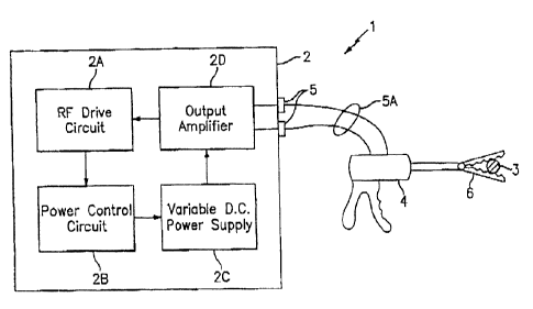
An electrosurgical system 1, which can be used to practice this invention, is
shown in
Fig. 2. The system 1 can be used for sealing vessels 3 and other tissues of a
patient,
including ducts, veins, arteries and vascular tissue. The system 1 includes an
electro-
surgical generator 2 and a surgical tool, also referred to herein as a
surgical instrument 4.
The surgical instrument 4 is illustrated by way of example, and as will become
apparent
from the discussion below, other instruments can be utilized. The
electrosurgical
generator 2, which is of most interest to the teachings herein, includes
several
interconnected sub-units, including an RF drive circuit 2A, a power control
circuit 2B, a
variable D.C. power supply 2C and an output amplifier 2D. The surgical
instrument 4 is
electrically connected to the electrosurgical generator 2 using a plug 5 for
receiving
CA 02475178 2004-07-20
1.6 ..
i
controlled electrosurgical power therefrom. The surgical
instrument A has some type of end effector member 6, such as
a forceps or hemostat, capable of grasping and holding the
vessels and tissues of the patient. The member 6, also
referred to simply as end effector 6, is assumed, in this
embodiment, to be capable of applying and maintaining a
relatively constant level of pressure on the vessel 3.
The member 6 is provided in the form of bipolar
electrosurgiaal forasps using two generally opposing
electrodes disposed on inner opposing surfaces of the member
6, and which are both electrically coupled to the output of
the electrosurgical generator 2. Durinq use, different
electric potentials are applied to each electrode. In that
tissue is an electrical conductor, when the forceps are
utilised to clamp or grasp the vessel 3 therebetween, the
electrical energy output from the electrosurgical generator 2
is transferred through the intervening tissue. Hoth open
surgical procedures and endoscopic surgical procedures can be
performed with suitably adapted .surgi.cal inst~uaents 9. It
should also be noted that the member 6 could be monopolar
forceps that utilize one active electrode, with the other
(return) electroda'ox pad being attached externally to the
patient, or a combination of bipolar and monopalar forceps.
By way of further explanation, Fig. 3 is a perspective view
of one embodiment of the surgical instrument 4 having a
bipolar end effector implemented as forceps 6A while Fig. 4
is an enlarged, perspective view of a distal end of the
bipolar forceps 6A shown in Fig. 3.
CA 02475178 2004-07-20
17
Referring now to Figs. 3 and 9, a~bipolar surgical instrument
9 for use with open surgical procedures includes s mechanical
forceps 20 and an electrode assembly 21. In the drawings and
in the description which follows, the term "proximal", as is
traditional, refers to the end of the instrument 4 which is
closer to the user, while the term "distal~ refers to the end
Nhich is fuxthex from the user.
Mechanical forceps 20 includes first and second members 9 and
11 which each have an elongated shaft 12 and 14,
respectively. Shafts 12 and Z4 each include a proximal end
and a distal end. Each proxi~aal end of each shaft portion
12, 14 includes a handle member 16 and 18 attached thereto to
allow a user to effect movement of the two shaft portions 7.2
and 14 relative to one another. Extending from the distal
end of each shaft portion 12 and 14 are end effactors 22 and
24, respectively. The end effectors 22 and 29 are movable
relative to one another in response to movement of handle
members 16 and 18. These end effectors members 6A can be
referred to collectively as bipolar forceps.
Preferably, shaft portions 12 and 14 are affixed to one
another at a point proximate the end effectors 22 and 24
about a pivot 25. As such, movement of the handles 16 and I8
imparts movement of the end effectors 22 and 24 from an open
position, wherein the end ef~ectors 22 and 24 are disposed in
spaced relat~.on relative to one another, to a clamping or
closed position, wherein the end effectors 22 and 24
cooperate to grasp the tubular vessel 3 therebetween. Either
one or both of the end effectors 22, 24 can be u~ovabls.
CA 02475178 2004-07-20
18
As is best seen in Fig. 4, end ei~fector 24 includes an upper
or first jaw member 44 which has an inner facing surface and
a plurality of mechanical interfaces disposed thereon which
are dimensioned to releasable engage a portion of an
electrode assembly 21, which may be disposable. Preferably,
the mechanical interfaces include sockets 41 which are
disposed at least partia7.ly through the inner facing surface
of jaw member 44 and which are dimensioned to receive a
complimentary detent attached to an upper electrode 21A of
the disposable electrode assembly 21. The upp~r eleetxode 21A
is disposed across from a corxespoading lower electrode 218.
The and effectvr 22 includes a second or lower jaw member 42
which has an inner facing surface which opposes the inner
facing surface of the first jaw member 44.
Preferably, shaft members 12 and 14 of the mechanical forceps
are designed to transmit a particular desired force to the
opposing inner facing surfaces of the jaw members 22 and 24
when clamped. In paxticular, since the shaft members 12 and
20 14 effectively act together in a spring~like manner (i.~.,
bending that behaqee like a~ spring), the length, width,
height and deflection of the shaft members 12 and 19 directly
iu~aCts the overall transmitted force imposed on opposing jaw
members 42 and 94. Preferably, jaw members 22 and 24 are
more rigid than the shaft members 12 and 14 and the strain
energy stored i.n the shaft members 12 and 14 provides a
constant closure force between the jaw members 42 and 49.
Each shaft member 12 and l4 also includes a ratchet portion
32 and 34. Preferably, each ratchet, e.g., 32, extends from
the pxoxirnal end of its respective shaft member 12 towards
CA 02475178 2004-07-20
19
the other ratchet 34 in a generally vertically aligned manner
such that the inner facing surfaces of each ratchet 32 and 34
abut one another when the end effectors 22 and.24 are moved
from the open position to the closed position. Each ratchet
32 and 34 includes a plurality of flanges which project from
the Inner facing surface of each ratchet 32 and 34 such that
the ratchets 32 and 34 can interlock in at least one
position. In the embodiment shown in Eig, 3, the ratchets 32
and 34 interlock at several different positions. Preferably,
each ratchet position ho~.ds a specific, i.e., constant.
strain energy in the shaft members 12 and 14 which, in turn,
transmits a specific force to the end effectors 22 and 24
and, thus, to the electrodes 21A and 21B. Also, preferably a
stop is provided on one or both of the end effectors 22, 24
to maintain a lareferred gap between the ~ aws . .
In some cases it may be preferable to include other
mechanisms to control and/or limit the movement of the jaw
members 42 and 44 relative to one another. For example, a
24 ratchet and pawl system could be~utilized to~segment the
movement of the two handles into discrete units which, in
turn, impart discrete movexaent to the jaw members 42 and 44
relative to one another.
Fig. 5 is a perspective view of an embodiment of the surgical
instrument 4 having end eftector members or forceps 6B that
are suitable for an endoscopic surgical procedure. The end
effeetor member 6B is depicted as sealing the tubular vessel
3 through a cannula assembly 130, 132.
CA 02475178 2004-07-20
The surgical instrument 4 fvr use With endosscopic surgical
procedures includes a drive rod assembly 50 which is coupled
to a handle assembly 54. The drive rod assembly 50 includes
an elongated hollow shaft portion 52 having a proximal end
5 and a distal end. An end effector assembly 68 is attached to
the distal end of shaft 52 and includes a pair of opposing
jaw members. Preferably, handle assembly 54 is attached to
the proximal end of sh~att 52 and includes an activator 56 for
imparting movement of the forceps jaw members of end effect:or
10 ~nober 6B from an open position, wherein the jaw members area
disposed in spaced relation relative to one another, to a
clamping or closed position, wherein the jaw members
cooperate to grasp tissue tharebetween.
Z5 Activatar 56 includes a movable handle 58 having an aperture
60 defined therein for receiving at least one of the
operator's fingers and a fixed handle 62 having an aperture
64 defined therein for receiving an operator's thumb.
Movable handle 58 is selectiqely moveable from a first
20 position relative to fined handle 62 to a second position in
the fixed handle 62 to close the jaw a~abexs. preferably,
fixed handle 62 includes a ehannnl 66 which extends
proximally for receiving a ratchet 66 which i9 coupled to
movable handle 58. This structure allows for progressive
closure of the end effector assembly, as well as a locking
engagement of the opposing jaw members. In some cases it may
be preferable to include other mechanisms to control and/or
limit the movement of handle 58 relative to handle 62 such
as, e.g., hydraulic, semi-hydraulic and/or gearing systems.
As with instrument 4, a stop can also be prov~,ded to caaintain
a preferred.gap between the jaw members.
CA 02475178 2004-07-20
21
The handle 62 i.nciudes handle sectians~62a and 62b, and is
generally hollow such that a cavity is formed therein for
housing various internal caatt~onents. For example, the cavity
can house a PC board which connects the electrosuzgical
energy being transmitted from'the electrosurgical generator 2
to each haw member, via connector 5. More particularly,
electrosurgical energy generated from the electrosurgical
. generator 2 is transmitted to the handle PC board by a cable
5A. The PC.board diverts the electrosurgicai energy from
' the generator into two different electrical potentials which
are transmitted to each jaw member by a separate terminal
clip. The handle 62 may also~house circuitry that .
conmnunicate8 With~the generator 2, far example, identifying
characteristics of the electrosuxqical tool 4 for use by the
- electrosurgical generator 2, whe=e the electrosurg~.cal
generator 2 may select a particrilar seal parameter lookup w
table based on those characteristics (as described below).
Preferably, a lost motion mechanism 3.s positioned between
each of the handle sections 62a and 62b'for maintaining a
.~ predetermined or maxi~aum clamping force for' sealing'"tis~ua
between the jaw members.
Having thus described two exemplary and non~limiting
embodiments of surgical instruments 4 that can be employed ,
with the eleatrosurgical generator.2, a description will noW
be provided of various aspects of the inventive
electrosurgical generator 2.
CA 02475178 2004-07-20
22~
Fig. 6A~is a block diagram that~iLlustrates the power control
circuit 2B of Fig. Z in greater detail. The power control
. . circuit ZB includes a suitably prograasued data processor.?Q .~ ,
' . that is preferai~ly implemented, as one or more~microcontroiler
devices. In a most preferred embodiment there are two
pr3.ncipal mi.crocontrollers, referred to as a main
~miarocoatroller ?OA and a feedback microcontroller 709. These
. twQ u4icrocontrolhrs are. capable of comcsxnicating. using .
shared data that is scored and retrieved fro~a a shared .
. 10 .vread/write memory ?Z. A control program fox the data
. processor 70~'is stored in a program memory 74, and includes . ~ .,
' ' software 'routines-and algorithms for ~eontxolling the overall
'operation of the eleatrosuiQical ge~xator 2. xn general, the
~~~~feedback microcontroller ?0B has a digital. output bus Coupled
. .15 to an input of a digital to analog converter (DAC) block '?6
.. . . ' ~xhich outputs aa' ana3.og siguai: This, ~s a system control _
. ~ ~ ,... _
voltage ~SCv),;~which is applied to the variable DC power
..supply 2C to'oontrcl the magnitude of the voltage and current
y ~ . of output RF poises . ~ ' . .
.. -. .~ An~an$Zog to digital converter (ADC) block ?8 reaeivea analog
inputs and sources a digital, input bus of the feedback
microoontroller ?0B. using the ADC block,?B the
microcontroller ?0B is apprised of the value of the actual
output voltage and the actual butput current, thereby closing, .
the feedback loop with the 8CY signal. The values of th$
output voltage and carte»t,~n be_used for determining tissue '
wimpedance, power and energy delivery for the overall, . , .
;general control of the applied RF energy waveform.. .
It should be noted that at least the ADC block 78'can
be an internal block of the feedback microcontroller .
' 7oB, and need not be a separate, external .
CA 02475178 2004-07-20
23
cobc~panent. It should be further dated that the same analog
signals can be digitized and read into the master
microcontroller 70A, thereby providing redundancy. The.ma$ ter
mierocontrolJ.er 70A controls the state ton/off) of the high
voltage (e. g., 190V mar) pow~r supply as a safety precaution,
controls the front panel display(s), such as a Seai Intensity
display, described below and shown in Fig. 9A, and also
receives various input switch closures, such as a Seal
Intensity selected by an operator.
.10
It is noted that in a preferred embod~.ment of the
electrosurgical generator 2 a third (waveform)
rnicrocontroller 74C is employed to generate the desired 970
kHz sinusoidal waveform tihat forms the basis of the RF pulses
applied to the tissue to ba sealed, such as the vessel 3
(Fig. 2). The waveform microcontroller 70C is controlled by
the feedback microcontroller 708 and is progranQaed thereby.
An output signal line from the feedback microcontroller 70B
is coupled to a Reset input of the waveform mi.croeontroller~
70C to easeritially turn the wavefor~a microcontrollsr 70C on
and off to provide the pulsed RF signnl in accordance with an
aspect of this disclosure. This particular arrangement is, of
course, not to be viewed in a limiting sense upon the '
. practice of this system, as those skilled in the art may
derive a number of methods and circuits for generating the
desired RF pulses in accordance with the teachings found
herein.
As an overview, the software algorithm$ executed by the data
processor 70 provide the following features. First, and
referring now also to the preferred waveform depicted in Fig.
CA 02475178 2004-07-20
.
24
?, a low power initial pulse of RF~energy is used to sense at .
least one electrical characteristic of the tissue prior to
starti.ng~ the seal cycle. Second. the sensed electrical
characteristic of the tissue is used as an input into tha~
5~ deteratination of the initial sealing parameters, thereby
making the sealing procedure adaptive to the characteristics . ,
of the tissue to be sealed. Thixd, the technique measures the
time required for the tissue to begin desiccating, preferably
by observing as electrical transient , to determine and/or
modify further seal parameters..Fourth, the technique
performs a tissue tes~perature~ control function' by adjusting
the duty cycle of RF pulses applied to the tissue, thereby . ..
avoiding excessive tissue heating and the.pxoblems that arise .
fr~a excessive tissue heating. This ie preferably .
~15 accomplished by~usi.ng at least one calculated seal parameter
related to the time required tar the tissue to begin
, desiccating. Fifth, the technique controllably changes the
RF pulse voltage with each pulse of RF energy DEL as the
tissue desiccates and shrinks tthercby' reducing .the spacing,
between the surgical instru~aent.electrodes~; arcing between
the instrument electrodes (e.g. 2IA,and 218. of Fig. 4) is
avoided, as is the tissue 'destruct~:oii that may re$ult from .
such uncontrolled arc~.ng. This is also preferably
accomplished by using at least one calculated seal parameter
that is related to the time required for the tissue to begin
desiccating. Sixth, the above-mentioned Seal Intensity front
panel control (Fig. 9A1 enables the operator to control the
sealing of tissue by varying parameters other than simply the
RF power. These various aspects of this disclosure are now
described in further detail.
CA 02475178 2004-07-20
v ~ ~~ a . ~ 25 ~ . . . .
Referring now also to the logic flow diagram of Fig. 13, the
impedance sensing feature is imphemented~at the beginning of
' . the seal cycle, wherein the eiectrosurgical generator 2 , ..
senses at least one ~e7.eetric~tl characteristic of the tissue,
.5 for example, impedance, I y phase rotation, or the output '
Current, by using n short burst of RF energy (Fig, 13, Steps
A and 8). The electrical characteristic of the tissue may be
measured at any freguent~ or power level, but preferably is
per~for~ned at the sa~ae fregvency as the . intended working ' ~ ~ . .
14.~ frequency.(e.Q., 470 kEZz). In a most preferred case the short .
burst of RF.energy (prefBrably less than about'2ti0
millisecond, at~d more pret~rably about 100 ~ millisecond) . is a .
~- 470 kHz else wave pith approximaxely 5i1 of power. The initial
pulse RF power is made low, and the pulse time is made. as
15 short as possible, to~~enable an~~.nitial tissue electrical .
characteristic maasuremdcnt to be made .t~~.thout excessively
heating the tissue.
.~ .
In a most preferred embodiment the electr~.cal characteristic
20 .serised'is the tissue impedance Which is employed to determine
an initial set of parameters that are input ~o the sealing
algorithm, and which are used to control the selection of
sealing parameters, including the starting power, current ~
. aad~voltage (Fig. Z3, Step C). Other sealing parameters may
2~ i include duty cycle and pulse width: Generally, if the sensed
impedance is in the lower ranges, then the initial power and
:. starting voltage are made relatively lower, the assumption
being that the tissue will desiccate faster and require less
energy. Tf the sensed impedance is in the higher ranges, the
~a initial pawer and starting voltage are made relatively higher,
the assumption being that the tissue will desiccate slower and
require more energy.
CA 02475178 2004-07-20
26
i
In other embodiments at least one of~any'other tissue
electrical characteristic, for example, the voltage or
current, can be used to set the parameters.,Thesa initial
parameters are prefgiably modified in accordance With the
setting of the Seal intensity control input (Fig. 13, Step
D), as will be described in further detail .below.
.Referring again to Fig. 13, Step C, the sensed i~npedance~ is
~10 employed to determine ~rh3ch set of values axe used from~a
seal parameter lookup table (LUT) 80 (eee Figs. 6A and 6H). '
The seal parameter look up table iaay one of a~plurality that' .
are stored in~the generator or accessible to'the generator.
. Furthermore, the seal parameter table may be~seleetec~ ,
15~ manually or automatically, based on, for~exartple, the
electrosurgical tool or ~r~strumant be~5.ng ~aplnyed. The,
specific values read from the heal parameter LtTT 80 Fig. 68) .
. are then adjusted based on the Seal Intensity front panel
setting 82' (Fig. Z3, Step D) , as 'is ~ shown sabre clearly in .
20 Figa. 9A and 9H. xn a preferred, but sot limiting embodiment,
the values read fro~a the seal parameter'hDT 80 comprise the
power, the maximum voltage'; starting voltage, minimum voltage,
voltage decay, voltage ramp, maximum RF on time, maximum cool
scale factor, pulse minimum, pulse dwell time, pulse off time,
. 25.~ current and the desired pulse width. In a preferred, but not
' limiting embodiment, the seal parameter values adjusted by the
Seal Intensity front panel setting 82 (Figs. 9A and 9B)
comprise the power, starting voltage, voltage decay, and pulse
dwell time.
SO
CA 02475178 2004-07-20
~ , . . , ~ 2?
Figure 1B is a graph that plots autput power versus_fmpeda=ice
in ohms for the disclosed electrosurgieal,generator. The
. ~ plot labeled "Intensity Bar 1" shows the electrosurgical .
.generator por~er output versus impedance when the "VLOW"
. 5 setting 82A (Fig. 9A} of the Seal Intensity.front~panel.
setting 82 is selected. The plot labeled intensity Bar 2 .
shows the power output of the electrosurgical generator when
the "LOW" settin~i 82B of the Seai Intensity front panel
setting 92 is Selected. The,plot labeled,Intensity Bars~3', ~~ ,
. XO 4, 5,_sho'tvs the power output of the electxosurgical venerator
- . ~ ~ when the "1~D" 82C, ~ "IiIGH"' $2D or YEixGHa 82E Seal , Intensit:y '
' .~ .front panel settings 82 are selected: The 8ea1 Intensity ~ - , . .
front ,.panel , settings .82 ad just the sea~.~.paraineter values as
. , . ;;
. shown in Fig. 9B. These values may be adjusted depending on
15 instrument used, tissue characteristics or surgical intent.
r . .
. Discussing this aspect of the disclosure now in further
. ' ' ' detail, and ~sferri~g as well to Fig's: T ~ and ~8, the selected
Seal Parameter Table, adjusted by'the Seal Intensity .front
.pa~e3...settings is then utilized by the RF energy geaer~tion
~systera and'.an initial RF.sealing pulse is then staxted. v
'~ Asweachwpulse i~WRF -energy is ~-applied to the tissue, the
current initially rises tc~ a ma~ci~aum (Pulse Peak), end .t#~en,
as the tissue desiccates and the impedance rises due to loss
of moisture in the tissue, the current falls. Reference in
th~.s xegard can be had to the circled areas designated as "A'~s
. : in the Ice, waveform of F~.g. 8 , The actual width of the
resulting electrical transient, preferably a current
. transient "A", is an important factor in determining what
type and amount'of tissue is between the jaws (electrodes).~f
the surgical instrument 4 (measur~d from "Full Power RF
CA 02475178 2004-07-20
28
Start" to "Poise Low and Stable~.)~The actual current ., . '
transient or pulse width is also. employed to detarcnine the
changes to, ar the values of, the parameters of the poise
duty cycle ~"Ih~tell.Time") and tc the change of the pulse .
voltage, as well as other parameters. This parameter can
also~be used to determine whether the tissue aeal~has been
. completed, or if the surgical instrument 4 has shorted.
As an alternative to directly measuring the pulse width, the
rate of change of an electrical characteristic (for.exanipie
. current, voltage, ~impedanoe,~ete.) of the transient aA"
(shown in Fig:~~7B) may be measured periodically (indicated by
the reference number 90 shown in Fi.g. 7H) over the tune the
transient occurs. The~rate of change of the .electrical ~ ..
characteristic may be proportional.to the width Ot 95 of the ,
tzansieat '"A", defined by the relationship:
At cc deldt
ZO where de/dt is the change in the electrical charaeteri9tic
over time: .This rate of change may then be used to provide
an indication of the Width of the transient "A" in
detexzaining.the type and amount of tissue that is between the
haws (electrodes) of th~ surgical instrument 4,~as Well as '
the $ubsecluent pulse duty cycle ("Dwell Time"), the amount of
subsequent pulse volt~.ge reduction, as well as other
parameters.
~efe~ring to-E'ig. 13.~Step E, a subsequent RF enexgy pulse is
applied to the~tissue, and the pulse width of the leading
edge current transient'is,.m~asured'(Fig. 13, Step F). A
CA 02475178 2004-07-20
29
determination'is made if the current transient is present.. If
it is, control passes via connector "a" to Step H, otherwise
control passes via connector "b" to Step K.
Assuming that the current transient i.s present, and
referring to Fig. 13, Step A, if the current transient pule a . .
is wide, for example, approximately in the range of 500-1000
ms, th~n one can assume the presence of a large amount of
tissue, or tissue that requires mare RF energy to desiccate.
Thus, the Dwell Time is increased, and an increase or small
reduction is made in the amplitude of the next RF pulse (see
the Vrms waveform in Fig. 8, and Fig. 13, Step I). If the
current transient pulse is narrow, for example, about 250 ms
or less (indicating that the tissue impedance rapidly rose),
then one can assume a small amount of tissue, of a tissue type
that requires little RF energy to desiccate is present_ Other
ranges of current transient pulse widths can also be used.
The relationship between the current transient pulse width and
the tissue characteristics may be empirically derived. In
this case the Dwell Time can be made shorter, and a larger
reduction in the amplitude of the next RF pulse can be made as
well (Fig. 13, Step J).
Zf a current pulse is not observed at Fig. 13, Step G, it may
_25 be assumed that either the instrument 4 has shorted, the
tissue has not yet begun to desiccate,~or that the tissue has
been fully desiccated and, thus, the seal cycle is comp3ete.
The determ~.nation of which of the above has occurred is
preferably made by observing the tissue impedance at fig.~l3,
Steps K and M. If the impedance is less than a low~threshold
value (THREBHL), then a shorted instrument 9 is assumed (Fig.
CA 02475178 2004-07-20
13, Step L), while if the impedance~is greater than a high
threshold value (THRESHH), then a complete tissue seal is
assumed (Fig. 13, Step N).
5 If the tissue impedance is otherwise found to be between the
high and low threshold values, a determination is made as to
whether the Max RF On Time has been exceeded. If the Max RF
On Time has been exceeded, it is assumed that the seal cannot
be successfully completed for some reason and the sealing
10 procedure is terminated. If the Max RF On Time has not been
exceeded then it is assumed that the tissue has not yet
received enough RF energy to start desiccation, and the seal
cycle continues (connector "c").
15 After the actual pulse width measurement has been completed,
the Dwell Time is determined based on the actual pulse Width
and on the Dwell Time field in the seal parameter LUT 80(see
Fig. 6B.) The RF pulse is continued until the Dwell Time has
elapsed, effectively determining the total time that RF
20 energy is delivered for that pulse. The RF pulse is then
turned off or reduced to a very low Level
for an amount of time specified by the Pulse Off field. This
low level allows some moisture to return to the tissue.
25 Based on the initial Desired Pulse Width field of the seal
parameter LUT 80 for the first pulse, or, for subsequent
pulses, the actual pulse width of the previous pulse, the
desired voltage limit kept constant or adjusted based on the
Voltage Decay and Voltage Ramp fields. The desired voltage
limit Is kept constant or raised during. the pulse if the actual
pulse width is greater than the Desired Pulse Width field (or
last actual)
CA 02475178 2004-07-20
pulse width), and is kept constaht or lowered if the actual
pulse width is less than the Desired Pulse Width field <or the '
last actual pulse width).
When the Desired Voltage has'been reduced to the Minimum
,Voltage field, then the~RF energy pulsing is terminated and
the electrosurgicai generator 2 enters a cool-down period
having a duration that is set by the Maximum Cool SF field
end the actual pulse w~.dth of the first pulse.
xo~
Several of the ~oragoing and other terms are defined with
greater specificity as follows (see also Fig. 7).
The Actual Pulse width is the t~rne from pulse. start to pulse
1~5 low. The Pulse Peak is the point where the current reaches ~a
maximum value, and does not exceed this value for some
predeteriai:ned period of tine (measured in mil7.isecoads) . The '
peak value of the ~u3.se, Peak eaa be reached until the Pulse
Peak-X% value is reached, which is the point.whera the
20 ' current bas~decrea$ed to souse predetermined determi»ed
percentage, X,of the value of Pulse Peak. Pulse 3~ow is the .~ ~ '
point where the current reaches a low point, and 'does not go.
.lower for another predetermined period of time. The value of
the. Maximum RF On Time or~MAX Pulsa Time is preferably
25 ~ preprogranened to some value that cannot be readily changed.
The RF pulse is terminated automatically if the Pulse Peak is .
' reached but tha Pulse Peak-X% value is not obtained with-the~
duration set by the Maximum RF On Time field of the seal
para~oneter LUT 80.
30'
CA 02475178 2004-07-20
' 32
' Referring to Fig. 68, the seal par~uaeter hUT.80 is employed
by the feedback microcontroller 70H in determining how to set
the various outputs that impact the RF output of the ..
electxoaurgical generator 2. The seal parameter Zv'r 80 is .
partitioned into a plurality of storage regions, each being
associated with a particular measured initial impedance.
More particularly, the Impedance Range defines a plurality of
impedance breakpoints (in ohms) Which are employed to
determine which net of variables are to be used for a
particular sealing cyche. The particular Impedance Range that
~.s selected ie.basatl on the above described dance Sense
State (Fig. 7) that is executed at the start of the aeah
cycle. The individual data fields of the'seal parameter LDT
84 are defined as foliaws.
The actual values for the Impedanco Ranges of Low, Med how,
. Med High, or High, are preferably contained.in one of a '
plurality of. tables stored in the generator 2, o~ othexwise~.~
.. . accessible tQ the generator 2.. A speci'fis table may be "
selected automatically, for example, based on signals
received from the electroaurgical tool 4 being used, ar by
the operator indicating what electrosurgical tool is iri.'use. ' ' ..
Power is the RF power setting to be ussd (in Watts~~:..Max
.. ,. 25 . Voltage is the greatest value that the output, voltage can
achieve (e.g., range 0 - about~190Yy. Start Voltage is the .
greatest valu~a that the first pulse voltage can adhieve
(e.c~.,,.x~nge 0 - about 190V). Subsequent pulse voltage values. -
are typically modified downwards from this value. The
SO Minimum Voltage is the voltage endpoint, and the seal
' cycle can be assumed to be complete when the RF pulse
. voltage has been reduced to
CA 02475178 2004-07-20
33
this value. The Voltage Decay scale factor is the rate (in
volts) at which the desired voltage is lowered if the ct~rre~nt
Actual E~ulse Width is less than the Desired Pulse Width. The.
Voltage Ramp scale factor is the rate at which the desired
voltage will be ineroaaed if the Actual Pulse l~idth is
greater than the Desired Pulse Width. The Maximum RF On Time
is the maximum amount of time (e. g., about 5-20 seconds!) that
the RF power can be delivered, as desoribed above. The
Msxiawm Cool Down Time det~rminem the generator oool down
time, also as described above. Pulse Minimum establishes the
minimum Desired Pulse ~iidth value. It c~ be noted that for
eaoh RF pulse, the Dssirad, Pulse Width is. equal to the Actual
Pulse Width from the previous pulse, or the Desired Pulse
field if the first pulse. Tha Dwell Time scale factor was
also discussed previously, and is the Lime (iri milliseconds)
that the RF pulse is continued after the current drops to the
Pulse Low and Stable point (see Fig. 7). Pulse Ott is the off
time (in milliseconds) between RF pulses. Desired Pulse Width
is a targeted pulse Width and determines when the Desired
Voltage (Vast) is raised, lowered or kept constant. If the~Actual Pulse
Width is less'fihan th~a Desired Pulse Width, then Vset is
decreased, while if the Actual Pulse Width is gxeater than
the Desired Pulse Width, then Vset is increased. If the
Actual Pulse Width ie equal to the Desired Pulse Width,
~25 then Vset ie kept constant. The Desired Pulse Width is
used ae the Desired Pul$e'T~idth for each sequential pulse.
.In general, a new Desired Pulse Width cannot be greater than
a previous Desired Pulse width, and cannot be less than
Pulse Minimum.
Hy applying the series of RF pulses to the tissue, the
surgical generator 2 effectively raises the tissue
CA 02475178 2004-07-20
34 .
temperature to a certain level, and than maintains the
teiaperature relatively constant. If the RF pulse width is
too long, then the tissu~ may be excessively heated and may
stick to the electrodes 21A, 218 of the surgical instrument
4, and/or an explosive vaporization of tissue fluid may
damage the tissue, such as the vessel 3. If the RF pulse
width is too narrow, then the tissue will not reach a
temperature that is high enough t4 properly seal. As such,
it cetn be appreciated that a proper balance of duty cycle to
tissue .type is important.
During the pulse off cycle that is made possible in
accordance with the teachings herein, the tissue relaxes,
thereby allowing the steam to exit without tissue
destruction. They tissue responds by rehydrating, which in
turn i.owers the tissue impedance. The lower impedance allows
the delivery of more current in the next pulse. This type of
pulsed operation thus tends to regulate the tissue
temperature so that the temperature does not rise to nn
undesirable level, while still performing the desired
electrosurgical procedure, and may also allow more energy to
be delivered, and thus achieving better desiccation.
As each RF pulse is delivered to the tissue., the tissue
desiccates and shrinks due to pressure being applied by the
jaws of the surgical instrument 4. The inventors have
realized that if the voltage applied to the tissue is not
reduced, then as the spacing between the haws Qf the surgical
instrument 9 is gradually reduced due to shrinking of the
tissue, an undesirable arcing can develop which may vapor~.ze
the tissue, resulting in bleeding.
CA 02475178 2004-07-20
r
As is made evident in the V~ trace of Fig. 8, and as was
described above, the voltage of each successive RF pulse can
be controllably decreased, thereby compensating for the
5 desiccation-induced narrowing of the gap between the surgica 1
instrument electrodes 21A and 218. That is, the difference in
electric potential between the electrodes is decreased as the
gap between the electrodes decreases., thereby avoiding
arcing. ~ .
As was noted'previous7.y, the Seal Intensity front panel
adjustment is.not a simple RF power control. The adjustment .'
of the seal intensity is acco~aplished.by adjusting the power
of the electrosurgical generator 2, as.weZl as the generator . .y
voltage, the duty cycle of the RF pulses, the length of time
of the seal cycle (e.g.y number of RF pulses), and the rata
.of voltage reduetion for suceessive RF pulses. Figs. 9H and 9C
. illustrate an exemplary set of~parameters (Power, Start
Voltage, Voltage Decay and Dwell Time) ,' and 'how they modify.. ~ . ... .
the contents of the seal parameter LUT 80 depending on the ~~
setting.of the Seal Intensity control B2 shown in Fig. 9A.
Generally, high~r~settings of the Seal Intensity control 82.
increase the seal time. and the energy delivered while lower
settings decrease the seal~time and the energy delivered.
In the Fig. 9B embodiment, it is instinctive to note that
for the Medium, High and Very High Seal Intensity settings
the RF Power remains unchanged, while variations are made w
instead in the Start Voltage, Voltage Decay and Dwell Time '
30' Parameters.
CA 02475178 2004-07-20
36
Based on the foregoing it can be appreciated that ari aspect
of this disclosure is a method far eleatroaurgically sealing
a tissue. Referring to Fig. 12, the method includes steps of:
(A) applying an initial pulse of RF energy to the tiasue,~the
pulse having characteristics ealeeted so as not to
ex~Pssively'heat the tissue: (9) measuring at least one
electrical characteristic of the tissue in response to the
'applied pulse: (C) in accordance with the measured electrical
characteristic , determining an initial set of pulse
parameters for use during a first RF energy pulse that is
applied to .the tissue: and (D) varying the. pulse par~neters
.of individual ones of subsequent RF energy pulses in
accordance with tit least one aharaoteristic of an electric
current transient that occurs at the beginning of each
individual one (pulses) of the subsequent RF energy pulses.
The method can terminate the generation of subsequent RF
energy pulses upon a determination that the current transient
is absent yr that the voltage has been reduced to a
predefined'level. In another embodiment of the present
invention, the initial pulse may be combined with at least
the first subsequent pulse..
Reference is now made to Figs. 10 and 11 for a description of
a novel over-voltage limit and transient energy suppression
'25 aspect of the system disclosed herein.
. A bi-directional transorb T91 normally is non-operational.
As long as the operating RF output levels stay below the
turn-on threshaid of TS1, eleatrosurgical energy is provided
at a controlled~rate of tissue desiaoation. However, in the
event that rapid biasue desiccation oaaurs, or that arcing is
present in the surgical tissue field, the RF output may
exhibit operating voltage levels in excess of the normal RF
CA 02475178 2004-07-20
37
levels used to achieve the contxolled rate of tissue
desiccation. rf the excess voltage present is left
unrestrained, the tissue 3 racy begin to exhibit undesirable
clinical effects contrary to the desired clinical outcome.
The TS1 is a strategic threshold that is set to turn on above
normal operating levels, but below and just prior to the RF
output reaching an excess voltage Level where undesirable
tissue effects begin to occur. The voltage applied across
TS1 is proportionately scaled to follow the RF output voltage
delivered to the tissue 3. The transorb TSl is selected such
that its turn on response is faster than the generator source
RF signal. This allows the transorb TSl to automatically
track and respond quickly in the first cycle of an excess RF
output overvoltaqe condition.
z~
Note should be made in Fig. 10 of the capacitor components or
network C2, C3, arid C4 that parallel the magnetic dxive
network (MDNl) which has an inductive characteristic and is
contained within the eleatrosurgical generator 2. The
comdbination of the inductive MON1 and the cspacitive networks
forms a resonant tuned network which yields the waveshape
canfiguration of the RF source signal shown in Fig. 11:
A turn on of tranaorb device TS1, which functions as a
voltage controlled switch, instantaneously connects the
serial capacitance C1 across the capacitor netwoxk C2, C3,
and Gg. Rn immediate change then appears in the tuning of
the resonant network iasntioned above, which then
instantaneously sltezs the waveshape of the RF source signal
shown in Fig. 11. The time base T1 of the nominally half-
sine signal shown increases incrementally in width out to
CA 02475178 2004-07-20
38
time T2, which automatically lowers the peak voltage of the
AF output signal. The peak voltage decreases because the
Voltage-Time product of the signal shown in Fig. ll~is
constant for a given operating quiescence. The concept of a
Voltage-Time product is well known to those skilled in the
art, and is not further discussed h4rein.
As the peak voltage decreases, the excess overvoltage is
automatically limited and is restricted to operating levels
below that which cause negative clinical effects. trace the
excess RF output voltage level falls below the tranaorb
threshold, the TSl device turns off and the electrosurgical
generator 2 returns to a controlled rate of tissue
desiccation.
In the event that arcing is present in the surgical tissue
field, undesirable excess transient RF energy may exist and
may be reflected in the RF output of the electrosurgiCal
generator 2. This in turn may generate a cozreaponding
excess RF output voltage that creates sufficient transient
overvaltage to turn on the transorb TS1. In this condition
the cycle repeats as described above, Where T81 turns on,
alters the resonant tuned network comprised of the magnetic
and capacitive components, and thus also alters the RF source
signal waveshape. This automatically reduces the excess
overvoltage.
In accordance with this aspect of the disclosure, the excess
RF transient en~rgy is suppressed and the overvoltage is
limf.ted by the dynamic, real-time automatic datuning of the
RF energy delivered to the tissue being treated.
CA 02475178 2004-07-20
_ . .39
2t should be noted that the embodiment of Figs. 10 and 11 can
be used to improve the operation of conventional
electrosurgical generators, as well as with the novel pulsed
output electrosurgical generator 2 that was described
previously. ' ,~ .
~~In an additional embodiment the measured electrical
characteristic of the tissue,. preferably the. impedance (Zi),
and the RMS cuz~rent pulse width (P") may be used to detertaine
a fixed voltage reduction factor (V~) to be. used for' .
Su~SeQueIlt pulses, and to determine a.fixed number of pulses
~(Pa) to be delivered for the sealing procedure. The , , . .
relationship among ire voltage reduction factor, the measured ~..
1S impedance ~nd.the liMS current pulse width may be defined as '
' V~ = F (Z=, P") , and the relationship , among the number of
' purses, the measured impedance arid the RMS curirent pulse .
. width may be defined' as Pf s F~ (Z=. P") ~ . Zn Fig. 14 ~s fixed
number of pulses. P~~ 100 -determined fra~a the'maasured
impedance and the RMS.~urrent pulse width are shown,' Each
subsequent pulse may be reduced by the fixed voltage
reduction factor (vae~) 110, also determined from the
. ' measured impedance and the RMS current'pulse width.
CA 02475178 2004-07-20
In a further additional embodiment, tissue sealing is accomplished by the
electrosurgical system
described above by continuously monitoring or sensing the current or tissue
impedance rate of
change. If the rate of change increases above a predetermined limit, then 1:ZF
pulsing is
automatically terminated by controlling the electrosurgical generator 2
accordingly and any
previously changed pulse parameters (e.g., power, voltage and current
increments) are reset to the
original default values. In this embodiment, the ending current or tissue
impedance, i.e., the
current or tissue impedance at the end of each RF pulse, is also continuously
monitored or
sensed. The ending values are then used to determine the pulse parameters for
the subsequent RF
pulse; to determine if the seal cycle should end (based on the ending values
of the last few RF
pulses which did not change by more than a predetermined amount); and to
determine the duty
cycle of the subsequent 1:ZF pulse.
Further, in this embodiment,1',tF power, pulse width, current andlor voltage
levels of subsequent
RF pulses can be kept constant or modified on a pulse-by-pulse basis depending
on whether the
tissue has responded to the previously applied RF energy or pulse (i.e., if
the tissue impedance
has begun to rise). For example, if the tissue has not responded to a
previously applied RF pulse,
the 1ZF power output, pulse width, current and/or voltage levels are increased
for the subsequent
RF pulse.
Hence, since these RF pulse parameters can subsequently be modified following
the initial RF
pulse, the initial set of RF pulse parameters, i.e., a magnitude of a starting
RF power level, a
magnitude of a starting voltage level, a magnitude of the starting pulse
width, and a magnitude of
a starting current level, are selected accordingly such that the fast or
initial RF pulse does not
excessively heat the tissue. One or more of these starting levels are modified
during subsequent
RF pulses to account for varying tissue properties, if the tissue has not
responded to the
previously applied RF pulse which includes the initial 1tF pulse.
The above functions are implemented by a seal intensity algorithm represented
as a set of
programmable instructions configured for being executed by at least one
processing unit of a
3o vessel sealing system. The vessel sealing system includes a Seal Intensity
control panel for
manually adjusting the starting voltage level, in a similar fashion as
described above with
reference to Figs. 9A and 9B.
CA 02475178 2004-07-20
41
As shown in Fig. 15, a preferred Seal Intensity control panel of the present
inventive embodiment
includes six settings, i.e., "Off' 1SOA, "VLOW' 1S0B, "LOW" 1SOC, "MED" 1SOD,
"HIGH"
150E and "VHIGH" 1SOF. The Seal Intensity front panel settings 1S0 adjust the
seal parameter
values of the Seal Parameter Table as shown by Figs. 9B and 9C. The selected
Seal Parameter
Table, adjusted by the Seal Intensity front panel settings 1S0 is then
utilized by an RF generation
system, as described above, and an initial ItF sealing pulse is then started.
The Seal Intensity front panel settings, as shown in Figs. 9B and 9C,
represent approximate
parametric values of several preferred embodiments, identified as an example
to achieve vessel
1 o sealing performance in clinical procedures. The variety of tissue types
and surgical procedures
requires the use of one or more Seal Intensity front panel settings.
Fig. 16 is a logic flow diagram that illustrates a method in accordance with
the vessel sealing
system. At step A', a 1tF pulse is applied to tissue. At step B', the current
or tissue impedance
t s rate of change is continuously monitored. At step C', a determination is
made whether the tissue
impedance rate of change has passed a predetermined Iimit. If yes, at step D',
RF pulsing is
terminated and any previously changed pulse parameters are reset back to the
original defaults. If
no, the process proceeds to step E'.
2o At step E', a determination is made as to whether the ItF pulse has ended.
If no, the process
loops back to step B'. If yes, the process proceeds to step F'. At step F',
the ending current or
tissue impedance is measured. At step G', the measured ending values are used
for determining
if the seal cycle should end (based on the current level or ending impedance
of the last few 1tF
pulses which did not change by more than a predetenmined amount). If yes, the
process
2S terminates at step H'. If no, the process continues at step I', where the
ending values are used for
determining the pulse parameters, i.e., the power, pulse width, current and/or
voltage levels, and
the duty cycle of the subsequent 1tF pulse from an entry in one of a plurality
of lookup tables.
The process then loops back to step A'. One of the plurality of lookup tables
is selected
manually or automatically, based on a choice of an electrosurgical tool or
instrument.
CA 02475178 2004-07-20
42
While the system has been particularly shown and~described with respect to
preferred
embodiments thereof, it will be understood by those skilled in the art that
changes in form and
details may be made therein without departing from its scope and spirit.