Note: Descriptions are shown in the official language in which they were submitted.
CA 02475348 2004-08-06
WO 03/068906 PCT/US03/01027
SYSTEMS AND METHODS FOR RAPIDLY CHANGING THE SOLUTION
ENVIRONMENT AROUND SENSORS
Field of the Invention
The invention relates to systems and methods for rapid and programmable
delivery of aqueous streams to a sensor, such as a cell-based biosensor. In
particular,
the invention provides methods and systems for high throughput patch clamp
analysis.
Background of the Invention
Ion-channels are important therapeutic targets. Neuronal communication,
heart function, and memory all critically rely upon the function of ligand-
gated and
voltage-gated ion-channels. In addition, a broad range of chronic and acute
pathophysiological states in many organs such as the heart, gastrointestinal
tract, and
brain involve ion channels. Indeed, many existing drugs bind receptors
directly or
indirectly connected to ion-channels. For example, anti-psychotic drugs
interact with
receptors involved in dopaminergic, serotonergic, cholinergic and
glutamatergic
neurotransmission.
Because of the importance of ion-channels as drug targets, there is a need for
methods which enable high throughput screening (HTS) of compounds acting on
ligand-gated and voltage-gated channels (see e.g., Sinclair et al., 2002,
Anal. Chem.
74: 6133-6138). However, existing HTS drug discovery systems targeting ion
channels generally miss significant drug activity because they employ indirect
methods, such as raw binding assays or fluorescence-based readouts. Although
as
many as ten thousand drug leads can be identified from a screen of a million
compounds, identification of false positives and false negatives can still
result in a
CA 02475348 2004-08-06
WO 03/068906 PCT/US03/01027
potential highly therapeutic blockbuster drug being ignored, and in
unnecessary and
costly investments in false drug leads.
Patch clamp methods are superior to any other technology for measuring ion
channel activity in cells, and can measure currents across cell membranes in
ranges as
low as picoAmps (see, e.g., Neher and Sakmann, 1976, Nature 260: 799-802;
Hamill,
et al., 1981, Pflugers Arch 391: 85-100; Sakmann and Neher, 1983, In Single-
Channel
Recording pp. 37-52, Eds. B. Sakmann and E. Neher. New York and London, Plenum
Press, 1983). However, patch clamp methods generally have not been the methods
of
choice for developing HTS platforms.
Summary of the Invention
The invention provides microfluidic systems for altering the solution
environment around a nanoscopic or microscopic object, such as a sensor, and
methods for using the same. The invention can be applied in any sensor
technology in
which the sensing element needs to be exposed rapidly, sequentially, and
controllably,
to a large number of different solution environments (e.g., greater than 10
and
preferably, greater than about 96 different environments) whose
characteristics may
be known or unknown. In contrast to prior art microfluidic systems, the
interval
between sample deliveries is minimized, e.g., on the order of microseconds and
seconds, permitting rapid analysis of compounds (e.g., drugs).
In one aspect, the invention provides a system comprising a substrate for
changing the solution environment around a nanoscopic or microscopic object,
such
as a sensor. The substrate comprises an open-volume chamber for the sensor,
and a
plurality of channels. Each channel comprises an outlet for delivering a
substantially
separate aqueous stream into the chamber. In one aspect, the outlets are
substantially
parallel, i.e., arrayed linearly in a single plane. The dimensions of the
outlets can
vary; however, in one aspect, where the sensor is a biological cell, the
diameter of
each of the outlets is, preferably, at least about the diameter of the cell.
Preferably, a
plurality, if not all, of the channels programmably deliver a fluid stream
into the
chamber.
2
CA 02475348 2004-08-06
WO 03/068906 PCT/US03/01027
In a preferred aspect, each channel of the substrate comprises at least one
inlet
for receiving solution from a reservoir, conforming in geometry and placement
on the
substrate to the geometry and placement of wells in a mufti-well plate. For
example,
the substrate can comprise 96-1024 reservoirs, each connected to an
independent
channel on the substrate. Preferably, the center-to-center distance of each
reservoir
corresponds to the center-to-center distance of wells in an industry standard
microtiter
or mufti-well plate.
In a further aspect, the substrate comprises one or more treatment chambers or
microchambers for delivering a treatment to a cell placed within the treatment
chamber. The treatment can comprise exposing the cell to a chemical or
compound,
(e.g. drugs or dyes, such as calcium ion chelating fluorogenic dyes), exposing
the cell
to an electrical current (e.g., electroporation, electrofusion, and the like),
or exposing
the cell to light (e.g., exposure to a particular wavelength of light). A
treatment
chamber can be used for multiple types of treatments which may be delivered
sequentially or simultaneously. For example, an electrically treated cell also
can be
exposed to a chemical or compound and/or exposed to light. Treatment can be
continuous over a period of time or intermittent (e.g., spaced over regular or
irregular
intervals). 'The cell treatment chamber can comprise a channel with an outlet
for
delivering a treated cell to the sensor chamber or directly to a mechanism for
holding
the cell connected to a positioner (e.g., a micropositioner or nanopositioner)
for
positioning the cell within the chamber.
Preferably, the base of the sensor chamber is optically transmissive and in
one
aspect, the system further comprises a light source (e.g., such as a laser) in
optical
communication with the open volume chamber. The light source can be used to
continuously or intermittently expose the sensor to light of the same or
different
wavelengths. The sensor chamber and/or channels additionally can be equipped
with
control devices. For example, the sensor chamber and/or channels can comprise
temperature sensors, pH sensors, and the like, for providing signals relating
to
chamber and/or channel conditions to a system processor.
The sensor chamber can be adapted for receiving a variety of different
sensors.
In one aspect, the sensor comprises a cell or a portion of a cell (e.g., a
cell membrane
3
CA 02475348 2004-08-06
WO 03/068906 PCT/US03/01027
fraction). In another aspect, the cell or cell membrane fraction comprises an
ion
channel, including, but not limited to, a presynaptically-expressed ion
channel, a
ligand-gated channel, a voltage-gated channel, and the like. In a further
aspect, the
cell comprises a receptor, such as a G-Protein-Coupled Receptor (GPCR), or an
orphan receptor for which no ligand is known, or a receptor comprising a known
ligand.
A cultured cell can be used as a sensor and can be selected from the group
consisting of CHO cells, NIH-3T3 cells, and HEK-293 cells, and can be
recombinantly engineered to express a sensing molecule such as an ion channel
or
receptor. Many other different cell types also can be used, which can be
selected
from the group consisting of mammalian cells (e.g., including, but not limited
to
human cells, primate cells, bovine cells, swine cells, other domestic animals,
and the
like); bacterial cells; protist cells; yeast cells; plant cells; invertebrate
cells, including
insect cells; amphibian cells; avian cells; fish; and the like.
A cell membrane fraction can be isolated from any of the cells described
above, or can be generated by aggregating a liposome or other lipid-based
particle
with a sensing molecule, such as an ion channel or receptor, using methods
routine in
the art.
The cell or portion of the cell can be positioned in the chamber using a
mechanism for holding the cell or cell portion, such as a pipette (e.g., a
patch clamp
pipette) or a capillary connected to a positioner (e.g., such as a
micropositioner or
nanopositioner or micromanipulator), or an optical tweezer. Preferably, the
positioner
moves the pipette at least in an x-, y-, z-, direction. Alternatively or
additionally, the
positioner may rotate the pipette. Also, preferably, the positioner is coupled
to a drive
unit which communicates with a processor, allowing movement of the pipette to
be
controlled by the processor.
In one aspect, the base of the chamber comprises one or more depressions and
the cell or portion of the cell is placed in a depression which can be in
communication
with one or more electrodes (e.g., the sensor can comprise a planar patch
clamp chip).
4
CA 02475348 2004-08-06
WO 03/068906 PCT/US03/01027
Non-cell-based sensors also can be used in the system. Suitable non-cell
based sensors include, but are not limited to: a surface plasmon energy
sensor; an
FET sensor; an ISFET; an electrochemical sensor; an optical sensor; an
acoustic wave
sensor; a sensor comprising a sensing element associated with a Quantum Dot
particle; a polymer-based sensor; a single molecule or an array of molecules
(e.g.,
nucleic acids, peptides, polypeptides, small molecules, and the like)
immobilized on a
substrate. The sensor chamber also can comprise a plurality of different types
of
sensors, non-cell based and/or cell-based. A sensor substrate can be affixed
to the
base of the chamber or the substrate can simply be placed on the base of the
chamber.
Alternatively, the base of the chamber itself also can serve as the sensor
substrate and
one or more sensing elements can be stably associated with the base using
methods
routine in the art. In one aspect, sensing elements are associated at known
locations
on a substrate or on the base of the sensor chamber.
However, an object placed within a chamber need not be a sensor. For
example, the object can be a colloidal particle, beads, nanotube, a non-
sensing
molecule, silicon wafer, or other small elements.
The invention also provides a system comprising a substrate which comprises
at least one chamber for receiving a cell-based biosensor, a plurality of
channels, at
least one cell storage chamber and at least one cell treatment chamber.
Preferably,
each channel comprises an outlet for delivering a fluid stream into the
chamber, and
the cell treatment chamber is adapted for delivering an electrical current to
a cell
placed within the cell treatment chamber. In one aspect, the cell treatment
chamber
further comprises a channel with an outlet for delivering a cell to the sensor
chamber
for receiving the cell-based biosensor. The system can be used to rapidly and
programmably change the solution environment around a cell which has been
electroporated and/or electrofused, and/or otherwise treated within the cell
treatment
chamber. Alternatively, or additionally, the sensor chamber also can be used
as a
treatment chamber and in one aspect, the sensor chamber is in electrical
communication with one or more electrodes for continuously or intermittently
exposing a sensor to an electric field.
5
CA 02475348 2004-08-06
WO 03/068906 PCT/US03/01027
In one aspect, a system according to the invention further comprises a
scanning mechanism for scanning the position of a sensor relative to the
outlets of the
channels. The scanning mechanism can translate the substrate relative to a
stationary
sensor, or can translate the sensor relative to a stationary substrate, or can
move both
sensor and substrate at varying rates and directions relative to each other.
In one
aspect, the sensor is positioned relative to an outlet using a mechanism for
holding the
sensor (e.g., such as a pipette or capillary) coupled to a positioner (e.g., a
micropositioner or nanopositioner or micromanipulator). Thus, the positioner
can be
used to move the sensor across a plurality of fluid streams exiting the
outlets of the
channels by moving the mechanism for holding the sensor. Alternatively, or
additionally, scanning also can be regulated by producing pressure drops
sequentially
across adjacent microchannels.
Preferably, the scanning mechanism is in communication with a processor and
translation occurs in response to instructions from the processor (e.g.,
programmed
instructions or instructions generated as a result of a feedback signal). In
one aspect,
the processor controls one or more of: the rate of scanning, the direction of
scanning,
acceleration of scanning, and number of scans. Thus, the system can be used to
move
nanoscopic and microscopic objects in a chamber to user-selected, or system-
selected
coordinates, for specified (e.g., programmable) lengths of time. Preferably,
the
system processor also can be used to locate the position of one or more
objects in the
chamber, e.g., in response to previous scanning actions and/or in response to
optical
signals from the objects detected by the system detector. In one aspect, the
system
further comprises a user device in communication with the processor which
comprises
a graphical user display for interfacing with a user. For example, the display
can be
used to display coordinates of objects) within the chamber, or optical data or
other
data obtained from the chamber.
The invention additionally provides a substrate comprising a chamber for
receiving a cell-based biosensor which comprises a receptor or ion channel. In
one
aspect, the system sequentially exposes a cell-based biosensor for short
periods of
time to one or several ligands which binds to the receptor/ion channel and to
buffer
without ligand for short periods of time through interdigitated channels of
the
substrate. For example, selective exposure of a cell biosensor to these
different
6
CA 02475348 2004-08-06
WO 03/068906 PCT/US03/01027
solution conditions for short periods of time can be achieved by scanning the
cell-
based biosensor across interdigitated channels which alternate delivery of one
or
several ligands and buffer. The flow of buffer and sample solution in each
microfluidic channel is preferably a steady state flow at constant velocity.
However, in another aspect, the system delivers pulses (e.g., pulsatile on/off
flow) of buffer to a receptor through a superfusion capillary positioned in
proximity to
both the cell-based biosensor or other type of sensor and to an outlet through
which a
fluid is streaming. For example, the system can comprise a mechanism for
holding
the sensor which is coupled to a positioner (e.g., a micropositioner,
nanopositioner,
micromanipulator, etc.) for positioning the c sensor in proximity to the
outlet and a
capillary comprising an outlet in sufficient proximity to the mechanism for
holding
the sensor to deliver a buffer from the capillary to the sensor. A scanning
mechanism
can be used to move both the capillary and sensor simultaneously, to maintain
the
appropriate proximity of the capillary to the sensor. The capillary also can
be coupled
1 S to a pumping mechanism to provide pulsatile delivery of buffer to the
sensor. In
another aspect, the flow rate of buffer from the one or more superfusion
capillaries in
proximity to one or more sensors can be higher or lower than the flow rate of
fluid
from the channels.
The invention further provides a substrate which comprises a circular chamber
for receiving a sensor, comprising a cylindrical wall and a base. In one
aspect, the
substrate comprises a plurality of channels comprising outlets whose openings
are
radially disposed about the circumference of the wall of the chamber (e.g., in
a
spokes-wheel configuration), for delivering samples into the chamber.
Preferably, the
substrate also comprises at least one output channel for draining waste from
the
chamber. In one aspect, at least one additional channel delivers buffer to the
chamber. Preferably, the angle between the at least one additional channel for
delivering buffer and the output channel is greater than 10°. More
preferably, the
angle is greater than 90 °. The channel "spokes" may all lie in the
same plane, or at
least two of the spokes may lie in different planes.
Rapid, programmed exchange of solutions in the chamber is used to alter the
solution environment around a sensor placed in the chamber and multiple output
7
CA 02475348 2004-08-06
WO 03/068906 PCT/US03/01027
channels can be provided in this configuration. For example, there may be an
output
channel for each channel for delivering sample/buffer. The number of channels
for
delivering also can be varied, e.g., to render the substrate suitable for
interfacing with
an industry standard microtiter plate. For example, there may be 96 to 1024
channels
for delivering samples. In another aspect, there may be an additional, equal
number
of channels for delivering buffer (e.g., to provide interdigitating fluid
streams of
sample and buffer).
The invention also provides a mufti-layered substrate for changing the
solution
environment around a sensor, comprising: a first substrate comprising channels
for
delivering fluid to a sensor; a filter layer for retaining one or more sensors
which is in
proximity to the first substrate; and a second substrate comprising a waste
reservoir
for receiving fluid from the filter layer. One or more sensors can be provided
between
the first substrate and the filter layer. In one aspect, at least one of the
sensors is a
cell. Preferably, the system further comprises a mechanism for creating a
pressure
differential between the first and second substrate to force fluid flowing
from
channels in the first substrate through the filter and into the waste
reservoir, i.e.,
providing rapid fluid exchange through the filter (i.e., sensor) layer.
The invention additionally provides a substrate which comprises a chamber for
receiving a sensor, a first channel comprising an outlet intersecting with the
chamber,
and a plurality of sample delivery channels intersecting with the first
channel. The
first channel also is connected to a buffer reservoir (e.g., through a
connecting
channel). In one aspect, the longitudinal axes of the sample delivery channels
are
parallel with respect to each other, but are angled with respect to the
longitudinal axis
of the first channel (e.g., providing a "fish bone" shape). Rapid flow of
solution
through the first channel and/or sample channels can be achieved through a
positive
pressure mechanism in communication with the buffer reservoir and/or sample
channels. Passive one-way valves can be provided at the junction between
sample
delivery channels and the first channel to further regulate flow rates. In one
aspect, at
least one of the sample reservoirs is sealed by a septum which can comprise a
needle
or tube inserted therein.
8
CA 02475348 2004-08-06
WO 03/068906 PCT/US03/01027
The invention further provides a substrate which comprises a chamber for
receiving a sensor, a plurality of delivery channels comprising outlets for
feeding
sample or buffer into the chamber, and a plurality of drain channels
comprising inlets
opposite the outlets of the delivery channels. The longitudinal axes of the
delivery
channels can be in the same, or a different plane, from the longitudinal axes
of the
drain channels. In one aspect, the plurality of drain channels is on top of
the plurality
of inlet channels (i.e., the substrate is three-dimensional).
Any of the systems described above can further comprise a pressure control
device for controlling positive and negative pressure applied to at least one
microchannel of the substrate. In systems where substrates comprise both
delivery
channels as well as output channel(s), the system preferably further comprises
a
mechanism for applying a positive pressure to at least one delivery channel
while
applying a negative pressure to at least one output channel. Preferably,
hydrostatic
pressure at at least one of the channels can be changed in response to a
feedback
signal received by the processor.
The system can thus regulate when, and through which channel, a fluid stream
is withdrawn from the chamber. For example, after a defined period of time, a
fluid
stream can be withdrawn from the chamber through the same channel through
which
it entered the system or through a different channel. When a drain channel is
adjacent
to a delivery channel, the system can generate a U-shaped fluid stream which
can
efficiently recycle compounds delivered through delivery channels.
As described above, multiple delivery channel configurations can be provided:
straight, angled, branched, fish-bone shaped, and the like. In one aspect,
each
delivery channel comprises one or more intersecting channels whose
longitudinal axes
are perpendicular to the longitudinal axis of the delivery channels. In
another aspect,
each delivery channel comprises one or more intersecting channels whose
longitudinal axes are at an angle with respect to the delivery channel.
In general, any of the channel configurations described above are
interfaceable
with containers for delivering samples to the reservoirs or sample inlets
(e.g., through
capillaries or tubings connecting the containers with the reservoirs/inlets).
In one
aspect, at least one channel is branched, comprising multiple inlets.
Preferably, the
9
CA 02475348 2004-08-06
WO 03/068906 PCT/US03/01027
multiple inlets interface with a single container. However, multiple inlets
also may
interface with several different containers.
Further, any of the substrates described above can be interfaced to a mufti-
well
plate (e.g., a microtiter plate) through one or more external tubings or
capillaries. The
one or more tubings or capillaries can comprise one or more external valves to
control
fluid flow through the tubings or capillaries. In one aspect, a plurality of
the wells of
the mufti-well plates comprise known solutions. The system also can be
interfaced
with a plurality of microtiter plates; e.g., the plates can be stacked, one on
top of the
other. Preferably, the system further comprises a micropump for pumping fluids
from
the wells of a microtiter plate or other suitable containers) into the
reservoirs of the
substrate. More preferably, the system programmably delivers fluids to
selected
channels of the substrate through the reservoirs.
In one aspect, a system according to the invention further comprises a
detector
in communication with a sensor chamber for detecting sensor responses. For
example, the detector can be used to detect a change in one or more of: an
electrical,
optical, or chemical property of the sensor. In one aspect, in response to a
signal from
the detector, the processor alters one or more of: the rate of scanning, the
direction of
scanning, acceleration of scanning, number of scans, and pressure on one or
more
channels.
The invention also provides a method for changing an aqueous solution
environment locally around a nanoscopic or microscopic object (e.g., such as a
sensor). The method comprises providing a substrate which comprises an open
volume chamber comprising a nanoscopic or microscopic object and an aqueous
fluid.
The substrate further comprises a plurality of channels, each channel
comprising an
outlet intersecting with the open volume chamber. Substantially separate
aqueous
streams of fluid are delivered into the open volume chamber, at least two of
which
comprise different fluids.
Preferably, fluid streams exiting from the at least two adjacent channels are
collimated and laminar within the open volume. However, the system can
comprise
sets of channels (at least two adjacent channels) wherein at least one set
delivers
collimated laminar streams, while at least one other set delivers non-
collimated, non-
CA 02475348 2004-08-06
WO 03/068906 PCT/US03/01027
laminar streams. In one aspect, the streams flow at different velocities.
Fluid can be
delivered from the channels to the chamber by a number of different methods,
including by electrophoresis and/or by electroosmosis and/or by pumping.
In one aspect, the longitudinal axes of the channels are substantially
parallel.
The channels can be arranged in a linear array, in a two-dimensional array, or
in a
three-dimensional array, can comprise treatment chambers, sensor chambers,
reservoirs, and/or waste channels, and can be interfaced with containers) or
multi-
well plates) as described above. In one aspect, output channels can overly
input
channels (i.e., in a three-dimensional configuration). Preferably, the
longitudinal axis
of at least one output or drain channel is parallel, but lying in a different
plane,
relative to the longitudinal axis of at least one input channel. By applying a
positive
pressure to an input channel at the same time that a negative pressure is
applied to an
adjacent output or drain channel, a U-shaped fluid stream can be generated
within the
chamber. In this way, an object within the chamber can be exposed to a
compound in
a fluid stream from an inlet channel which can, for example, be recycled by
being
withdrawn from the chamber through the adjacent output or drain channel. The U-
shaped fluid streams can, preferably, be used to create local well-defined
regions of
fluid streams with specific composition in a large-volume reservoir or open
volume.
Preferably, the object is scanned sequentially across the at least two aqueous
fluid streams, thereby altering the aqueous solution environment around the
object.
Scanning can be performed by moving the substrate and/or the object, or, can
be
mediated by pressure drops applied to the channels.
The open volume chamber can comprise a plurality of objects; preferably,
each object is scanned across at least two streams. Scanning can be performed
by a
scanning mechanism controlled by a processor as described above. The open
volume
can, additionally have inlets and outlets for adding and withdrawal of
solution. For
example, fresh buffer solution can be added to the recording chamber by using
a
peristaltic pump.
In one aspect, the method further comprises modifying one or more scanning
parameters, such as the rate of scanning, the direction of scanning,
acceleration of
scanning, number of scans, and pressure across one or more channels. Scanning
11
CA 02475348 2004-08-06
WO 03/068906 PCT/US03/01027
parameters can be modified in response to a feedback signal, such as a signal
relating
to the response of an object to one or more of aqueous streams. Scanning also
can be
coordinated with other system operations. For example, in a system comprising
a
cell-based biosensor, scanning can be coordinated with exposure of the
biosensor to
an electrical current, i.e., inducing pore formation in a cell membrane of the
biosensor, as the biosensor is scanned past one or more sample outlets.
Hydrostatic pressure at one or more channels also can be varied by the
processor according to programmed instructions and/or in response to a
feedback
signal. In one aspect, hydrostatic pressure at each of the plurality of
channels is
different.
In another aspect, the viscosity of fluids in at least two of the channels is
different. In yet another aspect, fluid within at least two of the channels
are at a
different temperature. In a further aspect, the osmolarity of fluid within at
least two
of the channels is different. In a still further aspect, the ionic strength of
fluid within
at least two of the channels is different. Fluid in at least one of the
channels also can
comprise an organic solvent. By changing these parameters at different
outlets,
sensor responses can be optimized to maximize sensitivity of detection and
minimize
background. In some aspects, parameters also can be varied to optimize certain
cell
treatments being provided (e.g., such as electroporation or electrofusion).
The invention also provides a method for rapidly changing the solution
environment around a nanoscopic or microscopic object which comprises rapidly
exchanging fluid in a sensor chamber comprising the nanoscopic or microscopic
object. In one aspect, fluid exchange in the chamber occurs within less than
about 1
minute, preferably, with less than about 30 seconds, less than about 20
seconds, less
than about 10 seconds, less than about 5 seconds, or less than about 1 second.
In
another aspect, fluid exchange occurs within milliseconds. In another aspect
fluid
exchange occurs within nanoseconds.
In one aspect, the method comprises providing a chamber comprising the
object (which may be a sensor or even a single molecule), wherein the chamber
comprises a plurality of inlet channels for delivering a fluid into the
chamber and a
plurality of outlet channels for draining fluid from the chamber. Preferably,
the
12
CA 02475348 2004-08-06
WO 03/068906 PCT/US03/01027
longitudinal axes of the drain channels are at an angle with respect to the
longitudinal
axes of the delivery channels. In one aspect, the longitudinal axis of at
least one drain
channel is > 90° with respect to the longitudinal axis of a delivery
channel.
Preferably, the angle is about 180°. Fluid entering the chamber is
withdrawn from the
chamber after a predetermined period of time or in response to a feedback
signal. By
controlling the velocity of fluid flow through the inlet channels and the
output or drain
channels, complete exchange of fluid in the chamber can occur in less than
about 30
seconds, and preferably, in milliseconds.
Preferably, the velocity of fluids in the channels at an angle with respect to
each other is different. In one aspect, the hydrostatic pressure of fluids in
the
channels at an angle with respect to each other is different. In another
aspect, the
viscosity of fluids in the channels at an angle with respect to each other is
different.
In still another aspect, the osmolarity of fluids in the channels at an angle
with respect
to each other is different. In a further aspect, the ionic strength of fluids
in the
channels at an angle with respect to each other is different. In yet a further
aspect, the
channels at an angle with respect to each other comprise different organic
solvents.
The chamber can be circular, comprising a cylindrical wall and a base and the
outlets can be radially disposed around the circumference of the wall, i.e.,
in a two-
dimensional or three-dimensional spokes-wheel configuration. Other
configurations
are also possible. For example, each delivery channel can comprise an
intersecting
inlet channel whose longitudinal axis is perpendicular to the delivery
channel.
The method can generally be used to measure responses of a cell or portion
thereof to a condition in an aqueous environment, by providing a cell or
portion
thereof in the chamber of any of the substrates described above, exposing the
cell or
portion thereof to one or more aqueous streams for creating the condition, and
detecting and/or measuring the response of the cell or portion thereof to the
condition.
For example, the condition may be a chemical or a compound to which the cell
or
portion thereof is exposed and/or can be the osmolarity and/or ionic strength
and/or
temperature and/or viscosity of a solution in which the cell or portion
thereof is
bathed.
13
CA 02475348 2004-08-06
WO 03/068906 PCT/US03/01027
The composition of the bulk solution in the sensor chamber in any of the
substrates described above can be controlled, e.g., to vary the ionic
composition of the
sensor chamber or to provide chemicals or compounds to the solution. For
example,
by providing a superfusion system in proximity to the sensor chamber, a
chemical or a
compound, such as a drug, can be added to the sensor chamber during the course
of an
experiment.
In one aspect, exposure of the cell or portion thereof to the condition occurs
in
the sensor chamber. However, alternatively, or additionally, exposure of the
cell or
portion thereof to the condition can occur in a microchamber which connects to
the
sensor chamber via one or more channels. The cell or portion thereof can be
transferred to the sensor chamber in order to measure a response induced by
changing
the conditions around the cell.
In one aspect, the invention also provides a method for generating an
activated
receptor or ion channel in order to detect or screen for antagonists. The
method
comprises delivering a constant stream of an agonist to a cell-based biosensor
in a
sensor chamber through a plurality of microchannels feeding into the sensor
chamber
(e.g., using any of the substrates described above). Preferably, the cell-
based
biosensor expresses receptor/ion channel complexes which do not desensitize or
which desensitize very slowly. Exposure of the biosensor to the agonist
produces a
measurable response, such that the receptor is activated each time it passes a
microchannel delivering agonist. Preferably, a plurality of the agonist
delivering
microchannels also comprise antagonist whose presence can be correlated with a
decrease in the measurable response (e.g., antagonism) when the cell-based
biosensor
passes by these microchannels. In one aspect, a plurality of microchannels
comprises
equal amounts of agonist but different concentrations of antagonist.
Inhibition of the
measurable response can thus be correlated with the presence of a particular
dose of
antagonist. In another aspect, a plurality of microchannels comprise equal
amounts of
agonist, but one or more, and preferably all of the plurality of
microchannels,
comprises different kinds of antagonists. In this way the activity of
particular types of
antagonists (or compounds suspected of being antagonists) can be monitored.
14
CA 02475348 2004-08-06
WO 03/068906 PCT/US03/01027
In one aspect, a periodically resensitized receptor is provided using the
superfusion system described above to deliver pulses of buffer to the cell-
based
biosensor, to thereby remove any bound agonist or modulator desensitizing the
receptor, before the receptor is exposed to the next channel outlet containing
agonists
or receptor modulators. In detection of antagonists, the pulsated superfusion
system
can also periodically remove the constantly applied agonist. A transient peak
response
(which is desensitized to a steady state response) is generated when the
resensitized
biosensor is exposed to the agonist. The generation of this peak response can
provide
a better signal-to-noise ratio in detection of antagonists.
In another aspect, ion-channels in a cell-based biosensor are continuously
activated or periodically activated by changing the potential across the cell-
membrane. This provides a sensor for detection of compounds or drugs
modulating
voltage-dependent ion-channels.
Responses measured by the systems or methods will vary with the type of
sensor used. When a cell-based biosensor is used, the agonist-, antagonist-,
or
modulator-induced changes of the following parameters or cell properties can
be
measured: cell surface area, cell membrane stretching, ion-channel
permeability,
release of internal vesicles from a cell, retrieval of vesicles from a cell
membrane,
levels of intracellular calcium, ion-channel induced electrical properties
(e.g., current,
voltage, membrane capacitance, and the like), optical properties, or
viability.
In one aspect, the sensor comprises at least one patch-clamped cell. For
example, the method can be performed by combining the system with a
traditional
patch clamp set-up. Thus, a cell or cell membrane fraction can be positioned
appropriately relative to channel outlets using a patch clamp pipette
connected to a
positioner such as a micropositioner or nanopositioner.
Alternatively, a patch-clamped cell or patch-clamped cell membrane fraction
can be positioned in a depression in the base of the chamber which is in
communication with one or more electrodes (e.g., providing a patch clamp
chip).
The systems and methods according to the invention can be used to perform
high throughput screening for ion channel ligands and for drugs or ligands
which act
CA 02475348 2004-08-06
WO 03/068906 PCT/US03/01027
directly or indirectly on ion channels. However, more generally, the systems
and
methods can be used to screen for compounds/conditions which affect any
extracellular, intracellular, or membrane-bound target(s). Thus, the systems
and
methods can be used to characterize, for example, the effects of drugs on
cell.
Examples of data that can be obtained for such purposes according to the
present
invention includes but is not limited to: dose response curves, ICSO and ECso
values,
voltage-current curves, on/off rates, kinetic information, thermodynamic
information,
etc.
Thus, the system can, for example, be used to characterize if an ion channel
or
receptor antagonists is a competitive or non-competetive inhibitor. The
systems and
methods according to the invention also can be used for toxicology screens,
e.g., by
monitoring cell viability in response to varying kinds or doses of compound,
or in
diagnostic screens. The method can also be used to internalize drugs, in the
cell
cytoplasm, for example, using electroporation to see if a drug effect is from
interaction with a cell membrane bound outer surface receptor or target or
through an
intracellular receptor or target. It should be obvious to those of skill in
the art that the
systems according to the invention can be used in any method in which an
object
would benefit from a change in solution environment, and that such methods are
encompassed within the scope of the instant invention.
Brief Description of the Figures
The objects and features of the invention can be better understood with
reference to the following detailed description and accompanying drawings. The
Figures are not to scale.
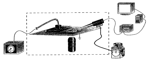
Figures 1 A and 1 B show a schematic of a system according to one aspect of
the invention showing integration of a microfluidic chip with patch clamp
recordings
of ion channel activity. Figure lA is a perspective view of a microfluidic
chip in
which a cell is positioned in proximity to microchannel outlets of the chip
using a
patch clamp micropipette connected to a positioner. Figure 1B is a side view,
partially in section, of Figure 1 A. Figure 1 C is a side view, partially in
section, of a
chip-based patch clamp system. In operation, the chip is preferably covered.
16
CA 02475348 2004-08-06
WO 03/068906 PCT/US03/01027
Figures 2A-C show top views of different embodiments of microfluidic chips
according to aspects of the invention illustrating exemplary placements of
reservoirs
for interfacing with 96-well plates. Figure 2A shows a chip comprising ligand
reservoirs (e.g., the reservoirs receive samples of ligands from a 96-well
plate).
Figure 2B shows a chip comprising alternating or interdigitating ligand and
buffer
reservoirs {e.g., every other reservoir receives samples of ligands from one
96-well
plate, while the remaining reservoirs receive samples of buffer from another
96-well
plate). As shown in Figure 2C, additional reservoirs can be placed on chip for
the
storage and transfer of cells or other samples of interest.
Figure 3 is a perspective view of a kit in accordance with one aspect of the
invention illustrating a process for dispensing fluids from 96-well plates
onto a
microfluidic chip comprising interdigitating reservoirs using automated array
pipettors and cell delivery using a pipette.
Figures 4A-C comprise a top view of a microfluidic chip structure for HTS of
1 S drugs according to one aspect of the invention, for scanning a sensor such
as a patch-
clamped cell or cells across interdigitated ligand and buffer streams. Figure
4A
depicts the overall chip structure for both a 2D and 3D microfluidic system.
Figure
4B shows an enlarged view of the reservoirs of the chip and their individual
connecting channels. Figure 4C shows an enlarged view of interdigitating
microchannel whose outlets intersect with the sensor chamber of the chip.
Figure 5A schematically depicts a top view of the interdigitating channels of
a
microfluidic chip, with a patch-clamped cell being moved past the outlets of
the
channels. Figures SB and SC depict side views of alternate embodiments of the
outlets and microchannels. Figure SB and SC are side views showing a 2D and 3D
microfluidic chip design, respectively. Figure SD is a perspective view of a
3D chip
design according to one aspect of the invention, in which the chip comprises a
bottom
set and top set of channels. Figure SE is a side view of Figure SD, showing
fluid flow
can be controlled through pressure differentials so that fluid flowing out of
a channel
in the bottom set will make a "U-turn" into an overlying channel. Figure SF is
a top
view of Figure SD and shows cell scanning across the "U-turn" fluid streams.
17
CA 02475348 2004-08-06
WO 03/068906 PCT/US03/01027
Figure 6A is a perspective view showing a 3D array of microchannel outlet
arrangements for increased throughput in HTS applications. Figure 6B depicts
the
use of a microchannel array as depicted in Figure 6A, but with a plurality of
patch-
clamped cells. The arrows in the Figures indicate directions in which the
patch-
clamped cells) can be scanned.
Figures 7A -N are schematics showing chip designs for carrying out cell
scanning across ligand streams using buffer superfusion to provide a
periodically
resensitized sensor. Figure 7A is a perspective view of the overall chip
design and
microfluidic system. Figures 7B-G show enlarged views of the outlets of
microchannels and their positions with respect to a superfusion capillary and
a patch
clamp pipette, as well as a procedure for carrying out cell superfusion while
scanning
a patch-clamped cell across different fluid streams. "P" indicates a source of
pressure
on fluid in a microchannel or capillary. Bold arrows indicate direction of
movement.
Figures 7H-7N show a different embodiment for superfusing cells. As shown in
the
perspective view in Figure 7H, instead of providing capillaries for delivering
buffer, a
number of small microchannels placed at each of the outlets of the ligand
delivery
channels are used for buffer delivery. As a patch-clamped cell is moved to a
ligand
channel and the system detects a response, a pulse of buffer can be delivered
via the
small microchannels onto the cell for superfusion. The advantage to using this
system
is that the exposure time of the patch-clamped cell to a ligand can be
precisely
controlled by varying the delay time between signal detection and buffer
superfusion.
Figure 7I is a cross-section through the side of a microfluidic system used in
this way
showing proximity of a patch-clamped cell to both ligand and buffer outlets.
Figure
7J is a cross section, front view of the system, showing flow of buffer
streams. Figure
7K is a cross-section through a top view of the device showing flow of ligand
streams
and placement of the buffer microchannels. Figures 7L-7M show use of pressure
applied to a ligand and/or buffer channel to expose a patch clamped cell to
ligand and
then buffer.
Figures 8A-I are top views of microchannel outlets in relationship to a patch-
clamped cell, collectively showing different methods by which a patch-clamped
cell
can be moved in relation to the fluid streams. Figures 8A-C show mechanical
scanning of the patched cell across stationary microchannel outlets. Figures
8D-F
18
CA 02475348 2004-08-06
WO 03/068906 PCT/US03/01027
show mechanical scanning of microchannel outlets relative to a stationary
patch-
clamped cell. Figures 8G-I show a method for sweeping fluid streams across an
immobilized patched cell by controlled variation of the pressure across, and
flow rates
through, each individual microchannel.
Figures 9A-C are top views of one design of a microfluidic chip for carrying
out cycles of rapid delivery and withdrawal of compounds into and from a cell
chamber for housing a patch-clamped cell. Figure 9A shows the overall
arrangements
of the microchannels feeding the cell chamber. Figure 9B is an expanded view
of
reservoirs and the individual channels through which they are accessed. Figure
9C
shows an enlarged view of microchannel outlets which feed into the cell
chamber.
Figure 10 is an enlarged top view of the cell chamber of Figure 9A, depicting
the arrangement of microchannels around a cell chamber comprising a patch-
clamped
cell.
Figures 11A-C are top views showing a microfluidic chip for carrying out
rapid and sequential exchange of fluids around a patch-clamped cell. Figure
11A
shows the overall arrangement of channels feeding into, and draining from, a
cell
chamber. The drain channels feed into a plurality of reservoirs such that the
pressure
drops across each channel can be independently controlled. Figure 11 B shows
an
enlarged view of reservoirs and their connecting channels. Figure 11C shows an
enlarged view of microchannel outlets which feed into the cell chamber.
Figure 12 is an enlarged illustration of Figure 1 lA, depicting the
arrangement
of and flow directions of fluids in microchannels around a cell chamber with a
patch-
clamped cell in a planar 2D microfluidic system according to one aspect of the
invention.
Figure 13 is an enlarged perspective view of the system of Figure 11 A
depicting the arrangement of microchannels, and flow directions in a 3D
microfluidic
system according to one aspect of the invention.
Figures 14A-C are top views depicting the chip structure of a fishbone design
for carrying out rapid and sequential exchange of fluids around a patch-
clamped cell
19
CA 02475348 2004-08-06
WO 03/068906 PCT/US03/01027
(not shown) according to one aspect of the invention. In the example shown in
Figure
14A, a single drain channel is provided which feeds into a single waste
reservoir.
Figure 14B shows an enlarged view of reservoirs for providing sample to the
microchannels. Figure 14C shows an enlarged view of a plurality of inlet
channels
intersecting with a central "spine" channel which feeds sample into the sensor
chamber. In this enlarged view, intersecting channels are perpendicular to the
spine
channel rather than slanted; either configuration is possible.
Figure 15 is a schematic illustration of an enlarged view of Figure 14A
depicting arrangements of, and flow directions in, microchannels, and a patch-
clamped cell in a chip according to one aspect of the invention, as well as
the
presence of passive one-way valves, which are schematically depicted as
crosses.
Figures 16A and B are microphotographs showing flow profiles at the outlet
of a single microchannel (Figure 16A) and an array of microchannels (Figure
16B).
Fluid flow was imaged under fluorescence using a fluorescent dye (fluorescein)
as a
flow tracer. The channels were 100-pm wide, 50 ~rn thick, with an inter-
channel
spacing of 25 Vim; the flow rate was 4 mm/s.
Figure 17 is a schematic illustrating the arrangement of the outlets of an
interdigitating array of microchannels in which varying dilutions of a sample
(e.g., a
drug) are provided in every other microchannel. By scanning a patch-clamped
cell
across the outlets of the channels, dose-response measurements can be
obtained.
Figures 18A-I show schematics of systems for obtaining dose-response
measurements based on high-frequency superfusion and re-sensitization of a
patch-
clamped cell. Superfusion can be achieved through a capillary co-axially
placed with
respect to a patch-clamp pipette, or through any capillary placed adjacent to
the patch
pipette which is suitable for superfusion, while translating the patch-clamped
cell
across a concentration gradient created by streams exiting microchannel
outlets.
Figures 18A-C show a concentration gradient generated by diffusion broadening
of a
ligand plug in a microchannel. Figures 18D-F show lateral diffusion spreading
of a
ligand stream as it exits a microchannel. Figure 18G-H show the use of
networks of
CA 02475348 2004-08-06
WO 03/068906 PCT/US03/01027
microchannels. "P" indicates a source of pressure applied at one or more
microchannels of the systems.
Figures 19A-C show scanning electron micrographs of microchannels
fabricated in silicon. Figure 19A shows simple microchannel arrangements in
which
the patch-clampled cell or cells can be scanned across interdigitated ligand
and buffer
streams. Figure 19B shows a simple planar radial spokes-wheel structure for
carrying
out cycles of rapid delivery and withdrawal of compounds into and from a cell
chamber housing a patch-clamped cell. Figure 19C shows a simple fishbone
arrangement of microchannel outlets for carrying out rapid and sequential
exchange
of fluids around a patch-clamped cell.
Figure 20 shows whole cell patch clamp recordings of transmembrane current
responses elicited by manual repeated scanning of a cell across the channel
outlet
where it was superfused by buffer into an open reservoir containing
acetylcholine
(1mM). A train of peaks are produced by repeated manual scanning of the
patched
cell across the superfusion-generated gradient. The cell was scanned back and
forth at
an average scan rate of 100 pm/s and at a maximum rate of up to 150 ~m/s
across the
entire outlet of the microchannel depicted in the inset.
Figures 21A-D show patch clamp current responses of a whole cell to 1 mM
acetylcholine as the patch-clamped cell is scanned across the outlets of a
parallel 7-
channel structure (same structure as that shown in Fig 16B). Channels 1, 3, 5
and 7
were filled with PBS buffer, while channels 2, 4 and 6 were filled with
acetylcholine.
The channel flow rate was 6.8 mm/s and the cell scanning speeds in the Figures
were
A) 0.61 mm/s, B) 1.22 mm/s, C) 2 mm/s and in D) 4mm/s
Figure 22 shows patch clamp current responses of a whole cell to 1 mM
acetylcholine as the patch-clamped cell was scanned across the outlets of a 7-
channel
structure (same structure as that shown in Fig 16B). Channels 1, 3, 5 and 7
were
filled with PBS buffer; channels 2, 4 and 6 with acetylcholine. The channel
flow rate
was 2.7 mm/s and the cell scanning speed was 6.25 pm/s.
Figure 23 shows concentration-dependent patch clamp current responses of
whole cells to 1 pM, 12 p.M and 200 pM nicotine as the patch-clamped cell was
21
CA 02475348 2004-08-06
WO 03/068906 PCT/US03/01027
scanned across the outlets of a 7-channel structure (same structure as that
shown in
Fig 16B); channels l, 3, 5 and 7 were filled with PBS buffer; channel 2 with 1
~M , 4
with 12 ~M and 6 with 200 pM nicotine respectively. The flow rate was 3.24
mm/s
and the cell scanning speed was 250 pm/s.
Figures 24A-C show agonist screening according to one method of the
invention using a microfluidic chip comprising 26 outlets feeding into a
sensor
chamber. As shown in Figure 24A, the screen is performed linearly from channel
outlet position 1 to 26. The scans can be repeated until a sufficient number
of scans
are performed. A simulated trace and score sheet are shown in Figures 24B and
C for
a single forward scan across microfluidic channel outlets. From this analysis,
a 6 is
the agonist with highest potency, followed by a 2.
Figures 25A-C show a method for antagonist screening according to one
aspect of the invention using a microfluidic chip comprising 26 outlets
feeding into a
sensor chamber. As shown in Figure 25A, the screen is performed linearly from
position 1-to-26. The scans can be repeated until a sufficient number of scans
are
performed. As shown in the simulated trace and score sheet, Figures 25B and C,
respectively, for a single forward scan across microfluidic channel outlets, ~
3 is the
antagonist with highest potency followed by ~ 5.
Figures 26A-C show a method for dose-response screening using a
microfluidic chip comprising 28 outlets feeding into a sensor chamber. As
shown in
Figure 24A, the screen is performed linearly from channel outlet position 1 to
28.
The scans can be repeated until a sufficient number of scans are performed. A
simulated trace and score sheet are shown in Figures 26B and C for a single
forward
scan across microfluidic channel outlets. From these data, a dose-response
curve can
be created for the unknown agonist a.
Figures 27A-C show a method for agonist screening using a microfluidic chip
comprising 14 outlets feeding into a sensor chamber and high repetition rate
buffer
superfusion using a fluidic channel placed close to a patch-clamped cell. As
shown in
Figure 27A, the screen is performed linearly from channel outlet position 1 to-
14.
The scans can be repeated until a sufficient number of scans are performed. A
22
CA 02475348 2004-08-06
WO 03/068906 PCT/US03/01027
simulated trace for a single forward scan across microfluidic channel outlets
and score
sheet are shown in Figures 27B-C. A plurality of peak responses are obtained
per
single microchannel outlet. From this analysis, a 3 is the agonist with
highest
potency, followed by a, 5.
DETAILED DESCRIPTION OF THE INVENTION
The invention provides a system and method for rapidly and programmably
altering the local solution environment around a sensor, such as a cell-based
biosensor. 'The invention further provides a system and method for interfacing
microfluidics with patch-clamp detection.
Definitions
The following definitions are provided for specific terms which are used in
the
following written description.
As used herein, a "microchannel" refers to a groove in a substrate comprising
two walls, a base, at least one inlet and at least one outlet. In one aspect,
a
microchannel also has a roof. The term "micro" does not imply a lower limit on
size,
and the term "microchannel" is generally used interchangeably with "channel".
Preferably, a microchannel ranges in size from about 0.1 pm to about 500 pm,
and
more preferably ranges from, 1 p.m to about 150pm.
As used herein, a "positioner" refers to a mechanism or instrument that is
capable of moving an object or device (e.g., a substrate, a sensor, a cell, a
mechanism
for holding a sensor, etc.) to which it is coupled. Preferably, the positioner
can
control movement of an object over distances such as nanometers (e.g., the
petitioner
is a nanopositioner), micrometers (e.g., the positioner is a micropositioner)
and/or
millimeters. Suitable positioners move at least in an x-, y-, or z- direction.
In one
aspect, positioners according to the invention also rotate about any pivot
point defined
by a user. In a preferred aspect, the positioner is coupled to a drive unit
that
communicates with a processor, allowing movement of the object to be
controlled by
the processor through programmed instructions, use of joysticks or other
similar
instruments, or a combination thereof.
23
CA 02475348 2004-08-06
WO 03/068906 PCT/US03/01027
As used herein, "a mechanism for holding a sensor" refers to a device for
receiving at least a portion of a sensor to keep the sensor in a relatively
stationary
position relative to the mechanism. In one aspect, the mechanism comprises an
opening for receiving at least a portion of a sensor. For example, such
mechanisms
include, but are not limited to: a patch clamp pipette, a capillary, a hollow
electrode,
and the like.
As used herein, the term "moving a sensor" refers to moving the sensor
directly or through the use of a mechanism for holding the sensor which is
itself
moved.
As used herein, a "chamber" refers to an area formed by walls (which may or
may not have openings) surrounding a base. A chamber may be "open volume"
(e.g.,
uncovered) or "closed volume" (e.g., covered by a coverslip, for example). A
"sensor
chamber" is one which receives one or more sensors and comprises outlets in
one or
more walls from at least two microchannels. However, a sensor chamber
according to
the invention generally can receive one or more nanoscopic or microscopic
objects,
without limitation as to their purpose. A sensor chamber can comprise multiple
walls
in different, not necessarily parallel planes, or can comprise a single wall
which is
generally cylindrical (e.g., when the chamber is "disc-shaped"). It is not
intended that
the geometry of the sensor chamber be a limiting aspect of the invention. One
or
more of the walls) and/or base can be optically transmissive. Generally, a
sensor
chamber ranges in size but is at least about 1 pm. In one aspect, the
dimensions of the
chamber are at least large enough to receive at least a single cell, such as a
mammalian cell. The sensor chamber also can be a separate entity from the
substrate
comprising the microchannels. For example, in one aspect, the sensor chamber
is a
petrie dish and the microchannels extend to a surface of the substrate opening
into the
petrie dish so as to enable fluid communication between the microchannels and
the
petrie dish.
As used herein, a "sensor" refers to a device comprising one or more
molecules capable of producing a measurable response upon interacting with a
condition in an aqueous environment to which the molecule is exposed (e.g.,
such as
the presence of a compound which binds to the one or more molecules). In one
24
CA 02475348 2004-08-06
WO 03/068906 PCT/US03/01027
aspect, the molecules) are immobilized on a substrate, while in another
aspect, the
molecules) are part of a cell (e.g., the sensor is a "cell-based biosensor").
As used herein, "a nanoscopic or microscopic object" is an object whose
dimensions are in the nm to mm range.
As used herein, the term, "a cell-based biosensor" refers to an intact cell or
a
part of an intact cell (e.g., such as a membrane patch) which is capable of
providing a
detectable physiological response upon sensing a condition in an aqueous
environment in which the cell (or part thereof) is placed. In one aspect, a
cell-based
biosensor is a whole cell or part of a cell membrane in electrical
communication with
an electrically conductive element, such as a patch clamp electrode or an
electrolyte
solution.
As used herein, the term "receptor" refers to a macromolecule capable of
specifically interacting with a ligand molecule. Receptors may be associated
with
lipid bilayer membranes, such as cellular, golgi, or nuclear membranes, or may
be
present as free or associated molecules in a cell's cytoplasm or may be
immobilized
on a substrate. A cell-based biosensor comprising a receptor can comprise a
receptor
normally expressed by the cell or can comprise a receptor which is non-native
or
recombinantly expressed (e.g., such as in transfected cells or oocytes).
As used herein, "periodically resensitized" or "periodically responsive"
refers
to an ion-channel which is maintained in a closed (i.e., ligand responsive)
position
when it is scanned across microchannel outlets providing samples suspected or
known
to comprise a ligand. For example, in one aspect, an receptor or ion-channel
is
periodically resensitized by scanning it across a plurality of interdigitating
channels
providing alternating streams of sample and buffer. The rate at which the
receptor/ion
channel is scanned across the interdigitating channels is used to maintain the
receptor/ion-channel in a ligand-responsive state when it is exposed to a
fluid stream
comprising sample. Additionally, or alternatively, the receptor/ion channel
can be
maintained in a periodically resensitized state by providing pulses of buffer,
e.g.,
using one or more superfusion capillaries, to the ion channel, or by providing
rapid
exchange of solutions in a sensor chamber comprising the ion channel.
CA 02475348 2004-08-06
WO 03/068906 PCT/US03/01027
As used herein, a "substantially separate fluid stream" refers to a flowing
fluid
in a volume of fluid (e.g., such as within a chamber) that is physically
continuous with
fluid outside the stream within the volume, or other streams within the
volume, but
which has at least one bulk property which differs from and is in non-
equilibrium
from a bulk property of the fluid outside of the stream or other streams
within the
volume of fluid. A "bulk property" as used herein refers to the average value
of a
particular property of a component (e.g., such as an agent, solute, substance,
or a
buffer molecule) in the stream over a cross-section of the stream, taken
perpendicular
to the direction of flow of the stream. A "property" can be a chemical or
physical
property such as a concentration of the component, temperature, pH, ionic
strength, or
velocity, for example.
As used herein, the term "in communication with" refers to the ability of a
system or component of a system to receive input data from another system or
component of a system and to provide an output response in response to the
input
data. "Output" may be in the form of data, or may be in the form of an action
taken
by the system or component of the system. For example, a processor "in
communication with a scanning mechanism" sends program instructions in the
form
of signals to the scanning mechanism to control various scanning parameters as
described above. A "detector in communication with a sensor chamber" refers to
a
detector in sufficient optical proximity to the sensor chamber to receive
optical signals
(e.g., light) from the sensor chamber. A "light source in optical
communication" with
a chamber refers to a light source in sufficient proximity to the chamber to
create a
light path from the chamber to a system detector so that optical properties of
the
chamber or objects contained therein can be detected by the detector.
As used herein, "a measurable response" refers to a response which differs
significantly from background as determined using controls appropriate for a
given
technique.
As used herein, an outlet "intersecting with" a chamber or microchamber
refers to an outlet that opens or feeds into a wall or base or top of the
chamber or
microchamber or into a fluid volume contained by the chamber or microchamber.
26
CA 02475348 2004-08-06
WO 03/068906 PCT/US03/01027
As used herein, "superfuse" refers to washing the external surface of an
object
or sensor (e.g., such as a cell).
The System
In one aspect, the system provides a substrate comprising a plurality of
microchannels fabricated thereon whose outlets intersect with, or feed into, a
sensor
chamber comprising one or more sensors. The system further comprises a
scanning
mechanism for programmably altering the position of the microchannels relative
to
the one or more sensors and a detector for monitoring the response of the
sensor to
exposure to solutions from the different channels. In a preferred aspect, the
sensor
chamber comprises a cell-based biosensor in electrical communication with an
electrode and the detector detects changes in electrical properties of the
cell-based
biosensor.
The system preferably also comprises a processor for implementing system
operations including, but not limited to: controlling the rate of scanning by
the
scanning mechanism (e.g., mechanically or through programmable pressure drops
across microchannels), controlling fluid flow through one or more channels of
the
substrate, controlling the operation of valves and switches that are present
for
directing fluid flow, recording sensor responses detected by the detector, and
evaluating and displaying data relating to sensor responses. Preferably, the
system
also comprises a user device in communication with the system processor which
comprises a graphical interface for displaying operations of the system and
for
altering system parameters.
The Substrate
In a preferred aspect, the system comprises a substrate that delivers
solutions
to one or more sensors at least partially contained within a sensor chamber.
The
substrate can be configured as a two-dimensional (2D) or three-dimensional
(3D)
structure, as described further below. The substrate, whether 2D or 3D,
generally
comprises a plurality of microchannels whose outlets intersect with a sensor
chamber
that receives the one or more sensors. The base of the sensor chamber can be
optically transmissive to enable collection of optical data from the one or
more
27
CA 02475348 2004-08-06
WO 03/068906 PCT/US03/01027
sensors placed in the sensor chamber. When the top of the sensor chamber is
covered,
e.g., by a coverslip or overlying substrate, the top of the chamber is
preferably
optically transmissive.
Each microchannel comprises at least one inlet (e.g., for receiving a sample
or
a buffer). Preferably, the inlets receive solution from reservoirs (e.g.,
shown as circles
in Figures 2A and B) that conform in geometry and placement on the substrate
to the
geometry and placement of wells in an industry-standard microtiter plate. The
substrate is a removable component of the system and therefore, in one aspect,
the
invention provides kits comprising one or more substrates for use in the
system,
providing a user with the option of choosing among different channel
geometries.
Non-limiting examples of different substrate materials include crystalline
semiconductor materials (e.g., silicon, silicon nitride, Ge, GaAs), metals
(e.g., Al, Ni),
glass, quartz, crystalline insulators, ceramics, plastics or elastomeric
materials (e.g.,
silicone, EPDM and Hostaflon), other polymers (e.g., a fluoropolymer, such as
Teflon~, polymethylmethacrylate, polydimethylsiloxane, polyethylene,
polypropylene, polybutylene, polymethylpentene, polystyrene, polyurethane,
polyvinyl chloride, polyarylate, polyarylsulfone, polycaprolactone,
polyestercarbonate, polyimide, polyketone, polyphenylsulfone, polyphthalamide,
polysulfone, polyamide, polyester, epoxy polymers, thermoplastics, and the
like),
other organic and inorganic materials, and combinations thereof.
Microchannels can be fabricated on these substrates using methods routine in
the art, such as deep reactive ion etching (described further below in Example
1 ).
Channel width can vary depending upon the application, as described further
below,
and generally ranges from about 0.1 ~m to about 10 mm, preferably, from about
1 pm
to about 150 pm, while the dimensions of the sensor chamber generally will
vary
depending on the arrangement of channel outlets feeding into the chamber. For
example, where the outlets are substantially parallel to one another (e.g., as
in Figures
2A-C), the length of the longitudinal axis of the chamber is at least the sum
of the
widths of the outlets which feed into the chamber. In one aspect, where a
whole cell
biosensor is used as a sensor in the sensor chamber, the width of one or more
outlets
28
CA 02475348 2004-08-06
WO 03/068906 PCT/US03/01027
of the microchannels is at least about the diameter of the cell. Preferably,
the width of
each of the outlets is at least about the diameter of the cell.
In one aspect, a cover layer of an optically transmissive material, such as
glass, can be bonded to a substrate, using methods routine in the art,
preferably
leaving openings over the reservoirs and over the sensor chamber when
interfaced
with a traditional micropipette-based patch clamp detection system.
Preferably, the
base of the sensor chamber also is optically transmissive, to facilitate the
collection of
optical data from the sensor.
The Sensor
Cell-Based Biosensors
The system can be used in conjunction with a cell-based biosensor to monitor
a variety of cellular responses. The biosensor can comprise a whole cell or a
portion
thereof (e.g., a cell membrane patch) which is positioned in the sensor
chamber using
a mechanism for holding a sensor (which may be stationary or movable) such as
a
pipette, capillary, or column connected to a positioner, such as a
micropositioner, a
nanopositioner or a micromanipulator, or an optical tweezer, or by controlling
flow or
surface tension, thereby exposing the cell-based biosensor to solution in the
chamber.
The biosensor can be scanned across the various channels of the substrate by
moving
the substrate, i.e., changing the position of the channels relative to the
biosensor, or by
moving the cell (e.g., by scanning the micropositioner or by changing flow
and/or
surface tension).
In one aspect, the cell-based biosensor comprises an ion channel and the
system is used to monitor ion channel activity. Suitable ion channels include
ion
channels gated by voltage, ligands, internal calcium, other proteins, membrane
stretching (e.g., lateral membrane tension) and phosphorylation (see e.g., as
described
in Hille B., In Ion Channels of Excitable Membranes 1992, Sinauer, Sunderland,
Massachusetts, USA). In another aspect, the ion-gated channel is a voltage-
gated
channel. Voltage-gated channels open in response to a threshold transmembrane
voltage. Voltage-gated sodium, potassium, and calcium channels are all
essential for
conducting an action potential (or a nerve pulse) down an axon and to another
nerve
29
CA 02475348 2004-08-06
WO 03/068906 PCT/US03/01027
cell (or neuron). These ion channels typically comprise a transmembrane
sequence
with a lysine and/or arginine-rich S4 consensus sequence. The positive amino
acids
within the S4 sequence are thought to "sense" voltage across a cell membrane,
causing an ion channel containing the sequence to either open or close under
different
voltage conditions.
In another aspect, the ion channel in the cell-based biosensor is a ligand-
gated
channel. Ligand-gated channels gate (open or close) in response to ligand
binding.
There are two types of ligand-gated channels, those gated when bound by
ligands
inside the cell and those gated by ligands outside the cell. Ion channels
gated by
ligands from outside of the cell are very important in chemical synaptic
transmission.
These types of ion channels are gated by neurotransmitters, which are the
small
molecules that actually carry the signal between two nerve cells. Ion channels
gated
from the inside of the cell are generally controlled by second messengers,
which are
small signaling molecules inside the cell. Intracellular calcium ions, cAMP
and
cGMP are examples of second messengers. The most common calcium-gated channel
is the calcium-gated potassium channel. This ion channel can generate
oscillatory
behavior (e.g., for frequency tuning of hair cells in the ear) upon changes in
membrane voltage when placed in a positive feedback environment.
In yet another aspect, the ion channel is gated by another protein. Certain
signaling proteins have been found to directly gate ion channels. One example
of this
is a potassium channel gated by the beta-gamma subunit of the G protein, which
is a
common signaling protein activated by certain membrane receptors.
In a further aspect, the ion channel is gated by phosphorylation.
Phosphorylation can be mediated by a protein kinase (e.g., a serine,
threonine, or
tyrosine kinase), e.g., as part of a signal transduction cascade.
In still a further aspect, the cell-based biosensor comprises a
mechanotransduction channel that can be directly gated by a mechanical
trigger. For
example, the cell-based biosensor can comprise the cation channel of an inner
ear hair
cell, which is directly gated by a mechanical vibration such as sound. Bending
of the
hair bundle in a particular direction will affect the probability of channel
gating, and
therefore, the amplitude of a depolarizing receptor current.
CA 02475348 2004-08-06
WO 03/068906 PCT/US03/01027
In another aspect, the cell-based biosensor comprises a receptor, preferably,
a
receptor involved in a signal transduction pathway. For example, the cell-
based
biosensor can comprise a G Protein Coupled Receptor or GPCR, glutamate
receptor, a
metabotropic receptor, a hematopoietic receptor, or a tyrosine kinase
receptor.
Biosensors expressing recombinant receptors also can be designed to be
sensitive to
drugs which may inhibit or modulate the development of a disease.
Suitable cells which comprise biosensors include, but are not limited to:
neurons; lymphocytes; macrophages; microglia; cardiac cells; liver cells;
smooth
muscle cells; and skeletal muscle cells. In one aspect, mammalian cells are
used;
these can include cultured cells such as Chinese Hamster Ovary Cells (CHO)
cells,
NIH-3T3, and HEK-293 cells and can express recombinant molecules (e.g.,
recombinant receptors and/or ion channels). However, bacterial cells (E. colt,
Bacillus sp., Staphylococcus aureus, and the like), protist cells, yeast
cells, plant cells,
insect and other invertebrate cells, avian cells, amphibian cells, and
oocytes, also can
be used, as these are well suited to the expression of recombinant molecules.
. Cells
generally are prepared using cell culture techniques as are know in the art,
from cell
culture lines, or from dissected tissues after one or more rounds of
purification (e.g.,
by flow cytometry, panning, magnetic sorting, and the like).
Non-Cellular Sensors
In one aspect, the sensor comprises a sensing element, preferably, a molecule
which is cellular target (e.g., an intracellular receptor, enzyme, signalling
protein, an
extra cellular protein, a membrane protein, a nucleic acid, a lipid molecule,
etc.),
which is immobilized on a substrate. The substrate can be the base of the
sensor
chamber itself, or can be a substrate placed on the base of the chamber, or
can be a
substrate which is stably positioned in the chamber (e.g., via a
micropositioner) and
which is moveable or stationary.
The sensor may consist of one or several layers that can include any
combination o~ a solid substrate; one or more attachment layers that bind to
the
substrate, and a sensing molecule for sensing compounds introduced into the
sensor
chamber from one or more channel outlets. Suitable sensors according to the
invention, include, but are not limited to, immunosensors, affinity sensors
and ligand
31
CA 02475348 2004-08-06
WO 03/068906 PCT/US03/01027
binding sensors, each of which can respond to the presence of binding partners
by
generating a measurable response, such as a specific mass change, an
electrochemical
reaction, or the generation of an optical signal (e.g., fluorescence, or a
change in the
optical spectrum of the sensing molecule). Such sensors are described in U.S.
Patent
No. 6,331,244, for example, the entirety of which is incorporated by reference
herein.
In one aspect, the sensor comprises a microelectrode which is modified with a
molecule which transports electrons. In response to a chemical reaction caused
by
contact with one or more compounds in an aqueous stream from one of the
microchannels, the molecule will produce a change in an electrical property at
the
electrode surface. For example, the molecule can comprise an electron-
transporting
enzyme or a molecule which transducer signals by reduction or oxidation of
molecules with which it interacts (see, e.g., as described in, Gregg, et al.,
J. Phys.
Chem. 95: 5970-5975, 1991; Heller, Acc. Chem. Res. 23 5 : 128-134, 1990; In
Diagnostic Biosensor Polymers. ACS Symposium Series. 556; Usmani, A M, Akmal,
N; eds. American Chemical Society; Washington, D.C.; pp. 47-70, 1994; U.S.
Patent
No. 5,262,035). Enzymatic reactions also can be performed using field-effect-
transistors (FETs) or ion-sensitive field effect transistors (ISFETs).
In another aspect, the sensor comprises a sensing molecule immobilized on a
solid substrate such as a quartz chip in communication with an electronic
component.
The electronic component can be selected to measure changes in any of:
voltage,
current, light, sound, temperature, or mass, as a measure of interaction
between the
sensing element and one or more compounds delivered to the sensor chamber
(see, as
described in, Hall, Int. J. Biochem. 2~: 357-62, 1988; U.S. Patent No.
4,721,677;
U.S. Patent No. 4,680,268; U.S. Patent No. 4,614,714; U.S. Patent No.
6,879,11). For
example, in one aspect, the sensor comprises an acoustic wave biosensor or a
quartz
crystal microbalance, on which a sensor element is bound. In this embodiment,
the
system detects changes in the resonant properties of the sensor upon binding
of
compounds in aqueous streams delivered from the microchannels to the sensor
element.
In another aspect, the sensor comprises an optical biosensor. Optical
biosensors can rely on detection principles such as surface plasmon resonance,
total
32
CA 02475348 2004-08-06
WO 03/068906 PCT/US03/01027
internal reflection fluorescence (TIRF), critical angle refractometry,
Brewster Angle
microscopy, optical waveguide lightmode spectroscopy (OWLS), surface charge
measurements, and evanescent wave ellipsometry, and are known in the art (see,
for
example, U.S. Patent No. 5,313,264; EP-A1-0 067 921; EP-A1-0 278 577; Kronick,
et
al., 1975, J. Immunol. Meth. 8: 235-240).
For example, for a sensor employing evanescent wave ellipsometry, the
optical response related to the binding of a compound to a sensing molecule is
measured as a change in the state of polarization of elliptically polarized
light upon
reflection. The state of polarization is related to the refractive index,
thickness, and
surface concentration of a bound sample at the sensing surface (e.g., the
substrate
comprising the sensing element). In TIRE, the intensity and wavelength of
radiation
emitted from either natively fluorescent or fluorescence-labelled sample
molecules at
a sensor is measured. Evanescent wave excitation scattered light techniques
rely on
measuring the intensity of radiation scattered at a sensor surface due to the
interaction
of light with sensing molecules (with or without bound compounds). Surface
plasmon resonance (SPR) measures changes in the refractive index of a layer of
sensor molecules close to a thin metal film substrate (see, e.g., Liedberg, et
al., 1983,
Sensors and Actuators 4: 299; GB 2 197 068). Each of these sensing schemes can
be
used to provide useful sensors according to the invention.
In yet another aspect, the sensor comprises a sensing molecule associated with
a fluorescent semiconductor nanocrystal or a Quantum DotTM particle. The
Quantum
Dot particle has a characteristic spectral emission which relates to its
composition and
particle size. Binding of a compound to the sensing element can be detected by
monitoring the emission of the Quantum Dot particle (e.g., spectroscopically)
(see,
e.g., U.S. Patent No. 6,306,610).
The sensor further can comprise a polymer-based biosensor whose physical
properties change when a compound binds to a sensing element on the polymer.
For
example, binding can be manifested as a change in volume (such as swelling or
shrinkage), a change in electric properties (such as a change in voltage or
current or
resonance) or in optical properties (such as modulation of transmission
efficiency or a
change in fluorescence intensity).
33
CA 02475348 2004-08-06
WO 03/068906 PCT/US03/01027
It should be obvious to those of skill in the art that a variety of different
types
of sensors may be adapted for use in present invention, and the examples above
are
intended to be non-limiting.
In general, the measurement outputs of one or more sensors are connected to a
control and evaluating device which is in electrical communication with a
detection
device and/or system processor. The control and evaluating device can be
integrated
with the substrate of the sensor andJor with the base of the sensing chamber.
The
control and evaluating device can comprise various electronic components such
as
microprocessors, multiplexers, IO units, ete. (see, e.g., as described in U.S.
Patent No.
6,280,586).
Microfluidics
In a preferred aspect, the substrates according to the invention are adapted
for
microfluidic transport of sample and/or buffer to a sensor chamber.
Interfacing Microfluidic Structures with Well Plates
Samples (i.e., drugs, etc.) contained in sample-well plates (e.g., industry-
standard microtiter plates such as 96-well plates) are manipulated and
transferred,
preferably, using robotic automated array pipettors as are known in the art
(see, e.g.,
Beckman's Biomek 1000 & 2000 automated workstations, available from Beckman
Coultcr, Inc., Fullerton, CA).
To be able to leverage the same sample transfer platform used to array a
sample
in a well plate, one important design parameter is to ensure the reservoir
arrangements
in the chip described above are compatible for use with such array pipettors.
For
example, preferably, the reservoirs in the microfluidic chip are arranged such
that the
center-to-center distance between each reservoir is identical to the center-to-
center
distance between each well of the well plate to which the chip interfaced.
Preferably,
each reservoir has a diameter suitable for receiving a fluid stream from an
array
pipetter without significantly impeding the flow of fluid from the array
pipettor.
In addition to array pipettors, there are other suitable automated devices for
transferring samples from well plates onto chips, such as robotic sequential
pipettors.
34
CA 02475348 2004-08-06
WO 03/068906 PCT/US03/01027
It is important to note that the use of these other devices may permit more
flexible
placement of reservoirs and microchannels on the chip, providing more
flexibility in
the design of channel parameters. Although a substrate suitable for
interfacing
between 96-well array pipettors is described in more detail below owing to the
widespread use of these pipettors, it should be obvious to those of skill in
the art that
the general design of the chip and placement of reservoirs can be modified for
interfacing with any desirable sample transfer platform, as such platforms
evolve. In
general, reference to 96-well plates is not intended to be limiting.
Figures 2A and 2B show examples of microfluidic chips according to the
invention that are suitable for interfacing with a 96-well plate. Figure 2A
illustrates
reservoir arrangements for which no buffer reservoirs are required. Figure 2B
illustrates reservoir arrangements for applications in which alternating
(i.e.,
interdigitating) streams of buffer and sample are provided to a sensor. In
this
arrangement, the center-to-center distances for both the ligand and buffer
reservoirs
are identical to the center-to-center distance of the wells of a 96-well
plate. To
compensate for doubling the number of reservoirs on chip, the diameter of all
reservoirs are decreased by half.
Figure 3 illustrates how sample solutions can be transferred from the wells of
a 96-well plate into reservoirs on a chip according to one aspect of the
invention using
traditional robotic automated array pipettors. For a microchip with
interdigitated
ligand and buffer reservoirs (e.g., as shown in Figure 2B), buffer solution
can be
transferred from a bath, where only one buffer is needed, or from a 96-well
plate, with
wells comprising the same or different buffers.
In addition to the reservoirs needed for interfacing with sources of sample
and/or buffer (e.g., such as well plates), there may be additional reservoirs
placed on
the chip for storing and transferring cells or other samples of interest.
Figure 2C
illustrates the possible placement of additional reservoirs and microchannels
for
storing and transporting cells into reservoirs or the sensor chamber of the
chip,
according to one aspect of the invention.
The cell chambers can be adapted for performing on-chip manipulation of
cells. In one aspect, the chip provides one or more cell treatment chambers
for
CA 02475348 2004-08-06
WO 03/068906 PCT/US03/01027
performing one or more of: electroporation, electroinjection, and/or
electrofusion.
Chemicals and/or molecules can be introduced into a cell within a chamber
which is
in electrical communication with a source of current. For example, one or more
electrodes may be placed in proximity to the chamber, or the chamber can be
configured to receive an electrolyte solution through which current can be
transmitted,
for example, from an electrode/capillary array as described in WO 99/24110,
the
entirety of which is incorporated by reference herein.
Suitable molecules which can be introduced into a cell in the cell treatment
chamber include, but are not limited to: nucleic acids (including gene
fragments,
cDNAs, antisense molecules, ribozymes, and aptamers); antibodies; proteins;
polypeptides; peptides; analogs; drugs; and modified forms thereof. In a
preferred
aspect, the system processor controls both the delivery of molecules to the
one or
more cell treatment chambers (e.g., via capillary arrays as described above)
and
incubation conditions (e.g., time, temperature, etc.). For example, a cell can
be
incubated for suitable periods of times until a desired biological activity is
manifested,
such as transcription of an mRNA; expression of a protein; inactivation of a
gene,
mRNA, and/or protein; chemical tagging of a nucleic acid or protein;
modification or
processing of a nucleic acid or protein; inactivation of a pathway or toxin;
and/or
expression of a phenotype (e.g., such as a change in morphology).
The treated cells can be used to deliver molecules of interest to the sensor
in
the sensor chamber, e.g., exposing the sensor to secreted molecules or
molecules
expressed on the surface of the cells. In this aspect, the system can be
programmed to
release a cell from a cell treatment chamber into a channel of the system
intersecting
with the sensor chamber, thereby exposing a sensor in the sensor chamber to
the
molecule of interest.
Alternatively, or additionally, when the system is used in conjunction with a
cell-based biosensor, the cell treatment chamber can be used to prepare the
biosensor
itself. In one aspect, a cell is delivered from the treatment chamber to a
channel
whose outlet intersects with the sensor chamber. In one aspect, the scanning
mechanism of the system is used to place a micropositioner in proximity to the
outlet
so that the micropositioner can position the cell within the sensor chamber.
In another
36
CA 02475348 2004-08-06
WO 03/068906 PCT/US03/01027
aspect, fluid flow or surface tension is used to position a cell in a suitable
position.
For example, the system can be used to deliver the cell to the opening of a
pipette
which is part of a patch clamp system.
In another aspect, a cell can be delivered to the sensor chamber to
periodically
replace a cell-based biosensor in the sensor chamber. In this aspect, the cell
can be
untreated, e.g., providing a substantially genetically and pharmacologically
identical
cell (i.e., within the range of normal biological variance) as the previous
sensor cell.
Alternatively, the replacement cell can be biochemically or genetically
manipulated to
be different from the previous sensor cell, to enable the system to monitor
and
correlate differences in biochemical and/or genetic characteristics of the
cells with
differences in sensor responses. The biochemical or genetic difference can be
known
or unknown.
The system can be programmed to deliver cells from the cell treatment
chamber at selected time periods based on control experiments monitoring
uptake of
chemicals and molecules by cells. Alternatively, the system can monitor the
phenotype of cells and deliver cells when a certain phenotype is expressed.
For
example, in one aspect, the cell treatment chamber is in communication with an
optical sensor which provides information relating to optical properties of
the cell to
the system processor, and in response to optical parameters indicating
expression of a
particular phenotype, the system can trigger release of the cell from the cell
treatment
chamber. Optical parameters can include the uptake of a fluorescent reporter
molecule or optical parameters identified in control experiments.
The combination of on-chip electroporation with microfluidics and patch
clamp (or other methods for monitoring cell responses) facilitates screening
for
molecules (e.g., ligands or drugs) which modulate the activity of
intracellular targets.
In one aspect, the system is used to deliver a cell-impermeant molecule into
the
interior of a cell by transiently electroporating the cell. In this way, the
molecule can
be introduced to intracellular receptors, intracellular proteins,
transcriptional
regulators, and other intracellular targets. The cell can be delivered to the
sensor
chamber and the response of the cell can be monitored (e.g., by patch clamp or
by
37
CA 02475348 2004-08-06
WO 03/068906 PCT/US03/01027
fluorescence, if the molecule is tagged with a fluorescent label).
Alternatively, the
sensor chamber can be modified to perform both treatment and response
detection.
In a further aspect, the system can be modified to perform electroporation by
scanning. For example, a cell can be repeatedly electroporated as it is being
translated
or scanned across a plurality of different fluid streams containing different
compounds. In one aspect, pores are introduced into one or more cells as they
come
into contact with a sample stream, enabling compounds in the sample stream to
be
taken up by the cell.
Rapid Alterations of the Solution Environment Around a Sensor
Central to the present invention is the use of two-dimensional (2D) and three-
dimensional (3D) networks of microfabricated channels for the complex
manipulation
of compounds or reagents contained in the fluid in a way that permits repeated
and
rapid delivery of different solutions to the sensor in the sensor chamber. For
example,
the microfluidics used with the system enables the system to programmably
deliver a
ligand to a cell-based biosensor comprising a receptor. This enables the
system to be
used for HTS screening of samples (e.g., such as compound libraries) to
monitor the
effects of compounds on the responses of the biosensor. In one aspect,
electrical
properties of a cell-based biosensor are monitored using voltage clamp or
patch clamp
techniques.
Because the system provides a scanning mechanism for changing the position
of channels relative to a sensor, the system can be used to flush a cell-based
biosensor
with buffer after exposure to a sample compound, enabling a receptor or ion
channel
that is part of the biosensor to be resensitized prior to exposure to the next
compound.
Thus, the system can provide a periodically resensitized receptor for exposure
to
potential modulators of receptor function (e.g., such as agonists or
antagonists). For
receptors that do not desensitise, the system is still advantageous for
providing pulsed
delivery of buffer to a receptor, e.g., to remove unbound ligand from the
receptor, to
enhance the specificity and/or decrease background of a response.
38
CA 02475348 2004-08-06
WO 03/068906 PCT/US03/01027
The geometry of different network structures of microchannels is designed to
exploit the unique characteristic of fluid behavior in micro-dimensions. Three
exemplary designs are described below.
The first design relies on the system's ability to transport one or more
sensors,
rapidly across different streams of fluid flowing from channel outlets by
translating
the sensors across the channels or by translating the substrate comprising the
channels
relative to the one or more sensors. The system also can sweep different fluid
streams
across a stationary sensor by varying pressure drops across individual
channels of the
substrate. This design is derived from the discovery of a new and unique
fluidic
behavior; i.e., that lateral interactions and couplings between neighbouring
fluid
streams as they exit from a set of closely spaced microchannels into an open
volume
can extend dramatically the distance over which these streams remain
collimated.
The second design exploits the reversibility of fluid behavior at low Reynolds
numbers while the third design is based on the ability to rapidly exchange
fluids in
microchannels and chambers.
The theme that runs through these designs is a microfluidic-based approach
for rapidly and efficiently altering the local solution microenvironment in
which one
or more biosensors reside, providing complete or near-complete solution
exchange.
The system requires small sample volumes (nLs to ~.Ls) and can be easily
automated
and programmed for HTS applications.
(1) The Rapid Transport of Sensors Across Different Streams of Fluids
Adjacent fluid streams exiting the plurality of microchannels of a substrate
according to the invention have a low Reynolds number and undergo minimal
mixing
by diffusion. For example, a small molecule with a diffusion coefficient of
about
5 X 10⁻⁶ cm2/s would take approximately 0.1 seconds to diffuse 10 Vim, but 10
s to
diffuse 100 Vim, owing to the square dependence of distance on diffusion time
(xz =
2Dt, where D is the diffusion coefficient). Similarly, for typical proteins
having D
10~~ cmZ/s, it will take 0.5 second to diffuse 10 ~m and 50 seconds for 100
pm.
However, flow rates in microchannels can vary dramatically from many
meters per second to micrometers per second. The flow rate in the present
system is
39
CA 02475348 2004-08-06
WO 03/068906 PCT/US03/01027
limited to the maximum flow rate that can be used without disturbing the
activity of
the sensor. For example, when using a patch clamp sensor, flow rate is
typically on
the order of hundreds of p,m/s to mm/s for a patch-clamped cell (see below for
discussion), in order to prevent dislodgement of the patch-clamped cell from
the
pipette which positions it at a channel opening.
Flow Profiles Of Multiple Fluid Streams Exiting Into the Sensor Chamber
When a plurality of microchannels is used, an understanding of the flow
profile of the multiple streams of fluid at the interface between the outlets
of the
microchannels and the open-volume reservoir is essential. Figures 16A and 16B
show photomicrographs of flow profiles of a fluid comprising 500 N.M of a
fluorescent dye (fluorescein) from a single channel (Figure 16A) and multiple
channels (Figure 16B). Excitation of the fluorescent tracer was carned out
using the
488-nm line of an Argon Ion laser in an epi-illumination configuration and
fluorescence of the tracer was collected and imaged using a CCD camera. As
shown
in Figure 16A, in the absence of adjacent microchannels and fluid streams, the
fluid
exits the single channel and disperses at the channel outlet in a semicircular
fashion.
Figure 16B shows a flow profile of interdigitated buffer and fluorescein fluid
streams
exiting from a plurality of channel outlets. The dimensions of microchannels
were
100 N.m wide, 50 ~m thick, with an interchannel interval of 25 N.m. The flow
rate in
the microchannels in both Figures 16A and 16B was 4 mm/s.
As shown in Figure 16B, the fluid stream exiting multiple microchannels into
an open volume is collimated for at least a distance that is about 4-5 times
the width
of the microchannels (e.g., about several hundred micrometers) at a flow rate
of about
4 mm/s. In this range of low flow rates (i.e., mm/s), the rate of fluid flow
is still much
faster than diffusion. For example, at a flow rate of mm/s, channel width of
10 pm,
and channel intervals (the space between channels) of 10 Vim, different
streams of
fluid containing small molecules (D = 5 x 10⁻⁶ cm2/s) exiting different
channel outlets
will not be fully mixed until at a distance of at least about 0.4 mm
downstream of the
channel outlets. This is more than sufficient for making measurements of a
typical
mammalian cell having a diameter of about 10 to 20 ~m which is placed 10 to 20
~m
outside the outlet of a channel. Since diffusion time varies with the square
of the
CA 02475348 2004-08-06
WO 03/068906 PCT/US03/01027
distance, doubling the width and spacing of microchannels to 20 pm extends the
downstream distance at which the cell can be placed to at least about 1.6 mm.
'The
average linear velocity of the flow will vary depending on the exact
application, with
typical flow rates ranging from about 100 pm/s to about 10 mm/s for a 10 p.m
cell.
Thus, a sensor can be scanned across substantially distinct and separate
aqueous
streams of fluid which exit from the microchannel outlets.
At the preferred flow rates for use with patch clamp measurements and at a
cell-to-outlet distance of about 20 pm or less, the different fluid streams
are
essentially distinct and separate and are undisturbed by the presence of a
patch-
clamped cell. Even at much lower flow rates (e.g., < 100 p.m/s) that may be
used with
patch clamp measurements, different fluid streams are still well separated.
This
observed behaviour (e.g., collimation of fluid streams) of fluid flow at the
exits of
microchannels into an open volume sensor chamber facilitates HTS applications
which require relatively rapid translation of patched cells with respect to
different
fluid streams. Spacing between microchannel outlets can be optimised to
optimise
separation between fluid streams, as can flow rate. For example, the more
rapid the
flow rate, the less mixing is observed. Preferably, flow rate and interchannel
spacing
are optimised to minimize the width of a boundary zone (i.e., an area of
mixing).
Preferably, a boundary zone is less than about 50% of the width of a fluid
stream, or
less than about 40%, less than about 30%, less than about 20%, less than about
10%,
less than about S%, or less than about 1 % of the width of the stream. In one
aspect,
the boundary zone is about 2-3 microns. Optimal fluid flow rates and
interchannel
spacings can be devised readily using one or more tracer dyes as described
above.
To exploit the unique behaviour of fluid flow into open volumes, the pressure
applied to each of a plurality of microchannels can be individually varied for
precise
manipulation of each flow stream. For example, in the extreme case in which
positive
pressure is applied to one channel and negative pressure is applied to an
adjacent
channel, the fluid stream can be made to make a "U-turn", going from the
channel
with positive pressure to the one with negative pressure while drawing in a
sheath of
buffer into the channel with negative pressure. Therefore, the position,
width,
collimation, direction, and rate of flow, as well as the composition of the
fluid
streams, can be controlled by varying the relative pressure applied to each
channel.
41
CA 02475348 2004-08-06
WO 03/068906 PCT/US03/01027
As shown in Figure SD-F, this can be used to create a U-shaped fluid stream
which has the advantage that sample delivered onto a cell from a channel
experiencing positive pressure can be withdrawn into a waste channel
experiencing
negative pressure. This minimizes the accumulation of ligands in the open
volume
where the patch-clamped cell resides. In situations where a sample (e.g., a
drug,
ligand, and the like) is in low concentration and/or is expensive, the system
further
can be used to recycle ligand and/or to feed ligand back into the system
(i.e., the U-
shaped stream can be turned into a closed loop).
By controlling pressure, the system can control the velocity (both amplitude
and direction) of fluid streams. Velocity control also may be exercised by
controlling
the resistance of each channel without changing the pressure or by changing
both
resistance and pressure. Fluid shear also can be varied by using solutions of
different
viscosity (e.g., adding different amounts of a sugar such as sucrose to a
fluid stream)
in both the microchannels and sensor chamber. Thus, by varying a number of
1 S different parameters, the flow profile of different fluid streams can be
precisely tuned.
Patch Clamp Under Fluid Flow
The ability to rapidly scan patch-clamped cells across interdigitated streams
of
receptor modulators (agonists or antagonists) and buffer depends on the
mechanical
stability of the patched cell under the required flow conditions as well as
scan speeds.
Here, the stability of the "giga seal" and ion-channel activities of patch-
clamped cells
under a range of flow conditions is described.
The effects of liquid flow on a patch-clamped cell arise from the force
(Stokes
drag) exerted by the flow on the cell. This Stokes drag can be calculated from
the
following equation:
Force = (frictional coefficient) x (velocity of the flow)
Where the frictional coefficient (f) can be calculated from:
f = 6~r~,
42
CA 02475348 2004-08-06
WO 03/068906 PCT/US03/01027
where r is the radius of the cell and p. is the viscosity of the solution.
This relationship
is valid for low Reynolds number flow and for particles that are spherical.
Both
conditions are adequately met in the methods and devices utilized in
connection with
the present invention.
S For water at room temperature, ~. is ~ 1 centipoise ( I centipoise = 0.01
g/[cm
s]) and for a typical mammalian cell, r = 5 Vim. Using these values and for
flow rates
of 1 mm/s, Force = 9.4 x 10-~ ~ N or 94 picoNewtons. Since force is linearly
proportional to the flow rate, at 0.1 mm/s, Force is 9.4 picoN. To put this
number in
perspective, micropipettes can routinely exert nano- and micro- Newtons on a
small
particle such as a cell. In addition to the force that arises owing to the
drag on the cell
from fluid flow, the scanning of the cell at a certain velocity exerts a
similar drag
force in the direction of cell translation, which is typically orthogonal to
the direction
of fluid flow. Scanning of a cell at 1 mm/s under no flow typically has the
same
effect as keeping the cell stationary while flowing the fluid at the same
rate.
For applications that require extremely high flow rates in which cell
dislodgement may become an issue, patch-clamped cells) may be put into a
recessed
region or well in the sensor chamber that matches the dimension of the cell.
This
design will permit the use of high flow rates while preventing cell
dislodgement
because the flow profile in a channel or chamber is parabolic, owing to no-
slip
boundary conditions at the interface of a fluid and a solid surface (i.e., the
velocity at
the interface of the fluid and the solid surface is zero). By placing cells)
in wells)
having similar dimensions as the cell, the cell is essentially "shielded" from
the high
velocity flow region that is located away from the well and the solid surface.
Therefore, although the average flow rate and the flow velocity away from the
solid
surface can be extremely high, the flow velocity near the well in which the
patched
cell is placed can be very small. By using this strategy, very high average
flow rates
can be used.
As discussed above, fluid flow also can be used to maximize the sensitivity of
patch clamp. As shown in Figure SD-F, a U-shaped fluid stream created by two
parallel channels can be used to create an optimal seal between a cell and a
patch
clamp micropipette.
43
CA 02475348 2004-08-06
WO 03/068906 PCT/US03/01027
Scanning Mechanisms
Scanning can be mediated mechanically (by moving the substrate while the
sensor is stationary or by moving the sensor while the substrate is stationary
or by
moving both the substrate and sensor at different speeds andlor in different
directions)
or by pressure drops across different microchannels. Figures 8A-I
schematically
depict how each of these methods may be carried out. The mechanical movement
and
scanning of a sensor can be readily accomplished by translating a
micropositioner
positioning the sensor. For example, a cell-based biosensor can be physically
attached to a micropipette by suction, which in turn is attached to a
micromanipulator.
Most micromanipulators can be controlled manually as well as actuated
electronically, so preprogrammed movement can be easily achieved. A number of
parameters that can be programmed, include, but are not limited to, the linear
velocity
for constant velocity scans; accelerations for variable velocity scans;
trajectories of
the scan, both in two dimensions and three dimensions; and the number of
repeated
scans. For scans based on real-time signal feedback from one or more sensors
in the
sensor chamber, programmable parameters include, but are not limited to, the
time
delay between signal detection and the change of scan settings. A variety of
signal
processing and computational functions can be performed to determine correct
feedback parameters to output for the scan.
The mechanical movement and scanning of a platform on which the substrate
(e.g., a chip) is resting also can be readily achieved, thereby moving the
substrate
relative to a stationary biosensor. For example, computer-controlled
microscope
stages with different designs (e.g., ones based on piezo-electric crystals, or
electronically actuated thread screws) and having the needed specifications
are
commercially available (for example, from Prior Scientific Inc., 80 Reservoir
Park
Drive, Rockland, MA). Suitable scanning parameters can be programmed using the
system processor which is in communication with the platform, e.g., in
response to
user directions or feedback signals from one or more sensors in the sensor
chamber,
as described above.
In one aspect, one or more sensors are moved rapidly over the distance between
the outlets of closely spaced channels in the sensor chamber, exposing the one
or
44
CA 02475348 2004-08-06
WO 03/068906 PCT/US03/01027
more biosensors in the chamber to different streams of fluid exiting the
outlets. For
example, the sensor can be a cell or a membrane patch attached to a
micropipette
which is programmed to move across the outlets of the channels. In another
aspect,
one or more stationary cells are immobilized in the sensor chamber (e.g., in a
planar-
patch clamp format) and different streams of fluid are made to sweep over the
stationary cell(s), e.g., by adjusting pressures and flow rates at each
individual
channel through which the different streams exit. Alternatively, channel
outlets can
be moved physically past a stationary cell as described above.
Because the aqueous solutions flowing through the channels are non-
compressible (unlike air), the width and placement of each fluid stream
depends on
the relative flow rate through each microchannel. Therefore, fluid streams
from the
microchannels also can be made to move and translate by varying the flow rate
through each channel. This is most easily achieved by controlling the pressure
drops
across each channel or by changing the resistance of each channel. The ability
to
move fluid streams by pressure variations (or other means) is particular
useful in
applications in which the sensors) are cell-based and are immobilized on the
chip,
such that such that mechanical movements of the cells) relative to the chip
are not
possible. As with mechanical scanning, the pressure and resistances of each
channel
can be programmed, using the system processor. Parameters which can be
programmed include, but are not limited to, linear changes in the pressure and
resistance of each channel, stepwise or constantly variable changes in the
pressure and
resistance of each channel, and the sequence of changes among the different
channels.
In addition, pressure and resistance changes can be based on real-time
feedback
signals, and these signals may be processed and computed prior to outputting
new
pressure and resistance parameters.
Scanning speed can be adjusted depending on the application. For example,
when the sensor comprises a receptor which is desensitized upon continued
exposure
to an agonist, the sensor can be moved from a sample-containing stream to a
buffer-
containing stream to allow the receptor to resensitize. By sequentially
sweeping a
sensor across sample streams and buffer streams (mechanically or through
pressure
differentials), pulsed delivery of agonist and buffer can be provided, thereby
generating a periodically resensitized receptor.
CA 02475348 2004-08-06
WO 03/068906 PCT/US03/01027
Scanning speed can be adjusted in this scenario to accommodate the amount of
time necessary for resensitization, which, in the case of an ion channel, is
often on the
order of milliseconds (depending on ligand-ion channel pair). In general, the
exposure time of the sensor to solution can be controlled and can range from
microseconds to hours. For drug-receptor pairs having long equilibration
times,
however, throughput may be limited by the length of equilibration time.
Similarly,
the responses of a cell-based biosensor may depend on transduction of a signal
through a biochemical pathway, which can require from milliseconds to longer
intervals (e.g., minutes). Scan rates therefore will be adjusted to
accommodate the
type of sensor being used and can be determined readily from control
experiments,
such as time course experiments.
Preferably, therefore, the scanning mechanism (whether it moves the sensor,
or the chip, or acts by controlling pressure drops across channels) is
controlled by the
system processor to move the position of the sensor relative to the chip, at a
user-
selected or system-selected rate. For example, a user can implement a system
program which alters translation parameters of the scanning mechanism, e.g.,
by
selecting an action button on a graphical interface displayed on a computer in
communication with the system processor. Alternatively, scanning rates can be
modified by the system processor in response to a feedback signal (e.g., such
as a
patch clamp recording of a cell indicating desensitization). The scanning
mechanism
can be programmed for linear or stepwise scanning (e.g., moving a sensor to a
channel outlet which is not adjacent to the previous outlet to which the
sensor was
exposed).
A sensor may comprise a receptor/ion channel which does not desensitize,
eliminating the need to resensitize the receptor. However, the system may
still be
used to provide pulsed delivery of buffer, for example, to wash a cell free of
unbound
compounds. In this scenario, the scan rate can be adjusted based on "noise"
observed
in the response. For example, the scan rate can be adjusted to achieve a
linear dose-
response over certain concentrations of sample compound.
A ligand also may irreversibly block a sensor, rendering it unresponsive to
other ligands in other fluid streams. In this case, pulsing with buffer will
have no
46
CA 02475348 2004-08-06
WO 03/068906 PCT/US03/01027
effect. It is straightforward to ascertain whether the cell is inactivated by
introducing
compounds of known effect periodically to the cell and verifying whether an
appropriate response is obtained. Preferably, the system is able to sense a
lack of
response by a sensor as it is scanned past a selected number of sample fluid
streams.
For example, the system can provide a feedback signal when no response is
observed
in patch clamp recordings over as a sensor is scanned past a selected number
of
consecutive fluid streams.
Alternatively, or additionally, devices can be provided in the sensor chamber
to monitor sensor function. In one aspect, an optical sensor is provided in
communication with the sensor chamber for monitoring the viability of a cell-
based
biosensor. For example, spectroscopic changes associated with cell death
(e.g., such
as from chromatin condensation) may be observed, or the uptake of a dye by a
dead or
dying cell can be monitored.
In one aspect, the system executes certain program instructions when a
selected number of scanning intervals in which no sensor signal has been
received
have gone by. For example, the system can vary pressure at particular channels
to
stop flow in those channels, thereby minimizing sample waste. In another
aspect, in
response to an absence of a response signal from a sensor over a threshold
period, one
or more replacement biosensors are delivered to the sensor chamber (e.g., from
the
cell treatment chambers described above).
If a sensor is translated at a constant speed compared to flow rate from
channel
outlets (e.g., mm/s), then the screening rate (e.g., compounds screened per
second) for
channels having a width and spacing of about 10 p.m will be approximately 25
Hz.
Using about 100 pm wide channels with channel intervals of about 10 pm, the
screening rate will be about 4.5 Hz. If the translation speed is increased,
the scan
range may be in the range of hundreds of Hz. For some applications, e.g.,
where the
sensors comprise rapidly desensitizing ion channels, fluidic channels with
narrow
outlets are preferred as these can provide sharp concentration profile over
short
periods of time. Preferably, such channels range from about 1 p.m to about 100
Eun in
width.
47
CA 02475348 2004-08-06
WO 03/068906 PCT/US03/01027
Scanning rates can be uniform or non-uniform. For example, scanning rates
across channels providing sample streams (e.g., providing agonists) can differ
from
scanning rates across channels providing buffer streams. Variable scanning
rates can
be based on preprogramming or on feedback signals from the sensor
measurements,
e.g., such as from patch clamp measurements. T'he actual scan rate will vary
depending on the exact screening system, but a typical linear scan rate will
range from
between about 100 pm/s to hundreds of mm/s for a sensor comprising a mammalian
cell having a diameter of about 10 pm.
A two-dimensional microfluidic system is shown in Figures 4A and SA. The
system comprises a substrate comprising a plurality of microchannels
corresponding
in number to the number of wells in an industry-standard microtiter plate to
which the
microchannels will be interfaced, e.g., 96 channels. When the system is used
to
provide alternating streams of sample and buffer to a sensor, at least 96
sample and 96
buffer microchannels (for a total of at least 192 channels) are provided.
Wells of a
microtiter plate, or of another suitable container, are coupled to reservoirs
which feed
sample or buffer to channels, e.g., for the system described previously, the
substrate
comprises 192 reservoirs, each reservoir connecting to a different channel.
Additional
reservoirs can be provided for cell storage and delivery, e.g., to provide
cells for patch
clamp recordings.
In the embodiment shown in Figures 4A and SA, microchannels are
substantially parallel, having widths of about 100 pm and thicknesses of about
50 pm.
The exact thickness of channels may be varied over a wide range, but
preferably is
comparable to, or greater than, the diameter of the sensor, e.g., the diameter
of a
patched cell. In the Figure, inter-channel spacings of about 10 pm are
provided.
Pressure can be applied simultaneously to all microchannels such that a steady
state flow of solutions is made to flow through all microchannels at the same
rate into
the open volume that houses the sensor. In this way, steady state
concentrations of
different solutions containing ligands or pure buffer can be established at
the
immediate outlet of each of the microchannels. The width of each microchannel
may
be adjusted to achieve the desired flow rate in each microchannel.
48
CA 02475348 2004-08-06
WO 03/068906 PCT/US03/01027
Although the fluid streams exiting from the parallel channels enter an open
volume sensor chamber in the embodiment discussed above, it may be more
convenient and desirable to provide a set of parallel drain channels opposite
the set of
sample and buffer channels. A groove having an appropriate width (e.g., about
SO
S Eun) can be placed in between, and orthogonal to, the two sets of channels
(i.e., the
delivery and drain channels) to accommodate scanning of a sensor in the sensor
chamber. To establish an appropriate flow profile, a negative pressure may be
applied
to all the drain channels while simultaneously applying a positive pressure to
the
delivery channels. This induces fluid exiting the delivery channels to enter
the set of
drain channels.
Figure SC shows a three-dimensional microfluidic system. The main
difference between this 3D structure and the planar structure shown in Figure
SB is
the displacement along the z axis of fluid flowing between the outlet of the
parallel
array channels (e.g., interdigitated sample and buffer channels) and the inlet
of the
waste channels. In this embodiment, a positive pressure is applied to all
sample and
buffer channels while a negative pressure is simultaneously applied to all
waste
channels. Consequently, a steady state flow is established between the outlets
of the
sample/buffer channels and the inlets of the waste channels. In this
configuration, a
sensor, such as a patch-clamped cell, is scanned across the z-direction flow
of fluid,
preferably close to the outlet of the sample/buffer microchannels.
Although the fabrication of this 3D structure is more complex than the planar
structure, the presence of z-direction flow in many cases will provide better
flow
profiles (e.g., sharper concentration gradients) across which to scan a
sensor, such as a
patch-clamped cell. The length over which z-direction flow is established
should be
significantly greater than the diameter/length of a sensor used. For example,
the
length of z-direction flow of a cell-based biosensor, such as a patch-clamp
cell, should
preferably range from about 10 ~.m to hundreds of pm.
Another strategy for providing alternating sample streams and buffer streams,
in addition to scanning, is shown in Figures 7A-N. In this embodiment, rather
than
providing interdigitating outlets which feed sample and buffer, respectively,
into the
sensor chamber, all outlet streams are sample streams. Buffer superfusion is
carried
49
CA 02475348 2004-08-06
WO 03/068906 PCT/US03/01027
out through one or more capillaries placed in proximity to one or more
sensors. In
Figure 7A, the sensor shown is a patch-clamped cell positioned in proximity to
an
outlet using a patch clamp pipette connected to a positioner, such as a
micropositioner
or a nanopositioner or micromanipulator. A capillary is placed adjacent to the
patch
clamp pipette and can be used for superfusion, e.g., to resensitize a
desensitized cell.
By this means, a cell-based biosensor comprising an ion channel can be
maintained in
a periodically responsive state, i.e., toggled between a ligand non-responsive
state
(e.g. bound to an agonist when exposed to drugs) and an ligand responsive
state (e.g.
ligand responsive after superfusion by buffer).
Programmed delivery of buffer through this co-axial or side-capillary
arrangement can be pre-set or based on the feedback signal from the sensor
(e.g., after
signal detection, buffer superfusion can be triggered in response to
instructions from
the system processor to wash off all bound ligands), providing pulsed delivery
of
buffer to the sensor. In one aspect, the longitudinal axis of the capillary is
at a 90°
angle with respect to the longitudinal axis of a patch clamp micropipette,
while in
another aspect, the longitudinal axis, is less than 90°.
Microchannel outlets themselves also may be arranged in a 3D array (e.g., as
shown in Figures 6A-B). A 3D arrangement of outlets can increase throughput
(e.g.,
increasing the number of samples that can be screened) and therefore increase
the
amount of biological information that the sensor can evaluate. In one aspect,
the
microfluidic system is used to obtain pharmacological information relating to
cellular
targets, such as ion channels.
There are several advantages to performing HTS in this format over the
scanning format described in the preceding paragraphs: (1) ligand exposure
time is
determined by the inter-superfusion period (e.g., time between pulses of
buffer) rather
than by the scan speed and width of the ligand streams; (2) buffer superfusion
and re-
sensitization time also is determined by the duration of the superfusion pulse
rather
than by residence time in the buffer stream; (3) higher packing density of the
number
of ligand streams can be provided, thus resulting in the ability to scan a
large number
of ligands per experiment.
CA 02475348 2004-08-06
WO 03/068906 PCT/US03/01027
(2) Cycles of Rapid Delivery
Another feature of the system according to the invention is that fluid can be
rapidly delivered through the channels into the sensor chamber, enabling
compounds
to be introduced into the microenvironment of a sensor and withdrawn from that
microenvironment rapidly.
Fluid flows inside micron-sized channels are laminar and reversible, a
property that can be gauged by a dimensionless number, called the Reynolds
number
(Re): For example, typically, fluid flow having a low Re number is reversible,
while
at high Re numbers, fluid flow becomes turbulent and irreversible. The
transition
between laminar reversible flow and turbulent flow appears to occur at a Re
number
of about 2000, an estimation based on flow through a smooth circular channel
(e.g.,
approximating flow through a microchannel). Even at high flow rates (m/s), Re
for
channels measuring a few microns in width is ~ < 10. This means that fluid
flow in
micron-sized channels fall well within the laminar reversible regime. The key
feature
of fluidic behaviour exploited herein is the reversibility of fluid flow.
In one aspect, positive pressure is applied at a microchannel to introduce a
compound or drug into the sensor chamber housing the biosensor, preferably a
patch-
clamped cell. After a suitable incubation time to allow interaction between
the
compound/drug and the biosensor, a negative pressure is applied to withdraw
the
compound/drug from the chamber. Because fluid flow is completely reversible
and
also because diffusion is negligible under conditions used (e.g., relatively
fast flow),
the drug is completely withdrawn from the chamber back into the microchannel
from
which it came. In this way, each compound delivered onto the cell to screen
for
potential interactions, can be subsequently withdrawn from the cell so the
cell is again
bathed in buffer, re-sensitized, and ready for interaction with the next
compound
delivered via a different microchannel.
This scheme is particularly useful because of the small channel and chamber
dimensions used in particular aspects of the invention. A number of channel
geometries can be suitable to implement this scenario, particularly, the
spokes-wheel
configuration described above and shown in Figures 9A-C and 10. As can be seen
from the Figures, an array of microchannels is arranged in a spokes-wheel
format in
51
CA 02475348 2004-08-06
WO 03/068906 PCT/US03/01027
which the microchannels converge in a circular sensor chamber at the center.
The
number of microchannels used depends on the number of sample wells in the
sample-
well plate to which the microchannels are to be interfaced. For example, a 96
sample-
well plate will require at least 96 microchannels. The center sensor chamber
can
house one or more sensors, such as a patch-clamped cell, which can be patch-
clamped
using a micropipette or patch-clamped on a chip. Figures 9A-C show the layout
of
the overall chip structure for interfacing with a 96 sample-well plate, in
which
solutions from 96-well plates can be pipetted directly from a sample-well
plate onto
the corresponding reservoirs of the chip using standard array pipettors.
Figure 10 schematically depicts the enlarged region around the central
chamber. The dimension of the chamber may vary depending on the exact
application
(e.g., whether the sensor comprises a cell or is another type of sensor), with
typical
diameters ranging from about 10 to hundreds of pm. The width of the
microchannels
will also vary depending on the diameter of the center chamber, with typical
widths
ranging from about 1 to about 20 ~.m. The thickness of the microchannels is
less
critical and will in most cases be range from about 1 to about 50 microns.
Flow rates
also can vary, with typical flow rates inside microchannels ranging from mm/s
to cm/s
and with corresponding flow rates in the open chamber across the sensor
ranging from
pm/s to mm/s. Positive and negative pressure applied to each of the
microchannels
can be controlled individually by the system processor such that positive
pressure
applied to one microchannel will cause its solution content to perfuse over
the sensor
while negative pressure will cause the withdrawal of this solution back to its
respective microchannel, thereby leaving the biosensor bathed in its original
buffer
solution.
(3) Rapid Exchange of Fluids
This design relies on the fact that solutions contained in the microchannels
and
sensor chamber (and/or cell treatment chambers) can be rapidly and efficiently
replaced and exchanged. Rapid solution exchange can be achieved using a
variety of
different microchannel network geometries. In one aspect, a plurality of
microchannels converge or feed into the sensor chamber, while in another
aspect, a
plurality of microchannels converge into a single channel which itself
converges into
52
CA 02475348 2004-08-06
WO 03/068906 PCT/US03/01027
the sensor chamber. The plurality of microchannels can comprise
interdigitating
channels for sample and buffer delivery respectively. In a preferred aspect,
the design
is integrated with a patch clamp system. Three exemplary constructions are
described
below.
i) Planar Radial Spokes-Wheel Format
In this construction, a large number (e.g. 96-1024) of microchannels are
arranged as radial spokes which converge into a chamber with dimensions
ranging
from about 10 pm to about 10 mm which houses the sensor. The number of
microchannels used are selected to accommodate the number of sample wells in
an
industry-standard microtiter plate, e.g., 96 to 1024 wells. In addition to the
number of
microchannels that matches the number of inputs from the well plates, there
are
preferably, at least two additional microchannels, one for the delivery of
buffer for
superfusion/re-sensitization and the other for waste removal. For patch clamp
using
micropipettes, this construction also contains an open volume region for
accessing the
cell(s); however, for chip-based patch clamp measurements (such as described
in WO
99/31503 and WO 01/25769), there will preferably not be any open volume
regions.
To prevent cells) from being dislodged by fluid flow from the microchannels,
it is
preferable that the cells) be placed in a recessed region or well that matches
the
dimensions of the cell(s). For membrane patches having dimensions much smaller
than that of a cell, dislodging of patch by fluid flow is not an issue because
the force
exerted by Stokes drag is inversely proportional to the dimension of the
object (i.e.,
patch).
In order to provide for efficient replacement of fluids contained in the
chamber
by incoming fluids from the channels, the angle between the input channel and
waste
channel is optimized. Fluid mixing and replacement is optimal when this angle
is
about 180° and gets progressively worse as this angle decreases towards
0 degrees.
For high flow rates (cm/s to m/s), the effect of this angle becomes
progressively more
important, while for low flow rates, the angle between the input channel and
waste
channel is less important.
To maximize efficient replacement of fluids at high flow rates, the number of
radial channels can be increased such that each input channel will have a
53
CA 02475348 2004-08-06
WO 03/068906 PCT/US03/01027
corresponding waste channel, rather than having all input channels share a
common
waste channel. In this format, all angles between input and output channels
are about
180 degrees, ensuring optimal fluid replacement. A second strategy is to
construct a
three-dimensional radial spokes-wheel channel network, while a third strategy
involves the use of branched channel geometries. These strategies are
described
further below.
One preferred embodiment of a 2D radial spokes-wheel format for rapid
solution exchange is shown in Figures 11 A-C and Figure 12. In this
embodiment, an
array of microchannels is arranged in a spokes-wheel format and converges in a
circular sensor chamber at the center. The number of microchannels used will
depend
on the number of wells in the well plate to which the microchannels are to be
interfaced. For example, a 96 sample-well plate will require at least 96
microchannels
for ligand delivery and an additional 96 microchannels for waste, with each
waste
microchannel oriented at about 180° with respect to its corresponding
sample delivery
microchannel. In addition to these 192 microchannels, there is one pair of
microchannels used for buffer superfusion and buffer waste, which brings the
total
number of channels to 194 for interfacing to 96 sample-well plates. A sensor,
such as
a patch-clamped cell is housed in the center chamber, which may be open
volume, if
interfaced with a traditional micropipette-based patch clamp system, or which
may be
closed volume, if interfaced with a chip-based patch clamp system. Figures 1
lA-C
show the structure of this microfluidic system, which again is designed to be
compatible for interfacing with a 96-well plate. Several spokes-wheel
microfluidic
arrangements, each having a patch-clamped detector cell, can be used on the
same
chip structure to obtain parallel measurements.
Figure 12 shows an enlarged view of the sensor chamber. The dimensions of
this center chamber may vary depending on the exact application, with typical
diameters ranging from about 10-100 p.m. The width of the microchannels will
also
vary, depending on the diameter of the center chamber, with typical widths
ranging
from about 1-l Opm. The thickness of the microchannels is less critical and
will in
most cases ranges from about 1-10~.m. The flow rates also can vary, with
typical flow
rates inside microchannels ranging from pm/s to cm/s, with corresponding flow
rates
in the center chamber ranging from pm/s to mm/s.
54
CA 02475348 2004-08-06
WO 03/068906 PCT/US03/01027
ii). Three-Dimensional Radial Spokes-Wheel Format
A three-dimensional radial spokes-wheel arrangement also can be used to
efficiently replace fluids entering the sensor chamber. In this construction,
one or
more sensors (e.g., such as cells) are placed on a filter membrane sandwiched
between
a substrate comprising radial channels and a substrate comprising a waste
reservoir.
In this format, fluids are forced to flow down from the top layer where the
radial
channels reside (e.g., through input channels which feed into the radial
channels), past
the sensor(s), then through the filters and into the waste channel. The filter
thus
permits the sensors) to be superfused with fast fluid flow while supporting
the
sensors (e.g., such as cells), so they are not carried away or dislodged by
the flow. In
addition, the fluids are forced to flow past the sensors and to replace all
the fluids that
surround the sensors.
There are a number of advantages offered by this 3D design: (1) fluids around
the cells are completely, efficiently, and rapidly exchanged; (2) sensors,
such as cells,
are firmly placed on the filter and will not be dislodged by fluid flow even
at
extremely high flow speed, because in the axial or z-direction, the flow
pushes the
cells against the filter; and (3) a minimal number of radial channels is
required in
comparison with the planar radial design described above. The main
disadvantage of
this design in comparison with other planar designs is increased complexity in
the
micro-fabrication.
One preferred embodiment of the 3D radial spokes-wheel format is shown in
Figure 13. The main difference between this 3D structure and the planar
structure
shown in Figure 12 is the presence of z-direction flow of fluids from the
outlets of the
microchannels to the inlet of the waste microchannel. Another difference is
the
presence of a porous membrane on which the sensors) (e.g., cells) are placed,
which
provides mechanical support for the sensors as the z-direction flow pushes the
cell
against the membrane. In this embodiment, the arrangements and dimensions of
the
microchannels are comparable to that of the 2D planar format (Figure 12).
Although
the fabrication of this 3D structure is more complex than the planar
structure, the
presence of the z-direction flow in many cases provides better flow profiles,
especially for open volume reservoirs. Because the sensors are placed
immediately
CA 02475348 2004-08-06
WO 03/068906 PCT/US03/01027
outside (i.e., on top) of the inlet of the waste channels, both ligand streams
and
superfusion streams are forced to flow past the sensor(s), which result in
more
efficient and complete dosing of the sensors) by the different fluid streams.
Also, the
presence of the porous membrane support permits the use of higher flow rates
and
thus higher throughput.
iii) Branched Channel Format
In this design, preferably only two channels are placed directly adjacent to
one
or more sensors (e.g., such as patch-clamped cells), one for the delivery of
compounds and the other for waste. Rather than separating all the input
channels and
converging the outlets of each input channel so they feed into a center sensor
chamber, channels are arranged in a branched geometry. To interface with 96-
1024
well plates, the single delivery channel adjacent to the sensors) is connected
to a
multitude of input microchannels, each input channels receiving input from a
different
well of the 96-1024 well plate. This format has the advantage that the channel
delivering compounds and the waste channel can be placed in very close
proximity to
the sensor(s), thereby ensuring a rapid response from the system. The delivery
of the
large number of compounds onto the sensors) in rapid succession is achieved by
the
controlled and multiplexed delivery of fluids containing compounds into the
single
channel feeding directly into the sensor chamber.
One preferred embodiment of this design is shown in Figures 14A-C and 15.
In this embodiment, a "fish-bone" structure is fabricated with each "bone"
corresponding to a sample (e.g., a ligand) delivery microchannel which
intersects with
a main "spine" microchannel which is connected to a buffer reservoir. The
rapid and
sequential delivery of sample and buffer onto one or more sensors in a sensor
chamber is achieved by first applying a positive pressure to one of the sample
delivery
microchannels, thus introducing a plug of sample (e.g., such as a ligand) from
that
microchannel into the main microchannel containing the buffer. This plug is
introduced onto the cell by applying positive pressure to the buffer
reservoir, which
carries the plug onto the sensor, and then washes the sensor (e.g.,
resensitizing it) with
the buffer solution. This cycle of delivery of sample and buffer superfusion
is
repeated with different samples contained in different microchannels. The
layout of
56
CA 02475348 2004-08-06
WO 03/068906 PCT/US03/01027
this chip design is shown in Figures 14A-C. In the embodiment shown in the
Figures,
the chip can be interfaced with a 96-well plate.
Figure 1 S is an enlarged view of the area around the main buffer channel and
the sensor chamber. The dimensions (width and thickness) of the microchannel
(for
both sample delivery and buffer delivery) can be highly variable, with typical
dimensions ranging from about 1-100 pm, and preferably from about 10-90 Eun.
Flow
rate also may be varied with preferred flow rates ranging from pm/s to cm/s.
Pressure is isotropic, therefore, upon application of a positive or negative
pressure, fluids will flow along any pressure drop without preference to any
particular
direction. Therefore, preferably, passive one-way valves are integrated at the
junction
between sample delivery microchannels and the main buffer channel. The purpose
of
these integrated one-way valves is to prevent any flow from the main buffer
channel
into each of the sample delivery microchannels upon application of a positive
pressure
to the buffer reservoir, while allowing flow from each of the sample delivery
1 S microchannels into the main buffer channels when positive pressure is
applied to
reservoirs providing sample to these microchannels. There are numerous
suitable
designs for microfluidic valves as well as pumping mechanisms.
Although the discussion below emphasizes pressure driven flow owing to its
simplicity of implementation, a number of appropriate means can be designed
for
transporting liquids in microchannels, including but not limited to, pressure-
driven
flow, electro-osmotic flow, surface-tension driven flow, moving-wall driven
flow,
thermo-gradient driven flow, ultrasound-induced flow, and shear-driven flow.
These
techniques are known in the art.
Valuing and Pumping
Scheme 1: Using Septums To Address Individual Microchannels
In this scheme, the reservoirs that connect to each of the microchannels are
sealed by a septum, for example, using polydimethyl siloxane (PDMS) for
sealing or
another suitable material as is known in the art. Because the septum forms an
airtight
seal, application of a positive pressure (e.g., with air or nitrogen) via a
needle or a
57
CA 02475348 2004-08-06
WO 03/068906 PCT/US03/01027
tube inserted through the septum will cause fluid to flow down the
microchannel onto
one or more sensors in a sensor chamber (e.g., to the center of a spokes-wheel
where
radial microchannels converge). Application of a negative pressure with a
small
suction through the needle or tubing inserted through the septum will cause
fluid to be
S withdrawn in the opposite direction (e.g., from the chamber at the center of
the
spokes-wheel to the reservoir feeding into the microchannel).
An array of such needle-septum arrangements allows each reservoir to be
individually addressed, and therefore, each microchannel. The use of this
scheme
permits the simultaneous and sequential pumping and valuing of the fluids
contained
within each of the microchannels. By exercising precise control over positive
and
negative pressure applied to each of the microchannels, controlled fluid flow
and
compound delivery onto the one or more sensors can be achieved. For designs
that do
not require individual addressing of the microchannels (e.g., design 1- the
rapid
transport of patched cells across different streams of fluids), a single or a
few septa
1 S with a single or a few pressure control devices will suffice.
Scheme 2: Controlling Fluidic Resistance by Varying Channel Dimensions
Although the above design using individual septa offers great flexibility and
control, for certain applications in which the sequence of compound delivery
and fluid
flow is predetermined, a simpler design offers simplicity and ease of
implementation.
In this scheme, equal positive pressure is applied to all reservoirs, for
example, by
using pressurized air applied homogeneously to all reservoirs via a single
septum, or
through the use of gravity flow caused by the difference in height between
inlet and
outlet reservoirs. The rapid sequential delivery of compounds from each
microchannel onto one or more sensors is accomplished by varying the fluidic
resistance of each microchannel, which is easily achieved by varying the width
and
length of the each microchannel.
Fluidic resistance increases linearly with length and to the fourth power of
the
diameter for a circular capillary. By gradually and systematically varying the
dimension of each microchannel, the time delay among the microchannels in
their
delivery of compounds onto one or more sensors in a sensor chamber can be
controlled. This scheme is especially pertinent to high-throughput drug
screening
58
CA 02475348 2004-08-06
WO 03/068906 PCT/US03/01027
applications in which a large number of compounds are to be delivered
sequentially
and rapidly onto patched cell/cells with pre-determined time delays.
Scheme 3: Control of Fluid Flow With External Valves
In this configuration, compounds from each of the wells of an array well plate
are introduced through external tubings or capillaries which are connected to
corresponding microchannels. External valves attached to these external
tubings or
capillaries can be used to control fluid flow. A number of suitable external
valves
exist, including ones actuated manually, mechanically, electronically,
pneumatically,
magnetically, fluidically, or by chemical means (e.g., hydrogels).
Scheme 4: Control of Fluid Flow With Internal Valves
Rather than controlling fluid flow with external valves, there are also a
number of chip-based valves that can be used. These chip-based valves can be
based
on some of the same principles used for the external valves, or can be
completely
different, such as ball valves, bubble valves, electrokinetic valves,
diaphragm valves,
and one-shot valves. The advantage of using chip-based valves is that they are
inherently suited for integration with microfluidic systems. Of particular
relevance
are passive one-way valves, which are preferred for implementing some of the
designs mentioned in above (e.g., such as the branched channel format).
Other suitable geometries may be integrated with any of the above systems. In
one aspect, at least one channel of a microfluidic system described above is a
mixing
channel which receives two or more separate streams of fluid from two or more
other
channels. The mixing channel can be used to combine the separate streams in a
single
channel. Such a configuration can be used to establish a concentration
gradient of a
substance provided in different concentrations in the two or more separate
streams as
is described in WO 02/22264.
Interfacing Patch Clamp Detection With Microfluidics
The system can be used to monitor cellular responses by measuring changes in
electrical properties of cells. In one aspect, the sensor chamber of the chip
comprises
a cell-based biosensor and the system comprises a detector for monitoring the
59
CA 02475348 2004-08-06
WO 03/068906 PCT/US03/01027
response of the biosensor to solution flow from the channels. One response
which
can be monitored is a change in an electrical property of the biosensor in
response to
gating of an ion channel. For example, a change in current flowing across the
membrane of the biosensor can be measured using a voltage clamp technique.
Currents can be in the range of a few picoampere (pA) (e.g., for single ion-
channel
openings) to several pA (for cell membranes of larger cells such as Xenopus
oocytes).
Among voltage clamp techniques, patch clamp is most suitable for measuring
currents in the pA range (see e.g. Neher and Sakmann, 1976, supra; Hamill, et
al.,
1981, supra, Sakmann and Neher, 1983, supra). The low noise property of patch
clamp is achieved by tightly sealing a glass microelectrode or patch clamp
pipette
onto the plasma membrane of an intact cell thereby producing an isolated
patch. The
resistance between the pipette and the plasma membrane is critical to minimize
background noise and should be in excess of 109 ohm to form a "giga seal". The
exact mechanism behind the formation of the "giga seal" is debated, but it has
been
suggested that various interactions such as salt-bridges, electrostatic
interactions, and
van der Waal forces mediate the interaction between the glass surface of the
pipette
and the hydrophilic heads in the lipid layer of the cell membrane (see, e.g.,
Corey and
Stevens, 1983, In Single-Channel Recording, pp. 53-68, Eds. B. Sakmann and E.
Neher. New York and London, Plenum Press). Variations of patch clamp
techniques
can be utilized such as whole-cell recording, inside-out recording, outside-
out
recording, and perforated patch recording as are known in the art.
In whole-cell recording, the cell membrane covering the electrode tip is
ruptured by suction in order to establish an electrical connection (and a
chemical
pathway) between the cell interior and the electrode solution. Because
electrode
solution is in great excess compared to the amount of cytosol in the cell
(about 10 pl
vs. about 1 pl), changing ionic species in the electrode solution will create
concentration gradients across the cell membrane, providing a means to control
the
direction and magnitude of the transmembrane ionic flow for a given
receptor/ion-
channel complex.
In inside-out and outside-out patch clamp configurations, the cytosolic
environment is lost by excision of a membrane patch from the entire cell (see,
e.g.,
CA 02475348 2004-08-06
WO 03/068906 PCT/US03/01027
Neher and Sakmann, 1976, supra; Sakmann and Neher, 1983, supra). To obtain an
excision of a patch in both the inside-out and the outside-out configurations,
the cells
are preferably attached to the bottom of the sensor chamber. In the case of
acutely
isolated cells, for example, poly-L-lysine can be used to fix the cells to the
bottom of
the chamber.
The inside-out configuration allows exposure of the cytosolic side of the
membrane to solution in the chamber. It is therefore a method of choice for
studying
gating properties of second-messenger activated ion-channels at the single-
channel
level. Thus, the effects of cytosolic signaling molecules or enzymatic
activity on ion-
channel function can be studied by means of this configuration. The outside
out
configuration, on the other hand, allows exposure of the extracellular side of
the
patch. It can therefore be used to monitor the activity of ligand-gated or
receptor-
operated ion-channels.
Low noise levels provide better signal-to-noise ratios where modulators (e.g.,
such as agonists or antagonists). Under optimal conditions, single-channel
currents in
the higher femto-ampere ( 10-15 A) range can be resolved. Strategies to
decrease noise
(e.g., such as caused by a bad seal between the electrode and the cell) to
facilitate
formation of GS2 -seals include, but are not limited to, fire polishing of the
glass
electrode or treating the surface the glass electrode using agents such as
sigmacote.
Dielectric noise and capacitive-resistive charging noise also can be decreased
by
selecting an expedient electrode/pipette geometry, using quartz glass, and by
coating
of the glass surface of the pipette with Sylgard~ (silicone, PDMS) in order to
insulate
the pipette tip as much as possible.
One frequently used modification of the whole-cell configuration, the
perforated patch mode, also can be used (see, e.g., as described in Pusch and
Neher,
1988, supra). In this technique, holes are selectively made in the cell
membrane
using a pore-building protein, such as amphotericin or nystatin (see, e.g.,
Akaike et
al., 1994, Jpn. J. Physiol. 44: 433-473; Falke, et al., 1989, FEBS Lett. 251:
167;
Bolard, et al., 1991, Biochemistry 30: 5707-5715) to create increased
conductivity
across the patched cell membrane without the loss of intracellular signalling
molecules.
61
CA 02475348 2004-08-06
WO 03/068906 PCT/US03/01027
In addition to measuring ion currents across ion channels at constant
membrane potential, the patch clamp technique can be used to measure membrane
voltage at a known constant or time-varying current. This patch clamp
configuration,
referred to as "current clamp", measures the change in membrane potential
caused by
activation of ligand-gated ion-channels or by voltage-gated ion channels and
is
particularly suited for creating a biosensor which can be used to monitor the
effects of
agents (e.g., drugs) on action potentials (i.e., frequency, duration, and
amplitude).
This technique also can be used to study the effect of an agent to study an
agent's
impact on the excitability of a nerve cell. Therefore, in one aspect, the
system is used
to monitor the modulation of the voltage threshold (e.g., hyperpolarizing or
depolarizing) of a cell-based biosensor in a current clamp mode when an action
potential is triggered.
In another aspect, the system is used to monitor capacitance changes in cell
membranes by providing a cell-based biosensor in the open volume reservoir and
measuring impedance of the membrane across the membrane of the biosensor in an
AC mode. For example, the system can be used to monitor the effect of agents
on the
release of vesicles from a cell (i.e., exocytosis) and/or on the uptake of
vesicles by a
cell (i.e., endocytosis).
One preferred embodiment for interfacing microfluidic systems with
electrophysiological patch clamp recordings is shown in Figure lA. In Figure
lA, a
single patch-clamped cell is shown; however, several patch-clamped cells can
be used
simultaneously. External pumps and fluid control equipments are placed
adjacent to a
standard microscope. The entire integrated system preferably is computer-
controlled
and automated. The different components of the system (i.e. microfluidics,
scanning
mechanism, patch clamp, and the like) may be controlled separately using
separate
controllers and separate software, but most preferably these components are
all
controlled by a single system processor as described above.
The system can be readily adapted for use with a conventional patch clamp
pipette or micropipette. In one aspect, a cell or a fraction of a cell (e.g.,
a cell
membrane) is positioned at the opening of a patch clamp micropipette. Patch
clamp
micropipettes are known in the art and are available, for example, from World
62
CA 02475348 2004-08-06
WO 03/068906 PCT/US03/01027
Precision Instruments, Inc. (Sarasota, Florida 34240 USA; at
http://www.wpiinc.com/WPI Web/Glass-Holders/Patch Clamp Glas.html). Suction
is applied to the patch clamp micropipette until a giga-seal (giga-ohms) is
created
between the end of the micropipette and the membranes of the cell. Preferably,
a
change in one or more electrical properties of the cell is monitored as a
means of
determining the presence of a ligand or other compound in a fluid stream
coming into
contact with the cell. For example, an electrical signal can be detected by an
electrode in the micropipette and transmitted, preferably with amplification,
to the
system processor. A reference electrode, which contacts solution in the sensor
chamber, also is required.
Various supporting solutions can be adapted for use in sensor chamber. The
type of solution will depend on the sensor and compounds being evaluated. For
example, a sensor solution can be a recording solution used for traditional
patch
clamp analysis of an ion channel. In general, the exact composition of a
solution for
patch clamp recording will vary depending on the type of channel being
evaluated
(see, e.g., U.S. Patent No. 6,333,337, for potassium channels; U.S. Patent No.
6,323,191, for Cl- channels, and PCT/LTS99/02008, for sodium channels); such
solutions are well known in the art.
In one aspect of the invention, patch clamp recording is automated and
controlled by the system processor. For example, the system processor may
direct the
movement of one or more micropipettes to pre-programmed locations. In another
aspect, the system processor directs the movement of the one or more
micropipettes in
response to image analyses of cells in the sensor chamber (e.g., the system
monitors
the delivery of cells to the micropipette(s) from one or more treatment
chambers). In
a preferred aspect, acquisition and analysis of patch clamp data, followed by
a
feedback control to vary microfluidic settings (e.g., pressure, valves and
switches) and
to control scanning parameters (e.g., speed and trajectory of scanning,
pressure drops
across channels), is implemented by the system processor.
In addition to integrating with traditional patch clamp systems, the
microfluidics platform according to the invention also is ideally suited for
interfacing
with chip-based patch clamp, as described, for example, in WO 99/31503; WO
63
CA 02475348 2004-08-06
WO 03/068906 PCT/US03/01027
01/25769; WO 01/59447; and WO 99/66329. This embodiment is shown in Figure
1 C and can eliminate such system components as a microscope, micropipette,
micromanipulators, and the like. Chip-based patch clamp integrate readily with
the
substrates of the invention. Chip-based patch clamp systems also provide the
ability
to patch clamp several cells together on a single substrate.
Methods of Using The System
The invention exploits the potential for using microfluidic systems to control
the delivery of a large number of different biologically active molecules and
compounds (e.g., candidate drugs) to a sensor comprising a target molecule.
Suitable
molecules/compounds which can be evaluated include, but are not limited to,
drugs;
irritants; toxins; proteins; polypeptides; peptides; amino acids; analogs and
modified
forms of proteins; polypeptides, peptides, and amino acids; antibodies and
analogs
thereof; immunological agents (e.g., such as antigens and analogs thereof,
haptens,
pyrogens, and the like); cells (e.g., such as eukaryotic cells, prokaryotic
cells, infected
cells, transfected cells, recombinant cells, bacteria, yeast, gametes) and
portions
thereof (e.g., cell nuclei, organelles, secretogogues; portions of cell
membranes);
viruses; receptors; modulators of receptors (e.g., agonists, antagonists, and
the like);
enzymes; enzyme modulators (e.g., such as inhibitors, cofactors, and the
like);
enzyme substrates; hormones; metabolites and analogs thereof; nucleic acids
(e.g.,
such as oligonucleotides; polynucleotides; fibrinotides; genes or fragments,
including
regulatory sequences, and/or introns, and/or coding regions; allelic variants;
RNA;
antisense molecules, ribozymes, nucleotides, aptamers), including analogs and
modified forms thereof; chemical and biological warfare agents; metal
clusters; and
inorganic ions.
Combinations of two or more of any of these molecules also can be delivered,
sequentially or simultaneously, to one or more sensors in the sensor chamber.
Compounds also can be obtained from synthetic libraries from drug companies
and
other commercially available sources known in the art (e.g., including, but
not
limited, to the LeadQuest~ library comprising greater than 80,000 compounds,
available through http://www.tripos.com/compounds/; ChemRx Diversity Library,
comprising 1000 to 5000 compounds per scaffold, available through
64
CA 02475348 2004-08-06
WO 03/068906 PCT/US03/01027
http://www.chemrx.com; the Nanosyn Pharma library, available through Nanoscale
Combinatorial Synthesis Inc., Menlo Park, CA, and the like) or can be
generated
through combinatorial synthesis using methods well known in the art. In
aspects in
which molecules are delivered to cells, any of the molecules described above
may be
taken up by cells by transiently exposing the cells to an electric field
(e.g., in a cell
treatment chamber or in a sensor chamber which is adapted for electroporation)
as
described above.
Providing Periodically Resensitized lon Channel Sensors
Binding a compound (such as an agonist or modulator or drug) to a broad
range of ion channels not only evokes conformational changes in these
channels,
allowing a flux of ions across a cell membrane, but also causes the ion
channel to
desensitize, i.e., to reside in a long-lasting, ligand-bound, yet shut-off and
non-
conducting state (see, e.g., Jones and Westbrook, 1996, GL Trends Neurosci.
19: 96-
101 ). Desensitization of many types of ion-channels usually occurs within a
few
milliseconds and is thought to be one of the mechanisms by which synaptic
information in the central nervous system is processed and modified.
Densitization
also may serve as a negative feedback mechanism that prevents excitotoxic
processes
caused by excessive activation of ion channels by neurotransmitters or other
neuromodulators (see, e.g., Nahum-Levy, et al., 2000, Biophys J. 80: 2152-
2166;
Swope, et al., 1999, Adv. Second Messenger Phosphoprotein. Res. 33: 49-78).
In one aspect, to achieve high screening rates in, for example, HTS
applications, patch-clamped cells) in the sensor chamber are moved from the
outlet
of one microchannel to the next in rapid succession. To achieve rapid
resensitizaton
of ion channels and receptors, microchannels delivering samples comprising
suspected modulators, agonists, or drugs of receptor/ion channels are
interdigitated
with microchannels delivering buffer for resensitization of the receptor/ion
channels
(e.g., buffer free of any agonist). In addition to resensitizing ion channels
and
receptors, this delivery of buffer onto cells between ligand and drug exposure
serves
to wash out ligands and drugs previously administered to the cell. Thus, in
this
aspect, the system is used to screen for an agonist or modulator or drug of a
specific
ion-channel by providing a periodically responsive ion channel sensor. For
example,
CA 02475348 2004-08-06
WO 03/068906 PCT/US03/01027
by providing pulsed or steady-state flow delivery of buffer to the sensor, the
system
provides a cell that is resensitized when exposed to a channel outlet
delivering a
candidate agonist or modulator or drug. Figures 24A-C show simulated
screenings of
unknown agonists according to one method using a microfluidic chip comprising
26
outlets feeding into a sensor chamber. The contents of each channel are shown
in
Figure 24A. Agonists with known pharmacological action (e.g., known efficacy,
or
potency) have been included in certain channels to serving as internal
controls or
standards. The score sheet for this experiment, i.e., the patch clamp response
obtained
for each microchannel is shown in Figure 24 C.
In another embodiment, an additional superfusion pipette proximal to the
patch-clamped cell, e.g., in an arrangement that is adjacent to or coaxial
with respect
to the patch pipette (as detailed below), is used to continuously
resensitize/wash
receptors/ion channels on the cell surface. This enables cells to be extremely
rapidly
resensitized and washed (e.g., within ms) and enables several different
readings/registrations of ion channel activation to be made as a cell moves
across a
channel outlet. Figures 27A-C show a simulated method of rapid resensitization
used
for screening of agonists which combines the use of a microfluidic chip
comprising
14 outlets feeding into a sensor chamber with pulsed superfusion of agonist-
free
buffer solution using a fluidic channel (or micropipet) placed coaxial or
orthogonal or
otherwise in close proximity to a patched-clamped cell. The contents of each
microfluidic channel are shown in Figure 27A. Agonists with known
pharmacological action (e.g., known efficacy, or potency) have been included
in
certain channels to serve as internal standards or test compounds.
The simulated trace, shown in Figure 27B, for a linear, single, forward scan
of
a cell-based biosensor across microfluidic channel outlets, show a plurality
of peak
responses obtained per single microchannel outlet. The score sheet for this
experiment, i.e., the patch clamp response obtained for each microchannel is
shown in
Figure 27C. In this case, a Gaussian-distributed response is obtained because
it was
modeled that the ligands exiting microchannels into the open volume had a
gaussian
distribution. Many other types of distributions can be obtained depending on
substrate geometry and experimental parameters, such as level of collimation
of
flows. However, this type of repeated superfusion of cells during their
passage across
66
CA 02475348 2004-08-06
WO 03/068906 PCT/US03/01027
a single microchannel outlet allows dose-response information and high signal-
to-
noise ratios to be obtained for receptors/channels that rapidly desensitize.
To obtain desired data, variable scan rates of cells) across individual
streams
of sample and buffer and variable pressure drops across each microchannel can
be
implemented by the system, either from pre-programmed instructions or in
response
to feed-back signals from a detector in electrical communication with the
patch clamp
electrode (e.g., based on a detected signal or in real-time).
The system thus can be used to change microenvironments rapidly around a
cell comprising a receptor/ion-channel. For example, the system can provide a
periodically responsive ion channel. Because of the small dimensions of the
substrates and microchannels used herein, which allows for rapid mass
transport, the
system enables a user to screen for drugs, in some instances, at the rate of
hundreds
per second (i.e., millions per hour) using one patch clamp sensor, provided
drugs and
resensitization solutions are delivered sequentially at a comparable rate to
the sensor.
As discussed above, scanning rates can be modified to account for the
physiological
responses of a cell-based sensor, e.g., providing slower scanning rates for
receptors
that equilibrate slowly.
Generating Dose-Response Curves and Analyzing lon-Channel
Pharmacology
Dose-response curves provide valuable information regarding the actions and
potencies of drugs. Obtaining dose-response curves using traditional methods
involving micropipettes often can be time consuming and tedious. The present
invention, which uses microfluidics for the rapid and controlled manipulation
of the
microenvironemnt around cell(s), is uniquely suited for dose-response
measurements.
Dose-response relationships most often follow a sigmoidal curve in a lin-log
plot, and
can be described by the Hill logistic functions:
I = Im~ / ( 1+ (ECsp/C)"
Where I is the whole-cell current, C is the concentration of ligands, Imp is
the
maximal current (i.e., when all channels are in the open state), ECso is the
half
67
CA 02475348 2004-08-06
WO 03/068906 PCT/US03/01027
maximal value (i.e., when half of the receptor population is activated, and
often
equals KD, the dissociation constant of the ligand), and n is the Hill
coefficient that
reflects the stoichiometry of ligand binding to the receptor.
In one aspect, to achieve dose-response information for agonists, patch-
y clamped cells) in the sensor chamber are moved from the outlet of one
microchannel
to the next in rapid succession. Microchannels delivering agonists at
different
concentration are interdigitated with microchannels delivering buffer free of
agonist
(e.g., to resensitize receptors/ion channels and/or to wash out compounds
previously
administered to the cell, as described above). Preferably, the serially or
sequentially
diluted agonists are loaded into different channels. Figure 26A is an example
of such
a loading scheme in a 56-channel substrate. Agonist is present at highest
concentration in channel 52 and then is serially diluted at each subsequent
channel
until channel 6. Agonists with known pharmacological action (e.g., known
efficacy,
or potency) have been included in certain channels to serve as internal
standards.
Preferably, the agonist concentration from the channel with the highest
concentration
to the channel with the lowest concentration covers many orders of magnitude.
Figure 26B show simulated patch clamp recordings of agonists at different
concentration as described above. From the score sheet for this simulated
experiment,
i.e., the patch clamp response obtained for each microchannel as shown in
Figure 26
C, a dose-response curve can be constructed.
Similarly, with some modifications, dose-response curves can be obtained for
antagonists as well using the system which is described in more detail below.
Furthermore, as described above, the combination of microfluidics with patch
clamp
can provide a wide range of information about the actions of modulators on ion-
channels, e.g., such as the association and dissociation constants of a ligand
for its
receptor, and whether a modulator is an agonist or an antagonist of a
receptor. It is
also possible, however, to obtain dose-response information from accumulated
responses of ligands without washing or resensitizing the receptors with
interdigitated
flows of buffer. In this aspect, the microchannels need only contain ligand
solutions
at different concentrations.
(i) Detection and Characterization ofAgonists
68
CA 02475348 2004-08-06
WO 03/068906 PCT/US03/01027
Partial Agonists
The ability of a drug molecule to activate a receptor-mediated response is a
graded property, rather than an all-or-nothing property. If a series of
chemically
related agonists acting on the same receptor are tested on a cell, the maximal
response
(i.e., the largest response that can be produced by an agonist in high
concentration)
generally differs from one agonist to another. Some compounds (known as "full
agonists") can produce a maximal response whereas others, referred to "partial
agonists", can only produce a submaximal response. An "partial agonist" can
therefore act as a "weak antagonist" by hampering a full agonist from binding
a
receptor. Thus, by using a defined ion-channel together with a known agonist
that
produces a maximal response, the grade of an agonist's activity can be
monitored (see,
e.g., Figure 24).
(ii) Detection and Characterization of Antagonists
In one aspect, the system is used to screen for antagonists of ion-channel
activity. Suitable ion-channels which can be evaluated include: (i) ion
channels that
do not de-sensitize; (ii) ion-channels that desensitize (iii) ion-channels
that desensitize
but which mediate large current fluctuations when activated; and (iv) ion-
channels
whose desensitizing property is blocked by irreversible binding of an
allosteric
modulator (e.g., such as a lectin). To detect antagonists, the ion-channels or
receptors
expressed by a biosensor need to be activated or "tested" by an agonist
during, before,
or after, application of the antagonist. For example, different antagonists
can be
applied together with a well-defined agonist with known pharmacological
properties.
Antagonists at different concentrations also can be loaded into microchannels
together
with agonists at a constant concentration.
To achieve rapid resensitizaton of ion channels and receptors, microchannels
containing agonist and antagonist (e.g., such as ligands and drugs) can be
interdigitated with microchannels delivering buffer free of any agonist or
antagonist
(e.g., buffer for resensitization of the receptor/ion channels). In addition
to
resensitizing ion channels and receptors, exposure of cells to buffer between
periods
of exposure to ligands and drugs serves to wash out ligands and drugs
previously
administered to the cell. Thus, in this aspect, the system is used to provide
a
69
CA 02475348 2004-08-06
WO 03/068906 PCT/US03/01027
periodically responsive ion channel sensor. Antagonists are detected in this
system by
their inhibition of the agonist-induced response.
In another aspect, the system is used to screen for antagonists which can be
detected through attenuation in the signal mediated by constantly pre-
activated
receptors/ion-channels. In this particular setup, different channels are
loaded with
different antagonists, or with the same antagonist at different
concentrations, or a
combination of both, while each channel comprising antagonist comprises
agonist at a
constant concentration. To achieve continuous activation of receptors and ion
channels, microchannels containing agonist and antagonist are interdigitated
with
microchannels delivering buffer and agonist at the same concentration as in
the
channels supplemented with antagonist. This delivery of buffer supplemented
with
agonist onto cells between ligand and drug exposure serves to wash out ligands
and
drugs previously administered to the cell and also can serve to resensitise a
receptor/ion channel.
A simulation of such an experiment is shown in Figures 25A-C. The contents
of each channel is shown in Figure 25A. Antagonists with known pharmacological
action (blocking potency) have been included in certain channels to serve as
internal
standards. The simulated trace shown in Figure 25 B represents a linear single
forward scan of a cell-based biosensor across microfluidic channel outlets. As
shown
in the Figure, a plurality of peak responses are obtained per single
microchannel
outlet. The score sheet for this experiment, i.e., the patch clamp response
obtained for
each microchannel, is shown in Figure 25C, from which the antagonist with the
highest blocking potency can be identified.
Competitive Antagonism
This type of antagonism refers to competition between agonists and
antagonists at the same binding site on the receptor. Reversible competitive
antagonism is characterized by a shift in the slope of a dose response curve
to higher
concentrations while maintaining the same maximum response and the slope of
the
curve. In irreversible competitive antagonism, no change in antagonist
occupancy is
observed when the cell is exposed to agonist.
CA 02475348 2004-08-06
WO 03/068906 PCT/US03/01027
Non-Competitive Antagonism
Non-competitive antagonism describes the situation where the antagonist
blocks, at some point, the chain of events that leads to the production of a
response by
the agonist. In this type of antagonism, the agonist and antagonist either
bind to
different sites on the receptor/ion channel or the antagonists simply block
the ion
channel pore. The net effect is to reduce the slope and maximum of the
agonist's
dose-response curve.
Isosteric Inhibition
This type of antagonism refers to the self inhibition of agonists above a
certain
concentrations; that is, an agonist will start to antagonize its own action at
a
sufficiently high concentration. A bell-shaped dose-response curve often
signals the
presence of this kind of antagonism.
Detection of Modulators of Presynaptically Expressed lon-Channels
In another aspect, the system is used to detect a modulator of a
presynaptically
expressed ion-channel. Strategies for studying presynaptically localized ion-
channels
often include patch clamp recordings of synaptosomes (i.e., pinched-off nerve
terminals produced by homogenizing brain tissue) inserted in proteoliposomes
or
planar phospholipid bilayers (see, as described in Farley and Rudy, 1988,
Biophys. J.
53: 919-934; Hirashima and Kirino, 1988, Biochim Biophys Acta 946: 209-214,
for
example). The method of Hirashima and Kirino, 1988, supra, is particularly
preferred, as it is a simple and rapid technique for generating giant
proteoliposomes
comprising presynaptically expressed ion-channels which can be used as
biosensors
for patch clamp analysis in the system according to the invention.
Detection of Ligands Acting on Orphan Receptorsllon-Channels
Conventional drug discovery approaches often are initiated by the discovery of
ligand's biological activity which is subsequently used to characterize its
tissue
71
CA 02475348 2004-08-06
WO 03/068906 PCT/US03/01027
pharmacology and physiological role. Typically, after the ligand is
characterized, the
con esponding receptor is identified as target for drug screening in HTS
applications.
A relatively novel strategy for characterizing orphan receptors (i.e.,
receptors with an
undefined biological activity) is often referred to as a "reverse
pharmacology"
approach.
The reverse approach starts with an orphan receptor of unknown function that
is used as target for detection of its ligand. The ligand is then used to
explore the
biological and pathophysiological role of the receptor. High-throughput
screening is
initiated on the receptor at the same time that the ligand is being
biologically
characterized in order to develop antagonists that will help determine the
therapeutic
value of the receptor.
The present invention is particularly useful for a reverse pharmacological
approach. In one aspect, the system comprises a cell-based biosensor which is
a non-
native cell line which expresses an exogenous orphan receptor (e.g., such as
an ion
channel). Suitable native cell lines, include, but are not limited to, HEK-
293, CHO-
KI , and COS-7.
There are several benefits coupled to screening ion channels in a non-native
cell background. First, a transfected cell line containing a null background
(e.g.,
which does not ordinarily express the orphan receptor) allows one to be
certain of the
molecular identity of the gene responsible for the observed signal. Second,
the
orphan receptor can be over-expressed, thus improving the signal-to-noise of
the
screening read-out. Third, host cells with low background conductances can be
chosen to allow very sensitive assays of certain types of ion channels.
Finally, these
cell lines are relatively easy to culture and are robust enough to be handled
by
automated screening systems.
Detection of Modulators of Neurotransmitter Vesicular Release
Patch-clamp techniques to measure membrane capacitance, developed over
ten years ago (see, e.g., Neher and Marty, 1982, Proc. Natl. Acad. Sci. USA
79:
6712-6716), provide a powerful tool to study the underlying mechanism and
control
of exocytosis.
72
CA 02475348 2004-08-06
WO 03/068906 PCT/US03/01027
The surface area of a cell depends on the balance between exocytosis and
endocytosis. Exocytosis results in the discharge of vesicle contents (i.e.,
such as
neurotransmitters) into the extracellular space and the incorporation of
vesicle
membrane into the plasma membrane, leading to an increase in cell surface
area.
During endocytosis, parts of the plasma membrane are retrieved, resulting in a
decrease in the surface area. Changes in net exocytotic and endocytotic
activity thus
can be monitored by measuring changes in cell surface area.
Membrane capacitance is an electrical parameter of the cell that is
proportional to the plasma membrane area. Thus, providing the specific
capacitance
remains constant, changes in plasma membrane area resulting from drug-induced
modulation of exocytotic and endocytotic activity through presynaptically
located
ion-channels, can be monitored by measuring membrane capacitance by means of
patch clamp in the open sensor chamber of the system.
Determining Permeability Properties of a Cell
When a cell used in a screening procedure expresses a broad range of ion-
channel types, characterizing the ion permeability properties of the cell's
activated
ion-channels can be used to characterize a drug's interaction with the cell.
Information about permeability properties of an ion-channel can be determined
by
monitoring reversal potential which can be determined by evaluating current-to-
voltage relationships, created from measurements of agonist-evoked currents at
different holding potentials. By employing the reversal potential and
knowledge
about infra- and extra-cellular ion concentrations, the relative ion-channel
permeability properties are determined from different models.
Noise Analysis of Current Traces
Analysis of current-traces from ion-channels activated by agonists can be
performed on both an ensemble- and single-channel level for further
characterization
of an agonist-ion-channel interaction. The Fourier transformation of the
autocorrelation function obtained for the total current recorded with whole-
cell patch
clamp yields power spectra that can be fitted by single or double Lorentzian
functions.
These fits provide information about mean single-channel conductances and ion-
73
CA 02475348 2004-08-06
WO 03/068906 PCT/US03/01027
channel kinetics (e.g., mean single channel open time) through analysis of the
frequency dependence of the current response (i.e., corner frequency). In
principle,
although a more difficult and tedious technique, recordings obtained from
outside-out
patch-clamp configurations also can be analysed to measure single-channel
opening
intervals and different conductance levels mediated by the same receptor-ion
channel
complex.
Examples
The invention will now be further illustrated with reference to the following
examples. It will be appreciated that what follows is by way of example only
and that
modifications to detail may be made while still falling within the scope of
the
invention.
Example 1. Microfabrication of a Substrate
Figure 19 shows examples of microchannels fabricated in silicon by deep
reactive ion etching in SF6_ Masks for photolithography were produced using
standard e-beam writing on a JEOL JBX-SDII electron beam lithography system
(medium reflective 4" chrome masks and Shipley UVS resists, 50 keV acc.
voltage,
dose 15 pC/cm z, exposure current 5 nA). The resist was spin coated at 2000
rpm for
60 s giving 250 nm of resist and soft baked for 10 minutes at 130 °C on
a hotplate
before exposure. The pattern was post exposure baked for 20 minutes in an oven
at
130 °C and developed for 60 s in Shipley MF24-A, rinsed in DI water and
etched in a
reactive ion etcher (Plasmatherm RIE m-95, 30 s, 50 W, 250 mTorr, 10 ccm Oz).
The
chrome was etched for 1-2 minutes in Balzers chrome etch #4, the mask was
stripped
of the remaining resist using Shipley 1165 remover and rinsed in acetone,
isopropanol
and DI water. A 3", [100], two sides polished, low N-doped Silicon wafers with
700
nm of thermally grown silicon dioxide and a total thickness of 380 pm was
cleaned in
a reactive ion etcher Plasmatherm RIE m-95 (30 s, 50 W, 250 mTorr, 10 ccm OZ),
spin coated with Shipley S-1813 photoresist at 4000 rpm, giving 1.3 ~m of
resist, and
exposed for a dose of
110 mJ/crri Z at 400 nm wavelength on a Carl Suss MA6 mask aligner. The
wafer was developed for 45 s in Shipley MF319 rinsed in DI water and asked in
a
74
CA 02475348 2004-08-06
WO 03/068906 PCT/US03/01027
reactive ion etcher (Plasmatherm RIE m-95, 30 s, 50 W, 250 mTorr, 10 ccm OZ).
The
wafer was hard baked for 10 minutes at 130 °C, the silicon dioxide was
etched with
SioTech buffered oxide etch and rinsed in DI water. The wafer was stripped of
the
remaining resist with acetone, rinsed in isopropanol and DI water. The other
side of
the wafer was spin coated with Shipley AZ4562 photoresist at 3000 rpm for 30
seconds giving approximately 8 pm of resist, soft baked for 3 minutes at 100
°C on a
hotplate and exposed for a dose of 480 mJ/cm-2 at 400 nm wavelength on a Carl
Suss
MA6 mask aligner. The pattern was developed for 200 seconds in Shipley MF312
and DI water in 50:50 mix, rinsed in DI water, and ashed in a reactive ion
etcher
(Plasmatherm RIE m-95, 30 seconds, 50 W, 250 mTorr, 10 ccm 02).
The pattern defined in the photoresist AZ4562, the recording chamber and the
combined access holes and sample wells was etched in a STS Multiplex deep
reactive
ion etcher using SF6 as etching gas and C4F's as passivation gas at 600 W of
RF power
and 30 W of platen power. The system was operating at a constant APC angle of
74%
and the etching time was 12 seconds with an overrun time of 1 second, and the
passivation time 8 seconds with an overrun time of 1 second. The etching rate
was
approximately 4.9 ~m/minute and the etching time 60 minutes resulting in a
depth of
approximately 300 pm. The wafer was stripped of the remaining resist in
acetone,
rinsed in isopropanol and DI water.
The pattern in silicon dioxide defining the microchannels was etched with the
same system as before but with 800 W of RF power, at a constant APC angle of
68%
and the etching time was 7 s with an overrun time of 0.5 s, and the
passivation time 4
second with an overrun time of 1 second. The etching rate was approximately
3.3
~m/min and the etching time 30 minutes resulting in a depth of 100 ~tm. The
wells
and the recording chamber were completely etched through resulting in holes in
the
wafer at these points. The channels were sealed to a 3", 1000 pm thick wafer
of
Corning #7740 borosilicate glass using anodic bonding at a temperature of 450
°C and
a voltage of 1000 V. The maximum current during bonding was typically 500 ~A.
CA 02475348 2004-08-06
WO 03/068906 PCT/US03/01027
Example 2. Re-sensitization of Patch-Clamped Cells Using Microfluidic-Based
Buffer Superfusion and Cell Scanning
Microchannels were molded in a polymer, polydimethylsiloxane (PDMS),
which were then sealed irreversibly onto a glass coverslip to form an enclosed
channel having four walls.
The procedure used is the following:
(1) A silicon master used for molding PDMS was fabricated by first cleaning
the wafer to ensure good adhesion to the photoresist, followed by spin coating
a layer
(~ 50 Vim) of negative photoresist (SU 8-50) onto the wafer. This layer of
negative
photoresist was then soft baked to evaporate the solvents contained in the
photoresist.
Photolithography with a mask aligner was carried out using a photomask having
the
appropriate patterns that were prepared using e-beam writing. The exposed
wafer was
then baked and developed by washing away the unexposed photoresist in an
appropriate developer (e.g. propylene glycol methyl ether acetate).
(2) This developed wafer (master) was surface passivated by silanizing in
vacuo with a few hundred microliters of tridecafluoro-1,1,2,2-tetrahydrooctyl-
1-
trichlorosilane for a few hours.
(3) Degassed PDMS prepolymer was poured on top of the silicon master and
left in an oven to cure at 60 °C for two hours, (4) The cured PDMS mold
containing
the microchannel features was then sealed irreversibly to a glass substrate
after
oxidization in an oxygen plasma for ~ 1 min. Channel dimensions we used in
this
example were approximately 100 pm wide and 50 pm deep.
The experiments described here used a simple single-channel structure. This
microchannel was interfaced to a polyethylene tubing by first punching a
smooth hole
through the PDMS with a sharp hole-puncher having the appropriate dimensions.
Polyethylene tubing having an outer diameter slightly greater the punched hole
was
inserted into the hole, and the tubing formed a pressure seal owing to the
elastomeric
nature of PDMS. The polyethylene tubing was connected to a syringe needle
having
the appropriate size (gauge), which was connected to a syringe. Controlled
pressure
76
CA 02475348 2004-08-06
WO 03/068906 PCT/US03/01027
for driving fluid flow was accomplished with a high precision syringe pump
(CMA/100, Microinjection pump, Carnegei Medicin).
Patch clamp experiments were carried out in the whole-cell configuration.
The pipettes for whole-cell recording were fabricated from thick-walled
borosilicate
glass capillaries having an outer diameter of 1.5 mm and an inner diameter of
0.86
mm (Harvard Apparatus LTD Edenbridge, Kent, UK). The diameters and the
resistances of the tips were ~ 2.5 pM and 5-15 MS2, respectively. The
estimated
series resistance was always < 50 MS2 and holding potentials were corrected
for
voltage errors due to series resistance. The patch clamp electrode solution
contained
100-mM KCI, 2-mM MgCl2, 1-mM CaCl2, 11-mM EGTA, and 10-mM HEPES; pH
was adjusted to 7.2 with KOH. All experiments were performed at room
temperature
(18 - 22 °C).
Signals were recorded with an Axopatch 200 A (Axon inc. California, U.S.A)
patch-clamp amplifier, at a holding potential of-70 mV, and were digitized and
stored on the computer hard drive (sample frequency 10 kHz, filter frequency
200Hz
using a 8 pole Bessel filter) and analyzed using a PC and Clampfit 8.1
software (Axon
inc.). The experimental chamber containing the microchannel structure was
mounted
on an inverted microscope stage equipped with 40x and 10 x objectives (Nikon,
Japan). Mounted to the microscope was a CCD camera (Hamamatsu) connected to a
video for recording of the scan rates, the sampling rate for the video was 25
Hz. This
equipment together with micromanipulators (Narishigi, Japan) was placed on a
vibration-isolated table inside a Faraday cage. The patch clamp amplifier, the
Digidata board, filters, the video and PCs, were kept outside the cage to
minimize
interference from line frequency.
Adherent PC-12 cells were cultivated on circular cover slips in Petrie dishes
for 2-6 days (DMEM/F12 medium supplemented with antibiotics and antimyocotin
(0.2%), fetal calf serum (10%), and L-glutamine). Before the patch clamp
experiments, cells were washed and detached in a HEPES-saline buffer,
containing
(in mM): 10 HEPES, 140 NaCI, 5 KCI, 1 CaClz, 1 MgCl2, 10 D-glucose (pH 7.4),
and
placed in the open buffer reservoir at the outlet of the microchannel.
77
CA 02475348 2004-08-06
WO 03/068906 PCT/US03/01027
The strength of the seals was tested with cells that were patched-clamped
without entering into a whole-cell configuration. A membrane holding potential
of -
70 mV was applied and the cell was positioned 10 ~m away from the channel
outlet.
Different flow rates, which varied between 0.3 - 21 mm/s, were applied while
the seal
was continuously monitored. The patched seal was stable (no shift in the
current
trace) for flow rates up to 6.7 mm/s, in this particular experiment.
For the re-sensitization experiment, agonist was added to the open reservoir
where the cell was patched while buffer was delivered from the syringe into
the
microchannel and exits the microchannel into the open reservoir. The patch-
clamped
cell was placed ~ 10 ~.m away from the outlet of the microchannel. The
reservoir in
which the patch-clamped cell resides was filled with 1 mM acetylcholine
(agonists).
Buffer was delivered by the syringe pump into the microchannel and was
continuously flown through the microchannel at ~ 3 mm/s.
No current was observed while the giga Ohm seal was stable (S-20 Gohm) as
the cell was moved, in a direction parallel to the microchannel, from ~ 10 p.m
to ~ 80
~m from the outlet of the microchannel. This fact means the patch-clamped cell
was
superfused by the buffer exiting from the microchannel and thus was not in
contact
with the agonists in the open reservoir. At ~ 80 ~m from the outlet of the
microchannel, the patched cell was scanned repeatedly at ~ 100 pm/s, in a
direction
perpendicular to the microchannel, between the reservoir containing agonists
and the
microchannel outlet (Figure 20).
De-sensitization of the current response could be observed after exposure to
the agonist for longer periods of time (> 5 s) as a decrease of the mean whole-
cell
current. No de-sensitization of the cells was seen for the shorter exposure
times (<5 s)
to the agonist nor for repeatedly short exposures as long as the patched cell
was re-
sensitized in agonist free buffer between each exposure.
Example 3. Rapid Scanning Of A Patch-Clamped Cell Across Interdigitated
Streams Of Ligands And Buffer For HTS Applications
One preferred embodiment for implementing HTS using the current invention
is to scan a patch-clamped cell rapidly across interdigitated streams of
buffer and
78
CA 02475348 2004-08-06
WO 03/068906 PCT/US03/01027
ligands, with each ligand stream corresponding to a different drug. In these
applications, as discussed above, both the flow rate of the fluids exiting the
microchannels and the scan rate of the patch clamped cell are important.
Figures
21A-D show the response of patch-clamped whole cells after being scanned
across the
S outlets of a 7-channel structure. The width of each channel is 100 pm, the
thickness
is 50 pm, and the interchannel spacing is 25 pm. This 7-channel structure is
identical
to that shown in Figure 16B. The procedure used for fabricating the
microchannels
and for patch clamping are identical to that described in Example 2 (see
above). The
patch clamped cell used was a PC-12 cell, which was placed between 10 to 20
micrometers away from the outlets of the microchannels. Channels 1, 3, 5 and 7
were
filled with PBS buffer, while channels 2, 4 and 6 were filled with
acetylcholine. The
flow rate of the fluid streams was 6.8 mm/s.
In Figures 21A-D, a patch-clamped cell was scanned across interdigitated
streams at four different scan rates: A, 0.61 mm/s; B, 1.22 mm/s; C, 2 mm/s;
and D, 4
mm/s. The difference in the scan rate is reflected in the width of the whole
cell
current response peaks, the wider the width, the longer the transit time and
the wider
the peak width. In addition, for slow scan rates (e.g., Figure 21 A), the
maximal
response for each peak decreases as the patch-clamped cell is scanned from one
acetylcholine stream to the next. This decrease in the peak response is caused
by
desensitisation of the patch-clamped cell as a result of the slow scan rate
that led to a
longer residence time for the cell in the acetylcholine stream.
From Figure 21 A, it can seen the decrease in height from the second to third
peak is greater when compared to the decrease from the first to second peak.
This is
consistent with the fact that the longer residence time (i.e., larger peak
width) of the
patch-clamped cell in the second stream causes more desensitisation. As the
scan rate
increases (Figures 21 C and 21 D), the residence time in the acetylcholine
stream
decreases and desensitisation is no longer an issue. For fast scan rates
(e.g., tens of
ms) as shown, for example, in Figure 21 D, no desensitisation can be detected.
Figure
22 shows the opposite scenario in which the scan rate is slow (seconds), and
desensitisation is pronounced as the patch-clamped cell is scanned across the
width of
the acetylcholine stream.
79
CA 02475348 2004-08-06
WO 03/068906 PCT/US03/01027
From these experiments, it is clear that controlling the scan rate is critical
for
achieving optimal performance of the system for HTS applications. Scanning
rates
can be controlled by any of the mechanisms described above or by other methods
known in the art.
Data obtained by the system relating to the dynamics of desensitisation and re-
sensitization can be exploited to provide useful information in elucidating
ion-channel
pharmacology, kinetics and identity.
Example 4. Dose-Response Measurements By Rapid Scanning Of A Patch-
Clamped Cell Across Interdigitated Streams Of Buffer And
Ligands Having Different Concentrations
The channel structure and experimental setup used in Example 3 can be used
to carry out dose-response measurements, in which the concentrations of the
ligands
in each of the ligand streams differ by predetermined amounts. Figure 23 shows
the
result of one such experiment, in which three different concentrations (1 ~M,
12 ~M
and 200 pM) of nicotine were applied to a patch-clamped cell. In this 7-
channel
structure, channels 1, 3, S and 7 were filled with PBS buffer, whereas
channels 2, 4,
and 6 were filled with 1 ~M, 12 pM, and 200 ~M nicotine, respectively. The
flow
rate used was 3.24 mm/s and the cell-scanning speed was 250 ~m/s. The patch-
clamped cell was placed between 10 to 20 ~m away from the outlet of the
microchannel.
At 1-~M concentration of nicotine, the whole-cell current response was barely
discernible in the patch-clamp trace. The current peak for 12 ~M was detected
with
good signal-to-noise ratio, and the peak that corresponds to 200 ~M was
approximately 15 to 20 times that of the peak for 12-pM. With these
measurements,
a dose-response curve can be generated that provides valuable information
about drug
action and ion-channel pharmacology. It should be emphasized that a number of
on-
chip techniques for gradient generation as well as off chip methods for
preparing
different concentrations of ligands can be used (see, e.g., Dertinger, et al.,
2001,
Analytical Chemistry 73: 1240-1246). In addition, the number of different
concentrations used for constructing dose-response curves will in most cases
be
CA 02475348 2004-08-06
WO 03/068906 PCT/US03/01027
greater than that used in this example, and will depend on the required
concentration
resolution and range desired for a particular application.
Variations, modifications, and other implementations of what is described
herein will occur to those of ordinary skill in the art without departing from
the spirit
and scope of the invention. The publications, patents, applications and other
references cited herein are all incorporated by reference in their entirety
herein.
What is claimed is:
81