Note: Descriptions are shown in the official language in which they were submitted.
CA 02475456 2004-07-20
METHOD AND DEVICE TO OPTIMIZE ANALYTE AND ANTIBODY
SUBSTRATE BINDING BY LEAST ENERGY ADSORPTION
Field of the Invention
The invention relates to assay devices and methods for constructing assay
devices for
detecting the presence of an analyte in a biological sample and the quantity
of same.
Background of the Invention
Methods of analysis for immunodiagnostic assays are concepts currently
employed in
attempts to measure errors resulting from faulty assay technique when assays
are
carried out. For example, the concept of assay sensitivity attempts to
characterize
1o sensitivity by classical statistical analysis based on repeated measurement
of low
concentration samples to confirm that the sample result is not statistically
different
from zero. As the standard error incurred is inversely proportional to the
square root
of the number of actual measurements, this method doe:. not measure the
inherent
assay sensitivity.
Further refinement has led to some improvements. Known in the art as
analytical
sensitivity, the zero standard is measured several times and the limit of
sensitivity
becomes a concentration equating to 2 - 3 SD (Standard Deviation) from M (the
MEAD. However, the precision for this theoretical determination may be
incorrect
by an order of magnitude. The concomitant fitting of any derived calibration
curve
2o does not create a true values dose response curve. This results in
considerable error in
the actual sensitivity.
To further measure the accuracy of such analytical measurement, accuracy is
used to
define how close the average measured value is to the tree value. The
difference in
measurement is known as the bias or degree of accuracy. Bias may vary over the
range of the assay. It is known in the art that methods for measuring this
true value
need to be developed.
The repeatability of an assay or the estimated error in an analytical assay is
known in
the art as the percentage coefficient of variation (%CV). Automated assay
analysis
machines can be affected by variations in sample concentration, temperature,
heat and
-1-
CA 02475456 2004-07-20
edge effects, incomplete suspension of particles and solid phase
precipitation.
Precision effects also result from fraction separation and counting errors. In
optical
systems error is due to effects of turbidity, presence of fluorophores,
deterioration of
Iamps and detectors and the deterioration, over time, of reagents. These
factors
generally Lead to significant decreases in signal to noise ratio. Mechanical
manipulation errors can result from poor pipetting and instrument stand-by
periods.
As a direct result, the assessment for precision of any analytical method
requires the
measurement of resulting variability at known and relevant concentrations by
using
defined or standard control solutions to create baseline calibration
standards. Accurate
1o determination of such calibrators is based on measurement of known
concentrations
in dilution series at predetermined intervals, which are then interpolated.
Commercially available, as well as in-house prepared reference solutions or
reference
standards are available, but are often calibrated with standard or pooled
matrices,
which may vary considerably from actual patient test samples. Part of the
solution to
overcome these errors is to plot the precision against a wide range of
concentrations to
obtain a precision profile, or calibration, of the assay.
Cross reactivity, assay specificity, bias causing interference, alterations in
antigen,
antibody, binding sites, low dose (competitive assay) and high dose (sandwich
assay)
hook effects, heterophilic antibody interference, endogenous interfering auto-
2o antibodies, complement, rheumatoid factor, interference in solid phase
antibody
binding, endogenous signal generating substances, enzyme inhibitors, catalysts
and
co-factors have also been shown to express confounding activity in assays,
including
cross reactivity, matrix effects and carry over of sample in automated
immunoassay
instruments and samplers.
For clinical applications, the quality control samples may not reflect actual
concentrations in the patient, rnay not reflect the spectnzm of present
analytes and
interfere with the sample matrix to no longer reflect the content of the
patient
samples. The quality control samples may measure performance at discrepant
intervals of concentration which may not reflect clinical decision points.
3o Recently, the Applicant has developed microarray assays that provide rapid
detection
of the presence of analytes in a sample. These are described in described in
US Patent
-2-
CA 02475456 2004-07-20
Application No. filed on May 28, 2004 entitled "Method and Device for
Rapid Detection and Quantitation of Macro and Micro Matrices" which is hereby
incorporated by reference. The assays permit rapid quantitative and
qualitative
measurements of analyte concentration in a sample. The an.alyte is labeled
with a first
antibody that is conjugated with a detectable marker. A typical assay device
defines a
chamber between the loading portion and the reading portion such that a liquid
portion of the sample moves from the loading portion to the reading portion by
capillary action. Test dots are printed on the reading portion. The test dots
include a
second antibody that is bound to the surface of the assay device and that is
adapted to
to bind to analyte fragments The assay device also has calibration dots with
predetermined amounts of bound analyte for reaction with excess amounts of the
first
antibody labeled with the detectable marker. Once the fragments are bound to
the test
dots, the presence of the analyte fragments in the test dot can be determined
by
comparison with the calibration dots and construction of appropriate
calibration
curves.
Immuno-assays in general, depend implicitly on the direct detection and
measurement
of the signal generated by the number of antigen to antibody adsorption sites.
Non-
competitive assays identify these adsorption sites by using a secondary
labeled
antibody, whereas competitive assays measure unoccupied adsorption sites. As
2o immuno-assays are a function of antibody concentration, volume and affinity
constants, only if these values axe held constant will it be possible to
obtain
comparatively accurate measurements. The actual quantity of analyte in the
sample
still needs to be measured. The analyte concentration is measured by
comparison to
the pre-calibrated concentration in reference standards.
The use of these pre-calibrated, external reference standard concentrations to
create
external calibration curves provides a consistent source of error in the
conversion of
interpolated detection signal into analyte concentration now assumed to be
present in
the test sample. Further error in antibody to analyte measurement may be
induced by
the use of a solid support or substrate on which either antigen or antibody is
adsorbed.
3o Although enhancing binding charge on the substrate further compounds
external
reference errors, ambient analyte assays are equally distorted.
-3-
CA 02475456 2004-07-20
Although adsorption to solid support surfaces is enhanced by addition of
binding
forces, surface modification including co-valent bonds, cause significant
changes to
protein i.e. analyte structure, as well as antibody structure, to
significantly alter
complex formation. Structural change leading to alteration in complex
formation has a
direct effect on the quanta of signal e.g. photon counting errors, measured by
a
detector.
There is therefore a need for a device for the testing of fluids for analyte
produced
under certain conditions to optimize the diagnostic value of the device. There
is a
need for such a device having secure and reliable attachrr~ent of micro-dots
to a solid
to support without aberration to improve the sensitivity of such immuno-assays
to have a
dynamic detection range of femtomol to nanomol per ml analyte concentrations.
Summary of the Invention
The invention relates to a method of printing test dots having a captive
antibody for
binding to a protein analyte and calibration dots having the protein analyte
onto a
is planar surface of an assay device. The assay device is preferably made of a
polymeric
material. A proportion of hydrophilic charged linking sites to hydrophobic
binding
sites on the surface of the assay device are modulated in order to optimize
the
sensitivity of the assay.
The immuno-diagnostic device has a solid support, the surface of which has
been
2o modified to optimize countervailing hydrophilic and hydrophobic forces.
This
modification permits the optimal adsorption of analyte protein and antibody.
Printing
of diagnostic dots, 5 micrometers to 500 micrometers iiz diameter, with a
preferred
range of 50 micrometers to 125 micrometers in diameter, is also a function of
constant
relative humidity. Surprisingly, the calibration of the test is internal by
printing
25 analyte known concentrations in dots and determining the proportional
measurement
by use of surplus analyte-antibody-detectable marker complexes. The method
pertains
to this combination of parameters to establish the optima,I configuration for
obtaining
more accurate diagnostic test results.
According to one aspect of the present invention there is provided a method of
3o making an assay device for conducting an assay to detect a concentration of
an
-4-
CA 02475456 2004-07-20
analyte protein in a sample fluid, said assay device having a substantially
planar
surface, said assay device having a plurality of calibration dots containing
pre-
determined quantities of the analyte protein printed thereon and a test dot
containing a
capture antibody for binding said analyte protein, said method including the
following
steps:
~ modifying said planar surface to provide hydrophobic binding sites and
hydrophilic linking sites;
~ printing said test spots and said calibration dots ox said planar surface;
~ applying the sample fluid to the assay device;
to ~ testing a sensitivity of the assay;
altering a ratio of said hydrophobic binding sites to said hydrophilic linking
sites in
order to optimize the sensitivity of the assay.
Brief Description of the Drawings
Figure 1 is a perspective view of an assay device of the present invention for
carrying
out fixed array tests.
Figure 2 is a photograph of a top surface of an alternate embodiment of the
assay
device of the present invention;
Figure 3 is a graph depicting dose/response curves to realize an internal
dynamic
calibration curve;
Figure 4 is a graph showing a correlation of charge density on binding to
modified
solid support surface; and
Figure 5 is a photograph of as assay device showing adequate surface
modification of
a support surface of the device.
-5-
CA 02475456 2004-07-20
Detailed Descrilation of the Invention
The present method is preferably carried out in association with an assay
device
having calibration dots and test dots printed thereon. A preferred assay
device 1 is
shown in Figure 1. The assay device 1 has a substantially planar surface 18.
The
substantially planar surface 18 includes a sample loading area 14 and a
reading area
16 printed thereon. At least one and preferably at least two test dots 20 are
printed in
the reading area. More preferably there are a plurality of test dots for
detecting the
presence of the analyte are printed on the reading area 16. The test dots 20
preferably
include bound antibodies that specifically bind to a protein analyte. The
bound
antibodies are preferably spaced apart to make each bound antibody available
for
binding to the test antigen free of stearic hindrance from adjacent antigen
complexes.
Preferably, a non-reactive protein separates the bound antibodies in the test
dots.
The reading area 16 has calibration dots 22 printed thereon. The calibration
dots
include a pre-determined amount of said analyte for reacting with unreacted
reagent
from the vessel that is bound to a detectable marker. The calibration dots
allow the
intensity of the label to be correlated to the amount of the antigen present.
The
intensity of label in the test dots is then used to derive the quantity of
antigen present.
The assay device is preferably made of plastic support substrates such as
polystyrenes
and polypropylenes. The polymer surface can be readily modified to adsorb
antigen
2o and antibody. Protein adsorption requires hydrophobic binding sites for
optimal
attachment to the surface of the assay device, whereas antibody binds well to
hydrophilic charge linkages, including covalent bonds. These test dots are
adsorbed to
the reading area surface as a function of surface modification resulting from
increase
of unit area charge density. Increase in bond density also enhances the
hydrophilic
property of the reading area surface and allows faster fluid flow on the
reading area
surface.
The enhanced flow also is a function of relative humidity as the humidity
increases,
drying time of the spots increases which allows larger spot formation on the
surface
for a similar dispensed volume of printed fluid. The net effect results in
much thinner
3o spots, thereby approaching the ideal single molecular layer adsorbate which
forms a
more sensitive molecular layer for optimal analyte capture by the capture
antibody.
-6-
CA 02475456 2004-07-20
The analyte to be captured and measured is identified by a label conjugated to
an
analyte specific marker antibody. Fluorescent dyes are known in the art as
detectable
markers for providing accurate labeling. The analyte-antibody-dye complex
provides
a measure of analyte concentration when equated to a baseline derived from
standard
external analyte calibrators. The use of external calibrators is the major
source of
accepted error commonly found in immuno-assays.
The excess dye conjugated anti-analyte antibody, which is not bound to the
respective
analyte, is normally considered to be redundant and is washed away as is known
in
the art. Surprisingly, this method uses the anti-analyte antibody, to create
an internal,
to in device calibration line.
Along with the known capture antibody dots to capture the analyte, a series of
test
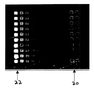
dots is printed directly adjacent to the test dots, as shown in Figure 2. This
second set
of dots (22) consists of decreasing per dot concentration of known
concentration of
analyte. Thus the assay, when carried out on the device, has now been reduced
to a
single step assay, with no need for intermediary washing steps to remove
excess anti-
analyte antibody, as the excess pre-conjugated label antibody background
concentration is effectively reduced by binding to the predominant spots of
calibration
antigen.
When the test fluid containing the analyte-antibody-dye complex as well as
surplus
2o anti-analyte-antibody-dye complex, is flowed over the two types of dots
printed in the
assay device, the surplus complex binds with the known analyte spots to create
the
reference concentrations for calibration when the dye concentration is read in
a
reader. The analyte-antibody-dye complex binds with the capture antibody spots
and
provides a reading for the to be determined concentration of analyte in the
test
solution. This method of internal dynamic calibration (TDCTM) integrates a
comparative external calibration standard curve with the comparative test
curve as
shown in Figure 3. The actual concentration of test fluid analyte is
accurately
determined because the test specific internal calibration standard is
simultaneously
provided.
CA 02475456 2004-07-20
The method when used in conjunction with the device provides an empirical
determination of the correct enhancement of charges induced on the assay
device for
sufficiently binding assay components without causing loss of detection signal
as a
result of high density binding events, i.e. number of bonds per unit surface
area of the
assay device.
In order to attain maximum sensitivity in the assay device of the present
invention, the
assay device has known concentrations of antigen in the calibration dots. The
increasing antigen concentrations are read during the assay as a result of
binding with
surplus antigen-antibody plus fluorescent label calibration complex. The
antigen is a
1o requisite protein and needs to be firmly attached to the preferably
polystyrene surface
of the assay device to prevent becoming soluble and being washed away during
the
course of the assay process. Attachment of a protein micro-array is optimum
with a
maximum of hydrophobic adsorption and minimal with excessive surface charges
on
the modified polystyrene surface.
Surprisingly, the degree of surface modification acts in reverse for antibody
adsorption as shown in Figure 4. Optimal density of surface charges binds
antibody
firmly to the charge-modified surface to such an extent that although antibody
is
present, no antigen binding events are found. As a direct consequence, no free
antigen-label complex is captured.
2o Direct binding of the antibodies to a polymer such as polystyrene results
in
conformational changes that reduce its affinity for the analyte. Proteins
passively
adsorb onto plastic surfaces. The proportion of protein bound can range from
5% to
95%, so careful optimization of the coating process is important. Binding of
protein to
plastic occurs because water molecules have a much stronger affinity for each
other
than for hydrophobic regions. The exclusion of hydrophobic sites from the
solution
causes parts of proteins to adsorb to plastic.
The solid support surface of the assay device is modified to have a balance of
hydrophilic enhancement for antibody binding and test fluid flow
characteristics
versus maintenance of hydrophobic binding for protein adsorption. Printing of
the
3o calibration and test dots on the modified surface is carried out under
constant
_g_
CA 02475456 2004-07-20
humidity control to ensure that the dots tend to minimal thickness leading to
the
formation of molecular layer thickness dots.
As shown in Figure 5, an internal calibration method of t:he assay consists of
multiple
repeats to support high confidence limits for accurate diagnosis. An effective
balance
of the applied parameters for attachment of the dots to the modified surface
is
confirmed by performing immuno-diagnosis of test sample fluid. When optimal
conditions for a specific assay have been confirmed, mass production commences
with quality control tests incorporated at required intervals.
Examples
l0 Example 1
Multiple arrays of spots were printed onto the modified solid support surface
substrate.
As shown in Figure 2, an embodiment of the assay device shows ten printed
columns
of calibration dots 22 of the same analyte concentration. 'fhe final two
columns of test
dots 20, on the right, are capture antibody dots with adsorbed test samples of
concentration to be determined. Each row represents a single assay with IDB
(internal
dynamic calibration). The test row is repeated 10 times. The entire test
matrix is 10
spots x 10 dots for IDB and 2 x 10 spots for test sample s. The measured
fluorescence
intensity determines the relative concentrations of analyte.
Example 2: Humidity Spot Size
Surface modified solid supports were tested for hydrophilic flow effects at 24
C at
18% humidity and 47% humidity. Dots were measured. for comparative increase in
diameter. Surface area increased by 51 % at higher humidity for the same
volume of
dot fluid, leading to an approximate 50% reduction in spot thickness.
Example 3: Dynamic Internal Calibration
As shown in Figure 3, curve A represents the typical variations in dose
response when
test samples are measured using an internal, generated concentration curve.
Curve B
is the derived external calibration line, erroneously interpolated as
determined by a
-9-
CA 02475456 2004-07-20
measured low dose external calibration reading and a high dose external
calibration
reference reading. The respective values in between the low and high points
are then
integrated by drawing a line i.e. curve B. The Internal Dynamic Calibration
produces
a line plot based on several plots resulting in a curve that represents the
actual
calibrated response of the test sample as in Figure 3.
Example 4. Device tested to compare the effect of high density surface charge
with
optimal surface modification with a balanced charge.
To compare dose response of optimized device solid support (upper "Optimal"
curve)
against highly charged solid support modified surface (lower "Charged" curve).
The
dynamic spot CK-MB analyze protein concentration ranged from 26 picogram/ml to
26,200 picogram/ml as shown in Figure 4. The highly charged surface area
effectively prevented analyze binding at concentrations less than 2620
picogram/ml.
Example 5: Method Applied to Production Test Device
Figure 5 illustrates a typical device surface, modified to show comparative
print array
matrices and to confirm adequate surface modification of solid support for
both
analyze and antibody adsorption. The method compares relative dot sizes with
increment in dispensed spot volume.
Those skilled in the art will recognize, or be able to .ascertain using no
more than
routine experimentation, many equivalents to the embodiments of the invention
2o described specifically above. Such equivalents are intended to be
encompassed in the
scope of the following claims.
-10-