Note: Descriptions are shown in the official language in which they were submitted.
CA 02475690 2004-08-10
WO 03/074735 PCT/IB02/00648
1
Diagnostic method for Glaucoma
Technical Field
The present invention relates to a method for
the diagnosis and/or prediction of glaucoma as well as to
an array of nucleic acid probes.
Background Art
Glaucoma is an optic neuropathy in which some
retinal ganglion cells (RCG) die through an apoptotic
process.
Primary Open Angle Glaucoma ("POAG") is the
most common form of glaucoma. The disease is character-
ized by the alteration of the trabecular meshwork, lead-
ing to obstruction of the normal ability of aqueous humor
to leave the eye without closure of the space. A charac-
teristic of such obstruction in this disease is an in-
creased intraocular pressure ("IOP"), resulting in pro-
gressive visual loss and blindness if not treated appro-
priately and in a timely fashion.
Another form of glaucoma is characterized by
progressive optic nerve damage and visual field loss with
a statistically normal intraocular pressure (IOP < 21 mm
Hg). This form of glaucoma is classified as normal ten-
sion glaucoma (NTG).
In the past, different diagnostic in vivo and
ex vivo methods for the diagnosis of glaucoma has been
3o described.
Patent application WO 98/44108 discloses in
vivo and in vitro methods for diagnosing glaucoma wherein
said methods are based on the determination of the ex-
pression of a trabecular meshwork induced glucocorticoid
response protein (TIGR).
Patent application WO 98/36098 describes an
in vitro method for the diagnosis of glaucoma based on
CA 02475690 2004-08-10
WO 03/074735 PCT/IB02/00648
2
the detection of a mutation in the gene cytochrome
P450B1.
Although the above identified prior art de-
scribes diagnostic methods for glaucoma, there is cur-
s rently no method available allowing an exact identifica-
tion of patients with a predisposition for glaucoma de-
velopment or for the progression of the disease.
There exists therefore an urgent need for a
reliable method for the diagnosis and/or prediction of
1o glaucoma and for means suitable for the use in said
method.
Disclosure of the Invention
15 Hence, it is a general object of the inven-
tion to provide an ex vivo method for the diagnosis
and/or prediction of glaucoma. Said method comprises de-
tecting in a tissue and/or blood sample of a human indi-
vidual an altered gene expression pattern of genes se-
20 lected from at least the group of genes related to tissue
remodeling.
The term "altered,gene expression" encom-
passes an increased gene expression as well as a de-
creased gene expression of genes of interest compared to
25 an average gene expression level observed in healthy sub-
jects. The determination of the health state of a person
is usually based on the subjective health state descrip-
tion of the patient, an interview by a physician and a
physical examination of the patient.
3o The term glaucoma as used herein comprises
all forms of glaucoma observed in the clinics.
In a preferred embodiment of the method said
gene expression pattern further comprises genes selected
from the following gene groups: genes related to DNA re-
35 pair, genes related to cell adhesion, genes related to
ischemia/reperfusion injury or genes in consequence of
the glaucomatous damage.
CA 02475690 2004-08-10
WO 03/074735 PCT/IB02/00648
3
In a further preferred embodiment of the
method said gene expression pattern comprises a total of
at least 4 genes, wherein at least one gene from each of
the four gene groups. In a more preferred embodiment said
gene expression pattern comprises a total of at least 8
genes, wherein at least 2 genes from each of the four
gene groups.
It has to be understood that any combination
of genes of said four gene groups is suitable for the use
in the method according to the present invention. It is
e.g. possible to use 2 genes of the first group, 1 gene
of the second group, 4 genes of the third group and 3
genes of the fourth group.
Said altered gene expression of the genes of
interest is preferably determined at the transcriptional
level.
Preferred genes of the gene group which re-
late to tissue remodeling are the following genes:
metalloproteinases, metalloproteinase inhibi-
2o tots and proteinase 3.
Preferred genes of the group of genes related
to DNA-repair are the following genes:
XPGC, 14-3-3 6 (Stratifin), p53, MDR-X (ABC
(ATP-binding cassette)-transporter), survivin, DEAD box X
isoform protein (DBX), X-linked retinopathy protein, STMT
gene familial Alzheimer's disease, MRCK (myotonic dystro-
phy kinase-related cdc42 binding kinase), thioredoxin,
NFkappB, inhibitor of apoptosis protein 1 (HIAP1, API1),
IAP homolog C, TNFR2-TRAF signaling complex protein,
MIHC, cyclin A1, guanine nucleotide-binding-protein
G(I)/G(S)/G(T)beta subunit 1 (GNB1), transducin beta-1
subunit.
Preferred genes of the gene group which re-
late to cell adhesion are the following genes:
E-cadherin, cytochrome P450, cyclooxygenase-
2, rho GDP dissociation inhibitor 1, rho GDI alpha,
ARHGDIA, thymosin beta, VEGEFR 1, tyrosine protein kinase
CA 02475690 2004-08-10
WO 03/074735 PCT/IB02/00648
4
receptor SFLT, Phospholipase C gamma 1, 1-
phosphatidylinositol-4,5-bisphosphate-phosphodiesterase
gamma 1, PLC-II, PLC-148, 68 kDa type I phosphatidyl-
inositol-4-phosphate-5-kinase alpha kinase, 1-
phosphatidylinositol-4-phosphate kinase, diphospho-
inositide kinase, G protein-activated inward rectifier
potassium channel 3, KIR 3.3, guanine nucleotide-binding
protein G(I)/G(S)/G(T) beta-subunit 1, transducin beta-1
subunit, Rac alpha serine/threonine kinase, protein ki-
nase B, c-akt, akt 1.
Preferred genes of the group of genes related
to ischemia/reperfusion injury or genes in consequence of
the glaucomatous damage are the following genes:
20S proteosome, NTP, Jun-D, c-jun N-terminal
kinase (JNKK), JNK activating kinase 1 (JNKK1), MAP ki-
nase 4 (MKK4), SRp20 splicing factor, lymphocyte-IgE-
receptor, thromboxan A2 receptor, Na+/K+-ATPase, ITK, al-
kal. phosphatase.
In a particular preferred embodiment of the
2o present invention said altered gene expression is deter-
mined in white blood cells, preferably peripheral lympho-
cytes, monocytes and stem cells.
Another object of the present invention is an
array of nucleic acid probes immobilized on a solid sup-
port, wherein said array comprises nucleic acid probes of
genes selected from the group of genes related to tissue
remodeling.
Said array according to the present invention
preferably further comprises nucleic acid probes of genes
3o selected from the following gene groups: genes related to
DNA repair, genes related to cell adhesion, genes related
to ischemia/reperfusion injury or genes in consequence of
the glaucomatous damage.
In a preferred embodiment said array com-
prises nucleic acid probes of genes selected from each of
the above identified four gene groups.
CA 02475690 2004-08-10
WO 03/074735 PCT/IB02/00648
Another preferred embodiment relates to an
array which only comprises nucleic acid probes of genes
selected from the four above defined gene groups.
An array according to the present invention
5 can be used in an ex vivo method for the diagnosis andlor
prediction of glaucoma, preferably in a method for the
diagnosis and/or prediction of glaucoma according to the
present invention.
A further object of the present invention is
1o the use of genes of the above defined three gene groups
for the somatic gene therapy of glaucoma.
Brief Description of the Drawings
The invention will be better understood and
objects other than those set forth above will become ap-
parent when consideration is given to the following de-
tailed description thereof. Such description makes refer-
ence to the annexed drawings, wherein:
2o Figure 1 shows the results of a Comet assay,
Figure 2 shows the results of a dot blot as
say (1: glaucoma patients, 2: healthy controls),
Figure 3 shows the results of a RT-PCR ampli-
fication of XPGC transcripts,
Figure 4a shows the results of a RT-PCR am-
plification of (3-actin transcripts in control individu-
als,
Figure 4b shows the results of a RT-PCR am-
plification of (3-actin transcripts in glaucoma patients,
Figure 4c shows the results of a RT-PCR am-
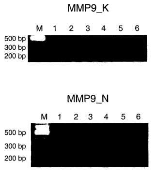
plification of maxtrix-metalloproteinase 9 (MMP-9) tran-
scripts in control individuals,
Figure 4d shows the results of a RT-PCR am-
plification of maxtrix-metalloproteinase 9 (MMP-9) tran-
scripts in glaucoma patients,
CA 02475690 2004-08-10
WO 03/074735 PCT/IB02/00648
6
Figure 4e shows the results of a RT-PCR am-
plification of membrane type maxtrix-metalloproteinase 1
(MT1-MMP) transcripts in control individuals,
Figure 4f shows the results of a RT-PCR am-
plification of membrane type maxtrix-metalloproteinase 1
(MTZ-MMP) transcripts in glaucoma patients,
Figure 4g shows the results of a RT-PCR am-
plification of metalloproteinase inhibitor 1 precursor 1
(TIMP-1) transcripts in control individuals,
Figure 4h shows the results of a RT-PCR am-
plification of metalloproteinase inhibitor 1 precursor 1
(TIMP-1) transcripts in glaucoma patients,
Figure 5a shows a restriction analysis of the
209 by PCR fragment of (3-actin,
Figure 5b shows a restriction analysis of the
289bp MMP-9 PCR fragment,
Figure 5c shows a restriction analysis of the
295bp MT1-MMP PCR fragment and
Figure 5d shows a restriction analysis of the
393bp TIMP-1 PCR fragment.
Modes for Carrying Out the Invention
The determination of expression patterns of
specific genes according to the method of the present in-
vention allows an exact diagnosis of patients with glau-
coma as well as an exact identification of patients with
a predisposition for chronic glaucoma development or for
3o the progression of the disease. The method of the present
invention offers a number of diagnostic advantages. For
example, said method is highly sensitive and minimal-
invasive by just taking/collecting a small tissue sample
and/or a small amount of a body fluid, in particular
blood, from a patient.
The man skilled in the art knows suitable
gene expression detection methods which can be employed
CA 02475690 2004-08-10
WO 03/074735 PCT/IB02/00648
7
in a method of the present invention. Said methods com-
prise e.g, northern blot analysis, RT-PCR, real time
quantitative PCR, immunohistochemical methods, ELISA, Dot
blot analysis.
In a preferred embodiment the gene expression
level is determined at the transcriptional level i.e. the
amount of a RNA transcript is determined. A biological
sample of a patient e.g. a tissue sample, preferably
blood, is processed to isolate mRNA using one of the es-
1o tablished methods for mRNA isolation and purification.
The RNA is preferably isolated from peripheral blood leu-
kocytes. The isolated mRNA is then transcribed to a DNA
in a reaction with a reverse transcriptase. The resulting
cDNA can then be analyzed by the method of the present
invention. A particularly suitable method for the use in
the present invention is RT-PCR which allows a fast de-
termination of expression levels of genes. The primers
for the RT-PCR are preferably chosen so that a non-
conserved region of the genes are amplified.
2o A preferred means for the detection of said
RNA transcripts is a DNA microarray. The construction of
DNA microarrays and their use is well known in the art.
For references see e.g. DNA Microarrays: A practical ap-
proach, Edited by M. Schena, Oxford University Press, 0x-
ford, UK, 1999; Lemieux et al., Overview of DNA Chip
Technology, Molecular Breeding 1998, 4, p. 277-289; and
the Internet site http://www.gene-chip.com and references
cited therein.
An array comprises nucleic acid probes immo-
3o bilized on a solid support. The term "nucleic acid probe"
as used herein encompasses single stranded nucleic acids
capable of binding to a target nucleic acid of complemen-
tary sequence by base pairing e.g. oligonucleotides, par-
tial or complete cDNAs. A nucleic acid probe can include
natural or modified bases. Nucleic acid probes can be be-
tween 10-500, 10-250, 10-150, 10-75, 10-50 and 10-25
bases long. The specific length of the used probes de-
CA 02475690 2004-08-10
WO 03/074735 PCT/IB02/00648
8
pends on the specific gene and said length has to be de-
termined for each gene by the man skilled in the art.
In a preferred embodiment said nucleic acid
probes stem from non-conserved domains of the protein of
interest, more preferably from a non-conserved N-terminal
domain or a non-conserved C-terminal domain of the pro-
tein of interest.
An exemplary embodiment of the method accord-
ing to the present invention using a DNA array comprises
1o the following steps: preparation of a sample of nucleic
acids, hybridization of the sample of nucleic acids to an
array, detection of hybridized nucleic acids and analysis
of hybridization patterns.
Nucleic acid sample preparation typically in-
cludes the following steps: mRNA isolation and purifica-
tion from a tissue and/or a body fluid sample, reverse
transcription to cDNA and optionally second strand syn-
thesis. Synthesized cDNA is typically labeled. Label can
e.g. be introduced by one of the nucleotides being incor-
2o porated. Detectable labels suitable for use include e.g.
spectroscopic, photochemical, biochemical or immunochemi-
cal means.
In one method of detection, denatured labeled
nucleic acid derived from mRNA of the sample is applied
to an array. Said nucleic acid hybridizes to complemen-
tary probes immobilized on the array and hybridization is
identified by detecting label. The position of label is
detected for each probe in the array and the concentra-
tion of each sequence that is complementary to a probe on
3o the array is determined by measuring e.g. the fluores-
cence intensity using a reader. Comparison of the hy-
bridization pattern of a patient sample to a control sam-
ple indicates which probes hybridize to nucleic acid
strands that derive from mRNAs that are differentially
expressed between the two samples. An expression pattern
of the patient sample differing from the expression pat-
CA 02475690 2004-08-10
WO 03/074735 PCT/IB02/00648
9
tern of the control is indicative for glaucoma or a pre-
disposition for glaucoma.
Genes of the above defined groups of genes
related to glaucoma or anti-sense oligonucleotides
thereof can be used for gene therapy of glaucoma.
For gene therapy a nucleic acid coding for a
protein of the above identified groups is introduced into
a suitable vector, preferably an adenoviral vector, al-
lowing the expression of a said protein in the addressed
1o target cells, preferably ganglion cells of the optical
nerve. Such a vector suitable for gene therapy and allow-
ing expression of the specific gene comprises the encod-
ing nucleic acid under the control of a target cell spe-
cific promoter. Gene therapy methods and vector systems
are e.g. described in Gene Therapy, T. Blankenstein, 1998
and Gene Therapy - from laboratory to the clinic, edited
by Kam M. Hui.
The invention is now further illustrated by
means of examples.
Experimental part
Purpose
In order to investigate specific differences
on the molecular level between patients with vasospastic
syndrom, Normal Tension Glaucoma (NTG) patients and High
Tension Glaucoma (HTG) patients have been compared to
healthy controls.
Results
1. Comet Assay
Evaluation of the initial DNA damage. Di-
rectly after thawing of the vital cells the majority (55
0) of the healthy controls exhibited a slight degree in
DNA damage (class 2), while only 5 0 of this group show
severe DNA damage (class 4). No DNA damage (class 1)
CA 02475690 2004-08-10
WO 03/074735 PCT/IB02/00648
could be observed in 25 0, while 15 0 of the healthy con-
trols could be classified for class 3. In NTG patients a
shift towards a higher degree of DNA damage could be ob-
served. Cells derived from NTG patients could be mainly
5 detected in the state of class 3 (intermediate DNA dam-
age) with an percentage of 41 %. Compared to controls,
also the percentage of cells in the state with severe DNA
damage (class 4) increased up to 26 %. The amount of
cells without DNA damage remained rather stable (29),
10 while the amount of cells in the state class 2 decreased
to 4 %. The distribution pattern of HTG patients was
rather similar to the group of NTG patients: the majority
of the cells (47 %) exhibited a DNA damage of the inter-
mediate state (class 3), 21 0 of the cells exhibited no
DNA damage, 10 o a mild (class 2) and 22 o a severe DNA
damage (class 4). In figure 1 a distribution pattern of
NTG-patients and healthy controls is shown. A patient
with Ataxia telangiecasia served a positive control for
DNA damage.
Evaluation of the 7~NA damage after in vitro-
incubation. After thawing the cells got a regenerating
period of 3 hours at 37°C in phosphate buffered saline
(PBS). Afterwards, the status of the DNA was examined.
Compared to the evaluation directly after thawing the ma-
jority of the cells derived from healthy controls shifted
towards undamaged DNA (31 0; class 1) and mild DNA damage
(26 %; class 2), respectively. In addition, 27 % of cells
exhibited intermediate DNA damage (class 3) and 16 % se-
were DNA damage (class 4). In contrary, within the group
of NTG patients no increase of the amount of cells with
undamaged DNA could be observed. In average there. was
following distribution: 13 ~ with undamaged DNA (class
1), 29 % with mild DNA damage (class 2), 32 o with inter-
mediate DNA damage (class 3) and 26 o with severe DNA
damage (class 4). Within the group of HTG patients the
majority of the cells could exhibit the status of inter-
CA 02475690 2004-08-10
WO 03/074735 PCT/IB02/00648
11
mediate DNA damage (class 3) with 54 o and severe DNA
damage (class 4) with 36 0. In average, 10 0 of the cells
could be classified for class 1 (undamaged DNA) and 5
for class 2 (mild DNA damage).
2. Subtractive Hybridization and dot blots.
The subtracted cDNAs showed very similar pat-
tern for all NTG patients. The subtracted cDNAs have been
cloned and sequenced. The comparison of their sequences
1o with data Genbank revealed homologies with genes coding
for the following known proteins listed up in table 1 and
2. For better visualization of the different expression
pattern of NTG patients compared to healthy controls dot
blots were performed with 6 NTG patients and 6 healthy
controls. Results are presented in figure 2: compared to
controls NTG patients exhibited on the level of mRNA ex-
pression a slight increase in p53 as well as in 20S-
proteasome subunit, and a stronger increase in the ex-
pression of the neuronal thread protein (NTP). In con-
trary, mRNA expression decreased for XPGC (Xeroderma Pig-
mentosum group complementing factor), survivin and a new
identified gene NCR-X.
Table 1
Homology of the up-regulated genes in lympho-
cytes of NTG patients (hs-Homo sapiens)
Name of the gene EMBL Organism Length o similarity
of
accession the cDNA of amino acid
number (bP) sequence
p53 X02469 hs 796 100
cellular tumor antigen
20S-proteasome subunit
AF022815 hs 168 99
XAPC7
neuronal thread pro-
tein AD7c-NTP (with
Alu-repeats-containing
domains) related
to
Alzheimer's disease
AF 010144 hs 213 87
CA 02475690 2004-08-10
WO 03/074735 PCT/IB02/00648
12
Table 2
Homology of the down-regulated genes in lym-
phocytes of NTG patients (hs-Homo Sapiens)
Name of the gene E1~L Organism Length % similarity
of
accession the cDNA of amino
acid
number (bP) sequence
apoptosis inhibitorU75285 hs 312 100
survivin gene
XPGC gene X71347 hs 174 100 (cDNA)
hypothetical ABC
transporter ATP-
P44656 Haemophilus 327 g3
binding protein
HI0354; MDR-X
influenza
Amplification of the XPGC-Transcript. In ad-
dition to the dot blot experiments, RT-PCR was performed
to evaluate XPGC gene expression. As shown in figure 3
1o all NTG patients exhibited a lack of XPGC gene expres-
sion. In contrary, in all healthy controls - with the ex-
ception of one volunteer (no. 5) - XPGC expression was
detectable. Re-examination of volunteer no. 5 revealed a
glaucomatous excavation of the optic nerve head, however
the visual field was normal. This indicates that the per-
son is a) at risk for developing glaucomatous damage or
b) already suffers from preperimetry glaucoma.
3. Atlas cDNA Expression Arrays
The spectrum of screened genes of glaucoma
patients vs. healthy controls was extended by using cDNA
Expression Arrays in combination with an Imagine System.
The results revealed an altered gene expression of 92
genes in glaucoma patients compared to controls. 33 of
genes exhibited down-regulation, while 59 genes were up-
regulated. Focussing on metalloproteinases - a group of
proteins, which are essential for tissue remodeling -
following expression patterns of genes have been found to
3o be altered in leukocytes of glaucoma patients: up-
CA 02475690 2004-08-10
WO 03/074735 PCT/IB02/00648
13
regulation of matrix-metalloproteinase 9 (MMP-9) and mem-
brane-type matrix-metalloproteinase 1 (MT1-MMP), and dys-
regulation of metalloproteinase inhibitor 1 precursor 1
(TIMP-1). The results for MMP-9 and MT1-MMP could be con-
s firmed by subtractive hybridization and real time QPCR
(quantitative PCR) confirmed. Data of the screened genes
mentioned above are listed up in table 3:
Table 3
Name of gene Genbank Organism Length % simi-
of
the cDNA larity
of
accession (bh) amino
number
acid se-
quence
Matrix-metalloproteinaseBC006093 hs 683 100
9 (MMP-9; gelatinase
B;
92-kDa type IV collage-
nase precursor)
Membrane-type MAtrix-
metalloproteinase 1 gg3535 hs 239 100
(MT1-
MMP,MMP-14 precursor)
Metalloproteinase inhibi- done onlydone only
for 1 precursor 1 (TIMP-gp3124 hs bY expres-by ex-
1) sion arraypression
array
4. Confirmation of target gene expression us-
ing specific RT-PCR
Amplification of cDNA fragments of MMP-9 and
MT1-MMP by RT-PCR confirmed an induction of their expres-
sion in circulating leukocytes of glaucoma patients in
contrast to healthy controls (Fig. 4C to F). As an inter-
nal control for cDNA synthesis the housekeeping gene (3-
2o actin was amplified (Fig. 4A and B).
5. Restriction analysis
The amplification of the target PCR products
has been confirmed by restriction analysis. Restriction
analysis of the target RT-PCR products in 3 % "wide
range" agarose gel. In figure 5 lane 1 belongs to a non-
CA 02475690 2004-08-10
WO 03/074735 PCT/IB02/00648
14
digested amplification product; lane 2 and 3, and in ad-
dition 4 and 5 belong to amplifications products obtained
by digestion with selected endonucleases. Restriction
analysis was performed using AluI to get fragments with
58 and 151 basepairs ((bp); Fig. 5A), AvaI for fragments
with 45 and 164 bp, HaeIII for fragments 15, 44 and 159
bp, RsaI for fragments 77 and 132 bp, all from the 209 by
beta-actin amplification product. To get fragments from
the 289 by MMP-9 amplification product restriction analy-
1o sis was performed using AluI to get fragments with 40,
123 and 125 by fragments, HpaII to get fragments with 46
and 243 bp, PvuII to get fragments with 123 and 166, and
RsaI to get fragments with 142 and 147 by (Fig. 5B). To
get fragments from the 295 by MT1-MMP amplification prod-
uct restriction analysis was performed using HaeIII for
fragments with 6, 54, 58, 69 and 108 bp, and RsaI for
fragments with 125 and 170 by (Fig. 5C). To get fragments
from the 393 by TIMP-1 amplification product restriction
analysis was performed using HaeIII for fragments with
2,30,96 and 265 bp, HindII for fragments with 154 and 239
bp, HpaII for fragments with 152 and 241 by and PstI for
fragments with 28, 29 and 336 (Fig. 5D).
5. Quantitative analysis of gene expression
using real-time PCR
Relative gene expression was calculated bas-
ing on the individual CT values of genes of interest and
the housekeeping gene (3-actin. Although in contrast to
healthy volunteers, the leukocytes of all glaucoma pa-
tients demonstrated an expression of the MMP-9 gene, the
transcription level differs up to 5 times among the ex-
treme cases. Also the transcriptional level of TIMP-1 is
very heterogeneous for these patients and differs from
sample to sample up to 25x. Furthermore there is no cor-
relation in increase of transcription between MMP-9, and
TIMP-1. MT1-MMP is highly expressed in 5 glaucoma pa-
tients and weak expressed in one patient.
CA 02475690 2004-08-10
WO 03/074735 PCT/IB02/00648
Conclusion
1. Depending on the degree of fragmented DNA
normal tension glaucoma (NTG) patients show a less suffi-
5 cient ability of DNA repair compared to normals.
2. These patients also differ from normals in
the expression pattern of various genes. The genes be-
longs to the gene families involved in a) tissue remodel-
1o ing, b) DNA repair, c) ischemia-reperfusion and d) adhe-
sion.
Materials and Methods
15 1. Blood samples
Blood samples were collected from patients
with NTG and HTG as well as from healthy controls. All
glaucoma patients had bilateral typical glaucomatous op-
tic nerve head cupping and visual field defects. In NTG
2o patients intraocular pressure (IOP) never exceeded 21 mm
Hg, but after local cooling of the fingers all these NTG
patients exhibited a stop in blood flow for more than 20
sec, which was detected by nailfold capillaromictroscopy
(indicative for vasospasm). In contrast, HTG patients ex-
hibited an IOP higher than 21 mm Hg, but no vasospastic
response. Ophthalmological examination of healthy con-
trols yielded unremarkable results and also no vasospas-
tic response. No patient had received either a systemic
or a locally applied ocular therapy at least four weeks
3o before blood draw.
2. Leukocyte isolation
Leukocytes were isolated from heparinized
blood by density gradient centrifugation as previously
described (Kalmar et al., 1988). After isolation pellets
of PBS-washed leukocytes were stored at -70°C either as
CA 02475690 2004-08-10
WO 03/074735 PCT/IB02/00648
16
dry pellet or frozen in DMSO-containing culture medium as
vital cells.
3. Comet assay
Sample preparation. The rate of cells con-
taining fragmented DNA was evaluated by the use of a
Comet assay. The principle of Comet assay (Trevigen INC.,
USA) or single cell electrophoresis is based on the abil-
ity of denatured, cleaved DNA fragments to migrate our of
1o the cells under the influence of an electric field. Un-
damaged DNA migrates slower and remains within the con-
fines of the nucleus when current is applied. Evaluation
of the DNA "comet" tail shape and migration pattern al-
lows for assessment of DNA damage (Fig. 1). In detail the
method was described by Ostling & Johanson (1984). In
brief, isolated leukocytes in a density of 200-300 cells
per sample were immobilized in a bed of low melting aga-
rose. After cell lysis samples were treated with alkali
to unwind and denature the DNA and hydrolyze sites of
2o damage. After electrophoresis samples were stained with
SYBR Green, a fluorescent DNA intercalating dye.
Sample analysis. The comets were visualized
under the fluorescent microscope (Olympus) at a magnifi-
ration of 200x. Excitation wavelength of 515-560 nm and a
barrier filter for 590 nm were used. At least 100 comets
were analyzed for each data point. For quantification of
the DNA damage the total length of the comet (head and
tail) were measured and the degree of the damage was cal-
culated according to the criteria of McKelvey-Martin et
al. (1993). The degree of the damage was assigned to 4
classes (1-4) based on the visual aspect of the comets,
considering the extent of DNA migration (Visvardis et
al., 1997). As shown in figure 1 comets with a bright
head and no tail were classified as class 1 (intact DNA)
while comets with a small head and a long diffuse tail
were classified as class 4 (severely DNA damage). Inter-
CA 02475690 2004-08-10
WO 03/074735 PCT/IB02/00648
17
mediate characteristics were assigned to class 2 and 3.
Cell loss greater then the average calculated for healthy
donors was assigned to class 5. Initial DNA damage (DD)
and DNA damage after incubation in BPS for 3 hours at
37°C (DD3) were estimated quantitatively using the modi-
fied equation (1) described by Jaloszynski et al. (1997):
DD = (nz + 2n3 + 3n4 + 4n5) / (S/100) ,
where DD: DNA damage, n2 - n4: amount of calculated comets
in class 2, 3 and 4, respectively; S: total number of
scored comets including class 1.
Statistical analysis. Initial DNA damage and
repair capacity after 3 hours of incubation were compared
between three groups by nonparametric two-way ANOVA. All
statistical analysis were done using the Graphpad Prism
software (version 2.01). Statistical significance was
calculated by the two sided, unpaired Student's t-test.
4. "Gene hunting" by subtractive hybridiza-
tion
Isolation of mRNA. Isolation of mRNA was per-
formed using the Quick Prep Micro mRNA Purification Kit
(Pharmacia Biotech, Uppsala, Sweden) according to the
manufactures protocol. Quality-check was performed by
First-strand cDNA Synthesis Kit (Pharmacia Biotech,
Uppsala, Sweden) using the incorporation of [cx-32P] dATP
(Amersham, Buckinghamshire, UK) with subsequent electro-
phoresis on a 1 % agarose gel followed by autoradiography
(Sambrock et al., 1981). The reflection film (NEN Life
Science Products) was exposed to the gel four 2 hours at
room temperature.
Construction of the subtractive library. In
principle, construction of the subtractive library is
CA 02475690 2004-08-10
WO 03/074735 PCT/IB02/00648
18
based on the cloning of the transcripts (in form of cDNA)
of those genes, which are activated/suppressed to become
up-or down-regulated under pathological conditions. To
identify these genes two pools of transcripts are used:
the complete pool of mRNA from patients and the complete
pool of mRNA from healthy controls. Both pools of tran-
scripts - called induced and uninduced pool of mRNA - un-
dergo the molecular biological comparison with the subse-
quent subtraction of the difference between two pools
representing the transcripts activated or suppressed due
to the disease. Construction of the subtractive library
was done as follows: mRNA from leukocyte samples and con-
trols were biotinylated by W radiation according to the
instruction manual of the Subtractor Kit (Invitrogen,
Leek, NL). To avoid false positive results the mRNA of
the uninduced pool was added in excess. Equal quantity of
each mRNA pool was subjected to reverse transcription
with subsequent denaturation of mRNA-template. Each newly
synthesized cDNA pool (induced pool) was hybridized with
2o the corresponding uninduced biotinylated mRNA-pool at
68°C for 48 hours. The hybridization mixture was incu-
bated with streptavidin and thus all the biotinylated
molecules (uninduced as well as RNA/DNA hybrids) were
complexed with streptavidin. The streptavidin nucleic
acid-complexes were removed by phenol-chloroform extrac-
tion and subtracted cDNAs were precipitated with ethanol
(Sine & John, 1988). For each pair of NTG patient/control
both pools were subtracted: the induced "NTG-genes" and
the induced "normal genes". The 2nd strand cDNA synthesis
was performed with the cDNA Synthesis Kit (Boehringer
Mannheim, FRG). The aim of the constructed libraries was
to compare gene expression individually and in the groups
of NTG-patients with healthy controls. Only those genes
which have been subtracted from both the individual pairs
and the corresponding groups have been considered as
relevant.
CA 02475690 2004-08-10
WO 03/074735 PCT/IB02/00648
19
Cloning of subtracted cDNA. Subtracted cDNAs
were cloned by using the pSPORT 1 cloning vector (GIBCO,
Life Technologies, Eggenstein, FRG) according to the
cloning methods described by Sambrook et al. (1989). To
enable visualization of the subtracted cDNAs the cloned
cDNAs were amplified using the universal primers I-5'
GTAAAACGACGGCCAGT 3' (Seq. Id. No. 1) and II-5'
ACAGCTATGACCATG 3' (Seq. Id. No. 2) restricting the mul-
tiple cloning site of pSPORT 1 vector. The amplificates
were analyzed in a 1 % agarose gel. The corresponding
cDNAs were cut off from the gel cleaned with the DNA
Clean Kit (AGS, Heidelberg, FRG) and recloned in Sma I-
site of pUC 18 vector. The recombinant molecules were
used for transformation in INVaF Eschericia coli cells
(Invitrogen, Leek, NL). Recombinant plasmid DNAs were
analysed for the length of the inserted fragment using
restriction analysis. Plasmid DNAs were purified using
QiaFilter Plasmid Midi System (Quiagen, Hilden, FRG).
Plasmid DNAs were sequenced by MWG-Biotech (Ebersberg,
2o FRG) .
Gene identification. Homologies were deter-
mined by computer assisted comparison of data with DNA
and protein gene banks (EMBL and SWISS-PROT, Heidelberg,
FGR). Alignments were prepared using "DNASIS"-programs
from MWG-Biotech (Ebersberg, FRG).
Dot blot analysis. For the quantification of
the specific transcripts the individual cloned and se-
3o quenced cDNAs have been used as specific labeled probes
for dot blot hybridization. After cloning and purifica-
tion each probe was denatured prior labelling at 95°C for
5 min and subsequently labeled with fluorescein-12-dUTP
using the Renaissance Random Primer Fluorescein-12-UTP
Labeling Kit (NEN Life Science Products). Aliquots of the
isolated mRNA-pools have been applied for the hybridiza-
tion with the specific probes using dot blots technique
CA 02475690 2004-08-10
WO 03/074735 PCT/IB02/00648
according to the protocol of White & Bancroft (1982). In
brief, samples of the mRNA-pools were placed onto a posi-
tively charged nylon membrane. After fixation, the nylon
membrane was incubated in a pre-hybridization solution
5 containing a block reagent (NEN Life Science Products)
for 3 hours at 65°C in a hybridization oven. The membrane
was hybridized step-wise with each labeled probe over-
night at 65°C. After hybridization, non-specifically
bound material was removed by washing the membrane two
1o times with pre-hybridization butter. The membrane was
then blocked with blocking reagent and incubated with an-
tifluorescein HRP-antibody (1:1000; (NEN Life Science
Products) for 1 hour at 37°C. After washing the membrane
was incubated in Nucleic Acid Chemiluminiscence Reagent
15 (NEN Life Science Products) for 1 hour and afterwards ex-
posed to an autoradiography reflection film (NEN Life
Science Products) for 1 hour at room temperature. Each
hybridization was performed in the same manner. Between
the individual hybridisations the membrane was stripped
20 according to the manufacturer's protocol. Densiometry of
the films was performed using a densiometer and the quan-
tification software program from MTniG-Biotech (FRG) .
Statistical analysis. The ANVA with subse-
quent Kruskal Wallis Test and Student's t-test were ap-
plied and linear regression analysis was performed. The
level of significance was at p < 0.05.
5. "Gene hunting" by hybridization of cDNA
probes to expression arrays
AtlasTM Human 1.2 Array (Clontech, Palo Alto,
USA) designed for the evaluation of different molecular
expression patterns was used. The AtlasTM Human 1.2 Array
includes 1176 human cDNAs, nine housekeeping control
cDNAs, and negative controls all immobilized on a nylon
membrane. Synthesis of cDNA from isolated mRNA derived
CA 02475690 2004-08-10
WO 03/074735 PCT/IB02/00648
21
from glaucoma patients and healthy controls was performed
using the First-Strand cDNA Synthesis Kit from Pharmacia
(Uppsala, S). After synthesis and labeling with fluores-
cein-12-dUTP (see above) the AtlasTM Human 1.2 Arrays was
hybridized with each individual labeled cDNA probe. After
hybridization non-specific bound material was removed by
several washing steps and the membrane was then blocked
with blocking reagent. Afterwards, the membrane was incu-
bated with anti-fluorescein HRP-antibody (NEN Life Sci-
1o ence Products) for 1 hour at 37°C followed by a washing
procedure and the incubation with the chemiluminiscence
reagent (NEN Life Science Products). Then, the membrane
was exposed to an autoradiography reflection film (NEN
Life Science Products) for 1 hour at room temperature.
Evaluation was performed using the Atlas Image 2.0 soft-
ware developed specifically for the analysis of Atlas
cDNA Expression Arrays (Clontech, Palo Alto, USA).
6. Polymerase Chain reactions
Reverse Transcriptase Polymerase Chain reac-
tion (RT-PCT). In order to detect qualitatively an ex-
pression of the target genes and to optimize individual
reaction conditions for the Real-Time Quantitative PCR
(see below) RT-PCR was performed with specific primers
designed for MMP-2, MMP-9, MT1-MMP and TIMP1 genes. Syn-
thesis of cDNA from isolated mRNA derived from glaucoma
patients and healthy controls was performed using the
First-Strand cDNA Synthesis Kit from Pharmacia (Uppsala,
3o S). PCRs were performed using a hot-start Taq-polymerase
(Abgene, Hamburg, FRG) and were run for 35 cycles. PCRs
without DNA served as negative controls and PCRs with se-
quences templates as positive controls. For analysis
ethidium bromide-stained PCR products, which had been
separated in an agarose gel, were visualized under UV il-
lumination.
CA 02475690 2004-08-10
WO 03/074735 PCT/IB02/00648
22
Real-Time Quantitative PCR (RT-QPCR). In or-
der to profile changes in the expression of genes of in-
terest RT-QPCR has been performed by using SYBER Green I
as intercalation dye and fluorescent reporter molecule to
detect the accumulation of amplified double-stranded
products in an iCycler iQTM Detection System (Bio-Rad
LAboratories, USA). RT-PCR was performed as described
above, only with one exception: hot-red Taq-polymerise
(Abgene, Hamburg, FRG) was substituted by Taq DNA poly-
1o merase (Roche, CH) to avoid color signal disturbances.
The algorithm of the iCycler iQTM Detection System normal-
izes the reporter signal (non-intercalated SYBER Green)
to a passive reference and multiplies the SD of the back-
ground signal in the first few cycles by a defaulter faC-
for of 10 to determine the threshold. The cycle at which
the baseline level is exceeded is defined as threshold
cycle (CT). CT depends on the initial template copy number
and is proportional to the log of the starting amount of
nucleic acid (Held et al.). By subtracting differences of
2o the CT values between the genes of interest and the
housekeeping genes ((3-actin) the data have been normal-
ized. Relative levels were calculated for gene expression
in NTG samples to control samples based on the differ-
ences in CT values (Held et al., 1996).
Statistical analysis. All values are ex-
pressed as mean ~SEM. Values were compared using Stu-
dent's t-test for parametriC data. A p value of less than
0.05 was considered as significant.
Restriction analysis. Target PCR products
were identified using specific restriction analysis. The
target amplification products underwent an extraction
from the agarose gel using the DNA isolation kit (DNA-
Clean, Hybaid-AGS, FRG) before digestion. They were di-
gested in a final volume of 50 ~,l with 20 units of each
restriction endonuclease for 4 hours, according to the
CA 02475690 2004-08-10
WO 03/074735 PCT/IB02/00648
23
protocol of the manufacturer (Roche, CH). Digested DNA
fragments were separated in an agarose gel and visualized
after staining with ethidium bromide by UV-light (Fig.
5) .
While there are shown and described presently
preferred embodiments of the invention, it is to be dis-
tinctly understood that the invention is not limited
thereto but may be otherwise variously embodied and prac-
ticed within the scope of the following claims.
CA 02475690 2004-08-10
WO 03/074735 PCT/IB02/00648
24
References
Heid CA, Stevens J, Livak KJ, Williams PM.
Real time quantitative PCR. Genome Res 1996 Oct; 6(10):
986-94
Jaloszynski P, Kujawski M, Czub-Swierczek M,
Markowska J, Szyfter K, Bleomycin-induced DNA damage and
its removal in lymphocytes of breast cancer patients
studied by comet assay. Mutat Res 1997 Dec; 385(3): 223-
33
Kalmar JR, Arnold RR, Warbington ML, Gardner
MK. Superior leukocyte separation with a discontinuous
one-step Ficoll-Hypaque gradient for the isolation of hu-
man neutrophils. J Immunol Methods 1988 Jun 13;110(2):
275-81
McKelvey-Martin VJ, Green MH, Schmezer P,
Pool-Zobel BL, De Meo MP, Collins A. The single cell gel
electrophoresis assay (comet assay): a European review.
Mutat Res 1993 Jul; 288/1):47-63
Ostling O, Johanson KJ. Microelectrophoretic
study of rediation-induced DNA damages in individual mam-
malian cells. Biochem Biophys Res Commun 1984 Aug 30;
123(1): 291-8
Sambrok J, Fritsch EF, Maniatis T. Molecular
cloning: A laboratory manual. In: Cold Spring Harbor
Laboratory, Cold Spring Harbor. 1989; second edition:
1.53-1.73
Sive HL & St John T. A simple subtractive hy-
bridization technique employing photoactivatable biotin
3o and phenol extraction. Nucleic Acids Res 1988 Nov 25;
15(22): 10937
White BA & Bancroft FC. Cytoplasmic dot hy
bridization. Simple analysis of relative mRNA levels in
multiple small cell or tissue samples. J Biol Chem 1982
Aug 10; 257(15): 8569-72
Visvardis EE, Tassiou AM, Piperakis SM. Study
of DNA damage induction and repair capacity of fresh and
CA 02475690 2004-08-10
WO 03/074735 PCT/IB02/00648
Cryopreserved lymphocytes exposed to H202 and gamma-
irradiation with the alkaline comet assay. Mutat Res 1997
Jan 31; 383(1): 71-80.
CA 02475690 2004-08-10
WO 03/074735 PCT/IB02/00648
SEQUENCE LISTING
<110> Flammer, Josef
<120> Diagnostic method for glaucoma
<130> 05750PC
<160> 2
<170> PatentIn version 3.1
<210> 1
<211> 17
<212> DNA
<213> Artificial Sequence
<220>
<223> forward PCR Primer
<400> 1
gtaaaacgac ggccagt 17
<210> 2
<211> 14
<212> DNA
<213> artificial sequence
<220>
<223> reverse PCR Primer
<400> 2
acagctatgc catg 14
Page 1