Note: Descriptions are shown in the official language in which they were submitted.
CA 02476817 2004-08-18
WO 2003/070085 PCT/US2003/005019
-1-
DEVICE AND METHOD FOR INTERNAL LIGATION
OF TUBULAR STRUCTURES
TECHNICAL FIELD
The present invention relates to methods for blocking tubular anatomical
structures. In particular, the present invention relates to methods for
ligating the
fallopian tube to achieve sterilization. The present invention pertains in
addition to
devices for performing tubal ligations.
BACKGROUND
Occlusion of tubular anatomical structures is desirable for various medical
treatments. One important application of occlusion techniques is blockage of
the
fallopian tubes in the female or vas deferens in the male to achieve
sterilization and
prevent undesired pregnancies.
Various methods for producing occlusion or blockage of tubular anatomical
structures have been considered for contraceptive purposes. A cormnonly used
method
for blocking the fallopian tube is to tie off or clamp the fallopian tube. The
tube may be
tied in two locations and the intermediate portion of tube removed. A similar
result
may be obtained by grasping and folding over a portion of the tube and tying
off a loop
of tube that does not communicate with the remainder of the tube. The folded
segment
of tube may be blocked by a loop of suture material, a elastic ligating band
or O-ring, or
a clamp. Access to the fallopian tube is usually gained through endoscopic
surgery,
either through the abdominal wall or, less commonly, through the wall of the
vagina.
Such methods are less invasive than conventional surgical methods, but still
have an
undesirably high rislc of infection and tissue damage, and are accompanied by
an
undesirable recovery time and level of discomfort.
To eliminate the need for endoscopic or other, more invasive, surgery, a
number
of approaches have been devised for blocking the lumen of the fallopian tube
after
accessing the interior of the fallopian tube by inserting a catheter into the
lumen of the
tube via the vagina and uterus.
CA 02476817 2004-08-18
WO 2003/070085 PCT/US2003/005019
-2-
One approach is to block the fallopian tube by inj ecting an adhesive or
sealant,
typically a polymeric material, into the fallopian tube to form a plug.
Another approach
is to insert a pre-formed occlusive device or plug into the lumen of the
fallopian tube or
the utero-tubal junction. However, either type of plug may separate or
dislodge from
the wall of the fallopian tube, resulting in unreliable or impermanent
blockage.
Another approach for blocking the fallopian tube or other tubular anatomic
structures is to induce the formation of sclerosis or scar tissue to block the
tube. Tissue
damage may be induced chemically or thermally. However, this method is
relatively
difficult to accomplish successfully and requires skilled personnel and
specialized
equipment, malting it unsuited for use in certain settings.
Improvements over the prior art desirably will provide a method and system for
applying a ligating structure to the interior of a tubular anatomical
structure. Desirable
improvements will cause a reliable occlusion of a tubular anatomical
structure. Such
occlusion of a tubular anatomical structure desirably is permanent in certain
applications, such as in reproductive contraception. An inexpensive method for
occluding a tubular anatomical structure is also desired. An improvement
mayprovide
a partially or completely disposable device for performing occlusion of a
tubular
anatomical structure. It would be a further advance to provide an improved
method for
performing tubal ligations which requires only minimally invasive surgery,
thereby
reducing damage to vascular and reproductive tissues and reducing post-
surgical
discomfort and recovery time. A method for performing tubal ligations which
further
reduces the risk of infection is also desirable.
BRIEF SUMMARY OF THE INVENTION
In accordance with the invention as embodied and broadly described herein, a
device is provided for applying ligating bands to tissue in the interior of a
tubular
anatomical structures. The invention also includes a method of using the
device
The device may be embodied as a surgical instrument for contraception of
female reproduction by occluding the fallopian tubes. Such a device has a
proximal and
a distal end, the device being generally elongated and configured to permit
insertion of
the distal end into a fallopian tube via the vagina and uterus, while the
device is held
and controlled external to the patient, at the proximal end.
CA 02476817 2004-08-18
WO 2003/070085 PCT/US2003/005019
-3-
The female contraceptive device generally includes an elongated tube having a
central, longitudinally extending lumen and a grasper carried on an end of an
elongated
member slidably disposed in the lumen. The grasper is capable of extending
distally
from the distal end of the tube, grasping tissue on the interior of a
fallopian tube, and
retracting proximally with the grasped tissue. Structure, including active
mechanisms,
may be provided at the distal end of the tube to assist in creating a
circumferential fold,
or an invagination of the fallopian tube, forming a tissue bundle or peduncle.
One or
more ligating bands are typically carried near the distal end of the tube. A
ligating band
may be released from the distal end of the tube to contract as a sphincter
about the tissue
bundle and thereby occlude a passageway through the fallopian tube. One way to
release a ligating band is by driving the band distally, with a distal end of
a sleeve
slidably deployed around the tube, moving the band off from band supporting
structure.
The proximal end of the device can be provided with a handle or base, and a
number of controls thereon for controlling extension and retraction of the
grasper with
respect to the tube, actuation of the grasper, and release of ligating bands
onto a tissue
bundle, among other operations. The device may be provided with a current
source for
supplying current to cauterize tissue held by the grasper, or to separate the
grasper from
an extension member. The device may also be provided with an additional lumen
for
delivering drugs or other compounds, such as antibiotics, topical anesthetics,
or
chemical cauterizing agents, in the vicinity of the ligation.
A method of using the device includes the steps of inserting the distal end of
the
device into a tubular anatomical structure, causing the grasper to extend
distally out of
the tube, grasping tissue in the interior of the tubular anatomical structure
with the
grasper, retracting the grasper proximally, forming an inner tissue bundle,
and releasing
a ligating band from the distal end of the tube to contract as a sphincter
around the inner
tissue bundle. The method may further include the steps of withdrawing the
device to a
new position within the tubular anatomical structure and repeating the
preceding steps
to apply one or more additional ligating bands.
BRIEF DESCRIPTION OF THE SEVERAL VIEWS OF THE DRAWINGS
In the drawings, which illustrate what are currently considered to be the best
modes for carrying out the invention:
CA 02476817 2004-08-18
WO 2003/070085 PCT/US2003/005019
-4-
FIG. 1 is a view of an embodiment of the device inserted into the fallopian
tube
of a patient, with the controls for the device shown in schematic form;
FIG. 2 is a perspective view of an embodiment of the device positioned in a
fallopian tube, with the grasper shaft unextended;
FIG. 3 is a longitudinal cross-sectional view taken along line 3-3 in FIG. 2;
FIG. 4 shows an alternative pusher mechanism for releasing a ligating band;
FIG. 5 is a transverse cross sectional view taken along line 5-5 in FIG. 3;
FIG. 6 a perspective view of the device of FIGS. 2 through 5, showing the
balloon deflated and catheter extended;
FIG. 6.5 is a longitudinal cross section view of an alternative embodiment of
the
device;
FIG. 6.6 is a perspective view of an embodiment of the device utilizing
suction
tubes as graspers;
FIG. 7 depicts an alternative embodiment of the device tip having two o-rings
carried on the device and an alternative grasper;
FIG. 8 depicts a further alternative embodiment of the device tip having two o-
rings carried on the device and another alternative grasper;
FIG. 9 is a longitudinal cross-sectional view taken along line 9-9 in FIG. 6;
FIG. 10 is a longitudinal cross-sectional view of the device shown in FIGS. 2-
9,
depicting inflation of the balloon to force the barbs into the wall of the
fallopian tube;
FIG. 11 is a longitudinal cross-sectional view of the device showing deflation
of
the balloon to draw the wall of the fallopian tube radially inward;
FIG. 12 is a longitudinal cross-sectional view of the device showing
retraction of
the grasper into the outer tube, drawing a fold of the fallopian tube with it
into the outer
tube;
FIG. 13 is a longitudinal cross-sectional view of the device showing expansion
of the pusher balloon to push the ligating band off the end of the outer tube
and onto the
fold of fallopian tube;
FIG. 14 is a longitudinal cross-sectional view of the ligated fallopian tube;
FIG. 15 is a longitudinal cross-sectional view of the fallopian tube following
application of a second ligating band;
FIG. 16 is a plan view of an alternate embodiment of the invention;
CA 02476817 2004-08-18
WO 2003/070085 PCT/US2003/005019
-5-
FIG. 17 is a longitudinal view, partially in section, of a tip portion of the
device
placed in a fallopian tube for tubal ligation;
FIG. 18 is a view similar to FIG. 17, but with the balloon expanded during a
preliminary stage of tubal ligation;
FIG. 19 is a view similar to FIG. 18, but with the balloon partially retracted
proximally in an intermediate stage of tubal ligation;
FIG. 20'is a cross-section view similar to FIG. 19, but in a configuration
near
completion of tubal ligation.
BEST MODE OF THE INVENTION
FIG. 1 depicts one embodiment of the inventive device, generally indicated
at 20, for performing internal ligation of tubular structures. Device 20
includes an
elongated tubular element 21 having a proximal end 22 and distal end 23.
Proximal
end 22 of tubular element 21 is connected to control segment 24, which
includes
controls 25, 26, 27, and 28 for controlling the device, and which also is used
for
supporting the device during use. Control segment 24 may be configured as a
handle to
be held in the hand of a person using device 20, or may be configured for
mounting on
an examination table or other base. Device 20 is supported and controlled by
control
segment 24 while distal end 23 is inserted into the lumen 30 of fallopian tube
31 of a
patient via the vagina 32, lumen 33 of uterus 34, and uterine horn 35. Ovaries
36 are
also shown in FIG. 1. Proximal end 22 may include an access port 37 to permit
injection of anesthetics, antibiotics, or other substances into tubular
element 21 for
infusion into the fallopian tube in the vicinity of the ligation.
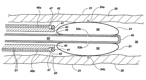
FIG. 2 shows detail of additional components of device 20 at distal end 23 of
tubular element 21, from circled region 2 in FIG. 1. Tubular element 21 is
shown
positioned within the lumen 30 of fallopian tube 31, with the fallopian tube
wall 39
shown in cross-section. Distal end 23 of tubular element 21 includes lip 40,
on which is
held a ligating band 41. Ligating band 41 may be of the type lmown for use in
performing tubal ligations, formed of rubber, silicone, and other suitable
materials.
Other ligating structures, such as suture loops or clamps, may be used as
well. Just
proximal to ligating band 41 is pusher 42, which in this example is a pusher
balloon
having a generally toroidal shape. Pusher balloon 42 can be expanded distally
to push
CA 02476817 2004-08-18
WO 2003/070085 PCT/US2003/005019
-6-
ligating band 41 off the distal end 23 of tubular element 21. The distal tip
43 of grasper
shaft 44 of grasper 38 is visible in lumen 45 of tubular element 21. Grasper
shaft 44 is
shown in its unextended position, so that tip 43 does notproject
significantlybeyond the
distal end 23 of tubular element 21. Grasper shaft 44 is preferably maintained
in an
unextended position while device 20 is inserted into the fallopian tube of the
patient.
FIG. 3 is a cross-sectional view of device 20 taken along section line 3-3 in
FIG. 2. Grasper 38 is slidably disposed in lumen 45 of tubular element 21. In
the
embodiment of the invention shown here, grasper 3 8 includes grasper shaft 44,
which is
hollow with a central lumen 50, and balloon 51, which is attached to grasper
shaft 44.
Lumen 50 of grasper shaft 44 communicates with the interior 52 of balloon 51
via fluid
channels 53a and 53b. In use, balloon 51 is inflated to a selected pressure or
volume by
the injection of a fluid with a syringe or other pressurized source. In this
context, fluid
is intended to mean liquids and gases. The fluid in grasper shaft 44 and the
interior 52
of balloon 51 could be, for example, air or saline. Balloon 51 may be inflated
in the
same way as balloon angioplasty catheters. A plurality of barbs, of which only
54a and
54b are visible in the present cross section, are attached to the exterior of
balloon 51.
Channels 46a and 46b in tubular element 21 communicate with the interior 47 of
pusher
balloon 42. Air or fluid from a syringe or other pressurized source connected
at the
proximal ends of channels 46a and 46b is forced into pusher balloon 42 to
cause it to
expand and push ligating band 41 off of lip 40.
FIG. 4 depicts an alternative embodiment of the invention in which a pusher
disk 48, driven by pusher rods 49a and 49b, is used in place of pusher balloon
42.
Pusher rods 49a and 49b are slidably disposed in channels 46a and 46b and are
driven
by a mechanical actuator (not shown) located at the proximal end of the
device, at
control segment 24. Various actuation mechanisms may be devised by those of
ordinary
skill in the art for causing pusher rods 49a and 49b to move pusher disk 48 to
push
ligating band 41 (not shown) off of band support structure at lip 40.
FIG. 5 is a transverse cross section taken at section line 5-5 in FIG. 3.
Channels 46a and 46b in tubular element 21 can be seen, as can as fluid
channels 53a,
53b, 53c, and 53d, which provide fluid communication between grasper shaft
lumen 50
and interior 52 of balloon 51. Fluid channels 53c and 53d were not visible in
the cross
section shown in FIG. 3. Also, all of the plurality of barbs 54a, 54b, 54c,
etc., are
CA 02476817 2004-08-18
WO 2003/070085 PCT/US2003/005019
_7_
visible in this cross section. Although two channels 46a and 46b and four
fluid
channels 53a, 53b, 53c; and 53d are shown, the numbers of channels are merely
exemplary, and embodiments of the device having different numbers of channels
are
considered to fall within the scope of the invention. Similarly, the number of
barbs 54a,
54b, 54c, etc., attached to balloon 51 may be varied.
FIG. 6, which depicts grasper shaft 44 extended out of the distal end 23 of
tubular element 21, more clearly shows the shape ofballoon 51. Balloon 51 is
generally
cylindrical in shape, with its inner surface attached to the exterior of
grasper shaft 44. A
plurality of barbs 54a, 54b, 54c, etc., are attached to the exterior of
balloon 51. As
noted previously, when balloon 51 is inflated so that its outer diameter is
substantially
equal to the diameter of lmnen 30 of fallopian tube 31, barbs 54a, 54b, 54c,
etc. are
forced into fallopian tube wall 39. Each barb has a shaft 90 that is attached
to the
exterior of balloon 51 at a first end 91 and which has a tip 55 at second end
92 which
allows it to be readily pushed into the tissue of fallopian tube wall 39.
Backward
extending points 56 are attached at or near tip 55 and extend back toward
first end 91 of
shaft 90, and serve to engage the tissue to prevent withdrawal of the barb
from the
fallopian tube wall 39. These features are specifically pointed out on barb
54a, but all
barbs 54a, 54b, 54c, etc. may include these features. The combination
ofballoon 51 and
barbs 54a, 54b, 54c, etc. and grasper shaft 44 function together as grasper
3~.
FIG. 6.5 depicts a farther alternative embodiment of the invention in which
ligating band 41 is pushed off of distal end 23 of tubular element 21 by
sleeve 93, which
is a tubular sleeve that is slidably disposed around tubular element 21 and
can be slid
distally to push ligating band 42 off of tubular element 21. In this and the
other
embodiments shown herein, ligating band 41 is released by being pushed off of
distal
end 23 of tubular element 21. However, the invention is not limited to
embodiments in
which the ligating band or other ligating structure is released by being
pushed. Other
mechanisms for releasing a ligating structure may be devised, for example,
tubular
element 21 could be retracted within sleeve 93, so that ligating band 41 is
maintained in
place while tubular element 21 is withdrawn from under it, thus allowing the
ligating
band to contract onto a grasped tissue bundle. Further, other means for
holding a
ligating band or other ligating structure at the end of tubular element 21 and
then
CA 02476817 2004-08-18
WO 2003/070085 PCT/US2003/005019
_g_
releasing it onto the grasped tissue bundle may be devised and are considered
to fall
within the scope of the invention.
The embodiment of the invention shown in FIG. 6.5 also shows an alternative
version of grasper 38, in which the elongated catheter formed by grasper shaft
44 and
balloon 51, as shown in FIGS. 3, S and 6, is replaced by an elongated catheter
comprising inflatable catheter 95, which has a closed end 96 and interior
lumen 97.
Inflatable catheter 95 is formed of a pliable material that is sufficiently
elastic that when
the pressure of the fluid in interior lumen 97 is increased, inflatable
catheter 95 inflates
or balloons out at end region 98. When the pressure of the fluid in interior
lumen 97 is
reduced, end region 98 of inflatable catheter 95 returns to its original
diameter.
Inflatable catheter 97 is substantially functionally equivalent to the
combination of
grasper shaft 44 and balloon 51 as shown in FIGS. 3, 5 and 6.
Also shown in FIG.6.5 are hooked wires 100, which provide an alternative
hooking structure to the barbs used in the embodiment of FIGS. 3, 5, and 6.
Two can be
seen in the cross section, but a plurality of hooks (for example, four or
five) would be
used. When inflatable catheter 95 is u~linflated, hooked wires 100 conform to
the
exterior of inflatable catheter 95, so inflatable catheter 95 and hooked wires
100 fit
inside tubular element 21. When inflatable catheter 95 is inflated, hooked
wires 100 are
splayed outward to be pushed into and grasp the inner wall of the fallopian
tube (not
shown). When inflatable catheter 95 is deflated, hooked wires 100 return to
their
original position.
A further alternative grasper 38 is shown in FIG. 6.6. W flatable catheter 95
is as
shown in FIG. 7, as is sleeve 93. Hooked wires 100 shown in FIG. 6.5 are
replaced by
suction tubes 101, each of which has an opening at or near its tip 102. In
FIG. 6.6,
openings 103 are positioned laterally, and tip 102 is closed. When inflatable
catheter 95
is inflated, suction tubes 101 are urged outward to contact the wall of the
fallopian tube
(not shown). Generation of a vacuum in suction tubes 101, from an external
vacuum
source connected to device 20 at control segment 24 and communicating with
suction
tubes 101, causes suction tubes 101 to grasp the fallopian tube by drawing the
tissue of
the fallopian tube to opening 103 and holding it there for as long as the
vacuum is
maintained.
CA 02476817 2004-08-18
WO 2003/070085 PCT/US2003/005019
-9-
The inventive device may be constructed with various other alternative grasper
mechanisms. For example, a forceps-like mechanism could be used to grasp
tissue in
the interior of the fallopian tube, or other grasper mechausms, for example,
as shown in
FIGS. 7 and 8, could be used. In FIG. 7, grasper 38 includes a grasper shaft
57 having a
plurality of hooks 58a, 58b, 58c, and 58d. In this embodiment of the
invention,
grasping is accomplished when one or more of hooks 58a, 58b, 58c, and 58d
catch on
the wall of the fallopian tube. In the alternative embodiment ofthe invention
shown in
FIG. 8, grasper 38 includes grasper shaft 60 and a plurality of pivoting hooks
61a and
61b having angled points 62a and 62b. Pivoting hooks 61 a and 61b would be
held in a
closed position (shown in dashed lines) while grasper 38 was in its retracted
position in
lumen 45 of tubular element 21, but when grasper 38 was extended, pivoting
hooks 61 a
and 61b would be moved to their open position (shown in solid lines) and then
closed
again to grasp tissue on the interior of the fallopian tube. Pivoting hooks
61a and 61b
pivot on pivot points 63a and 63b, actuated by actuation mechanisms 64a and
64b
located in the lumen 65 of grasper shaft 60. Actuation mechanisms 64a and 64b
could
be, for example, drive rods which pass through grasper shaft 60 to control
segment 24,
where they are moved by a lever or trigger mechanism.
FIGS. 7 and 8 feature illustrate another variation in the design of the
device, as
well. More than one ligating band may be held at distal end 23 of tubular
element 21,
on lip 40 or in some other manner. W FIGS. 7 and 8, two ligating bands 41a and
41b
are shown, but a larger number could be used as well. As will be described in
below, by
providing two ligating bands 41a and 41b, it is possible to make two legations
in a
fallopian tube, in order to provide more reliable blockage of the tube. In
order to release
ligating bands 41 a and 41b in sequence, pusher balloon 42 (in FIG. 7) or
pusher disk 48
(in FIG. 8) must be extended a first distance sufficient to push ligating band
41 a off
lip 40, and then be extended a second distance sufficient to push ligating
band 41b off
lip 40. Pusher balloon 42 would be expanded to a first volume, and then to a
second,
larger volume in order to push the two ligating bands sequentially. Similarly,
pusher
disk 48 would be extended to two different positions sufficient to release
ligating
bands 41a and 41b sequentially. It would be possible to use the two ligating
bands to
perform ligation of the two fallopian tubes sequentially, with the same
device, but this is
not preferred, because the withdrawal of the device from one fallopian tube,
followed by
CA 02476817 2004-08-18
WO 2003/070085 PCT/US2003/005019
-10-
reinsertion of the device into the second fallopian tube, provides an
opportunity for
contamination of the device and introduction of contaminants or infectious
agents into
the uterus or second fallopian tube.
It may be desirable to infuse antibiotics, topical anesthetics, or other drugs
into
the area of the ligation. Referring back to FIG. 2, drugs can be infused from
the tip 23
of tubular element 21 into fallopian tube 31. One or more drug delivery lumens
may be
provided. For example, lumen 45 of tubular element 21 may function as a drug
delivery
lumen. Alternatively, one or more drug delivery lumens may be provided in the
wall of
tubular element 21, comparable to channels 46a and 46b shown in FIG. 5. As a
further
alternative, a drug delivery lumen may be provided by adding a second tubular
element
surrounding, and coaxial with tubular element 21, thereby forming a drug
delivery
lumen between tubular element 21 and the second tubular element. Drugs would
be
injected into the drug delivery lmnen via access port 37, shown in FIG. 1,
which would
be connected to the drug delivery lumen.
If desired, an electrical current maybe passed through grasper 38 to cauterize
the
grasped tissue. For example, current could be passed through barbs 54a, 54b,
54c, etc.
of the device of FIGS. 2-6, hooked wires 100 of the device of FIG. 6.5, or
through
hooks 58a, 58b, 58c, 58d or 61a, 61b, etc. of the grasper as shown in FIGS. 7
and 8.
Cauterization of tissue may be of use to reduce bleeding and to burn away
small
amounts of tissue to facilitate freeing of the fallopian tube from grasper 38.
Cauterization of tissue may also be accomplished by delivery of a chemical
cauterizing
agent through a drug delivery lumen as discussed above.
The method of using the inventive device includes the following steps,
described
in the context of ligation of a fallopian tube, but applicable to the ligation
of other
tubular anatomical structures, as well. In the discussion of the methods
steps, specific
reference is made to the embodiment of the invention shown in FIGS. 1-3, 5 and
6, but
the steps may be readily generalized to other embodiments of the invention.
1) INSERTION OF DEVICE. The first step is the insertion of the device
into the fallopian tube, as shown in FIGS. 1 - 3. The grasper 38 maintained in
the
unextended position within tubular element 21 during the insertion step in
order to
prevent damage to the components of grasper 38 and to facilitate insertion
ofthe device
by having the relatively smooth, readily inserted distal end 23 of tubular
element 21
CA 02476817 2004-08-18
WO 2003/070085 PCT/US2003/005019
-11-
leading during insertion. Referring now to FIG. 1, a person performing the
procedure
holds device 20 by control segment 24 and inserts distal end 23 into the
vagina 31 ofthe
patient, and then into the lumen 33 of the uterus 34. Distal end 23 is then
guided into a
uterine horn 35 and into the lumen 30 of fallopian tube 31. Correct placement
of distal
end 23 may be determined by monitoring the length of tubular element 22
inserted after
distal end 23 has passed the uterine horn 35 and entered the fallopian tube
31, as
determined by change in resistance to insertion. Insertion of tubular element
22 into
uterus 34 and fallopian tube 31 may also be performed with hysteroscopic
guidance.
Device 20 may include control wires (not shown) for steering distal end 23, or
other
steering methods utilized with catheters, with steering control 25 on control
segment 24
used for steering distal end 23 during insertion.
2) EXTENSION OF GRASPER. As shown in FIGS. 6 and 9, once the
distal end 23 of tubular element 21 has been positioned properly within the
fallopian
tube 31, grasper 38 is extended out of tubular element 21. Grasper 38 is thus
passed
through the central opening of ligating band 41. FIG. 9 is a cross-section of
the device,
taken along section line 9-9 in FIG. 6. Extension and retraction of grasper
shaft 44 may
be controlled by extension control 26 on control segment 24 in FIG. 1 which
may be,
for example, a trigger causing movement of a mechanical linkage. Various
mechanisms
may be devised for causing grasper shaft 44 to extend out of tubular element
21 by a
predetermined distance, and the practice of the invention is not limited to a
particular
mechanism.
3) GRASPING OF TISSUE. Once grasper 38 has been extended out of
tubular element 21, grasper 38 is activated to grasp tissue on the interior of
fallopian
tube wall 39. Control segment 24, shown in FIG. 1, may include a grasp control
27 for
controlling grasping. As shown in FIG. 8, balloon 51 is inflated by fluid
flowing
through grasper shaft 44 until the outer diameter of balloon 51 is
substantially as large
as the inner diameter of fallopian tube 31. Barbs 54a, 54b, etc. are then
pushed into and
grasp or engage fallopian tube wall 39. Naturally, grasping of tissue could
also be
accomplished with an alternative grasper mechanism, such as those shown in
FIGS. 6.5,
3 0 6.6, 7, and 8.
4) RETRACTION OF GRASPER SHAFT AND GRASPED TISSUE. As
shown in FIG. 11, once tissue has been grasped by barbs 54a, 54b, etc.,
balloon 51 is
CA 02476817 2004-08-18
WO 2003/070085 PCT/US2003/005019
-12-
deflated, drawing the fallopian tube wall 39 radially inward toward grasper
shaft 44.
Referring now to FIG. 12, following deflation of balloon 51, grasper 38 is
retracted into
distal end 23 of tubular element 21. A tissue bundle 70 from the fallopian
tube wall 39,
is drawn into distal end 23 of tubular element 21 by grasper 38. When tissue
bundle 70
is drawn into distal end 23 of tubular element 21, it is at the same time
drawn through
the central opening of ligating band 41.
5) RELEASING OF LIGAT1NG BAND ONTO TISSITE BUNDLE. As
shown in FIG. 13, ligating band 41 is pushed off of lip 40 by the expansion of
pusher
balloon 42. Pusher balloon 42 may be expanded by air or liquid, such as water
or saline
solution, forced into pusher balloon 42 via charmels 46a and 46b. Once pushed
off of
lip 40, ligating band 41 contracts around tissue bundle 70. An alternative
release
mechanism, such as the pusher mechanisms shown in FIG. 4 or 6.5, could be used
at
this step, instead. The pusher mechanism may be controlled by a push
controller 28
located on control segment 24 in FIG. 1.
If tissue bundle 70 includes tissue from around the circumference of the
tubular
anatomical structure, application of ligating band 41 to tissue bundle 70 will
produce
blockage of fallopian tube 31. If, on the other hand, tissue bundle 70
includes tissue
from only one side of the fallopian tube 31, ligation of tissue bundle 70 will
only
separate tissue bundle 70 from the remainder of fallopian tube 31, but not
block
fallopian tube 31. This may be desirable in certain medical applications, such
as
ligating damaged or cancerous tissue, but of course would not be effective for
contraception. A grasper which grasps tissue around the circumference of the
tube will
form a tissue bundle 70 that includes tissue from around the circumference of
the tube.
It may also be possible to form a tissue bundle that includes tissue from
around the
circumference of the tube by grasping tissue around only a part of the
circumference of
the tube, if the amount of tissue grasped is large enough that the stiffness
of the tube
causes the entire circmnference of the tube to fold in to form the tissue
bundle.
6) FREEING OF GRASPED TIS SITE. Following application of a ligating
band or bands, tissue bundle 70 must be freed from grasper 38. This may be
accomplished by simply tearing barbs 54a, 54b, etc. from tissue bundle 70.
Since tissue
bundle 70 is separated from the main portion of the fallopian tube by the
ligation, tissue
damage caused by tearing out of the barbs is not of great concern.
Cauterization of the
CA 02476817 2004-08-18
WO 2003/070085 PCT/US2003/005019
-13-
tissue by passing current through the barbs, hooks, or other portion of the
grasper
contacting the tissue, or by delivering a chemical cauterizing agent, may
facilitate
freeing of tissue and reduce bleeding.
7) WITHDRAWAL OF DEVICE. Following ligation of tissue bundle 70
by ligating band 41, and freeing of tissue bundle 70 from grasper 38, the
device may be
withdrawn. FIG. 14 shows the ligated fallopian tube 31, with tissue bundle 70
secured
by ligating band 41. The lumen of fallopian tube 31 is now divided into two
sections
separated by the ligation: distal lumen 71, on the side closer to the ovary;
and proximal
lumen 72, on the side closer to the uterus. If it is desired that only a
single ligating band
be applied to the fallopian tube, the device is now withdrawn completely from
the
fallopian tube.
8) APPLICATION OF ADDITIONAL LIGATING BANDS. Refernng now
to FIG. 15, if it is desired that more than one ligating band be applied to
the fallopian
tube, after the application of first ligating band 41a to first tissue bundle
70a, tubular
element 21 is withdrawn only partially, to a new, more proximal position
within the
fallopian tube, and steps 2 through 5 are repeated at the new, more proximal
position, to
apply second ligating band 41b to second tissue bundle 70b to produce a double
ligation. Lumen 72 is now between the first and second ligations, and lumen 73
is
located most proximally on the side closer to the uterus. Steps 6 through 8
may be
repeated as many times as desired to apply multiple ligating bands to one
fallopian tube;
however, it is anticipated that reliable ligation would be provided by one to
three
ligating bands, and larger numbers of ligating bands would not be necessary or
desirable.
To accomplish sterilization, it is of course necessary to ligate both
fallopian
tubes. Thus the procedure would be repeated for the second tube in a similar
manner.
As noted above, it is preferred that the same device not be withdrawn from the
first
fallopian tube and then reinserted into the second fallopian tube, due to the
risk of
infection. Therefore, two sterilized devices are preferably provided to
perform ligation
of both fallopian tubes. It is within contemplation to manufacture the device
having
some or all components being disposable.
One alternate embodiment of the device, generally indicated at 200 in FIG. 16,
is
formed, in part, by three co-axial catheters 202, 204, and 206, referenced in
numerical
CA 02476817 2004-08-18
WO 2003/070085 PCT/US2003/005019
-14-
order corresponding to increasing diameter. The device 200 has been designed
for tubal
ligation as an office procedure using locally applied topical anesthetics.
Device 200 fits
within the operating channel of a hysteroscope (2.2 mm ID), so that standard
hysteroscopic techniques can be used to locate the fallopian tube opening
(ostium) and
feed the device 200 into the fallopian tube.
Placement of device 200 without hysteroscopic equipment may be effected to
provide non-surgical sterilization options for women in rural or
underdeveloped nations.
Using a cammla bent at a 140° angle, the device 200's tip is manually
guided through the
uterine horn and grossly positioned near the utero tubal junction. The device
200 is
then pushed from the cammla into the ostium and extended about 5 cm.
Resistance to
insertion requires tip manipulation to search for the tubal opening within the
minimal
surface area at the cannula tip. Verification of tubal entry can be achieved
via 20 ml
saline instillation through the catheter 204, where saline leakage into the
cannula or
cervical os is indicative of uterine (rather than tubal) catheter placement.
Catheters 202, 204, and 206 employed in devices 200 used for sterilization
procedures desirably are formed from extruded nylon, or any other suitable
medical
grade polymer. The inner catheter 202 is an elongate member. The distal tip
20~ of
catheter 202 desirably is flexible and forms, or carries, an inflatable
balloon 210. The
distal end 212 of middle catheter 204 has an expandable tip, generally
indicated at 214,
and also carries an O-ring 216. The outer catheter 206 is used to push or
deploy the
O-ring 216 over an invaginated tissue peduncle (tissue bundles 70a and 70b in
FIG.15).
Handles or grip-assisting structure, not illustrated, may be located on a
proximal, or
other convenient, location on each of catheters 202, 204, and 206, to
facilitate
manipulation of the device 200 and its components.
The distal tip 220 of the device 200 desirably includes about a 1 cm length of
double hulled tubing 222. Tubing 222 desirably flexes to reduce the risk of
tubal
perforation during positioning of device 200, and also functions as an
inflatable
balloon 210 during a ligation procedure. Tubing 222 maybe made from Silastic
tubing,
or some other operable material.
A minimal volume of cyanoacrylate or other adhesive may be contained between
the double hulls of a Silastic balloon 210, to function as a grasping aide.
Cyanoacrylate
is the adhesive of choice based upon its minimal viscosity, long shelf life
(in a dry
CA 02476817 2004-08-18
WO 2003/070085 PCT/US2003/005019
-15-
environment) and ability to cure upon tissue contact. Several cyanoacrylate
compounds
are approved for human use, but TrufillTM (Cordis Neurovascular) is currently
approved
for internal (non-superficial) use. Since scaring side effects are desirable
for this
application, the device 200 may not be limited to using Trufill adhesive.
An inner hull of balloon 210 may be nonporous, with the outer hull, in a
relaxed
state, having pores sized small enough to prevent premature passage of the
adhesive. A
syringe 225 can be provided in a device 200 to inflate the balloon 210. Other
inflation
devices are also operable. Inflation of the balloon 210 stretches the pores in
the outer
hull and allows local adhesive delivery through the pores in the outer balloon
for
adhesion of the balloon 210 to an inside wall section of a tube to be
occluded. Pores in
the outer hull may be arranged to produce spaced apart and axially aligned
strips of
adhesive on the balloon 210, to facilitate collapse of an inflated and adhered
balloon 210.
FIGS. 17-20 illustrate operation of the device during a ligation procedure. In
FIG. 17, distal tip 220 of device 200 has been inserted to a desired location
for creation
of an occlusion in a tube 230. The optimal sphincter location for female
contraceptive
ligation is just distal to the ampullary-isthmic junction (4-5 cm from the
ostium), where
the inner diameter of the fallopian tube abruptly increases from about 2 mm to
about 5
mm. The ratio of wall thickness to inner diameter in the ampullary tube makes
this the
first region that is appropriate for invagination. Following positioning in
the ampullary
tube, the tip 214 of catheter 204 is expanded to 5 mm, approximating the inner
diameter
of the fallopian tube. The device 200 is then drawn back (proximally) until
resistance to
the expanded tip 214 prevents further withdrawal. This procedure ensures
appropriate
positioning at the ampullary-isthmic junction. FIG. 18 depicts inflation
ofballoon 210
to place adhesive in contact between the balloon 210 and an inner surface 232
of
tube 230. Balloon 210 desirably is inflated with a formalin solution, such as
10%
formalin. Furthermore, expandable tip 214 is illustrated in FIGS. 17 and 18 as
having a
plurality of legs 235, each leg 235 being in a substantially collapsed, or
retracted,
position for insertion into a tube.
FIG. 19 illustrates proximal retraction of the balloon 210 with respect to a
fold
mechanism 237 formed, in part, from structure of expanding tip 214. Fold
mechanism 237 increases a diameter of tube 230, proximal to a grasped section,
to
CA 02476817 2004-08-18
WO 2003/070085 PCT/US2003/005019
-16-
assist in evening the tube 230. In the illustrated device 200, proximal
retraction of
balloon 210 simultaneously activates fold mechanism 237. As shown in FIG 20, a
balloon 210 desirably is collapsed to assist in forming a compact peduncle
250,
although such is not a requirement. FIG. 20 also shows catheter 206 has been
advanced
distally to deploy O-ring 216 as a legator band around tissue bundle 250. In
some cases,
a distal end of catheter 206 may additionally act as a passive fold assist
mechanism, or
to compact the peduncle 250.
After the O-ring is deployed, the tube 230 irmnediately is sealed for a shoe
term,
at least until the peduncle 250 dies and its tissue sloughs off. The balloon
210 may be
pulled from its attachment, promoting local scarring to provide a long term
occlusion of
tube 230. At present it is desired to leave balloon 210 attached, and for
formalin
leakage from balloon 210 to promote scarring in the tube proximal the O-ring
to provide
long term contraception. Permanent tubal occlusion is maintained through the
formation of scar tissue at the site of the ligation sphincter. Chronic
exposure to the
elastomeric ligature 216 causes a sustained inflammatory response leading to
more
stable scar tissue formation. In addition, instillation of 10% formalin
solution proximal
to the tubal sphincter prevents epithelial regeneration and aids in permanent
scar
formation. It is within contemplation alternatively, or additionally, to
provide a current
source to cauterize tissue of the peduncle and potentially to assist in
separating a
balloon 210 from a catheter 202. Alternatively, catheter 202 may be coupled to
a
balloon 210 in a way preferentially to separate at a known weak link under a
given
amount of tension in catheter 202.
Illustrated fold mechanism 237 includes a plurality of legs 235 spaced apart
around a centerline, each leg 235 having a knee 239 between a thigh 241 and a
shin 243.
A leg 235 desirably is sized such that a thigh 241 will have an axial length
equal to, or
greater than, a corresponding length of a shin 243. Such relative lengths
assist in
forming a circumferential fold in a wall of tube 230 during proximal
retraction of
balloon 210. A thigh 241 longer than a shin 243 causes a wrap to form in a
wall of
tube 230, thereby evening proximal and distal tubular portions of tube 230, as
the
shin 239 is displaced rotatingly towards the thigh 241. A long thigh 241 also
forms a
ramp, or surface guide, assisting in deployment of O-ring 216. Although such
is not
CA 02476817 2004-08-18
WO 2003/070085 PCT/US2003/005019
-17-
currently preferred, it is within contemplation for a fold mechanism 237 to
have a single
active component, such as a single leg 235.
Illustrated active fold mechanism 237 may be considered as forming one or
more four-bar linkages, and includes structure of catheter 202, expanding tip
214, and
catheter 204. A distal portion of expanding tip 214 is rotatably attached to a
distal end
of catheter 202. A proximal displacement of catheter 202, while holding
catheter 204
fixed, causes knees 239 to buckle and deflect radially outward, expanding the
tip 214
and increasing a diameter of a localized portion of tube 230. W use, grasping
structure,
such as balloon 210, maintains (or even reduces) a diameter of a first tubular
portion 245 of tube 230. A diameter of a second, and proximal, tubular portion
249 of
tube 230 is increased by the transverse motion of knees 239. As illustrated in
FIG. 20,
the second portion 249 is folded over the first portion (evening the tube) to
create a
tissue bundle or peduncle 250.
An active mechanism generally can be defined as connected structure arranged
actively to convert one form of work or a displacement in one direction, to
another form
of work or displacement in a different direction. W the case of the
illustrated fold
mechanism 237, a proximal (axial) displacement of catheter 202 is actively
convened to
a radial displacement of knees 239. In turn, knees 239 expand a cross-section
of
tube 230 to an effective diameter larger than a diameter grasped by the
balloon 210.
Such an active mechausm 237 can be contrasted to the essentially fixed
geometry of a
passive fold assist mechanism, such as a distal end of catheter 206. The
distal open end
of catheter 206 may assist in folding a tube 230, or in compacting a partially
folded
peduncle 250, but no active reduction in radial displacement occurs in the
catheter 206
itself. In fact, a distal end of catheter 206 may be required to expand to
accommodate
insertion of a peduncle 250. In such case, thighs 241 may act as wedges to
compact the
diameter of a peduncle 250.
Although hysterosalpingography can be used to confirm complete tubal
occlusion, these procedures are expensive and typically not available in
developing
nations. An inexpensive alternative to visualization exploits the fact that
the distal end
of the fallopian tube is naturally open to the peritoneal cavity. Following
uterine
injections of methylene blue, the dye dissipates into the peritoneal cavity
then is
CA 02476817 2004-08-18
WO 2003/070085 PCT/US2003/005019
-18-
processed and excreted by the kidneys in less than 30 minutes. Only completely
occluded tubes can prevent dye dispersion and excretion within this time
frame.
While the present invention has been described and illustrated in terms of
certain
specific embodiments, those of ordinary skill in the art will understand and
appreciate
that it is not so limited. Additions to, deletions from and modifications to
these specific
embodiments may be effected without departing from the scope of the invention
as
defined by the claims. Furthermore, features and elements from one specific
embodiment may be likewise applied to another embodiment without departing
from
the scope of the invention as defined herein.