Note: Descriptions are shown in the official language in which they were submitted.
CA 02477054 2010-05-25
WO 03072166 PCT/US03/05145
Title of the Invention
Vesicular shunt for the Drainage of Excess Fluid
Field of the Invention
The invention is a transvesicular drainage device designed to drain
excessive fluid from a bodily cavity into the bladder.
Background of the Invention
The present invention pertains to a chronic excess fluid drainage
device. More particularly, the present invention pertains to a vesicular
drainage device permitting unidirectional flow of excess fluid collections
into the bladder.
In medicine there are a variety of conditions which result in
pathologic chronic collection of bodily fluids. Chronic pericardial
effusions, normopressure hydrocephalus, hydrocephalus, chronic
pulmonary effusion, and ascites are but a few of the conditions in which
is chronic fluid collections persist and result in increased morbidity and
mortality.
These conditions currently are treated by one of two methods:
1) external drainage with a high-risk of infection and long-term
requirement for multiple punctures, 2) drainage to another body-
-1-
CA 02477054 2004-08-23
WO 03/072166 PCT/US03/05145
cavity, or 3) various drugs. For pericardial effusions and
hydrocephalus of all types, the treatment of choice is drainage
to another region of the body. For pericardial effusions this
entails a pericardial window, a highly invasive procedure in
which a large section of the external heart cavity is removed.
For hydrocephalus, the treatment typically involves the use of a
ventriculo-peritoneal shunt draining the: cerebrospinal fluid
into the peritoneal cavity. This device frequently becomes
clogged due to the proteinaceous environment of the peritoneal
cavity and requires removal or revision.
One conception of the present invention pertains to an
ascites drainage device. More specifically, said conception
pertains to a peritoneovesicular drainage device permitting
unidirectional flow of peritoneal fluid from the peritoneal
cavity into the bladder.
Ascites is a highly debilitating complication associated with
many medical conditions including liver failure and congestive
heart failure. Untreated ascites can result in respiratory
compromise, compression of the inferior vena cava (a vital blood
vessel) and spontaneous bacterial peritonitis (a life-
threatening condition). In order to treat chronic ascites,
medicine has turned to both drugs and surgery.
The drugs required to treat ascites are typically long-term
and frequently result in complications. The most common
pharmaceutical treatment of ascites involves the use of
diuretics to remove fluid from patient's body through their
urine. The difficulty with this treatment, though, is that
fluid is removed from the entire body, including the circulating
volume of blood, and can result in excessive loss of fluid
required to perfuse the vital organs of the human body. Thus,
- 2 -
CA 02477054 2004-08-23
WO 03/072166 PCT/US03/05145
even with religious application, though, the medicines
frequently fail. In this case, surgical, or invasive,
procedures are indicated.
Currently the treatment of choice is called paracentesis. In
paracentesis, the peritoneal fluid is drained through the
abdominal wall via the insertion of a needle through the
abdominal wall into the peritoneal cavity. This procedure,
though, is only a temporary fix as the ascites quickly refills
the peritoneal cavity in most chronic conditions. Furthermore,
repeated paracenteses put the patient at increased risk for a
life-threatening infection of their peritoneal cavity. Other
surgical/invasive procedures involve treatment of the cause of
the ascites (for example the Transjugular Intrahepatic
Portosystemic Shunt) but these measures also frequently result
in complications, which are often serious, and are thus
performed hesitantly.
The present invention avoids the difficulties associated with
the current therapies for chronic ascites, namely, the procedure
allows the drainage of peritoneal fluid without 1) the serious
complications of pharmaceuticals, 2) the inconvenience, the
substantial costs and the increased risk of infection associated
with frequent paracenteses and 3) the multiple severe
complications associated with more invasive and risky surgical
operations to treat the cause of ascites.
None of the existing devices are able to drain the peritoneal
cavity except through temporary transabdominal insertion of a
drainage catheter. These devices provide little improvement
over the intermittent punctures of paracentesis and result in
increased rates of infection if left in place for any length of
time. The present invention will obviate the need for a long-
- 3 -
CA 02477054 2010-05-25
WO 03/072166 PCT/US03/05145
term abdominal incision and, therefore, will eliminate the
associated increased risk of serious infection.
Summary of the Invention
The present invention is a device designed for implantation
in the wall of the bladder that permits the drainage of
excessive fluid into the bladder.
The device consists of a hollow, cylindrical column with
flanges at both ends to provide secure anchorage in the bladder
wall. Preferably there is a mechanism to provide unidirectional
flow of fluid and prevent reflux of urine inside the column. A
preferred embodiment of the device provides a passive ball-valve
mechanism which allows for drainage of fluid into the bladder
whenever a certain pressure is achieved at the collection site.
A second preferred embodiment of the device provides an active
valve mechanism which allows for controlled drainage of fluid
into the bladder whenever the valve is actuated. The most
preferred embodiment provides a pump in addition to an active
valve mechanism.
For ascites, the device can be implanted either through a
transurethral or transabdominal route. In order to drain other
sites, the bladder component is implanted as above, and a
flexible tube or other conduit may be incorporated to place the
receptacle end of the device in a fashion tailored to the region
to be drained.
In all embodiments, it is preferred that the device is
constructed with biocompatible materials.
4 -
CA 02477054 2010-05-25
WO 03072166 PCT/US03/05145
In a preferred embodiment, the vesicular shunt comprises: a hollow
cylinder, having an inside and an outside, and also having an inflow end
and an outflow end; a valve, located inside said hollow cylinder, wherein
said valve regulates the flow of fluid within the hollow cylinder such that
fluid may flow from the inflow end of said hollow cylinder to the outflow
end of said hollow cylinder; wherein said valve is an active-valve
controlled through an electric signal; and a flexible tube, having an inflow
and an outflow end, the outflow end of said flexible tube being in fluid
communication with the inflow end of said hollow cylinder.
In another preferred embodiment, the vesicular shunt comprises: a
hollow cylinder, having an inside and an outside, and also having an
inflow end and an outflow end; a valve, located inside said hollow
cylinder, wherein said valve regulates the flow of fluid within the hollow
cylinder such that fluid may flow from the inflow end of said hollow
cylinder to the outflow end of said hollow cylinder; wherein said valve is
an active-valve controlled through an EMF signal; and a flexible tube,
having an inflow and an outflow end, the outflow end of said flexible tube
being in fluid communication with the inflow end of said hollow cylinder.
In a further preferred embodiment, the vesicular shunt for draining
bodily fluids comprises: a tube, wherein said tube is designed to be
implanted in the wall of the bladder; a means for preventing the passage
of solids through said tube; a means for anchoring said tube in the wall of
the bladder; and a means for ensuring unidirectional flow through said
tube.
Brief Description of the Drawings
Fig. 1 shows a cross-sectional view of the device.
-4a -
CA 02477054 2004-08-23
WO 03/072166 PCT/US03/05145
FIG. 2 shows a cross-sectional view of the implanted device,
designed to treat ascites, when the peritoneal pressure is
sufficient to permit drainage.
FIG. 3 shows a cross-sectional view of the implanted device,
designed to treat ascites, when the peritoneal pressure is not
sufficient to open the valve and no fluid flow occurs.
FIG. 4 is an illustration of an example of an insertion
device through which the current invention can be implanted in
the bladder wall.
FIG. 5 is an illustration of alternative embodiments of the
invention with differing valve types, differing valve
positioning and differing number of valves.
FIG. 6 is an illustration of an alternative embodiment of the
device in which an active, externally or internally controlled
valve is utilized.
FIG. 7 is an illustration of an alternative embodiment of the
device in which a pump is included along the length of the
tubing and placed subcutaneously for external control of
drainage with a passive valve.
FIG. 8 is an illustration of a few of the alternative
embodiment of the device in which the peritoneal cavity, the
pulmonary space and the ventricular space are able to be drained
(pericardial drainage device not shown).
Description of the Preferred Embodiments
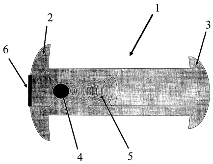
As can be seen in FIG. 1, the present invention provides a
novel vesicular drain 1 for implantation in the bladder wall 9
which will provide for unidirectional drainage of fluid into the
- 5 -
CA 02477054 2004-08-23
WO 03/072166 PCT/US03/05145
bladder. The drain 1 provides two flanges at its ends 2, 3
which allow the device to be firmly anchored once placed across
the bladder wall 9. Alternative embodiments of the device may
use other anchoring mechanisms, including, but not limited to: a
screw thread on the outside of 1, staples, sutures, an adhesive
compound, and/or one or more barbs.
The hollow shaft of the device contains a ball-valve 4
through which a positive closing pressure is provided by an
attached spring 5.
The fluid collection interface of the device 1 may optionally
include a large pore mesh 6 to allow for free flow of fluid
while preventing incarceration of tissues at the drainage site.
As can be seen in FIG. 2, once the pressure of the fluid
collection (in this case the peritoneal cavity) 7 exceeds the
combined force of the spring 5 and the pressure of the fluid-
filled bladder cavity 8, the peritoneal fluid 19 flows into the
bladder cavity 8 through displacement of the ball-valve 4.
There, the peritoneal fluid mixes with the urine 20.
If the pressure of the bladder cavity 8 and the force of the
spring 5, though, are greater than the pressure of the fluid
collection (in this case the peritoneal cavity) 7, then the
valve 4 will remain closed preventing reflux of urine 19 into
the peritoneal cavity as depicted in FIG. 3.
The device is designed to be placed transurethrally or
transabdominally via an insertion device 10 such as that
depicted in FIG. 4. The method of insertion allows for a single
invasive procedure to provide a long-term solution to the
otherwise difficult problem of refractory, chronic ascites.
- 6 -
CA 02477054 2004-08-23
WO 03/072166 PCT/US03/05145
Alternatively, the device may contain a length of tubing 11
or other means of fluid transport to reach the fluid collection
as well as an optional perforated receptacle 12, 17 and 18
through which the fluid collection will drain into the tubing.
Such other means of fluid transport include, but are not limited
to, conduit, catheter, channel, lumen, hose, pipe, duct, artery
or vessel. The device may contain one or more valves of a
variety of types including passive valves 4, 13 (flapper-valve),
14 (in FIG. 5), or active valves 15 (in FIG. 6) for tighter
control of fluid drainage.
The device is also designed to be able to incorporate a pump
mechanism 16 in FIG. 7 which, when placed subcutaneously, can be
actuated to provide an active pumping mechanism with the passive
valves 4, 13, 14, or with an active valve 15. A third
embodiment of the invention involves a unidirectional pump in
place of the valve, controlling the flow of fluid through the
device. A fourth conception of the invention involves a single
unidirectional valve controlling the flow of fluid through the
device.
Alternatively, maneuvers which increase the pressure of the
fluid cavity can also be utilized with the passive valves 4, 13,
14 to affect drainage such as bearing down to increase
intraabdominal pressure to drain the peritoneal cavity or
application of a girdle designed to increase abdominal pressure.
The device will be designed to drain a variety of different
fluid collections including, but not limited to, the peritoneal
cavity FIG. 8A, pulmonary effusions FIG. 8B and excessive
cerebrospinal fluid FIG. 8C. Pericardial effusion drain is not
shown.
7 -
CA 02477054 2004-08-23
WO 03/072166 PCT/US03/05145
Of particular interest to the inventors is the use of the
invention to drain pulmonary effusions and other fluid
collections in the lungs, in FIG. 8B.
While these are the preferred embodiments, the device could
employ any mechanism which provides a unidirectional passive or
active valve for the drainage of any body fluid into the urinary
bladder. This could involve filtration of the fluid through a
polymer so as to sequester albumin and other proteins in the
fluid collection while allowing flow of water and ions across
the semi-permeable membrane. This could also involve an
electronic valve triggered via communication across the tissues
of the human body through EMFs such as radio, electricity,
pressure, mechanical, magnetism, or other means of
communication, allowing drainage only at selected times. The
valve of the device can take many shapes and the device can be
manufactured from any of a variety of materials with the only
requirement being that of biocompatibility. Alternatively, the
device, in either the active or passive embodiment, may
incorporate anti-infective components in order to prevent the
spread of infection between the body cavities. Such anti-
infective components include, but are not limited to,
bacteriostatic materials, bacteriocidal materials, one or more
antibiotic dispensers, antibiotic eluting materials, entrained
radioisotopes, a heating element, bioactive plastics, surfaces
which encourage epithelialization, and coatings which prevent
bacterial adhesion. Alternatively, the device, in either the
active or passive embodiment, may incorporate anti-clogging
components. Such anti-clogging components include, but are not
limited to, an active ultrasonic component, an inner and outer
sleeve which, when actively agitated, disrupt the inner lumen,
surfaces which encourage epithelialization, enzyme eluting
8 -
CA 02477054 2004-08-23
WO 03/072166 PCT/US03/05145
materials, enzyme eluting materials which specifically target
the proteinaceous components of ascites, chemical eluting
surfaces, an intermittent plunger mechanism, and coatings which
prevent adhesion of proteinaceous compounds.
While the device is primarily contemplated for use in human
patients, the inventors also contemplate that the invention will
have veterinary uses or product development purposes in equine,
bovine, canine, feline, and other mammalian species.
- 9 -