Note: Descriptions are shown in the official language in which they were submitted.
CA 02477239 2004-08-27
WO 03/072065 PCT/US03/06382
DRUG SIGNATURES
This application claims the benefit of U.S. Provisional Application No.
60/360,728,
filed February 28, 2002.
Field of the invention
This invention relates to the fields of genomics, chemistry, and drug
discovery.
More particularly, the invention relates to methods and systems for grouping
and classifying
compounds by their activity and genomic effect in vivo, and methods and
systems for
Io predicting the activity and side effects of a compound in vivo.
Background of the Invention
Genomic sequence information is now available for several organisms, and
additional data is added continuously. However, only a small fraction of the
open reading
is frames now sequenced correspond to genes of known function: the function of
most
polynucleotide sequences, and many encoded proteins, is still unknown. These
genes are
now studied by means of, inter alia, polynucleotide arrays, which quantify the
amount of
mRNA produced by a test cell (or organism) under specific conditions.
"Chemogenomic
annotation" is the process of determining the transcriptional and bioassay
response of one or
2o more genes to exposure to a particular chemical, and defining and
interpreting such genes in
terms of the classes of chemicals for which they interact. A comprehensive
library of
chemogenomic annotations would enable one to design and optimize new
pharmaceutical
lead compounds based on the probable transcriptional and biomolecular profile
of a
hypothetical compound with certain characteristics. Additionally, one can use
as chemogenomic annotations to determine relationships between genes (for
example, as
members of a signal pathway or protein-protein interaction pair), and aid in
determining the
causes of side effects and the like. Finally, presenting the drug design
researcher with a
body of chemogenomic annotation information will generate research hypotheses
that will
stimulate follow-on experimental design.
so Several genomic database models have been disclosed. Sabatini et al., US
5,966,712
disclosed a database and system for storing, comparing and analyzing genomic
data.
Maslyn et al., US 5,953,727 disclosed a relational database for storing
genomic data.
Kohler et al., US 5,523,208 disclosed a database and method for comparing
polynucleotide
sequences and the predicted functions of their encoded proteins. Fujiyama et
al., US
1
CA 02477239 2004-08-27
WO 03/072065 PCT/US03/06382
5,706,498 disclosed a database and retrieval system, for identifying genes of
similar
sequence.
Sabry et al., WO00/70528 disclosed methods for analyzing compounds for drug
discovery using a cellular informatics database. The system images cells that
have been
s manipulated or exposed to test compounds, converting the resulting data into
a database.
Sabry further describes constructing a database of "cellular fingerprints"
comprising
descriptors of cell-compound interactions, where the descriptors are a
collection of
identified data/phenotype variations that characterize the interaction with
compounds of
known action, constructing a phylogenetic tree from the descriptors, and
determining the
io statistical significance of each descriptor. The descriptor for a new
compound can be
compared to the phylogenetic tree to determine its most likely mode of action.
Winslow et al., WO00/65523, disclosed a system comprising a database
containing
biological information which is used to generate a data structure having at
least one
associated attribute, a user interface, an equation generation engine
operative to generate at
is least one mathematical equation from at least one hierarchical description,
and a
computational engine operative on the mathematical equation to model dynamic
subcellular
and cellular behavior. The system is intended to access and tabulate genetic
information
contained within proprietary and nonproprietary databases, combine that data
with
functional information regarding the biochemical and biophysical role of gene
products, and
Zo based on this information formulate, solve and analyze computational models
of genetic,
biochemical and biophysical processes within cells.
Gould-Rothberg et al., WO00/63435, disclosed a method for identifying
hepatotoxic
agents by exposing a test cell population comprising a cell capable of
expressing one or
more nucleic acids sequences responsive to troglitazone (an anti-diabetes drug
discovered to
2s cause liver damage in some patients during phase III trials), contacting
the test cell
population with the test agent and comparing the expression of the nucleic
acids sequences
in a reference cell population. An alteration in expression of the nucleic
acids sequences in
the test cell population compared to the expression of the gene in the
reference cell
population indicates that the agent is hepatotoxic. Gould-Rothberg et al.,
WO00/37685,
so disclosed a method for identifying psychoactive agents that lack motor
involvement, by
identifying genes transcriptionally activated in rat brain striatum in
response to haloperidol.
Compounds that do not induce these genes are believed to not result in side
effects.
2
CA 02477239 2004-08-27
WO 03/072065 PCT/US03/06382
Thorp, W099/06839, describes a protein database, for screening with
combinatorial
chemical libraries. The database relates target and reference proteins,
compounds and
assays. The protein descriptors include molecular weight, activity,
hydrophobicity, etc., and
also their binding patterns with aptamers. Similarity of a target protein to a
reference
s protein is used to weight the combinatorial libraries examined more toward
compounds that
bind the most similar reference proteins.
Friend et al., US 6,203,987, discloses a method for comparing array profiles
by
grouping genes into co-regulated sets ("genesets"). Friend et al. disclose an
embodiment in
which the expression profile obtained in response to a drug is projected into
a geneset, and
io compared with other genesets to determine the biological pathways affected
by the drug. In
another embodiment, the projected profiles of drug candidates are compared
with the
profiles of known drugs to identify possible replacements for existing drugs.
Tamayo et al., EP 1037158, disclosed a method for organizing genomic data
using
Self Organizing Maps to cluster gene expression data into similar sets. The
method can be
is used to identify drug targets, by identifying which genes move from their
expression
clusters after a test cell is exposed to a given compound.
Tryon et al., WO01/25473, disclosed a method for constructing expression
profiles
of genes in response to a drug. In this method, a number of genes are selected
on the basis
of their expected interaction with the drug or condition to be examined, and
their expression
Zo in cell culture is measured in response to administration of the drug.
Summary of the Invention
One aspect of the invention is a method for creating a Group Signature for a
plurality of compounds having related activities, said method comprising: a)
providing a
zs plurality of expression datasets, each expression dataset comprising the
expression response
of a first plurality of genes in a subject cell following exposure to a
compound, wherein said
plurality of expression datasets comprises an expression dataset for each of a
plurality of
test compounds having a similar or identical biological activity, and an
expression dataset
for each of a plurality of control compounds that lack the biological activity
of the test
3o compounds; b) deriving a discrimination metric that distinguishes the
plurality of test
compounds from the control compounds based on gene expression to provide a
distinctive
gene set; and c) selecting a second plurality of genes from said distinctive
gene set to
provide a Group Signature for said plurality of test compounds.
3
CA 02477239 2004-08-27
WO 03/072065 PCT/US03/06382
Another aspect of the invention is a method for creating a Group Signature for
a
plurality of compounds having related activities, said method comprising: a)
providing a
plurality of test compounds having a similar or identical biological activity,
and a plurality
of control compounds that lack the biological activity of the test compounds;
b) contacting
s each compound with a subject cell; c) measuring the expression response of a
first plurality
of genes for each subject cell to provide an expression dataset for each
compound; d)
ordering the expression datasets by Principal Component Analysis to provide a
plurality of
principal components; e) identifying the Principal Component that
distinguishes the
plurality of test compounds from the plurality of control compounds to the
greatest degree,
io to provide a test Principal Component; f) identifying the genes that
distinguish the test
Principal Component from the control compounds to the greatest degree to
provide a
distinctive gene set; and g) selecting a second plurality of genes from said
distinctive gene
set to provide a Group Signature for said plurality of test compounds.
Another aspect of the invention is a method for creating a Drug Signature
capable of
is distinguishing the activity of a selected drug compound from a plurality of
compounds
having related activities, said method comprising: a) providing a plurality of
expression
datasets, each expression dataset comprising the expression response of a
plurality of genes
in a subject cell following exposure to a compound, wherein said plurality of
expression
datasets comprises an expression dataset for said selected drug compound and
an expression
zo dataset for each of a plurality of test compounds having a similar or
identical biological
activity; b) deriving a discrimination metric that distinguishes the selected
drug compound
from the plurality of test compounds based on gene expression to provide a
distinctive gene
set; and c) selecting a plurality of genes from said distinctive gene set to
provide a Drug
Signature for said selected drug compound.
zs Another aspect of the invention is a method for creating a Drug Signature
capable of
distinguishing the activity of a selected drug compound from a plurality of
compounds
having related activities, said method comprising: a) providing said selected
drug compound
and a plurality of test compounds having a similar or identical primary
biological activity;
b) contacting each compound with a subject cell; c) measuring the expression
response of a
3o first plurality of genes for each subject cell to provide an expression
dataset for each
compound; d) ordering the expression datasets by Principal Component Analysis
to provide
a plurality of principal components; e) identifying the Principal Component
that
distinguishes the selected drug compound from said plurality of test compounds
to the
4
CA 02477239 2004-08-27
WO 03/072065 PCT/US03/06382
greatest degree, to provide a distinguishing Principal Component; f)
identifying the genes
that contribute to the distinguishing Principal Component to the greatest
degree to provide a
distinguishing gene set; and g) selecting a second plurality of genes from
said distinguishing
gene set to provide a Drug Signature for said selected drug compound.
Another aspect of the invention is a Group Signature database comprising: a
plurality of Group Signature records, wherein each Group Signature record
comprises
indicia of at least one compound, wherein all compounds within a Group exhibit
a similar or
identical primary bioactivity; indicia of a set of genes, wherein the
expression of said genes
is modulated in response to exposure to a compound having a primary
bioactivity similar or
~o identical to the primary bioactivity of a compound indicated in the Group
record, and
wherein said set of genes distinguishes said Group from all other Groups
within said Group
Signature database. A further aspect of this invention is a Group Signature
database
comprising stress records, wherein each stress record comprises: an indicia of
a stress; and
indicia of a set of genes, wherein expression of said genes is modulated in
response to said
~s stress, and wherein said set of genes distinguishes said stress from all
other stresses and
Groups within said Group Signature database.
Another aspect of the invention is a Drug Signature database comprising: a
plurality
of Drug Signature records, wherein each Drug Signature record comprises
indicia of one
compound; and indicia of a set of genes, wherein expression of said genes is
modulated in
2o response to exposure to said compound, and wherein said set of genes
distinguishes said
compound from all other compounds within said Drug Signature database.
Another aspect of the invention is a method for determining the activity of a
drug
candidate, said method comprising: a) providing a Group Signature database,
said Group
Signature database comprising a plurality of Group Signature records, wherein
each Group
zs Signature record comprises indicia of at least one compound, wherein all
compounds within
a Group exhibit a similar or identical primary bioactivity; and indicia of a
set of genes,
wherein expression of said genes is modulated in response to exposure to a
compound
having a primary bioactivity similar or identical to the primary bioactivity
of a compound
indicated in the Group record, and wherein said set of genes distinguishes
said Group from
3o all other Groups within said Group Signature database; b) providing a drug
candidate
expression dataset for said drug candidate, said drug candidate expression
dataset
comprising the expression response of a plurality of genes in a subject cell
following
exposure to said drug candidate; c) comparing said drug candidate expression
dataset with
CA 02477239 2004-08-27
WO 03/072065 PCT/US03/06382
each Group Signature; d) selecting the Group Signature most similar to said
drug candidate
expression dataset; e) identifying the activity of the drug candidate to be
the primary
bioactivity exhibited by the compounds within the most similar Group
Signature.
Another aspect of the invention is a method for designing a Group Signature
s reagent, comprising: a) providing a plurality of expression datasets, each
expression dataset
comprising the expression response of a first plurality of genes in a subject
cell following
exposure to a compound, wherein said plurality of expression datasets
comprises an
expression dataset for each of a plurality of test compounds having a similar
or identical
biological activity, and an expression dataset for each of a plurality of
control compounds
io that lack the biological activity of the test compounds; b) deriving a
discrimination metric
that distinguishes the plurality of test compounds from the control compounds
based on
gene expression to provide a distinctive gene set; c) selecting a second
plurality of genes
from said distinctive gene set to provide a Group Signature for said plurality
of test
compounds; and d) providing a set of polynucleotide probes capable of
hybridizing
is specifically to one or more sequences of said second plurality of genes in
said Group
Signature to provide a Group Signature probe set. The invention further
includes the probe
sets designed by the above methods and kits, containing such probe sets.
Another aspect of the invention is a method for designing a Drug Signature
reagent,
comprising: a) providing a plurality of expression datasets, each expression
dataset
Zo comprising the expression response of a plurality of genes in a subject
cell following
exposure to a compound, wherein said plurality of expression datasets
comprises an
expression dataset for said selected drug compound and an expression dataset
for each of a
plurality of test compounds having a similar or identical biological activity;
b) deriving a
discrimination metric that distinguishes the plurality of test compounds from
the control
zs compounds based on gene expression to provide a distinctive gene set; c)
selecting a
plurality of genes from said distinguishing gene set to provide a Drug
Signature for said
selected drug compound; and d) providing a set of polynucleotide probes
capable of
hybridizing specifically to the sequences of said genes in said Drug Signature
to form a
Drug Signature probe set. The invention further includes the probe sets
designed by the
3o above methods and kits, containing such probe sets.
Another aspect of the invention is a method for determining the activity of a
drug
candidate, said method comprising: a) providing a Group Signature Array, said
Group
Signature Array comprising a solid support having affixed thereto a plurality
of Group
6
CA 02477239 2004-08-27
WO 03/072065 PCT/US03/06382
Signature probe sets, wherein each Group Signature probe set comprises a set
of
polynucleotide probes capable of hybridizing specifically to the sequences of
the genes in
each Group Signature, wherein said Group Signatures are obtained by: i)
providing a
plurality of expression datasets, each expression dataset comprising the
expression response
s of a plurality of genes in a subject cell following exposure to a compound,
wherein said
plurality of expression datasets comprises an expression dataset for each of a
plurality of
test compounds having a similar or identical biological activity, and an
expression dataset
for each of a plurality of control compounds that lack the biological activity
of the test
compounds; ii) deriving a discrimination metric that distinguishes the
plurality of test
io compounds from the control compounds based on gene expression to provide a
distinctive
gene set; iii) selecting a plurality of genes from said distinctive gene set
to provide a Group
Signature for said plurality of test compounds; and iv) repeating steps i) -
iii) for each
Group Signature; b) contacting a subject cell with said drug candidate; c)
extracting mRNA
from said subject cell; d) reverse-transcribing said mRNA to cDNA; e)
contacting said
is Group Signature Array with said cDNA; and f) determining whether any Group
Signature
probe set exhibits increased binding of cDNA. The invention also includes
applying this
method to a library of compounds and selecting a drug candidate, wherein the
Group
Signature probe set exhibits increased binding to the cDNA resulting from
contacting the
subject cell with said drug candidate.
zo Another aspect of the invention is a polynucleotide probe set for detecting
fibrate-
like activity, the set comprising: a plurality of polynucleotides capable of
hybridizing
specifically to genes selected from the group consisting of Rat for cytochrome
P452, Rat
cytochrome P450, Rat cytochrome P450-LA-omega (lauric acid omega-hydroxylase),
Rat
Sulfotransferase K2, Rat cytochrome P450-LA-omega (lauric acid omega-
hydroxylase),.Rat
zs Cyp4a locus, encoding cytochrome P450 (IVA3), Rat cytochrome P450, Rat
mitochondria)
3-2-trans-enoyl-CoA isomerase, Rat carnitine octanoyltransferase, Rat Wistar
peroxisomal
enoyl hydratase-like protein (PXEL), Rat mitochondria) long-chain 3-ketoacyl-
CoA thiolase
(3-subunit of mitochondria) trifunctional protein, Rat liver fatty acid
binding protein
(FABP), Rat pyruvate dehydrogenase kinase isoenzyme 4 (PDK4), Rat
mitochondria)
3o isoform of cytochrome b5, Hypothetical protein Rv3224, Rat peroxisomal
enoyl-CoA:
hydrotase-3-hydroxyacyl-CoA bifimctional enzyme, Rat peroxisomal membrane
protein
Pmp26p (Peroxin-11), Rat acyl-CoA hydrolase, Rat acyl-CoA oxidase, Rat acyl-
CoA
hydrolase, Rat 2,4-dienoyl-CoA reductase precursor, Rat mitochondria) 3-
hydroxy-3-
7
CA 02477239 2004-08-27
WO 03/072065 PCT/US03/06382
methylglutaryl-CoA synthase, Rat peroxisomal enoyl-CoA: hydrotase-3-
hydroxyacyl-CoA
bifunctional enzyme, and Mouse peroxisomal long chain acyl-CoA thioesterase Tb
(Ptelb).
Another aspect of the invention is a polynucleotide probe set for detecting
gemfibrozil-like activity, the set comprising: a plurality of polynucleotides
capable of
s hybridizing specifically to genes selected from the group consisting of Rat
fatty acid
synthase, Rat cholesterol 7a-hydroxylase, Mouse acetyl-CoA synthetase, Mouse
Vanin-1,
Rat kidney-specific protein (KS), Rat 2,3-oxidosqualene:lanosterol cyclase,
Rat aldehyde
dehydrogenase, and Rat thymosin (3-10.
Another aspect of the invention is a method for screening drug candidates for
fibrate
to activity, the method comprising: a) contacting a subject cell with a drug
candidate; b)
extracting mRNA from said subject cell; c) reverse-transcribing said mRNA into
cDNA; d)
hybridizing said cDNA to a fibrate signature probe set, said probe set
comprising a plurality
of polynucleotides capable of hybridizing specifically to a fibrate signature
gene, wherein
said fibrate signature genes are selected from the group consisting of Rat for
cytochrome
is P452, Rat cytochrome P450, Rat cytochrome P450-LA-omega (lauric acid omega-
hydroxylase), Rat Sulfotransferase K2, Rat cytochrome P450-LA-omega (lauric
acid
omega-hydroxylase), Rat Cyp4a locus, encoding cytochrome P450 (IVA3), Rat
cytochrome
P450, Rat mitochondria) 3-2-traps-enoyl-CoA isomerase, Rat carnitine
octanoyltransferase,
Rat Wistar peroxisomal enoyl hydratase-like protein (PXEL), Rat mitochondria)
long-chain
zo 3-ketoacyl-CoA thiolase ~i-subunit of mitochondria) trifunctional protein,
Rat liver fatty
acid binding protein (FABP), Rat pyruvate dehydrogenase kinase isoenzyme 4
(PDK4), Rat
mitochondria) isoform of cytochrome b5, Hypothetical protein Rv3224, Rat
peroxisomal
enoyl-CoA: hydrotase-3-hydroxyacyl-CoA bifunctional enzyme, Rat peroxisomal
membrane protein Pmp26p (Peroxin-11), Rat acyl-CoA hydrolase, Rat acyl-CoA
oxidase,
zs Rat acyl-CoA hydrolase, Rat 2,4-dienoyl-CoA reductase precursor, Rat
mitochondria) 3-
hydroxy-3-methylglutaryl-CoA synthase, Rat peroxisomal enoyl-CoA: hydrotase-3-
hydroxyacyl-CoA bifunctional enzyme, and Mouse peroxisomal long chain acyl-CoA
thioesterase Ib (Ptelb); and e) determining if said subject cell exhibits
increased expression
of a fibrate signature gene.
so Another aspect of the invention is a database product, comprising: a
computer-
readable medium, said medium storing thereon a Group Signature database, said
database
comprising a plurality of Group Signature records, wherein each Group
Signature record
comprises indicia of at least one compound, wherein all compounds within a
Group exhibit
CA 02477239 2004-08-27
WO 03/072065 PCT/US03/06382
a similar or identical primary bioactivity; and indicia of a set of genes,
wherein expression
of said gene is modulated in response to exposure to a compound having a
primary
bioactivity similar or identical to the primary bioactivity of a compound
indicated in the
Group record, and wherein said set of genes distinguishes said Group from all
other Groups
within said Group Signature database.
Brief Description of the Figures
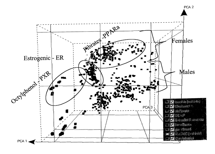
Fig. 1 is a projection of a Principal Component Analysis output, showing the
grouping of fibrate compounds along PCA1, split into male and female subjects
along
~o PCA2, and distinguished from octylphenol along PCA3. Fig. lA and Fig. 1B
are rotated
views of the same data.
Fig. 2 is a graph illustrating the specificity of a fenofibrate Drug
Signature. The
Drug Signature was based on four fenofibrate experiments compared to four
control/vehicle
experiments, and was then used to sort 677 other experiments. The sort was
according to
is the similarity score S = IIXReIRkX. The sorted list was then graphed,
assigning a value of
1.0 to each fenofibrate experiment, a value of 0.5 to each fibrate other than
fenofibrate, and
a value of 0 to each non-fibrate control. The graph demonstrates that this
minimal
fenofibrate Drug Signature correctly sorts most fenofibrate experiments to the
top of the list,
most fibrate experiments near the top of the list (although lower than
fenofibrate
zo experiments), and all control experiments below the fenofibrate experiments
(and below
most of the fibrate experiments).
Fig. 3 graphically presents bioassay results for seven nuclear receptor
agonists (z
axis from front to back: estradiol, bisphenol A, clofibrate, bis(2-ethyl-
hexyl)phthalate
(DEHP), fenofibrate, gemfibrozil, and octylphenol). Bioassays were selected
from a panel
zs of 123 assays performed if any of the selected compounds demonstrated
activity: the 26
selected bioassays were (x axis) acetylcholinesterase (a); adenosine A2A (b);
adenosine A3
(c); adrenergic a1D (d); adrenergic a2B (e); adrenergic a2C (f); adrenergic
(33 (g);
norepinephrine transporter (h); calcium channel type L (i); cyclooxygenase COX-
2 (j);
dopamine transporter (k); estrogen receptor (1); glucocorticoid receptor (m);
lipoxygenase
30 15-LO (n); muscarinic receptor M1 (o); muscarinic receptor M2 (p);
muscarinic receptor
M3 (q); S/T kinase p38a (r); Y kinase EGF receptor (s); serotonin S-HT2A (t);
serotonin 5-
HT2C (u); serotonin transporter (v); sodium channel-site 2 (w); tachykinin NKZ
(x);
9
CA 02477239 2004-08-27
WO 03/072065 PCT/US03/06382
testosterone receptor (y); thromboxane synthetase (z). The activity is shown
as 1/ICso (Y
axis), with all values <50% inhibition binned to 0.
Detailed Description
Definitions:
The term "test compound" refers in general to a compound to which a test cell
is
exposed, about which one desires to collect data. Typical test compounds will
be small
organic molecules, typically drugs and/or prospective pharmaceutical lead
compounds, but
can include proteins, peptides, polynucleotides, heterologous genes (in
expression systems),
io plasmids, polynucleotide analogs, peptide analogs, lipids, carbohydrates,
viruses, phage,
parasites, and the like.
The term "control compound" refers to a compound that is not known to share
any
biological activity with a test compound, which is used in the practice of the
invention to
contrast "active" (test) and "inactive" (control) compounds during the
derivation of Group
is Signatures and Drug Signatures. Typical control compounds include, without
limitation,
drugs used to treat disorders distinct from the test compound indications,
vehicles, known
toxins, known inert compounds, and the like.
The term "biological activity" as used herein refers to the ability of a test
compound
to affect a biological system, for example to modulate the effect of an
enzyme, block a
2o receptor, stimulate a receptor, alter the expression of one or more genes,
and the like. Test
compounds have similar or identical biological activity when they have similar
or identical
effects on an organism in vivo or on cells or proteins in vitro. For example,
fenofibrate,
clofibrate, and gemfibrozil have similar biological activities because all
three are prescribed
for hyperlipoproteinemia. Similarly, aspirin, ibuprofen, and naproxen all have
similar
2s activities as all three are known to be non-steroidal anti-inflammatory
compounds. The
terms "primary bioactivity" and "primary biological activity" refer to the
most pronounced
or intended effect of the compound. For example, the primary bioactivity of an
ACE
inhibitor is the inhibition of angiotensin-converting enzyme (and the
concomitant reduction
of blood pressure), regardless of secondary bioactivities or side effects.
so The term "subject cell" refers to a biological cell or a model of a
biological system
capable of reacting to the presence of a test compound, typically a live
animal, eukaryotic
cell or tissue sample, or a prokaryotic organism.
CA 02477239 2004-08-27
WO 03/072065 PCT/US03/06382
The term "expression response" refers to the change in expression level (if
any) of a
gene in response to administration of a test compound or control compound (or
other test or
control condition). The expression level can be measured directly, for example
by
quantifying the amount of protein encoded by the gene that is produced using
proteomic
s techniques. A variety of methods for detecting protein levels may be used,
including, but
limited to, Western blots and ELISA. The expression level can also measured as
the change
in mRNA transcription, or by any other quantitative means of measuring gene
activation.
The expression response can be weighted or scaled as necessary to normalize
data, and can
be reported as the absolute increase or decrease in expression (or
transcription), the relative
io change (for example, the percentage change), the degree of change above a
threshold level,
and the like.
The term "expression dataset" as used herein refers to data indicating the
identity of
genes affected by administration of the test or control compound, and the
change in
expression that resulted. The expression dataset typically contains a subset
of genes,
is preferably the subset of genes that displayed the greatest changes in
expression response.
The term "discrimination metric" refers to a method or algorithm for
distinguishing
the expression data in response to test compounds from the expression data in
response to
control compounds. The method can be selecting genes on the basis of the
eigenvalues for
the genes from the PCA output (selecting the principal component axis that
separates the
zo test compounds from the control compounds), or can include mathematical
analysis to
determine which gene or combination of genes best discriminates between the
test and
control compounds, for example using Golub's distinction metric, Student's t-
test or the
like.
The terms "PCA" and "principal component analysis" refer to mathematical
zs methods for transforming a number of correlated variables into a number of
uncorrelated
(independent) variables called principal components. The first principal
component
accounts for as much of the variability in the data as possible, and each
succeeding
component accounts for as much of the remaining variability as possible. "PCA"
as used
herein further includes variations of principal component analysis such as
kernel PCA and
3o the like.
The term "Group Signature" as used herein refers to a data structure
comprising a
group identifier and one or more gene identifiers. The group identifier
indicates a family of
compounds having similar activity (for example, "fibrates"), or can directly
indicate the
11
CA 02477239 2004-08-27
WO 03/072065 PCT/US03/06382
activity (for example, PPARa inhibition). It is often simply the "name" of the
group. The
group identifier can further indicate the identity of compounds known to
belong to the
group. Gene identifiers indicate which gene expression rates are modulated
(upregulated or
downregulated) by exposure to a compound belonging to the group, and which are
so
s characteristic of the group, or so distinctive, that modulation of the
expression of these
genes according to the signature is sufficient to distinguish the compound
administered as
belonging to the Group (rather than to another Group, or wholly lacking known
activity).
The gene identifiers can identify genes by sequence, name, reference to an
accession
number, reference to a clone or position within a DNA array, and the like.
Gene identifiers
io can further comprise the direction and degree of expression modulation, in
absolute or
relative terms. For example, a gene identifier can include the requirement
that expression
decrease by at least 10%, or that expression increase by between 100% and
500%. The
gene identifier can further include time restrictions: for example, a Group
Signature can
require that gene "X" be upregulated by at least 250% within 8 hours of
administration, or
is at not less than 4 hours but no more than 16 hours, or the like. Although
the Group
Signature may comprise any number of genes, it typically comprises up to 50
gene
identifiers of varying degrees of specificity, from which subsets of varying
specificity can
be derived. Preferably, the Group Signature consists of no more than 50 genes.
More
preferably, the Group Signature consists of no more than 25 genes. In
addition, the Group
zo Signature will comprise preferably at least three genes, more preferably at
least 5 genes,
even more preferably at least 10 genes and most preferably at least 15 genes.
In some cases
the Group Signature may consist of three or fewer. For example, the most
specific signature
for one group may comprise 20 gene identifiers: this signature contains a
plurality of sub-
signatures having similar (or somewhat less) specificity derived by omitting
one or more of
zs the gene identifiers. The Group Signature can further comprise bioassay
data, for example
indicating the bioactivity observed for compounds in the group against a panel
of standard
assays. Bioassay data can be used to identify the potential members of a Group
prior to
genomic experiments, particularly where a number of drug candidates are to be
screened.
Bioactivity data is particularly useful for distinguishing between compounds
having
3o unrelated structures, but which induce similar genomic expression patterns.
The data
structure can be stored physically or electronically, for example within a
database on a
computer-readable medium. Alternatively, the data structure can be embodied in
an array in
12
CA 02477239 2004-08-27
WO 03/072065 PCT/US03/06382
full or in part, such as a polynucleotide probe array having a separate region
of probes
specific for each Group Signature.
The term "Group Signature database" refers to a collection of data comprising
a
plurality of Group Signatures. A number of formats exists for storing data
sets and
s simultaneously associating related attributes, including without limitation,
tabular,
relational, and dimensional. The tabular format is most familiar, for example
spreadsheets
such as Microsoft Excel~ and Corel Quattro Pro~ spreadsheets. In this format,
association
of data points with related attributes occurs by entering a data point and
attributes related
thereto in a unique row. Relational databases typically support a set of
operations defined
~o by relational algebra. Such databases typically include tables composed of
columns and
rows for the data included in the database. Each table in the database has a
primary key,
which can be any column or set of columns, the values for which uniquely
identify the rows
in the table. The tables in a relational database can also include a foreign
key that is a
column or set of columns, the values of which match the primary key values of
another
is table. Typically, relational databases support a set of operations (for
example, select, join,
combine) that form the basis of the relational algebra governing relations
within the
database. Suitable relational databases include, without limitation, Oracle~
(Oracle Inc.,
Redwood Shores, CA) and Sybase~ (Sybase Systems, Emeryville, CA) databases.
The term "Drug Signature" as used herein refers to a data structure similar to
the
zo Group Signature, but specific to a single compound (or a plurality of
essentially identical
compounds, such as salts or esters of the same compound). The gene identifiers
of a Drug
Signature are selected to distinguish the selected compound from other
compounds with
which it shares activity(ies): Drug Signatures distinguish between members of
a Group
Signature, and also distinguish between the drug compound and unrelated
compounds.
2s The term "gene expression profile" refers to a representation of the
expression level
of a plurality of genes in response to a selected expression condition (for
example,
incubation in the presence of a standard compound or test compound). Gene
expression
profiles can be expressed in terms of an absolute quantity of mRNA transcribed
for each
gene, as a ratio of mRNA transcribed in a test cell as compared with a control
cell, and the
30 like. As used herein, a "standard" gene expression profile refers to a
profile already present
in the primary database (for example, a profile obtained by incubation of a
test cell with a
standard compound, such as a drug of known activity), while a "test" gene
expression
profile refers to a profile generated under the conditions being investigated.
The term
13
CA 02477239 2004-08-27
WO 03/072065 PCT/US03/06382
"modulated" refers to an alteration in the expression level (induction or
repression) to a
measurable or detectable degree, as compared to a pre-established standard
(for example,
the expression level of a selected tissue or cell type at a selected phase
under selected
conditions).
The term "correlation information" as used herein refers to information
related to a
set of results. For example, correlation information for a profile result can
comprise a list of
similar profiles (profiles in which a plurality of the same genes are
modulated to a similar
degree, or in which related genes are modulated to a similar degree), a list
of compounds
that produce similar profiles, a list of the genes modulated in said profile,
a list of the
~o diseases and/or disorders in which a plurality of the same genes are
modulated in a similar
fashion, and the like. Correlation information for a compound-based inquiry
can comprise a
list of compounds having similar physical and chemical properties, compounds
having
similar shapes, compounds having similar biological activities, compounds that
produce
similar expression array profiles, and the like. Correlation information for a
gene- or
is protein-based inquiry can comprise a list of genes or proteins having
sequence similarity (at
either nucleotide or amino acid level), genes or proteins having similar known
functions or
activities, genes or proteins subject to modulation or control by the same
compounds, genes
or proteins that belong to the same metabolic or signal pathway, genes or
proteins belonging
to similar metabolic or signal pathways, and the like. In general, correlation
information is
zo presented to assist a user in drawing parallels between diverse sets of
data, enabling the user
to create new hypotheses regarding gene and/or protein function, compound
utility, and the
like. Product correlation information assists the user with locating products
that enable the
user to test such hypotheses, and facilitates their purchase by the user.
"Similar", as used herein, refers to a degree of difference between two
quantities that
zs is within a preselected threshold. For example, two genes can be considered
"similar" if
they exhibit sequence identity of more than a given threshold, such as for
example 20%. A
number of methods and systems for evaluating the degree of similarity of
polynucleotide
sequences are publicly available, for example BLAST, FASTA, and the like. See
also
Maslyn et al. and Fujimiya et al., supra, incorporated herein by reference.
The similarity of
3o two profiles can be defined in a number of different ways, for example in
terms of the
number of identical genes affected, the degree to which each gene is affected,
and the like.
Several different measures of similarity, or methods of scoring similarity,
can be made
available to the user: for example, one measure of similarity considers each
gene that is
14
CA 02477239 2004-08-27
WO 03/072065 PCT/US03/06382
induced (or repressed) past a threshold level, and increases the score for
each gene in which
both profiles indicate induction (or repression) of that gene. We utilize a
similarity score
that takes into account, for each gene, the level of regulation achieved by
that gene in the
experimental profile relative to all other experiments in the dataset. For a
given gene, one
s can rank its level of regulation in the experimental profile relative to all
other profiles (RkX).
The relative rank (ReIRkX RkX/n , where n=number of profiles) is that rank
divided by total
number of profiles. A similarity score may then be defined as the product of
these relative
ranks for all genes in the profile or S = TIX ReIRkX. A small value of S
reflects an
experimental profile that matches a reference profile on multiple genes and
where the
io amplitude of regulation for each gene is large. Similarity between a test
profile and a
signature can be determined using a variety of metrics, a preferred one being
defined as S =
IIX ReIRkX. The similarity score may also be referred to as a "specificity
score" as it
measures how rare the match of the experimental to the reference profile is
relative to the
rest of the dataset. Other statistical methods are also applicable.
~s The term "hyperlink" as used herein refers to feature of a displayed image
or text
that provides information additional and/or related to the information already
currently
displayed when activated, for example by clicking on the hyperlink. An HTML
HREF is an
example of a hyperlink within the scope of this invention. For example, when a
user
queries the database of the invention and obtains an output such as a list of
the genes most
Zo induced or repressed by a selected compound, one or more of the genes
listed in the output
can be hyperlinked to related information. The related information can be, for
example,
additional information regarding the gene, a list of compounds that affect
gene induction in
a similar way, a list of genes having a known related function, a list of
bioassays for
determining activity of the gene product, product information regarding such
related
2s information, and the like.
The terms "polynucleotide," "oligonucleotide," "nucleic acid" and "nucleic
acid
molecule" are used herein to include a polymeric form of nucleotides of any
length, either
ribonucleotides or deoxyribonucleotides. This term refers only to the primary
structure of
the molecule. Thus, the term includes triple-, double- and single-stranded
DNA, as well as
3o triple-, double- and single-stranded RNA. It also includes modifications,
such as by
methylation and/or by capping, and unmodified forms of the polynucleotide.
More
particularly, the terms "polynucleotide," "oligonucleotide," "nucleic acid"
and "nucleic acid
molecule" include polydeoxyribonucleotides (containing 2-deoxy-D-ribose),
CA 02477239 2004-08-27
WO 03/072065 PCT/US03/06382
polyribonucleotides (containing D-ribose), any other type of polynucleotide
which is an N-
or C-glycoside of a purine or pyrimidine base, and other polymers containing
non-
nucleotidic backbones, for example, polyamide (e.g., peptide nucleic acids
(PNAs)) and
polymorpholino (commercially available from the Anti-Virals, Inc., Corvallis,
Oregon, as
s Neugene) polymers, and other synthetic sequence-specific nucleic acid
polymers providing
that the polymers contain nucleobases in a configuration which allows for base
pairing and
base stacking, such as is found in DNA and RNA.
As used herein, the term "probe" or "oligonucleotide probe" refers to a
structure
comprised of a polynucleotide, as defined above, that contains a nucleic acid
sequence
~o capable of hybridizing to a nucleic acid sequence present in the target
nucleic acid analyte.
The polynucleotide regions of probes may be composed of DNA, and/or RNA,
and/or
synthetic nucleotide analogs. Probes of dozens to several hundred bases long
can be
artificially synthesized using oligonucleotide synthesizing machines, or they
may be derived
from various types of DNA cloning. A probe can be single-stranded or double-
stranded.
~s Probes are useful in the detection, identification and isolation of
particular gene sequences
or fragments. It is contemplated that any probe used in the present invention
can be labeled
with a reporter molecule so that it is detectable using a detection system,
such as, for
example, ELISA, EMIT, enzyme-based histochemical assays, fluorescence,
radioactivity,
luminescence, spin labeling, and the like. The critical aspects are that the
probe must
zo contain a nucleic acid strand that is at least partially complementary to
the target sequence
to be detected, and the probe must be labeled so that its presence can be
visualized.
The terms "hybridize" and "hybridization" refer to the formation of complexes
between nucleotide sequences which are sufficiently complementary to form
complexes via
Watson-Crick base pairing. It will be appreciated that the hybridizing
sequences need not
zs have perfect complementarity to provide stable hybrids. Further, the
ability of two
oligonucleotides to hybridize will be dependent on the experimental
conditions. For
example, the temperature and/or salt concentration will affect the percentage
of
complementary base pair matches required for hybrid duplexes to remain intact.
Conditions
that favor hybridization are referred to as less "stringent" than conditions
that require a
3o greater degree of sequence complementarity to maintain a stable duplex. In
many
situations, stable hybrids will form where fewer than about 10% of the bases
are
mismatches, ignoring loops of four or more nucleotides. Accordingly, as used
herein the
term "capable of hybridizing" refers to an oligonucleotide that can form a
stable duplex with
16
CA 02477239 2004-08-27
WO 03/072065 PCT/US03/06382
its "complement" under appropriate assay conditions, generally where there is
about 90% or
greater homology.
The terms "array", "polynucleotide array", "microarray", and "probe array" all
refer
to a surface on which is attached or deposited a molecule capable of
specifically binding a
polynucleotide of a given sequence. Typically the molecule will be a
polynucleotide having
a sequence complementary to the polynucleotide to be detected, and capable of
hybridizing
to it.
General Method:
io The method of the invention employs chemogenomic expression data and
bioassay
data in order to characterize and predict the biological activity of
compounds. The method
of the invention provides a way to cluster expression data meaningfully, and
to extract
relevant information from the sea of data that typically results from a
genomic expression
experiment.
~s The invention is based on the use of chemogenomic expression data,
collected in
response to an experimental condition, preferably contact with a compound or
bioactive
substance. Suitable compounds include known pharmaceutical agents, known and
suspected toxins and pollutants, proteins, dyes and flavors, nutrients, herbal
preparations,
environmental samples, and the like. Other useful experimental conditions to
examine
zo include infectious agents such as viruses, bacteria, fungi, parasites, and
the like,
environmental stresses such as starvation, hypoxia, temperature, and the like.
It is presently
preferred to analyze a variety of compounds and/or experimental conditions
simultaneously,
particularly where many of the compounds and/or conditions are related by
activity or
therapeutic effect. The experimental conditions are applied to a cell having a
genome,
zs preferably a mammalian cell. Eukaryotic cells can be tested either in vivo
or in vitro.
Suitable eukaryotic cells include, without limitation, human, rat, mouse, cow,
sheep, dog,
cat, chicken, pig, goat, and the like. It is presently preferred to examine
mammalian cells
derived from a plurality of different tissue types, for example, liver,
kidney, bone marrow,
spleen, and the like. The subject cells are preferably exposed to a plurality
of experimental
3o conditions, for example, to a plurality of different concentrations of a
compound, and
examined at a plurality of time points.
The chemogenomic response can be obtained by any available means, for example
by employing a panel of reporter cells, each group of cells having a reporter
gene
17
CA 02477239 2004-08-27
WO 03/072065 PCT/US03/06382
operatively connected to a different selected regulatory region.
Alternatively, one can
employ primary tissue isolates, cells or cell lines lacking reporter genes,
and can determine
the expression of a plurality of genes directly.
Direct detection methods include direct hybridization of mRNA with
s oligonucleotides or longer DNA fragments such as cDNA or even fragments of
cloned
genomic DNA (whether in solution or bound to a solid phase), reverse
transcription
followed by detection of the resulting cDNA, Northern blot analysis, and the
like.
Primers and probes for use in the determination of expression levels herein
are
derived from gene sequences and are readily synthesized by standard
techniques, e.g., solid
io phase synthesis via phosphoramidite chemistry, as disclosed in U.S. Patent
Nos. 4,458,066
and 4,415,732, incorporated herein by reference; Beaucage et al. (1992)
Tetrahedron
48:2223-231 l; and Applied Biosystems User Bulletin No. 13 (1 April 1987).
Other
chemical synthesis methods include, for example, the phosphotriester method
described by
Narang et al., Meth. Enzymol. (1979) 68:90 and the phosphodiester method
disclosed by
~s Brown et al., Meth. Enzymol. (1979) 68:109. Poly(A) or poly(C), or other
non-complementary nucleotide extensions may be incorporated into probes using
these
same methods. Hexaethylene oxide extensions may be coupled to probes by
methods
known in the art. Cload et al. (1991) J. Am. Chem. Soc. 113:6324-6326; U.S.
Patent No.
4,914,210 to Levenson et al.; Durand et al. (1990) Nucleic Acids Res. 18:6353-
6359; and
ao Horn et al. (1986) Tet. Lett. 27:4705-4708.
While the length of the primers and probes can vary, the probe sequences are
selected such that they have a lower melt temperature than the primer
sequences. Hence,
the primer sequences are generally longer than the probe sequences. Typically,
the primer
sequences are in the range of between 10-75 nucleotides long, more typically
in the range of
2s 20-45. The typical probe is in the range of between 10-50 nucleotides long,
such as 15-40,
18-30, and so on, and any length between the stated ranges.
If a solid support is used, the oligonucleotide probe may be attached to the
solid
support in a variety of manners. For example, the probe may be attached to the
solid
support by attachment of the 3' or 5' terminal nucleotide of the probe to the
solid support.
3o More preferably, the probe is attached to the solid support by a linker
which serves to
distance the probe from the solid support. The linker is usually at least 15-
30 atoms in
length, more preferably at least 15-50 atoms in length. The required length of
the linker
18
CA 02477239 2004-08-27
WO 03/072065 PCT/US03/06382
will depend on the particular solid support used. For example, a six atom
linker is generally
sufficient when highly cross-linked polystyrene is used as the solid support.
A wide variety of linkers are known in the art which may be used to attach the
oligonucleotide probe to the solid support. The linker may be formed of any
compound
s which does not significantly interfere with the hybridization of the target
sequence to the
probe attached to the solid support. The linker may be formed of a
homopolymeric
oligonucleotide which can be readily added on to the linker by automated
synthesis.
Alternatively, polymers such as functionalized polyethylene glycol can be used
as the
linker. Such polymers are preferred over homopolymeric oligonucleotides
because they do
~o not significantly interfere with the hybridization of probe to the target
oligonucleotide.
Polyethylene glycol is particularly preferred.
The linkages between the solid support, the linker and the probe are
preferably not
cleaved during removal of base protecting groups under basic conditions at
high
temperature. Examples of preferred linkages include carbamate and amide
linkages.
~ s Examples of preferred types of solid supports for immobilization of the
oligonucleotide
probe include controlled pore glass, glass plates, polystyrene, avidin- coated
polystyrene
beads, cellulose, nylon, acrylamide gel and activated dextran.
Moreover, the probes may be coupled to labels for detection. As used herein,
the
terms "label" and "detectable label" refer to a molecule capable of detection,
including, but
zo not limited to, radioactive isotopes, fluorescers, chemiluminescers,
chromophores, enzymes,
enzyme substrates, enzyme cofactors, enzyme inhibitors, chromophores, dyes,
metal ions,
metal sols, ligands (e.g., biotin, avidin, strepavidin or haptens) and the
like. The term
"fluorescer" refers to a substance or a portion thereof which is capable of
exhibiting
fluorescence in the detectable range. There are several means known for
derivatizing
zs oligonucleotides with reactive functionalities which permit the addition of
a label. For
example, several approaches are available for biotinylating probes so that
radioactive,
fluorescent, chemiluminescent, enzymatic, or electron dense labels can be
attached via
avidin. See, e.g., Broken et al., Nucl. Acids Res. (1978) 5:363-384 which
discloses the use
of ferritin-avidin-biotin labels; and Chollet et al. Nucl. Acids Res. (1985)
13:1529-1541
so which discloses biotinylation of the S' termini of oligonucleotides via an
aminoalkylphosphoramide linker arm. Several methods are also available for
synthesizing
amino-derivatized oligonucleotides which are readily labeled by fluorescent or
other types
of compounds derivatized by amino-reactive groups, such as isothiocyanate,
19
CA 02477239 2004-08-27
WO 03/072065 PCT/US03/06382
N-hydroxysuccinimide, or the like, see, e.g., Connolly (1987) Nucl. Acids Res.
15:3131-3139, Gibson et al. (1987) Nucl. Acids Res. 15:6455-6467 and U.S.
Patent No.
4,605,735 to Miyoshi et al. Methods are also available for synthesizing
sulfhydryl-derivatized oligonucleotides which can be reacted with thiol-
specific labels, see,
s e.g., U.S. Patent No. 4,757,141 to Fung et al., Connolly et al. (1985) Nucl.
Acids Res.
13:4485-4502 and Spoat et al. (1987) Nucl. Acids Res. 15:4837-4848. A
comprehensive
review of methodologies for labeling DNA fragments is provided in Matthews et
al., Anal.
Biochem. (1988) 169:1-25.
Probes may be fluorescently labeled by linking a fluorescent molecule to the
~o non-ligating terminus of the probe. Guidance for selecting appropriate
fluorescent labels
can be found in Smith et al., Meth. Enzymol. (1987) 155:260-301; Karger et
al., Nucl. Acids
Res. (1991) 19:4955-4962; Haugland (1989) Handbook of Fluorescent Probes and
Research Chemicals (Molecular Probes, Inc., Eugene, OR). Preferred fluorescent
labels
include fluorescein and derivatives thereof, such as disclosed in U.S. Patent
No. 4,318,846
is and Lee et al., Cytometry (1989) 10:151-164, and 6-FAM, JOE, TAMRA, ROX,
HEX-1,
HEX-2, ZOE, TET-1 or NAN-2, and the like.
Additionally, probes can be labeled with an acridinium ester (AE) using the
techniques described below. Current technologies allow the AE label to be
placed at any
location within the probe. See, e.g., Nelson et al. (1995) "Detection of
Acridinium Esters
zo by Chemiluminescence" in Nonisotopic Probing, Blotting and Sequencing,
Kricka L.J.(ed)
Academic Press, San Diego, CA; Nelson et al. (1994) "Application of the
Hybridization
Protection Assay (HPA) to PCR" in The Polymerase Chain Reaction, Mullis et al.
(eds.)
Birkhauser, Boston, MA; Weeks et al., Clin. Chem. (1983) 29:1474-1479; Berry
et al., Clin.
Chem. (1988) 34:2087-2090. An AE molecule can be directly attached to the
probe using
zs non-nucleotide-based linker arm chemistry that allows placement of the
label at any
location within the probe. See, e.g., U.S. Patent Nos. 5,585,481 and
5,185,439.
It is presently preferred to measure the genomic response by means of a
nucleotide
array, such as, for example, GeneChip~ probe arrays (Affymetrix Inc., Santa
Clara, CA),
CodeLinkTM Bioarray (Motorola Life Sciences, Northbrook, IL), and the like.
3o Polynucleotide probes for interrogating the tissue or cell sample are
preferably of sufficient
length to specifically hybridize only to appropriate, complementary genes or
transcripts.
Typically, the polynucleotide probes used for this method will be at least 10,
12, 14, 16, 18,
20 or 25 nucleotides in length. In some cases, longer probes of at least 30,
40, or 50
CA 02477239 2004-08-27
WO 03/072065 PCT/US03/06382
nucleotides will be desirable. The genes examined using the array can comprise
all of the
genes present in the organism, or a subset of sufficient size to distinguish
the genomic
expression modulation due to compounds to the degree of resolution and/or
confidence
desired. The method of the invention is also useful for determining the size
of a sufficient
s subset of genes necessary for this purpose.
One can employ target amplification methods (for example, PCR amplification of
cDNA using Taqman~ polymerase, and other enzymatic methods) and/or signal
amplification methods (for example, employing highly-labeled probes,
chromogenic
enzymes, and the like) to determine the expression of the plurality of genes.
Transcription -
io mediated amplification (TMA) is described in detail in, e.g., U.S. Patent
No. 5,399,491, the
disclosure of which is incorporated herein by reference in its entirety. In
one example of a
typical assay, an isolated nucleic acid sample is mixed with a buffer
concentrate containing
the buffer, salts, magnesium, nucleotide triphosphates, primers,
dithiothreitol, and
spermidine. The reaction is optionally incubated at about 100 °C for
approximately two
~ s minutes to denature any secondary structure. After cooling to room
temperature, reverse
transcriptase, RNA polymerase, and RNase H are added and the mixture is
incubated for
two to four hours at 37 °C. The reaction can then be assayed by
denaturing the product,
adding a probe solution, incubating 20 minutes at 60 °C, adding a
solution to selectively
hydrolyze the unhybridized probe, incubating the reaction six minutes at 60
°C, and
zo measuring the remaining chemiluminescence in a luminometer.
TMA provides a method of identifying target nucleic acid sequences present in
very
small amounts in a biological sample. Such sequences may be difficult or
impossible to
detect using direct assay methods. In particular, TMA is an isothermal,
autocatalytic
nucleic acid target amplification system that can provide more than a billion
RNA copies of
zs a target sequence. The assay can be done qualitatively, to accurately
detect the presence or
absence of the target sequence in a biological sample. The assay can also
provide a
quantitative measure of the amount of target sequence over a concentration
range of several
orders of magnitude. TMA provides a method for autocatalytically synthesizing
multiple
copies of a target nucleic acid sequence without repetitive manipulation of
reaction
3o conditions such as temperature, ionic strength and pH.
Generally, TMA includes the following steps: (a) isolating nucleic acid,
including
RNA, from the biological sample of interest; and (b) combining into a reaction
mixture (i)
the isolated nucleic acid, (ii) first and second oligonucleotide primers, the
first primer
21
CA 02477239 2004-08-27
WO 03/072065 PCT/US03/06382
having a complexing sequence sufficiently complementary to the 3' terminal
portion of an
RNA target sequence, if present (for example the (+) strand), to complex
therewith, and the
second primer having a complexing sequence sufficiently complementary to the
3' terminal
portion of the target sequence of its complement (for example, the (-) strand)
to complex
s therewith, wherein the first oligonucleotide further comprises a sequence 5'
to the
complexing sequence which includes a promoter, (iii) a reverse transcriptase
or RNA and
DNA dependent DNA polymerases, (iv) an enzyme activity which selectively
degrades the
RNA strand of an RNA-DNA complex (such as an RNase H) and (v) an RNA
polymerase
which recognizes the promoter.
io The components of the reaction mixture may be combined stepwise or at once.
The
reaction mixture is incubated under conditions whereby an
oligonucleotide/target sequence
hybrid is formed, including DNA priming and nucleic acid synthesizing
conditions
(including ribonucleotide triphosphates and deoxyribonucleotide triphosphates)
for a period
of time sufficient to provide multiple copies of the target sequence. The
reaction
~s advantageously takes place under conditions suitable for maintaining the
stability of
reaction components such as the component enzymes and without requiring
modification or
manipulation of reaction conditions during the course of the amplification
reaction.
Accordingly, the reaction may take place under conditions that are
substantially isothermal
and include substantially constant ionic strength and pH. The reaction
conveniently does
zo not require a denaturation step to separate the RNA-DNA complex produced by
the first
DNA extension reaction.
Suitable DNA polymerases include reverse transcriptases, such as avian
myeloblastosis virus (AMV) reverse transcriptase (available from, e.g.,
Seikagaku America,
Inc.) and Moloney murine leukemia virus (MMLV) reverse transcriptase
(available from,
zs e.g., Bethesda Research Laboratories).
Promoters or promoter sequences suitable for incorporation in the primers are
nucleic acid sequences (either naturally occurring, produced synthetically or
a product of a
restriction digest) that are specifically recognized by an RNA polymerase that
recognizes
and binds to that sequence and initiates the process of transcription whereby
RNA
3o transcripts are produced. The sequence may optionally include nucleotide
bases extending
beyond the actual recognition site for the RNA polymerase which may impart
added
stability or susceptibility to degradation processes or increased
transcription efficiency.
Examples of useful promoters include those which are recognized by certain
bacteriophage
22
CA 02477239 2004-08-27
WO 03/072065 PCT/US03/06382
polymerises such as those from bacteriophage T3, T7 or SP6, or a promoter from
E. coli.
These RNA polymerises are readily available from commercial sources, such as
New
England Biolabs and Epicentre.
Some of the reverse transcriptases suitable for use in the methods herein have
an
s RNase H activity, such as AMV reverse transcriptase. It may, however, be
preferable to
add exogenous RNase H, such as E. coli RNase H, even when AMV reverse
transcriptase is
used. RNase H is readily available from, e.g., Bethesda Research Laboratories.
The RNA transcripts produced by these methods may serve as templates to
produce
additional copies of the target sequence through the above-described
mechanisms. The
io system is autocatalytic and amplification occurs autocatalytically without
the need for
repeatedly modifying or changing reaction conditions such as temperature, pH,
ionic
strength or the like.
As mentioned above, the primers and probes described above may be used in
polymerise chain reaction (PCR)-based techniques to determine the expression
levels of the
is plurality of genes. PCR is a technique for amplifying a desired target
nucleic acid sequence
contained in a nucleic acid molecule or mixture of molecules. In PCR, a pair
of primers is
employed in excess to hybridize to the complementary strands of the target
nucleic acid.
The primers are each extended by a polymerise using the target nucleic acid as
a template.
The extension products become target sequences themselves after dissociation
from the
20 original target strand. New primers are then hybridized and extended by a
polymerise, and
the cycle is repeated to geometrically increase the number of target sequence
molecules.
The PCR method for amplifying target nucleic acid sequences in a sample is
well known in
the art and has been described in, e.g., Innis et al. (eds.) PCR Protocols
(Academic Press,
NY 1990); Taylor (1991) Polymerise chain reaction: basic principles and
automation, in
is PCR: A Practical Approach, McPherson et al. (eds.) IRL Press, Oxford; Saiki
et al. (1986)
Nature 324:163; as well as in U.S. Patent Nos. 4,683,195, 4,683,202 and
4,889,818, all
incorporated herein by reference in their entireties.
In particular, PCR uses relatively short oligonucleotide primers which flank
the
target nucleotide sequence to be amplified, oriented such that their 3' ends
face each other,
3o each primer extending toward the other. The polynucleotide sample is
extracted and
denatured, prefer-ably by heat, and hybridized with first and second primers
which are
present in molar excess. Polymerization is catalyzed in the presence of the
four
deoxyribonucleotide triphosphates (dNTPs -- dATP, dGTP, dCTP and dTTP) using a
23
CA 02477239 2004-08-27
WO 03/072065 PCT/US03/06382
primer- and template-dependent polynucleotide polymerizing agent, such as any
enzyme
capable of producing primer extension products, for example, E. coli DNA
polymerase I,
Klenow fragment of DNA polymerase I, T4 DNA polymerase, thermostable DNA
polymerases isolated from Thermus aquaticus (Taq), available from a variety of
sources (for
s example, Perkin Elmer), Thermus thermophilus (United States Biochemicals),
Bacillus
stereothermophilus (Bio-Rad), or Thermococcus litoralis ("Vent" polymerase,
New
England Biolabs). This results in two "long products" which contain the
respective primers
at their 5' ends covalently linked to the newly synthesized complements of the
original
strands. The reaction mixture is then returned to polymerizing conditions,
e.g., by lowering
~o the temperature, inactivating a denaturing agent, or adding more
polymerase, and a second
cycle is initiated. The second cycle provides the two original strands, the
two long products
from the first cycle, two new long products replicated from the original
strands, and two
"short products" replicated from the long products. The short products have
the sequence of
the target sequence with a primer at each end. On each additional cycle, two
additional long
is products are produced, and a number of short products equal to the number
of long and
short products remaining at the end of the previous cycle. Thus, the number of
short
products containing the target sequence grow exponentially with each cycle.
Preferably,
PCR is carned out with a commercially available thermal cycler, e.g., Perkin
Elmer.
RNAs may be amplified by reverse transcribing the mRNA into cDNA, and then
zo performing PCR (RT-PCR), as described above. Alternatively, a single enzyme
may be
used for both steps as described in U.S. Patent No. 5,322,770. mRNA may also
be reverse
transcribed into cDNA, followed by asymmetric gap ligase chain reaction (RT-
AGLCR) as
described by Marshall et al. ( 1994) PCR Meth. App. 4:80-84.
An alternate method, the fluorogenic 5' nuclease assay, known as the TaqManTM
zs assay (Perkin-Elmer), is a powerful and versatile PCR-based detection
system for nucleic
acid targets. Hence, primers and probes can be used in TaqManTM analyses.
Analysis is
performed in conjunction with thermal cycling by monitoring the generation of
fluorescence
signals. The assay system dispenses with the need for gel electrophoretic
analysis, and has
the capability to generate quantitative data allowing the determination of
target copy
3o numbers.
The fluorogenic 5' nuclease assay is conveniently performed using, for
example,
AmpliTaq GoIdTM DNA polymerase, which has endogenous 5' nuclease activity, to
digest
an internal oligonucleotide probe labeled with both a fluorescent reporter dye
and a
24
CA 02477239 2004-08-27
WO 03/072065 PCT/US03/06382
quencher (see, Holland et al., Proc. Natl. Acad. Sci. USA (1991) 88:7276-7280;
and Lee et
al., Nucl. Acids Res. (1993) 21:3761-3766). Assay results are detected by
measuring
changes in fluorescence that occur during the amplification cycle as the
fluorescent probe is
digested, uncoupling the dye and quencher labels and causing an increase in
the fluorescent
s signal that is proportional to the amplification of target DNA. For a
detailed description of
the TaqManTM assay, reagents and conditions for use therein, see, e.g.,
Holland et al.,
Proc. Natl. Acad. Sci, U.S.A. (1991) 88:7276-7280; U.S. Patent Nos. 5,538,848,
5,723,591,
and 5,876,930, all incorporated herein by reference in their entireties.
The amplification products can be detected in solution or using solid
supports. In
io this method, the TaqManTM probe is designed to hybridize to a target
sequence within the
desired PCR product. The S' end of the TaqManTM probe contains a fluorescent
reporter
dye. The 3' end of the probe is blocked to prevent probe extension and
contains a dye that
will quench the fluorescence of the S' fluorophore. During subsequent
amplification, the 5'
fluorescent label is cleaved off if a polymerase with 5' exonuclease activity
is present in the
~ s reaction. Excision of the S' fluorophore results in an increase in
fluorescence which can be
detected. In particular, the oligonucleotide probe is constructed such that
the probe exists in
at least one single-stranded conformation when unhybridized where the quencher
molecule
is near enough to the reporter molecule to quench the fluorescence of the
reporter molecule.
The oligonucleotide probe also exists in at least one conformation when
hybridized to a
Zo target polynucleotide such that the quencher molecule is not positioned
close enough to the
reporter molecule to quench the fluorescence of the reporter molecule. By
adopting these
hybridized and unhybridized conformations, the reporter molecule and quencher
molecule
on the probe exhibit different fluorescence signal intensities when the probe
is hybridized
and unhybridized. As a result, it is possible to determine whether the probe
is hybridized or
is unhybridized based on a change in the fluorescence intensity of the
reporter molecule, the
quencher molecule, or a combination thereof. In addition, because the probe
can be
designed such that the quencher molecule quenches the reporter molecule when
the probe is
not hybridized, the probe can be designed such that the reporter molecule
exhibits limited
fluorescence unless the probe is either hybridized or digested.
3o The Ligase Chain Reaction (LCR) is an alternate method for nucleic acid
amplification and thus detection of expression levels. In LCR, probe pairs are
used which
include two primary (first and second) and two secondary (third and fourth)
probes, all of
which are used in molar excess to target. The first probe hybridizes to a
first segment of the
CA 02477239 2004-08-27
WO 03/072065 PCT/US03/06382
target strand, and the second probe hybridizes to a second segment of the
target strand, the
first and second segments being contiguous so that the primary probes abut one
another in
5' phosphate-3' hydroxyl relationship. Thus, a ligase can covalently fuse or
ligate the two
probes into a fused product. In addition, a third (secondary) probe can
hybridize to a
s portion of the first probe and a fourth (secondary) probe can hybridize to a
portion of the
second probe in a similar abutting fashion. If the target is initially double-
stranded, the
secondary probes will also hybridize to the target complement in the first
instance. Once
the ligated strand of primary probes is separated from the target strand, it
will hybridize
with the third and fourth probes which can be ligated to form a complementary,
secondary
io ligated product. By repeated cycles of hybridization and ligation,
amplification of the target
sequence is achieved. This technique is described in, e.g., European
Publication No.
320,308, published June 16, 1989 and European Publication No. 439,182,
published July
31, 1991
One preferable method of detecting the level of expression of a gene is the
use of
is target sequence-specific oligonucleotide probes The probes may be used in
hybridization
protection assays (HPA). In this embodiment, the probes are conveniently
labeled with
acridinium ester (AE), a highly chemiluminescent molecule. One AE molecule is
directly
attached to the probe using a non-nucleotide-based linker arm chemistry that
allows
placement of the label at any location within the probe. Chemiluminescence is
triggered by
2o reaction with alkaline hydrogen peroxide which yields an excited N-methyl
acridone that
subsequently collapses to ground state with the emission of a photon.
Additionally, AE
causes ester hydrolysis which yields the nonchemiluminescent -methyl
acridinium
carboxylic acid.
When the AE molecule is covalently attached to a nucleic acid probe,
hydrolysis is
2s rapid under mildly alkaline conditions. When the AE-labeled probe is
exactly
complementary to the target nucleic acid, the rate of AE hydrolysis is greatly
reduced.
Thus, hybridized and unhybridized AE-labeled probe can be detected directly in
solution,
without the need for physical separation.
HPA generally consists of the following steps: (a) the AE-labeled probe is
3o hybridized with the target nucleic acid in solution for about 15 to about
30 minutes. A mild
alkaline solution is then added and AE coupled to the unhybridized probe is
hydrolyzed.
This reaction takes approximately 5 to 10 minutes. The remaining hybrid-
associated AE is
detected as a measure of the amount of target present. This step takes
approximately 2 to 5
26
CA 02477239 2004-08-27
WO 03/072065 PCT/US03/06382
seconds. Preferably, the differential hydrolysis step is conducted at the same
temperature as
the hybridization step, typically at 50 to 70 °C. Alternatively, a
second differential
hydrolysis step may be conducted at room temperature. This allows elevated pHs
to be
used, for example in the range of 10-11, which yields larger differences in
the rate of
s hydrolysis between hybridized and unhybridized AE-labeled probe. HPA is
described in
detail in, e.g., U.S. Patent Nos. 6,004,745; 5,948,899; and 5,283,174, the
disclosures of
which are incorporated by reference herein in their entireties.
Nucleic acid sequence-based amplification (NASBA) may also be used in the
present invention for determining the expression of a plurality of genes. This
method is a
io promoter-directed, enzymatic process that induces in vitro continuous,
homogeneous and
isothermal amplification of a specific nucleic acid to provide RNA copies of
the nucleic
acid. The reagents for conducting NASBA include a first DNA primer with a 5'
tail
comprising a promoter, a second DNA primer, reverse transcriptase, RNase-H, T7
RNA
polymerase, NTP's and dNTP's. Using NASBA, large amounts of single-stranded
RNA are
i s generated from either single-stranded RNA or DNA, or double-stranded DNA.
When RNA
is to be amplified, the ssRNA serves as a template for the synthesis of a
first DNA strand by
elongation of a first primer containing an RNA polymerase recognition site.
This DNA
strand in turn serves as the template for the synthesis of a second,
complementary, DNA
strand by elongation of a second primer, resulting in a double- stranded
active
zo RNA-polymerase promoter site, and the second DNA strand serves as a
template for the
synthesis of large amounts of the first template, the ssRNA, with the aid of a
RNA
polymerase. The NASBA technique is known in the art and described in, e.g.,
Guatelli et
al. (1990) Proc. Natl. Acad. Sci. USA 87:1874-1878; Compton, J. Nature 350:91-
92;
European Patent 329,822, International Patent Application No. WO 91/02814, and
U.S.
zs Patent Nos. 6,063,603, 5,554,517 and 5,409,818, all of which are
incorporated herein in
their entireties.
Other known amplification and detection methods that can be utilized include,
but
are not limited to, Q-beta amplification; strand displacement amplification
(Walker et al.
Clin. Chem. 42:9-13 and European Patent Application No. 684,315); and target
mediated
so amplification (International Publication No. WO 93/22461).
Many of the described methods rely on complementarity between a probe or
primer
and a target nucleic acid. When ssDNA molecules form hybrids, the base
sequence
complementarity of the two strands does not have to be perfect. Poorly matched
hybrids
27
CA 02477239 2004-08-27
WO 03/072065 PCT/US03/06382
(i.e., hybrids in which only some of the nucleotides in each strand are
aligned with their
complementary bases so as to be able to form hydrogen bonds) can form at low
temperatures, but as the temperature is raised (or the salt concentration
lowered) the
complementary base-paired regions within the poorer hybrids dissociate due to
the fact that
s there is not enough total hydrogen bond formation within the entire duplex
molecule to hold
the two strands together under the new environmental conditions. The
temperature and/or
salt concentrations may be changed progressively so as to create conditions
where an
increasing percentage of complementary base pair matches is required in order
for hybrid
duplexes to remain intact. Eventually, a set of conditions will be reached at
which only
io perfect hybrids can exist as duplexes. Above this stringency level, even
perfectly matched
duplexes will dissociate. The stringency conditions for each unique fragment
of dsDNA in
a mixture of DNA depends on its unique base pair composition. The degree to
which
hybridization conditions require perfect base pair complementarity for hybrid
duplexes to
persist is referred to as the "stringency of hybridization." Low stringency
conditions are
~s those which permit the formation of duplex molecules having some degree of
mismatched
bases. High stringency conditions are those which permit only near-perfect
base
pair-matched duplex molecules to persist. Manipulation of stringency
conditions is key to
the optimization of sequence specific assays. It will be appreciated that the
present methods
do not require perfect base-pair-matched duplexes.
Zo More particularly, in the amplification-based methods described above, once
the
primers or probes have been sufficiently extended and/or ligated, they are
separated from
the target sequence, for example, by heating the reaction mixture to a "melt
temperature"
which dissociates the complementary nucleic acid strands. Thus, a sequence
complementary to the target sequence is formed. A new amplification cycle can
then take
zs place to further amplify the number of target sequences by separating any
double-stranded
sequences, allowing primers or probes to hybridize to their respective
targets, extending
and/or ligating the hybridized primers or probes and re-separating. The
complementary
sequences that are generated by amplification cycles can serve as templates
for primer
extension or fill the gap of two probes to further amplify the number of
target sequences.
so Typically, a reaction mixture is cycled between 20 and 100 times, more
typically between
25 and 50 times. In this manner, multiple copies of the target sequence and
its
complementary sequence are produced. Thus, primers initiate amplification of
the target
sequence when it is present under amplification conditions.
28
CA 02477239 2004-08-27
WO 03/072065 PCT/US03/06382
The "melting temperature" or "Tm" of double-stranded DNA is defined as the
temperature at which half of the helical structure of DNA is lost due to
heating or other
dissociation of the hydrogen bonding between base pairs, for example, by acid
or alkali
treatment, or the like. The Tm of a DNA molecule depends on its length and on
its base
s composition. DNA molecules rich in GC base pairs have a higher Tm than those
having an
abundance of AT base pairs. Separated complementary strands of DNA
spontaneously
reassociate or anneal to form duplex DNA when the temperature is lowered below
the Tm.
The highest rate of nucleic acid hybridization occurs approximately
25°C below the Tm.
The Tm may be estimated using the following relationship: Tm = 69.3 + 0.41
(GC)%
~o (Marmur et al. (1962) J. Mol. Biol. 5:109-118).
In another aspect of the invention, two or more of the tests described above
are
performed. For example, if the first test used the transcription mediated
amplification
(TMA) to amplify the nucleic acids for detection, then an alternative nucleic
acid testing
(NAT) assay is performed, for example, by using PCR amplification, RT PCR, and
the like,
is as described herein. As is readily apparent, design of the assays described
herein are
subject to a great deal of variation, and many formats are known in the art.
The above
descriptions are merely provided as guidance and one of skill in the art can
readily modify
the described protocols, using techniques well known in the art.
Detection, both amplified and nonamplified, may be performed using a variety
of
Zo heterogeneous and homogeneous detection formats. Examples of heterogeneous
detection
formats are disclosed in Snitman et al., U.S. Patent No. 5,273,882; Urdea et
al., U.S. Patent
No. 5,124,246; Ullman et al. U.S. Patent No. 5,185,243; and Kourilsky et al.,
U.S. Patent
No. 4,581,333, all of which are incorporated herein by reference in their
entireties.
Examples of homogeneous detection formats are described in Caskey et al., U.S.
Patent No.
is 5,582,989; and Gelfand et al., U.S. Patent No. 5,210,015, which are
incorporated herein by
reference in their entireties. Also contemplated and within the scope of the
present
invention is the use of multiple probes in hybridization assays, to improve
sensitivity and
amplification of the target signal. See, for example, Caskey et al., U.S.
Patent No.
5,582,989; and Gelfand et al., U.S. Patent No. 5,210,015; which are
incorporated herein by
3o reference in their entireties.
Protocols have been developed to evaluate rapidly multiple candidate compounds
in
a particular system and/or a candidate compound in a plurality of systems.
Such protocols
for evaluating candidate compounds have been referred to as high throughput
screening
29
CA 02477239 2004-08-27
WO 03/072065 PCT/US03/06382
(HTS). In one typical protocol, HTS involves the dispersal of a candidate
compound into a
well of a multiwell cluster plate, for example, a 96-well or higher format
plate, e.g., a 384-,
864-, or 1536-well plate. The effect of the compound is evaluated on the
system in which it
is being tested. The "throughput" of this technique, i.e., the combination of
the number of
s candidate compounds that can be screened and the number of systems against
which
candidate compounds can be screened, is limited by a number of factors,
including, but not
limited to: only one assay can be performed per well; if conventional dye
molecules are
used to monitor the effect of the candidate compound, multiple excitation
sources are
required if multiple dye molecules are used; and as the well size becomes
small (e.g., the
io 1536-well plate can accept about 5 pl of total assay volume), consistent
dispensing of
individual components into a well is difficult and the amount of signal
generated by each
assay is significantly decreased, scaling with the volume of the assay.
A 1536-well plate is merely the physical segregation of sixteen assays within
a
single 96 well plate format. It would be advantageous to multiplex 16 assays
into a single
is well of the 96 well plate. This would result in greater ease of dispensing
reagents into the
wells and in high signal output per well. In addition, performing multiple
assays in a single
well allows simultaneous determination of the potential of a candidate
compound to affect a
plurality of target systems. Using HTS strategies, a single candidate compound
can be
screened for activity as, e.g., a protease inhibitor, an inflammation
inhibitor, an anti-
Zo asthmatic, and the like, in a single assay.
In still another embodiment of the invention, an HTS assay using emission
labels as
multiplexed detection reagents is provided. The HTS assay is performed in the
presence of
various concentrations of a candidate compound. Emission is monitored as an
indication of
the effect of the candidate compound on the assay system. For example,
fluorescence
2s reading using a labeled ligand or receptor to monitor binding thereof to a
bead-bound
receptor or ligand, respectively, may be used as a flexible format to measure
emission
associated with the beads. The measure of emission associated with the beads
can be a
function of the concentration of candidate compound and, thus, of the effect
of the
candidate compound on the system. In addition, a multicolor scintillant can be
used to
so detect the binding of a radiolabeled ligand or receptor with a labeled
receptor or ligand,
respectively. A decrease in scintillation would be one result of inhibition by
the candidate
compound of the ligand-receptor pair binding. Thus a large number of genes can
be
evaluated using HTS techniques to prepare expression datasets.
CA 02477239 2004-08-27
WO 03/072065 PCT/US03/06382
The data obtained, whether resulting from the array experiments or otherwise,
is
generally expressed in terms of the amount or degree of gene expression, and
whether it is
significantly upregulated or down-regulated. The data may be subjected to one
or more
manipulations, for example to normalize data from an array (comparing data
from points in
s different regions of the physical array, to adjust for systematic errors).
Data is frequently
presented in the form of a ratio, for example the experimental expression
level compared to
the control level, where the control level can be the untreated expression
level for the same
gene, a historical untreated level, a pooled expression level for a number of
genes, and the
like. Each data point is associated with a compound (or control), a gene or
polynucleotide
io sequence corresponding to the mRNA detected, and an expression level, and
can further
comprise other experimental conditions such as, for example, time,
temperature, subject
animal species, subject animal gender, subject animal age, other treatment of
the subject
animal (such as fasting, stress, prior or concurrent administration of other
compounds, time
and manner of sacrifice, and the like), tissue or cell line from which the
data is derived, type
is of array and serial number, date of experiment, researcher or client for
whom the
experiment was performed, and the like.
When examining datasets derived from several hundred or more genes, it is
presently preferred to select the genes that exhibit the greatest variability
in expression level
during the experiment. We have found that for most compounds only a few genes
respond
zo to a high degree (for example, an increase in expression level by a factor
of five or more),
and approximately 100 to 500 exhibit a lesser but still substantial response.
Most genes do
not significantly respond, and can be excluded from the remainder of the
analysis without
loss of information. The observed variability in expression level can be
adjusted for the
available "dynamic range" of each gene: for example, if gene A exhibits a
maximum change
zs in expression level of only a factor of 2, and gene B exhibits a maximum
change in
expression level of a factor of 30, one expects that gene A at 2 is exhibiting
a stronger
relative response than gene B at 4. Accordingly, the genes can be selected
based on the
ratio of their observed variability (for example, standard deviation) to their
possible
variability (for example, the greatest degree of variation observed
historically, for all
3o experiments). It is presently preferred to order the genes by variability,
and to select the
200 most variable genes for the remainder of the analysis.
It is typical for genomic expression experiments to present data in the form
of a two-
dimensional table or matrix, where each gene is allotted a row, and each
column
31
CA 02477239 2004-08-27
WO 03/072065 PCT/US03/06382
corresponds to an experiment or experimental condition. In contrast, the
method of the
invention allots a row to each compound as the row variable, and a column to
each gene.
The data records are then clustered by compound, thus grouping all compounds
(and
optionally by experimental conditions) on the basis of similar gene expression
modulation.
s This permits one to directly identify which genes are most affected by the
presence of the
compounds used.
It is presently preferred to select a variety of related compounds (the
"experimental
group"), together with several compounds unrelated to the experimental group
("counter
group") for examination and analysis under a variety of experimental
conditions, such as,
~o for example, a plurality of time points post-administration. The compounds
included in the
experimental group are preferably related by virtue of having similar
mechanisms of action
(or are believed to act by the same pathway). For purposes of developing a
group signature,
it is presently preferred to select at least two compounds for the
experimental group, at a
plurality of different experimental conditions (for example, each compound
examined at
is several time points). The maximum number of compounds that can be included
in the
experimental group is typically limited by the number of related compounds
available, but
in any case is preferably limited to no more than 200. The number of compounds
included
in the counter group is preferably at least two, more preferably at least 10,
and preferably no
more than 200, preferably less than 100, most preferably less than 50.
Preferably, the
2o counter group is selected so that it does not contain a group of related
compounds larger
than the number of related compounds in the experimental group.
The compounds are tested and the resulting data is treated as described above,
and
then preferably analyzed by principal component analysis (PCA) to determine
the sets of
treatments (experiments) that form resolvable clusters. Once it is established
which
2s treatments can form resolvable clusters one can determine the genes or
groups of genes are
most responsible for the observed effect of the compound. Several methods for
achieving
this goal are presented below. If the compounds selected for the experimental
group are
related by activity, their data points will form a distinct cluster in PCA
analysis, separate
from the data points belonging to the counter group (which may or may not form
one or
so more clusters, depending on the compounds selected). The experimental group
will
typically dominate one PCA axis, with most or all of the counter group
situated at lower
values along the axis. The eigenvalues for the genes comprising the
corresponding PCA
axis can then be examined to determine which genes are modulated to the
greatest degree by
32
CA 02477239 2004-08-27
WO 03/072065 PCT/US03/06382
the experimental group: this group of genes provides a pool from which the
Group
Signature is determined. The Group Signature comprises a set of genes capable
of
distinguishing the group activity (the common biological activity exhibited by
the
compounds in the experimental group) from other activities. For example, the
Group
s Signature obtained for fibrates in Example 1 below is capable of
distinguishing between
compounds having fibrate activity (such as clofibrate, fenofibrate,
gemfibrozil, and the like)
from compounds having other activities (such as estrogenic compounds, phenols,
and the
like). If the genes included in the PCA axis that corresponds to the
experimental group
activity are sorted and ranked by eigenvalue (in other words, in order of
their contribution to
~o that principal component), the genes that sort to the top of the list will
comprise the Group
Signature. The Group Signature need not include all of the genes ranked at the
top, but
should include at least the top three, and preferably further includes at
least five of the top
ten, more preferably at least 10 of the top 20 genes.
Alternatively, the Group Signature can be defined by performing a
distinctiveness
is calculation to determine which genes distinguish the experimental group
best from the
counter group. For example, one can employ the distinction metric set forth by
T.R. Golub
et al., Science (1999) 286 5439 :531-37, where distinction is calculated as
means - mean2/(stdevl + stdev2)
where means and stdevl refer to the mean expression level and standard
deviation of
ao expression levels for gene "1 ". This calculation will generally produce a
very similar
(although not necessarily identical) set of genes for the Group Signature. It
is presently
preferred to use a modified form of the Golub metric, where distinction is
calculated as
means - meanz/(stdevl + stdevz + 0.01)
in order to avoid errors in cases where the standard deviation (stdev) terms
in the
2s denominator are zero or close to zero. This happens often by chance when a
small number
of experiments are used to define the groups. The problem is exacerbated when
the data is
filtered by a quality control metric and the ratios are reset to one (Log
ratio=0). The small
value of 0.01 added to the denominator can be modified for linear ratios (logo
of the ratio is
presently preferred).
3o If desired, the Group Signature can be further refined by comparing the
expression
patterns of two or more compounds at opposite ends of the PCA axis along which
they
spread, for example selecting a compound having a high degree of a known
bioactivity and
a second compound having a low degree of the same bioactivity. If the genes
(already
33
CA 02477239 2004-08-27
WO 03/072065 PCT/US03/06382
sorted for selection as part of the Group Signature) are then compared for
variation between
these two selected compounds, one can identify the genes that correlate most
closely with
the bioactivity of the compounds in the group.
It is sometimes helpful to examine the original data using PCA, to determine
if any
s systematic errors are present. For example, if the data clusters according
to experiment
date, lab technician, or the like, further analysis of the data is typically
warranted. It is
useful to note that a systematic bias can occur that separates all treatments
into subgroups
(along a PCA axis for example), yet this does not preclude the detection and
visualization of
additional real effects. This capacity of PCA to group experiments in three
dimensions, and
~o thus to visualize multiple simultaneous effects including systematic
biases, is a marked
advantage compared to other methods such 2D hierarchical clustering, where a
single
dimension is used to cluster experiments and the other dimension is used to
cluster the
genes.
The similarity between an experimental treatment and a signature can be
quantitated
is in a variety of ways. For example, in a signature consisting of upregulated
genes A, B, and
C, if the induction level for gene A in an experiment is reached (or
surpassed) 1% of the
time, the expression level for gene B is reached (or surpassed) 3% of the
time, and the
expression level for gene C is reached (or surpassed) 12% of the time, the
specificity would
be calculated 0.01 x 0.03 x 0.12 = 0.000036. If genes A, B, and C exhibited
their
zo expression levels more often, for example 4%, 6%, and 15% respectively, the
resulting
score would be lower (0.04 x 0.05 x 0.15 = 0.0003), because the gene
expression levels
would be less distinctive or characteristic. Generalizing this calculation for
a signature of
any length we obtain : S = IIX ReIRkX where RelRkx is the relative rank as
defined above.
The score can be further refined by weighting each gene's contribution: the
genes that are
2s ranked lower in the signature are less important, and less distinctive than
those ranked
higher. Thus, for example, one can calculate a weighted specificity by
dividing the
probability score for each gene by its rank in the signature, or by a multiple
or higher power
of the rank. For example, given a signature consisting of upregulated genes X,
Y, and Z,
wherein the induction level for gene X is reached in 1 % of the experiments,
the induction
30 level for gene Y is reached in 3% of the experiments, and the induction
level for gene Z is
reached in 12% of the experiments, a simple additive specificity would be
0.010 + 0.030 +
0.120 = 0.160. In a weighted specificity in which each term was divided by the
gene rank,
the specificity would be calculated (0.010/1) + (0.030/2) + (0.120/3) = 0.065.
A signature
34
CA 02477239 2004-08-27
WO 03/072065 PCT/US03/06382
in which the first gene was less predictive (higher probability) would have a
higher score
(indicating less specificity): for example, if the probabilities for genes X,
Y and Z were
reversed, the same specificity would be calculated (0.120/1) + (0.030/2) +
(0.010/3) _
0.138. The specificity score can be weighted more heavily by increasing its
dependence on
s gene rank, for example using the square or cube of the gene rank as the
divisor. Thus, for
example, the XYZ signature can be calculated as (0.010/1) + (0.030/4) +
(0.120/9) = 0.0308
using the square of the rank, or (0.010/1) + (0.030/8) + (0.120/27) = 0.0182
using the cube
of the rank. Again, comparing the results with the specificity scores obtained
with the
probabilities reversed (0.1286 and 0.1241, respectively), one can see that the
difference in
io score increases with increased weighting: the difference in specificity
score between XYZ
and "reversed" XYZ is 0.0723 for weighting by rank, 0.0978 for weighting by
the square of
the rank, and 0.1059 for weighting by the cube of the rank. Alternatively, one
can use other
weighting factors, such as for example, the gene rank raised to a non-integral
power (for
example, 2.1, 2.5, 4.2, and the like), the logarithm of the rank, a set of
arbitrarily-selected
is constants (for example, using as divisor 1, 2, 4, 8, and 10 for the first
five genes, and 15 for
each additional gene), and the like. One can use a power <1, such as square
root (=1/2): this
has the effect of decreasing the weight of the rank. This in effect allows
weighting over a
longer signature.
The Group Signature is useful for identifying the gene regulatory pathways
most
zo affected by the compounds in the experimental group, and by extension the
genes most
involved in the response to the compounds and/or the biological effect induced
by the
compounds, particularly when combined with bioassay information regarding the
effect of
the compounds on a variety of known enzymes and binding proteins.
The Group Signature is also useful for classifying or characterizing a new
compound
Zs based on its genomic expression pattern, and predicting the potential
therapeutic activity
thereof. Comparing the expression pattern of several thousand genes in
response to a
compound with the expression patterns of several thousand genes to a large
number of other
compounds is a very calculation-intensive activity. However, one can compile a
database of
Group Signatures, having one or more signatures for each class of therapeutic
compound
30 (for example, a fibrate signature, an ACE inhibitor signature, a caspase
inhibitor signature,
and the like), where each signature need only include, for example, 10 to 20
gene
expression patterns. The resulting Group Signature database is much smaller
than a
complete database of genomic expression patterns, and can be queried rapidly.
Genes that
CA 02477239 2004-08-27
WO 03/072065 PCT/US03/06382
have not been selected to comprise any Group Signature in the database need
not be
examined at all.
Further, Group Signatures can be directly "embodied" in a probe set (whether
in a
polynucleotide array or in solution phase) and other detection reagents. For
example, a
s substrate can be provided with a plurality of group areas, each group area
containing
polynucleotide sequences capable of specifically binding sequences present in
a specific
Group Signature. Thus, a Group Signature Chip may have a first region
containing probes
specific for the fibrate Group Signature, a second region containing probes
specific for the
phenyl-acetic acid (for example, aspirin, naproxen, ibuprofen) Group
Signature, and so
io forth. The probes for each Group Signature are preferably selected so that
they do not
overlap, or overlap to a minimal degree. Alternatively, if two or more Group
Signatures
include a common set of genes, the chip can be arranged to include probes for
the common
set as the intersection between two signatures, for example so that Signature
1 comprises
region 1 plus common region X, and Signature 2 comprises region 2 plus common
region
is X. The Group Signatures present on the chip can include both signatures
from therapeutic
drugs, and signatures of specific modes of toxicity. Thus, mRNA or cDNA can be
obtained
from a subject cell after exposure to a test compound, labeled, and applied
directly to the
Group Signature Chip: the activity(ies) and toxicity of the test compound (if
any) is then
identified directly by determining which Group Signatures exhibit binding.
Zo The above-described assay reagents, including the primers, probes, solid
support
with bound probes, as well as other detection reagents, can be provided in
kits, with suitable
instructions and other necessary reagents, in order to conduct the assays as
described above.
The kit will normally contain in separate containers the combination of
primers and probes
(either already bound to a solid matrix or separate with reagents for binding
them to the
Zs matrix), control formulations (positive and/or negative), labeled reagents
when the assay
format requires same and signal generating reagents (e.g., enzyme substrate)
if the label
does not generate a signal directly. Instructions (e.g., written, tape, VCR,
CD-ROM, etc.)
for carrying out the assay usually will be included in the kit. The kit can
also contain,
depending on the particular assay used, other packaged reagents and materials
(i.e., wash
so buffers and the like). Standard assays, such as those described above, can
be conducted
using these kits.
Individual compounds can be examined to provide specific Drug Signatures
capable
of distinguishing between members of the same group (to the extent that the
subject cells
36
CA 02477239 2004-08-27
WO 03/072065 PCT/US03/06382
are capable of exhibiting a distinct response between the members). By
selecting genes that
distinguish a selected compound from other compounds in its group from the
sorted list of
genes from which the Group Signature is derived, one can obtain a Drug
Signature that
indicates how the subject cell responds differently to the selected compound.
The Drug
s Signature is useful for identifying toxicities and side effects that are
peculiar to the selected
compound, as well as possible synergistic effects: i.e., the Drug Signature
can be used to
explain or determine why one compound has greater or lesser activity, and/or
why one
compound would be a better therapeutic choice for a particular patient (based
on the
patient's condition).
~o Fenofibrate, clofibrate, and gemfibrozil are fibric acid derivatives
commonly-
prescribed for treating hyperlipoproteinemia.
Fenofibrate
/ O
CI ~O
O
O
O~ Clofibrate
CI
)H
Gemfibrozil
is We have now determined a Group Signature for the fibrate group, which
comprises an
expression profile in which a combination of the genes set forth below are
strongly
upregulated:
Fibrate Group Signature
Clone Gene
ID
701507855Rat mRNA for cytochrome P452
700296865Rat cytochrome P450 mRNA, complete cds
701466373Rat cytochrome P450-LA-omega (lauric acid omega-hydroxylase)
mRNA, complete cds
701197528Rat mRNA for Sulfotransferase K2
701444552Rat cytochrome P450-LA-omega (lauric acid omega-hydroxylase)
mRNA, com lete cds
37
CA 02477239 2004-08-27
WO 03/072065 PCT/US03/06382
Clone ID Gene
701196893 Rat Cyp4a locus, encoding cytochrome P450 (IVA3)
mRNA,
complete cds
700296634 Rat cytochrome P450 mRNA, complete cds
700481210 Rat mRNA for mitochondria) 3-2-trans-enoyl-CoA
isomerase
701531239 Rat carnitine octanoyltransferase mRNA, complete
cds
701880740 Unnamed rotein product
700247611 Rat Wistar peroxisomal enoyl hydratase-like
protein (PXEL)
mRNA, complete cds
700397284 Rat mRNA for mitochondria) long-chain 3-ketoacyl-CoA
thiolase
-subunit of mitochondria) trifunctional rotein,
com lete cds
700505778 Rat liver fatty acid binding protein (FABP)
mRNA.
700187344 Rat pyruvate dehydrogenase kinase isoenzyme
4 (PDK4) mRNA,
com fete cds
700935253 Rat mRNA for mitochondria) isoform of cytochrome
b5
701826047 H othetical rotein Rv3224
701512411 Incyte EST
700935113 Rat peroxisomal enoyl-CoA: hydrotase-3-hydroxyacyl-CoA
bifunctional enzyme mRNA, complete cds
701512110 Rat peroxisomal membrane rotein Pmp26p (Peroxin-11)
700146486 Rat mRNA for acyl-CoA hydrolase, com lete cds
701646795 Rat acyl-CoA oxidase mRNA, complete cds
701466951 Rat mRNA for acyl-CoA hydrolase, complete cds
700628567 Rat mRNA for 2,4-dienoyl-CoA reductase precursor,
complete cds
700199767 Rat mitochondria) 3-hydroxy-3-methylglutaryl-CoA
synthase
mRNA, complete cds
701469162 Rat peroxisomal enoyl-CoA: hydrotase-3-hydroxyacyl-CoA
bifunctional enzyme mRNA, com fete cds
701606788 Mouse peroxisomal long chain acyl-CoA thioesterase
Ib (Ptelb)
gene, exon 3 and complete cds
The fibrate Group Signature includes at least three of the genes listed,
preferably at least
three of the first five genes listed, more preferably at least five of the
first ten genes listed,
more preferably at least fifteen genes including at least seven of the first
ten genes listed
above, or their equivalents. The Group Signature preferably contains no more
than 25
genes, more preferably from 20 to 25 genes. If desired, the Group Signature
can be further
refined by including time and dosage variation: for example, fibrate compounds
at a given
dosage may maximally stimulate expression of one gene at 12 hours, and of a
different gene
at 48 hours. The resulting refinements can be used to generate a more precise
Group
~o Signature.
The fibrate Group Signature is useful for identifying other compounds that
have a
biological activity similar or identical to the fibrates, i.e., that exhibit
PPARa agonist
38
CA 02477239 2004-08-27
WO 03/072065 PCT/US03/06382
activity. For example, a series of experimental compounds can be administered
to rat liver
tissue isolates at a variety of concentrations. At a variety of time points
a8er administration,
the liver cells are examined to determine which genes have been upregulated:
for example,
the total mRNA can be reverse-transcribed to provide cDNA, and the cDNA can
then be
subjected to hybridization with a set of polynucleotide probes, for example
bound to a solid
surface. The set of probes is selected to include polynucleotide sequences
corresponding to
the fibrate Group Signature: thus, any experimental compound that generates a
strong signal
(i.e., a signal that strongly matches the selected fibrate Group Signature) is
identified as
having PPARa agonist activity.
io The fibrate Group Signature can further be used to design probe sets and
reagents
for the detection of fibrate drugs, and for screening compounds for potential
PPARa
activity. The fibrate Group Signature probes can be provided as part of a
collection of
Group Signature probes designed to detect a variety of similar or different
activities. For
example, one can provide a kit comprising 20 polynucleotide probes selected
from the
is fibrate Group Signature alone, or alternatively one can provide a kit
comprising that probe
set in addition to one or more additional probe sets selected from other Group
Signatures.
The probe sets can further comprise additional probes, provided as controls
and/or to detect
other conditions, for example to monitor toxicity.
A distinct Drug Signature was derived for gemfibrozil, which is capable of
2o distinguishing gemfibrozil from the other fibrate compounds. This signature
was derived
from the top ten distinctive genes that were upregulated in response to
gemfibrozil:
Gemfibrozil Drug Signature:
Clone ID Gene
700532842 Unknown
700290539 Rat fatty acid synthase mRNA, com fete cds
701581809 Incyte EST
701436793 Rat cholesterol 7a-h drox lase gene, exon 6
700183232 Mouse acetyl-CoA synthetase mRNA, complete cds
700933512 Mouse mRNA for Vanin-1
700304757 Rat kidney-specific rotein (KS) mRNA, com lete
cds
701228305 Rat mRNA for 2,3-oxidos ualene:lanosterol cyclase,
com lete cds
701521645 Rat aldehyde dehydrogenase mRNA, complete cds
701562834 Rat thymosin (3-10 gene, complete cds
39
CA 02477239 2004-08-27
WO 03/072065 PCT/US03/06382
By selecting for genes that distinguish gemfibrozil from other fibrates, we
have essentially
subtracted the "fibrate activity" from the signature. The signature remaining
indicates an
additional activity, which in this case happens to correlate to a known side
effect:
gemfibrozil is known to induce an increase in LDL (low density lipoprotein)
levels in
s hypertriglyceridemic patients.
Various computer systems, typically comprising one or more microprocessors,
can
be used to store, retrieve, and analyze information obtained according to the
methods of the
invention. The computer systems can be as simple as a stand-alone computer
having a form
of data storage (i.e., a computer-readable medium, such as, for example, a
floppy disk, a
io hard drive, removable disk storage such as a ZIP~ drive, optical medium
such as CD-ROM
and DVD, magnetic tape, solid-state memory, magnetic bubble memory, and the
like).
Alternatively, the computer system can include a network comprising two or
more
computers linked together, for example through a network server. The network
can
comprise an intranet, an Internet connection, or both. In one embodiment of
the invention, a
is stand-alone computer system is provided with a computer-readable medium
containing a
Group Signature database thereon, said Group Signature database comprising one
or more
Group Signature records. The computer system preferably further comprises a
processor
and software that enables the system to compare gene expression and/or
bioassay data from
an experiment with the contents of the Group Signature database. In another
embodiment
zo of the invention, a computer is provided with a computer-readable medium
containing a
Group Signature database thereon (a database server), and a network connection
over which
other computers can connect (user systems). Preferably, the user systems are
provided with
processors and software for receiving and storing gene expression and/or
bioassay data from
one or more experiments, and for formulating database queries for transmission
over the
is network and execution on either the database server or on the user system.
The computer
system can further be linked to additional databases such as Genbank and
DrugMatrix
(Iconix Pharmaceuticals, Inc., Mountain View, CA).
Example
3o The following examples are provided as a guide for the practitioner of
ordinary skill
in the art. Nothing in the examples is intended to limit the claimed
invention. Unless
otherwise specified, all reagents are used in accordance with the
manufacturer's
recommendations.
CA 02477239 2004-08-27
WO 03/072065 PCT/US03/06382
Example 1
(Fibrate Drug Signature)
(A) Data Collection
s Sprague-Dawley CrI:CD(SD) BR strain (VAF plus) rats aged 4-6 weeks were fed
a
standard rodent diet and allowed tap water ad libitum. Animal procedures were
carried out
at Sequani Ltd. (Ledbury, Herefordshire, England).
All compounds were administered to groups of two male and two female rats for
each dose and time. Estradiol benzoate, bisphenol A ("BPA") and octylphenol
("OP") were
~o administered subcutaneously in arrachis oil; clofibrate, fenofibrate,
gemfibrozil and bis(2-
ethyl-hexyl)phthalate ("DEHP") were administered by oral gavage in 1 % NaCMC.
The
doses used were the maximum tolerated dose (MTD), 70% MTD, 50% MTD, and 10%
MTD for each compound. All MTDs were determined from the literature or based
on
experience. The MTDs used were: estradiol benzoate = 2 mg/kg; BPA = 150 mg/kg;
OP =
~s 450 mg/kg; clofibrate = 250 mglkg; fenofibrate = 1,000 mg/kg; gemfibrozil =
300 mg/kg;
DEHP = 1,000 mg/kg. Tissues were harvested at 3, 24, or 72 hours after the
initial dose.
For the 3 and 24 hour time points, animals were dosed at time 0 and euthanized
at 3 hours
and 24 hours, respectively. For the 72 hour time point, animals were dosed at
0, 24, and 48
hours, then euthanized at 72 hours. Tissues were collected and frozen on dry
ice prior to
2o storage at -80°C.
Homogenization of liver tissue, mRNA extraction, and probe labeling were
performed as described by H. Yue et al., Nuc Acids Res (2001) 29 8 :E41-l,
incorporated
herein by reference. Each sample was hybridized to duplicate Rat Toxicology
LifeArrays
(Incyte Genomics, Palo Alto, CA) as described by J.L. DeRisi et al., Science
(1997)
zs 278 5338 :680-86, incorporated herein by reference. The control mRNA was
derived from
a pool of livers obtained from age- and strain-matched untreated animals (40
male and 40
female). All 680 microarrays were analyzed simultaneously after mean total
signal intensity
normalization across both channels using GEM Tools~. Gene regulation was
expressed as
loge of the normalized ratios. Missing values were replaced with loge
ratios=0.
3o The 200 genes displaying the greatest variability was determined by the
standard
deviation of the ratios of a clone across all 680 experiments (listed in Table
1 below).
These genes were selected as variables for principal component analysis (PCA)
using
41
CA 02477239 2004-08-27
WO 03/072065 PCT/US03/06382
SpotfireTM DecisionSiteTM 6.3. The most important genes were identified by
sorting their
eigenvalue for each PCA dimension.
TABLE 1: Genes with highest variability to fibrates
Accession Clone ID Name
#
K03249 700935113 Rat peroxisomal enoyl-CoA: hydrotase-3-hydroxyacyl-CoA
bifunctional enzyme mRNA, com fete cds
J00738 700295656 Rat submaxillary gland alpha-2p globulin
mRNA, complete
cds
U41394 700523053 Mouse X inactivation transcript (Xist) gene,
cosmid MB4-
14A, fragment 1
M97167 700812060 Mouse X (inactive)-specific transcript (Xist)
5' repeat region,
partial mRNA sequence
M14972 701444552 Rat cytochrome P450-LA-omega (lauric acid
omega-
hydrox lase) mRNA, complete cds
X07259 701507855 Rat mRNA for cytochrome P452
AF037072 700820751 Rat carbonic anhydrase III (CA3) mRNA, complete
cds
CAC19029 700607235 liver regeneration-related protein 1
V01216 701192802 Rat al-acid lyco rotein (AGP mRNA, com fete
cds
M31363 700610331 Rat hydroxysteroid sulfotransferase mRNA,
complete cds
M13524 700610669 Mouse serum amyloid A seudogene (psi-SAA)
M29301 701879735 Rat senescence marker protein 2A gene, exons
l and 2
X79991 701257404 Rat CYP3 mRNA
X67156 700270866 Rat mRNA for (S)-2-hydroxy acid oxidase
U33500 700301147 Rat retinol dehydrogenase type II mRNA,
complete cds
AB017446 701430253 Rat mRNA for organic anion trans orter 3,
complete cds
M37828 700296634 Rat cytochrome P450 mRNA, com fete cds
M14972 701466373 Rat cytochrome P450-LA-omega (lauric acid
omega-
hydroxylase mRNA, complete cds
U31287 701727292 Rat a2u globulin mRNA, com lete cds
U41394 701441211 Mouse X inactivation transcript (Xist) gene,
cosmid MB4-
14A, fragment 1
M33936 701196893 Rat Cyp4a locus, encoding cytochrome P450
(IVA3) mRNA,
complete cds
X61184 700481210 Rat mRNA for mitochondria) 3-2trans-enoyl-CoA
isomerase
M37828 700296865 Rat cytochrome P450 mRNA, com lete cds
AJ224120 701512110 Rat eroxisomal membrane protein Pm 26 (Peroxin-11
X96721 700606819 Rat mRNA for P450IIIA23 protein
0 700305024 Incyte EST
M27883 701191029 Rat pancreatic secretory trypsin inhibitor
type II (PSTI-II)
mRNA, complete cds
AB010428 701466951 Rat mRNA for acyl-CoA hydrolase, com fete
cds
Y10420 700252601 Rat gene encoding 11 (3-hydroxysteroid dehydrogenase
1
U08976 700247611 Rat Wistar peroxisomal enoyl hydratase-like
protein (PXEL)
mRNA, complete cds
0 701461734 Incyte EST
42
CA 02477239 2004-08-27
WO 03/072065 PCT/US03/06382
Accession Clone ID Name
#
Ml 1794 700501633 Rat metallothionein-2 and metallothionein-1
genes, complete
cds
BAA91273 701428215 Unnamed protein product
BAA91069 700148731 Unnamed rotein product
X13295 700483986 Rat mRNA for a2u globulin-related protein.
Ml 1794 700176945 Rat metallothionein-2 and metallothionein-1
genes, complete
cds
AB017446 701263974 Rat mRNA for organic anion trans orter 3,
com lete cds
U26033 701531239 Rat carnitine octanoyltransferase mRNA,
complete cds
AF182168 700482728 Rat aldose-reductase-like protein MVDP/AKR1-B7
mRNA,
complete cds
U46118 700513352 Rat cytochrome P450 3A9 mRNA, com fete cds
J02752 701646795 Rat acyl-CoA oxidase mRNA, com lete cds
AAC36536 700532842 Unknown
K03243 700594016 Rat phosphoenolpyruvate carboxykinase (GTP)
gene exons 1-
3
K03249 701469162 Rat peroxisomal enoyl-CoA: hydrotase-3-hydroxyacyl-CoA
bifunctional enzyme mRNA, com lete cds
0 700503535 Incyte EST
X16359 700364565 Rat mRNA for SPI-3 serine rotease inhibitor
J03621 701193790 Rat mitochondria) succinyl-CoA synthetase
alpha subunit
(cytoplasmic precursor) mRNA, complete cds
J05035 700588986 Rat steroid 5a-reductase mRNA, com fete
cds
U04204 700182878 Mouse BALB/c aldose reductase-related protein
mRNA,
complete cds
X12595 700610052 Rat gene for cytochrome P450 f
M11794 700814596 Rat metallothionein-2 and metallothionein-1
genes, complete
cds
J03621 701195413 Rat mitochondria) succinyl-CoA synthetase
alpha subunit
(cyto lasmic recursor) mRNA, com fete cds
J02585 700330140 Rat liver stear 1-CoA desaturase mRNA, com
fete cds
AF180801 701606788 Mouse peroxisomal long chain acyl-CoA thioesterase
Ib
(Ptelb) gene, exon 3 and complete cds
CAB08313 700228072 h othetical protein Rv3224
M13508 700287180 Rat a olipo rotein A-IV gene, com lete cds
AAD34081 701258991 CGI-86 rotein
CAB08313 701826047 hypothetical protein Rv3224
J00732 700505778 Rat liver fatty acid binding rotein (FABP)
mRNA.
D13921 700370576 Rat mitochondria) acetoacetyl-CoA thiolase
mRNA, complete
cds
X91234 700606955 Rat mRNA for 17~i hydroxysteroid dehydrogenase
t a 2
AAF65568 700607496 Th us-ex ressed novel gene-3 rotein
0 700543841 Incyte EST
AF060490 700245238 Mouse TLS-associated protein TASR-2 mRNA,
complete cds
0 700480077 Incyte EST
L22339 701259952 Rat N-hydroxy-2-acetylaminofluorene (ST1C1)
mRNA,
complete cds
43
CA 02477239 2004-08-27
WO 03/072065 PCT/US03/06382
Accession Clone ID Name
#
AB030184 701342654 Mouse mRNA, complete cds, clone:)-44
K01933 700607255 Rat haptoglobin mRNA, partial a-, complete
~3-subunit and 3'
flank.
X52625 700147478 Rat mRNA for cytosolic 3-hydroxy 3-methylglutaryl
CoA
synthase (EC 4.1.3.5)
M11842 700508056 Rat ornithine aminotransferase mRNA, complete
cds
AAF52911 700302116 CG4995 gene roduct
BAA91273 701880740 unnamed rotein roduct
AF 198441 700483163 Rat urinary rotein 2 precursor, mRNA, com
fete cds
D78592 700937302 Rat mRNA for glucose-6-phosphatase catalytic
subunit,
complete cds
D90038 701427356 Rat liver 70-kDa peroxisomal membrane protein(PMP70)
mRNA
D28560 700860387 Rat mRNA for hosphodiesterase I
M33648 700199767 Rat mitochondria) 3-hydroxy-3-methylglutaryl-CoA
synthase
mRNA, com lete cds
AF121351 701878550 Mouse chromosome X clone BAC B22804, complete
sequence
M23995 701234495 Rat aldehyde dehydrogenase mRNA, com fete
cds
0 700638749 Incyte EST
AJ238392 701197528 Rat mRNA for Sulfotransferase K2
X05341 700147217 Rat mRNA for 3-oxoacyl-CoA thiolase
X96553 701519057 Rat mRNA for he atocyte nuclear factor 6a.
X07365 700181385 Rat mRNA for glutathione peroxidase
0 701882512 Incyte EST
M38179 700268926 Rat 3(3-hydroxysteroid dehydrogenase/0-5-D-4
isomerase type
II (3- -HSD) mRNA, com lete cds
0 701702593 Incyte EST
U87602 700610575 Rat L1 retrotransposon mlvi2-rn14, 5'UTR
and putative RNA
binding protein 1 gene, partial cds
AJ132098 700933512 Mouse mRNA for Vanin-1
K00034 700531210 rat u2 small nuclear RNA gene and flanks
X65083 700228203 Rat mRNA for cytosolic a oxide hydrolase
X85983 700435732 Mouse mRNA for carnitine acetyltransferase
M62642 700502986 Rat (clone pRHxl) hemopexin mRNA, complete
cds
J00734 701431517 rat fibrino en chain-a mRNA
X86561 700503328 Rat gene for a-fibrinogen.
AB009686 701244533 Rat CYP8B mRNA for sterol 12a-hydroxylase
P450, complete
cds
AAG36780 700607052 inorganic yro hosphatase
0 701436464 Incyte EST
AF169157 700938509 Mouse L-CaBP2 (Cabp2) mRNA, complete cds
U05675 700606793 Rat Sprague-Dawley fibrinogen B (3 chain
mRNA, complete
cds
M86758 701256292 Rat estrogen sulfotransferase mRNA, complete
cds
U26033 701227715 Rat carnitine octanoyltransferase mRNA,
com lete cds
AAA60043 700309689 endothelial cell growth factor
44
CA 02477239 2004-08-27
WO 03/072065 PCT/US03/06382
Accession Clone ID Name
#
AF044574 701030993 Rat putative peroxisomal 2,4-dienoyl-CoA
reductase (DCR-
AKL) mRNA, com lete cds
X13415 700290539 Rat fatty acid synthase mRNA, com fete cds
0 700484751 Incyte EST
D00569 700628567 Rat mRNA for 2,4-dienoyl-CoA reductase precursor,
complete
cds
AC020967 700483248 Mouse chromosome 18 clone RP23-16108, complete
se uence
0 701512411 Incyte EST
AF034577 700187344 Rat pyruvate dehydrogenase kinase isoenzyme
4 (PDK4)
mRNA, com lete cds
CAA72272 700528633 Phos hoenolpyruvate carboxykinase (GTP)
AF001896 700509013 Rat aldehyde dehydrogenase mRNA, com fete
cds
Y12517 700935253 Rat mRNA for mitochondria) isoform of cytochrome
b5
M58634 701186676 Rat IGF binding protein-1 (rIGFBP-1) mRNA,
complete cds
AAD45920 701336191 angio oietin-related protein 3
AF038870 700607442 Rat betaine homocysteine methyltransferase
(BHMT) mRNA,
complete cds
M23721 700198507 Rat carboxypeptidase (CA2) gene, exon 11
Y11283 700305148 Rat mRNA for lasma protein.
X53477 700304380 Rat 450Md mRNA for cytochrome P450
U15566 701560684 Mouse Tbx2 mRNA, complete cds
D90038 700288719 Rat liver 70-kDa peroxisomal membrane protein(PMP70)
mRNA
AF202115 701463794 Rat GPI-anchored ceruloplasmin mRNA, complete
cds
578221 700606373 nuclear protein TIF1 isoform (Mouse, mRNA,
4053 nt)
#N/A 700138684 Mouse L-CaBP2 (Cab 2) mRNA, complete cds
X53725 700329424 Rat MASH-1 mRNA expressed in neuronal precursor
cells
(mammalian achaete-scute homologue
U40397 700938882 Mouse serum amyloid A-4 protein (Saa4) gene,
complete cds
M23995 701521645 Rat aldehyde dehydrogenase mRNA, com lete
cds
0 700931483 Incyte EST
D28566 701192728 Hamster mRNA for carboxylesterase recursor,
complete cds
M13590 700147294 Rat glutathione S-transferase Yb2 subunit
mRNA, 3' end
AAF09483 701644022 E2IG4
0 700515449 Incyte EST
AB002558 700626043 Rat mRNA for glycerol 3-phosphate dehydrogenase,
complete
cds
AJ302031 700503842 Rat liver regeneration-related protein 1
mRNA, complete cds
D 16479 700397284 Rat mRNA for mitochondria) long-chain 3-ketoacyl-CoA
thiolase ~i-subunit of mitochondria) trifunctional
protein,
com fete cds
AE000664 700503071 Mouse T-cell receptor oc locus BAC clone
MBAC519 from
14D1-D2, com fete se uence
AB010428 700146486 Rat mRNA for acyl-CoA hydrolase, complete
cds
AF117887 700245634 Mouse protein arginine methyltransferase
(Carml) mRNA,
complete cds
CA 02477239 2004-08-27
WO 03/072065 PCT/US03/06382
Accession Clone ID Name
#
U43285 700368469 Mouse selenophos hate synthetase 2 mRNA,
com fete cds
U42719 701438090 Rat C4 complement protein mRNA, partial
cds
AAA65642 700502628 a oli o rotein F
583247 700233325 DA11=15.2 kDa fatty acid binding protein/FABP/C-FAPB
homolog (rats, Sprague-Dawley, sciatic nerve
traumatized,
dorsal root ganglia, mRNA Partial, 695 nt)
AAA36986 700608519 glutathione S-transferase subunit i
M59189 701436793 Rat cholesterol 7a-hydroxylase gene, exon
6
0 701644979 Incyte EST
AF116897 701193378 Mouse mahogany protein mRNA, complete cds
M80427 700303313 Syrian golden hamster androgen-dependent
expressed protein
mRNA, complete cds
M14201 700487123 Rat 11-Kd diaze am binding inhibitor (DBI),
artial cds
D88250 700372447 Rat mRNA for serine rotease, complete cds
#N/A 700063031 Rat VL30 element mRNA
D37920 700491942 Rat mRNA for squalene epoxidase, com fete
cds
U61266 700522707 Rat Rho-associated kinase (3 mRNA, com lete
cds
U02553 700187524 Rat protein tyrosine phos hatase mRNA, com
lete cds
AF062389 700304757 Rat kidney-specific rotein KS) mRNA, com
lete cds
D50559 700513027 Rat mRNA for RAMP-1, com fete cds
K02422 701193624 Rat cytochrome P450d methylcholanthrene-inducible
gene,
complete cds
X05684 701559151 Rat L-PK gene for L-type yruvate kinase
M11709 701345507 Rat L-t a yruvate kinase mRNA, com lete
cds
M20131 700502447 Rat cytochrome P450IIE1 gene, complete cds
X07266 700492544 Rat mRNA for gene 33 polypeptide
V01222 701431070 Messenger RNA for rat re roalbumin
J04632 700484528 Mouse glutathione S-transferase class p,
(GSTI-1) mRNA,
complete cds
J05430 701487679 Rat cholesterol 7a-hydroxylase CYP7) mRNA,
com lete cds
M77003 700331551 Mouse glycerol-3-phosphate acyltransferase
mRNA, complete
cds
J03734 701194460 Rat Ku ffer cell rece for mRNA, com lete
cds
250051 700610324 R. norvegicus mRNA for Bovine C4BP a-chain
rotein
0 701437076 Incyte EST
D90005 701430626 Rat endogenous retroviral se uence, 5' and
3' LTR
BAB 14526 701826510 oxidoreductase UCPA
U38419 700609878 Rat dopa/t osine sulfotransferase mRNA,
com fete cds
AF110477 701482962 Rat liver aldehyde oxidase female form (AOX1)
mRNA,
complete cds
S74802 700178702 Rat beta-globin gene, exons 1-3
M34561 700146495 Rat 70kd heat-shock-like rotein mRNA, com
fete cds
0 701440048 Incyte EST
X05341 700228787 Rat mRNA for 3-oxoacyl-CoA thiolase
AF172276 701649184 Mouse aldehyde oxidase homolog-1 (Aohl)
mRNA, complete
cds
46
CA 02477239 2004-08-27
WO 03/072065 PCT/US03/06382
Accession Clone ID Name
#
AF044574 701246587 Rat putative peroxisomal 2,4-dienoyl-CoA
reductase (DCR-
AKL) mRNA, complete cds
D90109 700527892 Rat mRNA for long-chain acyl-CoA synthetase
(EC 6.2.1.3)
#N/A 700137495 Rat cRC201 mRNA for pre-pro-com lement C3
X03430 700484501 Rat mRNA for L-type yruvate kinase
AF216873 700183232 Mouse acetyl-CoA synthetase mRNA, com fete
cds
M58404 701562834 Rat thymosin (3-10 gene, complete cds
M12516 700304405 Rat NADPH-cytochrome P450 reductase mRNA,
com fete cds
0 700501620 Incyte EST
K03252 700481289 Rat realbumin (transthyretin) mRNA, com
lete cds
X52984 700609873 Rat mRNA for alpha(1)-inhibitor 3, variant
I
0 700930555 Incyte EST
0 700328880 Incyte EST
232548 701430793 Mouse TRGC78 DNA 414 by
0 701518575 Incyte EST
BAA34502 700180621 KIAA0782 rotein
U49071 700304375 Rat complement component C9 precursor mRNA,
partial cds
AB012276 700528176 Mouse mRNA for ATFx, partial cds
AB010632 700480022 Rat mRNA for carboxylesterase precursor,
com lete cds
0 700483266 Incyte EST
J02861 701193056 Rat polymorphic, male-specific cytochrome
P450g mRNA,
complete cds
AF200357 701258381 Mouse pantothenate kinase 1 ~3 (panKl (3)
mRNA, complete
cds.
D45252 701228305 Rat mRNA for 2,3-oxidosqualene:lanosterol
cyclase, complete
cds
D17370 700307241 Rat mRNA for cystathionine gamma-lyase,
com fete cds
M17083 700293050 Rat major alpha-globin mRNA, complete cds
Molecular pharmacology assays were performed on all compounds in 130 different
assays selected from the MDS-Pharma Services catalog. The panel of assays was
chosen to
include important sites of drug action and drug toxicity. Those compounds that
exhibited a
s fractional inhibition of >_50% at 30 pM in a preliminary duplicate test were
further studied
using an eight-point triplicate concentration titration at 1/2-log intervals
from 30 ~M, to
determine an ICSO value.
(B) Analysis
Fig. 3 sets forth the results of the bioassay experiments. Compound
measurements
~o that resulted in <50% inhibition were binned as 0. Gemfibrozil, clofibrate
and DEHP
demonstrated no activity in the 123 assays completed. In contrast, OP
interacted in 16 of
the 123 assays conducted. Fenofibrate interacted weakly with the estrogen
receptor and the
site-2 sodium channel, and potently with 5HT2a and 5HT2c with Kds of about 600
nM.
47
CA 02477239 2004-08-27
WO 03/072065 PCT/US03/06382
This finding suggests other novel mechanisms of action and applications for
fibrates that
merit further investigation.
The 200 genes displaying the greatest difference in expression level between
experimental and control groups were selected for principal component analysis
(PCA).
Compounds (rather than genes) were sorted and clustered by PCA, and displayed
in a 3D
depiction as shown in Fig. 1. The results indicate that the expression
patterns cluster into
several distinct groups. Fibrates and other peroxisomal proliferator compounds
such as
DEHP cluster in one group, while estradiol benzoate and BPA (both pure
estrogen receptor
agonists) and the vehicle controls group into a second group. OP, a weak
estrogen receptor
io ("ER") agonist that also has activity on the PXR, separates from the other
compounds in a
unique position. Each of the groups was further split according to gender of
the test animal.
The three PCA components were then examined to determine which genes
contributed to each component. The results are set forth in Table 4 below,
which lists the
genes that contribute most to the first principal component, along with their
contribution to
~ s each principal component. The first PCA component is dominated by the
effect of
peroxisomal proliferator PPARa agonists (the fibrates and DEHP), and is
associated
primarily with fatty acid beta oxidation gene expression. The effect of gender
on the
expression of certain genes, particularly 4-12 X-chromosome specific
transcripts and certain
sex steroid metabolism genes dominate the second principal component. The
third
zo component was dominated by the effect of OP (PXR/ER mixed agonist), and is
associated
with extracellular and blood protein genes that might be indicative of stress
responses. The
ER-selective agonists (estradiol benzoate and BPA) and the vehicles were
unresolved.
TABLE 4: Genes by Principal Component Contribution sorted by PC(1) Eigenvalues
zs (Top X genes shown)
Clone ID Gene C(1 PC 2) PC 3)
700935113 Rat peroxisomal enoyl-CoA: hydrotase-3-0.26 -7.OOE-25.80E-2
hydroxyacyl-CoA bifunctional enzyme
mRNA, complete cds
701466373 Rat cytochrome P450-LA-omega (lauric0.216-S.OOE-20.101
acid
omega-hydroxylase) mRNA, com fete
cds
701507855 Rat mRNA for cytochrome P452 0.204-4.40E-28.80E-2
700296865 Rat cytochrome P450 mRNA, complete0.182-1.40E-28.60E-2
cds
701444552 Rat cytochrome P450-LA-omega (lauric0.182-3.80E-28.90E-2
acid
omega-hydroxylase) mRNA, complete
cds (2)
700296634 Rat cytochrome P450 mRNA, complete0.171-1.40E-29.SOE-2
cds (2) ~
48
CA 02477239 2004-08-27
WO 03/072065 PCT/US03/06382
Clone ID Gene C PC 2) PC 3)
1
700247611 Rat Wistar peroxisomal enoyl hydratase-like0.169-3.20E-23.40E-2
rotein (PXEL) mRNA, complete cds
701196893 Rat Cyp4a locus, encoding cytochrome0.168l .10E-29.40E-2
P450
(IVA3) mRNA, complete cds
700481210 Rat mRNA for mitochondria) 3-2-trans-enoyl-0.165-5.80E-22.SOE-2
CoA isomerase
701512110 Rat peroxisomal membrane protein 0.16 -4.40E-27.60E-2
Pmp26p
(Peroxin-11 )
700146486 Rat mRNA for acyl-CoA hydrolase, 0.159-6.90E-21.OOE-1
complete
cds
701646795 Rat acyl-CoA oxidase mRNA, complete0.151-3.40E-27.40E-2
cds
701469162 Rat peroxisomal enoyl-CoA: hydrotase-3-0.147-3.20E-25.80E-2
hydroxyacyl-CoA bifunctional enzyme
mRNA, complete cds (2)
701531239 Rat carnitine octanoyltransferase0.143-3.40E-25.60E-3
mRNA,
com lete cds
700295656 Rat submaxillary gland alpha-2~, 0.1370.139 -8.40E-2
globulin
mRNA, com fete cds
700370576 Rat mitochondria) acetoacetyl-CoA0.123l.lOE-2 -4.2E-2
thiolase
mRNA, complete cds
701826047 h othetical protein Rv3224 0.121-3.20E-23.30E-2
701880740 Unnamed protein product . 0.119-2.40E-2~ -4.40E-3~
The separation of the PPARa agonists in one component, estradiol and BPA in
another, and OP in a third correlates with the activities of these compounds
on several
receptors expressed in liver. DEHP and the fibrates potently stimulate PPARa,
and their
toxicity in liver requires the presence of PPARa (J.C. Corton et al., Ann.
Rev. Pharmacol.
Toxicol. (2000) 40:491-518; J.M. Ward et al., Toxicol. Pathol. (1998) 26 2
:240-46; S.A.
Kliewer et al., Science (1999) 284 5415 :757-60). These activities correlate
with the
clustering of the PPARa agonists in the PCA. Estradiol stimulates the estrogen
receptor
with an EDso near 10-~1 M (H. Masuyama et al., Mol. Endocrinol. (2000) 14 3
:421-28),
io while BPA, DEHP and nonylphenol (a homolog of OP) stimulate ER with ECsos
of about 1
pM. DEHP and nonylphenol stimulate PXR receptors with an ECso of approximately
0.5
pM, while estradiol and BPA are completely inactive on PXR (H. Masuyama et
al., supra).
ER-active compounds (estradiol and BPA) clustered with the vehicle controls,
perhaps
because liver shows weak estrogenic response. Because DEHP, which is active on
PXR,
is did not induce the same genes as OP, the distinction may arise from
activity on one or
multiple other receptors. The potential activity of OP on other receptors is
supported by its
49
CA 02477239 2004-08-27
WO 03/072065 PCT/US03/06382
promiscuity in the molecular pharmacology assays above (see also H. Masuyama
et al.,
supra).
To better understand which genes drive the discrimination of PPARa agonists
("PP") from the ER and ER/PXR compounds ("Non"), the data were also analyzed
using
the discrimination metric developed by T.R. Golub et al., Science (1999) 286
5439 :531-37.
These calculations identified a number of genes that uniquely discriminate the
PP set from
the Non set. Of the top 100 most discriminating genes for PP, 35 were readily
identified as
belonging to the fatty acid beta oxidation (FABO) pathway, and 25 were novel
genes. We
suggest that some or all of these novel genes are also member of the FABO
pathway,
io previously unrecognized. Table 5 below shows the distinctiveness value for
the top 25
genes identified as highly distinctive for fibrates, comparing fenofibrate vs.
vehicle (in
males), clofibrate vs. vehicle (in males), and (for comparison) the non-
fibrate octylphenol
vs. vehicle (in males). In this table, negative values indicate upregulation
and positive
values indicate down-regulation. It is clear from the table that fenofibrate
and clofibrate are
is closely correlated, differing mainly in the degree of upregulation, and
that both are
essentially non-correlated with octylphenol. This demonstrates that the method
of the
invention is capable of distinguishing different biological activities based
on gene
expression patterns, and that it is capable of identifying the relevant genes.
Further, it
demonstrates that the method of the invention is capable of finding genes
having previously
2o unknown activity (for example, the "Unnamed protein product"), and grouping
them with
genes of known activity.
TABLE 5: Distinctiveness
Clone ID Gene FenofibrateClofibrateOctylphenol
701507855 Rat mRNA for cytochrome -32.35 -11.54 1.43
P452
700296865 Rat cytochrome P450 mRNA,-25.96 -17.07 0.69
com lete cds
701466373 Rat cytochrome P450-LA-omega-24.45 -23.12 1.09
(lauric acid omega-hydroxylase)
mRNA, complete cds
701197528 Rat mRNA for Sulfotransferase-24.19 -29.46 1.16
1~
701444552 Rat cytochrome P450-LA-omega-22.11 -15.09 0.84
(lauric acid omega-hydroxylase)
mRNA, com fete cds
701196893 Rat Cyp4a locus, encoding-21.68 -15.66 -0.81
cytochrome P450 (NA3)
mRNA,
com fete cds
CA 02477239 2004-08-27
WO 03/072065 PCT/US03/06382
Clone ID Gene FenofibrateClofibrateOctyl henol
700296634 Rat cytochrome P450 mRNA,-19.23 -15.64 0.69
com lete cds
700481210 Rat mRNA for mitochondria)-19.18 -11.57 2.16
3-2-
trans-enoyl-CoA isomerase
701531239 Rat carnitine octanoyltransferase-18.56 -7.50 3.56
mRNA, com lete cds
701880740 Unnamed rotein roduct -17.88 -1.20 3.42
700247611 Rat Wistar peroxisomal -16.48 -10.02 8.18
enoyl
hydratase-like protein
(PXEL)
mRNA, com fete cds
700397284 Rat mRNA for mitochondria)-14.54 -4.48 1.46
long-chain 3-ketoacyl-CoA
thiolase ~i-subunit of
mitochondria) trifunctional
rotein, com fete cds
700505778 Rat liver fatty acid binding-14.06 -0.23 3.92
protein
(FABP) mRNA.
700187344 Rat pyruvate dehydrogenase-13.16 -16.42 -0.19
kinase isoenzyme 4 (PDK4)
mRNA, complete cds
700935253 Rat mRNA for mitochondria)-13.15 -4.98 1.37
isoform of cytochrome
b5
701826047 h othetical rotein Rv3224-12.66 -5.56 1.36
701512411 Incyte EST -11.10 -6.28 0.96
700935113 Rat peroxisomal enoyl-CoA:-10.93 -4.82 1.40
hydrotase-3-hydroxyacyl-CoA
bifunctional enzyme mRNA,
complete cds
701512110 Rat peroxisomal membrane -10.83 -8.16 1.31
protein Pmp26p (Peroxin-11)
700146486 Rat mRNA for acyl-CoA -10.45 -2.20 -2.66
hydrolase, complete cds
701646795 Rat acyl-CoA oxidase mRNA,-10.07 -8.02 0.81
com fete cds
701466951 Rat mRNA for acyl-CoA -9.93 -7.60 -1.06
hydrolase, com fete cds
700628567 Rat mRNA for 2,4-dienoyl-CoA-8.98 -2.16 2.72
reductase precursor, complete
cds
700199767 Rat mitochondria) 3-hydroxy-3--8.72 -9.90 1.97
methylglutaryl-CoA synthase
mRNA, com fete cds
701469162 Rat peroxisomal enoyl-CoA:-7.82 -5.72 1.22
hydrotase-3-hydroxyacyl-CoA
bifunctional enzyme mRNA,
com fete cds
701606788 Mouse peroxisomal long -7.74 -6.91 1.91
chain
acyl-CoA thioesterase
Ib (Ptelb)
gene, exon 3 and complete
cds
51
CA 02477239 2004-08-27
WO 03/072065 PCT/US03/06382
PCA and discrimination calculations identified a strongly overlapping set of
genes:
14 of the top 15 genes identified by PCA were also identified in the top 100
most distinctive
genes. The discrimination of PPARa agonists from the other drugs by two
methods
s provides a cross-validation of the results, suggesting that the FABO pathway
is a defining
effect of the PPAR-agonist drugs.
A group signature was derived by identifying the top 20 genes most capable of
discriminating PPARa compounds from non-PPARa compounds, and from this group
selecting the genes that responded most consistently for all members of the
PPARa-agonist
io group of compounds. For fibrate signature (determined on fenofibrate versus
vehicle) the
top ten genes upregulated genes work as well as the top 20. In essence, the
selection of only
a few genes from the group signature was sufficient to distinguish the common
activity of
the PPARa compounds from the activity of other compounds. Inclusion of
additional genes
selected from the group signature increases the degree of confidence. For
example, a
is signature based on distinguishing four fenofibrate experiments from four
vehicle/ control
experiments was capable of distinguishing essentially all of the fenofibrate
experiments
from the non-fibrate compounds and controls, and further sorted most of the
fibrate
compounds accurately as well.
Individual drug signatures were obtained for each PPARa compound by deriving
Zo signatures discriminating between all treatments relating to an individual
drug versus all
other treatments. Thus, the individual drug signature highlights the
differences in activity
between members of the same class of therapeutic compound, and can identify
potential
side effects and/or possible synergies. For example, gemfibrozil
administration induced 13
genes that were not induced by other PPARa agonists: 8 of the 13 genes are
involved in
zs cholesterol and fatty acid biosynthesis. This correlates with a known
clinical
contraindication. Fibrates are used to treat hyperlipoproteinemias, mainly by
elevating the
rate of fat oxidation in the liver, a mechanism corroborated by the up-
regulation of the
FABO pathway genes shown above. In many patients, particularly
hypertriglyceridemic
patients, gemfibrozil (but not other fibrates) induces an increase in LDL
levels. Elevated
3o fatty acid production raises VLDL and ILDL levels and subsequently LDL. The
observation that gemfibrozil increases fatty acid/cholesterol biosynthesis
gene expression
may provide a molecular explanation of the paradoxical clinical effect.
52
CA 02477239 2004-08-27
WO 03/072065 PCT/US03/06382
A fenofibrate Drug Signature was constructed in order to test the ability of
Drug
Signatures to select individual compounds and experiments. The Drug Signature
was
calculated by comparing four fenofibrate experiments compared to four
control/vehicle
experiments, and was then used to sort 677 other experiments (where each
combination of
s compound, dose, and time point constitutes an experiment). The sorted list
was then
graphed (Fig. 3), assigning a value of 1.0 to each fenofibrate experiment, a
value of 0.5 to
each fibrate other than fenofibrate, and a value of 0 to each non-fibrate
control. The graph
demonstrates that this minimal fenofibrate Drug Signature correctly sorts most
fenofibrate
experiments to the top of the list, most fibrate experiments near the top of
the list (although
io lower than fenofibrate experiments), and all control experiments below the
fenofibrate
experiments (and below most of the fibrate experiments).
53