Note: Descriptions are shown in the official language in which they were submitted.
CA 02478083 2004-09-07
WO 03/075765 PCT/US03/06730
DESCRIPTION
BIOSPECIFIC CONTRAST AGENTS
S This application claims priority to, and incorporates by reference, U.S.
Provisional Patent Application Serial No. 60/361,924, filed March 5, 2002 and
entitled "Biospecific Contrast Agents."
Ba~ound of the Invention
The government may own rights in the present invention pursuant to proposal
number Ol 19450 of the National Science Foundation (NSF).
1. Field of the Invention
The present invention relates generally to biological imaging. More
particularly, it concerns methods and apparatuses for using biospecific
contrast agents
to enhance the imaging of cells. Even more particularly, it concerns using
metal
nanoparticles and quantum dots attached to probe molecules with a high
affinity to a
specific biomarker on the surface of pre-cancerous and cancerous cells to
enhance the
imaging of those cells.
2. Description of Related Art
Cancer is the second leading cause of death in the U.S. exceeded only by heart
disease. The majority of cancers are of epithelial origin. Earlier detection
of pre-
invasive curable epithelial neoplasia remains the best way to ensure patient
survival
and quality of life. The American Cancer Society estimated that 1,200,000
people
would be diagnosed with cancer in 1999, resulting in 563,000 deaths.
Cervical cancer is the third most common cancer in women worldwide and the
leading cause of cancer mortality in women in developing countries. The
curable
precursor to cervical cancer is cervical intra-epithelial neoplasia (CIN). In
the U.S.
over $6 billion is spent annually in the evaluation and treatment of low-grade
-1-
CA 02478083 2004-09-07
WO 03/075765 PCT/US03/06730
precursor lesions. Approximately 50 million Pap smears are done annually in
the
U.S. to screen for cervical cancer and its precursor [1]; of these, the NCI
estimates 6-
7% are abnormal. Mass screening of asymptomatic women with the Pap smear is
considered one of the most successful public health measures in the prevention
of
cancer [2]; the decline in the incidence and mortality of cervical cancer over
the last
40 years have been attributed mainly to the introduction of this screening
test.
Cervical cancer goes undetected in developing countries because of the cost of
the tests and the lack of trained personnel and resources.. In the U.S.,
resources are
wasted on the evaluation and treatment of lesions not likely to progress to
cancer.
Both screening and detection could be vastly improved by in vivo optical
imaging
technologies that improve, automate, and decrease the cost of screening and
detection.
Despite the tremendous potential of optical techniques for identifying cancers
and pre-cancers (such as cervical cancers and pre-cancers), optical clinical
applications used for detection are still limited by low intrinsic contrast
between
normal and diseased tissues, especially at the earliest stages of pre-cancer
development. Notwithstanding that contrast may be increased using
conventional,
exogenous agents (such as Acetic Acid), conventional optical techniques could
be
greatly improved if even further contrast enhancing mechanisms could be
exploited.
In particular, it would be greatly beneficial if contrast could be increased
in a targeted
manner - i.e., if distinctive contrast agents could be associated with
specific
biomarkers (biospecific contrast agents).
Numerous studies using biopsy specimens have shown that cancer specific
biomarkers can significantly improve the ability to recognize and grade
cervical pre-
cancers and to use this information to predict whether the lesion will
progress to
higher grades of pre-cancer and cancer. However, all currently known
biomarkers
must be assessed in vitro - there is a large gap between clinically available
methods of
in vivo tissue analysis of tissue and the current techniques to quantitatively
assess
biomarkers.
-2-
CA 02478083 2004-09-07
WO 03/075765 PCT/US03/06730
An important new approach to treating cervical pre-cancer is
chemoprevention. Chemoprevention refers to. the use of chemical agents to
prevent or
delay the development of cancer in healthy populations or patients with
precancerous
S tissue changes. [3,4]. Several chemoprevention trials have been carned out
in patients
with CIN. [3]. Despite their promise, chemoprevention studies have several
inherent
problems. One is that many patients hesitate to enroll in such trials because
they
require multiple biopsies throughout the period when the chemopreventive agent
is
given; biopsies are processed to quantitatively measure biomarkers associated
with
cancer progression and assess drug response. A second problem is that the
biopsy
process itself can interrupt the natural progression of the lesion. Many times
these
lesions are small enough that the biopsy is the cure; frequent biopsies make
it difficult
to accurately assess drug response. Thus, tools to assess quantitative
biomarkers that
do not require biopsy could considerably improve chemoprevention studies.
In view of at least the foregoing, there is a need for new, improved
techniques
that at least (a) improve cancer and pre-cancer screening and detection
(including
cervical cancer and pre-cancer), (b) improve optical imaging techniques by
providing
increased contrast to targeted regions, (c) close the gap between clinically
available
methods of in vivo tissue analysis and techniques to quantitatively assess
biomarkers,
and (d) assess quantitative biomarkers, while not requiring biopsy, to improve
chemoprevention studies. Such techniques would be beneficial in, for example,
the
screening, detection, identification, monitoring, and diagnosis and
corresponding
treatment of a wide range of maladies including cancers and pre-cancers. Even
more
particularly, such techniques may be especially beneficial for cervical
cancers and
pre-cancers. With such techniques in place, it is hoped that the incidence of
cancers
such as cervical cancer, and the costs of detecting cancer and its precursors,
may be
reduced in the U.S. and in the developing world.
Any shortcomings referenced above are not intended to be exhaustive, but
rather are among many that tend to impair the effectiveness of previously
known
-3-
CA 02478083 2004-09-07
WO 03/075765 PCT/US03/06730
techniques concerning . Other noteworthy problems may also exist; however,
those
mentioned here are sufficient to demonstrate that methodology appearing in the
art
have not been altogether satisfactory and that a significant need exists for
the
techniques described and claimed herein.
Summary of the Invention
Shortcomings of the prior art are reduced or eliminated by the techniques
disclosed and claimed herein. These techniques are applicable to a vast number
of
applications, including but not limited to any application that would benefit
from the
use of biospecific contrast agents. Specifically, these techniques are
applicable to the
optical detection of cervical cancers and pre-cancers. More specifically,
these
techniques are applicable to the detection of cervical cancers and pre-cancers
using
reflectance and fluorescence imaging enhanced by biospecific contrast agents
made
up of reflective nanoparticles and quantum dots.
Embodiments of this invention involve in vivo optical imaging, modern nano-
chemistry, combinatorial chemistry and molecular engineering, permitting
optical
imaging with molecular specificity. In one embodiment, optically interrogated
contrast agents based on metal nanoparticles and quantum dots are attached to
probe
molecules with a high affinity to a specific biomarker on the surface of pre-
cancerous
and cancerous cells. This combination of optical imaging with cancer specific
contrast agents may increase optical contrast between normal and neoplastic
tissue
and provide useful molecular-specific information to assist clinicians in
earlier
detection and monitoring of pre-cancers. The techniques described here
accordingly
may significantly benefit health care by reducing the number of unnecessary
biopsies,
enabling combined diagnosis and therapy, and reducing the need for clinical
expertise.
Techniques of this invention address some of the major shortcomings of in
vivo optical imaging: low signal (especially in the case of fluorescence), low
contrast
-4-
CA 02478083 2004-09-07
WO 03/075765 PCT/US03/06730
between normal and diseased tissue, and lack of molecular specificity. To
address
these problems a combination of photonic probes (e.g., metal nanoparticles and
quantum dots) and cancer specific molecular probes may be used. This
combination
may result in contrast agents that provide bright optical signals with no or
very little
effects of photobleaching, enhanced contrast between normal and malignant
tissue,
and molecular specificity characteristic for histopathologic immunostains.
Embodiments of this invention may further improve optical detection and
monitoring of neoplasia, providing quantitative information about biomolecular
signatures of cancer in the living body. This, in turn, may reduce the number
of
unnecessary biopsies, enable combined diagnosis and therapy, and reduce the
need for
clinical expertise. Using techniques described herein may lead to at least
three
important clinical outcomes: (1) photonic probes with increased molecular
sensitivity
and specificity may lead to inexpensive, improved screening strategies that
can be
used in the U.S. and developing world to reduce the incidence of cancer; (2)
photonic
probes that specifically increase contrast between normal and pre-cancerous
tissue
may reduce the costs of detecting pre-cancers; and (3) photonic probes that
may be
quantitatively assessed without the need for biopsy may greatly facilitate
monitoring
of cancerous tissue in a wide range of applications, including but not limited
to
chemoprevention studies.
Optical interrogation according to embodiments described herein may provide
non-invasive, real-time assessment of tissue pathology, while contrast agents
may
give molecular specificity and selectivity. The combination of these optical
imaging
techniques with the cancer-specific contrast agents may increase optical
contrast
between normal and neoplastic tissue and provide useful molecular-specific
information to assist clinicians in earlier detection of pre-cancers. These
innovations
may significantly improve the specificity and selectivity of pre-cancer
detection.
As will be readily understood by those of skill in the art having the benefit
of
the present disclosure, the techniques described herein are not limited to
applications
-5-
CA 02478083 2004-09-07
WO 03/075765 PCT/US03/06730
involving the analysis of pre-cancerous or cancerous tissue. Rather, the
techniques
may be applied to a wide range of applications including but not limited to
the
analysis of unpurified human fluids such as whole blood, serum, or urine for
the
presence of circulating cancer cells and cancer related biomarkers. Such
applications
may thus be used to achieve novel approaches toward a more general form of
cancer
screening and diagnosis.
Although certain specific embodiments of this disclosure focus on cervical
cancers and pre-cancers, those having skill in the art will understand that
the
techniques described herein may be applied with equal success to various other
systems. The cervix has been focused upon for number of reasons. Cervical
lesions
have long been thought to be the best model for progression from mildly
dysplastic
lesions to severely dysplastic lesions to invasive cancer. These factors make
the
cervix a unique organ, well suited to the development of screening and
diagnostic
interventions. However, the proposed activities provide an example of a new
venue
for development of molecular optical imaging modalities for pre-cancer
detection
(and detection of other conditions) that can be extended to many organ sites,
as will
be understood by those of skill in the art having the benefit of the present
disclosure.
As used herein, "characteristic" as used in, for instance, "characteristic
optical
scattering" or "characteristic fluorescence excitation" shall be interpreted
broadly to
mean "distinctive" or "having a feature that helps to distinguish a thing." In
particular, "characteristic optical scattering" brought about by a metallic
nanoparticle
may be distinguished from optical scattering brought about by some other
matter.
Likewise, "characteristic fluorescence excitation" brought about by a quantum
dot
may be distinguished from excitation brought about by some other matter. As
used
herein, "biomarker" shall be interpreted broadly to a substance expressed,
produced,
or associated with a cell that distinguishes the cell from other cells in a
mixture of
cells such as tissues, organs, fluids, biological fluids, etc. The cell
associated with a
biomarker may distinguish cells that differ in growth state, cell lineage,
stage of
differentiation or de-differentiation, pathologic state (such as pre-
cancerous,
-6-
CA 02478083 2004-09-07
WO 03/075765 PCT/US03/06730
cancerous, neoplastic, hyperproliferative, or infected cells). A biomarker
may, for
example, distinguish endomertial cells that are abberently localized in non-
uterine
tissue or identify precancerous cells within a normal tissue. A biomarker may
include, but is not limited to proteins, nucleic acids, lipids, carbohydrates,
cellular
organelles, receptors, cell surface proteins, transporters, antigen presenting
complexes, and other molecules that are unique or over represented in certain
cell
types or growth states. As used herein, "molecular probe" shall be interpreted
broadly
to mean a molecule that preferentially binds a biomarker. A molecular probe
includes, but is not limited to a proteins, polypeptides, peptides, peptide
mimetics,
nucleic acids, pepto nucleic acids (PNAs), antibodies, aptamers, small
molecules
(folic acid or mimics thereof), growth factors, lipids, lipoproteins,
glycoproteins,
cabohydrates, etc.
Other features and associated advantages will become apparent with reference
to the following detailed description of specific embodiments in connection
with the
accompanying drawings.
Brief Description of the Drawing-s
The following drawings form part of the present specification and are included
to further demonstrate certain aspects of the present invention. The invention
may be
better understood by reference to one or more of these drawings in combination
with
the detailed description of specific embodiments presented herein.
FIG. 1 is a schematic diagram of in vitro selection of aptamers in accordance
with embodiments of the present disclosure.
FIG. 2 illustrates qdots attached to neuron using a site specific antibody in
accordance with embodiments of the present disclosure. Brightfield (left) and
fluorescence (right) images are shown. The bar represents 60 pm.
CA 02478083 2004-09-07
WO 03/075765 PCT/US03/06730
FIG. 3 is a schematic diagram showing the integration of various aspects of
embodiments of the present disclosure.
FIG. 4 is a UV-Vis spectra of isolated (a) and aggregated (b) metal
nanoparticles in accordance with embodiments of the present disclosure.
FIG. 5 illustrates a biotinilated bead labeled with streptavidin/particles
conjugates in accordance with embodiments of the present disclosure.
FIG. 6 illustrates the scattering of beads with high (left) and low (right)
density of metal nanoparticles in accordance with embodiments of the present
disclosure.
FIG. 7 illustrates silica coated nanoparticles in accordance with embodiments
of the present disclosure.
FIG. 8 is a schematic diagram showing the preparation of conjugates of
nanoparticles with antibodies and aptamers in accordance with embodiments of
the
present disclosure.
FIG. 9 illustrates size-dependent luminescence of Si nanocrystals in
accordance with embodiments of the present disclosure.
FIG. 10 shows scattering properties of gold nanoparticles.
FIG. 11 shows optical images of SiHa cells labeled with anti-EGFR/gold
conjugates.
FIG. 12 shows laser scanning confocal reflectance and confocal fluorescence
images of pre-cancerous and normal fresh cervical ex vivo tissue labeled with
anti
EGFR/gold conjugates.
_g_
CA 02478083 2004-09-07
WO 03/075765 PCT/US03/06730
FIG. 13 shows transmittance and reflectance images of engineered tissue
constructs labeled with anti-EGFR/gold conjugates.
FIG. 14 shows confocal reflectance (FIGS. 14A and 14C) and fluorescence
(FIGS. 14B and 14D) images of SiHa cells on collagen I labeled with anti-MMP-9
/gold conjugates. The area in the white square in (A) is shown in more detail
in (C).
Arrows show polarized cells.
FIG. 15 shows co-localized fluorescence and reflectance laser scanning
confocal microscopic images obtained from SiHa cells incubated in anti-E7 gold
nanoparticle conjugates with 10% PVP. Autofluorescence due to NAD(P)H is
observed in the cytoplasm, while strong backscattering due to contrast agents
is seen
in the nucleus.
Description of Illustrative Embodiments
Sensing cancer specific biomolecular signatures, or other specific
biomolecular signatures, may significantly improve screening, diagnosis and
prognosis, assist in design of treatment, and facilitate monitoring of
disease.
Currently, biomolecular signatures - such as cancer biomarkers - can only be
assessed
through invasive, painful biopsy. In this disclosure, techniques are divulged
that
combine the advantages of real-time, in vivo optical imaging with innovative,
molecular specific contrast agents to provide a unique opportunity for highly
selective
and sensitive detection of, for instance, cancer related biomarkers in vivo.
In one embodiment, optically interrogated contrast agents may be based on
metal nanocrystals and quantum dots attached to probe molecules with a high
affinity
to a specific biomarker on the surface of epithelial cancer cells. Optical
interrogation
may provide non-invasive real time assessment of tissue pathology, while
contrast
agents give molecular specificity and selectivity. The combination of optical
imaging
-9-
CA 02478083 2004-09-07
WO 03/075765 PCT/US03/06730
techniques with cancer specific contrast agents may increase optical contrast
between
normal and neoplastic tissue and provide useful molecular-specific information
to
assist clinicians in earlier detection of pre-cancers. Accordingly, the
techniques
disclosed herein may significantly improve the specificity and selectivity of
the
S detection of conditions such as pre-cancer.
Because aspects of this disclosure involve optical methods, it should be noted
that optical methods may be limited by relatively small penetration depth of
light
inside a turbid human tissue (about 1.5 mm); therefore, certain aspects of
this
disclosure may be better suited for application to epithelial tissue. The
majority of
cancers are of epithelial origin; hence, certain embodiments described herein
find
direct applicability to situations involving cancer. As is understood by those
having
skill in the art, however, there are several techniques that can be applied to
increase
penetration depth of light inside of tissue including but not limited to U.S.
Patent No.
1 S 6,275,726, which is hereby incorporated by reference.
Aspects of this invention involve concepts relating to optical imaging,
contrast
agents, biomarkers, and various types of probes. Therefore, it is useful to
first discuss
each of these topics, in turn, in a general manner. With this explanation
accomplished, attention may next be focused upon the application of those and
related
techniques to achieve even further exemplary, and therefore non-limiting,
embodiments of the present invention.
Optical Detection of Neoplasia
Optical technologies offer the ability to image tissue with unprecedented
spatial and temporal resolution using low cost, portable devices; thus, they
represent
an ideal approach to image early neoplasia. Multiple in vivo optical imaging
and
spectroscopic modalities, including mufti-spectral fluorescence imaging, [5,6]
multi-
spectral reflectance imaging with unpolarized [7] and polarized [8] light,
confocal
microscopy [9] and reflectance [10-14] and fluorescence [15-20] spectroscopy,
have
recently been explored as diagnostic tools in medicine. In the UV and visible
regions
-I 0-
CA 02478083 2004-09-07
WO 03/075765 PCT/US03/06730
of the spectrum, tissue reflectance spectra provide information about the
wavelength
dependent scattering of tissue as well as electronic absorption bands,
primarily those
of oxy- and deoxy hemoglobin. The most common naturally occurring fluorophores
include the aromatic amino acids, the co-factors NAD(P)H and FAD, which
describe
the tissue metabolic rate, crosslinks associated with collagen and elastin,
and
porphyrins.
Different research has led to optical techniques to identify certain pre-
malignant changes in the female genital tract. For example, optical techniques
have
been developed to address limitations of the Pap smear and colposcopy, the
follow up
test performed when a Pap smear is abnormal. [21-24]. Performance of some of
these algorithms exceed that of the Pap smear and are comparable to
colposcopy.
Another optical approach is in vivo confocal imaging, which provides the
ability to non-invasively image epithelial cells using reflected light. In
concept, this
is similar to histologic analysis of biopsies, except that 3D resolution may
be achieved
without removing tissue, and contrast is provided without stains. Confocal
images
can localize reflected light in 3D with enough resolution to image individual
cells and
infra-cellular structure. Changes in refractive index provide contrast to
sample intra-
cellular detail; in epithelium, contrast is provided primarily by fluctuations
in the
nuclear refractive index related to chromatin texture. Backscattering can be
enhanced
dramatically with simple contrast agents.
Exemplary optical detection methods are discussed in US Patents Nos.
5,562,100; 5,612,540; 5,623,932; 5,697,373; 5,699,795; 5,842,995; 5,920,399;
5.929,985; 5,991,653; 6,095,982; 6,135,965; 6,187,289; 6,241,662 and
6,258,576, all
of which are incorporated herein by reference.
Non-specific Contrast Agents for Optical Ima ink
Despite the tremendous potential of optical techniques for clinical
applications, they still are limited by low intrinsic contrast between normal
and
diseased tissues, especially at the earliest stages of pre-cancer development.
-11-
CA 02478083 2004-09-07
WO 03/075765 PCT/US03/06730
Therefore, many optical imaging techniques rely on the addition of exogenous
agents
to enhance intrinsic contrast. Acetic acid is commonly used during colposcopy
to
enhance contrast between normal and diseased regions in the cervix. [25].
Hypertonic
saline may also be used during colposcopy for increased visualization. [26].
Both of
these agents result in changes in the refractive index of the cell, making
them
potentially useful contrast agents for confocal reflectance imaging.
Cancer Related Biomolecular Signatures - Biomarkers
Although optical imaging techniques can be used to analyze tissue pathology
in situ in real time, they currently do not provide information about specific
biomolecular signatures associated with cancer development. These biomolecular
signatures or biomarkers can currently be assessed only through invasive
biopsy and
the use of quantitative immunochemical analyses in vitro. Researchers have
worked
extensively to assess cervical biomarkers, both for use in screening and
diagnosis and
in chemoprevention trials. [27,28]. A number of biomarkers of cancer
progression
have been identified in the cervix, including quantitative histology and
cytology,
PCNA, MIB-1, MPM-2, HPV viral load, EGFR, polyamines, and ploidy. [27].
Cervical biomarkers can be divided into several categories: cyto- and
histologic markers, markers indicating altered proliferation, regulation,
differentiation, and genomic instability. Cytologic and histopathologic
markers
include nuclear features, nucleolar features, and tissue architecture.
[29,30]. Nuclear
features of interest include grade, shape, area, optical density, texture,
nuclear
pleomorphism, and ploidy (as estimated by DNA content). Tissue architectural
measurements exploit the finding that disordered nuclei are crowded and
irregular.
One rationale for the use of proliferation markers is that cells with high
proliferative
activity are more likely to be associated with premalignant and malignant
tissues.
[31,32]. Proliferation can be studied with Ki-67 in frozen sections and MIB-1
(an
antibody to Ki-67) and proliferating cell nuclear antigen (PCNA) in archival
specimens.
-12-
CA 02478083 2004-09-07
WO 03/075765 PCT/US03/06730
Regulation markers include tumor suppressors, HPV viral load and
oncoproteins, oncogenes, growth factors and their receptors, polyamines, and
arachidonic acid. These agents in their normal states help regulate cell
growth. Their
measurement may provide clues to the process of carcinogenesis. HPV can be
quantitatively measured using PCR quantification of HPV 16 and 18 E7 mRNAs.
[33]. At present, the measurement of mRNA is labor-intensive and requires
sophisticated laboratory experience. Several types of selected protein kinases
and
their receptors have been identified to be important in the development of
cancer.
The tyrosine kinase subfamily includes epidermal growth factor receptor
(EGFR),
vascular endothelial growth factor (VEGF), platelet-derived growth factor
(PDGF),
Src, Lck, and others. Vascular atypia is the hallmark of colposcopic
progression of
CIN to cancer. Vascular growth factors have an important biologic role.
Fujimoto et
al [34] studied VEGF in normal cervix and all cell types of invasive cervical
cancer.
They observed increases in VEGF, which correlated with microvessel counts in
cancers.
Differentiation markers include fibrilar proteins (keratins, involucrin,
cornifin), adhesion molecules (cell-cell: lectins, gap junction, desmosomes;
cell-
substrate: integrins, cadherins, laminins, fibronectin, proteoglycans,
collagen), and
glycoconjugates (mucins, blood group substances, and glycolipids).
Molecular Probes of Biomarkers
Immunohistopathology
Significant benefits of quantitative assessment and monitoring of cancer
related biomarkers have stimulated development of probe molecules to
selectively
target biomarkers on tissue slices. For example, antibodies to epidermal
growth factor
(EGFR) are commercially available (Bigenex, CA). Immunostaning procedure to
specifically target the antibodies to their cellular targets in cell and
biopsy specimens
are routinely used in cytology and histopathology. [35].
-13-
CA 02478083 2004-09-07
WO 03/075765 PCT/US03/06730
Drug delivery
Selective delivery of therapeutic agents to cancer cells in a living body is
another area of research where targeting of cancer specific biomarkers is
intensively
studied. [36-38]. Immunoliposome-mediated targeting using monoclonal
antibodies
to folate receptor, [36,37] CA-125, [36] and HER2/neu antigen [39] have been
described. Problems associated with targeting delivery in vivo are thoroughly
addressed in these studies.
Aptamers
Traditionally, the identification of biomarkers and development of antibodies
for their specific targeting has been a difficult and time-consuming process
that does
not always provide the best result for a particular application. For example,
although
many cancer related biomarkers have been identified, only few of them have
shown
promising results for cancer screening and prognosis, and it has been
recognized that
it may be true that only combinations of these biomarkers can provide the best
discrimination between cancerous and normal tissue.
Numerous reviews have been written about the practice and products of in
vitro selection of aptamers. [40-43]. FIG. 1 summarizes some relevant
procedures
that may be used in carrying out embodiments of the present disclosure.
The methods of the present invention may utilize aptamers with unique or
improved binding characteristics to a target that is unique to or over
represented (as
compared to a normal or non-target cell) in, around or on a cell of interest.
An
"aptamer" as used herein refers to a nucleic acid that binds a target molecule
through
interactions or conformations other than those of nucleic acid
annealing/hybridization
described herein. Methods for making and modifying aptamers, and assaying the
binding of an aptamer to a target molecule may be assayed or screened for by
any
mechanism known to those of skill in the art (see for example, U.S. Patent
Nos.
6,111,095, 5,861,501, 5,840,867, 5,792,613, 5,780,610, 5,780,449, 5,756,291
-14-
CA 02478083 2004-09-07
WO 03/075765 PCT/US03/06730
5,631,146 and 5,582,981; as well as PCT Publication Nos. W092/14843,
W091/19813, and W092/05285, each of which is incorporated herein by
reference).
Aptamers are single- or double-stranded DNA or single-stranded RNA
molecules that recognize and bind to a desired target molecule by virtue of
their
shapes. See, e.g., PCT Publication Nos. W092114843, W091/19813, and
W092/05285. The SELEX procedure, described in U.S. Pat. No. 5,270,163 to Gold
et al., Tuerk et al. (1990) Science 249:505-510, Szostak et al. (1990) Nature
346:818-
822 and Joyce (1989) Gene 82:83-87, can be used to select for RNA or DNA
aptamers that are target-specific. In the SELEX procedure, an oligonucleotide
is
constructed wherein an n-mer, preferably a random sequence tract of
nucleotides
thereby forming a "randomer pool" of oligonucleotides, is flanked by two
polymerise
chain reaction (PCR) primers. The construct is then contacted with a target
molecule
under conditions which favor binding of the oligonucleotides to the target
molecule.
Those oligonucleotides which bind the target molecule are: (a) separated from
those
oligonucleotides which do not bind the target molecule using conventional
methods
such as filtration, centrifugation, chromatography, or the like; (b)
dissociated from the
target molecule; and (c) amplified using conventional PCR technology to form a
ligand-enriched pool of oligonucleotides. Further rounds of binding,
separation,
dissociation and amplification are performed until an aptamer with the desired
binding affinity, specificity or both is achieved. The final aptamer sequence
identified
can then be prepared chemically or by in vitro transcription.
The length of a random sequence tract can range from 20 to over 150 residues,
and can be even longer if multiple, random oligonucleotides are combined into
a
single pool by ligation or other methods. [44J. The number of individuals in a
random
sequence population is typically at least 10'3 and can easily be over 1015.
For most
pools, this means that upwards of all possible 25-mers are present, and a
proportionately smaller number of motifs longer than 25. Because of the
redundancy
of biological sequences, the sequence diversity of most random sequence pools
likely
rivals the sequence diversity of the Earth's biosphere.
-15-
CA 02478083 2004-09-07
WO 03/075765 PCT/US03/06730
Aptamers have been selected against a surprising range of targets, ranging
from ions to small organics to peptides to proteins to supramolecular
structures such
as viruses and tissues. [42,45-48]. In particular, aptamers have been selected
against
a wide variety of proteins, including many nucleic acid binding proteins, such
as T4
DNA polymerase [49] and HIV-1 Rev, [50] and multiple non-nucleic acid binding
proteins. In general, anti-protein aptamers seem to recognize basic patches on
protein
surfaces. For example, the arginine-rich motifs (ARMS) of many viral proteins
are
recognized by aptamers (reviewed in [51]), the phosphate-binding pockets of
both
kinases [52] and phosphatases, [53] and the heparin-binding sites on many
surface
proteins and cytokines, such as basic fibroblast growth factor [54,55] and
vascular
endothelial growth factor. [56,57].
Aptamers also seem to have an affinity for pockets or cusps on protein
surfaces, such as the combining sites of antibodies [58] or the active sites
of enzymes.
[59]. Almost all proteins have either surface pockets or basic patches
(indeed, even
proteins with negative pI's, such as T4 DNA polymerase, typically contain
sites that
can elicit aptamers). Most aptameraarget complexes have dissociation constants
in
the nanomolar range. Moreover, aptamers recognize their targets with high
specificity, and can typically discriminate between protein targets that are
highly
homologous or differ by only a few amino acids. [52,60,61].
Biophotonic Probes or Labels
Advances in nano-materials provide a wealth of optically-interrogatable
markers to explore for in vivo detection. In this disclosure, two embodiments
are
focused upon, although this disclosure is not limited thereto. One embodiment
is
based on metal nanoparticles that can be interrogated using optical
reflectance, and
the another embodiment is based on quantum dots, which can be interrogated
using
fluorescence. Both may be linked to aptamer-based, antibody-based, or peptide-
based
probe molecules, as well as other small molecules that are known to
preferentially
-16-
CA 02478083 2004-09-07
WO 03/075765 PCT/US03/06730
bind to proliferating cells or tissues, to provide selective labeling of, for
instance, pre-
cancerous, cancerous, neoplastic, or hyperproliferative cervical epithelial
cells.
Metal Nanoparticles
Colloidal gold and silver nanoparticles exhibit beautiful and intense colors
in
the visible spectral region. Without being bound by theory, it is believed
that these
colors are the result of excitation of surface plasmon resonances in the metal
particles
and are extremely sensitive to particles' sizes, shapes, and aggregation
state; dielectric
properties of the surrounding medium; adsorption of ions on the surface of the
particles; etc. [62].
The excitation of plasmon resonances leads to enhancement of the local
electromagnetic field near the surface of the particles. [63]. This effect may
serve as
the basis for enhancement of many optical phenomena including but not limited
to
Raman scattering, fluorescence intensity, and photochemistry in close vicinity
to the
metal surface. Harnessing the unique properties of the surface plasmon
resonances
has led to development of a variety of applications in biology and
bioanalytical
chemistry. Surface enhanced Raman scattering (SERS) spectroscopy has been used
to
solve a number of unique biologically relevant problems [64,65] and was
demonstrated to be capable of providing highly resolved vibrational
information at the
level of a single cell [65,66] and a single molecule. [67]. Surface enhanced
fluorescence (SEF) spectroscopy has shown a great potential for development of
sensitive bioanalytical procedures with reduced number of intermediate
processing
steps. [68].
In a new highly selective colorimetric DNA probe technique based on
reversible assembly of oligonucleotide-capped gold colloid, a detection limit
of about
10 femtomoles and sensitivity to a single base pair mismatch were achieved
[69].
That method exploited changes in plasmon resonances of gold particles upon
their
aggregation. In those experiments, gold nanoparticles were conjugated with
mercaptoalkyloligonucleotide probe molecules and were mixed with single
stranded
-17-
CA 02478083 2004-09-07
WO 03/075765 PCT/US03/06730
target oligonucleotides. The interactions between the target molecules and the
conjugated nanoparticles brought the nanoparticles in close vicinity, inducing
a
dramatic red-to-blue macroscopic color change. Because of the strong optical
absorption of gold particles, the proposed assay was about 50 times more
sensitive
than standard hybridization detection methods based on fluorescence detection.
Recently gold nanoshell-polymer composites were proposed as a candidate for
photothermally triggered drug delivery system [70]. The nanoshells consisted
of a
dielectric (gold sulfide or silica) core and a gold shell. [71]. The optical
resonances
of that material can be shifted from the visible to the near infrared region
by changing
the relative thickness of the core and the shell layers. When the gold
nanoshells are
embedded inside a polymeric matrix, their illumination at wavelengths of gold
plasmon resonances results in heat transfer to the local environment. This
photothermal effect may be used to optically induce drug release in an
implanted
nanoshell-polymer composite drug delivery material [70].
Besides certain specific optically based applications, gold nanoparticles have
been extensively used as molecular specific stains in electron microscopy of
cells and
tissues. [72,73]. In this field, the fundamental principle of interactions
between the
gold particles and biomolecules, especially proteins, have been thoroughly
studied.
As a result, well established protocols have been developed for the labeling
of a broad
range of biomolecules with colloidal gold, including protein A, avidin,
streptavidin,
glucose oxidase, horseradish peroxidase, and IgG (antibodies).
Among all the fascinating properties of metal nanoparticles, the ability to
resonantly scatter light at frequencies coinciding with the particles' surface
plasmon
resonances has, until now, yet to be explored or fully exploited for
biological
applications. The techniques of this disclosure, however, may use this
property in the
development of contrast agents for in vivo reflectance. In this disclosure,
there are
described innovative detection schemes that may allow users to fully harness
these
-18-
CA 02478083 2004-09-07
WO 03/075765 PCT/US03/06730
advantages for highly selective detection of, for instance, cancer related
biomolecular
signatures.
According to different embodiments of this disclosure, one may prepare
closely spaced assemblies of nanocrystals through self assembly and laser
photochemistry and/or photolithography. One may conduct chemical modification
of
prepared assemblies and consequent immobilization of mono- and multilayers of
bioorganic molecules on their surface (DNA, antibodies, avidin, phospholipids,
etc).
[68,74]. Metal nanocrystals, or other highly reflective nanocrystals, may be
prepared
with tailored optical properties. The nanocrystals may be characterized,
modified,
and conjugated with organic and bio-molecules. The prepared nanostructures may
be
applied for structure-functional characterization of complex biological
samples such
as proteins, synthetic bioactive polymers, nucleic acids, and single living
cells using
SERS and SEF spectroscopies. [65,68,74-79].
Quantum Dots
A variety of semiconductor nanocrystals with characteristic lengths typically
on the order of 1-10 nm were named quantum dots (qdots). These extremely small
nanoparticles are in the intermediate size range between the molecular and
macroscopic length scales. Many interesting properties of qdots result from
quantum-
size confinement including their luminescence. Fluorescence emission of qdots
is
size dependable and can range from 400 nm to 2 ~.m with very narrow typical
emission width of approximately 20-30 nm. [80,81].
But, perhaps, the most fascinating property of qdots' fluorescence is that it
can
be excited efficiently with any wavelength shorter than the emission
wavelength.
Therefore, qdots of different sizes that emit fluorescence at different
wavelength all
can be excited at a single wavelength at the same time. This provides a unique
opportunity to do multi-color imaging experiments with a single excitation
wavelength. Specific examples of ultrahigh-resolution imaging using this
approach
have been presented. [82].
-19-
CA 02478083 2004-09-07
WO 03/075765 PCT/US03/06730
Despite the obvious advantages of qdots as compared to conventionally used
fluorescence labels, their biological applications have been hampered by the
low
solubility of semiconductor materials which comprise qdots. Recently, two
chemical
strategies were developed to make water soluble qdots that immediately
resulted in
exciting demonstrations of specific labeling of cells using qdots labeled with
biospecific molecules. [83,84]. In [83] a surface of CdSe-CdS qdots was
modified
using silica layer to make the dots water-soluble. Nanocrystals of two
different sizes,
with 2 nm core and green fluorescence and 4 nm core and red fluorescence, were
used
to label 3T3 mouse fibroblast cells. Green particles were coated with
methoxysilylpropyl urea and acetate groups to bind in the cell nucleus and red
particles were labeled with biotin to bind to F-actin filaments pretreated
with
phalloidin-biotin and streptavidin. Both labels were simultaneously excited
using 363
nm. The nuclear membrane was nonspecifically colored resulting in an yellow
color
and actin filaments were specifically stained by the red qdots.
The second method was based on self adsorption of mercaptoacetic acid on the
surface of CdSe-ZnS qdots. [84]. The procedure resulted in water-soluble qdots
with
carboxy terminal groups which were stable in PBS buffer. Carboxygroups were
used
to conjugate the dots to transferrin and a human IgG. The transfernn-dot
conjugates
induced specific receptor-mediated activity on the surface of cervical cancer
cells
(Hella). Comparison of qdots to one of the brightest fluorescent molecules -
rhodamine 6G (R6G) - showed that the qdots were 20 times as bright, 100 times
as
stable against photobleaching, and one-third as wide in spectral linewidth.
The previous work [83, 84] has been extended by attaching qdots to SK-N-SH
(American Type Culture Collection #HTB-11) cells using an indirect
immunofluorescence approach without fixation. [85]. SK-N-SH cells are the most
prevalent human neuron studied in connection with nerve signaling. Integrin
a"(3~
(i.e., vitronectin receptor) is located on the exterior of the cell [86] and
has high
expression levels in the SK-N-SH cell type. [87]. In the procedure, a primary
-20-
Aptamers have been selected
CA 02478083 2004-09-07
WO 03/075765 PCT/US03/06730
antibody (1°Ab), targeting the a,, portion of the receptor (anti-CDS1,
Accurate
Chemical) was first attached to the cell surface. Quantum dots were covalently
bound
to Immunoglobin G (IgG) secondary antibodies (2°Ab) and exposed to
1°Ab-labeled
SK-N-SH cells (see FIG. 2). Qdots coat only the cell exterior where the
integrin
receptors are located. Without 1°Ab tagging of the cell, IgG/CdS qdots
do not bind.
Also, bare qdots (i.e., qdots coated with carboxyl groups only) do not attach.
These
results further confirm that antibodies may be used to attach qdots to living
cells.
In accordance with different embodiments, the inventors have used peptide
recognition sequences to attach the CdS qdots to the cell. The peptide
sequence, RGD
(Arg-Gly-Asp), is known to bind the a~(31 integrin, as well as other
integrins, and was
chosen as the recognition molecule. [88]. The terminal cysteine residue was
added to
covalently attach the recognition group to the particle surface through
exposed surface
Cd atoms. The three intermediate glycines serve as molecular spacers to reduce
steric
hindrance to binding resulting from the mercaptoacetic acid groups and the
nanocrystal itself. The qdots were coated with a mixture of mercaptoacetic
acid and
CGGGRGDS because the mercaptoacetic acid stabilizes the qdot size and prevents
unwanted particle aggregation, while the peptide groups supply sites for cell
surface
receptor binding. The procedure for attaching peptide-coated qdots to the cell
resembled that used for the antibody labeling, [85] one key difference being
that only
a primary incubation was needed, as peptide-coated qdots do not require an
intermediate linker. The qdots surrounded the exterior of the cell as
expected, given
the location of the integrin receptors. To ensure that the peptide sequences
indeed
recognize specific receptors on the cell surface, CdS nanocrystals were
synthesized
with a non-binding control peptide sequence, [88] CGGGRVDS (CTT Protein
Microanalysis Facility), and then exposed to the nerve cells. Qdot binding did
not
occur in this case.
Antibodies
It will be understood that polyclonal or monoclonal antibodies specific for a
molecule that is expressed or over-expressed in a cell, tissue, or organ
targeted for
-21-
CA 02478083 2004-09-07
WO 03/075765 PCT/US03/06730
imaging, such as pre-cancerous, cancerous, neoplastic, or hyperproliferative
cells;
tissues; or organs, may be used in the practice of the described invention.
Embodiments may include the in vivo or in vitro imaging, detection, or
diagnosis of
pre-cancerous, cancerous, neoplastic or hyperproliferative cells in a tissue
or organ.
S The compositions and methods of the invention may be used or provided in
diagnostic
kits for use in detecting and diagnosing cancer.
Thus, the invention may utilize antibodies specific for proteins,
polypeptides,
peptides, lipids, carbohydrates, lipoproteins, or other molecules that are
unique to or
over represented in, on, or around pre-cancerous, cancerous, neoplastic or
hyperproliferative cells in a tissue or organ. Means for preparing and
characterizing
antibodies are well known in the art (See, e.g., Antibodies: A Laboratory
Manual,
Cold Spring Harbor Laboratory, 1988; incorporated herein by reference).
Antibodies
used to detect, diagnose, identify or monitor a pre-cancerous, cancerous,
neoplastic or
1 S hyperproliferative cells in a tissue or organ, as well precursors or
derivatives of such
cells may be generated using such standard techniques.
Polyclonal Antibodies
Polyclonal antibodies to an antigen generally are raised in animals by
multiple
subcutaneous (sc) or intraperitoneal (ip) injections of the antigen and an
adjuvant. It
may be useful to conjugate the antigen or a fragment containing the target
amino acid
sequence or target molecule to a protein that is immunogenic in the species to
be
immunized, e.g. keyhole limpet hemocyanin, serum albumin, bovine
thyroglobulin, or
soybean trypsin inhibitor using a bifunctional or derivatizing agent, for
example
maleimidobenzoyl sulfosuccinimide ester (conjugation through cysteine
residues), N-
hydroxysuccinimide (through lysine residues), glytaraldehyde, succinic
anhydride,
SOCl2, or R, NCNR, where R and R~ are different alkyl groups.
Animals are immunized against the immunogenic conjugates or derivatives by
combining 1 mg or 1 pg of conjugate (for rabbits or mice, respectively) with 3
volumes of Freud's complete adjuvant and injecting the solution intradermally
at
-22-
CA 02478083 2004-09-07
WO 03/075765 PCT/US03/06730
multiple sites. One month later the animals are boosted with 1/5 to 1/10 the
original
amount of conjugate in Freud's complete adjuvant by subcutaneous injection at
multiple sites. 7 to 14 days later the animals are bled and the serum is
assayed for
antibody titer. Animals are boosted until the titer plateaus. An animal
boosted with
the conjugate of the same antigen, but conjugated to a different protein
and/or through
a different cross-linking reagent. Conjugates also can be made in recombinant
cell
culture as protein fusions. Also, aggregating agents such as alum are used to
enhance
the immune response.
Monoclonal Antibodies
Monoclonal antibodies are obtained from a population of substantially
homogeneous antibodies, i.e., the individual antibodies comprising the
population are
identical except for possible naturally-occurring mutations that may be
present in
minor amounts. Thus, the modifier "monoclonal" indicates the character of the
antibody as not being a mixture of discrete antibodies.
For example, the monoclonal antibodies of the invention may be made using
the hybridoma method first described by Kohler & Milstein, Nature 256:495
(1975),
or may be made by recombinant DNA methods (Cabilly, et al., U.S. Pat. No.
4,816,567).
In the hybridoma method, a mouse or other appropriate host animal, such as
hamster is immunized as herein above described to elicit lymphocytes that
produce or
are capable of producing antibodies that will specifically bind to the antigen
used for
immunization. Alternatively, lymphocytes may be immunized in vitro.
Lymphocytes
then are fused with myeloma cells using a suitable fusing agent, such as
polyethylene
glycol, to form a hybridoma cell (coding, Monoclonal Antibodies: Principles
and
Practice, pp.59-103 (Academic Press, 1986)).
DNA encoding a monoclonal antibody of the invention may be readily
isolated and sequenced using conventional procedures (e.g., by using
oligonucleotide
-23-
CA 02478083 2004-09-07
WO 03/075765 PCT/US03/06730
probes that are capable of binding specifically to genes encoding the heavy
and light
chains of murine antibodies). The hybridoma cells serve as a preferred source
of such
DNA. Once isolated, the DNA may be placed into expression vectors, which are
then
transfected into host cells such as simian COS cells, Chinese hamster ovary
(CHO)
cells, or myeloma cells that do not otherwise produce immunoglobulin protein,
to
obtain the synthesis of monoclonal antibodies in the recombinant host cells.
The
DNA also may be modified, for example, by substituting the coding sequence for
human heavy and light chain constant domains in place of the homologous murine
sequences, Mornson, et al., Proc. Nat. Acad. Sci. 81, 6851 (1984), or by
covalently
joining to the immunoglobulin coding sequence all or part of the coding
sequence for
a non-immunoglobulin polypeptide. In that manner, "chimeric" or "hybrid"
antibodies are prepared that have the binding specificity of an anti-cancer,
pre-cancer,
or hyperproliferative cell monoclonal antibody herein.
Typically such non-immunoglobulin polypeptides are substituted for the
constant domains of an antibody of the invention, or they are substituted for
the
variable domains of one antigen-combining site of an antibody of the invention
to
create a chimeric bivalent antibody comprising one antigen-combining site
having
specificity for a first antigen and another antigen-combining site having
specificity for
a different antigen.
Chimeric or hybrid antibodies also may be prepared in vitro using known
methods in synthetic protein chemistry, including those involving crosslinking
agents.
For example, immunotoxins may be constructed using a disulfide exchange
reaction
or by forming a thioether bond. Examples of suitable reagents for this purpose
include iminothiolate and methyl-4-mercaptobutyrimidate.
For diagnostic applications, the antibodies of the invention typically will be
labeled with a detectable moiety (optically interrogated moiety). The
detectable
moiety can be any one which is capable of producing, either directly or
indirectly, a
detectable signal when optically interrogated. In certain embodiments of the
-24-
CA 02478083 2004-09-07
WO 03/075765 PCT/US03/06730
invention the detectable moiety is a optical contrast agent, such as a metal
or
semiconductor nanoparticle. For example, the detectable moiety may be a gold,
silver, composite, silicon nanoparticle.
Any method known in the art for separately conjugating the antibody to the
detectable moiety may be employed, including those methods described by
Hunter, et
al., Nature 144:945 (1962); David, et al., Biochemistry 13:1014 (1974); Pain,
et al., J.
Immunol. Meth. 40:219 (1981); and Nygren, J. Histochem. and Cytochem. 30:407
(1982).
Humanized Antibodies
Methods for humanizing non-human antibodies are well known in the art.
Generally, a humanized antibody has one or more amino acid residues introduced
into
it from a source which is non-human. These non-human amino acid residues are
often
referred to as "import" residues, which are typically taken from an "import"
variable
domain. Humanization can be essentially performed following the method of
Winter
and co-workers (Jones et al., Nature 321, 522-525 (1986); Riechmann et al.,
Nature
332, 323-327 (1988); Verhoeyen et al., Science 239, 1534-1536 (1988)), by
substituting rodent CDRs or CDR sequences for the corresponding sequences of a
human antibody. Accordingly, such "humanized" antibodies are chimeric
antibodies
(Cabilly, supra), wherein substantially less than an intact human variable
domain has
been substituted by the corresponding sequence from a non-human species. In
practice, humanized antibodies are typically human antibodies in which some
CDR
residues and possibly some FR residues are substituted by residues from
analogous
sites in rodent antibodies.
It is important that antibodies be humanized with retention of high affinity
for
the antigen and other favorable biological properties. To achieve this goal
humanized
antibodies are prepared by a process of analysis of the parental sequences and
various
conceptual humanized products using three dimensional models of the parental
and
humanized sequences. Three dimensional immunoglobulin models are commonly
-25-
CA 02478083 2004-09-07
WO 03/075765 PCT/US03/06730
available and are familiar to those skilled in the art. Computer programs are
available
which illustrate and display probable three-dimensional conformational
structures of
selected candidate immunoglobulin sequences. Inspection of these displays
permits
analysis of the likely role of the residues in the functioning of the
candidate
immunoglobulin sequence, i.e. the analysis of residues that influence the
ability of the
candidate immunoglobulin to bind its antigen. In this way, FR residues can be
selected and combined from the consensus and import sequence so that the
desired
antibody characteristic, such as increased affinity for the target antigen(s),
is achieved.
In general, the CDR residues are directly and most substantially involved in
influencing antigen binding.
Human Antibodies
Human monoclonal antibodies can be made by the hybridoma method.
Human myeloma and mouse-human heteromyeloma cell lines for the production of
human monoclonal antibodies have been described, for example, by Kozbor, J.
Immunol. 133, 3001 (1984), and Brodeur, et al., Monoclonal Antibody Production
Techniques and Applications, pp.51-63 (Marcel Dekker, Inc., New York, 1987).
It is now possible to produce transgenic animals (e.g. mice) that are capable,
upon immunization, of producing a repertoire of human antibodies in the
absence of
endogenous immunoglobulin production. For example, it has been described that
the
homozygous deletion of the antibody heavy chain joining region (JH) gene in
chimeric
and germ-line mutant mice results in complete inhibition of endogenous
antibody
production. Transfer of the human germ-line immunoglobulin gene array in such
germ-line mutant mice will result in the production of human antibodies upon
antigen
challenge. See, e.g. Jakobovits et al., Proc. Natl. Acad. Sci. USA 90, 2551-
255
(1993); Jakobovits et al., Nature 362, 255-258 (1993).
Alternatively, the phage display technology (McCafferty et al., Nature 348,
552-553 (1990)) can be used to produce human antibodies and antibody fragments
in
vitro, from immunoglobulin variable (V) domain gene repertoires from
unimmunized
-26-
CA 02478083 2004-09-07
WO 03/075765 PCT/US03/06730
donors. According to this technique, antibody V domain genes are cloned in-
frame
into either a major or minor coat protein gene of a filamentous bacteriophage,
such as
M13 or fd, and displayed as functional antibody fragments on the surface of
the phage
particle.
Because the filamentous particle contains a single-stranded DNA copy of the
phage genome, selections based on the functional properties of the antibody
also
result in selection of the gene encoding the antibody exhibiting those
properties.
Thus, the phage mimics some of the properties of the B-cell. Phage display can
be
performed in a variety of formats; for their review see, e.g. Johnson, Kevin
S. and
Chiswell, David J., Current Opinion in Structural Biology 3, 564-571 (1993).
Several
sources of V-gene segments can be used for phage display. Clackson et al.,
Nature
352, 624-628 (1991) isolated a diverse array of anti-oxazolone antibodies from
a
small random combinatorial library of V genes derived from the spleens of
immunized mice. A repertoire of V genes from unimmunized human donors can be
constructed and antibodies to a diverse array of antigens (including self
antigens) can
be isolated essentially following the techniques described by Marks et al., J.
Mol.
Biol. 222, 581-597 (1991), or Griffith et al., EMBO J. 12, 725-734 (1993). In
a
natural immune response, antibody genes accumulate mutations at a high rate
(somatic hypermutation). Some of the changes introduced will confer higher
affinity,
and B cells displaying high-affinity surface immunoglobulin are preferentially
replicated and differentiated during subsequent antigen challenge. This
natural
process can be mimicked by employing the technique known as "chain shuffling"
(Marks et al., Bio/Technol. 10, 779-783 [ 1992]). In this method, the affinity
of
"primary" human antibodies obtained by phage display can be improved by
sequentially replacing the heavy and light chain V region genes with
repertoires of
naturally occurnng variants (repertoires) of V domain genes obtained from
unimmunized donors. This technique allows the production of antibodies and
antibody fragments with affinities in the nM range. A strategy for making very
large
phage antibody repertoires (also known as "the mother-of all libraries") has
been
described by Waterhouse et al., Nucl. Acids Res. 21, 2265-2266 (1993), and the
-27-
CA 02478083 2004-09-07
WO 03/075765 PCT/US03/06730
isolation of a high affinity human antibody directly from such large phage
library is
reported by Griffith et al., EMBO J. (1994), in press. Gene shuffling can also
be used
to derive human antibodies from rodent antibodies, where the human antibody
has
similar affinities and specificities to the starting rodent antibody.
According to this
method, which is also referred to as "epitope imprinting", the heavy or light
chain V
domain gene of rodent antibodies obtained by phage display technique is
replaced
with a repertoire of human V domain genes, creating rodent-human chimeras.
Selection on antigen results in isolation of human variable capable of
restoring a
functional antigen-binding site, i.e. the epitope governs (imprints) the
choice of
partner. When the process is repeated in order to replace the remaining rodent
V
domain, a human antibody is obtained (see PCT patent application WO 93/06213,
published Apr. 1, 1993). Unlike traditional humanization of rodent antibodies
by CDR
grafting, this technique provides completely human antibodies, which have no
framework or CDR residues of rodent origin.
Bispecific Antibodies
Bispecific antibodies are monoclonal, preferably human or humanized,
antibodies that have binding specificities for at least two different
antigens. One of
the binding specificities is for a first antigen and the other one is for a
second antigen.
Traditionally, the recombinant production of bispecific antibodies is based on
the coexpression of two immunoglobulin heavy chain-light chain pairs, where
the two
heavy chains have different specificities (Millstein and Cuello, Nature 305,
537-539
(1983)). Because of the random assortment of immunoglobulin heavy and light
chains, these hybridomas (quadromas) produce a potential mixture of 10
different
antibody molecules, of which only one has the correct bispecific structure.
The
purification of the correct molecule, which is usually done by affinity
chromatography
steps, is rather cumbersome, and the product yields are low. Similar
procedures are
disclosed in PCT application publication No. WO 93/08829 (published May 13,
1993), and in Traunecker et al., EMBO 10, 3655-3659 (1991).
-28-
CA 02478083 2004-09-07
WO 03/075765 PCT/US03/06730
For further details of generating bispecific antibodies see, for example,
Suresh
et al., Methods in Enzymology 121, 210 (1986).
Heteroconjugate Antibodies
Heteroconjugate antibodies are also within the scope of the present invention.
Heteroconjugate antibodies are composed of two covalently joined antibodies.
Such
antibodies have, for example, been proposed to target immune system cells to
unwanted cells (U.S. Pat. No. 4,676,980), and for treatment of HIV infection
(PCT
application publication Nos. WO 91/00360 and WO 92/200373; EP 03089).
Heteroconjugate antibodies may be made using any convenient cross-linking
methods. Suitable cross-linking agents are well known in the art, and are
disclosed in
U.S. Pat. No. 4,676,980, along with a number of cross-linking techniques.
Antibody Conjugates
Antibody conjugates in which an antibody that preferentially or specifically
binds pre-cancerous, cancerous, neoplastic, or hyperproliferative cells) is
linked to a
detectable labeling or contrast agent may also be used in certain embodiments
of the
invention. Diagnostic antibody conjugates may be used both in vitro
diagnostics, as
in a variety of immunoassays, and in vivo diagnostics, such as in imaging
technology
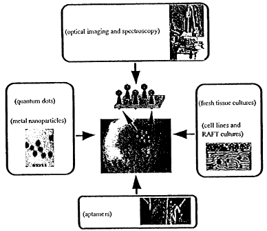
as described herein. Certain antibody conjugates include those intended
primarily for
use in vivo, where the antibody is linked to a optically interrogated agent.
The covalent binding can be achieved either by direct condensation of existing
side chains or by the incorporation of external bridging molecules. Many
bivalent or
polyvalent agents are useful in coupling protein molecules to other particles,
nanoparticles, proteins, peptides or amine functions. Examples of coupling
agents are
carbodiimides, diisocyanates, glutaraldehyde, diazobenzenes, and hexamethylene
diamines. This list is not intended to be exhaustive of the various coupling
agents
known in the art but, rather, is exemplary of the more common coupling agents
that
may be used.
-29-
CA 02478083 2004-09-07
WO 03/075765 PCT/US03/06730
In preferred embodiments, it is contemplated that one may wish to first
derivatize the antibody, and then attach the contrast agent to the derivatized
product.
As used herein, the term "derivatize" is used to describe the chemical
modification of
the antibody substrate with a suitable cross-linking agent. Examples of cross-
linking
agents for use in this manner include the disulfide-bond containing linkers
SPDP (N-
succinimidyl-3-(2-pyridyldithio)propionate) and SMPT (4-succinimidyl-
oxycarbonyl-
a-methyl-a(2-pyridyldithio)toluene).
***
Having described general aspects of concepts relating to optical imaging,
contrast agents, biomarkers, and various types of probes, attention may now be
focused upon the application of those and related techniques to achieve even
further
exemplary, and therefore non-limiting, embodiments of the present invention.
In different embodiments of this disclosure, the following specific steps may
be involved:
(1) A library of probe molecules for cancer specific targets associated with
pre-cancer/cancer cells may be created and characterized, using
molecular engineering and combinatorial chemistry approaches.
(2) Contrast agents may be made based on metal nanoparticles for in vivo
reflectance imaging. The agents may include two major parts:
optically interrogated labels - metal nanoparticles - and probe
molecules specific for cancer biomarkers. The optical properties of
these labels may be synthesized and tailored, and conjugation
chemistry may be used to couple labels and probe molecules.
(3) Contrast agents may be made based on quantum dots for in vivo
fluorescence imaging. These agents may contain quantum dot particles
as optically interrogated labels and probe molecules specific for cancer
-3 0-
CA 02478083 2004-09-07
WO 03/075765 PCT/US03/06730
biomarkers. The optical properties of these labels may be synthesized
and tailored, and conjugates may be made with probe molecules.
(4) If validation of the techniques of this disclosure is desired, molecular
specific contrast agents for pre-cancer detection may be validated in at
least two biological models. Suspensions of normal, pre-cancerous,
and cancerous cervical epithelial cells may be used to assess relative
binding efficiencies of contrast agents. Additionally, using three-
dimensional, tissue phantoms containing multiple layers of epithelial
cells atop a stroma, marker penetration and binding in model systems
of normal, pre-cancerous and cancerous epithelial tissue may be
examined.
(5) If specific testing of the techniques of this disclosure is desired, the
contrast agents and optical imaging techniques may be tested in living
normal and neoplastic cervical tissue. An ideal organ culture system of
1 S normal and pre-cancerous cervix may be used. Biopsies of normal and
neoplastic cervix may be obtained, and transverse sections may
immediately be prepared and maintained as an organ culture. Both
types of contrast agents may be applied and interrogated to determine
relative binding efficiency and penetration throughout the epithelium
in living human cervical tissue.
The activities listed above and throughout this disclosure provide an example
of a new venue for molecular-specific optical imaging modalities for disease
detection
that can be extended to many organ sites, including but not limited to the
cervix.
Methods
FIG. 3 illustrates a general embodiment showing the integration of inter-
disciplinary groups to make photonic probes and contrast agents for highly
sensitive
and selective detection of, for instance, pre-cancers in vivo. The approaches
of
combinatorial chemistry may be used to make a library of aptamer molecules
specific
for biomolecular targets on the surface of cervical cancerous and pre-
cancerous cells.
-31-
CA 02478083 2004-09-07
WO 03/075765 PCT/US03/06730
Aptamers exhibiting antiproliferative and antiangiogenic activity may be used.
Well-
established cervical cell lines at different stages of cancer development may
be used.
Photonic probes based on quantum dots and metal nanoparticles may be made.
They
may utilize custom-made aptamers or existing antibodies for well-known cancer
biomarkers currently used in clinical histopathology. The developed conjugates
may
be used as molecular specific contrast agents using optical microscopy and
spectroscopy. The cervical cancer cell lines, three-dimensional tissue
phantoms, and
fresh cervical tissue slices may all be used for imaging, testing, and/or
validation.
Experiments with all three biological systems representing properties of
normal and
neoplastic cervix at different levels of complexity may be used, if necessary,
to assess
and refine the performance and detection scheme for the contrast agents. This
refinement may include preparing bio-engineered aptamers with high affinity to
cancer specific targets, tailoring optical properties of metal nanoparticles
and quantum
dots, optimizing conjugation procedures, and/or generating optimal imaging
geometries.
The following sections describe even further details associated with four
major
components of embodiments of this disclosure.
Creation of Library of Aptamers Specific for Pre-Cancerous and Cancerous Cells
Recent advancement in combinatorial chemistry provide an excellent tool for
rapid screening of huge populations of biomolecules to find molecules with the
best
binding properties and selectivity to a specific target including whole cells.
In one
embodiment, one may use chemically engineered binding species based on short
nucleic acid sequences (aptamers) to create molecules with improved
selectivity to
pre-cancerous and cancerous cells as compared to the existing antibodies.
In related embodiments, the same methods that have been used to select
aptamers that bind tightly and specifically to protein targets may be used to
select
aptamers that bind to, for instance, tumor markers on the surfaces of cells.
In fact,
aptamers have previously been selected against cellular and organismal
targets. For
-32-
CA 02478083 2004-09-07
WO 03/075765 PCT/US03/06730
example, it has proven possible to use human red blood cell membranes as a
target for
the selection of single-stranded DNA aptamers. Several species of ssDNA were
isolated that recognize distinct targets within the membranes. [89]. In
addition,
aptamers have been selected against whole African trypanosomes. Three classes
of
RNA were selected that bind with high affinity to a protein within the
flagellar pocket
of the parasite. [90].
According to different embodiments, oligonucleotides may be generated that
contain a random sequence core that spans 60 residues and flanking regions
that allow
PCR amplification and in vitro transcription. Following amplification of the
nascent
DNA library to generate double-stranded transcription templates, RNA molecules
may be transcribed that contain 2' fluorinated pyrimidines. The presence of 2'
modified residues has been shown to substantially stabilize nucleic acids
against
endogenous nucleases or other perturbants. For example, RNA molecules
containing
2' modified pyrimidines have previously been stable for days in sera and
urine. [91].
RNA libraries may be gel-purified and directly used for selection. Roughly 100
micrograms (ca. 10'5 different sequences) may be applied to each of the target
cell
lines. Following the equilibration of binding species on the cell surfaces,
non-binding
or weakly binding species may be washed off using PBS. Binding species may be
eluted by homogenizing the cells with detergent. While this procedure of
course
releases cellular nucleic acids, many of those may be destroyed by endogenous
nucleases, may not be amplifiable with particular primer sets, and even if
they are
amplifiable may not be of the same size as the nucleic acid pool. Those
aptamers in
the extract may be directly amplified by reverse transcription and the
polymerase
chain reaction. Products of the correct size may be gel-isolated and used to
transcribe
the sieved RNA population for the next round of selection. In general,
radiolabel
(alpha-32P ATP) may be included in the transcription reaction, and the
fraction of
radioactive RNA that binds to a given cell line may be followed by
scintillation
counting. A relatively small fraction of the population may bind to cells in
the early
rounds of selection, but this fraction may progressively increase during the
course of
the selection.
-33-
CA 02478083 2004-09-07
WO 03/075765 PCT/US03/06730
The procedures described above may be used to identify aptamers that bind to
a given cell line. Those having skill in the art will recognize that any
number of
different cell lines may be targeted. In one embodiment, the following cell
lines may
S be focused upon: two cervical cancer cell lines (HeLa and SiHa), one HPV
infected
cell line (TCL-1), and a normal cervical primary culture from Clonetics (CrEC-
Ec).
In those cell lines, in order to specifically identify aptamers that bind to
pre-cancerous
or cancerous cells, a coupled negative, positive selection may be employed.
First, the RNA population may be mixed with the parental, non-transformed
line, CrEC-Ec, and those RNA species that do not bind to this cell line, or
that are
removed by the initial PBS washes, may be amplified. This delimited population
may
then be added to either the pre-cancerous (TCL-1) or cancerous (HeLa or SiHa
line),
and those RNA species that now bind may be selectively amplified. Multiple
rounds
of coupled negative and positive selection may yield aptamers that bind to
proteins or
epitopes specific to transformed cells. Sequence comparisons within and
between
these families may aid in identifying which residues, motifs, and secondary
structural
features are most significant for binding. Based on such comparative results,
a series
of minimal aptamers may be readily synthesized and assayed for their ability
to bind
to cells. Those minimal aptamers that show the best binding characteristics
may be
selected.
Following the selection, aptamers with terminal functional groups may be
synthesized to facilitate conjugation of the aptamers with nanoparticles.
There are
multiple different phosphoramidite reagents known in the art that can be
introduced
into the synthesis so that upon completion there are either alkyl amino or
alkyl thiol
groups at the 5' or 3' ends of the synthetic nucleic acid.
Contrast Agents Based on Metal Nanoparticles
A potential problem in targeting cancer related biomarkers to screen for and
detect neoplastic changes lies in the fact that many of the biomarkers are
only
-34-
CA 02478083 2004-09-07
WO 03/075765 PCT/US03/06730
overexpressed in tumors. It implies that they are also present in normal
tissue and
their amount increases with cancer development. Embodiments of this disclosure
involve a novel detection scheme that provides enhanced contrast between
normal and
malignant tissue. One concept of these techniques is based on changes in
optical
S properties of metal (or any other highly reflective) nanoparticles when they
form
closely spaced assembles. When gold or silver nanoparticles are brought in
close
vicinity, their plasmon resonances interact with each other. The interaction
results in
a red shift of plasmon resonances of particles' assemblies as compared to the
individual particles (see FIG. 4). The extinction of the assembly in the red
optical
region relative to the position of resonance of the isolated nanoparticles is
significantly higher than the sum of the extinction of individual particles
forming the
assembly. Therefore, excitation and collection conditions may be easily
optimized to
selectively detect closely spaced metal particles and their assemblies in the
presence
of the metal particles which are far apart. Application of this concept to
contrast
enhanced reflectance imaging of tissues is shown in FIG. 5 and FIG. 6.
First, the conjugates of metal nanoparticles with probe molecules specific for
cancer related biomolecular targets may be allowed to interact with tissue and
then the
excess of the unbound contrast agents may be washed. Closely spaced assemblies
of
metal nanoparticles may be formed on the surface of neoplastic cells due to
high
concentration of biomarkers on their surface, while only individual particles
spaced
further apart may be present on normal cells. In this situation, imaging with
a
wavelength optimized for spectral properties of the assemblies may provide
enhanced
contrast between normal and neoplastic tissue.
With respect to this concept, the inventors have conducted an experiment with
biotinilated polystyrene beads labeled with conjugates of silver particles
with
streptavidin. Beads with a high density (see FIG. 5) and a low density of
silver
particle conjugates were prepared and placed on the surface of a quartz prism.
Then
the beads were excited using S 14.5 nm wavelength of Ar+ laser in total
internal
reflection mode. For illustration purposes, the sensitivity of the detector
was reduced
-3 5-
CA 02478083 2004-09-07
WO 03/075765 PCT/US03/06730
so that scattering from the beads with low density of silver conjugates would
not be
seen (see FIG. 6). As can be seen, the beads with closely spaced assemblies of
silver
particles exhibit dramatic scattering while the beads with just a few
particles on the
surface are not visible. Although this experiment may not provide a
quantitative
comparison between scattering properties of individual nanoparticles and their
assemblies, because of significant differences in the amount of particles
adsorbed on
the surface of high and low density beads, it nevertheless illustrates and
describes the
use of metal nanoparticles as contrast probes in reflectance imaging.
In different embodiments, gold and/or silver nanoparticles may be used. Each
of these materials has their own advantages and disadvantages. It has been
reported
that silver particles exhibit higher extinction coefficients and provide
higher
enhancements of the local electromagnetic field and of other effects
associated with
optical excitation of surface plasmon resonances. [92]. However, silver
particles are
1 S not as stable and not as biocompatible as gold nanoparticles. This issue
can be
addressed by encapsulating silver particles inside an inert material. For this
purpose,
a silica coating may be used (see FIG. 7). [68]. This coating stabilizes
particles in
high ionic strength solutions and provides a well-characterized surface for
chemical
immobilization of biomolecules.
Gold and silver colloidal particles may be prepared from chloroauric acid
(HAuCl4) and silver nitrate (AgN03) respectively by using a variety of
reducing
agents including phosphorous, [93] ascorbic acid, [94] sodium citrate, [95-97]
borohydrate. [64,94]. In one embodiment, sodium citrate may be primarily used.
Highly uniform gold colloids with particle sizes ranging from about 10 nm to
ca. 100
nm may be prepared using sodium citrate reduction of chloroauric acid. [95].
This
colloid exhibits a single extinction peak ranging from 500 nm to about 540 nm
depending on the size of the particles (see FIG. 4). Sodium citrate reduction
of silver
nitrate results in a colloidal solution with about 35 nm diameter silver
particles and a
single peak at approximately 410 nm. [96,97]. The distribution of silver
particles is
-36-
CA 02478083 2004-09-07
WO 03/075765 PCT/US03/06730
significantly broader as compared to gold particles; however the procedure is
highly
reproducible from one preparation to another.
Silver particles with narrower distributions and different mean diameters may
be prepared using a starter hydrosol with small silver particles that provides
nucleation centers for growth of bigger silver colloid. [94]. In different
embodiments, any of several standard, well-established procedures may be used
in
conjunction with the production of silver particles.
Positions of surface plasmon resonances of gold and silver may be
significantly altered by using nanocomposite materials described in, for
instance,
[70,71]. The materials include a dielectric optically inert core particle and
an
optically active gold shell and can be prepared in a variety of sizes and in a
highly
uniform fashion. In different embodiments, one may use the methods developed
by
1 S Dr. N. J. Halas and Dr. J. L. West from Rice University in this regard.
FIG. 8 illustrates one embodiment for the preparation of conjugates of gold
particles with cancer specific probe molecules. At least two different types
of probe
molecules may be used: well established antibodies for molecular biomarkers of
cancer and aptamers specifically developed for cancer cells. Aptamers with
antiangiogenic activity may be used. Such aptamers may be used to make
conjugation chemistry with the metal particles for these type of molecules.
For
antibodies, conjugation protocols developed to prepare gold immunostains for
electron microscopy may be used. [72,73].
Briefly, the procedure is based on non-covalent binding of proteins at their
isoelectric point (point of zero net charge) to gold particles. The complex
formation
is irreversible and very stable. In fact, the shelf life of the conjugates is
so long that
the most commonly used gold immunostains can be routinely purchased from major
biochemical companies. However, the described conjugation approach is not
always
successful. [72,73]. Therefore a second conjugation strategy for antibodies
may
-37-
CA 02478083 2004-09-07
WO 03/075765 PCT/US03/06730
include preparation of biotinilated antibody molecules and their consequent
interaction with streptavidin/gold conjugates (see FIG. 8). This approach
takes
advantage of strong biospecific interaction between biotin and streptavidin
and a well-
developed protocol for immobilization of streptavidin on gold particles. [98].
Smaller aptamer molecules may not be directly adsorbed on the gold surface
because that could significantly change their conformation and therefore lead
to loss
of binding properties. Aptamers with thiol terminated alkyl chains may be
directly
attached to the surface of gold particles similar to the procedures described
in [69] for
preparation of DNA probes. For conjugation procedure, one may use a mixture of
thiol terminated aptamers and relatively small mercaptoacetic molecules to
avoid high
density immobilization of the aptamers (see FIG. 8).
The immobilization of antibodies and thiol terminated aptamers on silver
particles may be accomplished using the strategies known in the art. The
silver
conjugates do not have the same stability and shelf life as the gold
conjugates;
however, in one embodiment they may be used to evaluate the silver based
contrast
agents. If silver particles are well suited for a particular application, one
may use the
conjugation protocols for silica capped silver particles (see FIG. 8). The
silica layer
may be formed using tetraethyl orthosilicate (TEOS) and the procedure
described in
detail in [68]. Many silanization reagents (Gelest, Inc.) are available to
introduce
functional groups to silica surface for subsequent immobilization of proteins,
[99]
nucleic acids including aptamers, [100-102] etc. It was shown that binding
properties
of aptamers are preserved after immobilization on glass cover slides [101] and
silica
microspheres. [102].
Contrast Agents Based on Quantum Dots
The unique fluorescence properties of quantum dots (qdots) may be used to
make multi-color contrast agents for fluorescence imaging in vivo. A variety
of
semiconductor nanocrystals, or quantum dots (qdots), with relatively high
quality
optical properties may be produced using solution-phase methods. [81,103,105].
In
-38-
CA 02478083 2004-09-07
WO 03/075765 PCT/US03/06730
one embodiment, the nanocrystal preparations should yield nanocrystals with a
relatively tight size distribution (i.e., a size distribution sufficient to
eliminate
inhomogeneous broadening of the optical and electronic properties),
crystalline cores
with few compositional and structural defects, and well-passivated surfaces.
One successful route to synthesizing semiconductor nanocrystals has been
through arrested precipitation with subsequent size selective precipitation.
[81,103].
Arrested preparation methods rely on binding bulky "inert" ligands to the
particle
surfaces during growth. Thiols have been used as capping ligands in a
relatively
general way since they adsorb to a wide variety of semiconductor materials.
Other
capping ligands include but are not limited to phosphines, amines, and
carboxyl
groups, depending on the chemistry of the inorganic material. The ligand
extending
away from the particle surface determines the particle solubility. Particles
can be
functionalized with either hydrophobic (i.e., alkanes) or hydrophilic
(carboxyl or
amine groups for example) moieties. The nanocrystals are sufficiently stable
that,
once made, chemistry can be done to their surfaces.
In one embodiment, quantum dots may be synthesized using previously
published methods. [104]. Briefly, carboxyl-stabilized CdS nanocrystals may be
synthesized by arrested precipitation at room temperature in an aqueous
solution
using mercaptoacetic acid as the colloidal stabilizer. Nanocrystals may be
prepared
from a stirred solution of CdCl2 (1 mM) in pure water. The pH may be lowered
to 2
with mercaptoacetic acid, and then may be raised to 7 with concentrated NaOH.
Then, NaZS9H20 may be added to the mixture.
In one embodiment, chemical synthetic methods for Si nanocrystals with size-
tunable photoemission color may be used (see FIG. 9). Such methods yield
surface-
passivated Si nanocrystals that exhibit relatively high photoemission quantum
yields
(~23%) and discrete optical transitions in the absorbance spectra, indicative
of size-
monodisperse samples. [105]. High resolution transmission electron microscopy
(HR-TEM) reveals that the nanocrystals are single crystals with apparently
faceted
-39-
CA 02478083 2004-09-07
WO 03/075765 PCT/US03/06730
surfaces. In some embodiments, Si may prove to be more biologically useful due
to
its relative inertness.
Both CdS and Si have strong affinity to thiols; therefore, aptamers with thiol
terminated groups can be directly adsorbed on the qdots using the approach
described
for metal nanoparticles. Also, a variety of functional groups (e.g., carboxy)
can be
introduced on the surface of qdots for subsequent immolization of antibodies
using
cross-linking agents (e.g., ethyl-3-(dimethylaminopropyl)carbodiimide),
similar to the
procedure described in [84]. The qdots can be also encapsulated by a silica
layer and
then the reaction outlined on FIG. 8 may be applied for immobilization.
***
Described below are various testing techniques and issues relating, at least
in
part, to forms of testing that may be used in conjunction with one or more
embodiments of the present disclosure. Such techniques may, for example, allow
for
an assessment of the outcome of different immobilization protocols and/or
simply
verify that a study is proceeding as expected or desired.
Testing - Contrast Agents
According to different embodiments, one may measure absorption for all
prepared nanoparticles as well as excitation/emission spectra of qdots and
scattering
profiles of metal nanoparticles. To measure scattering properties of metal
nanoparticles, a reflectance spectrometer may be used to measure scattering of
cells
and tissue slices. [14]. The range of nanoparticle optical parameters
achievable with
existing preparation methods may be determined to identify
excitation/collection
geometries that can be used for in vivo imaging. The optical properties of
bare
nanoparticles, particles capped with a silica layer or mercaptoalkyl
molecules, and
particles conjugated with biomolecules may be compared. Immobilization of
biomolecules on nanoparticles may be verified using at least three different
techniques: UV-Vis spectroscopy (qdots, metal particles), anisotropic
fluorescence
-40-
CA 02478083 2004-09-07
WO 03/075765 PCT/US03/06730
spectroscopy (qdots), and conjugation assays. UV-Vis spectra of conjugates may
exhibit absorption spectra characteristic for both antibodies and particles;
controls will
have only absorbance peaks of nanoparticles. Heavier conjugates may have
slower
rotation as compared to bare qdots that will result in higher anisotropy in
their
fluorescence. Conjugation assays may also be carned out, where purified target
molecules which specifically bind to probe molecules attached to nanoparticles
may
be added to a suspension of probe/nanoparticles conjugates. This may result in
aggregation of the conjugates which can be easily be monitored by UV-Vis
(metal
nanoparticles) or fluorescence anisotropy (qdots) spectroscopy. All these
methods
may allow for a quick assessment of the outcome of different immobilization
protocols made in accordance with embodiments of this disclosure. Stability of
the
prepared conjugates and bare nanoparticles under physiological conditions (pH,
ionic
strength, etc.) may be also studied.
Biocompatibility of the Contrast Agents
Biocompatibility of the contrast agents disclosed herein may be a very
important issue for in vivo applications. While it has been widely recognized
that
gold and, probably, Si based materials are inert with respect to biological
tissue and
are biocompatible (especially, gold), there are some concerns regarding silver
and
most semiconductor based materials. To address this issue, one may use silica
capped
CdS and silver nanocrystals. Silica is considered to be a biocompatible
material
except for the lung, where it can cause silicoses. In one embodiment, a
topical
application of the contrast agents to cervical epithelium may be used. It is
not
anticipated that nanoparticles can penetrate inside the human body through
layers of
epithelial cells, basal membrane, and stroma. However, if desired, one may
thoroughly study this issue before in vivo measurements are performed on a
particular
living subject. In one embodiment, one may address issues relating to the
penetration
of the contrast agents using RAFT cultures and fresh tissue culture models.
One may
also perform standard cytotoxicity tests.
-41-
CA 02478083 2004-09-07
WO 03/075765 PCT/US03/06730
Testing - Biolo ical SXstems
According to different embodiments, one may assess the interaction between
labeled nanoparticles and normal and neoplastic cervical tissue using at least
3 biologically relevant models of cervical neoplasia: cell suspensions, RAFT
cultures,
and fresh tissue cultures. A similar approach may be used with all model
systems.
First, contrast agents may be applied to the model system by mixing (cell
suspensions) or by topical application (RAFT cultures and fresh tissue slices)
and may
be allowed to interact with their specific targets. The incubation may be
performed at
the temperature characteristic for, for instance, the cervix (37°C).
The contrast agents
may be added in a solution formulated to prevent nonspecific binding of the
probe
molecules to the epithelial cells. High relative concentrations of "non-
specific"
proteins such as bovine serum albumin (BSA) are commonly used for this
purpose.
To determine non-specific binding, one may use "bare" (without attached probe
molecules but with capping layers) qdots and metal nanoparticles as well as
particles
with attached biomolecules which do not specifically bind to cervical
epithelial cells
(e.g., BSA). After incubation, the excess solution of probe molecules may be
washed,
and the optical characteristics of the labeled biological models may be
measured.
Following is a more detailed description for each system.
Cell Suspensions
Suspensions of at least two cervical cancer cell lines (HeLa and SiHa) may be
used, one transformed, HPV infected cell line (TCL-1), and a normal cervical
primary
culture from Clonetics (CrEC-Ec). Quantitative comparison of binding of
different
contrast agents to the cell lines may be carried out. Such experiments may
identify or
confirm the best contrast agents for discrimination between normal, pre-
cancerous,
and cancerous cervical cells. A combination of qdots based contrast agents of
different sizes may be evaluated to improve detection of neoplastic cells in
multi-
color imaging strategies with a single excitation frequency (or multiple
excitation
frequencies). Excitation/emission wavelengths for fluorescence imaging with
qdots
and excitation wavelengths for reflectance imaging with metal nanoparticles
may be
optimized to provide the best contrast between normal and abnormal cells, to
provide
-42-
CA 02478083 2004-09-07
WO 03/075765 PCT/US03/06730
adequate penetration of the cervical epithelium and to match small,
inexpensive laser
diode or LED sources.
To quantify the binding of contrast agents to cells, one may use fluorescence
(qdots) and UV-Vis spectroscopy (metal nanoparticles). Scattering from cells
and
their relatively quick sedimentation in solution can significantly interfere
with
quantitative measurements. Furthermore, optical properties of particles can be
altered
as a result of binding. Therefore, one may centrifuge labeled cells and then
measure
the particles remaining in solution. The amount of bound agent may be
determined
based on the decrease in fluorescence and/or absorption. Standard confocal and
deconvolution fluorescent microscopes and a confocal reflectance microscope
may be
used to image individual cells. Heterogeneity of binding and manner in which
cells
interact with the contrast agents (e.g., if they undergo cellular uptake) may
be
determined.
Biocompatibility of the contrast agents may also be addressed by performing
standard cytotoxicity assays. Labeled cells may be grown using standard cell
culture
techniques to address possible long term effects of the contrast agents on the
cells.
Organotypic (RAFT) Cultures
Conventional cell cultures provide homogeneous samples that can be used to
study changes in normal and transformed epithelial cells. However, these
cultures do
not reflect the complex physical organization of the epithelium and underlying
stroma
present in real tissues. Recent developments in molecular biology, however,
may be
utilized to implement a series of progressively more complex culture models
which
have the desired physical and chemical properties and can be manipulated
through the
neoplastic process.
The maintenance of various tissue components in their normal anatomical
relationship is important for regulation of growth and differentiation.
[106,107].
Tumor cells, stromal fibroblasts, and endothelial cells may express a set of
genes in
-43-
CA 02478083 2004-09-07
WO 03/075765 PCT/US03/06730
situ that only partially overlaps the set expressed by each cell type in
isolation from
the other in primary cultures. Organotypic (RAFT) cultures have been developed
initially for skin and then adapted for a variety of epithelial cancers as an
approach to
provide the three dimensional growth including epithelial cell-cell
interactions that are
major features of solid carcinomas. The method is based on the growth of
epithelial
cells at the air-liquid interface on top of a collagen gel containing
fibroblasts (hence
the name RAFT cultures for floating on the liquid phase). This organ culture
provides
conditions that preserve tissue architecture, growth, and function. It can be
prepared
with different cell layers, different cell types and can be analyzed as a
tissue without
restrictions involved in obtaining actual surgical specimens from patients or
volunteers. RAFT cultures are also more reproducible than tissues obtained
from
different individuals.
In one embodiment, one may prepare RAFT cultures using normal, pre-
cancerous and cancerous cervical epithelial cells. Contrast agents may be
added,
washed, and then cultures may be examined using the optimized
excitation/collection
wavelengths determined from, for instance, experiments with cell suspensions.
The
contrast between images of RAFT cultures with normal, pre-cancerous and
cancerous
cells may be determined. This biological model of the epithelium may also
provide
an opportunity to measure the depth of penetration of the contrast agents
through
multiple layers of epithelial cells and to optimize binding kinetics before
undertaking
more difficult and resource-consuming experiments with human tissue samples.
The
same optical microscopic approaches as in the case of cells may be used.
Fresh Tissue Slices
According to different embodiments, the contrast agents may be tested using a
model system that most closely resembles living human epithelial tissue -
fresh tissue
slices. Dr. Richards-Kortum, Follen and Lotan have recently explored this
model
system to explore the biological basis for differences in the autofluorescence
of
normal and neoplastic cervix. [ 108,109]. To prepare fresh tissue slices,
cervical
biopsies may be obtained, and biopsies may be immediately placed in chilled
culture
-44-
CA 02478083 2004-09-07
WO 03/075765 PCT/US03/06730
medium, and then embedded in agarose. A Krumdieck Tissue Slicer may be used to
obtain transverse, 200 ~m thick fresh tissue slices, which can be maintained
alive in
culture for 7-10 days. Experiments with contrast agents may be performed
within 1.5
to 5 hours of biopsy. The tissue slices can remain in culture medium during
the
S imaging, and an image of a field of medium may be collected as a control.
Following
fluorescence microscopy, 4 pm sections may be made for histological evaluation
and
may be read by, for instance, a board certified pathologist to provide a
diagnosis.
At least two different series of experiments may be performed. In one,
contrast agents may be topically applied to the biopsy prior to preparation of
tissue
slices. Such studies allow one to evaluate the penetration depth of contrast
agents
inside the human epithelium. In the second, contrast agents may be applied to
prepared tissue slices. In this case, one may assess the binding profile of
different
contrast agents throughout the whole thickness of epithelium. Contrast between
normal, pre-cancerous and cancerous lesions in living cervical tissue may be
determined. The best combinations of antibodies and/or aptamers labeled with
qdots
and metal particles for discrimination of pre-cancerous and cancerous lesions
may be
identified or confirmed.
In a specific embodiment, one may initially conduct a pilot study of 18
patients, obtaining paired normal and abnormal biopsies from each patient.
These
data may be used to calculate the required sample size to achieve statistical
significance to determine the sensitivity and selectivity of nanoparticles as
compared
to gold standard of histopathology. One may use a commercially available
inverted
fluorescence microscope and confocal reflectance microscope, which may also be
used to image cervical biopsies and fresh tissue slices.
***
The following additional examples are included to demonstrate specific, non-
limiting embodiments of this disclosure. It should be appreciated by those of
skill in
-45-
CA 02478083 2004-09-07
WO 03/075765 PCT/US03/06730
the art that the techniques disclosed in the additional examples that follow
represent
techniques discovered by the inventors to function well in the practice of the
invention, and thus can be considered to constitute specific modes for its
practice.
However, those of skill in the art should, in light of the present disclosure,
appreciate
that many changes can be made in the specific embodiments which are disclosed
and
still obtain a like or similar result without departing from the spirit and
scope of the
invention.
Example l:
Abstract
Recent developments in photonic technology provide the ability to non-
invasively image cells in vivo; these new cellular imaging technologies may
dramatically improve the prevention, detection and therapy of epithelial
cancers.
Endoscope-compatible microscopies, such as optical coherence tomography and
reflectance confocal microscopy, image reflected light, providing a three-
dimensional
picture of tissue microanatomy with excellent spatial resolution (1 - 10
microns).
However, their ability to image molecular biomarkers associated with cancer
may be
limited. In this example, we describe a new class of molecular specific
contrast
agents for vital reflectance imaging based on gold nanoparticles attached to
probe
molecules with high affinity for specific cellular biomarkers. The application
of gold
bioconjugates for vital imaging of pre-cancers is demonstrated using cancer
cell
suspensions, three-dimensional cell cultures, and normal and neoplastic fresh
cervical
biopsies. We show that gold conjugates can be delivered topically for imaging
throughout the whole epithelium. These contrast agents may extend the ability
of
vital reflectance microscopies for in vivo molecular imaging. They may enable
combined screening, detection and therapy of disease using inexpensive imaging
systems; such tools may allow mass screening of diseases such as cancer in
resource-
poor settings.
-46-
CA 02478083 2004-09-07
WO 03/075765 PCT/US03/06730
Introduction
Early diagnosis of premalignant and malignant lesions is essential for
improving the current poor survival of patients with a variety of cancers. Non
invasive diagnostic methods are especially needed for the screening of large
populations for the identification of high risk individuals who can then be
followed up
frequently and/or enrolled in chemoprevention trials. In the past decade a
number of
microscopic techniques have been developed to image living tissue with sub-
cellular
resolution. Vital microscopies, such as optical coherence tomography (OCT) and
reflectance confocal microscopy (RCM), image reflected light, providing a
detailed
three-dimensional picture of tissue microanatomy without the need for physical
sectioning. These technologies provide excellent spatial resolution (1 - 10
microns)
with penetration depth ranging from 300 microns to 1-2 mm. The resulting
histologic-quality images can identify and monitor neoplastic changes in
epithelium.
Recently, endoscope-compatible fiber optic OCT and RCM systems have been
1 S developed to image tissue microanatomy in vivo in near real time. These
systems are
portable and inexpensive compared to other high resolution imaging
technologies
such as MRI microscopy; as such they are ideally suited for early screening
and
diagnosis of superficial disease.
Tissue reflectance is produced by refractive index mismatches; sources of
contrast in OCT and RCM images include structures with increased refractive
index
such as mitochondria, nuclear chromatin and melanin. Non-specific contrast
agents,
such as acetic acid, can perturb the nuclear refractive index distribution
increasing the
ability to visualize cellular anatomy. While OCT and RCM provide images of
tissue
microanatomy, their ability to image molecular changes associated with
carcinogenesis is limited.
In the last few years, global analysis of gene expression by genomic and
proteomic approaches have led to the discovery of new cancer related genes,
proteins
and biomarkers. Currently, most of these biomolecular signatures can only be
assessed through invasive, painful biopsy. The ability to noninvasively image
the
-47-
CA 02478083 2004-09-07
WO 03/075765 PCT/US03/06730
expression of these biomarkers may translate into improved ability to screen
and
detect neoplastic changes, better ability to select and monitor therapy, and
new tools
to understand the pathobiology of the disease.
Summary
In this example, we demonstrate a new class of molecular specific contrast
agents for vital optical imaging of pre-cancers and cancers, based on gold
nanoparticles conjugated to probe molecules with high affinity for cellular
biomarkers. Conventional gold nanoparticles have been extensively used as
molecular-specific stains in a different application-electron microscopy. As
result,
the fundamental principles of interactions between gold particles and
biomolecules
have been thoroughly studied. The nanoparticles also exhibit the ability to
resonantly
scatter visible and near infrared (NIR light). This property is the result of
excitation
of surface plasmon resonances and is extremely sensitive to the size, shape,
and
aggregation state of the particles. The ability to resonantly scatter visible
and NIR
light may be explored for vital microscopy in living specimens.
In this example, we describe bioconjugates of gold nanoparticles with
monoclonal antibodies against EGFR, a transmembrane 170 kDa glycoprotein that
is
overexpressed in epithelial pre-cancers, for molecular specific optical
imaging. A high
level of EGFR expression is often associated with enhanced aggressiveness of
epithelial cancers and poor prognosis. In these studies we used gold
nanoparticles
with ca. 12 nm in diameter. This size is approximately the same as the size of
antibodies that are routinely used for molecular specific labeling and
targeting.
To demonstrate the application of gold bioconjugates for vital reflectance
imaging
we used three biologically relevant models of cancer with increasing
complexity.
First, suspensions of cervical cancer cells were explored; SiHa cells are well-
characterized cervical epithelial cancer cells that overexpress EGFR. Next,
engineered tissue constructs, three-dimensional cell cultures that mimic major
features
of epithelial tissue, were explored. We prepared engineered tissue constructs
-48-
CA 02478083 2004-09-07
WO 03/075765 PCT/US03/06730
consisting of densely packed, multiple layers of SiHa cells atop a collagen
stroma.
Finally, we demonstrated the application of contrast agents in normal and
neoplastic
fresh cervical biopsies - the model system that most closely resembles living
human
epithelial tissue.
Methods
Preparation of gold bioconjugates:
Colloidal gold of various sizes was prepared using citrate reduction of
chloroauric acid (HAuCl4) according to the method described in Frens, G.
(1973)
Nature Physical Science 241, 20-22, which is incorporated by reference. To
prepare
conjugates colloidal gold was diluted twice in 20 mM HEPES buffer, pH 7.4 and
anti-
EGFR monoclonal antibodies (host mouse, Sigma) were reconstituted in the same
buffer at 100 pg/ml. Then the solutions were mixed at 1:1 volume ratio and
were
allowed to interact for 20 minutes at room temperature. Polyethyleneglycol
(PEG,
MW 20,000, Sigma) was added to the mixture up to a final concentration of 0.2
mg/mL and the solution was centrifuged twice at 5000 rpm for 2 hours to wash
unbound antibodies. After the second wash the pellet was resuspended in
phosphate
buffered saline.
Preparation of cells:
SiHa cells were grown inside tissue culture flasks covered with collagen type
I
(Roche) in DMEM plus S% FBS at 37°C under 5% C02. Cells were harvested
using
1 mg/mL collagenase (Roche) in phosphate buffered saline at 37°C for
approximately
20 minutes, or until the collagen substrate was entirely disassociated, and
were
washed in DMEM. The cell suspension was labeled with gold conjugate at room
temperature for ca. 30 minutes on a shaker to prevent sedimentation. The
labeled
cells were placed on top of a microscope slide coated with gelatin to
eliminate
background scattering from the glass substrate during reflectance imaging.
-49-
CA 02478083 2004-09-07
WO 03/075765 PCT/US03/06730
Preparation of epithelial tissue constructs:
To prepare the constructs a suspension of epithelial cells was spun down and a
very small amount of buffered collagen type I solution (3 mg/ml) was added to
the
pellet. The mixture was transferred to 6.5 mm Elisa plate wells and allowed to
gel at
37°C for 20 minutes. The volume of the mixture was adjusted to form
gels with
thickness between 400 and 600 Vim. The gel with embedded cells was kept in
DMEM
culture medium plus 5% FBS for 24-48 hours. During this time the cells
continued to
grow resulting in formation of a highly dense structure consisting of multiple
layers of
epithelial cells. The contrast agents were added on top of the tissue phantoms
in 10%
polyvinyl pyrrolidone (PVP) solution in PBS or in pure PBS. After incubation
for ca.
30 minutes at room temperature the phantoms were transversely sectioned with a
Krumdieck tissue dicer and the sections were imaged using Zeiss Leica inverted
laser
scanning confocal microscope.
Preparation of fresh cervical biopsies:
Colposcopically normal and abnormal cervical biopsies were obtained, with
written consent, from women seen in the University of Texas M.D. Anderson
Cancer
Center Colposcopy Clinic. Biopsies were immediately placed in chilled
(4°C) culture
medium (Dilbecco Modified Eagle Medium without phenol red), and then embedded
in 4% agarose. Subsequently, a Krumdieck Tissue Slicer was used to obtain
transverse, 200 ~m thick fresh tissue slices. The slices were placed in a
phosphate
buffered saline solution of anti-EGFR/gold conjugates for ca. 30 minutes at
room
temperature. After incubation with contrast agents the sections were washed in
PBS
and were imaged. After imaging the sections were submitted for H&E staining
and
histopathological analysis.
The wavelength dependence of light scattering was measured using the optical
set-up described in Sokolov, K., Drezek, R., Gossage, K., & Richards-Kortum,
R.
(1999) Optics Express 5 (13), 302-317, which is incorporated by reference.
Briefly,
samples were illuminated by a broad-band light source (halogen lamp, Dolan-
Jenner
Industries) and the scattered light was focused on the 250 pm entrance slit of
a single
-50-
CA 02478083 2004-09-07
WO 03/075765 PCT/US03/06730
grating spectrograph (F/3.8, 300 lines/mm grating, Monospec 18, Jarrel Ash)
coupled
to an intensified photodiode array detector (IRY-700, Princeton Instruments).
Spectra
were normalized by scattering from a "white" diffusely scattering substrate
(Labsphere) to account for the wavelength dependence of the light source and
the
spectrometer.
Confocal microscopy:
The series of through focus confocal images were acquired using Zeiss Leica
inverted epi-fluorescence/reflectance laser scanning confocal microscope with
a 40X
oil immersion objective or a lOX objective. The excitation was provided by a
Kr/Ar
mixed gas laser.
Figure Legends for Results and Discussion:
In the "Results and Discussion" section immediately below, reference is made
to FIGS. 10-13. Corresponding figure legend are as follows:
FIG. 10:
Scattering properties of gold nanoparticles are shown. FIGS. l0A and lOB
compare scattering of gold particles and polystyrene beads of approximately
the same
diameter. In FIG. l0A suspensions of gold particles (left) and the polymeric
spheres
(right) were illuminated by a laser pointer which provides light in 630-680 nm
region.
The images were obtained using a regular web camera at a 90° angle
relative to
illumination. To acquire the images of both suspensions under the same
conditions,
the concentration of the polymeric beads (in particles per ml) was increased 6
fold
relative to the concentration of the metal nanoparticles. FIG. lOB shows the
wavelength dependence of visible light scattering by the polystyrene spheres
and the
gold nanoparticles. The spectra were obtained from suspensions with the same
concentration of metal and polymeric nanospheres.
FIG. lOC compares scattering of isolated and closely spaced (agglutinated)
conjugates of 12 nm gold nanoparticles with monoclonal antibodies for
epidermal
-S1-
CA 02478083 2004-09-07
WO 03/075765 PCT/US03/06730
growth factor receptor (EGFR). Polyclonal antibodies specific for mouse IgG
(Sigma) were added to induce agglutination of the conjugates.
FIG. 71:
High (FIGS. 11A-11D) and low (FIGS. 11G-11I) resolution optical images of
SiHa cells labeled with anti-EGFR/gold conjugates are shown. Non-specific
labeling
using gold conjugates with BSA is shown in FIGS. 11E and 11F. Laser scanning
confocal reflectance (FIGS. 11 A, 11 C, and 11 E) and combined confocal
reflectance/transmittance (FIGS. 11B, 11D, and 11F) images of the labeled SiHa
cells
obtained with 40X objective. The scattering from gold conjugates is false-
colored in
red. In FIGS. 11A and 11B the focal plane is at the top of the cells. In FIGS.
11C
and 11D the middle cross-section of the cells is in focus. The confocal
reflectance
and transmittance images were obtained independently and then overlaid.
Reflectance images were obtained with 647 nm laser excitation. The scale bar
is ca.
20 pm (FIGS. 11A-11F).
FIGS. 11G-11I: a series of bright-field and reflectance images of the labeled
SiHa cells obtained with 20X objective using a combination of a white light
and a
laser-pointer illumination are shown: FIG. 11G white light illumination; FIG.
11H
white light with a laser-pointer illumination at grazing incidence; FIG. 11I
laser-
pointer illumination at grazing incidence. The scattering of gold conjugates
is false-
colored in red. The laser pointer emits light in 630-680 nm region with power
output
less than S mW. The laser pointer illuminated an area ca. 3-5 mm in diameter.
The
scale bar is ca. 30 pm.
FIG. 12:
Laser scanning confocal reflectance (FIGS. 12A, 12C, and 12E) and confocal
fluorescence (FIGS. 12B and 12D) images of pre-cancerous (FIGS. 12A and 12B)
and
normal (FIGS. 12C, 12D, and 12E) fresh cervical ex vivo tissue labeled with
anti-
EGFR/gold conjugates are shown. Reflectance images were obtained with 647 nm
excitation wavelength and fluorescence images using 488 nm excitation and 515
nm
-52-
CA 02478083 2004-09-07
WO 03/075765 PCT/US03/06730
long band-pass emission filter. Reflectance images FIG. 12A and 12C were
obtained
after labeling with gold conjugates under the same acquisition conditions.
FIG. 12E
was obtained after 6% acetic acid (AA) solution was added to the normal
cervical
biopsy and laser power was increased by ca. 6 fold. AA is a nonspecific
contrast
agent that is used in reflectance imaging of epithelium to increase scattering
from
nuclei. Confocal fluorescence images FIGS. 12B and 12D were obtained under the
same acquisition conditions. The reflectance images are false-colored in red.
The
scale bar is ca. 20 p.m.
FIG. 13:
Transmittance (FIGS. 13A, 13C, and 13E) and reflectance (FIGS. 13B and
13D) images of engineered tissue constructs labeled with anti-EGFR/gold
conjugates
are shown. The tissue constructs consist of densely packed, multiple layers of
cervical cancer (SiHa) cells. The contrast agents were added on top of the
tissue
phantoms in 10% polyvinyl pyrrolidone (PVP) solution in PBS (FIGS. 13A and
13B)
or in pure PBS (FIGS. 13C and 13D). After incubation for ca. 30 minutes at
room
temperature the phantoms were transversely sectioned with a Krumdieck tissue
slicer
and the sections were imaged using the Zeiss Leica inverted laser scanning
confocal
microscope with lOX (FIGS. 13A-13D) objective. A small spot on a tissue
construct
was imaged using 40X oil immersion objective to show high density of the
epithelial
cells in the phantom (FIG. 13E). Reflectance images were obtained with 647 nm
excitation. Arrows show the surfaces exposed to the contrast agents. The scale
bars
are ca. 200 ~.m (FIGS. 13A-13D) and ca. 20 pm (FIG. 13E).
Results and Discussion
The scattering cross section of gold nanoparticles is extremely high compared
to polymeric spheres of the same size (FIG. 10), especially in the red region
of the
spectrum. This property is important for development of contrast agents for
optical
imaging in living organisms because light penetration depth in tissue
dramatically
increases with increasing wavelength. Another interesting optical property of
gold
nanoparticles that can be exploited for vital optical imaging is the increase
in
scattering cross section per particle when the particles agglutinate (FIG.
lOC). These
-53-
CA 02478083 2004-09-07
WO 03/075765 PCT/US03/06730
changes produce a large optical contrast between isolated gold particles and
assemblies of gold particles. This increase in contrast improves the ability
to image
markers which are not uniquely expressed in diseased tissue, but are expressed
at
higher levels relative to normal tissue (such as EGFR), and to develop highly
sensitive
labeling procedures which do not require intermediate washing steps to remove
single
unbound particles.
The preparation of gold bioconjugates may be based on non-covalent binding
of the anti-EGFR IgG antibodies at their isoelectric point (point of zero net
charge of
the protein) to gold particles. The complex formation is irreversible and very
stable.
Specific optical changes in IJV-Vis spectrum of gold nanoparticles indicate
binding of
the antibodies: a characteristic red shift (ca. 6 nm) of the maximum of the
surface
plasmon resonance and ca. 10 % decrease in transmission. These optical changes
are
associated with alterations in the local refractive index around the particles
after
binding of the monoclonal antibodies. An additional indication of protein
binding to
the surface of the nanoparticles is their stability in phosphate buffered
saline (PBS).
The gold conjugates are monodispersed in the saline solution while a
suspension with
"bare" gold particles quickly changes its color from red to blue upon addition
of the
saline as a result of aggregation of the nanoparticles. The anti-EGFR/gold
complexes
also undergo molecular specific agglutination when anti-IgG polyclonal
antibodies
are added to the suspension of the conjugates. The agglutination results in
increased
scattering by the conjugates (FIG. l OC).
FIGS. 11A-D show confocal reflectance images and combined
transmittance/reflectance images of SiHa cells labeled with anti-EGFR/gold
conjugates. In a series of through focus confocal reflectance images of
labeled cells,
the bound conjugates first appear as randomly distributed bright spots at the
top of the
cells, then bright rings can be seen in the optical cross-sections through the
middle of
the cells. Comparison of the labeling pattern with transmittance images of the
cells
indicates that labeling predominately occurs on the surface of the cellular
cytoplasmic
membrane. The labeling pattern is consistent with the fact that the monoclonal
-54-
CA 02478083 2004-09-07
WO 03/075765 PCT/US03/06730
antibodies have molecular specificity to the extracellular domain of EGFR. The
intensity of light scattering from the labeled SiHa cells is ca. 50 times
higher than
from unlabeled cells. Therefore unlabeled cells cannot be resolved on the dark
background. No labeling was observed when gold conjugates with bovine serum
albumin (BSA) were added to the cells (FIGS. 11E and 11F).
We conducted reflectance imaging before and after the unbound gold
conjugates were washed from the cell suspension. The unbound gold particles
were
not visible before or after washing. UV-Vis measurements of a washed
suspension of
labeled cells showed an increase in extinction of the nanoparticles in the red
optical
region. This change is characteristic for agglutination of the nanoparticles
and
indicates that the particles form closely spaced assemblies on the surface of
the cells.
We demonstrated that agglutination of anti-EGFR gold conjugates results in
increase
of scattering of particles forming the assembly (FIG. lOC). We believe that
similar
effect can contribute to the contrast between the labeled cells and the
isolated,
unbound conjugates.
Using UV-Vis spectroscopy we estimated the average amount of gold
conjugates bound per cell. Scattering from cells, their relatively quick
sedimentation,
and changes of optical properties of the particles upon binding make it
difficult to
measure the amount of bound nanoparticles directly. Instead, we centrifuged
the
labeled cells and measured the decrease in optical density of the supernatant
relative
to the original suspension of the conjugates. Using this approach we
calculated that
approximately 5x10⁴ conjugates are bound per cell. Our results correlate well
with
previously published studies, which report that most cell types express from
2x10⁴ to
20x 104 EGF receptors per cell.
We observed heterogeneous labeling of SiHa cells in suspension. To ensure
that preparation of cell suspensions did not affect the extracellular domain
of EGFR
and produce heterogeneous labeling, we grew cells on collagen and used
collagenase
to harvest the cells. The same heterogeneity was also observed when the cells
were
-55-
CA 02478083 2004-09-07
WO 03/075765 PCT/US03/06730
labeled directly on the surface of the collagen matrix without harvesting.
Heterogeneity of protein expression in cell lines is not uncommon and has been
described before in the case of EGFR.
The light scattering from the labeled cells is so strong that it can be easily
observed using low magnification optics and an inexpensive light source such
as a
laser pointer. FIGS. 11G-1 lI show a series of images of labeled SiHa cells
placed on
a microscope slide obtained using a 20X objective. In bright-field
transmission, the
cells with bound gold conjugates appear darker due to light absorption by the
metal
nanoparticles in the green optical region and the unlabeled cells appear more
transparent (FIG. 11 G). When the sample is illuminated by a laser pointer at
grazing
incidence, the labeled cells appear bright due to scattered light (FIG. 11H).
Finally,
after bright-field illumination is turned off, only labeled cells can be seen
(FIG. 11I).
No scattering was observed when cells labeled using gold conjugates with BSA
were
illuminated by a laser pointer under the same conditions.
Bright "honey-comb" like structures can be seen in laser scanning confocal
reflectance images of abnormal cervical biopsies labeled with anti-EGFR/gold
complexes (FIG. 12A). Scattering from the labeled cytoplasmic membranes of
epithelial cells forms this pattern. No labeling of the normal biopsy can be
seen when
the sample is imaged under the same acquisition conditions as the abnormal
sample
(FIG. 12C). The morphology of the normal biopsy can be resolved after addition
of a
non-specific contrast agent - acetic acid - and increasing the laser power by
ca. 6 fold
(FIG. 12E). Acetic acid enhances fluctuations in the nuclear refractive index
related
to chromatin texture enhancing scattering from nuclei. An increase in
scattering of
stroma is also evident (FIG. 12E). There is no binding of anti-EGFR/gold
conjugates
to the stromal layer of cervical biopsies.
In corresponding autofluorescence confocal images obtained using 488 nm
excitation, the epithelial cells exhibit cytoplasmic fluorescence (FIGS. 12B
and 12D)
due to mitochondria) flavin adenine dinucleotide (FAD). In the fluorescence
image of
-56-
CA 02478083 2004-09-07
WO 03/075765 PCT/US03/06730
the abnormal cervical biopsy the epithelial cells appear to be surrounded by
black
contours (FIG. 12B). These contours are formed by the bound gold conjugates
which
strongly absorb visible light in the green optical region where most of the
autofluorescence signal is emitted (FIGS. lOB and lOC). The comparison of the
reflectance and the fluorescence confocal images of the abnormal biopsy
confirms
predominant binding of the anti-EGFR/gold conjugates to the cytoplasmic
membrane
of the epithelial cells (FIGS. 12A and 12B).
Thus, the contrast agents presented here, coupled with vital reflectance
microscopies, may yield both anatomic and molecular images of epithelial
pathology.
A particularly important application is the early detection of precancerous
lesions.
Early detection of curable precancers may dramatically reduce the incidence
and
mortality of cancer. In vivo application of these contrast agents requires the
ability to
deliver the agents throughout the epithelium in the organ site of interest.
Pre-cancers
of squamous epithelium originate at the basal layer, which can be located 300-
500 ~,m
beneath the tissue surface; therefore, to use diagnostic tools and to study
the earliest
molecular changes associated with cancer progression it is important to
deliver the
gold nanoparticles throughout the whole epithelium.
Using engineered tissue constructs, we demonstrated that penetration
enhancers used for topical drug delivery, such as polyvinyl pyrrolidone (PVP),
may
be used to deliver the gold nanoparticles throughout the epithelium (FIG. 13).
PVP is
approved by FDA for human use as an excipient in topical formulations (e.g.
Povidone). The anti-EGFR/gold conjugates were applied to the top of engineered
tissue constructs in pure PBS buffer and in PBS in the presence of 10% PVP.
After
ca. 30 minutes incubation, constructs were washed in PBS and 200 p.m thick
transverse sections were prepared and imaged using transmittance and confocal
reflectance microscopies (FIG. 13). When the conjugates are applied in the
presence
of PVP, uniform labelling is achieved throughout the whole depth (ca. 400 pm)
(FIGS. 13A and 13B). When gold conjugates are applied in PBS, only the surface
-57-
CA 02478083 2004-09-07
WO 03/075765 PCT/US03/06730
layer of epithelial cells in the engineered tissue constructs is labelled
(FIGS. 13C and
13D).
The contrast agents presented here indicate the ability to extend vital
reflectance microscopies for in vivo molecular imaging. Using these contrast
agents,
we demonstrated the ability to image the distribution of EGFR expression in
living
neoplastic cervical tissue - providing the ability to assess molecular
pathology in vivo.
Currently, the prognosis of patients with cancer is predicted mainly based on
microanatomic features of disease; however, the use of molecular markers has
recently shown promise to better predict patient outcomes and to select
therapies. In
the absence of contrast agents, OCM and RCM yield images of tissue
microanatomy
similar to that which can be obtained with conventional histopathology;
contrast
agents based on gold nanoparticles provide a strong source of signal with
molecular
specificity that is immune to photobleaching.
Other reflectance based technologies which been developed to image disease
in deeper tissues with lower spatial resolution may also benefit from these
contrast
agents. Diffuse optical tomography (DOT) allows noninvasive in vivo imaging of
oxygenated and deoxygenated hemoglobin and has been explored for detection of
breast cancer; coupling DOT with the contrast agents presented here may
provide
more sensitive detection of smaller lesions.
Many properties of contrast agents based on gold nanoparticles make them
ideally suited for vital imaging and in vivo diagnosis. By appropriately
adjusting the
size of the particles, surface plasmon resonances can be selected to take
advantage of
regions where tissue is most transparent depending on the degree of tissue
penetration
required. Using particles of different sizes conjugated to different probe
molecules,
mufti-color labeling for many targets can be achieved. The enhanced scattering
from
closely spaced gold particles confers important advantages for in vivo
imaging. First,
the scattering from aggregates of bound particles is greatly enhanced compared
to
background scattering from unbound particles. Additionally, many markers are
not
-58-
CA 02478083 2004-09-07
WO 03/075765 PCT/US03/06730
uniquely expressed in disease,. but are over- or under-expressed. The
scattering from
closely spaced aggregates associated with over-expression can magnify the
signal
difference owing to moderate levels of over-expression.
Contrast agents based on gold nanoparticle antibody conjugates may be put to
in vivo use, with topical or systemic delivery. The inherent biocompatibility
of gold
means that they can be used directly in vivo without the need for protective
layer
growth. In fact, long term treatment of rheumatoid arthritis utilizes gold (up
to a
cumulative dose of 1.2-1.8 g/year for up to 10 years). We anticipate that less
than 0.3
mg of gold may be required for diagnosis with topical delivery to the cervix.
Humanized antibodies, where a mouse antibody-binding site is transferred to a
human
antibody gene, are much less immunogenic in humans, and many humanized
antibodies are currently in clinical trials. Since 1997, the FDA has approved
more
than 10 monoclonal antibody based drugs, including Herceptin for metastatic
breast
cancer therapy. For surface lesions located in epithelial tissue, simple FDA
approved
agents, such as polyvinylpyrrolidone can be used to increase tissue
permeability and
deliver contrast agents topically.
Example 2:
Metal Nanoparticles and MMP
To demonstrate the imaging of metallo-proteases (MMPs) using contrast
agents based on metal nanoparticles, we prepared gold conjugates with
monoclonal
antibodies for MMP-2 (data not shown) and MMP-9. The conjugates were used to
label cervical epithelial cancer cells grown on two different substrates: a
pure collagen
I gel and a collagen I gel in the presence of 5% gelatin. SiHa cells were
placed on a
substrate and allowed to grow for S-24 hrs in DMEM with 5% FBS at 37°C
and 5%
CO2, and then antibody-gold conjugates were applied to a sample in PBS for 20-
30
minutes under sterile conditions. Excess contrast agent was removed and the
sample
was imaged using a confocal reflectance/fluorescence microscope without
intermediate washing. Reflectance images were obtained with 647 nm excitation
and
fluorescence was excited at 360 nm and collected using 405 band-pass emission
filter.
-59-
CA 02478083 2004-09-07
WO 03/075765 PCT/US03/06730
FIG. 14 shows the results of labeling of SiHa cells grown on collagen I
substrate with anti-MMP-9/gold conjugates. Significant labeling of cellular
cytoplasm was observed. Cytoplasmic labeling may be associated with
internalization of labeled MMP-9 molecules from the plasma membrane. Rapid
internalization and degradation of MMPs including MMP-9 is an important
mechanism in regulating extracellular proteinase activity. We also observed
strong
labeling of collagen fibers located along clusters of cells (FIG. 14A and
14C).
Previously, similar labeling was observed using fluorescent labeled antibodies
in
fixed 3D tissue cultures of cells inside collagen I matrix. It was attributed
to
membrane deposits which are shed by migrating cells along their tracks in
collagen
matrix. These deposits contain a variety of plasma membrane proteins including
MMPs. Our images show a number of elongated polarized cells, indicative of
cellular
migration. Little cell or ECM labeling was observed in areas with low cell
density.
Metal Nanoparticles and Intracellular Targets
We can deliver metal nanoparticle based contrast agents to image intracellular
targets. Contrast agents may be utilized to image molecular features of human
papillomavirus (HPV) induced cervical carcinogenesis. Persistence and
progression
of cervical cancer is clearly related to expression of the viral oncoproteins,
E6 and E7.
FIG. 1 S shows preliminary results obtained labeling SiHa cells with 10 run
gold nanoparticles conjugated to anti-E7 monoclonal antibodies. Cells were
incubated in contrast agent in PBS and in PBS with 10% PVP. Following
incubation,
cells were washed and imaged using laser scanning confocal microscopy. FIG. 15
shows the co-localized autofluorescence (green) and reflectance (white) images
from
SiHa cells incubated with PVP. Autofluorescence is limited to the cytoplasm,
whereas backscattering is produced by nanoparticles within the nucleus. No
backscattering was observed in cells incubated with contrast agent in PBS
alone.
Thus, delivery and detection of contrast agent is feasible. In data, we
observed at
least a three fold, statistically significant increase in nuclear
backscattering from
-60-
CA 02478083 2004-09-07
WO 03/075765 PCT/US03/06730
nanoparticles in cells with high E7 expression compared to cells with low E7
expression.
Safety of Metal Nanoparticles for In Yivo Use
Contrast agents based on gold nanoparticle antibody conjugates may be used
in vivo, with topical or systemic delivery. The inherent biocompatibility of
gold
implies they can be used directly in vivo without the need for protective
layer growth.
In fact, long term treatment of rheumatoid arthritis utilizes gold (up to a
cumulative
dose of 1.2-1.8 g/year for up to 10 years). We anticipate that less than 0.3
mg of gold
may be required for diagnosis with topical delivery to the cervix. Humanized
antibodies, where a mouse antibody-binding site is transferred to a human
antibody
gene, are much less immunogenic in humans, and many humanized antibodies are
currently in clinical trials. Since 1997, the FDA has approved more than 10
monoclonal antibody based drugs, including Herceptin for metastatic breast
cancer
therapy. For surface lesions located in epithelial tissue, simple FDA approved
agents,
such as polyvinylpyrrolidone can be used to increase tissue permeability and
deliver
contrast agents topically.
Optically active contrast agents to target the molecular signatures of
neoplasia.
Metal NPs
Vital reflectance imaging using both gold and silver nanoparticles may be
accomplished using the techniques of this invention. Contrast agents based on
gold
nanoparticles may be used because gold is biocompatible and can be used
directly for
in vivo applications. Silver particles may be used also. Silver may exhibit
higher
extinction coefficients and provide higher enhancements of the local
electromagnetic
field and exhibit other effects associated with optical excitation of surface
plasmon
resonances. Silver particles are not as stable and not as biocompatible as
gold
nanoparticles. This issue can be addressed by encapsulating silver particles
inside an
inert material. One may use a silica coating developed by Dr. Sokolov in
Sokolov,
K., G. Chumanov, and T.M. Cotton, Enhancement of Molecular Fluorescence near
the
Surface of Colloidal Metal Films. Analytical Chemistry, 1998. 70(18): p. 3898-
3905,
-61-
CA 02478083 2004-09-07
WO 03/075765 PCT/US03/06730
which is incorporated by reference. This coating stabilizes particles in high
ionic
strength solutions and provides a well characterized surface for chemical
immobilization of biomolecules.
Gold and silver colloidal particles can be prepared from chloroauric acid
(HAuCl4) and silver nitrate (AgN03) respectively by using a variety of
reducing
agents including phosphorous, ascorbic acid, sodium citrate, borohydrate. In
one
embodiment, sodium citrate may be used. Highly uniform gold colloids with
particle
sizes ranging from about 10 nm to ca. 100 nm can be prepared using sodium
citrate
reduction of chloroauric acid. This colloid exhibits a single extinction peak
ranging
from 500 nm to about 540 nm depending on the size of the particles. Sodium
citrate
reduction of silver nitrate results in a colloidal solution with about 35 nm
diameter
silver particles and a single peak at approximately 410 nm. The distribution
of silver
particles is significantly broader as compared to gold particles; however the
procedure
is highly reproducible from one preparation to another. Silver particles with
narrower
distributions and different mean diameters can be prepared using a starter
hydrosol
with small gold or silver particles that provides nucleation centers for
growth of
bigger silver colloid.
Scattering properties of metal nanoparticles depend on their size and shape.
By changing sizes metal nanoparticles that exhibit different colors in
reflected light
can be produced. Additionally, position of surface plasmon resonances of gold
and
silver can be significantly altered by using nanocomposite materials described
in (a)
Sershen, S.R., et al., Temperature-sensitive polymer-nanoshell composites for
photothermally modulated drug delivery. Journal of biomedical materials
research,
2000. 51(3): p. 293-8, and (b) Averitt, R.D., S.L. Westcott, and N.J. Halas,
The
Linear Optical Properties of Gold Nanoshells. J. Opt Soc Am B, 1999. 16: p.
1824-32,
each of which is incorporated by reference. The materials include a dielectric
optically inert core particle and an optically active gold shell and can be
prepared in a
variety of sizes and in a highly uniform fashion.
-62-
CA 02478083 2004-09-07
WO 03/075765 PCT/US03/06730
Another approach to tune optical properties of metal nanoparticles is based on
template synthesis such as that in Haes, A.J. and R.P. Van Duyne, A nanoscale
optical
biosensor: Sensitivity and selectivity of an approach based on the localized
surface
plasmon resonance spectroscopy of triangular silver nanoparticles. Journal of
the
American Chemical Society, 2002. 124(35): p. 10596-10604, which is
incorporated
by reference. In one method, metal nanoparticles of pyramidal shape with
different
sizes can be synthesized inside cavities formed by a dense monolayer of
polystyrene
beads on a flat substrate. After synthesis the nanoparticles can be removed
from the
surface by a simple one step procedure. These approaches may be used to
optimize
scattering properties of nanoparticles to take advantage of optical regions
where tissue
is most transparent depending on the degree of tissue penetration required.
Another venue may be to use particles of different sizes conjugated to
different probe molecules to achieve multi-color labeling for many targets.
FIG. 8 summarizes embodiments for preparation of conjugates of gold
particles with cancer specific probe molecules. At least two different types
of probe
molecules may be used: well established commercially available antibodies for
molecular biomarkers of cancer and peptides specifically developed to bind to
cancer
biomarkers. One may utilize humanized antibodies. For antibodies, one may use
conjugation protocols developed to prepare gold immunostains for electron
microscopy. See Horisberger, M., Colloidal gold : a cytochemical marker for
light
and fluorescent microscopy and for transmission and scanning electron
microscopy.
Scan Electron Microsc, 1981. 2: p. 9-31 and Geoghegan, W.D. and G.A. Ackerman,
Adsorption of horseradish peroxidase, ovomucoid and anti-immunoglobulin to
colloidal gold for the indirect detection of concanavalin A, wheat germ
agglutinin and
goat anti-human immunoglobulin G on cell surfaces at the electron microscopic
level:
a new method, theory and application. The journal of histochemistry and
cytochemistry : official journal of the Histochemistry Society, 1977. 25(11):
p. 1187-
200, each of which is incorporated by reference. Briefly, the procedure is
based on
non-covalent binding of proteins at their isoelectric point (point of zero net
charge) to
-63-
CA 02478083 2004-09-07
WO 03/075765 PCT/US03/06730
gold particles. The complex formation is irreversible and very stable. In
fact, the
shelf life of the conjugates is so long that the most commonly used gold
immunostains
can be routinely purchased from major biochemical companies. A colloidal gold
drug
delivery system was recently used to specifically target tumors in live mice.
The
reported results demonstrate stability of gold bioconjugates in vivo.
A second conjugation strategy for antibodies may include preparation of
biotinylated antibody molecules and their consequent interaction with
streptavidin/gold conjugates (FIG. 8). This approach takes advantage of strong
biospecific interaction between biotin and streptavidin and a well-developed
protocol
for immobilization of streptavidin on gold particles. Smaller peptide
molecules can
not be directly adsorbed on the gold surface because that could significantly
change
their conformation and lead to loss of binding properties. Peptides with thiol
terminated alkyl chains may be directly attached to the surface of gold
particles
similar to the procedures described in Elghanian, R., et al., Selective
colorimetric
detection of polynucleotides based on the distance-dependent optical
properties of
gold nanoparticles. Science, 1997. 277(5329): p. 1078-1080 (incorporated by
reference) for preparation of DNA probes. For conjugation procedure one may
use a
mixture of thiol terminated peptides and relatively small mercaptoacetic
molecules to
avoid high density immobilization of the peptides (FIG. 8).
The immobilization of antibodies and thiol terminated peptides on silver
particles may be accomplished using strategies similar to those utilized for
gold
particles. Silver conjugates may not have the same stability and shelf life as
the gold
conjugates, however they can be used to evaluate the silver based contrast
agents.
Conjugation protocols for silica capped silver particles may be formed (FIG.
8). The
silica layer may be formed using tetraethyl orthosilicate (TEOS). See Sokolov,
K., G.
Chumanov, and T.M. Cotton, Enhancement of Molecular Fluorescence near the
Surface of Colloidal Metal Films. Analytical Chemistry, 1998. 70(18): p. 3898-
3905,
which is incorporated by reference. Many silanization reagents (Gelest, Inc.)
are
-64-
CA 02478083 2004-09-07
WO 03/075765 PCT/US03/06730
available to introduce functional groups to silica surfaces for subsequent
protein
immobilization.
For vital imaging with contrast agents based on metal nanoparticles it may be
important to develop bioconjugates that have very low nonspecific binding and
are
not accumulated by the reticuloendothelial system (RES), namely the liver and
spleen.
To address this issue one may prepare hybrid conjugates by co-adsorbing
polyethylene glycol (PEG) and probe (antibodies, peptides) molecules on the
surface
on nanoparticles. This strategy has been recently demonstrated in experiments
on in
vivo molecular specific imaging of embryogenesis using quantum dots and in
colloidal gold drug delivery system in live mice. One may use commercially
available thiol terminated PEG from Shearwater Polymers, AL. In the case of
antibodies, one may first prepare conjugates of gold nanoparticles with the
proteins
and then expose the prepared complexes to thiol-PEG. Because strong
interactions
1 S between thiol groups and the metal surface can lead to replacement of
bound
antibodies by PEG molecules it is important to carefully control PEG co-
adsorption.
This can be achieved by adjusting PEG concentration and by using compounds
with
high affinity to thiol groups to timely terminate the reaction. Very small
(few
nanometers in diameter) gold nanoparticles may be used to inhibit free thiol
molecules. The smaller gold particles and their complexes with PEG may be
isolated
from the gold immunoconjugates by centrifugation. Another possibility is to
use zinc
(Znz+) ions which can form stable complexes with thiol groups. To co-adsorb
thiol
terminated peptide molecules and thiol-PEG, one may mix the two compounds
together and apply them to gold nanoparticles at the same time. In this case
the
reaction can be simply controlled by adjusting relative amount of peptides and
PEG
molecules.
***
With the benefit of the present disclosure, those having skill in the art will
comprehend that techniques claimed herein may be modified and applied to a
number
-65-
CA 02478083 2004-09-07
WO 03/075765 PCT/US03/06730
of additional, different applications, achieving the same or a similar result.
The
claims attached hereto cover all such modifications that fall within the scope
and
spirit of this disclosure.
-66-
CA 02478083 2004-09-07
WO 03/075765 PCT/US03/06730
References
Each of the following references is hereby incorporated by reference in its
entirety:
1. The 1988 Bethesda System for reporting cervical/vaginal cytologic
diagnoses.
National Cancer Institute Workshop. JAMA 1989;262:931-934.
2. Koss LG. The Papanicolaou test for cervical cancer detection. A triumph and
a
tragedy [see comment citation in Medline]. JAMA 1989;261:737-743.
3. Kelloff GJ, Boone CW, Crowell JA, Steele VE, Lubet R, Doody LA. Surrogate
endpoint biomarkers for phase II cancer chemoprevention trials. J Cell
Biochem Suppl 1994;19:1-9.
4. Daly MB. The chemoprevention of cancer: Directions for the future. Cancer
Epidemiol Biomarkers Prev 1993;2:509-12.
5. S. Lam, T. Kennedy, M. Unger, Y.E. Miller, D. Gelmont, V. Rush, B. Gipe, D.
Howard, J.C. LeRiche, A. Coldman, and A.F. Gazdar, "Localization of
Bronchial Intraepithelial Neoplastic Lesions by Fluorescence
Bronchoscopy,"Chest 113 (2), 696-702 (1998).
6. S. Lam, C. MacAulay, J. Hung, J. LeRiche, A.E. Profio, and B. Palcic,
"Detection
of Dysplasia and Carcinoma In Situ with a Lung Imaging Fluorescence
Endoscope Device," J of Thoracic & Cardiovascular Surgery 105 (6), 1035-40
(1993).
7. B.W. Pogue, G.C. Burke, J. Weaver, D.M. Harper, "Development of a
Spectrally
Resolved Colposcope for early detection of Cervical Cancer," in Biomedical
Optical Spectroscopy and Diagnostics Technical Digest (Optical Society of
America, Washington DC, 1998), 87-89.
8. S. L. Jacques, J.R. Roman, and K. Lee, "Imaging Superficial Tissues with
Polarized
Light," Lasers Surg. Med. 26, 119-129 (2000).
9. Smithpeter C, Dunn A, Drezek R, Collier T, Richards-Kortum R; Real Time
Confocal Microscopy of In Situ Amelanotic Cells: Sources of Signal, Contrast
Agents and Limits of Contrast, Journal of Biomedical Optics, 3:429-36, 1998.
-67-
CA 02478083 2004-09-07
WO 03/075765 PCT/US03/06730
10. L. T. Perelman, V. Backman, M. Wallace, G. Zonios, R. Manoharan, A.
Nusrat, S.
Shields, M. Seiler, C. Lima, T. Hamano, I. Itzkan, J. Van Dam, J. M.
Crawford, and M. S. Feld, "Observation of Periodic Fine Structure in
Reflectance from Biological Tissue: A New Technique for Measuring Nuclear
Size Distribution," Physical Review Letters, vol. 80, pp. 627-630, 1998.
11. J. R. Mourant, I. J. Bigio, J. Boyer, R. L. Corn, T. Johnson, and T.
Shimada,
"Spectroscopic Diagnosis of Bladder Cancer with Elastic Light Scattering,"
Lasers in Surgery and Medicine, vol. 17, pp. 350-357, 1995.
12. I. J. Bigio, J. R. Mourant, J. D. Boyer, T. M. Johnson, T. Shimada, and R.
L.
Conn, "Noninvasive Identification of Bladder Cancer with Subsurface
Backscattered Light," presented at Advances in Laser and Light Spectroscopy
to Diagnose Cancer and Other Diseases, Los Angeles, CA, 1994.
13. I. J. Bigio, J. D. Boyer, T. M. Johnson, J. Lacey, and J. R. Mourant,
"Detection of
Gastrointestinal Cancer by Elastic Scattering and Absorption Spectroscopies
with the Los Alamos Optical Biopsy System," presented at Advances in Laser
and Light Spectroscopy to Diagnose Cancer and Other Diseases II, San Jose,
CA, 1995.
14. Sokolov K., Drezek R., Gossage K., and Richards-Kortum R. Reflectance
Spectroscopy with Polarized Light: Is it Sensitive to Cellular and Nuclear
Morphology. - Optics Express, 1999, v. 5, No. 13, pp. 302-317.
15. R. R. Alfano, G. C. Tang, A. Pradham, W. Lam, D. S. J. Choy, and E. Opher,
"Fluorescence Spectra from Cancerous and Normal Human Breast and Lung
Tissues," IEEE Journ. Quart. Electron., vol. QE-23, pp. 1806-1811, 1987.
16. R. M. Cothren, R. Richards-Kortum, M. V. Sivak, M. Fitzmaurice, R. P.
Rava, G.
A. Boyce, M. Doxtader, R. Blackman, T. B. Ivanc, G. B. Hayes, M. S. Feld,
and R. E. Petras, "Gastrointestinal tissue diagnosis by laser-induced
fluorescence spectroscopy at endoscopy," Gastrointest. Endosc., vol. 36, pp.
105-111, 1990.
17. R. M. Cothren, M. V. Sivak, J. Van Dam, R. E. Petras, M. Fitzmaurice, J.
M.
Crawford, J. Wu, J. F. Brennan, R. P. Rava, R. Manoharan, and M. S. Feld,
-68-
CA 02478083 2004-09-07
WO 03/075765 PCT/US03/06730
"Detection of Dysplasia at Colonoscopy Using Laser-Induced Fluorescence: A
Blinded Study," Gastrointest. Endosc., pp. in press, 1995.
18. S. Lam, C. MacAulay, J. Hung, J. LeRiche, A. E. Profio, and B. Palcic,
"Detection of dysplasia and carcinoma in situ with a lung imaging
fluorescence endoscope device," J. Thorac. Cardiovasc. Surg., vol. 105, pp.
1035-1040, 1993.
19. K. Svanberg, S. Andersson-Engels, R. Berg, J. Johansson, S. Svanberg, and
I.
Wang, "Tissue Characterization in Different Malignant Tumors Utilizing
Laser-Induced Fluorescence," presented at Advances in Laser and Light
Spectroscopy to Diagnose Cancer and Other Diseases II, San Jose, CA, 1995.
20. T. Vo-Dinh, M. Panjehpour, B. F. Overholt, C. Farris, F. P. Buckley, and
R.
Sneed, "In Vivo Cancer Diagnosis of the Esophagus Using Differential
Normalized Fluorescence (DNF) Indices," Las. Surg. Med., vol. 16, pp. 41-47,
1995.
21. A. Mahadevan, M. Follen Mitchell, E. Silva, S. Thomsen, and R. Richards-
Kortum, "Study of the Fluorescence Properties of Normal and Neoplastic
Human Cervical Tissue," Lasers in Surgery and Medicine, vol. 13, pp. 647-
655, 1993.
22. N. Ramanujam, M. Follen Mitchell, A. Mahadevan, S. Thomsen, A. Malpica, T.
Wright, N. Atkinson, and R. Richards-Kortum, "Development of a
Multivariate Statistical Algorithm to Analyze Human Cervical Tissue
Fluorescence Spectra Acquired in vivo," Lasers in Surgery and Medicine, vol.
19, pp. 46-62, 1996.
23. N. Ramanujam, M. Follen Mitchell, A. Mahadevan-Jansen, S. L. Thomsen, G.
Staerkel, A. Malpica, T. Wright, N. Atkinson, and R. Richards-Kortum,
"Cervical Precancer Detection Using a Multivariate Statistical Algorithm
Based on LIF Spectra at Multiple Excitation Wavelengths," Photochemistry
and Photobiology, vol. 64, pp. 720-735, 1996.
24. K. Turner, N. Ramanujam, J. Ghosh, and R. Richards-Kortum, "Ensembles of
Radial Basis Function Networks for Spectroscopic Detection of Cervical Pre
Cancer," IEEE Transactions on Biomedical Engineering, pp. in press, 1998.
-69-
CA 02478083 2004-09-07
WO 03/075765 PCT/US03/06730
25. Anderson, M., Jordan, J., Morse, M., and Sharp, F. A Text and Atlas of
Integrated
Colposcopy. St. Louis: Mosby, 1991.
26. Burke, L., Antonioli, D., and Ducatman, B. "Colposcopy: Text and Atlas."
Norwalk, CT: Appleton&Lange, 1991.
27. M. Follen and D. Schottenfeld, "Surrogate Endpoint Biomarkers: their
Modulation
in Cervical Chemoprevention Trials," Cancer, submitted, 2001.
28. Xu XC, Mitchell MF, Silva E, Jetten A, Lotan R, "Decreased expression of
retinoic acid receptors, transforming growth factor beta, involucrin, and
cornifin in cervical intraepithelial neoplasia," Clin Cancer Res 1999
Jun;S(6):1503-8
29. Boone CW, Kelloff GJ, Steele VE. Natural history of intraepithelial
neoplasia in
humans with implications for cancer chemoprevention strategy. Cancer Res
1992;52:1651-59.
30. Boone CW, Bacus JW, Bacus JV, Steele VE, Kelloff GJ. Properties of
intraepithelial neoplasia relevant to cancer chemoprevention and to the
development of surrogate end points for clinical trials (44165). Proc Soc Exp
Biol Med 1997;216:151-65.
31. Heatley MK. Proliferation in the normal cervix and in preinvasive cervical
lesions [editorial]. J Clin Pathol 1996;49:957.
32. Heatley MK. What is the value of proliferation markers in the normal and
neoplastic cervix? Histol Histopathol 1998;13:249-54.
33. Ho GYF, Burk RD, Klein S, Kadish AS, Chang CJ, Palan P, et al. Persistent
genital human papillomavirus infection as a risk factor for persistent
cervical
dysplasia. J Natl Cancer Inst 1995;87:1365-71.
34. Fujimoto J, Sakaguchi H, Hirose R, Ichigo S, Tamaya T. Expression of
vascular
endothelial growth factor (VEGF) and its mRNA in uterine cervical cancers.
Br J Cancer 1999;80:827-33.
35. "Diagnostic Cytology," L.G. Koss, Ed., J.B. Lippincott Co., Philadelphia,
1992.
36. E. Mastrobattista, G.A. Koning, and G. Storm, "Immonoliposomes for the
Targeted Delivery of Antitumor Drugs, " Adv Drug Delivery Reviews 1999,
40: 103-27.
-70-
CA 02478083 2004-09-07
WO 03/075765 PCT/US03/06730
37. J. Sudimack and R.J. Lee, "Targeted Drug Delivery Via Folate Receptor,"
Adv
Drug Delivery Reviews 2000, 41: 147-62.
38. S.P. Vyas and V. Sihorkar, "Endogenous Carriers and Ligands in Non
Immunogenic Site-Specific Drug Delivery," Adv Drug Delivery Reviews
2000, 43: 101-64.
39. D.B. Kirpotin, J.W. Park, K. Hong, S. Zalipsky, W.L. Li, P. Carter, C.C.
Benz,
and D. Papahadjopoulos, "Sterically Stabilized anti-HER2 immunoliposomes:
design and targeting to human breast cancer cells in vitro, " Biochemistry
1997, 36: 66-75.
40. Conrad, R. C., L. Giver, et al. (1996). "In vitro selection of nucleic
acid aptamers
that bind proteins." Methods Enzymol 267: 336-67.
41. Osborne, S. E., I. Matsumura, et al. (1997). "Aptamers as therapeutic and
diagnostic reagents: problems and prospects." Curr Opin Chem Biol 1(1): 5-9.
42. Famulok, M. and G. Mayer (1999). "Aptamers as tools in molecular biology
and
immunology." Curr Top Microbiol Immunol 243: 123-36.
43. Hesselberth, J., M. P. Robertson, et al. (2000). "In vitro selection of
nucleic acids
for diagnostic applications [In Process Citation]." J Biotechnol 74(1): 15-25.
44. Bartel, D. P. and J. W. Szostak (1993). "Isolation of new ribozymes from a
large
pool of random sequences [see comment]." Science 261(5127): 1411-8.
45. Xu, W. and A. D. Ellington (1996). "Anti-peptide aptamers recognize amino
acid
sequence and bind a protein epitope." Proc Natl Acad Sci U S A 93(15): 7475-
46. Weiss, S., D. Proske, et al. (1997). "RNA aptamers specifically interact
with the
prion protein PrP." J Virol 71(11): 8790-7
25 47. Convery, M. A., S. Rowsell, et al. (1998). "Crystal structure of an RNA
aptamer-
protein complex at 2.8 A resolution." Nat Struct Biol 5(2): 133-9.
48. Homann, M. and H. U. Goringer (1999). "Combinatorial selection of high
affinity
RNA ligands to live African trypanosomes." Nucleic Acids Res 27(9): 2006-
14.
-71-
CA 02478083 2004-09-07
WO 03/075765 PCT/US03/06730
49. Tuerk, C. and L. Gold (1990). "Systematic evolution of ligands by
exponential
enrichment: RNA ligands to bacteriophage T4 DNA polymerase." Science
249(4968): 505-10.
50. Giver, L., D. Bartel, et al. (1993). "Selective optimization of the Rev-
binding
element of HIV-l." Nucleic Acids Res 21(23): 5509-16.
51. Ellington, A. D., F. Leclerc, et al. (1996). "An RNA groove [news]." Nat
Struct
Biol 3(12): 981-4.
52. Conrad, R., L. M. Keranen, et al. (1994). "Isozyme-specific inhibition of
protein
kinase C by RNA aptamers." J Biol Chem 269(51): 32051-4.
53. Bell, S. D., J. M. Denu, et al. (1998). "RNA molecules that bind to and
inhibit the
active site of a tyrosine phosphatase." J Biol Chem 273(23): 14309-14.
54. Jellinek, D., C. K. Lynott, et al. (1993). "High-affinity RNA ligands to
basic
fibroblast growth factor inhibit receptor binding." Proc Natl Acad Sci U S A
90(23): 11227-31.
55. Jellinek, D., L. S. Green, et al. (1995). "Potent 2'-amino-2'-
deoxypyrimidine RNA
inhibitors of basic fibroblast growth factor." Biochemistry 34(36): 11363-72.
56. Jellinek, D., L. S. Green, et al. (1994). "Inhibition of receptor binding
by high-
affinity RNA ligands to vascular endothelial growth factor." Biochemistry
33(34): 10450-6.
57. Green, L. S., D. Jellinek, et al. (1995). "Nuclease-resistant nucleic acid
ligands to
vascular permeability factor/vascular endothelial growth factor." Chem Biol
2(10): 683-95.
58. Tsai, D. E., D. J. Kenan, et al. (1992). "In vitro selection of an RNA
epitope
immunologically cross-reactive with a peptide." Proc Natl Acad Sci U S A
89(19): 8864-8.
59. Tuerk, C., S. MacDougal, et al. (1992). "RNA pseudoknots that inhibit
human
immunodeficiency virus type 1 reverse transcriptase." Proc Natl Acad Sci U S
A 89(15): 6988-92.
60. Eaton, B. E., L. Gold, et al. (1995). "Let's get specific: the
relationship between
specificity and affinity." Chem Biol 2(10): 633-8.
-72-
CA 02478083 2004-09-07
WO 03/075765 PCT/US03/06730
61. Hirao, L, M. Spingola, et al. (1998). "The limits of specificity: an
experimental
analysis with RNA aptamers to MS2 coat protein variants." Mol Divers 4(2):
75-89.
62. P. Mulvaney, "Surface Plasmon Spectroscopy of Nanosized Metal Particles,"
Langmuir 1996, 12:788-800.
63. M. Moscovits, "Surface-Enhanced Spectroscopy," Rev Mod. Phys. 1985, 57:783-
826.
64. Cotton, T.M. In Spectroscopy of Surfaces; Clark, R.J.H., Hester, R.E.,
Eds.;
Wiley: New York, 1988, 91.
65. Nabiev L, Sokolov K., Manfait M. Surface Enhanced Raman Spectroscopy and
its Biomedical Applications, in Biomolecular Spectroscopy. / Eds. R.J.H.
Clark and R.A. Hester. Chichester, New York, Brisbane, Toronto, Singapore:
John Wiley & Sons, 1993, v.20, p. 267-338.
66. Nabiev, I. R.; Morjani, H.; Manfait, M, "Selective analysis of antitumor
drug
interaction with living cancer cells as probed by surface-enhanced Raman
spectroscopy," Eur. Biophys. J. 1991, 19(6): 311-16.
67. S. Nie and S.R. Emory, "Probing Single Molecules and Single Nanoparticles
by
Surface-Enhanced Raman Scattering," Science 1997, 275: 1102-06.
68. Sokolov K., Chumanov G.D., and Cotton T.M. Enhancement of Molecular
Fluorescence near the Surface of Colloidal Metal Films. - Anal. Chem., 1998,
v. 70, pp. 3898-3905.
69. R. Elghanian, J.J. Storhoff, R.C. Mucic, R.L. Letsinger, and C. A. Mirkin,
"Selective Colorimetric Detection of Polynucleotides Based on the Distance-
Dependent Optical Properties of Gold Nanoparticles," Science 1997, 277:
1078-81.
70. S.R. Sershen, S.L. Westcott, N.L. Halas, and J.L. West, "Temperature-
Sensitive
Polymer-Nanoshell Composites for Photothermally Modulated Drug
Delivery," J. Biomed. Mater. Res. 2000, S 1 (3)
71. R.D. Averitt, S.L. Westcott, and N.J. Halas, "The Linear Optical
Properties of
Gold Nanoshells," J. Opt Soc Am B 1999, 16: 1824-32.
-73-
CA 02478083 2004-09-07
WO 03/075765 PCT/US03/06730
72. M. Horisberger, "Colloidal Gold: a Cytochemical Marker for Light and
Fluorescent Microscopy and for Transmission and Scanning Electron
Microscopt, " Scanning Electron Microscopy 1981, 11: 9-31.
73. W.D. Geoghegan and G.A. Ackerman, "Adsorption of Horseradish Peroxidase,
Ovomucoid and Anti-Immunoglobulin to Colloidal Gold for the Indirect
Detection of Concanavalin A, Wheat Germ Agglutination and Goat Anti
Human immunoglobulin G on Cell Surface at the Electron Microscope Level:
a New Method, Theory and Application," J of Histochemistry and
Cytochemistry 1977, 25: 1187-1200.
74. Sokolov K., Chumanov G., Cotton T.M. New Substrates for Enhanced
Spectroscopies, in Laser Techniques for Surface Science II. / Eds. J.M. Hicks,
W. Ho, and H.-L. Dai. Proc. SPIE, 1995, v. 2547, pp. 117-124.
75. D. Andreyev, T.M. Cotton, and K. Sokolov, "Template Synthesis of Thin
Films
and Particulates With Uniform Nanosized Channels," JACS, submitted 2001.
76. Fritzsche W., Sokolov K., Chumanov G.D., Cotton T.M., and Henderson E.
Ultrastructural Characterization of Colloidal Metal Films for Bioanalytical
Applications by SFM. - J. Vac. Sci. Technol. A, 1996, v. 14 (3), pp. 1766-
1769.
77. Chumanov G., Sokolov K., Cotton T.M. Unusual Extinction Spectra of
Nanometer-Sized Silver Particles Arranged in Two-Dimensional Arrays. - J.
Phys. Chem., 1996, v. 100, pp. 5166-S 168.
78. Sokolov K.V., Byramova N.E., Mochalova L.V., Tuzikov A.B., Shiyan S.D.,
Bovin N.V., Nabiev LR. Detection of sialic acid residues and studies of their
organization in normal and tumor D- acid glycoproteins as probed by surface-
enhanced Raman spectroscopy. - Appl. Spectrosc., 1993, v. 47, N 5, p. 535-
538.
79. Sokolov K.V., Khodorchenko P.V., Petukhov A.V., Nabiev LR., Chumanov G.,
Cotton T.M. Contribution of short-range and classical electromagnetic
mechanisms to surface-enhanced Raman scattering from several types of
biomolecules adsorbed on cold-deposited island films. - Appl. Spectrosc.,
1993, v. 47, p. 51 S-522.
-74-
CA 02478083 2004-09-07
WO 03/075765 PCT/US03/06730
80. U. Banin, C. J. Lee, A. A. Guzelian, A. V. Kadavanich, A. P. Alivisatos,
W.
Jaskolski, G. W. Bryant, Al. L. Efros, M. Rosen, "Size-dependent Electronic
Level Structure ofInAs Nanocrystal Quantum Dots: Test of Multiband
Effective Mass Theory," J. Chem. Phys. 109, 2306 (1998).
81. C. B. Murray, D. J. Norris, M. G. Bawendi, "Synthesis and Characterization
of
Nearly Monodisperse CdE (E = sulfur, selenium, tellurium) Semiconductor
Nanocrystallites," J. Am. Chem. Soc. 115, 8706-8715 (1993).
82. T. D. Lacoste, X. Michalet, F. Pinaud, D.S. Chemla, A.P. Alivisatos,
"Ultrahigh-
Resolution Multicolor Colocalization of Single Fluorescent Probes," PNAS
2000, 97: 9461-66.
83. M. Bruchez Jr., M. Maronne, P. Gin, S. Weiss, A. P. Alivisatos,
"Semiconductor
Nanocrystals as Fluorescent Biological Labels," Science 1998, 281, 2013.
84. W. C. W. Chan, S. Nie, "Quantum dot bioconjugates for ultrasensitive
nonisotopic
detection," Science 1998, 281, 2016.
85. M. C. Willingham, in Methods in Molecular Biology, Vol. 115:
Immunocytochemical Methods and Protocols, (Ed: L. C. Javois), Humana
Press Inc., Totowa, NJ, 1999, ch 16.
86. M. E. Hemler, "VLA proteins in the integrin family-structures, functions
and their
role on leukocytes," Annu. Rev. Immunol. 1990, 8, 365
87. T. Yoshihara, N. Esumi, M. J. Humphries, S. lmashuku, "Unique expression
of
integrin fibronectin receptors in human neuroblastoma cell lines," Int. J.
Cancer 1992, 51, 620.
88. M. D. Pierschbacher, E. Ruoslahti, "Cell attachment activity of
fibronectin can be
duplicated by small synthetic fragments of the molecule," Nature 1984, 309,
30.
89. Morris, K.N., Jensen, K.B., Julin, C.M., Weil, M. & Gold, L. High affinity
ligands
from in vitro selection: complex targets. Proc Natl Acad Sci U S A 95, 2902-
2907. (1998).
90. Homann, M. & Goringer, H.U. Combinatorial selection of high affinity RNA
ligands to live African trypanosomes. Nucleic Acids Res 27, 2006-2014.
( 1999).
-75-
CA 02478083 2004-09-07
WO 03/075765 PCT/US03/06730
91. Green, L. S., D. Jellinek, et al. (1995). "Nuclease-resistant nucleic acid
ligands to
vascular permeability factor/vascular endothelial growth factor." Chem Biol
2(10): 683-95.
92. E.J. Zeman and G.C. Schatz, "An Accurate Electromagnetic Theory Study of
Surface Enhancement Factors for Ag, Au, Cu, Li, Na, Al, Ga, In, Zn, and Cd,"
J Phys Chem 1987, 91: 634-43.
93. R.M. Wilenzick, D.C. Russell, and R.H. Mornss, "Uniform Microcrystals of
Platinum and Gold," J Chem Phys 1967, 47: 533-536.
94. S. Schneider, P. Halbig, H. Grau, and U. Nickel, "Reproducible Preparation
of
Silver Sols with Uniform Particle Size for Application in Surface-Enhanced
Raman Spectroscopy," Photochemistry and Photobiology 60 (6): 605-10.
95. G. Frens, "Controlled Nucleation for the Regulation of the Particle Size
in
Monodisperse Gold Suspensions," Nature Physical Science 1973, 241: 20-22.
96. P.C. Lee and T.J. Meisel, "Adsorption and Surface-Enhanced Raman of Dyes
on
Silver and Gold Soles," J Phys Chem 1982, 86: 3391-95.
97. P. Hildebrandt and M. Stockburger, "Surface-Enhanced Resonance Raman
spectroscopy of Rhodamine 6G Adsorbed on Colloidal Silver," J Phys Chem
1984, 88: 5935-5944.
98. G.R. Newman and J.A. Hobot, "Modern Acrylics for Post-Embedding
Immunostaining Techniques," J Hictochem Cytochem 1987, 35 (9): 971-81.
99. S.K. Bhatia, L.C. Shriver-Lake, K.J. Prior, J.H. Georger, J.M. Calvert, R.
Bredehorst, and F.S. Ligler, "Use of Thiol-Terminal Silanes and
Heterobifunctional Crosslinkers for Immobilization of Antibodies on Silica
Surfaces," Anal Biochem 1989, 178: 408-13.
100. L.A. Chrisey, G.U. Lee, and C.E. O'Ferrall, "Covalent Attachment of
Synthetic
DNA to Self Assembled Monolayer Films," Nucleic Acid Research 1996, 24
(15): 3031-39.
101. R.A. Potyrailo, R.C. Conrad, A.D. Ellington, and G.M. Hieftje, "Adapting
Selected Nucleic Acid Ligands (Aptamers) to Biosensors," Anal Chem 1998,
70: 3419-25.
-76-
CA 02478083 2004-09-07
WO 03/075765 PCT/US03/06730
102. M. Lee and D.R. Walt, "A Fiber-Optic Microarray Biosensor Using Aptamers
as
Receptors," Anal Biochem 2000, 282: 142-146.
103. Korgel and D. Fitzmaurice, "Small Angle X-Ray Scattering Study of Silver
Nanocrystal Disorder-Order Phase Transitions," Phys. Rev. B, 59, 14191
S ( 1999).
104. H. M. Chen, X. F. Huang, L. Xu, J. Xu, K. J. Chen, D. Feng, "Self
assembly and
photoluminescence of CdS-mercaptoacetic clusters with internal clusters,"
Superlattices and Microstructures 2000, 27, 1.
105. J. D. Holmes, K. J. Ziegler, R. C. Doty, L. E. Pell, K. P. Johnston, B.
A. Korgel,
"Highly Luminescent Silicon Nanocrystals With Discrete Optical Transitions,"
Journal of the American Chemical Society, (2001) in press.
106. F. R. Miller, D. McEachern, and B. E. Miller, "Growth Regulation of Mouse
Mammary Tumor Cells in Collagen Gel Cultures by Diffusible Factors
Produced by Normal Mammary Gland Epithelium and Stromal Fibroblasts,"
Cancer Research, vol. 49, pp. 6091-6097, 1989.
107. R. M. Hoffinan, "Three Dimensional Histoculture: Origin and Applications
in
Cancer Research," Cancer Cells, vol. 3, pp. 86-92, 1991
108. Brookner, C. K., M. Follen, I. Boiko, J. Galvan, S. Thomsen, A. Malpica,
S.
Suzuki, R. Lotan, and R.Richards-Kortum (2000) Autofluorescence patterns
in short-term cultures of normal cervical tissue. Photochem.Photobiol. 71,
730-736.
109. R. Drezek, C. Brookner, I. Pavlova, L, I. Boiko, A. Malpica, R. Lotan, M.
Follen,
and R. Richards-Kortum, "Autofluorescence microscopy of fresh cervical
tissue sections reveals alterations in tissue biochemistry with dysplasia,"
Photochem. Photobiol, accepted, 2001.
110. Durkin, Jaikumar, Richards-Kortum, "Dilute, Absorbing and Turbid Phantoms
for Fluorescence Spectroscopy," Appl Spec, 47, pp. 2114-2121 (1993).
111. Durkin, Jaikumar, Ramanujam, Richards-Kortum, "Relation Between
Fluorescence of Dilute and Turbid Samples," Appl Opt, 33, pp. 414-423
(1994).
_77_
CA 02478083 2004-09-07
WO 03/075765 PCT/US03/06730
112. Durkin and Richards-Kortum, "Methods to Determine Chromophore
Concentrations from Fluorescence Spectra," Las Sury Med, 19, pp. 75-89
( 1996).
113. Richards-Kortum, Durkin, Zeng, "Description and Performance
of a Fiber Optic
Confocal Spectrometer," App Spec, 48, pp. 350-355 (1994).
114. Dunn, Smithpeter, Welch, Richards-Kortum, "Sources
of Contrast in Confocal
Reflectance Imaging," App Op, 35, pp. 3441-3446 (1996).
115. Smithpeter, Dunn, Welch, Richards-Kortum, "Penetration
Depth Limits of in
vivo Confocal Reflectance Imaging," Appl Opt, 37, pp.
2749-2754 (1998).
116. Dunn, Smithpeter, Welch, Richards-Kortum, "FDTD Simulation
of Light
Scattering from Single Cells," J Bio Optics, 2, pp.
262-266 (1997).
117. U.S. Patent No. 6,258,576
118. U.S. Patent No. 6,241,662
119. U.S. Patent No. 6,187,289
120. U.S. Patent No. 6,135,965
121. U.S. Patent No. 6,111,095
122. U.S. Patent No. 6,095,982
123. U.S. Patent No. 5,991,653
124. U.S. Patent No. 5,929,985
125. U.S. Patent No. 5,920,399
126. U.S. Patent No. 5,861,501
127. U.S. Patent No. 5,842,995
128. U.S. Patent No. 5,792,613
129. U.S. Patent No. 5,780,449
130. U.S. Patent No. 5,756,291
131. U.S. Patent No. 5,699,795
132. U.S. Patent No. 5,697,373
133. U.S. Patent No. 5,631,146
134. U.S. Patent No. 5,623,932
135. U.S. Patent No. 5,612,540
136. U.S. Patent No. 5,562,100
_78_
CA 02478083 2004-09-07
WO 03/075765 PCT/US03/06730
137. U.S. Patent No. 6,275,726
***
138. Huang, D., Swanson, E. A., Lin, C. P., Schuman, J. S., Stinson, W. G.,
Chang,
W., Hee, M. R., Flotte, T., Gregory, K., & Puliafito, C. A. (1991) Science
254,
1178-1181.
139. Rajadhyaksha, M., Grossman, M., Esterowitz, D., Webb, R.H., & Anderson,
R.R. (1995) J. Invest. Dermatol. 104, 946-952.
140. Collier, T., Lacy, A., Malpica, A., Follen, M., & Richards-Kortum, R.
(2002)
Academic Radiology 9, 504.
141. Tearney, G.J., Brezinski, M. E., Bouma, B. E., Boppart, S. A., Pitris,
C.,
Southern, J. F., & Fujimoto, J. G. (1997) Science 276, 2037-2039.
142. Liang, C., Sung, K.-B., Richards-Kortum, R., & Descour, M.R. (2001)
Optics
Express 9, 821-830.
143. Drezek, R., Collier, T., Brookner, C. K., Malpica, A., Lotan, R.,
Richards-
Kortum, R., & Follen, M. (2000) AM. J. Obstet. Gynecol., 182, 1135-1139.
144. Horisberger, M. (1981) Scanning Electron Microscopy 11, 9-31.
145. Geoghegan, W.D. & Ackerman, G.A. (1977) J of Histochemistry and
Cytochemistry 25, 1187-1200.
146. Mulvaney, P. (1996) Langmuir 12, 788-800.
147. Carpenter, G. (1987) Ann. Rev. Biochem. 56, 881-914.
148. Maruo, T., Yamasaki, M., Ladines-Llave, C.A., & Mochizuki, M. (1992)
Cancer
69(5),1182-1187.
149. Pfeifer, D., Stellwag, B., Pfeifer, A., Borlinghaus, P., Meier, W. (1989)
Gynecol.
Oncol. 33, 146-150.
150. Todd, R. & Wong, D.T.W. (1999) Histol. Histopathol. 14, 491-500.
151. Wobus, M., Kuns, R., Wolf, C., Horn, L.-C., Koehler, U., Sheyn, L,
Werness, B.
A., & Sherman, L. S. (2001) Gynecol. Oncol. 83, 227-234.
152. Saltzman, M.W., Parkhurst, M.R., Parsons-Wingerter, P., & Zhu, W.-H.
(1992)
Annals New York Academy Sciences 665, 259-273.
153. Sokolov, K., Galvan, J., Myakov, A., Lacy, A., Lotan, R., & Richards-
Kortum,
R. (2002) J Biomedical Optics 7 (1), 148-156.
-79-
CA 02478083 2004-09-07
WO 03/075765 PCT/US03/06730
154. Drezek, R., Brookner, C., Pavlova, L, Boiko, L, Malpica, A., Lotan, R.,
Follen,
M., & Richards-Kortum, R. (2001) Photochem. Photobiol. 73(6), 636-641.
155. Frens, G. (1973) Nature Physical Science 241, 20-22.
156. Sokolov, K., Drezek, R., Gossage, K., & Richards-Kortum, R. ( 1999)
Optics
Express 5 (13), 302-317.
157. Monaghan, P., Ormerod, M.G., & O'Hare, M.J. (1990) Int. J. Cancer 46, 935-
943.
158. Grizzle, W.E., Manne, U., Jhala, N.C. & Weiss, H.L. (2001) Archives of
Pathology and Laboratory Medicine 125(1), 91-98.
159. Ntziachristos, V. & Chance, B. (2001) Breast Cancer Research 3, 41-46.
160. Schultz, S., Smith, D., Mock, J., & Schultz, D. (2000) Proc. Natl. Acad.
Sci.
U.S.A. 97(3), 996-1001.
161. Abrams, M.J. & Murrer, B.A. (1993) Science 261, 725-730.
162. Berkower, I. (1996) Current Opinion in Biotechnology 7(6), 622-628.
163. Holliger, P. & Bohlen, H. (1999) Cancer and Metastasis Reviews 18(4), 411-
419.
164. Weiner, L.M. (1999) Seminars in Oncology 26(4 Suppl 12), 43-51.
***
165. Organization, W.H., Cancer Control. 1996.
166. Cotran, R.S., Vinay Kumar, Tucker Collins, Pathologic basis of disease. 6
ed.
1999, Philadelphia: Saunders.
167. Fujimoto, J.G., et al., Optical coherence tomography: an emerging
technology
for biomedical imaging and optical biopsy. Neoplasia, 2000. 2(1-2): p. 9-25.
168. Richards-Kortum, R. and E. Sevick-Muraca, Quantitative optical
spectroscopy
for tissue diagnosis. Annual review of physical chemistry, 1996. 47: p. 555-
606.
169. Wagnieres, G.A., W.M. Star, and B.C. Wilson, In vivo fluorescence
spectroscopy and imaging for oncological applications. Photochemistry and
photobiology, 1998. 68(5): p. 603-32.
-80-
CA 02478083 2004-09-07
WO 03/075765 PCT/US03/06730
170. Pisani, P., F. Bray, and D.M. Parkin, Estimates of the world-wide
prevalence
of cancer for 25 sites in the adult population. International journal of
cancer,
2002. 97(1): p. 72-81.
171. Kurman, R.J., et al., Interim guidelines for management of abnormal
cervical
S cytology. The 1992 National Cancer Institute Workshop. JAMA : the journal
of the American Medical Association, 1994. 271(23): p. 1866-9.
172. Ollayos, C.W. and K.A. Swogger, Abnormal cervical smears in the military
recruit population. Military medicine, 1995. 160(11): p. 577-8.
173. Koss, L.G., The Papanicolaou test for cervical cancer detection. A
triumph and
a tragedy. JAMA : the journal of the American Medical Association, 1989.
261(5): p. 737-43.
174. Society, A.C., Cancer Facts and Figures, Publication No. 93-400M, No.
5008-
03. 1993.
175. Blair, E.A. and D.L. Callender, Head and neck cancer. The problem.
Clinics in
plastic surgery, 1994. 21 ( 1 ): p. 1-7.
176. Silverman, S., Jr., M. Gorsky, and F. Lozada, Oral leukoplakia and
malignant
transformation. A follow-up study of 257 patients. Cancer, 1984. 53(3): p.
563-8.
177. Greenlee, R.T., et al., Cancer statistics, 2001. CA: a cancer journal for
clinicians, 2001. 51(1): p. 15-36.
178. Welch, H.G., L.M. Schwartz, and S. Woloshin, Are increasing 5-year
survival
rates evidence of success against cancer? JAMA : the journal of the American
Medical Association, 2000. 283(22): p. 2975-8.
179. McCrory DC, M.D., Bastian L, Datta S, Hasselblad V, Hickey J, Myers E, et
al, Evaluation of cervical cytology: Evidence Report/Technology Assessment.
1999, Agency for Health Care Policy and Research (AHCPR). Rockville, MD.
180. Incze, J., et al., Premalignant changes in normal appearing epithelium in
patients with squamous cell carcinoma of the upper aerodigestive tract.
American journal of surgery, 1982. 144(4): p. 401-405.
181. Brennan, J.A. and D. Sidransky, Molecular staging of head and neck
squamous carcinoma. Cancer and metastasis reviews, 1996. 15(1): p. 3-10.
-81-
CA 02478083 2004-09-07
WO 03/075765 PCT/US03/06730
182. Kelloff, G.J., et al., Surrogate endpoint biomarkers for phase II cancer
chemoprevention trials. Journal of cellular biochemistry Supplement, 1994.
19: p. 1-9.
183. Daly, M.B., The chemoprevention of cancer: directions for the future.
Cancer
epidemiology, biomarkers & prevention : a publication of the American
Association for Cancer Research, cosponsored by the American Society of
Preventive Oncology, 1993. 2(6): p. 509-12.
184. Armstrong, W.B. and F.L. Meyskens, Jr., Chemoprevention of head and neck
cancer. Otolaryngology--head and neck surgery : official journal of American
Academy of Otolaryngology-Head and Neck Surgery, 2000. 122(5): p. 728
35.
185. Kelloff, G.J., et al., Perspectives on surrogate end points in the
development of
drugs that reduce the risk of cancer. Cancer epidemiology, biomarkers &
prevention : a publication of the American Association for Cancer Research,
cosponsored by the American Society of Preventive Oncology, 2000. 9(2): p.
127-37.
186. Geyer, C., V. Papadimitrakopoulou, and W.K. Hong, Chemoprevention in
head and neck cancer: basic science and clinical application. Seminars in
radiation oncology, 1998. 8(4): p. 292-301.
187. Sporn, M.B., The war on cancer. Lancet, 1996. 347(9012): p. 1377-81.
188. Sporn, M.B. and D.L. Newton, Chemoprevention of cancer with retinoids.
Federation proceedings, 1979. 38(11): p. 2528-34.
189. Greenwald, P., et al., Chemoprevention. CA: a cancer journal for
clinicians,
1995. 45(1): p. 31-49.
190. Li, N., et al., The chemopreventive effects of tea on human oral
precancerous
mucosa lesions. Proceedings of the Society for Experimental Biology and
Medicine Society for Experimental Biology and Medicine, 1999. 220(4): p.
218-24.
191. Papadimitrakopoulou, V.A., et al., Biochemoprevention for dysplastic
lesions
of the upper aerodigestive tract. Archives of otolaryngology -- head & neck
surgery, 1999. 125(10): p. 1083-9.
-82-
CA 02478083 2004-09-07
WO 03/075765 PCT/US03/06730
192. Mao, L., et al., Phenotype and genotype of advanced premalignant head and
neck lesions after chemopreventive therapy. Journal of the National Cancer
Institute, 1998. 90(20): p. 1545-51.
193. Lam S, l.J., Zheng Y, Coldman A, MacAulay CE, Hawk E, Kelloff G, Gazdar
S AF, Sex-related differences in bronchial epithelial changes associated with
tobacco smoking. J Natl Cancer Inst, 1999. 91: p. 691-696.
194. Wistuba II, L.S., Behrens C, Virmani AK, Fong KM, LeRiche JC, Samet J,
Srivastava S, Minna JD, Gazdar AF, Molecular damage in the bronchial
epithelium of current and former smokers. J Natl Cancer Inst, 1997. 89: p.
1366-1373. .
195. Takaha, N., et al., High mobility group protein I(Y): a candidate
architectural
protein for chromosomal rearrangements in prostate cancer cells. Cancer Res,
2002.
196. Condeelis, J., The biochemistry of animal cell crawling, in Motion
Analysis of
Living Cells, D. Soll, Editor. 1988, Wiley Liss, Inc. p. 85-100.
197. Badly, M., et al., Relationship between Arp2/3 complex and the barbed
ends
of actin filaments at the leading edge of carcinoma cells after epidermal
growth factor stimulation. J Cell Biol, 1999.
198. Condeelis, J.S., J. Wyckoff, and J.E. Segall, Imaging of cancer invasion
and
metastasis using green fluorescent protein. European journal of cancer, 2000.
36(13 Spec No): p. 1671-80.
199. Gohongi, T., et al., Tumor-host interactions in the gallbladder suppress
distal
angiogenesis and tumor growth: involvement of transforming growth factor
betal. Nature medicine, 1999. 5(10): p. 1203-8.
200. Brown, E.B., et al., In vivo measurement of gene expression, angiogenesis
and
physiological function in tumors using multiphoton laser scanning
microscopy. Nature medicine, 2001. 7(7): p. 864-8.
201. Carmeliet, P., Jain, RK, Angiogenesis in cancer and other diseases.
Nature(London), 2000. 407(6801): p. 249-257.
-83-
CA 02478083 2004-09-07
WO 03/075765 PCT/US03/06730
202. van de Vijver, M.J., et al., A Gene-Expression Signature as a Predictor
of
Survival in Breast Cancer. The New England journal of medicine, 2002.
347(25): p. 1999-2009.
203. Ang, K.K., et al., Impact of Epidermal Growth Factor Receptor Expression
on
Survival and Pattern of Relapse in Patients with Advanced Head and Neck
Carcinoma. Cancer research, 2002. 62(24): p. 7350-7356.
204. Hanahan, D. and R.A. Weinberg, The hallmarks of cancer. Cell, 2000.
100(1):
p. 57-70.
205. Lin, C.R., et al., Expression cloning of human EGF receptor complementary
DNA: gene amplification and three related messenger RNA products in A431
cells.
206. Ulrich, A., Nature, 1984. 309: p. 418-425.
207. Xu, Y.H., et al., Human epidermal growth factor receptor cDNA is
homologous to a variety of RNAs overproduced in A431 carcinoma cells.
Nature, 1984. 309(5971 ): p. 806-10.
208. Kersemaekers, A.M., et al., Oncogene alterations in carcinomas of the
uterine
cervix: overexpression of the epidermal growth factor receptor is associated
with poor prognosis. Clinical cancer research : an official journal of the
American Association for Cancer Research, 1999. 5(3): p. 577-86.
209. Chang, J.L., et al., The expression of type I growth factor receptors in
the
squamous neoplastic changes of uterine cervix. Gynecologic oncology, 1999.
73(1): p. 62-71.
210. Nishioka, T., et al., Prognostic significance of c-erbB-2 protein
expression in
carcinoma of the cervix treated with radiotherapy. Journal of cancer research
and clinical oncology, 1999. 125(2): p. 96-100.
211. Mathevet, P., L. Frappart, and W. Hittelman, Dysplasies du col uterin:
etude
des expressions des genes Rb et p53, et correlation avec factivite mitotique.
Gynecologie, obstetrique & fertilite, 2000. 28(1): p. 44-50.
212. Shin, D.M., et al., Expression of epidermal growth factor receptor,
polyamine
levels, ornithine decarboxylase activity, micronuclei, and transglutaminase I
in
-84-
CA 02478083 2004-09-07
WO 03/075765 PCT/US03/06730
a 7,12-dimethylbenz(a)anthracene-induced hamster buccal pouch
carcinogenesis model. Cancer research, 1990. 50(8): p. 2505-10.
213. Todd, R. and D.T. Wong, Epidermal growth factor receptor (EGFR) biology
and human oral cancer. Histology and histopathology, 1999. 14(2): p. 491-
500.
214. Averbuch, S.D., Lung cancer prevention: retinoids and the epidermal
growth
factor receptor-a phoenix rising? Clinical cancer research : an official
journal
of the American Association for Cancer Research, 2002. 8(1): p. 1-3.
215. Sen, S., et al., A comparative study of telomerase activity in sputum,
bronchial
washing and biopsy specimens of lung cancer. Lung cancer, 2001. 33(1): p.
41-9.
216. Thomas, G.T., M.P. Lewis, and P.M. Speight, Matrix metalloproteinases and
oral cancer. Oral oncology, 1999. 35(3): p. 227-33.
217. Garzetti, G.G., et al., Microinvasive cervical carcinoma and cervical
intraepithelial neoplasia: biologic significance and clinical implications of
72
kDa metalloproteinase immunostaining. Gynecologic oncology, 1996. 61(2):
p. 197-203.
218. Talvensaari, A., et al., Matrix metalloproteinase 2 immunoreactive
protein
appears early in cervical epithelial dedifferentiation. Gynecologic oncology,
1999. 72(3): p. 306-11.
219. Daneri-Navarro, A., et al., Proteolytic activity in extracts of invasive
cervical
carcinoma and precursor lesions. Biomedicine & pharmacotherapy, 1995.
49(6): p. 304-10.
220. McCluggage, W.G., P. Maxwell, and H. Bharucha, Immunohistochemical
detection of metallothionein and MIB 1 in uterine cervical squamous lesions.
International journal of gynecological pathology : official journal of the
International Society of Gynecological Pathologists, 1998. 17(1): p. 29-35.
221. Davidson, B., et al., Expression of Matrix Metalloproteinase-9 in
Squamous
Cell Carcinoma of the Uterine Cervix--Clinicopathologic Study Using
Immunohistochemistry and mRNAin SituHybridization. Gynecologic
Oncology, 1999. 72(3): p. 380-386.
-85-
CA 02478083 2004-09-07
WO 03/075765 PCT/US03/06730
222. Sienel, W., et al., Prognostic impact of matrix metalloproteinase-9 in
operable
non-small cell lung cancer. International journal of cancer, 2003. 103(5): p.
647-651.
223. Passlick, B., et al., Overexpression of matrix metalloproteinase 2
predicts
unfavorable outcome in early-stage non-small cell lung cancer. Clinical cancer
research : an official journal of the American Association for Cancer
Research, 2000. 6(10): p. 3944-8.
224. Hrabec, E., et al., Activity of type IV collagenases (MMP-2 and MMP-9) in
primary pulmonary carcinomas: a quantitative analysis. Journal of cancer
research and clinical oncology, 2002. 128(4): p. 197-204.
225. Adachi, M., et al., Significance of integrin alphas gene expression as a
prognostic factor in node-negative non-small cell lung cancer. Clinical cancer
research : an official journal of the American Association for Cancer
Research, 2000. 6(1): p. 96-101.
226. Chatterjee, N. and A. Chatterjee, Role of alphavbeta3 integrin receptor
in the
invasive potential of human cervical cancer (SiHa) cells. Journal of
environmental pathology, toxicology and oncology : official organ of the
International Society for Environmental Toxicology and Cancer, 2001. 20(3):
p. 211-21.
227. Hamidi, S., et al., Expression of alpha(v)beta6 integrin in oral
leukoplakia.
British journal of cancer, 2000. 82(8): p. 1433-40.
228. Pomper, M.G., Molecular imaging: an overview. Academic radiology, 2001.
8(11): p. 1141-53.
229. Rajadhyaksha, M., et al., In Vivo Confocal Scanning Laser Microscopy of
Human Skin: Melanin Provides Strong Contrast. Journal of Investigative
Dermatology, 1995. 104(6): p. 946-952.
230. Rajadhyaksha, M., R.R. Anderson, and R.H. Webb, Video-rate Confocal
Scanning Laser Microscope for Imaging Human Tissues In Vivo. Applied
Optics, 1999. 38(10): p. 2105-2115.
-86-
CA 02478083 2004-09-07
WO 03/075765 PCT/US03/06730
231. Rajadhyaksha, M., et al., In Vivo Confocal Scanning Laser Microscopy of
Human Skin II: Advances in Instrumentation and Comparison with Histology.
Journal of Investigative Dermatology, 1999. 113(3): p. 293-303.
232. Rajadhyaksha, M., et al., Confocal examination of nonmelanoma cancers in
thick skin excisions to potentially guide mohs micrographic surgery without
frozen histopathology. The Journal of investigative dermatology, 2001.
117(5): p. 1137-43.
233. Rajadhyaksha, M., et al., Confocal Cross-Polarized Imaging of Skin
Cancers
to Potentially Guide Mohs Micrographic Surgery. Optics & Photonics News,
2001: p. 30.
234. Selkin, B., et al., In vivo confocal microscopy in dermatology.
Dermatologic
clinics, 2001. 19(2): p. 369-77, ix-x.
235. White, W.M., et al., Noninvasive Imaging of Human Oral Mucosa In Vivo by
Confocal Reflectance Microscopy. Laryngoscope, 1999. 109(10): p. 1709-
1717.
236. Gonzalez, S., et al., Confocal Reflectance Imaging of Folliculitis In
Vivo:
Correlation with Routine Histology. Journal of Cutaneous Pathology, 1999.
26: p. 201-205.
237. Gonzalez, S., et al., Allergic contact dermatitis: correlation of in vivo
confocal
imaging to routine histology. Journal of the American Academy of
Dermatology, 1999. 40(5) Pt 1: p. 708-13.
238. Gonzalez, S., et al., Characterization of psoriasis in vivo by
reflectance
confocal microscopy. Journal of medicine, 1999. 30(5-6): p. 337-56.
239. Gonzalez, S., et al., In vivo abnormal keratinazation in Darier-White's
disease
as viewed by real-time confocal imaging. Journal of cutaneous pathology,
1999. 26(10): p. 504-8.
240. Gonzalez, S., et al., Confocal imaging of sebaceous gland hyperplasia in
vivo
to assess efficacy and mechanism of pulsed dye laser treatment. Lasers in
surgery and medicine, 1999. 25(1): p. 8-12.
_87_
CA 02478083 2004-09-07
WO 03/075765 PCT/US03/06730
241. Huzaira, M., et al., Topographic variations in normal skin, as viewed by
in
vivo reflectance confocal microscopy. The Journal of investigative
dermatology, 2001. 116(6): p. 846-52.
242. Langley, R.G., et al., Confocal scanning laser microscopy of benign and
malignant melanocytic skin lesions in vivo. Journal of the American Academy
of Dermatology, 2001. 45(3): p. 365-76.
243. Collier, T., et al., Near Real-Time Confocal Microscopy of Amelanotic
Tissue: Detection of Dysplasia in ex Vivo Cervical Tissue. Academic
Radiology, 2002. 9(5): p. 504-512.
244. Collier, T., et al., Near Real Time Confocal Microscopy of Amelanotic
Tissue:
Dynamics of Aceto-Whitening Enable Nuclear Segmentation. Optics Express,
2000. 6(2): p. 40-48.
245. Collier, T., et al. Fiber-Optic Confocal Microscope for Biological
Imaging. in
Conference on Lasers and Electro-Optics. 1998. San Francisco, CA: Institute
of Electrical and Electronics Engineers.
246. Das, A., et al., High-resolution endoscopic imaging of the GI tract: a
comparative study of optical coherence tomography versus high-frequency
catheter probe EUS. Gastrointestinal endoscopy, 2001. 54(2): p. 219-24.
247. Pitris, C., et al. Cellular and Neoplastic Tissue Imaging with Optical
Coherence Tomography. in Conference on Lasers and Electro-Optics Europe.
1998. San Francisco, CA: Institute of Electrical and Electronics Engineers.
248. Pitris, C., et al., High-resolution imaging of gynecologic neoplasms
using
optical coherence tomography. Obstetrics and gynecology, 1999. 93(1): p.
135-9.
249. Jesser, C.A., et al., High resolution imaging of transitional cell
carcinoma with
optical coherence tomography: feasibility for the evaluation of bladder
pathology. The British journal of radiology, 1999. 72(864): p. 1170-6.
250. Li, X.D., et al., Optical coherence tomography: advanced technology for
the
endoscopic imaging of Barrett's esophagus. Endoscopy, 2000. 32(12): p. 921-
30.
_88_
CA 02478083 2004-09-07
WO 03/075765 PCT/US03/06730
251. Bouma, B.E., et al., High-resolution imaging of the human esophagus and
stomach in vivo using optical coherence tomography. Gastrointestinal
endoscopy, 2000. 51 (4) Pt 1: p. 467-74.
252. Porter, D.A., et al., A SAGE (serial analysis of gene expression) view of
S breast tumor progression. Cancer research, 2001. 61(15): p. 5697-702.
253. Polyak, K. and G.J. Riggins, Gene discovery using the serial analysis of
gene
expression technique: implications for cancer research. Journal of clinical
oncology : official journal of the American Society of Clinical Oncology,
2001. 19(11): p. 2948-58.
254. Grigorenko, E., ed. DNA Arrays: Technologies and Experimental Strategies.
2002, CRC Press: Boca Raton, FL.
255. Hunt, S.P. and F.J. Livesey, Functional Genomics: A Practical Approach.
2000, Oxford: Oxford University Press.
2~6. Miglani, G.S., Advanced Genetics. 2002, Pangbourne, U. K.: Alpha Science
1 S International Ltd.
257. Sutcliffe, J.G., et al., TOGA: an automated parsing technology for
analyzing
expression of nearly all genes. Proceedings of the National Academy of
Sciences of the United States of America, 2000. 97(5): p. 1976-81.
258. Diehn, M. and D.A. Relman, Comparing functional genomic datasets: lessons
from DNA microarray analyses of host-pathogen interactions. Current opinion
in microbiology, 2001. 4(1): p. 95-101.
259. Green, C.D., et al., Open systems: panoramic views of gene expression.
Journal of immunological methods, 2001. 250(1-2): p. 67-79.
260. Brent, R., Genomic biology. Cell, 2000. 100(1): p. 169-83.
261. Rampal, J.B., ed. DNA Arrays: Methods and Protocols. Methods in Molecular
Biology. Vol. 170. 2001, Humana Press: Totowa, N. J.
262. Leslie, R.A. and H.A. Robertson, eds. Differential Display: A Practical
Approach. Practical Approach Series. 2000, Oxford University Press: Oxford,
U. K.
-89-
CA 02478083 2004-09-07
WO 03/075765 PCT/US03/06730
263. Chen, B.Y. and H.W. Janes, eds. PCR Cloning Protocols: From Molecular
Cloning to Genetic Engineering. 2nd ed. Methods in molecular biology. Vol.
192. 2002, Humana Press: Totowa, N.J.
264. Kononen, J., et al., Tissue microarrays for high-throughput molecular
profiling
of tumor specimens. Nature medicine, 1998. 4(7): p. 844-7.
265. Datson, N.A., et al., MicroSAGE: a modified procedure for serial analysis
of
gene expression in limited amounts of tissue. Nucleic acids research, 1999.
27(5): p. 1300-7.
266. St Croix, B., et al., Genes expressed in human tumor endothelium.
Science,
2000. 289(5482): p. 1197-202.
267. Hibi, K., et al., Advances in Brief - Serial Analysis of Gene Expression
in
Non-Small Cell Lung Cancer. Cancer research : the official organ of the
American Association for Cancer Research, 1998. 58(24): p. 5690-5694.
268. Lal, A., et al., Advances in Brief - A Public Database for Gene
Expression in
Human Cancers. Cancer research : the official organ of the American
Association for Cancer Research, Inc. 59, no. 21, (1999): 5403 (5 pages),
1999. 59(21): p. 5403-5407.
269. Ewing, R., O. Poirot, and J.M. Claverie, Comparative analysis of the
Arabidopsis and rice expressed sequence tag (EST) sets. In silico biology,
1999. 1 (4): p. 197-213.
270. Ewing, B. and P. Green, Analysis of expressed sequence tags indicates
35,000
human genes. Nature genetics, 2000. 25(2): p. 232-4.
271. Kolonin, M., R. Pasqualini, and W. Arap, Molecular addresses in blood
vessels as targets for therapy. Current opinion in chemical biology, 2001.
5(3):
p. 308-13.
272. Arap, W., et al., Targeting the prostate for destruction through a
vascular
address. Proceedings of the National Academy of Sciences of the United
States of America, 2002. 99(3): p. 1527-31.
273. Smith, G.P. and J.K. Scott, Libraries of peptides and proteins displayed
on
filamentous phage.
-90-
CA 02478083 2004-09-07
WO 03/075765 PCT/US03/06730
274. Arap, W., R. Pasqualini, and E. Ruoslahti, Chemotherapy targeted to tumor
vasculature. Current opinion in oncology, 1998. 10(6): p. 560-5.
275. Brooks, P.C., R.A. Clark, and D.A. Cheresh, Requirement of vascular
integrin
alpha v beta 3 for angiogenesis. Science, 1994. 264(5158): p. 569-71.
276. Smith, G.P., Filamentous fusion phage: novel expression vectors that
display
cloned antigens on the virion surface. Science, 1985. 228(4705): p. 131 S-7.
277. Smith, G.P. and J.K. Scott, Libraries of peptides and proteins displayed
on
filamentous phage. Methods Enzymol, 1993. 217: p. 228-57.
278. Arap, W., R. Pasqualini, and E. Ruoslahti, Cancer treatment by targeted
drug
delivery to tumor vasculature in a mouse model. Science, 1998. 279(5349): p.
377-80.
279. Curnis, F., et al., Enhancement of tumor necrosis factor alpha antitumor
immunotherapeutic properties by targeted delivery to aminopeptidase N
(CD13). Nat Biotechnol, 2000. 18(11): p. 1185-90.
280. Ellerby, H.M., et al., Anti-cancer activity of targeted pro-apoptotic
peptides.
Nat Med, 1999. 5(9): p. 1032-8.
281. Giordano, R.J., et al., Biopanning and rapid analysis of selective
interactive
ligands. Nat Med, 2001. 7(11): p. 1249-53.
282. Hammes, H.P., et al., Subcutaneous injection of a cyclic peptide
antagonist of
vitronectin receptor-type integrins inhibits retinal neovascularization. Nat
Med, 1996. 2(5): p. 529-33.
283. Hashizume, H., et al., Openings between defective endothelial cells
explain
tumor vessel leakiness. Am J Pathol, 2000. 156(4): p. 1363-80.
284. Koivunen, E., et al., Identification of receptor ligands with phage
display
peptide libraries. J Nucl Med, 1999. 40(5): p. 883-8.
285. McDonald, D.M., G. Thurston, and P. Baluk, Endothelial gaps as sites for
plasma leakage in inflammation. Microcirculation, 1999. 6(1): p. 7-22.
286. Watson, C.A., et al., Variability among human umbilical vein endothelial
cultures. Science, 1995. 268(5209): p. 447-8.
-91-
CA 02478083 2004-09-07
WO 03/075765 PCT/US03/06730
287. Rajotte, D. and E. Ruoslahti, Membrane dipeptidase is the receptor for a
lung-
targeting peptide identified by in vivo phage display. J Biol Chem, 1999.
274(17): p. 11593-8.
288. Pasqualini, R., et al., Aminopeptidase N is a receptor for tumor-homing
peptides and a target for inhibiting angiogenesis. Cancer Res, 2000. 60(3): p.
722-7.
289. Boiko, LV., et al., Epidermal growth factor receptor expression in
cervical
intraepithelial neoplasia and its modulation during an alpha-
difluoromethylornithine chemoprevention trial. Clinical cancer research : an
official journal of the American Association for Cancer Research, 1998. 4(6):
p. 1383-91.
290. Boiko, LV., et al., DNA image cytometric measurement as a surrogate end
point biomarker in a phase I trial of alpha-difluoromethylornithine for
cervical
intraepithelial neoplasia. Cancer epidemiology, biomarkers & prevention : a
1 S publication of the American Association for Cancer Research, cosponsored
by
the American Society of Preventive Oncology, 1997. 6(10): p. 849-55.
291. Cho, S.H., et al., Sialyl-Tn antigen expression occurs early during human
mammary carcinogenesis and is associated with high nuclear grade and
aneuploidy. Cancer research, 1994. 54(24): p. 6302-5.
292. Dhingra, K., et al., Strategies for the application of biomarkers for
risk
assessment and efficacy in breast cancer chemoprevention trials. Journal of
cellular biochemistry Supplement, 1993. 17G: p. 37-43.
293. Follen, M., et al., A randomized clinical trial of 4-
hydroxyphenylretinamide
for high-grade squamous intraepithelial lesions of the cervix. Clinical cancer
research : an official journal of the American Association for Cancer
Research, 2001. 7(11): p. 3356-65.
294. Hamada, K., et al., Adenovirus-mediated transfer of a wild-type p53 gene
and
induction of apoptosis in cervical cancer. Cancer research, 1996. 56(13): p.
3047-54.
295. Hittelman, W.N., Clones and subclones in the lung cancer field. Journal
of the
National Cancer Institute, 1999. 91(21): p. 1796-9.
-92-
CA 02478083 2004-09-07
WO 03/075765 PCT/US03/06730
296. Hittelman, W.N., Genetic instability in epithelial tissues at risk for
cancer.
Annals of the New York Academy of Sciences, 2001. 952: p. 1-12.
297. Hittelman, W.N., Head and Neck Field Carcinogenesis, in Head and Neck
Cancers, J.F. Ensley, et al., Editors. 2003, Academic Press: San Diego.
298. Hittelman, W.N., et al., Detection of chromosome instability of tissue
fields at
risk: in situ hybridization. Journal of cellular biochemistry Supplement,
1996.
25: p. 57-62.
299. Hittelman, W.N., J.M. Kurie, and S. Swisher, Molecular Events in Lung
Cancer and Implications for Prevention and Therapy, in Anderson Associates
Monograph on Lung Cancer, F. Fosella, R. Komaki, and J. Putnam, J., Editors.
2002. p. 280-298.
300. Hittelman, W.N., et al., Persistent Genetic Instability Despite Decreased
Proliferation in Human Lung Tissue following Smoking Cessation.
Proceedings of the AACR, 1998. 39(336).
301. Hittelman, W.N., et al., Early genetic changes during upper aerodigestive
tract
tumorigenesis. Journal of cellular biochemistry Supplement, 1993. 17F: p.
233-6.
302. Hu, W., et al., Progressive dysregulation of proliferation during
cervical
carcinogenesis as measured by MPM-2 antibody staining. Cancer
epidemiology, biomarkers & prevention : a publication of the American
Association for Cancer Research, cosponsored by the American Society of
Preventive Oncology, 1997. 6(9): p. 711-8.
303. Kim, J., et al., Chromosome polysomy and histological characteristics in
oral
premalignant lesions. Cancer epidemiology, biomarkers & prevention : a
publication of the American Association for Cancer Research, cosponsored by
the American Society of Preventive Oncology, 2001. 10(4): p. 319-25.
304. Kurie, J.M., et al., Increased epidermal growth factor receptor
expression in
metaplastic bronchial epithelium. Clinical cancer research : an official
journal
of the American Association for Cancer Research, 1996. 2(10): p. 1787-93.
-93-
CA 02478083 2004-09-07
WO 03/075765 PCT/US03/06730
305. Lee, J.J., et al., Predicting cancer development in oral leukoplakia: ten
years of
translational research. Clinical cancer research : an official journal of the
American Association for Cancer Research, 2000. 6(5): p. 1702-10.
306. Lee, J.J., et al., Long-term impact of smoking on lung epithelial
proliferation
S in current and former smokers. Journal of the National Cancer Institute,
2001.
93(14): p. 1081-8.
307. Lee, J.S., et al., Detection of chromosomal polysomy in oral leukoplakia,
a
premalignant lesion. Journal of the National Cancer Institute, 1993. 85(23):
p.
1951-4.
308. Lippman, S.M., et al., Retinoic acid and interferon
combination studies in
human cancer. European journal of cancer, 1993. 29A
Suppl 5: p. S9-13.
309. Lippman, S.M., et al., Recent advances in cancer chemoprevention.
Cancer
cells, 1991. 3(2): p. 59-65.
310. Lippman, S.M., et al., p53 and retinoid chemoprevention
of oral
carcinogenesis. Cancer research, 1995. 55(1): p. 16-9.
311. Mao, L., et al., Frequent microsatellite alterations
at chromosomes 9p21 and
3p14 in oral premalignant lesions and their value in
cancer risk assessment.
Nature medicine, 1996. 2(6): p. 682-5.
312. Mitchell, M.F., et al., Chemoprevention trials and
surrogate end point
biomarkers in the cervix. Cancer, 1995. 76(10) Suppl:
p. 1956-77.
313. Mitchell, M.F., et al., The natural history of cervical
intraepithelial neoplasia:
an argument for intermediate endpoint biomarkers. Cancer
epidemiology,
biomarkers & prevention : a publication of the American
Association for
Cancer Research, cosponsored by the American Society
of Preventive
Oncology, 1994. 3(7): p. 619-26.
314. Mitchell, M.F., et al., Chemoprevention trials in the
cervix: design, feasibility,
and recruitment. Journal of cellular biochemistry Supplement,
1995. 23: p.
104-12.
315. Mitchell, M.F., et al., Polyamine measurements in the uterine cervix.
Journal
of cellular biochemistry Supplement, 1997. 28-29: p. 125-32.
-94-
CA 02478083 2004-09-07
WO 03/075765 PCT/US03/06730
316. Mitchell, M.F., et al., Phase I dose de-escalation trial of alpha-
difluoromethylornithine in patients with grade 3 cervical intraepithelial
neoplasia. Clinical cancer research : an official journal of the American
Association for Cancer Research, 1998. 4(2): p. 303-10.
317. Nishikawa, K., et al., Resistance of human cervical carcinoma cells to
tumor
necrosis factor correlates with their increased sensitivity to cisplatin:
evidence
of a role for DNA repair and epidermal growth factor receptor. Cancer
research, 1992. 52(17): p. 4758-65.
318. Papadimitrakopoulou, V., et al., Frequent inactivation of pl6INK4a in
oral
premalignant lesions. Oncogene, 1997. 14(15): p. 1799-803.
319. Poulin, N., et al., Nuclear morphometry as an intermediate endpoint
biomarker
in chemoprevention of cervical carcinoma using alpha-
difluoromethylornithine. Cytometry : the journal of the Society for Analytical
Cytology, 1999. 38(5): p. 214-23.
320. Shin, D.M., et al., p53 protein accumulation and genomic instability in
head
and neck multistep tumorigenesis. Cancer epidemiology, biomarkers &
prevention : a publication of the American Association for Cancer Research,
cosponsored by the American Society of Preventive Oncology, 2001. 10(6): p.
603-9.
321. Shin, D.M., et al., Activation of ribosomal protein S2 gene expression in
a
hamster model of chemically induced oral carcinogenesis. Carcinogenesis,
1993. 14(1): p. 163-6.
322. Shin, D.M., W.N. Hittelman, and W.K. Hong, Biomarkers in upper
aerodigestive tract tumorigenesis: a review. Cancer epidemiology, biomarkers
& prevention : a publication of the American Association for Cancer
Research, cosponsored by the American Society of Preventive Oncology,
1994. 3(8): p. 697-709.
323. Shin, D.M., et al., Activation of p53 gene expression in premalignant
lesions
during head and neck tumorigenesis. Cancer research, 1994. 54(2): p. 321-6.
-95-
CA 02478083 2004-09-07
WO 03/075765 PCT/US03/06730
324. Shin, D.M., et al., Dysregulation of epidermal growth factor receptor
expression in premalignant lesions during head and neck tumorigenesis.
Cancer research, 1994. 54(12): p. 3153-9.
325. Shin, D.M., et al., Sequential increases in proliferating cell nuclear
antigen
expression in head and neck tumorigenesis: a potential biomarker. Journal of
the National Cancer Institute, 1993. 85(12): p. 971-8.
326. Shin, D.M., et al., Accumulation of p53 protein and retinoic acid
receptor beta
in retinoid chemoprevention. Clinical cancer research : an official journal of
the American Association for Cancer Research, 1997. 3(6): p. 875-80.
327. Singletary, E., et al., Novel translational model for breast cancer
chemoprevention study: accrual to a presurgical intervention with tamoxifen
and N-[4-hydroxyphenyl] retinamide. Cancer epidemiology, biomarkers &
prevention : a publication of the American Association for Cancer Research,
cosponsored by the American Society of Preventive Oncology, 2000. 9(10): p.
1087-90.
328. Soria, J.C., et al., Effects of N-(4-hydroxyphenyl)retinamide on hTERT
expression in the bronchial epithelium of cigarette smokers. Journal of the
National Cancer Institute, 2001. 93(16): p. 1257-63.
329. Tae, K., et al., Expression of vascular endothelial growth factor and
microvessel density in head and neck tumorigenesis. Clinical cancer research
an official journal of the American Association for Cancer Research, 2000.
6(7): p. 2821-8.
330. Voravud, N., et al., Increased polysomies of chromosomes 7 and 17 during
head and neck multistage tumorigenesis. Cancer research, 1993. 53(12): p.
2874-83.
331. Drezek, R.A., et al., Laser scanning confocal microscopy of cervical
tissue
before and after application of acetic acid. American journal of obstetrics
and
gynecology, 2000. 182(5): p. 1135-9.
332. Delaney, P.M. and M.R. Harris, Fiber Optics in Confocal Microscope, in
Handbook of Confocal Microscopy, J.B. Pawley, Editor. 1995, Plenum Press:
New York. p. S 1 S-523.
-96-
CA 02478083 2004-09-07
WO 03/075765 PCT/US03/06730
333. Smithpeter, C.L., et al., Near Real Time Confocal Microscopy of Cultured
Amelanotic Cells: Sources of Signal, Contrast Agents, and Limits of Contrast.
Journal of Biomedical Optics, 1998. 3(4): p. 429-436.
334. Minsky, M., Microscopy Apparatus. 1961: U. S.
335. Liang, C., et al., Design of a high-numerical-aperture miniature
microscope
objective for an endoscopic fiber confocal reflectance microscope. Applied
Optics, 2002. 41 (22): p. 4603-4610.
336. Sung, K.B., et al., Near real time in vivo fibre optic confocal
microscopy: sub-
cellular structure resolved. Journal of microscopy, 2002. 207(Pt) 2: p. 137-
45.
337. Sung, K.-B., et al., Fiber-Optic Confocal Reflectance Microscope With
Miniature Objective for In Vivo Imaging of Human Tissues. IEEE
Transactions on Biomedical Engineering, 2002. 49(10): p. 1168-1172.
338. Hornbeck, L.J. Digital light processing and MEMS: Reflecting the digital
display needs of the networked society. in SPIE/EOS European Symposium
on Lasers, Optics, and Vision for productivity in Manufacturing 1, Conference
on Micro-Optical Tech. For measurement, Sensors & Microsystems,. 1996.
Besacon, France.
339. Lane, P.M., et al., Medical Optics and Biotechnology - Fiber-optic
confocal
microscopy using a spatial light modulator. Optics letters, 2000. 25(24): p.
1780-1782.
340. Descour, M.R., et al., Toward the development of miniaturized imaging
systems for detection of pre-cancer. IEEE Journal of Quantum Electronics,
2002. 38(2): p. 122-130.
341. Sokolov, K., G. Chumanov, and T.M. Cotton, Enhancement of Molecular
Fluorescence near the Surface of Colloidal Metal Films. Analytical Chemistry,
1998. 70(18): p. 3898-3905.
342. Ari H. O. Karkkainen, J.T.R., Michael R. Descour. Photolithographic
processing of hybrid glasses for micro-optics applications. in Optical EMS
IEEE/LEOS,. Okinawa, Japan.
343. J. D. Rogers, M.R.R.a.A.H.O.K. Fabrication and assembly of miniature
imaging systems using lithographically patterned micro-optics and silicon
-97-
CA 02478083 2004-09-07
WO 03/075765 PCT/US03/06730
microstructures. in Diffractive Optics and Micro-optics. 2000: Optical Society
of America.
344. Horisberger, M., Colloidal gold : a cytochemical marker for light and
fluorescent microscopy and for transmission and scanning electron
microscopy. Scan Electron Microsc, 1981. 2: p. 9-31.
345. Geoghegan, W.D. and G.A. Ackerman, Adsorption of horseradish peroxidase,
ovomucoid and anti-immunoglobulin to colloidal gold for the indirect
detection of concanavalin A, wheat germ agglutinin and goat anti-human
immunoglobulin G on cell surfaces at the electron microscopic level: a new
method, theory and application. The journal of histochemistry and
cytochemistry : official journal of the Histochemistry Society, 1977. 25(11):
p.
1187-200.
346. Mulvaney, P., Surface Plasmon Spectroscopy of Nanosized Metal Particles.
Langmuir, 1996. 12(3): p. 788-800.
347. Nabiev, LR., H. Morjani, and M. Manfait, Selective analysis of antitumor
drug
interaction with living cancer cells as probed by surface-enhanced Raman
spectroscopy. European biophysics journal : EBJ, 1991. 19(6): p. 311-6.
348. Kneipp, K., et al., Surface-Enhanced Raman Spectroscopy in Single Living
Cells Using Gold Nanoparticles. Applied Spectroscopy, 2002. 56(2): p. 150 -
154.
349. Nie, S. and S.R. Emory, Probing single molecules and single nanoparticles
by
surface-enhanced Raman scattering. Science, 1997. 275(5303): p. 1102-1106.
350. Elghanian, R., et al., Selective colorimetric detection of
polynucleotides based
on the distance-dependent optical properties of gold nanoparticles. Science,
1997. 277(5329): p. 1078-1080.
351. Yasuda, R., et al., Resolution of distinct rotational substeps by
submillisecond
kinetic analysis ofFl-ATPase. Nature, 2001. 410(6831): p. 898-904.
352. Schultz, S., et al., Single-target molecule detection with nonbleaching
multicolor optical immunolabels. Proceedings of the National Academy of
Sciences of the United States of America, 2000. 97(3): p. 996-1001.
-98-
CA 02478083 2004-09-07
WO 03/075765 PCT/US03/06730
353. Monaghan, P., M.G. Ormerod, and M.J. O'Hare, Epidermal growth factor
receptors and EGF-responsiveness of the human breast-carcinoma cell line
PMC42. International journal of cancer Journal international du cancer, 1990.
46(5): p. 935-43.
354. Carpenter, G., Receptors For Epidermal Growth Factor And Other
Polypeptide
Mitogens. Annual Review of Biochemistry, 1987. 56: p. 881-914.
355. Hahn-Dantona, E., et al., The low density lipoprotein receptor-related
protein
modulates levels of matrix metalloproteinase 9 (MMP-9) by mediating its
cellular catabolism. The Journal of biological chemistry, 2001. 276(18): p.
15498-503.
356. Friedl, P. and U.o.W.G.p.f.m.u.-w.d. Brocker Eb Affiliation: Department
of
Dermatology, The biology of cell locomotion within three-dimensional
extracellular matrix.
357. Abrams, M.J. and J.M.LW.C.P.A. Murrer Ba Affiliation: Biomedical Research
Worldwide, Metal compounds in therapy and diagnosis.
358. Berkower I Affiliation: Laboratory of Immunoregulation, D.o.A.P.,
O.o.V.R.C.f.B.F. Parasitology, and B.M.D.U.S.A.B.a.c.f.g. Drug
Administration, The promise and pitfalls of monoclonal antibody therapeutics.
359. Holliger, P. and C.U.K.p.m.-l.c.a.u. Bohlen H Affiliation: Mrc Laboratory
for
Molecular Biology, Engineering antibodies for the clinic.
360. Weiner, L.M., An overview of monoclonal antibody therapy of cancer.
Seminars in oncology, 1999. 26(4) Suppl 12: p. 41-50.
361. Soukos, N.S., et al., Epidermal growth factor receptor-targeted
immunophotodiagnosis and photoimmunotherapy of oral precancer in vivo.
Cancer research, 2001. 61(11): p. 4490-6.
362. Achilefu, S., et al. New approach to optical imaging of tumors. in
Proceedings
of SPIE: Biomarkers and Biological Spectral Imaging. 2001. San Jose, CA.
363. Achilefu, S., et al. Site-specific tumor targeted fluorescent contrast
agents. in
Proceedings of SPIE: Clinical Lasers and Diagnostics. 2001. Amsterdam.
-99-
CA 02478083 2004-09-07
WO 03/075765 PCT/US03/06730
364. Bugaj, J.E., et al., Novel fluorescent contrast agents for optical
imaging of in
vivo tumors based on a receptor-targeted dye-peptide conjugate platform.
Journal of biomedical optics, 2001. 6(2): p. 122-33.
365. Tung, C.H., et al., A receptor-targeted near-infrared fluorescence probe
for in
vivo tumor imaging. Chembiochem, 2002. 3(8): p. 784-786.
366. Becker, A., et al., Receptor-targeted optical imaging of tumors with near-
infrared fluorescent ligands. Nature biotechnology, 2001. 19(4): p. 327-31.
367. Bremer, C., C.H. Tung, and R. Weissleder, In vivo molecular target
assessment of matrix metalloproteinase inhibition. Nature medicine, 2001.
7(6): p. 743-8.
368. Tung, C.H., et al., In vivo imaging of proteolytic enzyme activity using
a
novel molecular reporter. Cancer research, 2000. 60(17): p. 4953-8.
369. Farina, K.L., et al., Cell motility of tumor cells visualized in living
intact
primary tumors using green fluorescent protein. Cancer research, 1998.
58(12): p. 2528-32.
370. Bornhop, D.J., et al., Advance in contrast agents, reporters, and
detection.
Journal of biomedical optics, 2001. 6(2): p. 106-10.
371. Desmettre, T., J.M. Devoisselle, and S. Mordon, Fluorescence properties
and
metabolic features of indocyanine green (ICG) as related to angiography.
Survey of ophthalmology, 2000. 45(1): p. 15-27.
372. Slakter, J.S., et al., Indocyanine-green angiography. Current opinion in
ophthalmology, 1995. 6(3): p. 25-32.
373. Haglund, M.M., M.S. Bergen and D.W. Hochman, Enhanced optical imaging
of human gliomas and tumor margins. Neurosurgery, 1996. 38(2): p. 308-17.
374. Anikijenko, P., et al., In vivo detection of small subsurface melanomas
in
athymic mice using noninvasive fiber optic confocal imaging. The Journal of
investigative dermatology, 2001. 117(6): p. 1442-8.
375. Ito, S., et al., Detection of human gastric cancer in resected specimens
using a
novel infrared fluorescent anti-human carcinoembryonic antigen antibody with
an infrared fluorescence endoscope in vitro. Endoscopy, 2001. 33(10): p. 849-
53.
-100-
CA 02478083 2004-09-07
WO 03/075765 PCT/US03/06730
376. Banin, U., et al., Size-dependent electronic level structure of InAs
nanocrystal
quantum dots: test of multiband effective mass theory. Journal of Chemical
Physics, 1998. 109(6): p. 2306.
377. Murray, C.B., D.J. Norris, and M.G. Bawendi, Synthesis and
Characterization
of Nearly Monodisperse CdE (E = sulfur, selenium, tellurium) Semiconductor
Nanocrystallites. Journal of the American Chemical Society, 1993. 115: p.
8706-8715.
378. Lacoste, T.D., et al., Ultrahigh-resolution multicolor colocalization of
single
fluorescent probes. Proceedings of the National Academy of Sciences of the
United States of America, 2000. 97(17): p. 9461-6.
379. Chan, W.C., et al., Luminescent quantum dots for multiplexed biological
detection and imaging. Current opinion in biotechnology, 2002. 13(1): p. 40-6.
380. Parak, W.J., et al., Cell motility and metastatic potential studies based
on
quantum dot imaging of phagokinetic tracks. Advanced Materials, 2002.
14(12): p. 882-885.
381. Akerman, M.E., et al., Nanocrystal targeting in vivo. Proceedings of the
National Academy of Sciences of the United States of America, 2002. 99(20):
p. 12617-21.
382. Dubertret, B., et al., In vivo imaging of quantum dots encapsulated in
phospholipid micelles. Science, 2002. 298(5599): p. 1759-1762.
383. Bruchez, M., Jr., et al., Semiconductor nanocrystals as fluorescent
biological
labels. Science, 1998. 281(5385): p. 2013-6.
384. Chan, W.C. and S. Nie, Quantum dot bioconjugates for ultrasensitive
nonisotopic detection. Science, 1998. 281 (5385): p. 2016-8.
385. Willingham, M.C., Immunocytochemical Methods and Protocols, in Methods
in Molecular Biology, L.C. Javois, Editor. 1999, Humana Press Inc.: Totowa,
NJ. p. 121-130.
386. Hemler, M.E., VLA proteins in the integrin family: structures, functions,
and
their role on leukocytes. Annual review of immunology, 1990. 8: p. 365-400.
-101-
CA 02478083 2004-09-07
WO 03/075765 PCT/US03/06730
387. Yoshihara, T., et al., Unique expression of integrin fibronectin
receptors in
human neuroblastoma cell lines. International journal of cancer, 1992. 51(4):
p. 620-6.
388. Pierschbacher, M.D. and E. Ruoslahti, Cell attachment activity of
fibronectin
can be duplicated by small synthetic fragments of the molecule. Nature, 1984.
309(5963): p. 30-3.
389. Fry, W.A., H.R. Menck, and D.P. Winchester, The National Cancer Data Base
report on lung cancer. Cancer, 1996. 77(9): p. 1947-55.
390. Henderson, S., et al. Serial Analysis of Gene Expression (SAGE) in Oral
Premalignant Lesions. in 31 st Annual Meeting and Exposition of the AADR.
2003.
391. Lonergan, K., et al., Comparing Expression Profiles of Lung Cancer
Progression by SAGE. American Association for Cancer Research
Proceedings, 2002. 43: p. 472.
392. Ma, S., et al., A concerted genomic and proteomic approach to
understanding
the early steps in the genesis of prostate cancer. American Association for
Cancer Research Proceedings, 2002. 43: p. 683.
393. Ma, S., et al., Optimization for integrity and yield of DNA extraction
from
laser capture microdissected cells from archival biopsies. American
Association for Cancer Research Proceedings, 2001. 42: p. 532.
394. Gorski, S., et al., A SAGE Approach to Discovery of Genes Involved in
Autophagic Cell Death. Current Biology, Accepted.
395. Arap, W., et al., Steps toward mapping the human vasculature by phage
display. Nat Med, 2002. 8(2): p. 121-7.
396. Rajotte, D., et al., Molecular heterogeneity of the vascular endothelium
revealed by in vivo phage display. J Clin Invest, 1998. 102(2): p. 430-7.
397. Koivunen, E., et al., Tumor targeting with a selective gelatinase
inhibitor. Nat
Biotechnol, 1999. 17(8): p. 768-74.
398. Wickham, T.J., et al., Targeting endothelium for gene therapy via
receptors
up-regulated during angiogenesis and inflammation. Cancer Immunol
Immunother, 1997. 45(3-4): p. 149-S1.
-102-
CA 02478083 2004-09-07
WO 03/075765 PCT/US03/06730
399. Zeman, E.J. and G.C. Schatz, An Accurate Electromagnetic Theory Study of
Surface Enhancement Factors for Ag, Au, Cu, Li, Na, Al, Ga, In, Zn, and Cd.
Journal of Physical Chemistry, 1987. 91 (3): p. 634-43.
400. Wilenzick, R.M., D.C. Russell, and R.H. Morriss, Uniform Microcrystals of
Platinum and Gold. Journal of Chemical Physics, 1967. 47: p. 533-536.
401. Schneider, S., et al., Reproducible Preparation of Silver Sols with
Uniform
Particle Size for Application in Surface-Enhanced Raman Spectroscopy.
Photochemistry and Photobiology, 1988. 60(6): p. 605-610.
402. Frens, G., Controlled Nucleation for the Regulation of the Particle Size
in
Monodisperse Gold Suspensions. Nature Physical Science, 1973. 241: p. 20-
22.
403. Lee, P.C. and T.J. Meisel, Adsorption and Surface-Enhanced Raman of Dyes
on Silver and Gold Soles. Journal of Physical Chemistry, 1982. 86: p. 3391-
95.
1 S 404. Hildebrandt, P. and M. Stockburger, Surface-Enhanced Resonance Raman
spectroscopy of Rhodamine 6G Adsorbed on Colloidal Silver. Journal of
Physical Chemistry, 1984. 88(24): p. 5935-5944.
405. Cotton, T.M. Spectroscopy of Surfaces, ed. R.E. Hester. 1988, New York:
Wiley. 91.
406. Sershen, S.R., et al., Temperature-sensitive polymer-nanoshell composites
for
photothermally modulated drug delivery. Journal of biomedical materials
research, 2000. S1(3): p. 293-8.
407. Averitt, R.D., S.L. Westcott, and N.J. Halas, The Linear Optical
Properties of
Gold Nanoshells. J. Opt Soc Am B, 1999. 16: p. 1824-32.
408. Haes, A.J. and R.P. Van Duyne, A nanoscale optical biosensor: Sensitivity
and
selectivity of an approach based on the localized surface plasmon resonance
spectroscopy of triangular silver nanoparticles. Journal of the American
Chemical Society, 2002. 124(35): p. 10596-10604.
409. G.F. Paciotti, L.D.M., T.H. Kim, S. Wang, H.R. Alexander, D. and a.L.T.
Weinrech. Colloidal gold: a novel colloidal
nanoparticle vector for tumor-directed drug dlivery. in Proceedings of
-103-
CA 02478083 2004-09-07
WO 03/075765 PCT/US03/06730
of AACR-NCI-EORTC International Conference on "Molecular Targets and
Cancer Therapeutics: Discovery, Biology, and Clinical Applications. 2001.
Miami
Beach, Florida.
410. Newman, G.R. and J.A. Hobot, Modern acrylics for post-embedding
immunostaining techniques. The journal of histochemistry and cytochemistry
official journal of the Histochemistry Society, 1987. 35(9): p. 971-81.
411. Bhatia, S.K., et al., Use of thiol-terminal silanes and
heterobifunctional
crosslinkers for immobilization of antibodies on silica surfaces. Analytical
biochemistry, 1989. 178(2): p. 408-13.
412. Deutscher, M.P., ed. Guide to protein purification. Methods in
enzymology.
Vol. 182. 1990, Academic Press: San Diego, CA.
413. Korgel, B.A. and D. Fitzmaurice, Small-angle x-ray-scattering study of
silver-
nanocrystal disorder-order phase transitions. Physical Review B: Condensed
Matter and Materials Physics, 1999. 59(22): p. 14191-14201.
414. Holmes, J.D., et al., Highly Luminescent Silicon Nanocrystals with
Discrete
Optical Transitions. Journal of the American Chemical Society, 2001.
123(16): p. 3743-3748.
415. Chen, H.M., et al., Self assembly and photoluminescence of CdS-
mercaptoacetic clusters with internal clusters. Superlattices and
Microstructures, 2000. 27(1).
416. Muguruma, N., et al., Antibodies labeled with fluorescence-agent
excitable by
infrared rays. Journal of gastroenterology, 1998. 33(4): p. 467-71.
417. Lehr, C.M., et al., Effects of the mucoadhesive polymer polycarbophil on
the
intestinal absorption of a peptide drug in the rat. The Journal of pharmacy
and
pharmacology, 1992. 44(5): p. 402-7.
418. Luessen, H.L., et al., Mucoadhesive polymers in peroral peptide drug
delivery.
II. Carbomer and polycarbophil are potent inhibitors of the intestinal
proteolytic enzyme trypsin. Pharmaceutical research, 1995. 12(9): p. 1293-8.
419. Lehr, C.M., Y.H. Lee, and V.H. Lee, Improved ocular penetration of
gentamicin by mucoadhesive polymer polycarbophil in the pigmented rabbit.
Investigative ophthalmology & visual science, 1994. 35(6): p. 2809-14.
-104-
CA 02478083 2004-09-07
WO 03/075765 PCT/US03/06730
420. Bernkop-Schnurch, A., S. Scholler, and R.G. Biebel,
Development of
controlled drug release systems based on thiolated
polymers. Journal of
controlled release, 2000. 66(1): p. 39-48.
421. Eouani, C., et al., In-vitro comparative study of buccal
mucoadhesive
performance of different polymeric films. European
Journal of Pharmaceutics
and Biopharmaceutics, 2001. 52(1): p. 45-55.
422. Kerec, M., et al., Mucoadhesion on pig vesical mucosa:
influence of
polycarbophil/calcium interactions. International journal
of pharmaceutics,
2002. 241(1): p. 135-43.
423. Chang, J.Y., et al., Rheological evaluation of thermosensitive
and
mucoadhesive vaginal gels in physiological conditions.
International journal
of pharmaceutics, 2002. 241(1): p. 155-63.
424. Yun Chang, J., et al., Prolonged antifungal effects
of clotrimazole-containing
mucoadhesive thermosensitive gels on vaginitis. Journal
of controlled release,
2002. 82(1): p. 39-50.
425. Park, H. and J.R. Robinson, Mechanisms of mucoadhesion
of poly(acrylic
acid) hydrogels. Pharmaceutical research, 1987. 4(6):
p. 457-64.
426. Lee, C.H. and Y.W. Chien, In vitro permeation study
of a mucoadhesive drug
delivery system for controlled delivery of nonoxynol-9.
Pharmaceutical
development and technology, 1996. 1 (2): p. 135-45.
427. Warren, S.J. and LW. Kellaway, The synthesis and in
vitro characterization of
the mucoadhesion and swelling of poly(acrylic acid)
hydrogels.
Pharmaceutical development and technology, 1998. 3(2):
p. 199-208.
428. Rossi, S., et al., Drug release and washability of
mucoadhesive gels based on
sodium carboxymethylcellulose and polyacrylic acid.
Pharmaceutical
development and technology, 1999. 4(1): p. 55-63.
429. Oechsner, M. and S. Keipert, Polyacrylic acid/polyvinylpyrrolidone
bipolymeric systems. I. Rheological and mucoadhesive
properties of
formulations potentially useful for the treatment of
dry-eye-syndrome.
European Journal of Pharmaceutics and Biopharmaceutics,
1999. 47(2): p.
113-8.
-105-
CA 02478083 2004-09-07
WO 03/075765 PCT/US03/06730
430. Chun, M.K., C.S. Cho, and H.K. Choi, Mucoadhesive drug carrier based on
interpolymer complex of polyvinyl pyrrolidone) and poly(acrylic acid)
prepared by template polymerization. Journal of controlled release, 2002.
81(3): p. 327-34.
431. Ugwoke, M.L, et al., Scintigraphic evaluation in rabbits of nasal drug
delivery
systems based on carbopol 971p((R)) and carboxymethylcellulose. Journal of
controlled release, 2000. 68(2): p. 207-14.
432. Singla, A.K. and M. Chawla, Chitosan: some pharmaceutical and biological
aspects--an update. The Journal of pharmacy and pharmacology, 2001. 53(8):
p. 1047-67.
433. Ferrari, F., et al., Characterization of rheological and mucoadhesive
properties
of three grades of chitosan hydrochloride. Farmaco, 1997. 52(6-7): p. 493-7.
434. Bernkop-Schnurch, A., Chitosan and its derivatives: potential excipients
for
peroral peptide delivery systems. International journal of pharmaceutics,
2000.
194(1): p. 1-13.
435. Filipovic-Grcic, J., N. Skalko-Basnet, and I. Jalsenjak, Mucoadhesive
chitosan-coated liposomes: characteristics and stability. Journal of
microencapsulation, 2001. 18(1): p. 3-12.
436. Dodane, V., M. Amin Khan, and J.R. Merwin, Effect of chitosan on
epithelial
permeability and structure. Int J Pharm, 1999. 182(1): p. 21-32.
437. Kotze, A.F., et al., Enhancement of paracellular drug transport with
highly
quaternized N-trimethyl chitosan chloride in neutral environments: in vitro
evaluation in intestinal epithelial cells (Caco-2). Journal of pharmaceutical
sciences, 1999. 88(2): p. 253-7.
438. Kotze, A.F., et al., N-trimethyl chitosan chloride as a potential
absorption
enhancer across mucosal surfaces: in vitro evaluation in intestinal epithelial
cells (Caco-2). Pharmaceutical research, 1997. 14(9): p. 1197-202.
439. Thanou, M., J.C. Verhoef, and H.E. Junginger, Oral drug absorption
enhancement by chitosan and its derivatives. Advanced Drug Delivery
Reviews, 2001. 52(2): p. 117-26.
-106-
CA 02478083 2004-09-07
WO 03/075765 PCT/US03/06730
440. Thanou, M., J.C. Verhoef, and H.E. Junginger, Chitosan and its
derivatives as
intestinal absorption enhancers. Advanced Drug Delivery Reviews, 2001. 50
Suppl 1: p. S91-101.
441. Senel, S. and A.A. Hincal, Drug permeation enhancement via buccal route:
possibilities and limitations. Journal of controlled release, 2001. 72(1-3):
p.
13 3-44.
442. Thanou, M., et al., N-trimethylated chitosan chloride (TMC) improves the
intestinal permeation of the peptide drug buserelin in vitro (Caco-2 cells)
and
in vivo (rats). Pharmaceutical research, 2000. 17(1): p. 27-31.
443. Kast, C.E., et al., Design and in vitro evaluation of a novel bioadhesive
vaginal drug delivery system for clotrimazole. Journal of controlled release,
2002. 81 (3): p. 347-54.
444. Genta, L, et al., Microparticulate drug delivery systems. Exs, 1999. 87:
p. 305-
13
445. McIntyre, G.M.M.a.K.J., Optical system design with diffractive optics, in
Diffractive Optics for Industrial and Commercial Applications, J.T.a.F.
Wyrowski, Editor. 1997, Akademie Verlag. p. 95.
446. Coghlan, L., et al., Fluorescence spectroscopy of epithelial tissue
throughout
the dysplasia-carcinoma sequence in an animal model: spectroscopic changes
precede morphologic changes. Lasers in surgery and medicine, 2001. 29(1): p.
1-10.
447. Brookner, C.K., et al., Autofluorescence patterns in short-term cultures
of
normal cervical tissue. Photochemistry and photobiology, 2000. 71 (6): p. 730-
6.
448. Drezek, R., et al., Autofluorescence microscopy of fresh cervical-tissue
sections reveals alterations in tissue biochemistry with dysplasia.
Photochemistry and photobiology, 2001. 73(6): p. 636-41.
449. Miller, F.R., D. McEachern, and B.E. Miller, Growth regulation of mouse
mammary tumor cells in collagen gel cultures by diffusible factors produced
by normal mammary gland epithelium and stromal fibroblasts. Cancer
research, 1989. 49(21): p. 6091-7.
-107-
CA 02478083 2004-09-07
WO 03/075765 PCT/US03/06730
450. Hoffman, R.M., Three-dimensional histoculture: origins and applications
in
cancer research. Cancer cells, 1991. 3(3): p. 86-92.
451. Krumdieck, C.L., J.E. dos Santos, and K.J. Ho, A new instrument for the
rapid
preparation of tissue slices. Analytical biochemistry, 1980. 104(1): p. 118-
23.
452. Smith, P.F., et al., Dynamic organ culture of precision liver slices for
in vitro
toxicology. Life sciences, 1985. 36(14): p. 1367-75.
453. Smith, P.F., et al., Maintenance of adult rat liver slices in dynamic
organ
culture. In vitro cellular & developmental biology : journal of the Tissue
Culture Association, 1986. 22(12): p. 706-12.
454. Gimenez-Conti, LB., et al., Changes in keratin expression during 7,12-
dimethylbenz[a]anthracene-induced hamster cheek pouch carcinogenesis.
Cancer research, 1990. 50(14): p. 4441-5.
455. Zenklusen, J.C., et al., Transforming growth factor-beta 1 expression in
Syrian
hamster cheek pouch carcinogenesis. Molecular carcinogenesis, 1994. 9(1): p.
10-6.
456. Hamada, K., et al., The nude rat as an orthotopic model for cervical
cancer.
submitted to Gynecologic Oncology, 2002.
457. Sumida, T., et al., Telomerase activation and cell proliferation during
7,12
dimethylbenz[a]anthracene-induced hamster cheek pouch carcinogenesis.
Molecular carcinogenesis, 1999. 25(3): p. 164-8.
458. Maekawa, K., et al., Inhibition of cervical lymph node metastasis by
marimastat (BB-2516) in an orthotopic oral squamous cell carcinoma
implantation model. Clinical & experimental metastasis, 2002. 19(6): p. 513-8.
459. Shinohara, M., et al., Expression of integrins in squamous cell carcinoma
of
the oral cavity. Correlations with tumor invasion and metastasis. American
journal of clinical pathology, 1999. 111(1): p. 75-88.
460. Yguerabide, J. and E. Yguerabide, Resonance Light Scattering Particles as
Ultrasensitive Labels for Detection of Analytes in a Wide Range of
Applications. Journal of Cellular Biochemistry, 2001. 537: p. 71-81.
-108-
CA 02478083 2004-09-07
WO 03/075765 PCT/US03/06730
461. Charous, S.J., et al., Expression of matrix metalloproteinases and tissue
inhibitor of metalloproteinases in head and neck squamous cell carcinoma.
The Annals of otology, rhinology, and laryngology, 1997. 106(4): p. 271-8.
462. Sutinen, M., et al., Expression of matrix metalloproteinases (MMP-1 and -
2)
and their inhibitors (TIMP-1, -2 and -3) in oral lichen planus, dysplasia,
squamous cell carcinoma and lymph node metastasis. British journal of
cancer, 1998. 77(12): p. 2239-45.
463. Ke, L.D., et al., Expression of human papillomavirus E7 mRNA in human
oral
and cervical neoplasia and cell lines.
464. Ke, L.D., et al., Differential expression of epidermal growth factor
receptor in
human head and neck cancers.
-109-