Note: Descriptions are shown in the official language in which they were submitted.
CA 02478418 2004-09-08
WO 2003/077795 PCT/US2003/007903
BLEND AND TRANSITION ZONES FOR CORNEAL ABLATIONS
CROSS-REFERENCES TO RELATED APPLICATIONS
[01] NOT APPLICABLE
STATEMENT AS TO RIGHTS TO INVENTIONS MADE UNDER
_FEDERALLY SPONSORED RESEARCH OR DEVELOPMENT
[02] NOT APPLICABLE
REFERENCE TO A "SEQUENCE LISTING," A TABLE, OR A COMPUTER
PROGRAM LISTING APPENDIX SUBMITTED ON A COMPACT DISK.
[03] NOT APPLICABLE
BACKGROUND OF THE INVENTION
[04] This invention generally relates to laser eye surgery, and in particular
provides
methods, devices, and systems for selectively ablating corneal tissue to
improve the vision of
patients having corneal irregularities or other vision defects.
[05] There are known laser-based systems and methods for enabling ophthalmic
surgery
on the cornea in order to treat vision defects. Typically, these systems and
methods perform
a process known as ablative photodecomposition, which involves selectively
exposing the
cornea to laser radiation to remove a microscopic layer of stromal tissue from
the cornea.
This ablation leads to a resculpting of the cornea, without causing
significant thermal damage
to adjacent and underlying tissues of the eye. Corneal shaping is intended to
change the
optical properties of an eye, and thus treat optical defects such as
refractive errors. Such
shaping is often performed in stromal tissue of the cornea, while a flap of
overlying tissue is
temporarily displaced in a procedure known as Laser In Situ Keratomileusis
(LASIK).
[06] The distribution of ablation energy across the cornea can be controlled
by a variety of
systems and methods, including ablatable masks, fixed and moveable apertures,
controlled
scanning systems, and the like. Optionally, eye movement tracking mechanisms
may also be
used to control the distribution of ablation energy across the cornea. In
known systems, the
CA 02478418 2004-09-08
WO 2003/077795 PCT/US2003/007903
laser beam often comprises a series of discrete pulses of laser light energy,
with the total
shape and amount of tissue removed being determined by factors such as, for
example, the
shape, size, location, or number of laser energy pulses impinging on the
cornea. A variety of
software and hardware combinations may be used to generate the pattern of
laser pulses that
reshape the cornea. Methods and systems may provide various forms of lasers
and laser
energies to effect the treatment, including, for instance, infrared lasers,
ultraviolet lasers,
femtosecond lasers, wavelength multiplied solid-state lasers, and the like.
[07] By using laser eye surgery to change the shape of the cornea, a broad
range of vision
defects, including myopia (nearsightedness), hyperopia (farsightedness), and
symmetrical
cylindrical astigmatisms, are now being treated. Many patients suffer from
optical defects
that are not easily treated using known corneal reshaping ablation techniques,
and in certain
circumstances it may not be possible or desirable to follow known ablation
profiles. For
example, in patients needing very high power corrections, as well as in
patients having a
particularly large pupil, the depth of tissue that must be removed using
current ablation
profiles may be greater than that which is considered safe.
[08] Standard ablation profiles may also be inappropriate for a patient having
an unusually
thin cornea. In these case, there is a need for providing an ablation profile
having a reduced
ablation depth that still results in useful treatment of the optical defect.
Relatedly, there are
circumstances where standard ablation profiles may be inappropriate for a
patient as known
shapes may introduce or amplify night glare problems. Likewise, certain
currently used
ablation profiles may complicate flap repositioning procedures. What is more,
standard
ablation profiles can give rise to ablation zones that have abrupt transitions
between the
treated and untreated portions of the cornea, for example when the depth of an
ablation
profile does not smoothly transition to zero at the edge of the ablation.
[09] In light of the above, it would be desirable to provide improved optical
ablation
systems and methods, particularly for use in patients needing high power
corrections, in
patients having large pupils, or in patients presenting other optical
characteristics that render
them difficult to treat with current approaches.
BRIEF SUMMARY OF THE INVENTION
[10] The present invention provides improved laser-based methods and systems
for
correction of vision defects. These systems and methods may be particularly
useful for
treating patients presenting certain anatomical features, such as large pupil
diameters or thin
2
CA 02478418 2004-09-08
WO 2003/077795 PCT/US2003/007903
corneal tissue. Advantageously, these techniques generally avoid abrupt
changes in ablation
depth of the ablation profile, and particularly toward the peripheral areas of
the ablation. The
form of the ablation profile can vary, depending in part on the type of
refractive treatment
being administered. The systems and methods of the present invention also are
advantageous
because they provide ablation zones having decreased angles at the ablation
edge, they may
reduce incidence of night vision problems, and they may enhance flap
positioning and fit in a
LASIK procedure. The present invention may be used to treat a wide variety of
optical
conditions, including, but not limited to, myopia, hyperopia, presbyopia,
astigmatism, and
irregular astigmatism. To achieve these advantages, the present invention
often provides for
the incorporation of depth reduction, blend region, and transition region
features into
treatment ablation profiles to more efficiently and effectively help patients
who have
heretofore been particularly difficult and/or impossible to treat with
conventional techniques.
[11] In a first aspect, a preferred embodiment of the present invention
provides a method
of repro filing a cornea of an eye by ablating a portion of the cornea to
create an ablation
zone. The ablation zone includes an optical zone having an optically correct
central optical
zone located in a central portion of the cornea, and a blend zone disposed
peripherally to the
central optical zone and at least partially within an optical zone of the eye,
wherein the blend
zone has an optical power that gradually diminishes with increasing radius
from the central
optical zone.
[12] Optionally, the area of the optical zone is equal to a maximum possible
pupil size of
the eye. The blend zone may have an optical power that diminishes as a linear
or nonlinear
function of increasing radius. The blend zone can have an optical power that
diminishes as a
linear or nonlinear monotonic function of increasing radius, the blend zone
terminating at an
ablation zone edge.
[13] The ablation zone may further comprise a transition zone disposed
peripherally to the
blend zone, the transition zone being defined by a set of radially oriented
cubic splines. The
shape of the surface of the cornea can be reprofiled from an initial shape to
a shape that
mitigates myopia. The shape of the surface of the cornea can also be
reprofiled from an
initial shape to a shape that mitigates hyperopia, presbyopia, astigmatism, or
irregular
astigmatism. Optionally, the dimension across the central optical zone may be
from about 6
mm to about 7 mm or from about 3 mm to about 9 mm, and/or the dimension across
the
ablation zone may be from about 7.5 mm to about 8.5 mm or from about 3 mm to
about 12
mm. In some instances, the ablation profile of the central optical zone may be
calculated
3
CA 02478418 2006-08-08
using a Munnerlyn equation or a wavefront equation. Munnerlyn equations known
in the art
and well published. For example, Munnerlyn equations are discussed in
Munnerlyn et al.,
"Photorefractive Keratectomy: A Technique For Laser Refractive Surgery," J.
Cataract
Refract. Surg. 14(1):46-52 (1998). Additional discussion of wavefront
equations can be
found in co-pending patent application entitled Direct Wavefront-Based Corneal
Ablation
Treatment Program,U.S. Patent Publication No. 2002/0135736.
Wavefront techniques are
well adapted for use with advanced topographies, as described in U.S. Patent
Nos. 6,271,914
and 6,271,915,
[141 In a second aspect, the present invention provides a method for
reprofiling a surface
of a cornea of an eye by determining a maximum target depth of a first
ablation profile,
determining a maximum allowed ablation depth of the cornea, and treating the
cornea with a
depth reduced ablation profile when the maximum target depth of the first
ablation profile is
greater than the maximum allowed ablation depth of the cornea.
[151 The method of the present invention may further include determining the
first ablation
profile in response to a size of an optical zone of the eye and an optical
power correction.
Treatment of the cornea with the depth reduced ablation profile can establish
in the cornea an
optically correct central optical zone and a blend zone peripheral to the
central optical zone,
wherein the blend zone may be at least partially within the optical zone of
the eye. The
optical power of the blend zone can gradually diminish with increasing radius
from the
central optical zone.
[161 Ina third aspect, the present invention may provide a system for
reprofiling a surface
of a cornea of an eye from an initial shape to a subsequent shape having
correctively
improved properties. The system can include a processor that generates an
ablation profile
having a blend region disposed peripherally to a central optical region, and
may further
include a laser system that directs laser energy onto the cornea according to
the ablation
profile, so as to reprofile a surface of the cornea from the initial shape to
the subsequent
shape. The ablation zone of the subsequent shape may have an optically correct
central
optical zone disposed in a central portion of the cornea, the central optical
zone being formed
by the central optical region of the ablation profile. The ablation zone also
may have a blend
zone disposed peripherally to the central optical zone and at least partially
within an optical
zone of the eye. The blend zone can have an optical power that gradually
diminishes as a
4
CA 02478418 2004-09-08
WO 2003/077795 PCT/US2003/007903
function of increasing radius from the central optical zone, and may be formed
by the blend
region of the ablation profile.
[17] The processor of the present invention can comprise a simulated annealing
module to
generate the ablation profile, and the ablation profile approximates an ideal
target shape. The
simulated annealing module will often comprise data processing software and/or
hardware,
and may be integrated with other data processing structures. The area of the
optical zone
may be equal to a maximum pupil size of the eye. The blend zone provided by
the present
invention may have an optical power that gradually diminishes as a linear or
nonlinear
function of increasing radius out toward an ablation zone edge. The optical
power of the
blend zone gradually can diminish as a linear or nonlinear monotonic function
of increasing
radius out toward the optical zone edge, and the blend zone may terminate at
an ablation zone
edge.
[18] The present invention may also include an ablation profile that includes
a transition
region disposed peripherally to the blend region, between an interface with
the blend region
and an ablation profile edge. Optionally, the transition region can be defined
by a set of
radially oriented cubic splines. A transition zone is effected by the
transition region of the
ablation profile. The maximum ablation depth of the ablation profile may be
less than or
equal to a maximum allowable ablation depth of the cornea.
[19] The shape of the surface of the cornea may be reprofiled from an initial
shape to a
shape that mitigates myopia. The shape of the surface of the cornea may also
be reprofiled
from an initial shape to a shape that mitigates hyperopia, presbyopia,
astigmatism, or
irregular astigmatism. The dimension across the central optical zone may be
from about 6
mm to about 7 mm, or from about 3 mm to about 9 mm.
[20] In a fourth aspect, the present invention can provide a method for
reprofiling a surface
of a cornea of an eye by ablating a portion of the cornea to create an
ablation zone that
provides an ablation surface. The ablation zone may include an ablated central
optical zone
disposed in a central portion of the cornea, wherein the central optical zone
provides an
optically correct central optical surface. The optical zone may further
include an ablated
blend zone disposed peripherally to the ablated central optical zone and at
least partially
within an optical zone of the eye. Respectively, the optical zone may provide
an optical
surface, and the ablated blend zone may provide a blend surface. The blend
surface can be
disposed peripherally to the central optical surface and at least partially
within the optical
5
CA 02478418 2004-09-08
WO 2003/077795 PCT/US2003/007903
surface of the eye, and can have an optical power that gradually diminishes as
a function of
increasing radius out toward an ablation surface edge. Additionally, the
ablation zone may
also include a transition zone disposed peripherally to the blend zone,
between an interface
with the blend zone and an ablation zone edge. The transition zone can provide
a transition
surface, the transition surface being disposed peripherally to the blend
surface and an ablation
surface edge.
[21] In a fifth aspect, the present invention may provide a method for
reprofiling a surface
of a cornea of an eye by determining an ablation profile having a central
optical region,
adding a blend region to the ablation profile to create a modified ablation
profile. In this
aspect, the blend region can be disposed peripherally to the central optical
region. The
method may further include treating the cornea with the modified ablation
profile.
Optionally, prior to the treatment step, the method may also include the step
of adding a
transition region to the modified ablation profile, the transition region
being disposed
peripherally to the blend region.
[22] In a sixth aspect, the present invention provides a kit that includes a
system for
reprofiling a surface of a cornea of an eye from an initial shape to a
subsequent shape having
correctively improved optical properties. The system can have a processor that
generates an
ablation profile having a blend region disposed peripherally to a central
optical region, and a
laser system that directs laser energy onto the cornea according to the
ablation profile so as to
reprofile a surface of the cornea from the initial shape to the subsequent
shape. The ablation
zone of the subsequent shape can include an optically correct central optical
zone disposed in
a central portion of the cornea. The central optical zone can be formed by the
central optical
region of the ablation profile. The ablation zone can also include a blend
zone disposed
peripherally to the central optical zone and at least partially within an
optical zone of the eye.
The blend zone can have an optical power that gradually diminishes as a
function of
increasing radius from the central optical zone, and the blend zone can be
formed by the
blend region of the ablation profile. The kit can also include instructions to
use the system in
reprofiling a surface of a cornea of an eye.
[23] The processor can have a simulated annealing module to generate the
ablation profile,
where the ablation profile approximates an ideal target shape. Also, an area
of the optical
zone can be equal to a maximum pupil size of the eye. Further, the blend zone
can have an
optical power that gradually diminishes as a linear function of increasing
radius out toward an
ablation zone edge. What is more, the optical power of the blend zone can
gradually
6
CA 02478418 2004-09-08
WO 2003/077795 PCT/US2003/007903
diminish as a linear monotonic function of increasing radius out toward the
optical zone edge,
and the blend zone can terminate at an ablation zone edge. The ablation
profile can further
include a transition region disposed peripherally to the blend region, between
an interface
with the blend region and an ablation profile edge, and the transition region
can be defined by
a set of radially oriented cubic splines. The ablation zone of the subsequent
shape can further
comprise a transition zone disposed peripherally to the blend zone, between an
interface with
the blend zone and an ablation zone edge, and the transition zone can be
effected by the
transition region of the ablation profile.
[24] Relatedly, a maximum ablation depth of the ablation profile can be less
than or equal
to a maximum allowable ablation depth of the cornea. Further, the shape of the
surface of the
cornea can be reprofiled from an initial shape to a shape that mitigates
myopia. Likewise, the
shape of the surface of the cornea can be reprofiled from an initial shape to
a shape that
mitigates hyperopia, presbyopia, astigmatism, or irregular astigmatism. Also,
a dimension
across the central optical zone can be from about 6 mm to 7 mm.
[25] In a seventh aspect, the present invention provides a method for
preparing to perform
an ablative surgical procedure, the surgical procedure making use of ablating
a portion of the
cornea to create an ablation zone. The method includes determining an ablation
zone
wherein the ablation zone comprises an optically correct central optical zone
disposed in a
central portion of the cornea, and a blend zone disposed peripherally to the
central optical
zone and at least partially within an optical zone of the eye. The blend zone
can have an
optical power that gradually diminishes with increasing radius from the
central optical zone.
[26] Relatedly, the present invention provides a method for preparing to
perform a surgical
procedure, the surgical procedure making use of reprofiling a surface of a
cornea of an eye.
The method includes determining a maximum target depth of a first ablation
profile,
determining a maximum allowed ablation depth of the cornea, and determining a
depth
reduced ablation profile. The maximum target depth of the first ablation
profile can be
greater than the maximum allowed ablation depth of the cornea.
[27] Likewise, the present invention provides a method for preparing to
perform an
ablative surgical procedure, the surgical procedure making use of ablating a
portion of the
cornea to create an ablation zone that provides an ablation surface. The
method can include
determining an ablation zone wherein the ablation zone comprises an ablated
central optical
zone disposed in a central portion of the cornea. The central optical zone can
provide an
7
CA 02478418 2005-06-06
optically correct central optical surface. The ablation zone can also comprise
an ablated
bland zone disposed peripherally to the ablated central optical zone and at
least partially
within an optical zone of the eye, wherein the optical zone provides an
optical surface. The
ablated blend zone can provide a blend surface, and the bleed surface can be
disposed
peripherally to the central optical surface and at least partially within the
optical surface of
the eye. The blend surface can have an optical power that gradually diminishes
as a function
of increasing radius out toward an ablation surface edge. Also, the ablation
zone can feather
comprise a transition zone disposed peripherally to the blend zone, between an
interface with
the blend zone and an ablation zone edge. The transition zone can provide a
transition
surface, and the transition surface can be disposed peripherally to the blend
surface between
an interface with the blend surface and an ablation surface edge.
[28J Stoll further, the present invention can provide a method for preparing
to perform an
ablative surgical procedure, the surgical procedure making use of reprofiling
a surface of a
cornea of an eye. The method can include determining an ablation profile
having a central
optical region, and adding a blend region to the ablation profile to create a
modified ablation
profile. I .U blend region can be disposed peripherally to the central optical
region.
Relatedly, the method can finther include adding a transition region to the
modified ablation
profile. The transition region can be disposed peripherally to the blend
region between an
interface with the blend region and an ablation profile edge. The transition
region can be
added before the blend region is added.
[29] In addition to ablating Imman comesi.tissue, the systems and methods of
the present
invention are well suited for ablating a wide variety of materially such as
plastic,
polyme hylac ylaote (PMMA), porcine and bovine corneal tissue, and the like.
8
CA 02478418 2005-06-06
In various embodiments, there is provided use of a laser system for ablating a
portion of the cornea to create an ablation zone, wherein the ablation zone is
determined
and comprises: (a) an optically correct central optical zone disposed in a
central portion of
the cornea, and (b) a blend zone disposed peripherally to the central optical
zone and at
least partially within an optical zone of the eye, the blend zone having an
optical power
that gradually diminishes with increasing radius from the central optical
zone.
In various embodiments, there is provided use of a system for reprofiling a
surface
of a cornea of an eye, wherein: (a) a maximum target depth of a first ablation
profile is
determined, (b) a maximum allowed ablation depth of the cornea is determined,
and (c) a
depth reduced ablation profile is determined when the maximum target depth of
the first
ablation profile is greater than the maximum allowed ablation depth of the
cornea.
In various embodiments, there is provided use of a laser system for ablating a
portion of the cornea to create an ablation zone that provides an ablation
surface, wherein
the ablation zone is determined and comprises: (a) an ablated central optical
zone disposed
in a central portion of the cornea, the central optical zone providing an
optically correct
central optical surface, and (b) an ablated blend zone disposed peripherally
to the ablated
central optical zone and at least partially within an optical zone of the eye,
wherein the
optical zone provides an optical surface; the ablated blend zone providing a
blend surface,
the blend surface disposed peripherally to the central optical surface and at
least partially
within the optical surface of the eye, the blend surface having an optical
power that
gradually diminishes as a function of increasing radius out toward an ablation
surface
edge.
In various embodiments, there is provided the use disclosed herein, wherein
the
ablation zone further comprises a transition zone disposed peripherally to the
blend zone,
between an interface with the blend zone and an ablation zone edge, the
transition zone
providing a transition surface, the transition surface disposed peripherally
to the blend
surface between an interface with the blend surface and an ablation surface
edge.
In various embodiments, there is provided use of a system for reprofiling a
surface
of a cornea of an eye, wherein: (a) an ablation profile having a central
optical region is
determined, and (b) a blend region is added to the ablation profile to create
a modified
ablation profile, the blend region being disposed peripherally to the central
optical region.
8a
CA 02478418 2005-06-06
In various embodiments, there is provided the use disclosed herein, wherein a
transition region is added to the modified ablation profile subsequent to
addition of the
blend region, the transition region being disposed peripherally to the blend
region between
an interface with the blend region and an ablation profile edge.
[301 For a fuller understanding of the nature and advantages of the present
invention,
reference should be had to the ensuing detailed description taken in
conjunction with the
accompanying drawings.
BRIEF DESCRIPTION OF THE DRAWINGS
[311 Fig.1 is a schematic diagram of a laser surgery system for incorporating
the present
invention.
[321 Fig. 2 is a block diagram of a laser surgery system for incorporating the
present
invention.
8b
CA 02478418 2004-09-08
WO 2003/077795 PCT/US2003/007903
[33] Fig. 3 is a schematic plan view illustrating a movable slit and variable
aperture used in
the laser system.
[34] Figs. 4 schematically illustrates an ablation profile for treating the
cornea.
[35] Figs. 5A-5D illustrate ablation profiles for treating the cornea
(myopia).'
[36] Fig. 5E illustrates an ablation profile for treating the cornea
(hyperopia).
[37] Fig. 6 illustrates the relationship between ablation depth and corneal
optical power in
myopia and hyperopia treatments.
[38] Figs. 7 and 8 schematically illustrates ablation profiles for treating
the cornea.
[39] Figs. 9A, 9B, 1OA, and l OB schematically illustrate transition zones
according to the
present invention.
[40] Fig. 11 schematically illustrates corneal ablation data for a series of
laser pulses.
DETAILED DESCRIPTION OF THE INVENTION
[41] The present invention is particularly useful for enhancing the accuracy
and efficacy of
laser eye surgical procedures, such as photorefractive keratectomy (PRK),
phototherapeutic
keratectomy (PTK), laser in situ keratomileusis (LASIK), laser assisted
epithelium
keratomileusis (LASEK), and the like. Preferably, the present invention can
provide
enhanced refractive procedures by improving the methodology for deriving or
generating a
corneal ablation profile.
[42] While the system and methods of the present invention are described
primarily in the
context of a laser eye surgery system, it should be understood that the
techniques of the
present invention maybe adapted for use in alternative eye treatment
procedures and systems
such as radial keratotomy (e.g., by attenuating an incision depth at the
periphery of a radial
keratotomy incision), intraocular lenses, collagenous corneal tissue thermal
remodeling,
removable corneal lens structures, and the like.
[43] According to the present invention, the region of the cornea to be
ablated may be
designated the ablation zone. Depending on the nature of the desired optical
correction, the
ablation zone may or may not be centered on the center of the pupil. A myopic
condition
may be treated, for example, by laser sculpting corneal tissue to reduce the
curvature of the
cornea. In contrast, a hyperopic condition may be treated by laser sculpting
corneal tissue to
steepen or increase the curvature, such as by providing an ablation profile
having an ablation
depth that increases with distance from the intended center of ablation. The
result is a
substantially spherical ablated shape for the cornea, of increased curvature,
with a maximum
depth of cut at the outer edge of the optically correct portion of the
ablation zone. Cylindrical
9
CA 02478418 2004-09-08
WO 2003/077795 PCT/US2003/007903
astigmatism, on the other hand, is typically treated by selectively removing
corneal tissue
according to a cylindrical ablation profile, in which the cylinder extends
laterally across the
optical axis of the eye. The optical zone of the cornea often corresponds to
the area defined
by a aximum pupil size of the eye, as when the pupil is fully and completely
dilated.
[44] The techniques of the present invention can be readily adapted for use
with existing
laser systems. By providing improved corneal ablation profiles for treating
optical defects,
the present invention may allow enhanced treatment of patients who have
heretofore
presented difficult or complicated treatment problems.
[45] Turning now to the drawings, Fig. 1 illustrates a laser eye surgery
system 10 of the
present invention, including a laser 12 that produces a laser beam 14. Laser
12 is optically
coupled to laser delivery optics 16, which directs laser beam 14 to an eye E
of patient P. A
delivery optics support structure (not shown here for clarity) extends from a
frame 18
supporting laser 12. A microscope 20 is mounted on the delivery optics support
structure, the
microscope often being used to image a cornea of eye E.
[46] Laser 12 generally comprises an excimer laser, ideally comprising an
argon-fluorine
laser producing pulses of laser light having a wavelength of approximately 193
nm. Laser 12
will preferably be designed to provide a feedback stabilized fluence at the
patient's eye,
delivered via delivery optics 16. The present invention may also be useful
with alternative
sources of ultraviolet or infrared radiation, particularly those adapted to
controllably ablate
the corneal tissue without causing significant damage to adjacent and/or
underlying tissues of
the eye. Such sources include, but are not limited to, solid state lasers and
other devices
which can generate energy in the ultraviolet wavelength between about 185 and
205 nm
and/or those which utilize frequency-multiplying techniques. Hence, although
an excimer
laser is the illustrative source of an ablating beam, other lasers may be used
in the present
invention. As mentioned above, the systems and methods of the present
invention are well
suited for ablating a wide variety of materials, such as plastic,
polymethylacrylate (PMMA),
porcine and bovine corneal tissue, and the like.
[47] As mentioned above, laser system 10 will generally include a computer or
programmable processor 22. Processor 22 may comprise (or interface with) a
conventional
PC system including the standard user interface devices such as a keyboard, a
display
monitor, and the like. Processor 22 will typically include an input device
such as a magnetic
or optical disk drive, an internet connection, or the like. Such input devices
will often be
CA 02478418 2004-09-08
WO 2003/077795 PCT/US2003/007903
used to download a computer executable code from a tangible storage media 29
embodying
any of the methods of the present invention. Tangible storage media 29 may
take the form of
a floppy disk, an optical disk, a data tape, a volatile or non-volatile
memory, RAM, or the
like, and the processor 22 will include the memory boards and other standard
components of
modem computer systems for storing and executing this code. Tangible storage
media 29
may optionally embody wavefront sensor data, wavefront gradients, a wavefront
elevation
map, a treatment map, a corneal elevation map, and/or an ablation table. While
tangible
storage media 29 will often be used directly in cooperation with a input
device of processor
22, the storage media may also be remotely operatively coupled with processor
by means of
network connections such as the internet, and by wireless methods such as
infrared,
Bluetooth, or the like.
[48] Laser 12 and delivery optics 16 will generally direct laser beam 14 to
the eye of
patient P under the direction of a computer 22. Computer 22 will often
selectively adjust
laser beam 14 to expose portions of the cornea to the pulses of laser energy
so as to effect a
predetermined sculpting of the cornea and alter the refractive characteristics
of the eye. In
many embodiments, both laser beam 14 and the laser delivery optical system 16
will be under
computer control of processor 22 to effect the desired laser sculpting
process, with the
processor effecting (and optionally modifying) the pattern of laser pulses.
The pattern of
pulses may by summarized in machine readable data of tangible storage media 29
in the form
of a treatment table, and the treatment table may be adjusted according to
feedback input into
processor 22 from an automated image analysis system in response to feedback
data provided
from an ablation monitoring system feedback system. Optionally, the feedback
may be
manually entered into the processor by a system operator. Such feedback might
be provided
by integrating the wavefront measurement system described below with the laser
treatment
system 10, and processor 22 may continue and/or terminate a sculpting
treatment in response
to the feedback, and may optionally also modify the planned sculpting based at
least in part
on the feedback. Measurement systems are further described in U.S. Patent No.
6,315,413,
the full disclosure of which is incorporated herein by reference.
[49] Laser beam 14 may be adjusted to produce the desired sculpting using a
variety of
alternative mechanisms. The laser beam 14 may be selectively limited using one
or more
variable apertures. An exemplary variable aperture system having a variable
iris and a
variable width slit is described in U.S. Patent No. 5,713,892, the full
disclosure of which is
incorporated herein by reference. The laser beam may also be tailored by
varying the size
11
CA 02478418 2006-08-08
and offset of the laser spot from an axis of the eye, as described in U.S.
Patent Nos.
5,683,379, 6,203,539, and 6,331,177.
[50] Still further alternatives are possible, including scanning of the laser
beam over the
surface of the eye and controlling the number of pulses and/or dwell time at
each location, as
described, for example, by U.S. Patent No. 4,665,913;
using masks in the optical path of laser beam 14 which
ablate to vary the profile of the beam incident on the cornea, as described in
U.S. Patent No.
5,807,379; hybrid
profile-scanning systems in which a variable size beam (typically controlled
by a variable
width slit and/or variable diameter iris diaphragm) is scanned across the
cornea; or the like.
The computer programs and control methodology for these laser pattern
tailoring techniques
are well described in the patent literature.
[51] Additional components and subsystems maybe included with laser system 10,
as
should be understood by those of skill in the art. For example, spatial and/or
temporal
integrators may be included to control the distribution of energy within the
laser beam, as
described in U.S. Patent No. 5,646,791.
Ablation effluent evacuators/filters, aspirators, and other ancillary
components of
the laser surgery system are known in the art. Further details of suitable
systems for
performing a laser ablation procedure can be found in commonly assigned U.S.
Pat. Nos.
4,665,913, 4,669,466, 4,732,148,4,770,172,4,773,414, 5,207,668, 5,108,388,
5,219,343,
5,646,791 and 5,163,9341
Basis data can be further characterized for particular lasers or operating
conditions, by taking into account localized environmental variables such as
temperature,
humidity, airflow, and aspiration.
[52] Referring now to Fig. 2, elements of an excimer laser system are shown.
The
subcomponents of laser surgery system 10 are known components and often
comprise the
elements of the VISX STAR, VISX STAR S2TT', or VISX STAR S3 ActiveTrakTM
Excimer Laser Systems as commercially available from VISX, Incorporated of
Sunnyvale,
Cali
[53] A computer control system 60 enables precise control of laser system 10
to sculpt a
surface shape specified in a laser treatment table 34. The controller 60,
which generally
12
CA 02478418 2006-08-08
comprises a PC workstation, makes use of a computer program stored on a
tangible media 29
to generate treatment table 34. Alternatively, treatment table 34 is contained
on tangible
media 29, while the computer program that generates the treatment data is
located externally.
An embedded computer 21 within laser system 10 is in electronic communication
with the
PC workstation, and may thereby comprise a portion of the overall controller.
Alternatively,
a PC workstation may be embedded in the laser system and function as both the
embedded
computer and PC workstation for directing the ophthalmic surgery. The
controller 60 further
includes an ablation profile module 41, a transition region module 43, a blend
module 45, and
a simulated annealing module 47. The modules may comprise data processing
software
and/or hardware, and may be integrated with other data processing structures.
[54] Embedded computer 21 is in electronic communication with a plurality of
sensors 36
and a plurality of motor drivers 46. The motor drivers are coupled to the
controller to vary
the position and configuration of many of the optical components of the
delivery optics 16
according to treatment table 34. For example, first and second scanning axis
62, 64 may
control the position of the offset lens to move the laser beam over the
surface of the cornea.
Optionally, the laser beam may comprise several overlapping beamlets, as
described in U.S.
Patent 6,331,177. Iris motor
38 controls the diameter of the overall beam, and in some cases, the length of
light
transmitted through a variable width slit. Similarly slit width driver 66
controls the width of
the variable slit. Slit angle driver 68 controls rotation of the slit about
its axis. Beam angle
driver 70 controls rotation of the beam, while excimer laser 48 is pulsed to
generate the laser
beam 14 after the various optical elements have been positioned to create a
desired crater on
eye E. Treatment table 34 may comprise a listing of all of the desired craters
to be combined
so as to effect a treatment therapy.
[55]. In addition, embedded computer 21 is couplable with the Excimer laser
48, such as an
argon-fluorine laser with a 193 nanometer wavelength output designed to
provide feedback
stabilized fluence of 160 mJoules per square centimeter at the cornea at the
patient's eye E
via the delivery system optics generally designated with reference numeral 16.
[56) Other lasers having a suitable wavelength may be used to make an ablative
energy for
removing a tissue from the eye. For example, solid state lasers such as a
yttrium aluminum
garnet (YAG) laser producing a fifth harmonic of a fundamental wavelength may
be used to
generate an ablative energy. Other ancillary components of the laser surgery
system 10
which are not necessary to an understanding of the invention, such as a high
resolution
13
CA 02478418 2006-08-08
microscope, a video monitor for the microscope, a patient eye tracking system,
and an
ablation effluent evacuator/filter, as well as the gas delivery system, have
been omitted to
avoid prolixity.
[57] Similarly, the keyboard, display, and conventional PC subsystem
components (e.g.,
flexible/floppy and hard disk drives, memory boards and the like) have been
omitted from the
depiction of the PC work station 30. If desired, embedded computer 21 may be
constructed
with PC work station components and built into laser surgery system 10. In
this case
embedded computer 21 may supplant PC workstation 30.
[58] For customizing ablations to treat irregular comeas, controller 60 will
preferably
include library 72 having a number of different photorefractive and/or
phototherapeutic
ablation profiles. Alternatively, library 72 may operate to generate the
ablation profiles.
These ablation profiles will often be used for treatment of spherical and/or
cylindrical
refractive errors of the eye by coaxially locating treatment center at the
center of pupil P. To
treat irregular corneas, these same ablation profiles may be directed to
laterally offset
treatment center using input device 74, as described, for example, in U.S.
Patent No.
6,245,059, . Conveniently, the
controller can modify the treatment table to offset the ablation profile by
adjusting each
ablation coordinate with the desired offset. Optionally, irregular corneal
features may be
treated with customized ablation profiles generated by wavefront techniques. A
preferred
method for solving for such ablation profiles is described below. Techniques
for measuring
corneal aberrations are described in U.S. Patent Nos. 6,271,914 and 6,271,915,
[59] While the input device 74 is here schematically illustrated as a
joystick, it should be
understood that a variety of input mechanisms may be used. Suitable offset
input
mechanisms may include trackballs, touch screens, or a wide variety of
alternative pointing
devices. Still further alternative input mechanisms include keypads, data
transmission
mechanisms such as an ethernet, intranet, intemet, a modem, or the like. These
or other input
mechanisms may be used to identify an offset treatment center which is offset
laterally from
the center of the pupil of the eye.
[601 The iris motor 38 is used to control the diameter of a variable diameter
iris
schematically depicted in Fig. 3. The astigmatism motor 66 is used to control
the separation
distance between a pair of cylinder blades 80, 82 which are mounted on a
platform 84 for
14
CA 02478418 2006-08-08
bi-directional translational motion in the direction of arrows 86, 88.
Platform 84 is rotatably
mounted on a second platform (not illustrated) and is rotationally driven by
astigmatism
angle motor 68 in a conventional way in order to enable alignment of the slit
axis (illustrated
in a vertical orientation in Fig. 3) with the appropriate coordinate axes of
the patient's eye. Iris
90 is driven by iris motor 38 in a known way to change the diameter of the
iris opening from
a fully opened position (the position illustrated in Fig. 3) to a fully closed
position in which
the aperture is closed to a minimum diameter of 0.8 mm. It is understood that
the variable
diameter iris 90 and the cylinder blades 80, 82 are positioned with respect to
the output of
laser 48 in such a manner as to intercept the beam prior to irradiation of the
corneal surface of
the patient's-eye E. For the purpose of this application, it may be assumed
that iris 90 and
cylinder blades 80, 82 are part of the delivery system optics subunit 16 shown
in Fig. 2.
[61] The system of Figs. 1-3 is used according to the invention to provide
presbyopic,
hyperopic, myopic, astigmatic, and other error treatments to the surface of
the cornea by
incorporating blend regions, depth reduction, or transition regions into laser
ablation profiles.
Other techniques besides the above may be used to generate the ablation
profile as desired for
a particular patient or treatment. For example, a lens may be used to profile
a laser beam
exiting from an aperture by focusing the beam to a suitably small area and
desired energy
profile as described in U.S. Pat. No. 4,718,418.
Also a diffractive optic may be used to adjust an energy profile of
the laser beam on the surface of the eye as described in co-pending
application entitled Laser
Delivery System and Method with Diffractive Optic Beam Integration, U.S.
patent
application Ser. No. 09/015,841 filed on Jan. 29, 1998 publicly available as
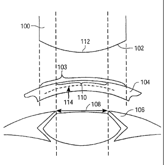
PCT Patent
Publication No. WO 99/039410
[62] Depth Reduction And Blend Zones
[63] A schematic illustration of a laser ablation treatment is shown in Fig.
4. Laser
ablation treatment 100 provides a laser ablation profile 102 that ablates a
corresponding
ablation zone 103 of the cornea 104, and thereby reshapes the cornea. The iris
106 of the
eye, when fully dilated, defines the maximum pupil size 108. The optical zone
110 is that
portion of the cornea 104 corresponding to the maximum pupil size. Relatedly,
the optical
region 112 is that portion of the ablation profile 102 corresponding to the
maximum pupil
size 108. The maximum allowed ablation depth for the cornea is 114.
CA 02478418 2004-09-08
WO 2003/077795 PCT/US2003/007903
[64] A representative illustration of a series of ablation profiles for
treating myopia is
shown in Fig. 5A. In laser treatments for myopia, the intent is to reduce the
curvature, and
thus the optical power, of the cornea. Accordingly, ablation profiles 120,
122, and 124 confer
progressively greater reductions in the optical power of the cornea, by
providing
progressively deeper ablations into the corneal tissue. As the curvature of
the ablation is
sharpened, the optical power of the cornea is progressively decreased. It is
the curvature of
the cornea that determines the optical power. Again, maximum allowed ablation
depth 114 is
the depth beyond which ablation may be unsafe or otherwise undesired, and so
an ablation as
deep as ablation profile 126 is acceptable. In some cases, a stronger
treatment or optical
correction may be advantageous. The deeper ablation profile 128 shown in Fig.
5B
represents the desired stronger treatment, yet because of its excessive depth,
the ablation may
be unsafe. However, by incorporating a blend region 130 into ablation profile
132, it is
possible to achieve a treatment equally as strong as ablation profile 128, yet
not ablate
beyond the maximum allowed ablation depth 114. The portion of the depth
reduced ablation
profile 132 central to the blend region 130 is the central optical region 134.
The blend region
is formulated by the blend region module.
[65] Fig. 5C further illustrates the concept that treatment with any ablation
profile creates a
corresponding ablation zone 103 in the cornea 104. Accordingly, depth reduced
ablation
profile 132 creates an ablation zone 103 that includes an optically correct
central optical zone
140 (corresponding to the central optical region 134), and a blend zone 142
(corresponding to
blend region 130), wherein the blend zone 142 is disposed peripherally to the
central optical
zone 140, and has an optical power that gradually diminishes with increasing
radius from the
central optical zone 140. In accordance with a general myopia treatment, the
original contour
144 of the cornea has been modified to a lower powered ablated contour 146.
[66] Fig. 5D illustrates a particular depth reduced ablation profile according
to the
principles of the present invention. Analogous to Fig. 5C, deeper ablation
profile 128 reaches
a maximum depth of approximately 130 pm. In some cases, this maximum depth may
exceed a maximum allowed ablation depth for a particular cornea. In order to
reduce the
maximum depth of the deeper ablation profile 128 to a more acceptable depth of
105 pm, and
still retain the same optical power provided by the deeper ablation profile
128, a peripherally
disposed blend region 130 is introduced. With the application of the blend
region 130, it is
possible to formulate the depth reduced ablation profile 132. As the figure
illustrates, both
the deeper ablation profile 128 and the depth reduced ablation profile 132
exhibit an optical
16
CA 02478418 2004-09-08
WO 2003/077795 PCT/US2003/007903
region 112 diameter of approximately 6.5 mm. As shown, the central optical
region 134 of
the depth reduced ablation profile 132 is approximately 5 mm in diameter,
however the
ablation profile may be formulated to provide central optical regions 134 of
varying sizes. In
some cases, the central optical region 134 may range from about 6 mm to about
7 mm in
diameter. In other cases, the central optical region 134 may range from about
3 mm to about
3 mm to about 9 mm in diameter.
[67] A representative graph illustrating a relationship between optical power
of the eye and
maximum ablation depth is shown in Fig. 6. The left side of the graph is
divided into three
sections: regular myopia ablation profile section 150, depth adjusted myopia
profile section
152, and invalid myopia refraction section 154. The horizontal axis represents
the optical
power of the cornea, and the vertical axis represents the depth of any given
ablation profile.
[68] The first section 150 demonstrates the principle that, in a series of
ablation profiles for
myopia (as shown in Fig. 5A), the optical power of the cornea decreases as the
depth of the
ablation profile increases. This correlation continues until the depth of the
ablation profile
reaches a maximum allowed ablation depth 114. Any ablation profile in this
section is not
depth adjusted. In a presently preferred embodiment, the maximum allowed
ablation depth is
determined by subtracting the minimum residual stroma from the stromal bed
depth.
[69] The second section 152 demonstrates the principle of depth reduction
discussed above
with respect to Fig. 5B. Here, the maximum ablation depth of a standard
ablation profile for
the desired correction would exceed the maximum allowed ablation depth of the
cornea. In
order to provide an ablation profile of reduced depth relative to the desired
standard ablation
profile, yet still achieve a decrease in optical power in the cornea, the
ablation profile is
modified by decreasing the size of the central optical region, and introducing
a peripherally
disposed blend region. In a preferred embodiment, the decrease in corneal
optical power
comes from ablation profiles having central optical regions with progressively
sharper
curvatures. Often, this will be accompanied by blend regions of increasingly
larger area.
[70] With a very high power correction, or in a patient presenting a large
optical zone or
maximum pupil size, the corrective ablation profile may have an optical region
that is smaller
than the optical zone. For example, if the size of the central optical region
is very small, it
may not be possible to incorporate a refractively acceptable blend region into
the ablation
profile and still provide an optical region that would span the area of the
optical zone of the
cornea. This situation is represented in section 154 of the graph.
17
CA 02478418 2004-09-08
WO 2003/077795 PCT/US2003/007903
[71] Fig. 5E illustrates many of these same principles in the application of
hyperopia
treatments. In laser treatments for hyperopia, the intent is to increase the
curvature, and thus
the optical power, of the cornea. Accordingly, ablation profiles 160 and 162
confer
progressively greater increases in the optical power of the cornea, by
providing progressively
deeper ablations into the corneal tissue. Once more, maximum allowed ablation
depth 114
provides a limit beyond which ablation may be unwanted. Thus, an ablation
profile as deep
as ablation profile 164 is acceptable. In some cases, a stronger treatment or
optical correction
may be advantageous. The deeper ablation profile 166 represents the desired
stronger
treatment, yet because of its excessive depth, the ablation may be unsafe.
However, by
incorporating a blend region 130 into ablation profile 166, it is possible to
achieve a treatment
with the corrective strength of ablation profile 166, yet not ablate beyond
the maximum
allowed ablation depth 114. As before, the optical region 112 of the ablation
profile is then
defined by the central optical region 134 and the blend zone 130.
[72] A representative graph illustrating a relationship between optical power
of the cornea
and maximum ablation depth in hyperopia is also shown in Fig. 6. The right
side of the graph
is divided into three sections: regular hyperopia ablation profile 170, depth
adjusted
hyperopia ablation profile 172, and invalid hyperopia refraction 174. The
horizontal axis
represents the optical power of the cornea, and the vertical axis represents
the depth of any
given ablation profile.
[73] Section 170 demonstrates the principle that, in a series of ablation
profiles for
hyperopia (shown as ablation profiles 160 and 164 in Fig. 5E), the optical
power of the
cornea increases as the depth of the ablation profile increases. This
correlation continues
until the depth of the ablation profile reaches a maximum allowed ablation
depth. Any
ablation profile in this section is not depth adjusted, because the maximum
ablation depth is
less than the maximum allowed ablation depth 114. Section 172 explains the
circumstance
when the maximum ablation depth of an ablation profile for a desired optical
correction is
greater than the maximum allowed ablation depth for the cornea.
[74] In order to provide an ablation profile of reduced depth relative to the
desired ablation
profile, yet still achieve an increase in optical power of the cornea, the
ablation profile is
modified by introducing a peripherally disposed blend region and decreasing
the size of the
central optical region. In a preferred embodiment, the increase in corneal
power comes from
ablation profiles having central optical regions with progressively sharper
curvatures. Often,
this will be accompanied by blend regions of increasingly larger size.
18
CA 02478418 2004-09-08
WO 2003/077795 PCT/US2003/007903
[75] With a very high power correction, or in a patient presenting a large
optically
functional region, the corrective ablation profile may have an optical zone
that is smaller than
the optically functional region. For example, if the size of the central
optical zone is very
small, it may not be possible to incorporate a refractively acceptable blend
zone and still
provide an optical zone that would span the area of the optically functional
region. This
situation is represented in section 174.
[76] Another illustrative plot of the application of a blend region into an
ablation profile
for myopia treatment is shown in Fig. 7. Ablation profile 180 represents a
standard
Munnerlyn shape of 6.5 mm diameter. For a patient having a large optical zone
(e.g. 8 mm
diameter), it may be preferable to treat the patient with an ablation profile
that covers the
entire 8 mm optical zone. This is accomplished by adding a blend region to the
standard 6.5
mm ablation profile 180. In order to do so, a standard 8 mm Munnerlyn ablation
profile 182
is calculated. A constant depth is then subtracted from ablation profile 182
inside the 6.5 mm
central optical region, and a transform is applied to the shape at the blend
region, between 6.5
mm and 8 mm. The result is ablation profile 184. In effect, this represents
the addition of a
blend region to a 6.5 mm optical region ablation profile, resulting in an
aspheric ablation
profile 184 having a deeper, blended 8 mm optical region. This approach is
also illustrated in
Fig. 8, effectively achieving the optical zone size of the standard 8 mm
ablation profile, but
minimizing the ablation depth required to do so.
[77] Characteristics of the resulting blend zone include: a precise
mathematically defined
relation to standard ablation shape; an optical power that changes smoothly as
a function of
radius; and flexibly defined parameters, in that the extent and amount of
optical power
change are specifiable.
[78] Transition Zone
[79] The function of the transition region is to bring the ablation depth
smoothly to zero
depth at the ablation profile edge. The transition zone is general in nature,
and is well suited
for implementation in a wide variety of ablation profiles, including arbitrary
wavefront
shapes. Whether a transition zone is needed or desirable often depends on the
shape of the
target ablation profile. For instance, treatment of mild spherical myopia
involves an ablation
profile which gradually tapers at its periphery, so that no transition zone
may be needed.
[80] In contrast, treatments for astigmatism involve a cylindrical ablation
profile that
would otherwise have abrupt axial ends. To avoid such discontinuities in the
ablated surface,
19
CA 02478418 2004-09-08
WO 2003/077795 PCT/US2003/007903
the apertures of the present invention may be varied, per the programming of
the system
controller, to impose a smooth astigmatism transition zone between the optical
zone and the
surrounding corneal surface. Generally, the shape of the transition zone
applied by the
present invention during a given treatment is based on the ablation profile.
[81] For forming the optical zone during hyperopic corrections, the variable
width slit and
variable diameter iris define an elongate rectangular beam. The rectangular
beam is rotated
about an ablation center with a major edge of the beam following a series of
circumferential
bands or arcs. A transition zone shape is generated by the outer edge of the
rectangular beam
as the inner edge rotates along an arc within the optical zone. Hence, the
shape of the
transition zone during a hyperopic refraction correcting procedure is an
artifact of the
mechanical arrangement used to define the optical zone.
[82] Still further transition zones shapes are generated by the method and
systems of the
present invention for other specific ablation shapes. For example, the
refractive treatment
used to correct for hyperopic astigmatism is ablation of a centrally
steepening cylindrical
shape from the optical zone. Absent any transition zone, the edges of the
optical zone might
comprise abrupt changes in ablation depth. As such discontinuities are
generally avoided, the
present invention imposes gradually tapering ablation depths beyond the
optical zone.
[83] As an illustrative example, Fig. 9A shows a myopic treatment with
astigmatism
having a transition zone on the major axis, but not on the minor axis.
Relatedly, Fig. 9B
shows a cylinder having a transition zone on the major axis but not on the
minor axis. This
shape has a larger transition zone than shown in Fig. 9A. In another example,
Fig. 10A
represents a mixed astigmatism treatment shape, having an even larger
transition zone. As
shown in Fig. 10B, the size of the transition zone is adjustable, and the
transition zone can be
made as gentle as desired.
[84] In one embodiment of the present invention, the transition region module
produces
the transition region shapes in a series of steps. First, the proper optical
region shape is
determined, either by Munnerlyn equations or wavefront techniques. Second, a
blend region
may optionally be applied. Third, the depth and radial slope of the ablation
shape at the outer
edge of the optical region, or at the blend region if present, is calculated.
Fourth, a cubic
spline is used to connect the outer edge of the optical region, or the blend
region, to the
ablation profile edge. Alternatively, in the case of the wavefront transition
region, the
transition region may be smoothed by pixel averaging, or by spatial averaging
of depth.
CA 02478418 2004-09-08
WO 2003/077795 PCT/US2003/007903
[85] Once the desired ablation shape has been determined, a next step is to
define the
parameters of the actual laser ablation required to administer the treatment
ablation profile.
A particularly useful way of determining these parameters is by using an
ablation equation,
such as the one shown below.
TotalPulses
[86] AblationShape = I (PulseShapeõ Positions )
[87] Essentially, this equation is based on the principle that a treatment
ablation is the sum
of each of the individual laser pulses. This equation has been empirically
verified on a
variety of materials including plastic, and bovine, porcine, and human corneal
tissue.
[88] In this equation, the AblationShape variable represents the desired
ablation shape. In
this sense, it is a known variable. The target shape can be, for example, a
simple sphere, an
ellipse, a cylinder for treating myopia or hyperopia, or even a saddle for
treating mixed
astigmatism. The target shape can be any arbitrary shape, such as the map from
a wavefront
type device or any other topography system. What is more, the target shape can
contain, for
example, a blend zone or a reduced depth profile. Further, the AblationShape
may or may
not include a transition zone.
[89] The PulseShape variable, which is also a known variable, represents the
ablation
shape of each laser pulse size to be used. The PulseShape typically varies for
different
ablated materials, such as plastic, animal cornea, or human cornea. The
PulseShape also
typically varies for each laser pulse diameter. An example of this type of
ablation data is
shown in Fig. 11. This figure shows different shapes of craters expected from
a single laser
pulse. There is a unique description for every unique pulse shape or size to
be used. By
systematically measuring the shape which each laser pulse ablates onto a
specific target
material, it is possible to generate such basis data for a variety of
materials, such as tissue or
plastic. For a given material, at a given diameter, the shape is generally
consistent from laser
system to laser system.
[90] A fixed spot laser may have only one description, while a variable spot
laser could
have as many as desired. There is no requirement that the crater shape be
flat, round, or
symmetric. As long as it can be described mathematically or with an array of
data, it can be
incorporated in the equation.
[91] In order to create the ablated surface, it is useful to determine the
locations where
each of the laser pulses will be applied. The Position variable, which
represents the exact
21
CA 02478418 2006-08-08
position of every laser pulse, is an unknown variable. This variable is
calculated by solving
the ablation equation. Put another way, the output is a set of instructions
for creating the
target ablation shape using the laser pulses. This is sometimes called a
treatment table. The
treatment table consists of a list of individual pulses, each containing the
size and offset, or
position, to be used for that pulse. When the laser fires according to the
instructions in the
treatment table, the target shape will be created.
[92] The target ablation shape is a theoretical construct; it is a
mathematically perfect
representation of a desired ablation outcome. Put another way, while the
application of
thousands of specifically placed brief laser pulses can create an actual
ablation shape that
approaches the ideal target ablation shape, in the end it is still an
approximation thereof.
[93] Therefore, solving for the Position variable can allow for the
formulation of a
corresponding ablation shape that approaches the target ablation shape as
closely as possible.
In this way each of the thousands of pulse positions are individually
determined so as to
minimize the difference between the ideal target ablation shape and the actual
resulting
ablation shape. In a system for ablating tissue using a scanning laser, a
presently preferred
computational technique for achieving this goal employs simulated annealing.
[94] Other mathematical approaches include, for example, the SALSA Algorithm.
SALSA is an acronym for Simulated Annealing Least Squares Algorithm. It is an
algorithm
that solves an equation having over 10,000 unknowns. The algorithm finds the
best solution
by selecting: the number of pulses, the size of each pulse, and the location
of each pulse. It
is an exact algorithm, and makes no statistical assumptions.
[95] Simulated Annealing is a recent, proven method to solve otherwise
intractable
problems, and may be used to solve the ablation equation discussed above. This
is more fully
described in PCT Application No. PCT/US01/08337, filed March 14, 2001,
See also W. H. Press et al.,
"Numerical Recipes in C" 2d Ed., Cambridge University Press, pp. 444-455
(1992). This
approach is also further discussed in co-pending U.S. Patent Publication No.
2002/0035359.
[96] Simulated annealing is a method used for minimizing (or maximizing) the
parameters
of a function. It is particularly suited to problems with very large, poorly
behaved function
spaces. Simulated annealing can be applied in the same way regardless of how
many
dimensions are present in the search space. It can be used to optimize any
conditions that can
= 22
CA 02478418 2004-09-08
WO 2003/077795 PCT/US2003/007903
be expressed numerically, and it does not require a derivative. It can also
provide an accurate
overall minimum despite local minima in the search space, for example.
[97] The methods and apparatuses of the present invention may be provided in
one or more
kits for such use. The kits may comprise a system for profiling an optical
surface, such as an
optical surface of an eye, and instructions for use. Optionally, such kits may
further include
any of the other system components described in relation to the present
invention and any
other materials or items relevant to the present invention. The instructions
for use can set
forth any of the methods as described above.
[98] While the above provides a full and complete disclosure of the preferred
embodiments
of the present invention, various modifications, alternate constructions and
equivalents may
be employed as desired. Therefore, the above description and illustrations
should not be
construed as limiting the invention, which is defined by the appended claims.
23