Note: Descriptions are shown in the official language in which they were submitted.
CA 02479956 2004-09-20
WO 03/086213 PCT/US03/10336
ORTHOPAEDIC FIXATION METHOD AND DEVICE WITH DELIVERY AND
PRESENTATION FEATURES
Inventors: Ed Austin, John Schneider, and Michael W. Mullaney
RELATED APPLICATION DATA
This application claims the benefit of USSN 60/370,201, filed April 5, 2002
entitled
"Orthopaedic Fixation Method and Device" which is incorporated herein by this
reference.
TECHNICAL FIELD
Embodiments of the invention are directed to treating musculoskeletal
conditions,
including skeletal fractures. More specifically, apparatuses and methods for
securing and
placing fragments of a fractured bone or bones on two sides of a joint in
desired locations are
disclosed. In some embodiments of the invention, apparatuses and methods are
used to
generate a computer model of a fixation device and bones or bone fragments.
Through
operations on the model, desired placement of the bones or bone fragments is
determined
quickly and accurately regardless of the initial configuration of the fixation
device. The
operations required to create the desired placement of the bones or bone
fragments may then
be enacted on a corresponding physical device to treat the musculoskeletal
condition.
BACKGROUND OF THE INVENTION
Devices and methods of treating skeletal fractures using ring external
fixation
structures are well known in the art. Smith & Nephew, Inc. has developed and
marketed a
number of SPATIAL FRAME'' brand external ring fixators based on the general
concept of a
Stewart platform. Smith & Nephew, Inc. owns U.S. Pat. Nos. 5,702,389;
5,728,095;
5,891,143; 5,971,984; 6,030,386; and 6,129,727 that disclose many basic
concepts of and
improvements to Stewart platform based external fixators. The disclosure of
those patents is
incorporated by reference herein.
3o As will be appreciated by one skilled in the art, mathematically solving
for the
relative positions of the members of a Stewart platform creates' a somewhat
cumbersome
equation. As an example, note the rotational matrix detailed in U.S. Pat. No.
5,971,984. This
rotational matrix is a means by which one Stewart platform fixation element
can be
transformed relative to another to align fragments of a bone with inputs
commonly obtainable
CA 02479956 2004-09-20
WO 03/086213 PCT/US03/10336
from a clinical examination. However, in order to use the rotational matrix, a
starting
position for the fixation elements must be known. Therefore, prior art systems
typically have
required a Stewart platform type ring fixator to either start or end its
transformation in a
neutral position. A neutral position is a position where all of the six struts
are the same
length, and consequently, the rings of the fixator are parallel to one
another. See Figures 4
and 5. A neutral position makes locating the starting positions of the
fixation elements
readily calculable. Once the frame moves beyond neutral, Cartesian coordinates
of the frame
components are difficult to find mathematically. This limitation results in
complexity with
regard to a mathematical solution for a Stewart platform. As a practical
matter, it means that
1o in the past, supposed correction solutions that did not in fact solve a
particular deformity were
very difficult to secondarily correct. This situation will be described in
more detail below as
a "crooked-frame/cxooked-bone" situation. A solution for the crooked-
frame/crooked-bone
situation will be described as a "total residual" solution.
The current SPATIAL FRAME'' brand external fixators include operating modes
for
"chronic" and "residual" corrections. A chronic correction is a correction
that starts with a
fixator frame that has been deformed to fit onto a deformed bone structure
such that when the
fixator is returned to a given neutral position, the deformed structure will
be corrected. In
other words, a chronic correction starts with a frame that has been deformed
identically to the
deformity of the bone.
2o For a residual correction, a neutral fixator frame is fit onto a deformed
structure, and
the struts of the fixator are adjusted until the deformity is corrected.
Therefore, in the case of
a residual correction, a straight-frame/crooked-bone is corrected to a crooked-
frame/straight-
bone. For a chronic correction, a crooked-frame/crooked-bone situation is
corrected to a
straight-frarne/straight-bone. Note that a "total residual" correction differs
from a "residual"
correction in that a residual must start with a neutral frame. A total
residual may start with
even a crooked frame.
The crooked-frame/crooked-bone complication exists where, at the end of a
correction, both the bone structure and the frame axe crooked. In other words,
the deformities
of the frame and the bone are different from one another. The current, known
mathematical
3o equations are only valid for going to or starting from a neutral frame.
Therefore, if a crooked
frame is on a crooked bone structure that is not corrected by returning the
frame to a neutral
position, the current equations will not solve the problem in a single step.
Specifically, some
of the initial values to plug into the equations cannot be determined. This
crooked-
frame/crooked-bone situation may result from inaccurate placement or
adjustment of a frame,
CA 02479956 2004-09-20
WO 03/086213 PCT/US03/10336
inaccurate x-rays or reading of x-rays used to generate deformity parameters,
or any number
of inaccurate applications of a device. Such inaccuracies are common and
expected,
especially in an environment such as a trauma operating room. In the case of a
crooked-
frame/crooked-bone situation, the surgeon could reset the frame back to
neutral and take new
x-rays that could be used to establish a new residual correction. However,
that would not be
optimal for the patient, especially where adjustment of the frame to neutral
would result in
increased skeletal deformity and pain.
Some crooked-frame/crooked-bone situations may also be solved with a deformity
simulator such as the one shown in French Pat. No. 2,576,774, Figure 6. As
shown, two rods
to that represent segments of bone are connected by hinges about two axes. By
setting the rods
relative to one another the way the bone segments are actually deformed,
noting the position
of the simulator, re-aligning the rods, and noting the changes in the
simulator, corrective
settings for an actual device may be derived. However, this simulator device
fails to account
for translations or for rotation about all three possible axes between the
segments. Both
modes of deformation commonly occur. Additionally, manipulation of the
mechanical
device in the loosened frame would be awkward and potentially require multiple
operators.
A total residual solution is highly advantageous over solutions that require
more
precise alignment of components of the frame with the patient's anatomy.
External ftxation
devices are often used in trauma situations where reduced initial operating
time is beneficial
2o to the patient. Total residual devices require relatively little time for
alignment and can be x
rayed or imaged and adjusted after the patient has been stabilized. Therefore,
an improved
device must provide methods and apparatuses for solving crooked-frame/crooked-
bone
situations.
What is needed are methods and apparatuses that are useful in quickly and
accurately
determining the strut settings that solve crooked-frame/crooked-bone
situations. Optimally,
solutions would be obtainable without substitution or experimentation, and all
possible
physical relationships of bone segments could be modeled. Improved methods and
apparatuses may also give a user visual representations of frame placement and
correction
results so that the parameters the user is inputting are visually verifiable
as correct prior to
3o adjustment of the frame on the patient. Visualization also would enable a
user to see if pins
and wires used in a frame will interfere with strut positions as a correction
is executed.
Improved methods and apparatuses may be implemented through software that is
operative to
be run, updated, and replaced over a network either by storage and use on
distributed
computers or a central computer or a combination of both.
CA 02479956 2004-09-20
WO 03/086213 PCT/US03/10336
SUMMARY OF THE INVENTION
According to the present invention there is provided an external orthopaedic
fixation
device in combination with a computer. In this embodiment, the combination is
for aligning
fragments of a fractured bone. The orthopaedic fixation device includes a
first fixation
element for coupling to a first bone fragment and a second fixation element
for coupling to a
second bone fragment. The device also includes six adjustable length struts
coupled at their
respective first ends to the first fixation element and coupled at their
respective second ends
to the second fixation element. When the first bone fragment and the second
bone fragment
1o are out of alignment, at least two of the first, second, third, fourth,
fifth, and sixth adjustable
length struts are different lengths. And in the same embodiment, if the first,
second, third,
fourth, fifth, and sixth adjustable length struts were the same length, the
first bone fragment
and the second bone fragment would be out of alignment. The combination is
operable to
bring the first bone fragment into alignment with the second bone fragment by:
storing the
relative locations of the first fixation element and the ftrst bone fragment,
storing the
locations of the couplings of the first ends of the first, second, third,
fourth, fifth, and sixth
adjustable length struts relative to the first fixation element, storing the
relative locations of
the second fixation element and the second bone fragment, storing the
locations of the
couplings of the second ends of the first, second, third, fourth, fifth, and
sixth adjustable
2o length struts relative to the second fixation element, spatially
associating, or correlating, the
stored location of the first f~ation element with the stored location of the
second fixation
element, aligning a computer generated representation of the stored location
of the first bone
fragment relative to a computer generated representation of the stored
location of the second
bone fragment, obtaining the respective distances in the aligned computer
generated
representations between the first and second ends of the first, second, third,
fourth, fifth, and
sixth adjustable length struts respectively, and providing the aligned lengths
of the first,
second, third, fourth, fifth, and sixth adjustable length struts to a user for
adjusting the
adjustable length struts of the external orthopaedic ftxation device.
There is further provided a method of configuring an orthopaedic fixation
device that
3o can be coupled to fragments of a fractured bone. The method of the
embodiment includes
representing a first fixation element of the ftxation device virtually in
three-dimensional
space, representing a first bone fragment virtually in three-dimensional
space, and spatially
associating, or correlating, the representation of the first fixation element
with the
representation of the first bone fragment. The method also includes
representing a second
CA 02479956 2004-09-20
WO 03/086213 PCT/US03/10336
fixation element of the fixation device virtually in three-dimensional space,
representing a
second bone fragment virtually in three-dimensional space, and spatially
associating, or
correlating, the representation of the second fixation element with the
representation of the
second bone fragment. The representation of the first bone fragment is also
spatially
associated , or correlated ,with the representation of the second bone
fragment. The method
then includes aligning the virtual representation of the first bone fragment
with the virtual
representation of the second bone fragment while tracking the spatially
associated, or
correlated, locations of the representation of first fixation element and the
representation of
the second fixation element, and configuring the orthopaedic fixation device
such that the
to first fixation element is in the same relative position to the second
fixation element as the
aligned representation of the first fixation element is with the aligned
representation of the
second fixation element.
Still another embodiment is a method of determining adjustments required to
align
fragments of a fractured bone coupled in an orthopaedic fixation device that
has a first
fixation element coupled to a second fixation element by at least three
struts, each strut
coupled at its first end to the first fixation element and at its second end
to the second fixation
element. 'The method in this embodiment includes representing the first
fixation element and
a first bone fragment in a computer, and spatially associating, or
correlating, the
representations of the first fixation element with the first bone fragment.
The method also
2o includes representing the second fixation element and a second bone
fragment in the
computer, and spatially associating, or correlating, the representation of the
second fixation
element with the representation of the second bone fragment. Further, the
method includes
spatially associating, or correlating, the representation of the first bone
fragment with the
representation of the second bone fragment, and aligning the representation of
the first bone
fragment with the representation of the second bone fragment. The location of
the
representation of the first fixation element relative to the representation of
the second fixation
element subsequent to the aligning of the representation of the first bone
fragment and the
representation of the second bone fragment is determined, and the distance
between the
couplings of each of the at least three struts to the representation of the
first fixation element
3o and the representation of the second fixation element is determined. The
amount to adjust
each of the at least three struts to equal the determined distance between
couplings may then
be determined.
Also according to the present invention there is provided . a digital
computing device
programmed to provide data to a user for adjusting an orthopaedic fixation
device that can be
CA 02479956 2004-09-20
WO 03/086213 PCT/US03/10336
coupled to fragments of a fractured bone. The digital computing device may
include a
motherboard, a central processing unit electrically coupled to the motherboard
for executing
program instructions, a monitor electrically coupled to the motherboard for
displaying
representations of the fixation device, and a memory device electrically
coupled to the
motherboard. The memory device stores program instructions that enable the
computing
device to represent a first fixation element of the fixation device virtually
in three-
dimensional space, represent a first bone fragment virtually in three-
dimensional space, and
spatially associate, or correlate, the virtual representation of the first
fixation element with the
virtual representation of the first bone fragment. Stored instructions also
enable the
to computing device to represent a second fixation element of the fixation
device virtually in
three-dimensional space, represent a second bone fragment virtually in three-
dimensional
space, and spatially associate, or correlate, the virtual representation of
the second fixation
element with the virtual representation of the second bone fragment. The
program
instructions also enable the computing device to spatially associate, or
correlate, the virtual
representation of the first bone fragment with the virtual representation of
the second bone
fragment, align the virtual representation of the first bone fragment with the
virtual
representation of the second bone fragment while tracking the spatially
associated, or
correlated, locations of the virtual repxesentation of first fixation element
and the virtual
representation of the second fixation element, and output data specifying how
the first
2o fixation element is to be positioned relative to the second fixation
element to align the first
bone fragment and the second bone fragment
According to the present invention there is further provided a program storage
device
containing instructions that enable a computer to provide data specifying how
to configure an
orthopaedic fixation device that can be coupled to fragments of a fractured
bone. Execution
of the instructions results in providing data specifying how to configure the
orthopaedic
fixation device such that a first fixation element is in the same relative
position to a second
fixation element as a virtual representation of the first fixation element is
with an aligned,
virtual representation of the second fixation element after virtual
representations of the bone
fragments have been aligned.
3o Still another embodiment of the invention is a method of configuring a
mufti-strut
orthopaedic fixation device that can be coupled to fragments of a fractured
bone. The method
includes at least representing a first fixation element of the fixation device
virtually in three-
dimensional space, representing a first bone fragment virtually in three-
dimensional space,
and spatially associating, or correlating, the representation of the first
fixation element with
CA 02479956 2004-09-20
WO 03/086213 PCT/US03/10336
the representation of the first bone fragment. The method also includes
representing a second
fixation element of the fixation device virtually in three-dimensional space,
representing a
second bone fragment virtually in three-dimensional space, and spatially
associating, or
correlating, the representation of the second fixation element with the
representation of the
second bone fragment. The representation of the first bone fragment with the
representation
of the second bone fragment are spatially associated, or correlated" and the
representation of
the multi-strut orthopaedic fixation device is configured to any neutral frame
configuration
while tracking the fixation elements and bone fragments. The method also
includes noting
virtual deformity parameters of the bone fragments with the multi-strut
orthopaedic fixation
1o device virtually configured to a neutral frame, and then solving for
correcting strut lengths by
aligning virtual representations of the virtually altered representations of
the bone fragments
while tracking the representations of the spatially associated, or correlated,
fixation elements
and calculating the resulting strut lengths.
An embodiment of the invention is a method of configuring an orthopaedic
fixation
device that can be coupled to bones on either side of a joint to move the
bones relative to one
another. Representations of a first fixation element and a first bone are
represented virtually
in three-dimensional space and spatially associated, or correlated.
Representations of a
second fixation element and a second bone are represented virtually in three-
dimensional
space and spatially associated, or correlated. The representation of the first
bone is
2o associated, or correlated, with the representation of the second bone and
the representations
are positioned while tracking the spatially associated, or correlated,
locations of the
representation of first fixation element and the representation of the second
fixation element.
The orthopaedic fixation device is configured such that the first fixation
element is in the
same relative position to the second fixation element as the positioned
representation of the
first fixation element is with the positioned representation of the second
fixation element.
Also according to the present invention there is provided a program storage
device
containing instructions that enable a computer to graphically represent
anatomical features in
relation to a fixation device. The instructions include at Ieast receiving
input regarding the
size and orientation of the fixation device, and receiving input regarding the
orientation of the
3o anatomical features. The fixation device and the anatomical features
relative to the fixation
device are graphically represented. The graphical representation of the
anatomical features
relative to the fixation device is presented in a perspective that corresponds
to a typical
medical diagnostic image.
CA 02479956 2004-09-20
WO 03/086213 PCT/US03/10336
Another embodiment of the invention is a method of planning the application of
a
multi-strut external fixation device. The method includes at least modeling
the size and
orientation of the multi-strut fixation device, including the maximum and
minimum lengths
of all struts that are a part of the multi-strut fixation device, and modeling
the orientation of
anatomical features to be manipulated by the multi-strut fixation device.
Further the method
includes virtually accomplishing a manipulation of the multi-strut fixation
device while
recording the lengths the struts of the multi-strut fixation device during the
manipulation, and
displaying a record of struts that exceed maximum or minimum lengths during
the virtual
manipulation of the mufti-strut fixation device.
BRIEF DESCRIPTION OF THE DRAWINGS
Figure 1 is a perspective view of an orthopaedic fixation device.
Figure 2 is a perspective view of an orthopaedic fixation device coupled to a
tibia.
Figure 3 is a system diagram of an orthopaedic fixation device in combination
with a
computer.
Figure 4 is a perspective view of a virtual representation of an orthopaedic
fixation
device.
Figure 5 is a perspective view of a virtual representation of an orthopaedic
fixation
device.
2o Figure 6 is an elevation view of a virtual representation of an orthopaedic
fixation
device.
Figure 7 is an elevation view of a virtual representation of an orthopaedic
fixation
device.
Figure 8 is a perspective view of a virtual representation of an orthopaedic
fixation
device with some elements removed for clarity.
Figure 9 is a screen shot illustrating an embodiment of the invention being
executed
on a Web browser where a user may login to the program.
Figure 10 is a screen shot illustrating an embodiment of the invention being
executed
on a Web browser where case information has been input by a user.
3o Figure 11 is a screen shot illustrating an embodiment of the invention
being executed
on a Web browser where deformity definitions are to be input by a user.
Figure 12 is a screen shot illustrating an embodiment of the invention being
executed
on a Web browser where deformity definitions have been input by a user.
CA 02479956 2004-09-20
WO 03/086213 PCT/US03/10336
Figure 13 is a screen shot illustrating an embodiment of the invention being
executed
on a Web browser where fixation device parameters have been input by a user.
Figure 14 is a screen shot illustrating an embodiment of the invention being
executed
on a Web browser where fixation device mounting parameters have been input by
a user.
Figure 15 is a screen shot illustrating an embodiment of the invention being
executed
on a Web browser where initial frame strut lengths have been input by a user.
Figure 16 is a screen shot illustrating an embodiment of the invention being
executed
on a Web browser displaying an enlarged initial frame AP view.
Figure 17 is a screen shot illustrating an embodiment of the invention being
executed
to on a Web browser displaying final frame strut lengths and configurations.
Figure 18 is a screen shot illustrating an embodiment of the invention being
executed
on a Web browser displaying an enlarged final frame lateral view.
Figure 19 is a screen shot illustrating an embodiment of the invention being
executed
on a Web browser displaying a prescription for an alignment.
15 Figure 20 is a screen shot illustrating an embodiment of the invention
being executed
on a Web browser displaying an enlargement of virtual representations of the
fixation device,
struts, and bone fragments at a point during the prescription for an
alignment.
Figure 21 is a screen shot illustrating an embodiment of the invention being
executed
on a Web browser displaying a prescription for an alignment.
2o Figure 22 is a perspective view of a virtual representation of a fixation
device with a
virtual representation of a bone fragment.
Figure 23 is a perspective view of a virtual representation of a fixation
device with a
virtual representation of a bone fragment, with some elements of the fixation
device removed
for clarity.
25 Figure 24 is a perspective view of a virtual representation of a fixation
device with
virtual representation of two bone fragments, with some elements of the
fixation device
removed for clarity.
Figure 25 is a perspective view such as Figure 24 with additional elements of
the
fixation device removed for clarity and to show association.
30 Figure 26 is a perspective view of a virtual representation of a fixation
device with
virtual representations of two bone fragments.
Figure 27 is a perspective view of a virtual representation of a fixation
device in a
neutral position with virtual representations of two bone fragments.
CA 02479956 2004-09-20
WO 03/086213 PCT/US03/10336
Figure 28 is a perspective view of a virtual representation of a fixation
device with
virtual representations of two bone fragments that have been aligned.
Figure 29 is a system diagram of a digital computing device.
DETAILED DESCRIPTION OF THE INVENTION
Figure 1 shows an external orthopaedic fixation device 100 useful for aligning
fragments of a fractured bone. The device shown is a Stewart platform based
ring fixator
device. Smith & Nephew, Inc. markets the particular device shown as a SPATIAL
FRAME'M
brand or TAYLOR SPATIAL FRAME'M brand external fixator. The fixation device
100
1o includes a proximal ring or first fixation element 10 and a distal ring or
second fixation
element 20. In other embodiments of the invention, the proximal ring could be
the second
fixation element and the distal ring could be the f~~rst fixation element. In
Figure 1, the first
fixation element 10 is coupled to the second fixation element 20 by six
adjustable length
struts 1-6. Each of the struts 1-6 is coupled at its first end to the first
fixation element 10 and
15 at its second end to the second fixation element 20.
Figure 2 illustrates the first fixation element 10 coupled to a first bone
fragment 11,
and the second fixation element 20 coupled to a second bone fragment 21. As
shown, the
bone fragments are coupled to the fixation elements using cantilevered bone
pins 13. In other
embodiments, wires, bilateral pins, or any variety of coupling devices
effective to secure a
2o bone relative to a fixation element may be used. The fixation device 100
shown is coupled to
a tibia; however, the device may be used on practically any bone on which it
could be placed.
For example, and without limitation, the device could also be used on a femur
or a humerus.
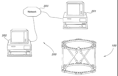
Figure 3 shows the fixation device 100 in combination with a computer 200.
Such a
combination is useful to align fragments of a fractured bone. The computer 200
may be an
25 autonomously operating computer system such as, for example, first computer
system 201.
All storage, processing, etc. necessary to align fragments of a fractured bone
may be
accomplished with the first computer system 201. In other embodiments, two or
more
computers may be linked together over a network to accomplish tasks necessary
to align the
fragments. As shown, computer systems 201 and 202 are linked over a network
203. The
3o network may be a local area network or a wide area network such as the
Internet. In some
embodiments, all of the programs that are run to accomplish the tasks may be
run on one or
more of the computer systems, and another of the computer systems may merely
be used to
display data. Alternatively, the programs run may be run partially on several
computer
systems, with data and instructions being shared over the network.
CA 02479956 2004-09-20
WO 03/086213 PCT/US03/10336
For example, in some embodiments, first computer system 201 runs a World Wide
Web browser that executes instructions and shares data through network 203
with a second
computer system 202 that is a server. This is advantageous in circumstances
where a larger
computer system is required to run a more complex or memory intensive program.
A
computer assisted engineering program is an example of such a program. In some
embodiments of the present invention, a server computer is used to run both a
computer
assisted engineering program and to serve or host a World Wide Web site. The
term
computer assisted engineering program includes both traditional computer aided
drafting
(CAD) programs, and programs that are capable of not only drafting, but
providing design
to solutions and other data useful in implementing a project. For example,
load capacities and
dynamic relationships of the components of a structure are provided with some
such
programs. One computer assisted engineering program useful in the present
invention is the
LTnigraphics program provided by EDS Corporation. Computer assisted
engineering and
Web hosting functions may themselves be dedicated to separate machines in some
embodiments. A served program arrangement may also be beneficial because the
supporting
programs in such a configuration may be updated by merely updating the program
at the
central computer or computers. Therefore, software updates become much less
complicated
and much less expensive.
As described in detail above, a particularly complex situation solved by the
present
2o invention is a crooked-frame/crooked-bone situation. Another way of
describing the
crooked-frame/crooked-bone situation is to say that when two bone fragments
are coupled in
a fixation device and the fragments are out of alignment, and at least two of
the first, second,
third, fourth, fifth, and sixth adjustable length struts are different
lengths, and if the struts
were adjusted until they were any same length, the bone fragments would still
be out of
alignment. Stated another way, a crooked-frame/crooked-bone situation occurs
when both
the frame and the attached bone are not neutral or aligned, and the bone would
not be aligned
if the frame were brought to any neutral position.
Figures 4-8 show geometric characteristics of the fixation device 100. What is
shown
in the figures are virtual representations of the device generated with the
aid of a computer
3o assisted engineering program or the like. A more detailed description of
these characteristics
will facilitate the discussion of applications of the device that follows. In
defining any spatial
system, arbitrary points of reference must be established from which to
reference the location
of components of the system. As illustrated in Figure 4, the hole between the
holes where the
proximal ends of struts 1 and 2 are coupled is referred to as a master tab 15.
The master tab
11
CA 02479956 2004-09-20
WO 03/086213 PCT/US03/10336
15 defines a point of origin in the plane of the first fixation element 10.
This point is
extended distally and projected posteriorly to define a frame centerline 16.
Figure 5 shows
the definition of the neutral frame height as the distance between the first
fixation element 10
and the second fixation element 20 when the struts 1-6 are neutral. Figure 6
illustrates an
origin I7. The origin 17 is placed at the center of the fractured end of a
bone fragment
coupled orthogonally to the first fixation element. In a Cartesian coordinate
system, the
origin 17 is defined as (0,0,0).
Figures 1 and 4 also show U joints near the end of each shut 1-6. Figure 1
shows the
U joints as they actually appear near the end of each strut 1-6, and Figure 4
shows each strut
l0 virtually represented with a sphere centered at the respective U joint's
center of rotation. For
example, struts 1 and 2 are shown to have proximal U joints I a and 2a, and
distal U j oints 1 b
and 2b. The proximal U j oints 1 a-6a define a plane A, and the distal U-j
oints lb-6b define a
plane B as illustrated in Figures 7 and 8.
The combination shown in Figure 3 is operable to bring a first bone fragment
into
15 alignment with a second bone fragment. To accomplish an alignment, a user
must provide
parameters regarding characteristics of the fixation device 100 used. In
addition,
characteristics of the deformity to be corrected and the way the device 100 is
mounted must
be input. Given this information from a user, embodiments of the invention
provide lengths
to which struts can be configured to achieve alignment.
20 Figures 9-21 illustrate an example of an alignment solution reached using a
program
that receives parameters from a user and outputs strut length settings. Figure
9 is a depiction
of a user login screen designed to provide secure and confidential access to
the program.
A user completes the fields shown in Figures IO-12 to provide information to
the
program regarding the deformity to be corrected. In Figure 10 next to
"Anatomy," a user
25 inputs whether a left or right limb is to be corrected. In the example, a
left limb is being
corrected.
Figures 11 and I2 show blanks to be filled in by a user regarding the
orientation and
extent of a deformity. A selection is required to define which fragment of
bone will be a
reference fragment, the proximal or the distal. The fragment defined as the
reference
30 fragment will be shown as remaining fixed and the~other fragment will be
brought into
alignment with the reference fragment. "Proximal" is selected in the example.
The
remainder of the parameters to be input are typical clinical parameters that
medical
professionals are familiar with obtaining. In the example illustrated, the
deformity as viewed
from the AP is defined by 15.0 degrees of valgus angulation and lS.Ornm of
medial
12
CA 02479956 2004-09-20
WO 03/086213 PCT/US03/10336
translation. As viewed from the lateral, there are 25.0 degrees of apex
anterior angulation,
and 30.Omm of anterior translation. Axially, the deformity has 10.0 degrees of
external
rotation and shows lS.Omm of shortened axial translation. It is usual to
obtain such
parameters from x-ray machines and other such imaging devices as well as by
observation
and physical measurement.
The graphical representations of the present invention labeled "Left AP View",
"Left
Lateral View", and "Left Axial View" are very useful because they provide the
user
immediate feedback as to whether the correct parameters have been input. The
Left AP View
and Left Lateral View are particularly familiar and efficient because they
correspond to
to typical x-ray images that the user will likely have available. The
embodiment illustrated
represents bones as cylindrical objects and a foot on the distal fragment as a
perpendicular
cylindrical object with a knob at the object's free end. Other embodiments of
the invention
represent bones with their actual anatomical shapes and proportions. Such
representations
can be useful to give a user further means of verifying the accuracy of data
being input and
15 solutions generated. The use of actual anatomical shapes is carried forward
throughout the
alignment process in some embodiments. In addition, in some embodiments, soft
tissue such
as but not limited to muscle, skin, vessels, arteries, and nerves are
represented graphically.
Figure 13 is an illustration of the input screen used to select what fixation
elements,
i.e., rings, and what struts will be used in the case. Since rings and struts
are stocked items,
2o pull-down menus are provided that only allow a user to select from a
limited number of
items. This reduces the likelihood of mistakes and increases the accuracy of
the alignment.
In the example illustrated, both distal and proximal rings are 180mm rings.
The struts
selected are standard medium struts, adjustable between 116mm and 178mm.
Figure 14 shows the input screen where a user selects how the first fixation
element,
25 or reference ring, of a frame has been or will be mounted on a first bone
fragment. This is
also where a user defines the operative mode: Total Residual, Chronic, or
Residual. As
discussed above, residual and chronic solutions generally are known iri the
art and require
going to or coming from a neutral frame. Consequently, for Residual and
Chronic modes,
either a neutral frame height or neutral strut lengths must be defined. The
Total Residual
3o mode is useful in aligning fragments in any circumstance, including the
crooked-
frarne/crooked-bone situation. The origin is defined as the center of the
fractured end of the
first bone fragment, and the position of the frame is defined relative to that
origin. Reference
points on the first fixation element of the frame are the center of the
fixation element for
lateral and AP views, the closest edge of the first fixation element to the
origin axially, and
13
CA 02479956 2004-09-20
WO 03/086213 PCT/US03/10336
the plane defining the master tab to the center of the fixation element for
rotary frame angle.
Adjustments are also provided to correct for non-orthogonal mountings AP and
laterally. In
the example shown, there is no AP offset or angulation, no lateral non-
orthogonal angulation,
and a 20.Omm posterior to origin frame offset. Axially there is no frame
rotary angulation,
but there is 100.Omm proximal to origin axial frame offset. As with the
deformity definition,
graphical displays of the mounted first fixation element and first bone
fragment are provided
so that the user may check for proper input of data. The input of data to the
stage so far
described allows for the relative locations of the first fixation element and
the first bone
fragment to be stored in the computer.
i0 Figure 15 shows the input screen for the initial frame strut lengths.
Typically, these
strut lengths are read from the six struts after the second fixation element
is coupled to the
second bone fragment. In the example that is shown, strut 1 was observed to
have a length of
122mm, strut 2, 140mm, strut 3, I47mm, strut 4, 132rnm, strut 5, 178mm, and
strut 6,
150mm. Once input, the program displays graphic representations of the
fixation and bone
fragments so that the user may verify the data thus far input into the
program.
Figure 16 shows an enlarged view of the Left AP View generated in Figure 15.
In
some embodiments of the invention, such enlargements are available for each of
the graphic
representations of Figure 15 by selecting the graphic representations. With
this data input,
the relative locations of the first fixation element and the first bone
fragment may be stored in
the computer. Additionally, the locations of the couplings of the first and
second ends of the
first, second, third, fourth, fifth, and sixth adjustable length struts
relative to the first and
second fixation elements respectively are stored after the orientation of the
fixation device is
defined by the placement of the second fixation element and the struts.
Spatial association
between the stored locations of first fixation element and the second fixation
element may
then be accomplished, thereby storing the locations of the two elements on a
common
coordinate system.
The results of solving for the Final Frame, i.e., spatial association and
alignment, are
illustrated in Figure 17. In this example, the resulting strut lengths are:
strut 1, 122mm, stxut
2, 161mm, strut 3, 176mm, strut 4, 211mm, strut 5, 241mm, and strut 6, 136mm.
To reach
3o the results illustrated, embodiments of the invention align the computer
generated
representations of the stored location of the first bone fragment and a
computer generated
representation of the stored location of the second bone fragment. Spatial
association and
aligning may be at least in part enabled by use of a computer assisted
engineering program.
For example, there exist in the prior art formulas for transforming one
fixation element of a
14
CA 02479956 2004-09-20
WO 03/086213 PCT/US03/10336
Stewart platform relative to another fixation element of the platform to
achieve an alignment
of bone fragments coupled to each fixation element. Smith & Nephew's U.S. Pat.
No.
5,971,984 and numerous Stewart platform manipulation algorithms provide
examples of such
transformation equations. However, coordinates for both fixation elements must
be known to
implement the formulas. With a crooked-frame/crooked-bone situation, there
previously was
no acceptable way to associate the fixation elements and enable the bone
fragments to be
aligned. A computer assisted engineering program can be used to provide the
coordinates of
a first fixation element relative to a second fixation element of a Stewart
platform where the
lengths of the struts are known. Therefore, by modeling a Stewart platform in
a computer
to assisted engineering system, knowledge of the strut lengths is equivalent
to knowing the
relative coordinates of both fixation elements. Given the coordinates of the
first fixation
element and the second fixation element and the frame parameters, deformity
parameters, and
mounting parameters, known transformation equations are used in some
embodiments to
determine strut lengths required to align the bone fragments. In other
embodiments it is
15 possible to achieve alignment by manipulation of graphical representations
of the fragments.
More specific examples of solving for the Final Frame strut lengths such as
those shown in
Figure 17 are provided below in association with Figures 22-28.
Recent improvements in computer assisted engineering programs have enabled the
programs to simultaneously track both the first and second fixation elements
and all six
2o struts. By use of such computer assisted engineering programs, direct use
of even the
previously applied transformation equations may be bypassed. Consequently,
these improved
programs have enabled graphical manipulation and measurement of the structures
with less
user intervention.
Strut lengths may also be solved for using trial and error or a similar
iterative method.
25 To implement a trial and error method, start with the assumption that the
parameters defining
how a bone is mounted on a frame are unchanged and correct. Deformity
parameters can be
substituted into the known mathematical equations of a residual mode
correction, i.e.,
transformation equations, until the actual crooked-frame strut lengths are
achieved. When
valid substitutes are found, the actual crooked-frame strut lengths and the
deformity
3o parameters of a bone if the bone would be corrected by a residual
correction are known. The
actual bone would not, however, be corrected by a residual correction because
the substituted
deformity parameters are not the actual deformity parameters. Another set of x-
rays must be
taken to determine the actual deformity. The actual deformity parameters
observed on the x-
rays are then subtracted from the deformity parameters obtained by
substitution. The
CA 02479956 2004-09-20
WO 03/086213 PCT/US03/10336
resulting deformity parameters are substituted into the mathematical equations
in a residual
correction mode, and final strut settings are output. The mathematical
equations may be
embodied in a computer program.
The lengths of the struts when the first and second fragments are aligned are
provided
in the output of Figure 17. In addition, graphical representations of the
solution are provided
so that the user may check the progress and accuracy of the alignment. Figure
18 shows an
enlarged view of the "Left Lateral View" generated in Figure 17. In some
embodiments of
the invention, such enlargements are available for each of the graphic
representations of
Figure 17 by selecting the graphic representations.
to Figures 19-21 illustrate a prescription for aligning the fragments over a
ten-day
period. The rate of alignment can be metered to not exceed a certain amount of
distance
moved in a given time or can be set to achieve completion in a given amount of
time. A
factor that is often important in determining a rate of alignment is whether
there may be
"structures at risk" during the alignment such as nerves, vessels, muscle,
skin, arteries, or
15 other tissue. A structure at risk is tissue that may be damaged by too
rapid of an alignment.
Therefore, the rate of alignment is controlled in some circumstances.
Embodiments of the invention not only allow for protection of structures at
risk by
controlling the rate at which alignments are made, but also enable the control
of the path
taken to achieve an alignment. A path may be chosen that minimizes stress on a
structure at
2o risk. Alternatively, a user can specify a path for bone fragments to travel
that causes a
fractured end of the first bone fragment to avoid contact with a fractured end
of second bone
fragment until immediately prior to completion of the alignment. The term
"immediately
prior" means within a later portion of the time period of the correction. For
example, the
bone fragments could be scheduled for a path that would prevent their ends
from contacting
25 one another and potentially creating further damage to the ends. However,
near the
completion of the alignment, the bone fragments would need to be brought into
contact for
proper healing of the bone. In other embodiments, the bone ends could be
initially brought
together and rotated into place while maintaining contact throughout the
alignment.
Figure 20 shows an example of an enlarged view that graphically represents the
3o progress of an alignment. Such views are available for each day of the
alignment by
selecting the "View" column to the far right of the prescription shown (Figure
19). This
feature is useful for checking progress and accuracy.
In some embodiments of the invention, frame configurations such as those shown
in
Figures 15 and 17, and the progress representations available by selecting
"View" are useful
16
CA 02479956 2004-09-20
WO 03/086213 PCT/US03/10336
in determining whether coupling structures such as pins and wires are likely
to interfere with
struts and fixation elements during the course of an alignment. A visual
inspection of the
representations is useful to determine interference in some circumstances.
Additionally, the
pins and wires themselves may be modeled and tracked in some embodiments of
the
invention.
Footnotes "a" and "b" (Figures 19 and 21) designate when struts must be
changed due
to struts needing to lengthen or shorten beyond the physical limits of a
strut. In some
embodiments of the invention, the configuration of the fixation device and
selection of struts
is optimized by the program itself. For example, during the process of
preoperative or
to intraoperative planing, if a proposed alignment was determined to result in
exceeding a strut
parameter before alignment would be achieved, placement of the second fixation
element
could be altered to avoid strut replacement. Such an embodiment avoids the
additional cost
of replacement struts.
Figures 22-28 illustrate aspects of methods of configuring an orthopaedic
fixation
15 device that can be coupled to fragments of a fractured bone. Such methods
are useful in
determining the adjustment required to align the fragments. Figures 22-25 and
26-28
respectively illustrate two ways of accomplishing embodiments of the
invention. As
described above, a user can input frame parameters, mounting parameters, and
strut settings
to virtually represent the fixation device and the fragments in three-
dimensional space. In
2o some embodiments of the invention, the representations of the fixation
device and the
fragments are accomplished by storing data in a computer.
With information regarding the representations of the fixation elements and
the bone
fragments known (e.g., frame parameters, mounting parameters, and strut
settings), spatial
associations among the representations of the fixation elements and bone
fragments are
25 determinable. Such a determination can be made numerically by use of a
Cartesian
coordinate system and the geometries of the fixation device components, or by
representing
the elements graphically, such as in a computer assisted engineering program.
Figure 22
shows representations of a first fixation element 10, a second fixation
element 20 and a first
bone fragment 11 represented in three-dimensional space and spatially
associated with one
3o another. As illustrated in Figure 23, this embodiment of the invention
further relates
representations of the first fixation element 10 with proximal U joints la-6a
and the second
fixation element 20 with distal U joints lb-6b. The first fixation element 10
and the proximal
U joints la-6a Cartesian coordinates are therefore determinable from the
mounting
parameters. Then, knowing the strut settings and, either by use of
transformation equations
17
CA 02479956 2004-09-20
WO 03/086213 PCT/US03/10336
or modeling in a computer assisted engineering program, the Cartesian
coordinates of the
distal U joints lb-6b are determinable. Because there is a constant and
predetermined spatial
relationship between the first fixation elements and their respective U
joints, tracking the
positions of the U joints is equivalent to tracking the positions of the
fixation elements.
Figure 24 depicts a representation of a second bone fragment 21 that is
spatially
associated with the other represented elements of the external fixation
device, including the
second fixation element 20 (Figure 22). The input deformity parameters enable
the
association of the second bone fragment 21. Spatial association is also made
between the
representations of the first bone fragment 11 and second bone fragment 21.
l0 Figure 25 shows the representation of the second fixation element 20
spatially
associated with the representation of the second bone fragment 21. By
transforming the
representation of the second bone fragment 21 to align with the representation
of the first
bone fragment 11, and tracking the representation of the second fixation
element 20 as it acts
with the representation of the second bone fragment 21, new coordinates for
the second
fixation element 20 can be determined.
Because the spatial associations of the representations of the first and
second fixation
elements 10 and 20 are known in the embodiment of the invention illustrated,
Cartesian
coordinates can be derived for the fixation elements, and the associated U
joints. In some
embodiments of the invention, a computer assisted engineering program is used
to determine
2o these coordinates. The coordinates may be used in conjunction with data
about the deformity
of the bone and known transformation equations to determine the amount that
the struts 1-6
must be adjusted to align the bone fragments. The transformation equations in
effect track
the spatially associated locations of the representations of the first
fixation element 10 and the
second fixation element 20 to provide strut lengths that will generate the
alignment of the
bone fragments.
The alignment of the virtual representations of the first bone fragment 11 and
the
second bone fragment 21 may also be accomplished by aligning virtual
representations of the
bone fragments, such as by manipulating images depicted by a computer assisted
engineering
program.
3o In a further example, consider the first bone fragment 11 as sitting along
a line
defined by points at the proximal and distal ends of the first bone fragment
11. The first
fixation element 10 is spatially associated in relation to the line along
which the first bone
fragment 11 sits. Likewise, the second bone fragment 21 rnay be defined as
sitting along a
line defined by points at the fragment's proximal and distal ends. 'The second
fixation
1s
CA 02479956 2004-09-20
WO 03/086213 PCT/US03/10336
element 20 is spatially associated in relation to the line along which the
second bone
fragment 21 sits. A computer assisted engineering program may be used to
establish the
relative positions of the first fixation element 10 and the second fixation
element 20, given
the strut lengths between the fixation elements. To align representations of
the first bone
fragment 11 and the second bone fragment 21, the proximal end of the second
bone fragment
21 is virtually moved to be coincident with the distal end of the first bone
fragment I 1. The
distal end of the second bone fragment 21 may then be rotated about the
proximal end of the
second bone fragment 21 until the distal end is located on the line defined by
the first bone
fragment 11. The distance, direction, and rotation of the transformation
required to move the
to second bone fragment 2I are applied to the second fixation element 20.
Transformations of
this type can be accomplished mathematically or by manipulating images
displayed through a
computer assisted engineering program. Note that in the art known prior to the
present
invention, these transformations were not possible with respect to the
fixation elements and
struts because the location of the second fixation element 20 relative to the
first fixation
element 10 was not determinable under the equations then applied, unless the
frame was a
neutral frame. With the transformed second fixation element 20 position known
relative to
the first fixation element 10, the lengths of the struts are readily
determinable mathematically
or graphically.
Figures 26-28 illustrate an alternate way of accomplishing an alignment under
2o embodiments of the invention. Figure 26 shows representations of the first
fixation element
10, the second fixation element 20, the first bone fragment 11, and second
bone fragment 21.
As in previous embodiments, the fixation elements and bone fragments are
virtually
represented or modeled and associated to one another based on the frame
parameters,
mounting parameters, and strut settings provided by a user. However, an
alternate method of
alignment is shown in Figures 27 and 28. With all of the elements and
fragments modeled,
the fixation device 100 can be virtually returned to any neutral frame (Figure
27). The
fixation elements and the bone fragments will continue to be tracked
virtually. Virtual
deformity parameters are then observed. Typical clinical views such as the AP,
lateral, and
axial views, like those shown in Figure 1 S, are observed in the virtual bone
fragments to
3o determine the virtual deformity parameters. The virtual deformity
parameters are then used
in known transformation equations to determine strut lengths for an alignment
as is shown in
Figure 28. Stated another way, once the virtual deformity parameters are
determined (Figure
27), a "Residual" rather than a "Total Residual" may be run to determine final
strut settings
needed for an alignment of the bone fragments.
19
CA 02479956 2004-09-20
WO 03/086213 PCT/US03/10336
In embodiments of the invention, a path for the fragments to travel may be
specified
so that desirable modes of alignment can be achieved as discussed above.
While the embodiments of the invention that have been specifically detailed
here
include six strut ring external fixation structures, it is important to note
that the apparatuses
and methods of the invention are applicable to many types of external fixation
devices. Many
variations of the Smith & Nephew, Inc. Stewart platform based external
fixators are noted in
the patents and documents incorporated by reference above. Apparatuses and
methods of the
invention are useful with any of these variations, including with external
fixators that have
only partial rings, reduced numbers of struts, or include clamp and bar
structures built into or
to built separately from the external fixation device. Apparatuses and methods
of the invention
are equally useful in configuring unilateral orthopaedic external fixation
devices. Varieties of
such unilateral devices are illustrated in Figures 28 and 29 of U.S. Pat. No,
5,702,389. The
illustrated devices also incorporate a six strut Stewart platform. However, a
unilateral
orthopaedic external fixation device within the claims of this invention would
not necessarily
include a Stewart platform. A device with the claims of this invention may
merely include a
combination of adjustments that allow the device to mimic some or all of the
degrees of
translation and rotation of the devices detailed above.
Figure 29 illustrates a digital computing device programmed to provide data to
a user
for adjusting an orthopaedic fixation device 100. A central processing unit 22
is shown
electrically coupled to a motherboard 23. The central processing unit 22 is
for executing
program instructions. A monitor 24 is also electrically coupled to the
motherboard 23. The
monitor 24 is for displaying representations of the fixation device 100. A
random access
memory device 25 is electrically coupled to the motherboard 23. A hard disk
drive 26 is
electrically coupled to the motherboard 23. A removable media disk drive 27 is
electrically
coupled to the motherboard 23. Each of the random access memory device 25, the
hard disk
drive 26, and the removable media disk drive 27 are capable of storing program
instructions
that enable actions to adjust the orthopaedic fixation device. In some
embodiments of the
invention, two or more of the central processing unit 22, the motherboard 23,
the monitor 24,
the random access memory device 25, the hard disk drive 26, and the removable
media disk
3o drive 27 may be integrated into a single component. Such components may be
referred to as
a system-on-a-chip.
The instructions executed by the digital computing device of Figure 29 are
consistent
with the apparatus and method embodiments described above. The digital
computing device
may be a single computer system such as computer system 201 illustrated in
Figure 3.
CA 02479956 2004-09-20
WO 03/086213 PCT/US03/10336
Alternatively, the digital computing device may be two or more computer
systems, such as
computer systems 201 and 202 connected through a network 203.
Another embodiment of the invention is a program storage device 28 (Figure 29)
containing instructions that enable a computer to provide data specifying how
to configure an
orthopaedic fixation device that can be coupled to fragments of a fractured
bone. The
instructions stored on the program storage device 28 are consistent with the
apparatus and
method embodiments described above.
Another use for an embodiment of the device is joint contracture or other such
exercise or articulation of a joint. In an instance where there has been
trauma, atrophy, or
1o some other abnormality experienced by a patient near a joint, soft tissue
may become
damaged. Soft tissue damage may include damage to muscles, skin, tendons,
ligaments,
cartilage, etc. A result of damage is sometimes an inability to fully flex or
extend a joint. An
embodiment of the invention is useful to couple fixation elements to bones on
either side of
the joint and use the fixation device to flex and/or extend the limb about the
joint. Just as
i5 with bone alignment, a prescription can be created to reposition.the
fixation elements relative
to one another. In embodiments for causing movement about a joint, the natural
center of the
joint would typically be set as a rotation point about which the fixation
device would operate.
21