Note: Descriptions are shown in the official language in which they were submitted.
CA 02480579 2004-09-28
WO 03/084582 PCT/US03/09408
EMBOLIZATION
TECHNICAL FIELD
This invention relates to embolization.
BACKGROUND
Therapeutic vascular occlusions (embolizations) are used to prevent or treat
pathological conditions in situ. Compositions including embolic particles are
used for
occluding vessels in a variety of medical applications. Delivery of embolic
particles
through a catheter is dependent on size uniformity, density and
compressibility of the
embolic particles.
SUMMARY
In a first aspect, the invention features an embolic composition. The
composition includes substantially spherical embolic particles having a
diameter of
about 1200 micron or less. The particles include polyvinyl alcohol and an
interior
having relatively large pores and a surface region with fewer relatively large
pores.
In another aspect, the invention features an embolic composition including
embolic polymer particles having a diameter of about 1200 micron or less and a
surface
with a predominant pore size of about 2 micron or less and pores interior to
surface of
about 10 micron or more.
In another aspect, the invention features an embolic composition including
2o embolic polymer particles including a surface region from about 0.8r to r,
the
predominant pore size in the surface region being smaller than the predominant
pore
size in a region C to 0.3r.
In another aspect, the invention features an embolic composition, including
embolic particles with a surface region defined primarily by relatively small
pores and
an interior region defined primarily of relatively large pores.
In another aspect, the invention features a method of manufacturing embolic
particles. The method includes generating drops of a base polymer and a
gelling
compound and combining the particles with a pharmaceutically acceptable
medium.
The method may optionally include reacting the base polymer and removing the
gelling
CA 02480579 2004-09-28
WO 03/084582 PCT/US03/09408
compomid. hi another aspect, the invention features forming embolic particles
by
nebulization such as vibratory nebulization.
In another aspect, the invention features embolic compositions including
particles formed by the processes described herein.
In another aspect, the invention features a method of delivering a therapeutic
agent to a patient. The method includes administering to a patient in need of
an
embolization a therapeutically effective amount of substantially spherical
embolic
polymer particles. The particles include polyvinyl alcohol and include an
interior
region having relatively large pores and a surface region having fewer
relatively large
o pores.
Embodiments may also include one or more of the following. The relatively
large pores are about 20 or 30 micron or more. The surface region is about r
to 0.8r.
The surface region is about r to 2/3r. The particles include a body region
from about
2/3r to r/3 including intermediate size pores and the body region has more
intermediate
15 size pores than the surface region. The center region is from about r/3 to
C, the outer
region including large size pores and the body region has fewer large size
pores than
the center region. The intermediate size pores are about 2 to 18 microns. The
surface
region is substantially free of pores greater than about 5 micron.
Embodiments may also include one of the following. The predominant pore
2o size progressively increases from surface to the center of the particle.
The predominant
pore size on the particle surface is about 1 micron or less. The particles
have a surface
region from about (2r)/3 to the surface wherein the predominant pore size is
in the
range of about 1 micron or less. The predominant pore size is about 0.1 micron
or less.
W terior of said surface region, the particles have a predominant pore size in
the range
25 of about 2 to 35 microns. The particles include a center region from about
r to r/3 in
which the predominant pore size is about 20 to 35 micron. The particles have a
body
region from r/3 to (2r)/3 in which the predominant pore size is about 2 to 18
micron.
The particles have a surface region from about (2r)/3 to the periphery and the
predominant pore size in the surface region is about 10% or less than the
predominant
3o pore size in the interior to the surface region. The particles include a
surface region
from about 0.8r to r wherein the predominate pore size is about 1 micron or
less. The
particles include a region from about C to 0.8r includes pores having a
diameter of 10
microns or more. The region C to 0.8r has a predominant pore size of about 3.5
to 2
CA 02480579 2004-09-28
WO 03/084582 PCT/US03/09408
micron. The particles have a density of about 1.1 to about 1.4 g/cm3. The
particles
have a density of about 1.2 to 1.3 g/cm3. The embolic particles have a
sphericity of
about 90% or more. The particles have an initial sphericity of about 97% or
more. The
particles have a sphericity of about 0.90 after compression to about 50%. The
particles
have a size uniformity of about + 15% or more.
Embodiments may also include one or more of the following. The particles
include about 1% or less polysaccharide. The polysaccharide is alginate. The
alginate
has a guluronic acid content of about 60% or greater. The embolic particles
are
substantially insoluble in DMSO. The embolic particles are substantially free
of
~ o animal-derived compounds. The polyvinyl alcohol is composed of
substantially
unmodified polyvinyl alcohol prepolymer. The polyvinyl alcohol is
predominantly
intrachain 1, 3-diols acetalized. The composition includes saline and/or
contrast agent.
The particles and/or composition are sterilized.
Embodiments may also include one or more of the following. The gelling
~5 compound is a polysaccharide The gelling compound is alginate. The alginate
has a
guluronic acid content of about 60% or more. The drops are contacted with a
gelling
agent. The gelling agent is a divalent cation. The cation is Ca+2. The base
polymer is
PVA. The PVA is reacted by acetalization. The PVA has a molecular weight of
about
75,000 g/mole or greater. The viscosity of the base polymer and gelling
compound is
2o modified prior to forming said drops. The viscosity is modified by heating.
The drops
are formed by vibratory nebulization.
Embodiments may also include one or more of the following. Administration is
by percutaneous injection. Administration is by a catheter. The particles are
introduced
to the body through a lumen, and the lumen has a smaller diameter than the
particles.
25 The composition is used for treatment of uterine fibroids. The composition
is used for
treatment of tumors, including hypervascular tumors and for arteriovenous
malformations (AVMs).
Embodiments of the invention may have one or more of the following
advantages. Some disorders or physiological conditions can be mediated by
delivery of
3o embolic compositions. Embolic compositions can be used, for example, in
treatment of
fibroids, internal bleeding AVMs and hypervascular tumors. Fibroids can
include
uterine fibroids which grow within the uterine wall, on the outside of the
uterus, inside
the uterine cavity, between the layers of broad ligament supporting the
uterus, attached
3
CA 02480579 2004-09-28
WO 03/084582 PCT/US03/09408
to another organ or on a mushroom-like stalk. Internal bleeding includes
gastrointestinal, urinary, renal and varicose bleeding. AVMs are, for example,
abnormal collections of blood vessels which shunt blood from a high pressure
artery to
a low pressure vein, resulting in hypoxia and malnutrition of those regions
from which
the blood is diverted.
Spherical embolic particles in the embolic compositions can be tailored to a
particular application by varying particle size, porosity gradient,
compressibility,
sphericity and density of the particles. The uniform size of the spherical
embolic
particles can, for example, fit through the aperture of a catheter for
administration by
o inj ection to a target site without partially or completely plugging the
lumen of the
catheter. The spherical embolic particles have a diameter of about 1200 micron
or less.
Size uniformity of + 15% of the spherical embolic particles allows the
particles to stack
evenly in the cylindrical lumen of the blood vessel to completely occlude the
blood
vessel lumen. Suspensions containing the embolic particles at density of about
1.1 to
~ 5 about 1.4 g/cm3 can be prepared in calibrated concentrations of the
embolic particles
for ease of delivery by the physician without rapid settlement of the
suspension.
Control in sphericity and uniformity of the embolic particles can result in
reduction in
aggregation caused, for example, by surface interaction of the particles. In
addition, the
embolic particles are relatively inert in nature.
2o The details of one or more embodiments of the invention are set forth in
the
accompanying drawings and the description below. Other features, objects, and
advantages of the invention will be apparent from the description and
drawings, and
from the claims.
DESCRIPTION OF DRAWINGS
25 FIG. lA is a schematic illustrating injection of an embolic composition
including embolic particles into a vessel, while FIG.1B is a greatly enlarged
view of
the region A in FIG. lA;
FIG. 2A is a Iight micrograph of a collection of hydrated embolic particles,
while FIG. 2B is a scanning electron microscope (SEM) photograph of the
embolic
3o particle surface and FIGS. 2C-2E are cross-sections of embolic particles;
FIG. 3A is a schematic of the manufacture of an embolic composition wlule
FIG. 3B is an enlarged schematic of region A in FIG. 3A;
4
CA 02480579 2004-09-28
WO 03/084582 PCT/US03/09408
FIG. 4 is a photograph of gel-stabilized drops;
FIG. 5 is a graph of embolic particle size uniformity; and
FIG. 6 is a schematic of an injection pressure testing equipment;
FIG. 7 is an infrared spectrum of embolic particles.
Like reference symbols in the various drawings indicate lilce elements.
DETAILED DESCRIPTION
Composition
Referring to FIGS. lA and 1B, an embolic composition 100, including embolic
particles 111 and carrier fluid, is injected into a vessel through an
instrument such as a
o catheter 150. The catheter is connected to a syringe barrel 110 with a
plunger 160. The
catheter 150 is inserted, for example, at the leg of a patient into a femoral
artery 120 to
deliver the embolic composition 100 to, for example, occlude a uterine artery
130
leading to a fibroid 140. The fibroid 140 is located in the uterus of a female
patient.
The embolic composition 100 is initially loaded into the syringe 110. The
plunger 160
of syringe 110 is compressed to deliver the embolic composition 100 through
the
catheter into lumen of the uterine artery 130.
Referring particularly to FIG.1B which is an enlarged view of section A of
FIG. lA, the uterine artery 130 is subdivided into smaller uterine vessels 170
(about 2
mm or less) which feed a uterine fibroid 180. The embolic particles 111 in
embolic
2o composition 100 partially or totally fill the lumen of uterine artery 130,
either partially
or completely occluding the lumen of the uterine artery 130 feeding the
uterine fibroid
140.
The particles are substantially formed of polymer such as a highly water
insoluble, high molecular weight polymer. As will be discussed below, a
preferred
polymer is high molecular weight polyvinyl alcohol (PVA) that has been
acetalized.
Preferably, the embolic particles are substantially pure intrachain 1,3
acetalized PVA
and substantially free of animal derived residue such as collagen. In
embodiments, the
particles include a minor amount, e.g. less than about 0.2 weight %, of
alginate or
another polysaccharide or gelling material.
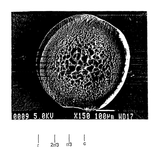
3o Referring to FIG. 2A, embolic particles 111 have a substantially uniform
spherical shape and size. Referring to FIG. 2B, each embolic particle has a
well-
defined outer spherical surface including relatively small, randomly located
pores. The
CA 02480579 2004-09-28
WO 03/084582 PCT/US03/09408
surface appears substantially smooth, with some larger surface morphology such
as
crevice-lilce features. Referring to FIGS. 2C-ZE, SEM images of cross-sections
through embolic particles, the body of the particle defines pores which
provide
compressibility and other properties to the embolic composition. Pores near
the center
of the particle are relatively large and pores neax the surface of the
particle are
relatively small.
The region of small pores near the periphery of the embolic particle is
relatively
stiff arid incompressible, which enhances resistance to shear forces and
abrasion. In
addition, the variable pore size profile produces a symmetric compressibility
and, it is
o believed, a compressibility profile such that the particles are relatively
easily
compressed from a maximum, at rest diameter to a smaller, compressed first
diameter
but compression to even smaller diameter requires substantially greater force.
A
variable compressibility profile is believed to be due to the presence of a
relative weak,
collapsible inter-pore wall structure in the center region where the pores are
large, and a
stiffer inter-pore wall structure near the surface of the particle, where the
pores are
more numerous and relatively small. The variable pore size profile also is
believed to
enhance elastic recovery after compression. The pore structure also influences
the
density of the embolic particles and the rate of carrier fluid or body fluid
uptake.
The embolic particles can be delivered through a catheter having a lumen area
2o that is smaller, e.g. 50% smaller or less, than the uncompressed cross-
sectional area of
the particles. As a result, the embolic particles must be compressed to pass
through the
catheter for delivery into the body. The compression force is provided
indirectly by
increasing the pressure applied to the carrier fluid by depressing the syringe
plunger.
The embolic particles are relatively easily compressed to diameters sufficient
for
delivery through the catheter into the body. The robust, rigid surface region
resists
abrasion when the embolic particles contact hard surfaces such as syringe
surfaces,
hard plastic or metal stopcock surfaces, and the catheter lumen wall (e.g.
Teflon) during
delivery. Once in the body, the embolic particles substantially recover to
original
diameter and shape for efficient transport in the carrier and body fluid
stream. At the
3o point of occlusion, the particles can again compress as they aggregate in
the occlusion
region. The embolic particles form a dense occluding mass. The compression in
the
body is determined by the force provided by body fluid flow in the lumen. The
CA 02480579 2004-09-28
WO 03/084582 PCT/US03/09408
compression may be limited by the compression profile of the particles and the
number
of embolic particles needed to occlude a given diameter may be reduced.
W embodiments, the particles have a diameter of about 1500 or 1200 microns or
less, and about 10 microns or more, e.g. about 400 microns or more and the
pores are
about SO or 3S to 0.01 micron. The embolic particles can be classified in size
ranges of
about 500-700 microns, about 700-900 microns, or about 900-1200 microns. The
particles typically have a mean diameter in approximately the middle of the
range and
variance of about 20% or less, e.g. 15% or 10% or less.
Referring particularly to FIG. 2C, the particles can be considered to include
a
o center region, C, from the center of the particle to a radius of about r/3,
a body region,
B, from about r/3 to about 2 r/3 and a surface region, S, from 2r/3 to r. The
regions can
be characterized by the relative size of the pores and the number of pores of
given
sizes. In embodiments, the center region has a greater number of relatively
large pores
than the body region and the surface region. The large pores are in the range
of about
20 micron or more, e.g. 30 micron or more, or in the range of about 20 to 3S
micron.
The body region has a greater number of intermediate size pores than the
surface
region. The intermediate size pores are in the range of about S to 18 micron.
In
embodiments, the regions may also have different densities, with the density
of the
surface region being greater than the density of the body region, and the
density of the
2o body region being greater than the density of the center region.
The size of the pores in each of the regions can also be characterized by a
distribution. In embodiments, the predominant pore sizes) in the center region
being
greater than the predominant pore sizes) in the body region and the
predominant pore
sizes) in the body region is greater than the predominant pore sizes) in the
surface
region. In embodiments, in the predominant pore size in the center region is
20 micron
or more, e.g. 30 microns or more, or in the range of about 20 to 3S microns.
The
predominant pore size in the body region is about 18 micron or less, e.g.
about 1S
micron or less, or in the range of about 18 to 2 micron. The pores in the
surface region
are preferably predominantly less than about 1 micron, e.g. about 0.1 to 0.01
micron.
3o In embodiments, the predominant pore size in the body region is about SO to
70% of the pore size in the center region and the pore size in the surface
region is about
10% or less, e.g. about 2% of the pore size in the body region. The size of
the pores on
the outer surface of the particle is predominantly in the range of about 1
micron or Less,
CA 02480579 2004-09-28
WO 03/084582 PCT/US03/09408
e.g. about 0.1 or 0.01 micron. In embodiments, the surface and/or surface
region is
substantially free of pores having a diameter larger than about 10 micron or
larger than
about 1 micron. In embodiments, the predominant pore size is in the region 0.8
or 0.9r
to r is about 1 micron or less, e.g. 0.5 to 0.1 micron or less. The region
from the center
of the particle to 0.8 or 0.9r has pores of about 10 micron or greater and/or
has a
predominant pore size of about 2 to 35 micron. In embodiments, the predominant
pore
size in the region 0.8 or 0.9r to r is about 5% or less, e.g. 1% or 0.3% or
less than the
predominant pore size in the region from the center to 0.9r, the largest pores
in the
particles can have a size in the range of 1% or 5% or 10% or more of the
particle
o diameter.
The size of the pores can be measured by viewing a cross-section as in Fig.
2C.
For irregularly shaped pores, the maximum visible cross-section is used. The
predominant pore sizes) can be found by measuring the size of the visible
pores and
plotting the number of pores as a function of size. The predominant pore
sizes) are the
~ 5 sizes that are about the maximum in the distribution. In Fig. 2C, the SEM
was taken on
wet particles including absorbed saline, which were frozen in liquid nitrogen
and
sectioned. (Fig. 2B was talcen prior to sectioning.) In Figs. 2D and 2E, the
particle was
freeze-dried prior to sectioning and SEM analysis.
The density of the particles is such that they are readily suspended in the
carrier
2o fluid such as a mixture of saline and contrast solution and remain
suspended during
delivery. In embodiments, the density is in about 1.1 - 1.4g/cm3. For
suspension in a
saline-contrast solution, the density is about 1.2 - 1.3g/cm3. The sphericity
after
compression in a catheter to about 50% or more of their cross-sectional area
is about
0.90 or 0.95 or greater. In embodiments, the particles can be manually
compressed,
25 essentially flattened, while wet to less than 50% of original diameter and
then, upon
exposure to fluid, regain a sphericity of about 0.9 or more. The carrier fluid
is a
pharmaceutically acceptable carrier such as saline or contrast agent. The
particles can
be sterilized prior to use.
3o Manufacture
Referring to FIG. 3A, a system for producing embolic particles includes a flow
controller 300, a drop generator 310, a gelling vessel 320, a reactor vessel
330, a gel
dissolution chamber 340 and a filter 350. The flow controller 300 delivers
polymer
s
CA 02480579 2004-09-28
WO 03/084582 PCT/US03/09408
solutions to a viscosity controller 305, which heats the solution to reduce
viscosity prior
to delivery to the drop generator 310. The drop generator 310 forms and
directs drops
into a gelling vessel 320, whexe drops are stabilized by gel formation. The
gel-
stabilized drops are transferred from the gelling vessel 320 to reactor vessel
330 where
the polymer in the gel-stabilized drops are reacted forming precursor
particles. The
precursor panicles are transferred to a gel dissolution chamber 340, where the
gel is
dissolved. The particles are then filtered in a filter 350 to remove debris,
sterilized, and
paclcaged as an embolic composition including embolic particles.
A base polymer and a gelling precursor are dissolved in water and mixed. The
o mixture is introduced to a high pressure pumping apparatus, such as a
syringe pump
(e.g., model PHD4400, Harvard Apparatus, Holliston, MA). Examples of base
polymers include polyvinyl alcohol, polyacrylic acid, polymethacrylic acid,
poly vinyl
sulfonate, carboxyrnethyl cellulose, hydroxyethyl cellulose, substituted
cellulose,
polyacrylamide, polyethylene glycol, polyamides, polyureas, polyurethanes,
polyester,
~ 5 polyethers, polystyrene, polysaccharide, polylactic acid, polyethylene,
polymethylmethacrylate and copolymers or mixtures thereof. A preferred polymer
is
polyvinyl alcohol. The polyvinyl alcohol, in particular, is hydrolyzed in the
range of 80
to 99%. The weight average molecular weight of the base polymer can be in the
range
of 9000 to 186,000, 85,000 to 146,000 or 89,000 to 98,000. Gelling precursors
include,
2o for example, alginates, alginate salts, xanthan gums, natural gum, agar,
agarose,
chitosan, carrageenan, fucoidan, furcellaran, laminaran, hypnea, eucheuma, gum
arabic,
gum ghatti, gum karaya, gum tragacanth, hyalauronic acid, locust beam gum,
arabinogalactan, pectin, amylopectin, other water soluble polysaccharides and
other
sonically crosslinkable polymers. A particular gelling precursor is sodium
alginate. A
25 preferred sodium alginate is high guluronic acid, stem-derived alginate
(e.g. about 50 or
60% or more guluronic acid with a low viscosity e.g. about 20 to 80 cps at
20°C) which
produces a high tensile, robust gel. High molecular weight PVA is dissolved in
water
by heating, typically above about 70°C, while alginates can be
dissolved at room
temperature. The PVA can be dissolved by mixing PVA and alginate together in a
3o vessel which is heated to autoclave temperature (about 121°C).
Alternatively, the PVA
can be disposed in water and heated and the alginate subsequently added at
room
temperature to avoid exposing the alginate to high temperature. Heat can also
be
applied by microwave application. In embodiments, for PVA/alginate, the
mixture is
CA 02480579 2004-09-28
WO 03/084582 PCT/US03/09408
typically about 7.5 to 8.5%, e.g. about 8% by weight PVA and about 1.5 to
2.5%, e.g.
about 2%, by weight alginate.
Referring to FIG. 3B, the viscosity controller 305 is a heat exchanger
circulating water at a predetermined temperature about the flow tubing between
the
pump and drop generator. The mixture of base polymer and gelling precursor
flows
into the viscosity controller 305, where the mixture is heated so that its
viscosity is
lowered to a level for efficient formation of very small drops. For a high
molecular
weight PVA/alginate solution, the temperature of the circulating water is less
than
about 75°C and more than about 60°C, for example, 65°C
which maintains the mixture
o at a viscosity of 90-200 centipoise. For spherical particles, the viscosity
of the drops is
maintained so they are captured in the gelling vessel without splintering or
cojoining
which can create irregular, fiberous particles. In other embodiments, the flow
controller and/or the drop generator can be placed in a temperature-controlled
chamber,
e.g. an oven, or a heat tape wrap, to maintain a desired viscosity.
The drop generator 310 generates substantially spherical drops of
predetermined
diameter by forcing a stream of the mixture of base polymer and gelling
precursor
through a nozzle which is subject to a periodic disturbance to break up the
jet stream
into drops. The jet stream can be broken into drops by vibratory action
generated for
example, by an electrostatic or piezoelectric element. The drop size is
controlled by
2o controlling the flow rate, viscosity, amplitude, and frequency at which the
element is
driven. Lower flow rates and higher frequencies produce smaller drops. A
suitable
electrostatic drop generator is available from NISCO Engineering, model NISCO
Encapsulation unit VAR D, Zurich, Switzerland. In embodiments, the frequency
is in
the range of about 0.1 to 0.8 kHz. The flow rate through the droplet generator
is in the
range of about 1 to 12 mL per minute. The drop generator can include charging
the
drops after formation such that mutual repulsion between drops prevents drop
aggregation as drops travel from the generator to the gelling vessels.
Charging may be
achieved by, e.g. an electrostatic charging device such as a charged ring
positioned
downstream of the nozzle.
3o Drops of the base polymer and gelling precursor mixture are captured in the
gelling vessel 320. The gelling vessel 320 contains a gelling agent which
interacts with
the gelling precursor to stabilize drops by forming a stable gel. Suitable
gelling agents
include, for example, a divalent cation such as allcali metal salt, alkaline
earth metal salt
CA 02480579 2004-09-28
WO 03/084582 PCT/US03/09408
or a transition metal salt th~,~ can ionically crosslinlc with the gelling
agent. An
inorganic salt, for example, a calcium, barium, zinc or magnesium salt can be
used as a
gelling agent. In embodiments, particularly those using an alginate gelling
precursor, a
suitable gelling agent is calcium chloride. The calcium cations have an
affinity for
carboxylic groups in the gelling precursor. The cations complex with
carboxylic
groups in the gelling precursor resulting in encapsulation of the base polymer
in a
matrix of gelling precursor.
Referring to FIG. 4, a photo-image of the gelled particles, the gelling agent
is in
an amount selected in accordance with the desired properties of the particles.
As
o evident, a pore stuucture in the particle forms in the gelling stage. The
concentration of
the gelling agent can control pore formation in the particle, thereby
controlling the
porosity gradient in the embolic particle. Adding non-gelling ions, for
example,
sodium ions, to the gelling solution can reduce the porosity gradient,
resulting in a more
uniform intermediate porosity throughout the particle. In embodiments, the
gelling
~5 agent is, for example, 0.01-10 weight percent, 1-5 weight percent or 2
weight percent in
deionized water. In embodiments, particles, including gelling agent and a pore
structure can be used in embolic compositions.
Following drop stabilization, the gelling solution is decanted from the solid
drops and the stabilized drops are transferred to the reactor vessel 330. In
the reactor
2o vessel 330, the stabilized drops are reacted to produce precursor
particles. The reactor
vessel includes an agent that chemically reacts with the base polymer, e.g. to
cause
crosslinking between polymer chains and/or within a polymer chain. The agent
diffuses into the stabilized drops from the surface of the particle in a
gradient which, it
is believed, provides more crosslinking near the surface of the stabilized
drop compared
25 to the body and center of the drop. Reaction is greatest at the surface of
the drop,
providing a stiff, abrasion resistant exterior. For polyvinyl alcohol, for
example, the
vessel 330 includes aldehydes, such as formaldehyde, glyoxal, benzaldehyde,
aterephthalaldehyde, succinaldehyde and glutaraldehyde for the acetalization
of
polyvinyl alcohol. The vessel 330 also includes an acid, for example, strong
acids such
3o as sulfuric acid, hydrochloric acid, ntric acid and weak acids such as
acetic acid,
formic acid and phosphoric acid. In embodiments, the reaction is primarily a
1,3
acetalization:
II
CA 02480579 2004-09-28
WO 03/084582 PCT/US03/09408
H+
--(-CH-CH2-CH-CH2-)-- + CH2=O ~ --(-CH-CH2-CH-CH2-)--
+H20
65C
s OH OH O O
CH2
This intra-chain acetalization reaction can be carried out with relatively low
probability of inter-chain crosslinking as described in John G. Pritchard
"Poly(Vinyl
Alcohol) Basic Properties And Uses (Polymer Monograph, vol. 4) (see p. 93-97),
~o Gordon and Breach, Science Publishers LTD., London, 1970, the entire
contents of
which is hereby incorporated by reference. Some OH groups along a polymer
chain
may remain unconverted since the reaction proceeds in a random fashion and
there will
be left over OH groups that do not react with adjacent groups.
Adjusting the amount of aldehyde and acid used, reaction time and reaction
temperature can control the degree of acetalization. In embodiments, the
reaction time
is e.g., 5 minutes to 1 hour, 10 to 40 minutes or 20 minutes. The reaction
temperature
can be 25°C to 150°C or 75°C to 130°C or
65°C. The reactor vessel is placed in a water
bath f tted with a orbital motion mixer. The crosslinlced precursor particles
are washed
several times with deionized water to neutralize the particles and remove any
residual
2o acidic solution.
The precursor particles are transferred to the dissolution chamber 340 to
remove
the gelling precursor, e.g. by an ion exchange reaction. In embodiments,
sodium
alginate is removed by ion exchange with a solution of sodium hexa-
metaphosphate
(EM Science). The solution can include, for example, ethylenediaminetetracetic
acid
25 (EDTA), citric acid, other acids and phosphates. The concentration of the
sodium
hexa-metaphosphate can be, for example, 1-20 weight %, 1-10 weight % or 5
weight
in deionized water. Residual gelling precursor, for example, sodium alginate,
can be
determined by assay for detection of uronic acids in, for example, alginates
containing
mannuronic and guluronic acid residues. Suitable assays include rinsing the
particles
so with sodium tetraborate in sulfuric acid solution to extract alginate and
combining the
extract with metahydroxydiphenyl colormetric reagent and determining
concentration
by UV/VIS spectroscopy. Testing can be carried out by alginate suppliers such
as
12
CA 02480579 2004-09-28
WO 03/084582 PCT/US03/09408
FMC Biopolymer, Oslo, Norway. Residual alginate may be present in the range of
about 20-35% by weight prior to rinsing and in the range of about 0.01-0.5% or
0.1-
0.3% or 0.18% in the particles after rinsing for 30 minutes in water at about
23°C.
The particles are filtered through filter 350 to remove residual debris.
Particles
of 500 to 700 microns are filtered through a sieve of 710 microns and then a
sieve of
300 microns. Particles of 700 to 900 microns are filtered through a sieve of
1000
microns and then a sieve of 500 microns. Particles of 900 to 1200 microns are
filtered
through a sieve of 1180 microns and then a sieve of 710 microns.
The filtered particles are sterilized by a low temperature technique such as e-
1 o beam irradiation, and packaged, typically about 1 to 5 ml of particles in
about 5 to 10
ml saline. In embodiments, electron beam irradiation can be used to
pharmaceutically
sterilize the particles to reduce bioburden. In e-beam sterilization, an
electron beam is
accelerated using magnetic and electric fields, and focused into a beam of
energy. This
resultant beam can be scanned by means of an electromagnet to produce a
"curtain" of
accelerated electrons. The accelerated electron beam penetrates the collection
of
embolic particles to confer upon them electrons which destroy bacteria and
mold to
sterilize and reduce the bioburden in the embolic particles. Electron beam
sterilization
can be carried out by sterilization vendors such as Titan Scan, Lima, Ohio.
13
CA 02480579 2004-09-28
WO 03/084582 PCT/US03/09408
Examples
Example 1
Embolic particles are manufactured from an aqueous solution containing 8
weight % of polyvinyl alcohol, 99+% hydrolyzed, average MW 89,000-120,000
(ALDRICH) and 2 weight% of gelling precursor, sodium alginate, PRONOVA
UPLVG, (FMC BioPolymer, Princeton, NJ) in deionized water and the mixture is
heated to about 121° C. The solution has a viscosity of about 310
centipoise at room
temperature and a viscosity of about 160 cps at 65°C. Using a syringe
pump (Harvard
Apparatus), the mixture is fed to drop generator (Nisco Engineering). Drops
are
directed into a gelling vessel containing 2 weight % of calcium chloride in
deionized
water and stirred with a stirring bar. The calcium chloride solution is
decanted within
about three minutes to avoid substantial leaching of the polyvinyl alcohol
from the
drops into the solution. The drops are added to the reaction vessel containing
a solution
of 4% by weight of formaldehyde (37 wt% in methanol) and 20% by weight
sulfuric
~5 acid (95-98% concentrated). The reaction solution is stirred at 65°C
for 20 minutes.
Precursor particles are rinsed with deionized water (3 X 300 mL) to remove
residual
acidic solution. The sodium alginate is substantially removed by soaking the
precursor
particles in a solution of 5 weight % of sodium hexa-methaphosphate in
deionized
water for 0.5 hour. The solution is rinsed in deionized water to remove
residual
2o phosphate and alginate. The particles are filtered by sieving, as discussed
above,
placed in saline (USP 0.9% NaCI) and followed by irradiation sterilization.
Particles were produced at the nozzle diameters, nozzle frequencies and flow
rates (amplitude about 80% of maximum) described in Table I.
25 TABLE 1
Bead Nozzle FrequencyFlow DensitySphericitySuspendability
Size Diameter(kHz) Rate
microns)(microns) (mL/min)(g/mL) (minutes)
500-700150 0.45 4 - 0.92 3
700-900200 0.21 5 1.265 0.94 5
900-1200300 0.22 10 - 0.95 6
Suspendability is measured at room temperature by mixing a solution of 2 ml of
particles in 5 ml saline with contrast solution (Omnipaque 300, Nycomed,
14
CA 02480579 2004-09-28
WO 03/084582 PCT/US03/09408
Buckinghamshire, UK) and observing the time for about 50% of the particles to
enter
suspension, i.e. have not sunk to the bottom or floated to the top of a
container (about
ml, 25 mm dia vial). Suspendability provides a practical measure of how long
the
particles will remain suspended in use. (Omnipaque is an aqueous solution of
Iohexol,
s N.N.-Bis (2,3-dihydroxypropyl)-T-[N-(2,3-dihydroxypropyl)-acetamide]-2,4,6-
trilodo-
isophthalamide; Omnipaque 300 contains 647 mg of iohexol equivalent to 300 mg
of
organic iodine per ml. The specific gravity of 1.349 of 37° C and an
absolute viscosity
11.8 cp at 20° C.) The particles remain in suspension for about 2 to 3
minutes.
Particle size uniformity and sphericity is measured using a Beckman Coulter
~ o RapidVLTE Image Analyzer version 2.06 (Beckman Coulter, Miami, FL).
Briefly, the
RapidVUE takes an image of continuous-tone (gray-scale) form and converts it
to a
digital form through the process of sampling and quantization. The system
software
identifies and measures particles in an image in the form of a fiber, rod or
sphere.
Sphericity computation and other statistical definitions are in Appendix A,
attached,
which is a page from the RapidV~TE operating manual.
Referring to FIG. 5, particle size uniformity is illustrated for particles 700
- 900
micron. The x-axis is the particle diameter. The y-axis is the volume
normalized
percentage of particles at each particle size. The total volume of particles
detected is
computed and the volume of the particles at each diameter is divided by the
total
2o volume. The embolic particles have distribution of particle sizes with
variance of less
than about + 15%.
Example 2
Referring to FIG. 6, a catheter compression test investigates the
injectability,
and indirectly, the compressibility of the particles. The test apparatus
includes a
2s reservoir syringe 610 and an injection syringe 620 coupled to a T-valve
630. Syringe
610 is a 20mL syringe while injection syringe 620 is a 3 mL syringe. T-valve
630 is
coupled in series to a second T-valve 640. T-valve 640 is coupled to a
catheter 650 and
a pressure transducer 660. Injection syringe 620 is coupled to a syringe pump
621
(Harvard Apparatus).
3o To test deliverability of the particles, syringe 610 and syringe 620 are
loaded
with embolic composition in saline and contrast (50!50 Ominipaque 300). The
embolic
composition in syringes 610 and 620 is intermixed by turning the T-valve to
allow fluid
between the syringes to mix and suspend the particles. After mixing, the
embolic
CA 02480579 2004-09-28
WO 03/084582 PCT/US03/09408
composition in syringe 620 flows at a rate of about l OmL/min. The back
pressure
generated in the catheter 650 is measured by the pressure transducer 670 in
millivolts to
measure the clogging of catheter 650. About 1 ml of the particles is mixed in
l OmL of
solution.
Results for several different catheters (available from Boston Scientific,
Natick,
MA) and particle sizes are shown in Table 2. The baseline pressure is the
pressure
observed when injecting carrier fluid only. The delivery pressure is the
pressure
observed while delivering particles in carrier fluid. The average is the
average of the
peak pressure observed in the three runs.
SIZE Delivery CatheterInner DiameterAvg. BaselineAvg. DeliveryTotal
number
(microns) (inches) Pressure Pressure of Clogs
(psia) (psia)
500-700 RENEGADE ~ 0.021 32.610 33.245 0
(533 micron)
700-900 FASTRACI~ER~ 0.024 11.869 13.735 0
(609 micron)
900-1200GL)DECATH ~ 0.038 0.788 0.864 0
(965 micron)
As evident, particles in each of the size ranges were successfully delivered
without clogging through catheters having a lumen diameter smaller than the
largest
particle size. The particles exhibit a post-compression sphericity of about
0.9 or more.
Example 4
Solubility is tested by mixing particles in a solution of solvent at room
temperature for about 0.5 hour and observing the mixture for visible signs of
dissolution. The particles are insoluble in DMSO (Dimethylsulfoxide), HFIP
(Hexafluoro-isopropanol), and THF (Tetrahydrafuran).
16
CA 02480579 2004-09-28
WO 03/084582 PCT/US03/09408
Example 5
Embolic particles include the following glass transition temperatures as
measured by differential scanning calorimetry data (DSC)
Product 500-700 microns 900-1200 microns
Glass transition109.30-110.14 108.30-111.87
temperature (Tg)
Example 6
Refernng to Fig. 7, an ATR infrared spectrum of dried particles is provided.
1 o Use
The embolic compositions can be used as pharmaceutically acceptable
compositions in the treatment of, for example, fibroids, tumors, internal
bleeding,
AVMs, hypervascular tumors, fillers for aneurysm sacs, endoleak sealants,
arterial
sealants, puncture sealants and occlusion of other lumens such as fallopian
tubes.
15 Fibroids can include uterine fibroids which grow within the uterine wall
(intramural
type), on the outside of the uterus (subserosal type), inside the uterine
cavity
(submucosal type), between the layers of broad ligament supporting the uterus
(interligamentous type), attached to another organ (parasitic type), or on a
mushroom-
like stalk (pedunculated type). Internal bleeding includes gastrointestinal,
urinary,
2o renal and varicose bleeding. AVMs are for example, abnormal collections of
blood
vessels, e.g. in the brain, which shunt blood from a high pressure artery to a
low
pressure vein, resulting in hypoxia and malnutrition of those regions from
which the
blood is diverted.
The magnitude of a therapeutic dose of the embolic composition can vary based
2s on the nature, location and severity of the condition to be treated and the
route of
administration. A physician treating the condition, disease or disorder can
determine
effective amount of embolic composition. An effective amount of embolic
composition
refers to the amount sufficient to result in amelioration of symptoms or a
prolongation
of survival of the patient. The embolic compositions can be administered as
3o pharmaceutically acceptable compositions to a patient in any
therapeutically acceptable
17
CA 02480579 2004-09-28
WO 03/084582 PCT/US03/09408
dosage, including those administered to a patient intravenously,
subcutaneously,
percutaneously, intratrachealy, intramuscularly, intramucosaly,
intracutaneously, intra-
articularly, orally or parenterally.
Compositions containing the embolic particles can be prepared in calibrated
s concentrations of the embolic particles for ease of delivery by the
physician. The
density of the composition can be from about 1.1 to 1.4 g/cm3, or from about
1.2 to
about 1.3 g/cm3 in saline solution. Suspensions of the embolic particles in
saline
solution can be prepared to form stable suspensions over duration of time. The
suspensions of embolic particles can be stable from 1 to 10 minutes, 2-7
minutes or 3 to
6 minutes. The physician can determine concentration of embolic particles by
adjusting the weight ratio of the embolic particles to physiological solution.
If weight
ratio of the embolic particles is too small, too much liquid could be injected
in a blood
vessel, possibly allowing the embolic particles to stray into lateral vessels.
In
embodiments, the weight ratio of the embolic particles to the physiological
solution is
~ 5 about 0.01 to 15% by weight. The embolic composition can include a mixture
of
particles including particles with the pore profiles discussed above and
particles with
other pore profiles or non-porous particles. Particles can be used for embolic
applications without removal of the gelling agent (e.g. alginate) for example
at the
stabilized drop stage or precursor particle stages described above. While
substantially
2o spherical particles are preferred, non-spherical particles can be
manufactured and
formed by controlling, e.g. drop formation conditions or by post-processing
the
particles, e.g. by cutting or dicing into other shapes. Particles can also be
shaped by
physical deformation followed by crosslinking. Particle shaping is described
in U.S.
Serial No. 10/116,330 filed April 4, 2002, the entire contents of which is
hereby
2s incorporated by reference.
Other embodiments are within the scope of the following claims.
18