Note: Descriptions are shown in the official language in which they were submitted.
CA 02480970 2004-10-O1
f25914.S01
Method and Arrangement for Optically Measuring Swelling of the Nose
Field of Use of the Invention
The invention relates to the fields of medicine and device construction and
relates to a
method and an arrangement for optically measuring swelling of the nose, which
can be
used, e.g., to measure nasal obstruction after allergen provocation.
Prior Art
From a medical point of view there is a need for an objectification of the
measurement o;f
swelling and of the course of swelling, e.g., in allergic reactions that are
triggered, e.g.,
by the nasal provocation test.
Nowadays the diagnosis of allergic rhinitis is made by calculating a symptom
score (itchy
nose, secretion, remote symptoms, such as watery eyes) and by measuring the
nasal
obstruction after allerge$ provocation with the aid of rhinomanomeixy (Clement
et al.:
l2hinornanometry = a review, ORL J. Otorhinola~yngol. Relat. Spec. 46, 173-91,
1984).
The disadvantage of rhinomanometry hereby is that the measurement cannot be
carried
out during the allergen. application. With severe nasal obstruction, patients
experience
the xhiz~omvaaometry as very unpleasant. Faulty' measurements occux frequently
with
uncooperative patients.
Another possibility for determining the swelling of the nose and in particular
of the nasal
mucous membrane is acoustic rhinometry (Fisher: Acoustic rhinometty, Clan.
OtolaryngQl. 22, 307-17, 1997). These measurements have a xelatiwely large
spread in
their results. Su~cient precision is achieved only for the front sections of
the nose. No
medication ox allergen provocation can take place during the measurement. A
continuous
measurement is not possible with this method, either.
Fuxthezrr~ore, it is not possible with either method to say whether a nasal
swelling is due
to a change in the microcirculation or the formation of an edema,
CA 02480970 2004-10-O1
P25914,501
T)escription of the Invention
The object of the invention is to disclose a method and an arrangement for
optically
measuring swelling of the nose with which a largely objective measurement of
the
swelling of the nose is ~'endered possible, in particular while provocation
tests are being
conducted.
The object is attained with the invention described in the claims.
Advantageous
embodiments are the subject matter of dependent claims.
The arrangement according to the invention for optically measuring swelling of
the nose
comprises a basic device with light-producing components and light-detecting
components and associated emitter and receiver electronic systems and
controllers.
Furthermore, at least one optical connection is implemented between the basic
device and
an optical emitter element, whereby the transmission of the light produced by
the Iight-
producing components is realized by optical elements in the optical connection
to the
emiti;er element: Furthermore, at least one optical' ~cormecti4rl fs'
pre~en't' b~tVVe~'ah '" ' ~ ~ " -' - '
optical receiver element and the light-detecting components. Emitter and
receiver
elements that are arranged on an application element are located outside the
basic device.
The application element thereby realizes an arrangement of the emitter and
receiver
elements that makes it possible for the light emitted by the emitter element
to pass
through the swellable tissue of at Ieast one side of the nose to the receiver
element.
Furthermore the application element can be placed in a. forIri~loc'king manner
at least on . '-'
the upper part of the nose.
'With the arrangement according to the invention the swehing of the nasal
tissue is
optically recorded. The nasal tissue is thereby irradiated from outside by a
light source
that is emitted from an emitter element, and the scattered light passing
through the tissue
is recorded by a detector, a receiver element, on either the same side of the
nose or on the
opposite side of the apse, Wlien passing through the nasal tissue, the light
passes tbz~ough
a number of tissue layers, such as skin, musculature, mucous membrane, bone,
cartilage,
and the airways. A part of the tissue penetrated is characterized by
swellability, in
CA 02480970 2004-10-O1
P25914.501
particular the nasal mucous membrane located above the bone of the nasal
cvncha. In the
course of the swelling an increase of the blood volume occurs in this part of
the tissue
due to the influx of blood into the cavernous bodies. The inflowing blood is
thereby
primarily of an arterial nature and thus normally 95% saturated with oxygen.
Furthermore, in the event of the formation of an edema possibly associated
with the
swelling, an increase in the tissue fluid volume occurs. rt is therefore
advantageous to
conduct the irradiation spectrometrically in order to be able to
quantitatively record
separately the volume proportions of the oxygenated and deoxygenated
hemoglobin and
of the tissue fluid. This can take place either by using a white-light source
and a
spectrometer detector (e.g., diode line spectrometer) or ~by using several
light sources
with discrete radiation spectra (LEDs, laser diodes).
Since the cited substances involved in the swelling have difFerent optical
absorption
spectra, a separate absolute or relative determination of the volume
proportions is
possible with corresponding mathematical methods. Such an arrangement has
hitherto
not bean described. ' '
One advantage of the arrangement according to the invention is 'that it is
characterized by
a non-invasive application from outside and by simple handling.
Best Way vf'Oarr~~ (3ut tlze'Izrv~erltion .. .
. . The in~ez~tiowwi~h be described in mare deteal~ bei~ow otr the basis of
exemplary
embodiments. The design of the azxangemeat has been adjusted to the purpose of
the
examination. The drawings show:
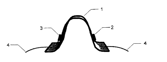
Fig. 1 An application element in front view (a) and in a view from above (b)
Fig. 2 Two embodiment variants of an application element with active emitter
and
receiver elements (a) and passive emitter and receiver elements (b)
Fig, 3 An application element and its mounting an the head and nose
Fig. 4 A basic device of an arrangement according to the invention
CA 02480970 2004-10-O1
P25914.501
Fig. 5 A, diagrammatic representation of a cross section of the nose, showing
the
position of the optical elements and the irradiation channel before and after
a
swelling
Fig. 6 A diagrammatic representation of the measured extinction values in the
course of
a swelling.
The arrangement according to the invention comprises at least one basic device
1Z with
the emitter 15 and receiver 16 electronic systems necessary to carry out the
measuring
task and an application element 1 ~ that is in direct contact with the nasal
tissue during the
measurement.
An application element 1 is shown in Fig. 1 a and b. It comprises a clamp-
shaped base
body, both sides of which can be placed on the sides of the nose in a fozzn-
locking
manner. The light-emitting element, optical emitter element 2, is arranged on
one side of
the application element 1, the light-receiving element, optical receiver
element 3, is _
arranged owthe oppt~site side. They are embodieu-eitlzer as~discrete radiation
sources anti-"
detectors, the optical axes of which are aligned in the direction of the
tissue (Fig. 2a) and
which are connected to the basic device 12 via current-carrying cables 4, or
otherwise as
optical connections 6, 7 that realixe the light transfer from and to the basic
device 12 and
that are either guided with their radiating suxfaces perpendicular to the
nasal tissue or that
~a~re aligrredlto the tissue by the a~rrangement~ of correslrondiag optical
de~lae~orr~lementw - y
(mirrors, microprisnls) (Fig. 2b). . . . . , , . , , ..
Mounting of the application element 1 takes place according to Fig. 3 with the
aid of a
headband 8 placed on the head. This ensures a stable position of the
application element
1 on the bridge of the nose during the measurement. The application element is
connected to the headband 8 via a clamp 10. The comc~ectioz~ is embodied such
that an
exact positioning of the application element on the bridge of the nose is
possible, e.g.,
through a lockable ball joint 9 or a flexible metal hose. Further relevant
variants of the
arrangement according to the invention can be embodied as follows:
~ The application element is adhesively attached to the nose;
CA 02480970 2004-10-O1
P25914.501
~ The application element is pressed directly onto the nose with the aid of an
elastic
belt or a belt that is adjustable in circumfexe~,ce to the size of tl~e head;
~ The application element is embodied as a spectacles-like frame that sits on
the
root of the nose and in which the optical emitter and receiver elements are
pressed
onto the nasal tissue by gravity;
~ An arrangement in which the emitter and receiver elements are arranged on
two
separate basic elements (pads) that are adhesively attached oz~ each side of
the
nose separately frox~a one another.
For a precise and reproducible measurement, in addition to the spatially
stable and
motion-free fixing of the application element to the nose, it is also
important to suppress
and/or calibrate out extraneous light influences. It is therefore advantageous
to use
optical filters or to cover the measuring site during the measurement by a
light-
im~pervious cap, which, e.g., as a plastic cap, can fixed to the headband as
well and can be
closed oven the field of measurement during the examination as required.
Fig. 4 shows a basic device 12 with light-producing components 13 inside the
device and
light-detecting components 14 to which an application element 1 described
above can be
connected via the optical connections 6, 7. The basic device 12 comprises an
emitter
electronic system 15 fox the optical light-producing components 13, a receiver
electronic
system 16 artd'a: bon'f~~l~~r--t'I '~b'whficlt
b~'~ei~'~'vices°can~bz'eoirin~~ted viTa'a-d'ata~~:
interface. ~t its output the emitter elecCto~iC system 1 ~' has several light-
producing
components 13, the light of which is concentrated tbzough an optical element
18. The
concentrated light is introduced into an optical connection 6.
A, light-detection component 14 is connected to the entry of the receiver
electronic
system 16 into which component light enters from the optical connection 7.
A, spectrometric measurement is advantageous for the optical measurement of
swelling
and the differentiation of the causes of swelling. Light sources with limited
spectrum
(LEDs, semiconductor lasers) and a photodetector that is adequately sensitive
for the
CA 02480970 2004-10-O1
P25914.501
selected spectral range (semiconductor photodetector, photomultiplier) can be
used for
this. Alternatively, a white-light source and a detector measuring in a
spectxometrically
resolving manner can be used. The object of the measurement is to defect light
attenuation 'values (optical density of the tissue) at individual wavelengths
of interest over
time. This results from the equation:
E(~~t) = logio rs (~~t)
ro (~~t)
where Is(a,,t) denotes the light intensity radiated at the emitter element and
ID('~.,t) denotes
the light irnensity arriving at the receiver element at the wavelength 7~ and
at the point of
time t. In general the extinction E(~,t) is a function of the light scattering
and the light
absorption in the tissue and thus provides a measured value for the geometric
and optical
change of the tissue. By subtraction E(~,i,t)-E(7vz,t) at two wavelengths, a
relative
measurement of the change can be determined, which reflects the ratio of the
volume
change values of individual tissue constituents and is largely free of
geometric effects.
Thus when using, e.g., a hemoglobin-sensitive wavelength of ~,~=800nm and an
Hz0- -
sensitive wavelength of ~,2~~70am,'the ratio between the blood'and tissue
fluid iac~eas~'
' can be shown. Furthermore, by using special optical measurement techniques
it is
possible to separately determine the scatter and absorption properties of the
tissue, To
this end photon time delay measurements are necessary with the aid of a high-
frequency
modulation technology (intensity modulation of the light sources) and
amplitude and
phase meastsremer~t csf 'the receives signal) -ci~W pul~e~ laser teciqog~"
(appl.i"cation~' of ~ ''
short laser pulses ~ arid time-resolved measurement of the receiver signal),
These
measuring methods and associated mathematical methods for determining optical
parameters from such measurement data are state of the art (e.g., Sevick et
al.,
Quantitation of time- and frequency resadved optical spectra for the
determination of
tissue oxygenataon, Anal. Biochem, 195, 330-51, 1991; Patterson et al., Tame
resolved
reflectance and transmittance for the non-i~tvasive measurement of tissue
optical
properties, ~ppl. Opt., 28, 2331-36, 1989).
The course of a measurement will be explained here using the example of a
provoked
allergic reaction (provocation test).
CA 02480970 2004-10-O1
P25914.541
After the person to be examined has been prepared, the application element is
fixed on
the bridge of the nose near the root of the nose such that the optical emitter
and receivex
elennents axe opposite one another on the tissue and the optical radiation
penetrates as ,
much swellable tissue inside the nose as possible (Fig. 5). Subsequently an
optimal
photometric signal is adjusted with the aid of a manual, automatic or
semit~automatic
adjustrnent of source intensity/intensities and/or detector sensitivity in a
range suitable for
the measurement by means of optomechanical, electronic and/or software
methods. The
data acquisition is then manually started by the operator. Controlled by the
controller 17
inside the basic device, a repeated sequential switching of the radiation
sources takes
place by the emitter electronic system 15 and simultaneously the measured
detector
values are acquired by the receiver electronic system 16, Through the separate
measurement of the ambient light (dark signal) with light sources switched ofF
or
alternatively through a measurement o~ the AC portion of a light signal of the
light
sources that is sufficiently highly modulated, it is ensured that only fihe
light produced by
the light sources is measured and not the ambient' light possibly entering the
measurement .
de-ice. , . . . ~ . . . . . .. . . _ , .. ... . .
Fig. 6 shows diagrammatically measured extinction values in the course of a
swelling.
The spectral light attenuation values ua fhe unprovoked condition represent
the baseline
of the measuzement, When this is detected in a timeframe of 1 to 2 minutes, an
allergenic
subst~anoe as ad~ist~red by~spray#ng ~ e~n~e ~ox .both x~os#ril~(s~ and the
m~asuaremer~..
tune is recorded, e,g., by operating a pedal switch at the. moment of
adminis~ra'tit~~p t~.
With an allergic reaction, a swelling of the nasal tissue then occurs, which
causes a
detectable increase in the spectral extinction. Fig. 6 shows spectral
extinction values
which were standardized at the starting point tP for easier comprehension. At
the point of
time tE the swelling reaches a stationary condition at which no further
swelling is
detectable. Only after a time t»tE-tp does the swelling subside again.
Diagnostically
utilizable information can be derived from the.time response of the spectral
extinction
values, These include in particular:
~ The increase ~E(~,~E(a,,t~-E(?~,tP) of the extinction for a wavelength as a
gauge
of the intensity of the swelling;
CA 02480970 2004-10-O1
P25914.501
~ The extinction value difference ~E('A,1)~~.E(7~z) at different wa~'elengths
as a gauge
of the increase of the volume pzoportions of different tissue constituents
relative
to one another;
~ The time difference ~trts-tP of the reaction from the moment of provocation
until
the stationary final condition as a gauge of the speed of the swelling and the
form
of the curves E(7~,,t) as indicator for the physiological course of the
swelling.
CA 02480970 2004-10-O1
P2S914.S01
List of Reference Numbers
1 Application element
2 Optical emitter element
3 Optical xeceiver element
4 Current-carrying cable
S Optical deflection element
6 Optical coxnnection to the emitter element
7 Optical connection to the receiver element
8 '~Teadband
9 Ball yoint
Attaching clamp
11 Irradiation channel
12 Basic device .
13 Light-producing components .
i i,ight-detecting component ~ ' ~ '
4
Emitter electronic system
16 Receiver electronic system
17 Conbcoller
18 Optical element
