Note: Descriptions are shown in the official language in which they were submitted.
CA 02481093 2004-10-O1
WO 03/085404 PCT/CA03/00495
MSH FILE: KLKB-OVA
TITLE: Methods for Detecting Ovarian Cancer
FIELD OF THE INVENTION
The invention relates to methods and compositions for detecting ovarian
cancer.
BACKGROUND OF THE INVENTION
Ovarian cancer represents a great clinical challenge in gynecological
oncology. Since most
patients are asymptomatic until the disease has metastasized, two-thirds are
diagnosed with advanced
disease (1). In the United States, around 23,000 new cases of ovarian cancer
and about 14,000 deaths from
the disease were expected for the year 2000 (2), giving it the highest
mortality rate of all gynecological
malignancies.
The human kallikrein gene family, a subfamily of serine proteases, is now
known to include
fifteen members (1;2). All genes in this family localize to chromosome 19q13.4
and share significant
similarities at both the DNA and amino acid level. The human kallikrein family
includes hK3/Prostate
specific antigen (PSA) which is the most important biomarker for prostate
cancer (3). Recently, three
other members of this family, hK6/neurosin, hKlO/normal epithelial cell
specific 1 (NES1) and
hKl1/trypsin like serine Protease (TLSP), have been shown to be potential
biomarkers for ovarian and,
prostate cancer (4-6). In addition, recent reports also suggest that many
other members of this family are
associated with cancers of the breast, ovary, prostate and testis, as well as
with diverse diseases of the
central nervous system, skin, etc. (reviewed in ref. (7)).
2 0 KLKB/neuropsin is a member of human kallikrein family (1;2) (note: KLKB,
gene; hKB, protein,
according to the official kallikrein gene nomenclature (8)). Originally, KLK8
was cloned from a human
skin cDNA library as a homolog of mouse neuropsin (9). The mouse homolog has
highest expression in
skin and brain, especially the hippocampus, and was assumed to be associated
with neural plasticity,
memory formation and some forms of epilepsy (10-13). KLKB mRNA is increased in
Alzheimer's disease
2 5 hippocampus compared to controls, which suggests that KLK8 may indeed have
a relationship with neural
'plasticity in humans (14). KLK8 transcripts in ovarian cancer tissues are
expressed at higher levels than in
controls (15). Two splice variants of KLK8 have been detected in ovarian
cancer (16).
To date, there is no literature describing any relationship between hK8
protein expression and
cancer.
3 0 SUMMARY OF THE INVENTION
Recombinant human kallikrein 8 was produced using a baculovirus expression
system, purified
using column chromatography, and injected into mice and rabbits for antibody
generation. These
antibodies were used to develop a highly sensitive and specific immunoassay
for hK8. The assay was
applied to the measurement of native hK8 in tissue extracts and biological
fluids. Applicants found
3 5 elevated levels of serum hK8 in patients with ovarian cancer. In addition,
hK8 was found in tumor extracts
and ascites fluid of patients with ovarian cancer.
Therefore, kallikrein 8 has particular application in the detection of ovarian
cancer. Thus,
kallikrein 8 constitutes a new biomarker for diagnosis and monitoring (i.e.
monitoring progression or
CA 02481093 2004-10-O1
WO 03/085404 PCT/CA03/00495
- 2 -
therapeutic treatment) of ovarian cancer. In accordance with an aspect of the
invention kallikrein 8 is used
for the diagnosis, monitoring, and prognosis of ovarian cancer, and it may be
used as a biomarker before
surgery or after relapse.
The presence of kallikrein 8 may be assessed, for example by detecting the
presence in the sample
of (a) polypeptides or polypeptide fragments corresponding to the marker;
and/or (b) metabolites which are
produced directly or indirectly by polypeptides corresponding to the marker.
In an aspect of the invention kallikrein 8 and agents that bind to kallikrein
8 may be used to detect
ovarian cancer and they can be used in the diagnostic evaluation of ovarian
cancer, and the identification of
subjects with a predisposition to such disorders. The present invention
therefore relates to a method for
diagnosing and monitoring ovarian cancer in a subject comprising detecting
kallikrein 8 in a sample from
the subject
The term "detect" or "detecting" includes assaying, identifying, imaging or
otherwise
establishing the presence or absence of the target kallikrein 8, subunits
thereof, or combinations of reagent
bound targets, and the like, or assaying for, imaging, ascertaining,
establishing, or otherwise determining
one or more factual characteristics of ovarian cancer, metastasis, stage, or
similar conditions. The term
encompasses diagnostic, prognostic, and monitoring applications for kallikrein
8.
In an aspect of the invention, a method for screening a subject for ovarian
cancer is provided
comprising (a) obtaining a biological sample from a subject; (b) detecting the
amount of kallikrein 8 in
said sample; and (c) comparing said amount of kallikrein 8 detected to a
predetermined standard, where
2 0 detection of a level of kallikrein 8 greater than that of a standard
indicates disease.
In an embodiment, the invention provides a method for detecting a kallikrein 8
polypeptide
associated with ovarian cancer in a patient comprising:
(a) obtaining a sample from a patient;
(b) detecting or identifying in the sample a kallikrein 8 polypeptide
associated with ovarian
2 5 cancer; and
(c) comparing the detected amounts with amounts detected for a standard.
In an aspect the invention provides a method of assessing whether a patient is
afflicted with
ovarian cancer (e.g. screening, detection of a recurrence, reflex testing),
the method comprises comparing:
(a) levels of a kallikrein 8 polypeptide associated with ovarian cancer in a
sample from the
3 0 patient; and
(b) normal levels of the kallikrein 8 polypeptide in a control non-ovarian
cancer sample.
A significant difference between the levels of the kallikrein 8 polypeptide in
the patient sample
and the normal levels is an indication that the patient is afflicted with
ovarian cancer.
In another aspect, the invention provides a method of assessing whether a
patient is afflicted with
3 5 or has a pre-disposition for ovarian cancer, the method comprising
comparing:
(a) levels of a kallikrein 8 polypeptide associated with ovarian cancer in a
sample from the
patient; and
CA 02481093 2004-10-O1
WO 03/085404 PCT/CA03/00495
- 3 -
(b) normal levels of the kallikrein 8 polypeptide in samples of the same type
obtained from
control patients not afflicted with ovarian cancer, wherein significantly
altered levels of the
kallikrein 8 polypeptide, relative to the corresponding normal levels of the
kallikrein 8
polypeptide, is an indication that the patient is afflicted with ovarian
cancer.
Kallikrein 8 may be measured using an agent or reagent that detects or binds
to kallikrein 8,
preferably antibodies specifically reactive with kallikrein 8 or a part
thereof.
In an aspect, the invention relates to a method for diagnosing and monitoring
ovarian cancer in a
subject by quantitating kallikrein 8 in a biological sample from the subject
comprising (a) reacting the
biological sample with a binding agent specific for kallikrein 8 (e.g. an
antibody) which is directly or
indirectly labelled with a detectable substance; and (b) detecting the
detectable substance.
Embodiments of the methods of the invention may comprise (a) reacting a
biological sample from
a subject with an antibody specific for kallikrein 8 which is directly or
indirectly labelled with an enzyme;
(b) adding a substrate for the enzyme wherein the substrate is selected so
that the substrate, or a reaction
product of the enzyme and substrate forms fluorescent complexes; (c)
quantitating kallikrein 8 in the
sample by measuring fluorescence of the fluorescent complexes; and (d)
comparing the quantitated levels
to levels obtained for other samples from the subject patient, or control
subjects. In an embodiment, the
quantitated levels are compared to levels quantitated for control subjects
without ovarian cancer wherein
an increase in kallikrein 8 levels compared with the control subjects is
indicative of disease.
In a particular embodiment of the invention, a method for detecting ovarian
cancer comprises the
2 0 following steps
(a) incubating a biological sample with a first antibody specific for
kallikrein 8 which is directly
or indirectly labeled with a detectable substance, and a second antibody
specific for kallikrein
8 which is immobilized;
(b) separating the first antibody from the second antibody to provide a first
antibody phase and a
2 5 second antibody phase;
(c) detecting the detectable substance in the first or second antibody phase
thereby quantitating
kallikrein 8 in the biological sample; and
(d) comparing the quantitated kallikrein 8 with levels for a predetermined
standard.
The standard may correspond to levels quantitated for samples from control
subjects without ovarian
3 0 cancer, with a different disease stage, or from other samples of the
subject. In accordance with an aspect of
the invention, increased levels of kallikrein 8 as compared to a standard is
indicative of ovarian cancer.
The invention also contemplates the methods described herein using multiple
markers for ovarian
cancer. Therefore, the invention contemplates a method for anaylzing a
biological sample for the presence
of kallikrein 8 and other markers that are specific indicators of ovarian
cancer. Other markers include
3 5 markers to kallikreins such as human stratum corneum chymotryptic enzyme
(HSCCE), kallikrein 2,
kallikrein 3, kallikrein 4, kallikrein 5, kallikrein 6, kallikrein 9,
kallikrein 10, and kallikrein 11; CA125,
CA15-3, CA72-4, CA19-9, OVXl, lysophosphatidic acid (LPA), creatin-kinase BB,
haptoglobin alpha,
prostasin, osteopontin, and carcinoembryonic antigen (CEA). The methods
described herein may be
CA 02481093 2004-10-O1
WO 03/085404 PCT/CA03/00495
- 4 -
modified by including reagents to detect the markers.
The invention further relates to a method of assessing the efficacy of a
therapy for inhibiting
ovarian cancer in a patient. This method comprises comparing:
(a) levels of a kallikrein 8 polypeptide associated with ovarian cancer in a
sample from the
patient; and
(b) levels of the kallikrein 8 polypeptide in a second sample obtained from
the patient following
therapy.
A significant difference between the levels of the kallikrein 8 polypeptide in
the second sample,
relative to the first sample, is an indication that the therapy is efficacious
for inhibiting ovarian cancer.
The "therapy" may be any therapy for treating ovarian cancer including but not
limited to
chemotherapy, immunotherapy, gene therapy, radiation therapy, and surgical
removal of tissue. Therefore,
the method can be used to evaluate a patient before, during, and after
therapy, for example, to evaluate the
reduction in tumor burden.
In an aspect, the invention provides a method for monitoring the progression
of ovarian cancer in
a patient, the method comprising:
(a) detecting in a patient sample at a first time point, a kallikrein 8
polypeptide associated with
ovarian cancer in a sample from the patient; and
(b) repeating step (a) at a subsequent point in time; and
(c) comparing the levels detected in (a) and (b), and therefrom monitoring the
progression of
2 0 ovarian cancer in the patient.
In another aspect, the invention provides a method for assessing the
aggressiveness or indolence
of ovarian cancer (e.g. staging), the method comprising comparing:
(a) levels of a kallikrein 8 polypeptide associated with ovarian cancer in a
sample from the
patient; and
2 5 (b) normal levels of the kallikrein 8 polypeptide in a control sample.
A significant difference between the levels in the sample and the normal
levels is an indication
that the cancer is aggressive or indolent.
The invention provides a method for determining whether an ovarian cancer has
metastasized or
is likely to metastasize in the future, the method comprising comparing:
3 0 (a) levels of a kallikrein 8 polypeptide associated with ovarian cancer in
a sample from the
patient; and ,
(b) normal levels (or non-metastatic levels) of the kallikrein 8 polypeptide
in a control sample.
A significant difference between the levels in the patient sample and the
normal levels is an
indication that the cancer has metastasized or is likely to metastasize in the
future.
3 5 The invention also provides a method for assessing the potential efficacy
of a test agent for
inhibiting ovarian cancer in a patient, and a method of selecting an agent for
inhibiting ovarian cancer in a
patient.
The invention further provides a method of inhibiting ovarian cancer in a
patient comprising:
CA 02481093 2004-10-O1
WO 03/085404 PCT/CA03/00495
- 5 -
(a) obtaining a sample comprising cancer cells from the patient;
(b) separately maintaining aliquots of the sample in the presence of a
plurality of test agents;
(c) comparing levels of a kallikrein 8 polypeptide associated with ovarian
cancer in each of the
aliquots; and
(d) administering to the patient at least one of the test agents which alters
the levels of the
kallikrein 8 polypeptide in the aliquot containing that test agent, relative
to other test agents.
The invention also contemplates a method of assessing the ovarian cancer
carcinogenic potential
of a test compound comprising:
(a) maintaining separate aliquots of ovarian cancer cells in the presence and
absence of the test
compound; and
(b) comparing levels of a kallikrein 8 polypeptide in each of the aliquots.
A significant difference between the levels of the kallikrein 8 polypeptide in
the aliquot
maintained in the presence of (or exposed to) the test compound relative to
the aliquot maintained in the
absence of the test compound, indicates that the test compound possesses
ovarian cancer carcinogenic
potential.
The invention also provides a diagnostic composition comprising a kallikrein 8
polypeptide or
agents that bind to the polypeptide. In an embodiment, the composition
comprises an agent that binds a
kallikrein 8 polypeptide or a fragment thereof. An agent may be labeled with a
detectable substance.
Still further the invention provides therapeutic applications for ovarian
cancer employing a
2 0 kallikrein 8 polypeptide and/or binding agents for the polypeptide.
In accordance with an aspect of the invention an in vivo method is provided
comprising
administering to a subject an agent that has been constructed to target one or
more kallikreins.
The invention therefore contemplates an ih vivo method comprising
administering to a mammal
one or more agent that carries a label for imaging and binds to a kallikrein,
preferably kallikrein 8, and
2 5 then imaging the mammal.
According to a preferred aspect of the invention, an in vivo method for
imaging ovarian cancer is
provided comprising:
(a) injecting a patient with an agent that binds to kallikrein 8, the agent
carrying a label
for imaging the ovarian cancer;
3 0 (b) allowing the agent to incubate in vivo and bind to kallikrein 8
associated with the
ovarian cancer; and
(c) detecting the presence of the label localized to the ovarian cancer.
In an embodiment of the invention the agent is an antibody which recognizes
the kallikrein. In
another embodiment of the invention the agent is a chemical entity which
recognizes the kallikrein.
3 5 The agent carries a label to image the kallikreins. Examples of labels
useful for imaging are
radiolabels, fluorescent labels (e.g fluorescein and rhodamine), nuclear
magnetic resonance active labels,
positron emitting isotopes detectable by a positron emission tomography
("PET") scanner,
chemiluminescers such as luciferin, and enzymatic markers such as peroxidase
or phosphatase. Short-
CA 02481093 2004-10-O1
WO 03/085404 PCT/CA03/00495
- 6 -
range radiation emitters, such as isotopes detectable by short-range detector
probes can also be employed.
The invention also contemplates the localization or imaging methods described
herein using
multiple markers for ovarian cancer. For example, a method for imaging ovarian
cancer may further
comprise injecting the patient with one or more of an agent that binds to
human stratum corneum
chymotryptic enzyme (HSCCE), kallikrein 2, kallikrein 3, kallikrein 4,
kallikrein 5, kallikrein 6, kallikrein
9, kallikrein 10, kallikrein 11, CA125, CA15-3, CA19-9, CA72-4, OVXl, creatin-
kinase BB, haptoglobin
alpha, lysophosphatidic acid (LPA), osteopontin, prostasin, or
carcinoembryonic antigen (CEA), preferably
CA125.
The invention also relates to kits for carrying out the methods of the
invention. In an embodiment,
the kit is for assessing whether a patient is afflicted with ovarian cancer
and it comprises reagents for
assessing kallikrein 8 polypeptides.
In another aspect the invention relates to a kit for assessing the suitability
of each of a plurality of
test compounds for inhibiting ovarian cancer in a patient. The kit comprises
reagents for assessing
kallikrein 8 polypeptides. The kit may also comprise a plurality of test
agents or compounds.
The invention contemplates a kit for assessing the presence of ovarian cancer
cells, wherein the
kit comprises antibodies specific for a kallikrein 8 polypeptide.
Additionally the invention provides a kit for assessing the ovarian cancer
carcinogenic potential
of a test compound. The kit comprises ovarian cancer cells and reagents for
assessing kallikrein 8.
Other objects, features and advantages of the present invention will become
apparent from the
2 0 following detailed description. It should be understood, however, that the
detailed description and the
specific examples while indicating preferred embodiments of the invention are
given by way of illustration
only, since various changes and modifications within the spirit and scope of
the invention will become
apparent to those skilled in the art from this detailed description.
DESCRIPTION OF THE DRAWINGS
2 5 The invention will now be described in relation to the drawings in which:
Figure 1: SDS-PAGE of purified recombinant hK8 at each chromatographic step.
Each lane
(except marker) was loaded with 3 p.g of total protein. hK8 is essentially
pure after the benzamidine step
(see also Table 1).
Figure 2: A typical calibration curve for the hK8 immunoassay. The background
fluorescence
3 0 (zero calibrator) was subtracted from all measurements. The dynamic range
of this assay is 0.2-20 p,g/L.
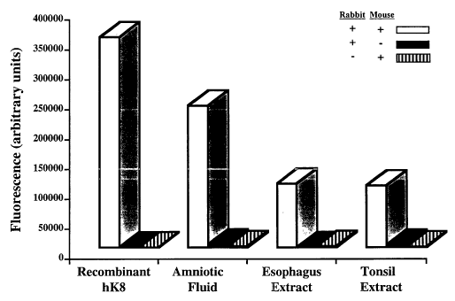
Figure 3: Specificity of the hK8 immunoassay. The assay was performed in the
presence of both
mouse and rabbit antibodies, or in the absence of mouse, or rabbit antibody
(substituted by non-immune
serum). Samples tested were amniotic fluid and tissue extracts from esophagus
and tonsil.
Figure 4: Hormonal regulation of hK8 in the prostate carcinoma cell line PC-3
(AR)6 and the
3 5 breast carcinoma cell line MCF-7. In the PC-3 (AR)6 cell line, hK8 is up-
regulated by norgestrel (an
androgenic progestin) and dihydrotestosterone (DHT). In the MCF-7 cell line,
hK8 is up-regulated by
estradiol.
CA 02481093 2004-10-O1
WO 03/085404 PCT/CA03/00495
Figure 5: Expression of hK8 in adult and fetal tissue extracts from males and
females.
Figure 6: Levels of hK8 (p.g/g of total protein) in ascites fluid of women
with advanced ovarian
cancer. Higher levels are seen in lower grade disease. The p value was
calculated by the Kruskal Wallis
test. Horizontal lines indicate median values.
Figure 7: Kaplan-Meier survival curves of patients with advanced ovarian
carcinoma. Patients
were categorized as hK8-negative (ascites fluid hK8 concentration < 25th
percentile; 0.75 p.g/g of total
protein) or hK8-positive (hk8 concentration > 0.75 p,g/g). The p value was
calculated by the log-rank test.
Figure 8: hK8 serum concentration in 26 ovarian cancer patients and 25 normal
females. At 95%
specificity (cutoff of 5.5 ~.g/L, indicated by the dotted lines), the
sensitivity is 54%.
Figure 9: Monitoring of an ovarian cancer patient with serum CA 125 and hK8.
Figure 10: High-performance liquid chromatographic separation on a gel
filtration column of a
serum from an ovarian cancer patient (A), an esophageal extract (B), an
aminiotic fluid (C) and a breast
milk (D). The peak represents the free 30 kDa form of hK8.
DETAILED DESCRIPTION OF THE INVENTION
l5 The invention relates to newly discovered correlations between expression
of kallikrein 8 and
ovarian cancer. The kallikrein 8 marker provides sensitive methods for
detecting ovarian cancer. The levels
of expression of kallikrein 8 correlate with the presence of ovarian cancer or
a pre-malignant condition in a
patient. Methods are provided for detecting the presence of ovarian cancer in
a sample, the absence of
ovarian cancer in a sample, the stage of an ovarian cancer, the grade of an
ovarian cancer, the benign or
2 0 malignant nature of an ovarian cancer, the metastatic potential of an
ovarian cancer, assessing the
histological type of neoplasm associated with the ovarian cancer, the
indolence or aggressiveness of the
cancer, and other characteristics of ovarian cancer that are relevant to
prevention, diagnosis,
characterization, and therapy of ovarian cancer in a patient. Methods are also
provided for assessing the
efficacy of one or more test agents for inhibiting ovarian cancer, assessing
the efficacy of a therapy for
2 5 ovarian cancer, monitoring the progression of ovarian cancer, selecting an
agent or therapy for inhibiting
ovarian cancer, treating a patient afflicted with ovarian cancer, inhibiting
ovarian cancer in a patient, and
assessing the carcinogenic potential of a test compound.
In accordance with the present invention there may be employed conventional
molecular biology,
microbiology, and recombinant DNA techniques within the skill of the art. Such
techniques are explained
3 0 fully in the literature. See for example, Sambrook, Fritsch, & Maniatis
(Molecular Cloning: A Laboratory
Manual, Second Edition (1989) Cold Spring Harbor Laboratory Press, Cold Spring
Harbor, N.Y); DNA
Cloning: A Practical Approach, Volumes I and II (D.N. Glover ed. 1985);
Oligonucleotide Synthesis (M..J.
Gait ed. 1984); Nucleic Acid Hybridization B.D. Hames & S.J. Higgins eds.
(1985); Transcription and
Translation B.D. Hames & S.J. Higgins edsr (1984); Animal Cell Culture R.I.
Freshney, ed. (1986);
3 5 Immobilized Cells and enzymes IRL Press, (1986); and B. Perbal, A
Practical Guide to Molecular Cloning
(1984). The invention may also employ standard methods in immunology known in
the art such as
described in Stites et al. (eds) Basic and Clinical Immunology, 8a' Ed.,
Appleton & Lange, Norwalk, Conn.
CA 02481093 2004-10-O1
WO 03/085404 PCT/CA03/00495
g _
(1994) and Mishell and Shigi (eds), Selected Methods in Cellular Immunology,
W.H. Freeman and Co.,
New York (1980).
Glossary
For convenience, certain terms employed in the specification and claims are
collected here.
The terms "sample", "biological sample", and the like mean a material known or
suspected of
expressing or containing a kallikrein 8 polypeptide associated with ovarian
cancer. The test sample can be
used directly as obtained from the source or following a pretreatment to
modify the character of the
sample. The sample can be derived from any biological source, such as tissues,
extracts, or cell cultures,
including cells (e.g. tumor cells), cell lysates, and physiological fluids,
such as, for example, whole blood,
plasma, serum, saliva, ocular lens fluid, cerebral spinal fluid, sweat, urine,
milk, ascites fluid, synovial
fluid, peritoneal fluid and the like. The sample can be obtained from animals,
preferably mammals, most
preferably humans. The sample can be treated prior to use, such as preparing
plasma from blood, diluting
viscous fluids, and the like. Methods of treatment can involve filtration,
distillation, extraction,
concentration, inactivation of interfering components, the addition of
reagents, and the like. Proteins may
be isolated from the samples and utilized in the methods of the invention. In
an embodiment, the sample is
a serum sample.
"Kallikrein 8 polypeptide(s)", "kallikrein 8 markers)", or "kallikrein 8"
includes native-sequence
polypeptides, isoforms, precursors, proproteins, and chimeric polypeptides.
A "native-sequence polypeptide" comprises a polypeptide having the same amino
acid sequence
2 0 of a polypeptide derived from nature. Such native-sequence polypeptides
can be isolated from nature or
can be produced by recombinant or synthetic means. A native-sequence
polypeptide may comprise a
proprotein or precursor.
The amino acid sequences for a native kallikrein 8 polypeptide employed or
detected in
accordance with the present invention include the sequences found in Yoshida
et al, and GenBank for
2 5 human kallikrein 8 ("hK8") at GenBank Accession Nos. NP 009127, BAA28676,
BAA28673,
NP_653088, NP 653089, NP 653090, 060259, and AAG23254 (see for example SEQ ID
NOs.l and 2) or
a portion thereof. Other useful kallikrein 8 polypeptides are substantially
identical to these sequences (e.g.
at least about 45%, preferably 50%, 55%, 60%, 65%, 70%, 75%, 80%, 85%, 90%,
and 95%, more
preferably at least 97%, 98%, or 99% sequence identity), and preferably retain
the immunogenic activity of
3 0 the corresponding native-sequence kallikrein 8 polypeptide.
The term "native-sequence polypeptide" also specifically encompasses naturally
occurring
truncated or secreted forms of a kallikrein 8 polypeptide, polypeptide
variants including naturally
occurring variant forms (e.g., alternatively spliced forms or splice
variants), and naturally occurring allelic
variants.
3 5 The term "polypeptide variant" means a polypeptide having at least about
70-80%, preferably at
least about 85%, more preferably at least about 90%, most preferably at least
about 95% amino acid
sequence identity with a native-sequence polypeptide, in particular having at
least 70-80%, 85%, 90%,
95% amino acid sequence identity to a sequence identified in Yoshida et al,
and GenBank Accession Nos.
CA 02481093 2004-10-O1
WO 03/085404 PCT/CA03/00495
- 9 -
NP 009127, BAA28676, BAA28673, NP 653088, NP 653089, NP 653090, 060259, and
AAG23254
(see for example SEQ ID NOs.l and 2). Such variants include, for instance,
polypeptides wherein one or
more amino acid residues are added to, or deleted from, the N- or C-terminus
of the full-length or mature
sequences of BAA28676, BAA28673, and AAG23254 (e.g. SEQ ID NOs.l and 2),
including variants
from other species, but excludes a native-sequence polypeptide.
An allelic variant may also be created by introducing substitutions,
additions, or deletions into a
nucleic acid encoding a native polypeptide sequence such that one or more
amino acid substitutions,
additions, or deletions are introduced into the encoded protein. Mutations may
be introduced by standard
methods, such as site-directed mutagenesis and PCR-mediated mutagenesis. In an
embodiment,
conservative substitutions are made at one or more predicted non-essential
amino acid residues. A
"conservative amino acid substitution" is one in which an ammo acid residue is
replaced with an amino
acid residue with a similar side chain. Amino acids with similar side chains
are known in the art and
include amino acids with basic side chains (e.g. Lys, Arg, His), acidic side
chains (e.g. Asp, Glu),
uncharged polar side chains (e.g. Gly, Asp, Glu, Ser, Thr, Tyr and Cys),
nonpolar side chains (e.g. Ala,
Val, Leu, Iso, Pro, Trp), beta-branched side chains (e.g. Thr, Val, Iso), and
aromatic side chains (e.g. Tyr,
Phe, Trp, His). Mutations can also be introduced randomly along part or all of
the native sequence, for
example, by saturation mutagenesis. Following mutagenesis the variant
polypeptide can be recombinantly
expressed and the activity of the polypeptide may be determined.
Polypeptide variants include polypeptides comprising amino acid sequences
sufficiently identical
2 0 to or derived from the amino acid sequence of a native polypeptide which
include fewer amino acids than
the full length polypeptides. A portion of a polypeptide can be a polypeptide
which is for example, 10, 15,
20, 25, 30, 35, 40, 45, 50, 60, 70, 80, 90, 100 or more amino acids in length.
Portions in which regions of a ,
polypeptide are deleted can be prepared by recombinant techniques and can be
evaluated for one or more
functional activities such as the ability to form antibodies specific for a
polypeptide.
2 5 A naturally occurring allelic variant may contain conservative amino acid
substitutions from the
native polypeptide sequence or it may contain a substitution of an amino acid
from a corresponding
position in a kallikrein polypeptide homolog, for example, the murine
kallikrein polypeptide.
Percent identity of two amino acid sequences, or of two nucleic acid sequences
identified herein is
defined as the percentage of amino acid residues or nucleotides in a candidate
sequence that are identical
3 0 with the amino acid residues in a kallikrein polypeptide or nucleic acid
sequence, after aligning the
sequences and introducing gaps, if necessary, to achieve the maximum percent
sequence identity, and not
considering any conservative substitutions as part of the sequence identity.
Alignment for purposes of
determining percent amino acid or nucleic acid sequence identity can be
achieved in various conventional
ways, for instance, using publicly available computer software including the
GCG program package
35 (Devereux J. et al., Nucleic Acids Research 12(1): 387, 1984); BLASTP,
BLASTN, and FASTA (Atschul,
S.F. et al. J. Molec. Biol. 215: 403-410, 1990). The BLAST X program is
publicly available from NCBI
and other sources (BLAST Manual, Altschul, S. et al. NCBI NLM NIH Bethesda,
Md. 20894; Altschul, S.
et al. J. Mol. Biol. 215: 403-410, 1990). Skilled artisans can determine
appropriate parameters for
CA 02481093 2004-10-O1
WO 03/085404 PCT/CA03/00495
- 10 -
measuring alignment, including any algorithms needed to achieve maximal
alignment over the full length
of the sequences being compared. Methods to determine identity and similarity
are codified in publicly
available computer programs.
Kallikrein 8 polypeptides include chimeric or fusion proteins. A "chimeric
protein" or "fusion
protein" comprises all or part (preferably biologically active) of a
kallikrein 8 polypeptide operably linked
to a heterologous polypeptide (i.e., a polypeptide other than a kallikrein 8
polypeptide). Within the fusion
protein, the term "operably linked" is intended to indicate that the
kallikrein polypeptide and the
heterologous polypeptide are fused in-frame to each other. The heterologous
polypeptide can be fused to
the N-terminus or C-terminus of the kallikrein polypeptide. A useful fusion
protein is a GST fusion protein
in which a kallikrein polypeptide is fused to the C-terminus of GST sequences.
Another example of a
fusion protein is an immunoglobulin fusion protein in which all or part of a
kallikrein polypeptide is fused
to sequences derived from a member of the immunoglobulin protein family.
Chimeric and fusion proteins
can be produced by standard recombinant DNA techniques.
A kallikrein 8 polypeptide may be part 'of a complex, in particular, a complex
with a protease
inhibitor.
Kallikrein 8 polypeptides may be isolated from a variety of sources, such as
from human tissue
types or from another source, or prepared by recombinant or synthetic methods,
or by any combination of
these and similar techniques.
The term "subject" or "patient" refers to a warm-blooded animal such as a
mammal which is
2 0 afflicted with ovarian cancer or condition as described herein.
Preferably, "subject" refers to a human.
"Kallikrein 8 polynucleotide(s)", " kallikrein 8 nucleic acids)", "KLK8", or
"KLK8 nucleic
acid(s)" includes nucleic acids that encode a native-sequence kallikrein 8
polypeptide, a polypeptide
variant including a portion of a kallikrein 8 polypeptide, an isoform,
precursor, and chimeric polypeptide.
The nucleic acid sequences encoding native kallikrein 8 polypeptides employed
in the present
2 5 invention include the nucleic acid sequences of Yoshida et al and GenBank
Accession Nos. NP 009127,
AB009849, AB012761, NM_007196, NM_144505, NM_144506, NM_144507, and AC011473
(for
example, SEQ ID NOs. 3 and 4), or a fragment thereof.
Polynucleotides encoding kallikrien 8 polypeptides include nucleic acid
sequences
complementary to these nucleic acids, and nucleic acids that are substantially
identical to these sequences
3 0 (e.g. at least about 45%, preferably 50%, 55%, 60%, 65%, 70%, 75%, 80%,
85%,90%, 95%, 97%, 98%, or
99% sequence identity).
Kallikrein polynucleotides also include sequences which differ from a nucleic
acid sequence of
Yoshida et al, and GenBank Accession Nos. NP 009127, AB009849, AB012761, NM
007196,
NM 144505, NM_144506, NM 144507, and AC011473 (for example, SEQ ID NOs. 3 and
4), due to
3 5 degeneracy in the genetic code. As one example, DNA sequence polymorphisms
within the nucleotide
sequence of a kallikrein 8 polypeptide may result in silent mutations that do
not affect the amino acid
sequence. Variations in one or more nucleotides may exist among individuals
within a population due to
natural allelic variation. DNA sequence polymorphisms may also occur which
lead to changes in the
CA 02481093 2004-10-O1
WO 03/085404 PCT/CA03/00495
- 11 -
amino acid sequence of a kallikrein 8 polypeptide.
I~allikrein 8 polynucleotides also include nucleic acids that hybridize under
stringent conditions,
preferably high stringency conditions to a nucleic acid sequence of Yoshida et
al, and GenBank Accession
Nos. NP 009127, AB009849, AB012761, NM 007196, NM-144505, NM-144506, NM-
144507, and
AC011473 (for example, SEQ ID NOs. 3 and 4). Appropriate stringency conditions
which promote DNA
hybridization are known to those skilled in the art, or can be found in
Current Protocols in Molecular
Biology, John Wiley & Sons, N.Y. (1989), 6.3.1-6.3.6. For example, 6.0 x
sodium chloride/sodium citrate
(SSC) at about 45°C, followed by a wash of 2.0 x SSC at 50°C may
be employed. The stringency may be
selected based on the conditions used in the wash step. By way of example, the
salt concentration in the
wash step can be selected from a high stringency of about 0.2 x SSC at
50°C. In addition, the temperature
in the wash step can be at high stringency conditions, at about 65°C.
I~allikrein polynucleotides also include truncated nucleic acids or nucleic
acid fragments and
variant forms of the nucleic acids that arise by alternative splicing of an
mRNA corresponding to a DNA.
Kallikrien polynucleotides are intended to include DNA and RNA (e.g. mRNA) and
can be either
double stranded or single stranded. A polynucleotide may, but need not,
include additional coding or non-
coding sequences, or it may, but need not, be linked to other molecules and/or
carrier or support materials.
The polynucleotides may be of any length suitable for a particular method.
Methods
A variety of methods can be employed for the diagnostic and prognostic
evaluation of ovarian
2 0 cancer involving a kallikrein 8 polypeptide, and the identification of
subjects with a predisposition to such
disorders. Such methods may, for example, utilize binding agents (e.g.
antibodies) directed against a
kallikrein 8 polypeptide, including peptide fragments. In particular, the
antibodies may be used, for
example, for the detection of either an over- or an under-abundance of
kallikrein 8 polypeptide relative to a
non- disorder state or the presence of a modified (e.g., less than full
length) kallikrein 8 polypeptide which
2 5 correlates with a disorder state, or a progression toward a disorder
state.
The invention also contemplates a method for detecting ovarian cancer
comprising producing a
profile of levels of kallikrein 8 polypeptides in cells from a patient and
comparing the profile with a
reference to identify a protein profile for the test cells indicative of
disease.
The methods described herein may be used to evaluate the probability of the
presence of
3 0 malignant or pre-malignant cells, for example, in a group of cells freshly
removed from a host. Such
methods can be used to detect tumors, quantitate their growth, and help in the
diagnosis and prognosis of
disease. The methods can be used to detect the presence of cancer metastasis,
as well as confirm the
absence or removal of all tumor tissue following surgery, cancer chemotherapy,
and/or radiation therapy.
They can further be used to monitor cancer chemotherapy and tumor
reappearance.
3 5 The methods described herein can be adapted for diagnosing and monitoring
ovarian carcinoma
by detecting a kallikrein 8 polypeptide in biological samples from a subject.
These applications require that
the amount of kallikrein 8 polypeptide quantitated in a sample from a subject
being tested be compared to
levels quantitated for another sample or an earlier sample from the subject,
or levels quantitated for a
CA 02481093 2004-10-O1
WO 03/085404 PCT/CA03/00495
- 12 -
control sample. Levels for control samples from healthy subjects or ovarian
cancer subjects may be
established by prospective and/or retrospective statistical studies. Healthy
subjects who have no clinically
evident disease or abnormalities may be selected for statistical studies.
Diagnosis may be made by a
finding of statistically different levels of kallikrein 8 polypeptide compared
to a control sample or previous
levels quantitated for the same subject.
"Statistically different levels" or "significant differences" in levels of a
kallikrien 8 polypeptide
in a patient sample compared to a control or standard (e.g, normal levels or
levels in other samples from a
patient) may represent levels that are higher or lower than the standard error
of the detection assay,
preferably the levels are at least about 1.5, 2, 3, 4, 5, or 6 times higher,
respectively, than the control or
standard.
Binding agents specific for a kallikrein 8 polypeptide may be used for a
variety of diagnostic and
assay applications. There are a variety of assay formats known to the skilled
artisan for using a binding
agent to detect a target molecule in a sample. (For example, see Harlow and
Lane, Antibodies: A
Laboratory Manual, Cold Spring Harbor Laboratory, 1988). In general, the
presence or absence of an
ovarian cancer in a subject may be determined by (a) contacting a sample from
the subject with a binding
agent for a kallikrein 8 polypeptide; (b) detecting in the sample levels of
polypeptide that bind to the
binding agent; and (c) comparing the levels of polypeptide with a
predetermined standard or cut-off value.
"Binding agent" refers to a substance such as a polypeptide or antibody that
specifically binds to a
kallikrein 8 polypeptide. A substance "specifically binds" to a polypeptide if
it reacts at a detectable level
2 0 with the kallikrein 8 polypeptide, and does not react detectably with
peptides containing unrelated
sequences or sequences of different polypeptides. Binding properties may be
assessed using an ELISA,
which may be readily performed by those skilled in the art (see for example,
Newton et al , Develop.
Dynamics 197: 1-13, 1993).
A binding agent may be a ribosome, with or without a peptide component, an RNA
molecule, or a
2 5 polypeptide. A binding agent may be a polypeptide that comprises a
kallikrein 8 polypeptide sequence, a
peptide variant thereof, or a non-peptide mimetic of such a sequence. By way
of example a kallikrein 8
polypeptide sequence may be a peptide portion of a kallikrein 8 polypeptide
that is capable of modulating a
function mediated by the kallikrein 8 polypeptide.
In certain preferred embodiments, the binding agent is an antibody.
3 0 In an aspect the present invention provides a diagnostic method for
monitoring or diagnosing
ovarian cancer in a subject by quantitating a kallikrein 8 polypeptide in a
biological sample from the
subject comprising reacting the sample with antibodies specific for a
kallikrein 8 polypeptide, which are
directly or indirectly labelled with detectable substances, and detecting the
detectable substances.
In an aspect of the invention, a method for detecting ovarian cancer is
provided comprising:
3 5 (a) obtaining a sample suspected of containing a kallikrein 8 polypeptide
associated with ovarian
cancer;
(b) contacting said sample with antibodies that specifically bind a kallikrein
8 polypeptide under
conditions effective to bind the antibodies and form complexes;
CA 02481093 2004-10-O1
WO 03/085404 PCT/CA03/00495
- 13 -
(c) measuring the amount of kallikrein 8 polypeptide present in the sample by
quantitating the
amount of the complexes; and
(d) comparing the amount of kallikrein 8 polypeptide present in the samples
with the amount of
polypeptide in a control, wherein a change or significant difference in the
amount of
polypeptide in the sample compared with the amount in the control is
indicative of ovarian
cancer.
In an embodiment, the invention contemplates a method for monitoring the
progression of ovarian
cancer in a subject, comprising:
(a) contacting antibodies which bind to a kallikrein 8 polypeptide, with a
sample from the subject
so as to form binary complexes comprising the antibodies and kallikrein 8
polypeptide in the
sample;
(b) determining or detecting the presence or amount of complex formation in
the sample;
(c) repeating steps (a) and (b) at a point later in time; and
(d) comparing the result of step (b) with the result of step (c), wherein a
significant difference in
the amount of complex formation is indicative of the stage and/or progression
of the ovarian
cancer in the subject.
The amount of complexes may also be compared to a value representative of the
amount of the
complexes from a subject not at risk of, or afflicted with, ovarian cancer at
different stages.
Thus, antibodies specifically reactive with a kallikrein 8 polypeptide, or
derivatives, such as
2 0 enzyme conjugates or labeled derivatives, may be used to detect kallikrein
8 polypeptides in various
samples (e.g. biological materials). They may be used as diagnostic or
prognostic reagents and they may be
used to detect abnormalities in the level of kallikrein 8 polypeptide
expression, or abnormalities in the
structure, and/or temporal, tissue, cellular, or subcellular location of
kallikrein 8 polypeptide. Antibodies
may also be used to screen potentially therapeutic compounds in vitro to
determine their effects on
2 5 disorders (e.g. ovarian cancer) involving a kallikrein 8 polypeptide, and
other conditions. Ih vit~~o
immunoassays may also be used to assess or monitor the efficacy of particular
therapies.
Antibodies may be used in any known immunoassays which rely on the binding
interaction
between an antigenic determinant of a kallikrein 8 polypeptide and the
antibodies. Examples of such
assays are radioimmunoassays, enzyme immunoassays (e.g. ELISA),
immunofluorescence,
3 0 immunoprecipitation, latex agglutination, hemagglutination, and
histochemical tests. The antibodies may
be used to detect and quantify a kallikrein 8 polypeptide in a sample in order
to diagnose and treat such
pathological states. These terms are well understood by those skilled in the
art. A person skilled in the art
will know, or can readily discern, other immunoassay formats without undue
experimentation.
In particular, the antibodies may be used in immunohistochemical analyses, for
example, at the
3 5 cellular and sub-subcellular level, to detect a kallikrein 8 polypeptide,
to localize it to particular ovarian
tumor cells and tissues, and to specific subcellular locations, and to
quantitate the level of expression.
Antibodies for use in the present invention include monoclonal or polyclonal
antibodies,
immunologically active fragments (e.g. a Fab or (Fab)2 fragments), antibody
heavy chains, humanized
CA 02481093 2004-10-O1
WO 03/085404 PCT/CA03/00495
- 14 -
antibodies, antibody light chains, genetically engineered single chain F,,
molecules (Ladner et al, U.S. Pat.
No. 4,946,778), chimeric antibodies, for example, antibodies which contain the
binding specificity of
murine antibodies, but in which the remaining portions are of human origin, or
derivatives, such as enzyme
conjugates or labeled derivatives.
Antibodies including monoclonal and polyclonal antibodies, fragments and
chimeras, may be
prepared using methods known to those skilled in the art. An isolated native
or recombinant kallikrein 8
polypeptide may be utilized to prepare antibodies. See, for example, Kohler et
al. (1975) Nature 256:495- ,
497; Kozbor et al. (1985) J. Immunol Methods 81:31-42; Cote et al. (1983) Proc
Natl Acad Sci 80:2026-
2030; and Cole et al. (1984) Mol Cell Biol 62:109-120 for the preparation of
monoclonal antibodies; Huse
et al. (1989) Science 246:1275-1281 for the preparation of monoclonal Fab
fragments; and, Pound (1998)
Immunochemical Protocols, Humana Press, Totowa, N.J for the preparation of
phagemid or B-lymphocyte
immunoglobulin libraries to identify antibodies. The antibodies specific for a
kallikrein 8 polypeptide used
in the methods of the invention may also be obtained from scientific or
commercial sources.
Preferably, antibodies used in the methods of the invention are reactive
against a kallikrein 8
polypeptide if they bind with a Ka of greater than or equal to 10-~ M.
An antibody that binds to a kallikrein 8 polypeptide may be labelled with a
detectable substance
and localised or detected in biological samples based upon the presence of the
detectable substance.
Examples of detectable substances include, but are not limited to, the
following: radioisotopes (e.g., 3H,
iaC~ ssS~ izsl isil)~ Euorescent labels (e.g., FITC, rhodamine, lanthanide
phosphors), luminescent labels
2 0 such as luminal, enzymatic labels (e.g., horseradish peroxidase, beta-
galactosidase, luciferase, alkaline
phosphatase, acetylcholinesterase), biotinyl groups (which can be detected by
marked avidin e.g.,
streptavidin containing a fluorescent marker or enzymatic activity that can be
detected by optical or
calorimetric methods), and predetermined polypeptide epitopes recognized by a
secondary reporter (e.g.,
leucine zipper pair sequences, binding sites for secondary antibodies, metal
binding domains, epitope tags).
2 5 In some embodiments, labels are attached via spacer arms of various
lengths to reduce potential steric
hindrance. Antibodies may also be coupled to electron dense substances, such
as ferritin or colloidal gold,
which are readily visualised by electron microscopy.
Indirect methods may also be employed in which the primary antigen-antibody
reaction is
amplified by the introduction of a second antibody, having specificity for the
antibody reactive against
3 0 kallikrein 8 polypeptide. The second antibody may be labeled with a
detectable substance to detect the
primary antigen-antibody reaction. By way of example, if the antibody having
specificity against a
kallikrein 8 polypeptide is a rabbit IgG antibody, the second antibody may be
goat anti-rabbit
gamma-globulin labelled with a detectable substance as described herein.
Methods for conjugating or labelling the antibodies discussed above may be
readily accomplished
3 5 by one of ordinary skill in the art. (See for example Inman, Methods In
Enzymology, Vol. 34, Affinity
Techniques, Enzyme Purification: Part B, Jakoby and Wichek (eds.), Academic
Press, New York, p. 30,
1974; and Wilchek and Bayer, "The Avidin-Biotin Complex in Bioanalytical
Applications,"Anal.
Biochem. 171:1-32, 1988 re methods for conjugating or labelling the antibodies
with enzyme or ligand
CA 02481093 2004-10-O1
WO 03/085404 PCT/CA03/00495
- 15 -
binding partner).
Cytochemical techniques known in the art for localizing antigens using light
and electron
microscopy may be used to detect a kallikrein 8 polypeptide. Generally, an
antibody may be labeled with a
detectable substance and a kallikrein 8 polypeptide may be localised in
tissues and cells based upon the
presence of the detectable substance.
In the context of the methods of the invention, the sample, binding agents
(e.g. antibodies) for a
kallikrein 8 polypeptide may be immobilized on a carrier or support. Examples
of suitable carriers or
supports are agarose, cellulose, nitrocellulose, dextran, Sephadex, Sepharose,
liposomes, carboxymethyl
cellulose, polyacrylamides, polystyrene, gabbros, filter paper, magnetite, ion-
exchange resin, plastic film,
plastic tube, glass, polyamine-methyl vinyl-ether-malefic acid copolymer,
amino acid copolymer, ethylene-
maleic acid copolymer, nylon, silk, etc. The support material may have any
possible configuration
including spherical (e.g. bead), cylindrical (e.g. inside surface of a test
tube or well, or the external surface
of a rod), or flat (e.g. sheet, test strip). Thus, the carrier may be in the
shape of, for example, a tube, test
plate, well, beads, disc, sphere, etc. The immobilized material may be
prepared by reacting the material
with a suitable insoluble carrier using known chemical or physical methods,
for example, cyanogen
bromide coupling. Binding agents (e.g. antibodies) may be indirectly
immobilized using second binding
agents specific for the first binding agent. For example, mouse antibodies
specific for a kallikrein.8
polypeptide may be immobilized using sheep anti-mouse IgG Fc fragment specific
antibody coated on the
carrier or support.
2 0 Where a radioactive label is used as a detectable substance, a kallikrein
8 polypeptide may be
localized by radioautography. The results of radioautography may be
quantitated by determining the
density of particles in the radioautographs by various optical methods, or by
counting the grains.
Time-resolved fluorometry may be used to detect a fluorescent signal. For
example, the method
described in Christopoulos TK and Diamandis EP Anal Chem 1992:64:342-346 may
be used with a
2 5 conventional time-resolved fluorometer.
Therefore, in accordance with an embodiment of the invention, a method is
provided wherein a
kallikrein 8 specific antibody is labelled with an enzyme, a substrate for the
enzyme is added wherein the
substrate is selected so that the substrate, or a reaction product of the
enzyme and substrate, forms
fluorescent complexes with a lanthanide metal. A lanthanide metal is added and
kallikrein 8 is quantitated
3 0 in the sample by measuring fluorescence of the fluorescent complexes. The
antibodies specific for
kallikrein 8 may be directly or indirectly labelled with an enzyme. Enzymes
are selected based on the
ability of a substrate of the enzyme, or a reaction product of the enzyme and
substrate, to complex with
lanthanide metals such as europium and terbium. Examples of enzymes and
substrates for enzymes that
provide such fluorescent complexes are described in U.S. Patent No. 5,312,922
to Diamandis. Examples of
3 5 suitable enzymes include alkaline phosphatase and (3-galactosidase. When
the antibody is directly or
indirectly labelled with alkaline phosphatase the substrate employed in the
method may be 4-
methylumbelliferyl phosphate, 5-fluorosalicyl phosphate, or diflunisal
phosphate. The fluorescence
intensity of the complexes is typically measured using a time-resolved
fluorometer e.g. a CyberFluor 615
CA 02481093 2004-10-O1
WO 03/085404 PCT/CA03/00495
- 16 -
Imunoanalyzer (Nordion International, Kanata, Ontario).
Antibodies specific for a kallikrein 8 polypeptide may also be indirectly
labelled with an enzyme.
For example, the antibodies may be conjugated to one partner of a ligand
binding pair, and the enzyme
may be coupled to the other partner of the ligand binding pair. Representative
examples include avidin=
biotin, and riboflavin-riboflavin binding protein. In an embodiment, the
antibodies are biotinylated, and the
enzyme is coupled to streptavidin. In another embodiment, antibodies specific
for the anti-kallikrein
antibodies are labeled with an enzyme.
In accordance with an aspect, the present invention provides means for
determining kallikrein 8
polypeptide in a sample by measuring kallikrein 8 polypeptide by immunoassay.
It will be evident to a
skilled artisan that a variety of immunoassay methods can be used to measure
kallikrein 8 polypeptide in a
sample in particular a serum sample. In general, a kallikrein 8 polypeptide
immunoassay method may be
competitive or noncompetitive. Competitive methods typically employ an
immobilized or immobilizable
antibody to kallikrein 8 polypeptide (anti-K8) and a labeled form of
kallikrein 8 polypeptide. Sample
kallikrein 8 polypeptide and labeled kallikrein 8 polypeptide compete for
binding to anti-K8. After
separation of the resulting labeled kallikrein 8 polypeptide that has become
bound to anti-K8 (bound .
fraction) from that which has remained unbound (unbound fraction), the amount
of the label in either
bound or unbound fraction is measured and may be correlated with the amount of
kallikrein 8 polypeptide
in the test sample in any conventional manner, e.g. by comparison to a
standard curve.
In another aspect, a non-competitive method is used for the determination of a
kallikrein 8
2 0 polypeptide, with the most common method being the "sandwich" method. In
this assay, two anti-K8
antibodies are employed. One of the anti-K8 antibodies is directly or
indirectly labeled (sometimes referred
to as the "detection antibody") and the other is immobilized or immobilizable
(sometimes referred to as the
"capture antibody"). The capture and detection antibodies can be contacted
simultaneously or sequentially
with the test sample. Sequential methods can be accomplished by incubating the
capture antibody with the
2 5 sample, and adding the detection antibody at a predetermined time
thereafter (sometimes referred to as the
"forward" method); or the detection antibody can be incubated with the sample
first and then the capture
antibody added (sometimes referred to as the "reverse" method). After the
necessary incubations) have
occurred, to complete the assay, the capture antibody is separated from the
liquid test mixture, and the
label is measured in at least a portion of the separated capture antibody
phase or the remainder of the liquid
3 0 test mixture. Generally it is measured in the capture antibody phase since
it comprises kallikrein 8
polypeptide bound by ("sandwiched" between) the capture and detection
antibodies. In an embodiment, the
label may be measured without separating the capture antibodies and liquid
test mixture.
In a typical two-site immunometric assay for kallikrein 8 polypeptide, one or
both of the capture
and detection antibodies are polyclonal antibodies or one or both of the
capture and detection antibodies
3 5 are monoclonal antibodies (i.e. polyclonal/polyclonal,
monoclonal/monoclonal, or monoclonal/polyclonal).
In a specific embodiment, mouse and rabbit polyclonal antibodies are utilized.
The label used in the
detection antibody can be selected from arty of those known conventionally in
the art. The label may be an
enzyme or a chemiluminescent moiety, but it can also be a radioactive isotope,
a fluorophor, a detectable
CA 02481093 2004-10-O1
WO 03/085404 PCT/CA03/00495
- 17 -
ligand (e.g., detectable by a secondary binding by a labeled binding partner
for the ligand), and the like.
Preferably the antibody is labelled with au enzyme which is detected by adding
a substrate that ~is selected
so that a reaction product of the enzyme and substrate forms fluorescent
complexes. The capture antibody
is selected so that it provides a means for being separated from the remainder
of the test mixture.
Accordingly, the capture antibody can be introduced to the assay in an already
immobilized or insoluble
form, or can be in a immobilizable form, that is, a form which enables
immobilization to be accomplished
subsequent to introduction of the capture antibody to the assay. An
immobilized capture antibody may
comprise an antibody covalently or noncovalently attached to a solid phase
such as a magnetic particle, a
latex particle, a microtiter plate well, a bead, a cuvette, or other reaction
vessel. An example of an
immobilizable capture antibody is antibody which has been chemically modified
with a ligand moiety,
e.g., a hapten, biotin, or the like, and which can be subsequently immobilized
by contact with an
immobilized form of a binding partner for the ligand, e.g., an antibody,
avidin, _or the like. In an
embodiment, the capture antibody may be immobilized using a species specific
antibody for the capture
antibody that is bound to the solid phase.
A particular sandwich immunoassay method of the invention employs two
antibodies reactive
against a kallikrein 8 polypeptide, a second antibody having specificity
against an antibody reactive against "
a kallikrein 8 polypeptide labelled with an enzymatic label, and a fluorogenic
substrate for the enzyme. In ,
an embodiment, the enzyme is alkaline phosphatase (ALP) and the substrate is 5-
fluorosalicyl phosphate.
ALP cleaves phosphate out of the fluorogenic substrate, 5-fluorosalicyl
phosphate, to produce 5-
2 0 fluorosalicylic acid (FSA). 5-Fluorosalicylic acid can then form a highly
fluorescent ternary complex of
the form FSA-Tb(3+)-EDTA, which can be quantified by measuring the Tb3+
fluorescence in a time-
resolved mode. Fluorescence intensity is measured using a time-resolved
fluorometer as described herein.
The methods described herein may utilize multiple markers for ovarian cancer.
Therefore, the
invention contemplates a method for anaylzing a biological sample for the
presence of kallikrein 8 and
2 5 other markers that are specific indicators of ovarian cancer. Other
markers include markers to kallikreins
such as human stratum corneum chymotryptic enzyme (HSCCE), kallikrein 2,
kallikrein 3, kallikrein 4,
kallikrein 5, kallikrein 6, kallikrein 9, lcallikrein 10, and kallikrein 11;
CA125, CA15-3, CA72-4, CA19-9,
OVXl, lysophosphatidic acid (LPA), creatin-kinase BB, haptoglobin alpha,
prostasin, osteopontin, and
carcinoembryonic antigen (CEA). Preferably the other markers are markers to
kallikreins. In an aspect of
3 0 the invention, the markers are one or more of kallikrein 6, kallikrein 10,
kallikrein 11, and CA125. The
methods described herein may be modified by including reagents to detect the
markers (e.g. binding agents
such as antibodies or nucleic acids specific for the markers).
The above-described immunoassay methods and formats are intended to be
exemplary and are not
limiting since, in general, it will be understood that any immunoassay method
or format can be used in the
3 5 present invention.
Computer Systems
Computer readable media comprising kallikrein 8 markers is also provided.
"Computer readable
media" refers to any medium that can be read and accessed directly by a
computer, including but not
CA 02481093 2004-10-O1
WO 03/085404 PCT/CA03/00495
- 18 -
limited to magnetic storage media, such as floppy discs, hard disc storage
medium, and magnetic tape;
optical storage media such as CD-ROM; electrical storage media such as RAM and
ROM; and hybrids of
these categories such as magnetic/optical storage media. Thus, the invention
contemplates computer
readable medium having recorded thereon kallikrein 8 markers identified for
patients and controls.
"Recorded" refers to a process for storing information on computer readable
medium. The skilled
artisan can readily adopt any of the presently known methods for recording
information on computer
readable medium to generate manufactures comprising information on kallikrein
8 markers.
A variety of data processor programs and formats can be used to store
information on kallikrein 8
markers on computer readable medium. For example, the information can be
represented in a word
processing text file, formatted in commercially-available software such as
WordPerfect and Microsoft
Word, or represented in the form of an ASCII file, stored in a database
application, such as DB2, Sybase,
Oracle, or the like. Any number of dataprocessor structuring formats (e.g.,
text file or database) may be
adapted in order to obtain computer readable medium having recorded thereon
the marker information.
By providing the marker information in computer readable form, one can
routinely access the
information for a variety of purposes. For example, one skilled in the art can
use the information in
computer readable form to compare marker information obtained during or
following therapy with the
information stored within the data storage means.
The invention provides a medium for holding instructions for performing a
method for
determining whether a patient has ovarian cancer or a pre-disposition to
ovarian cancer, comprising
2 0 determining the presence or absence of kallikrein 8 markers, and based on
the presence or absence of the
kallikrein 8 markers, determining whether the patient has ovarian cancer or a
pre-disposition to ovarian
cancer, and optionally recommending treatment for the ovarian cancer or pre-
ovarian cancer condition.
The invention also provides in an electronic system and/or in a network, a
method for determining
whether a subject has ovarian cancer or a pre-disposition to ovarian cancer
associated with kallikrein
2 5 markers, comprising determining the presence or absence of kallikrein
markers, and based on the presence
or absence of the kallikrein markers, determining whether the subject has
ovarian cancer or a pre-
disposition to ovarian cancer, and optionally recommending treatment for the
ovarian cancer or pre-ovarian
cancer condition.
The invention further provides in a network, a method for determining whether
a subject has
3 0 ovarian cancer or a pre-disposition to ovarian cancer associated with
kallikrein 8 markers, comprising: (a)
receiving phenotypic information on the subject and information on kallikrein
8 markers associated with
samples from the subject; (b) acquiring information from the network
corresponding to the kallikrein 8
markers; and (c) based on the phenotypic information and information on the
kallikrein 8 markers
determining whether the subject has ovarian cancer or a pre-disposition to
ovarian cancer; and (d)
3 5 optionally recommending treatment for the ovarian cancer or pre-ovarian
cancer condition.
The invention still further provides a system for identifying selected records
that identify an
ovarian cancer cell. A system of the invention generally comprises a digital
computer; a database server
coupled to the computer; a database coupled to the database server having data
stored therein, the data
CA 02481093 2004-10-O1
WO 03/085404 PCT/CA03/00495
- 19 -
comprising records of data comprising kallikrein 8 markers, and a code
mechanism for applying queries
based upon a desired selection criteria to the data file in the database to
produce reports of records which
match the desired selection criteria.
In an aspect of the invention a method is provided for detecting an ovarian
cancer cell using a
computer having a processor, memory, display, and input/output devices, the
method comprising the steps
of:
(a) creating records of kallikrein 8 markers isolated from a sample suspected
of containing an
ovarian cancer cell;
(b) providing a database comprising records of data comprising kallikrein 8
markers; and
(c) using a code mechanism for applying queries based upon a desired selection
criteria to the
data file in the database to produce reports of records of step (a) which
provide a match of the
desired selection criteria of the database of step (b) the presence of a match
being a positive
indication that the markers of step (a) have been isolated from a cell that is
an ovarian cancer
cell.
The invention contemplates a business method for determining whether a subject
has ovarian
cancer or a pre-disposition to ovarian cancer associated with kallikrein 8
markers comprising: (a) receiving
phenotypic information on the subject and information on kallikrein 8 markers
associated with samples
from the subject; (b) acquiring information from a network corresponding to
the kallikrein 8 markers; and
(c) based on the phenotypic information, information on the kallikrein 8
markers, and acquired
2 0 information, determining whether the subject has ovarian cancer or a pre-
disposition to ovarian cancer; and
(d) optionally recommending treatment for the ovarian cancer or pre-ovarian
cancer condition.
Imaging
Antibodies specific for kallikrein 8 may also be used in imaging methodologies
in the
management of ovarian cancer. The invention provides a method for imaging
tumors associated with one
2 5 or more kallikreins, preferably kallikreins associated with ovarian
cancer, most preferably kallikrein 8 and
optionally kallikrein 4, kallikrein 5, kallikrein 6, kallikrein 10, and
kallikrein 11.
The invention also contemplates imaging methods described herein using
multiple markers for
ovarian cancer. For example, a method for imaging ovarian cancer may further
comprise injecting the
patient with one or more of an imaging agent that binds to human stratum
corneum chymotryptic enzyme
3 0 (HSCCE), kallikrein 2, kallikrein 3, kallikrein 4, kallikrein 5,
kallikrein 6, kallikrein 9, kallikrein 10,
kallikrein 11,CA125, CA15-3, CA72-4, CA19-9, OVXl, lysophosphatidic acid
(LPA), haptoglobin alpha,
creatin-kinase BB, osteopontin, prostasin, or carcinoembryonic antigen (CEA),
preferably CA 125.
Preferably each imaging agent is labeled so that it can be distinguished
during the imaging.
In an embodiment the method is an ifz vivo method and a subject or patient is
administered one or
3 5 more imaging agents that carry an imaging label and that are capable of
targeting or binding to a kallikrein.
The imaging agent is allowed to incubate in vivo and bind to the kallikrein(s)
associated with a tumor,
preferably ovarian tumors. The presence of the label is localized to the
ovarian cancer, and the localized
label is detected using imaging devices known to those skilled in the art.
CA 02481093 2004-10-O1
WO 03/085404 PCT/CA03/00495
- 20 -
An imaging agent may be an antibody or chemical entity which recognizes the
kallikrein(s). In an
aspect of the invention the imaging agent is a polyclonal antibody or
monoclonal antibody, or fragments
thereof, or constructs thereof including but not limited to, single chain
antibodies, bifunctional antibodies,
molecular recognition units, and peptides or entities that mimic peptides. The
antibodies specific for the
kallikreins used in the methods of the invention may be obtained from
scientific or commercial sources, or
isolated native kallikrein or recombinant kallikrein may be utilized to
prepare antibodies etc as described
herein.
An imaging agent may be a peptide that mimics the epitope for an antibody
specific for a
kallikrein and binds to the kallikrein. The peptide may be produced on a
commercial synthesizer using
conventional solid phase chemistry. By way of example, a peptide may be
prepared that includes either
tyrosine, lysine, or phenylalanine to which N2S2 chelate is complexed (See
U.S. Patent No. 4,897,255).
The anti-kallikrein peptide conjugate is then combined with a radiolabel (e.g.
sodium 99mTc pertechnetate
or sodium 188Re perrhenate) and it may be used to locate a kallikrein
producing tumor.
The imaging agent carries a label to image the kallikreins. An imaging agent
may be labelled for
use in radionuclide imaging. In particular, an imaging agent may be directly
or indirectly labelled with a
radioisotope. Examples of radioisotopes that may be used in the present
invention are the following: z77Ac,
211At' 128Ba' 131Ba' 7Be' 204Bi' 205Bi' 206Bi' 76Br' 77Br' 82Br' 109Cd' 47Ca'
11~r' 14C' 361' 48~,r' SlCr' 62~ru' 64Cu~
67Cu' 165Dy' 155Eu' 18F' 153Gd' 66Ga' 67Ga' 68Ga' 72Ga' 198Au' 3H' 166H~'
111In' 113mIn' 115mIn' 123I' 125I' 131I'
189, 191mIr 192, 194Ir 52Fe 55Fe 59Fe 177Lu 15O 191m-191OS 109Pd 32P 33P 42I~
z26Ra ls6Re lssRe 82mRb
> > > > > > > > > > > > > > > > > >
0 153Sm~ 46SC~ 47~,C 72Se 75~,e~ loSAg~ zzNa~ 24Na 89sr~ 355 38s' 177Ta~
96T.~' 99mTC' 201T1, 202Tf 113S,n~ 117mS-,n~
lzlSn~ 166Yb~ 169Yb~ 175Yb~ aaY~ 901, 62Zn and 65Zn. Preferably the
radioisotope is 131h 125h 123h 111h 99mTc,
9oY~ IasRe~ lsBRe, 3zP~ Is3Sm~ 67Ga, 2oITl 77Br, or 18F, and is imaged with a
photoscanning device.
Procedures for labeling biological agents with the radioactive isotopes are
generally known in the
art. U.S. Pat. No. 4,302,438 describes tritium labeling procedures. Procedures
for iodinating, tritium
2 5 labeling, and 35S labeling especially adapted for murine monoclonal
antibodies are described by Goding, J.
W. (supra, pp 124-126) and the references cited therein. Other procedures for
iodinating biological agents,
such as antibodies, binding portions thereof, probes, or ligands, are
described in the scientific literature
see Hunter and Greenwood, Nature 144:945 (1962), David et al., Biochemistry
13:1014-1021 (1974), and
U.S. Pat. Nos. 3,867,517 and 4,376,110). Iodinating procedures for agents are
described by Greenwood, F.
3 0 et al., Biochem. J. 89:114-123 (1963); Marchalonis, J., Biochem. J.
113:299-305 (1969); and Morrison, M.
et al., Immunochemistry, 289-297 (1971). 99m Tc-labeling procedures are
described by Rhodes, B. et al. in
Burchiel, S. et al. (eds.), Tumor Imaging: The Radioimmunochemical Detection
of Cancer, New York:
Masson 111-123 (1982) and the references cited therein. Labelling of
antibodies or fragments with
technetium-99m are also described for example in U.S. Pat. No. 5,317,091, U.S.
Pat. No. 4,478,815, U.S.
35 Pat. No. 4,478,818, U.S. Pat. No. 4,472,371, U.S. Pat. No. Re 32,417, and
U.S. Pat. No. 4,311,688.
Procedures suitable for 111 In-labeling biological agents are described by
Hnatowich, D. J. et al., J. Immul.
Methods, 65:147-157 (1983), Hnatowich, D. et al., J. Applied Radiation, 35:554-
557 (1984), and Buckley,
R. G. et al., F.E.B.S. 166:202-204 (1984).
CA 02481093 2004-10-O1
WO 03/085404 PCT/CA03/00495
- 21 -
An imaging agent may also be labeled with a paramagnetic isotope for purposes
of an in vivo
method of the invention. Examples of elements that are useful in magnetic
resonance imaging include
gadolinium, terbium, tin, iron, or isotopes thereof. (See, for example,
Schaefer et al., (1989) JACC 14,
472-480; Shreve et al., (1986) Magn. Reson. Med. 3, 336-340; Wolf, G L.,
(1984) Physiol. Chem. Phys.
Med. NMR 16, 93-95; Wesbey et al., (1984) Physiol. Chem. Phys. Med. NMR 16,
145-155; Runge et al.,
(1984) Invest. Radiol. 19, 408-415 for discussions on in vivo nuclear magnetic
resonance imaging.)
In the case of a radiolabeled imaging agent, the agent may be administered to
the patient, it is
localized to the tumor having a kallikrein with which the agent binds, and is
detected or "imaged" iyz vivo
using known techniques such as radionuclear scanning using e.g., a gamma
camera or emission
tomography. [See for example, A. R. Bradwell et al., "Developments in Antibody
Imaging", Monoclonal
Antibodies for Cancer Detection and Therapy, R. W. Baldwin et al., (eds.), pp.
65-85 (Academic Press
1985)]. A positron emission transaxial tomography scanner, such as designated
Pet VI located at
Brookhaven National Laboratory, can also be used where the radiolabel emits
positrons (e.g., 11 C, is F~ is
O, and 13 N).
Whole body imaging techniques using radioisotope labeled agents can be used
for locating both
primary tumors and tumors which have metastasized. Antibodies specific for
kallikreins, or fragments
thereof having the same epitope specificity, are bound to a suitable
radioisotope, or a combination thereof,
and administered parenterally. For ovarian cancer, administration preferably
is intravenous. The bio-
distribution of the label can be monitored by scintigraphy, and accumulations
of the label are related to the
2 0 presence of ovarian cancer cells. Whole body imaging techniques are
described in U.S. Pat. Nos.
4,036,945 and 4,311,688. Other examples of agents useful for diagnosis and
therapeutic use which can be ,
coupled to antibodies and antibody fragments include metallothionein and
fragments (see, U.S. Pat. No. ,
4,732,864). These agents are useful in diagnosis, staging and visualization of
cancer, in particular ovarian
cancer, so that surgical and/or radiation treatment protocols can be used more
efficiently.
2 5 Screening Methods
The invention also contemplates methods for evaluating test agents or
compounds for their ability
to inhibit ovarian cancer or potentially contribute to ovarian cancer. Test
agents and compounds include
but are not limited to peptides such as soluble peptides including Ig-tailed
fusion peptides, members of
random peptide libraries and combinatorial chemistry-derived molecular
libraries made of D- and/or L-
3 0 configuration amino acids, phosphopeptides (including members of random or
partially degenerate,
directed phosphopeptide libraries), antibodies [e.g. polyclonal, monoclonal,
humanized, anti-idiotypic,
chimeric, single chain antibodies, fragments, (e.g. Fab, F(ab)2, and Fab
expression library fragments, and
epitope-binding fragments thereof)], and small organic or inorganic molecules.
The agents or compounds
may be endogenous physiological compounds or natural or synthetic compounds.
3 5 In an aspect, the invention provides a method for assessing the potential
efficacy of a test agent
for inhibiting ovarian cancer in a patient, the method comprising comparing:
(a) levels of kallikrein 8 markers in a first sample obtained from a patient
and exposed to the test
agent, and
CA 02481093 2004-10-O1
WO 03/085404 PCT/CA03/00495
_ ~~ _
(b) levels of the kallikrein 8 markers in a second sample obtained from the
patient, wherein the
sample is not exposed to the test agent, wherein a significant difference in
the levels of
expression of kallikrein 8 markers in the first sample, relative to the second
sample, is an
indication that the test agent is potentially efficacious for inhibiting
ovarian cancer in the
patient.
The first and second samples may be portions of a single sample obtained from
a patient or
portions of pooled samples obtained from a patient.
In another aspect, the invention provides a method of selecting an agent for
inhibiting ovarian
cancer in a patient comprising:
(a) obtaining a sample comprising ovarian cancer cells from the patient;
(b) separately maintaining aliquots of the sample in the presence of a
plurality of test agents;
(c) comparing kallikrein 8 markers in each of the aliquots; and
(d) selecting one of the test agents which alters the levels of the kallikrein
8 markers in the
aliquot containing that test agent, relative to other test agents. ,
' Still another aspect of the present invention provides a method of
conducting a drug discovery
business comprising:
(a) providing one or more methods or assay systems of the invention for
identifying agents that
inhibit ovarian cancer in a patient;
(b) conducting therapeutic profiling of agents identified in step (a), or
further analogs thereof, for
2 0 efficacy and toxicity in animals; and
(c) formulating a pharmaceutical preparation including one or more agents
identified in step (b) ,
as having an acceptable therapeutic profile.
In certain embodiments, the subject method can also include a step of
establishing a distribution ,.
system for distributing the pharmaceutical preparation for sale, and may
optionally include establishing a
2 5 sales group for marketing the pharmaceutical preparation.
The invention also contemplates a method of assessing the ovarian cancer
carcinogenic potential
of a test compound comprising:
(a) maintaining separate aliquots of ovarian cancer cells in the presence and
absence of the test
compound; and
3 0 (b) comparing kallikrein 8 markers encoding same in each of the aliquots.
A significant difference between the levels of the markers in the aliquot
maintained in the
presence of (or exposed to) the test compound relative to the aliquot
maintained in the absence of the test
compound, indicates that the test compound possesses ovarian cancer
carcinogenic potential.
Kits
3 5 The invention contemplates kits for carrying out the methods of the
invention. Such kits typically
comprise two or more components required for performing a diagnostic assay.
Components include but are
not limited to compounds, reagents, containers, and/or equipment.
In an aspect of the invention, a container with a kit comprises binding agents
as described herein.
CA 02481093 2004-10-O1
WO 03/085404 PCT/CA03/00495
- 23 -
In particular, the invention may provide a pre-packaged diagnostic kit
comprising a binding agent
described herein which may be conveniently used, e.g. in clinical settings, to
screen and diagnose patients,
and to screen and identify those subjects afflicted with or exhibiting a
predisposition to ovarian cancer. In
an embodiment, the kit may contain antibodies specific for a kallikrein 8
polypeptide, antibodies against
the antibodies labelled with enzymes, and substrates for the enzymes. The kit
may also contain microtiter
plate wells, standards, assay diluent, wash buffer, adhesive plate covers,
and/or instructions for carrying
out a method of the invention using the kit.
In an aspect of the invention, the kit includes antibodies or antibody
fragments which bind
specifically to epitopes of a kallikrein 8 polypeptide, and means for
detecting binding of the antibodies to
epitopes associated with tumor cells, either as concentrates (including
lyophilized compositions), which
may be further diluted prior to use or at the concentration of use, where the
vials may include one or more
dosages. Where the kits are intended for in vivo use, single dosages may be
provided in sterilized
containers, having the desired amount and concentration of agents. Containers
that provide a formulation
for direct use, usually do not require other reagents, as for example, where
the kit contains radiolabelled
antibody preparations for in vivo imaging.
The reagents suitable for applying the screening methods of the invention to
evaluate compounds
may be packaged into convenient kits described herein providing the necessary
materials packaged into
suitable containers.
Therapeutic Applications
2 0 Kallikrein 8 polypeptides are targets for ovarian cancer immunotherapy.
Such immunotherapeutic
methods include the use of antibody therapy, i~ vivo vaccines, and ex vivo
immunotherapy approaches.
In one aspect, the invention provides antibodies specific for kallikrein 8
polypeptides that may be
used systemically to treat ovarian cancer. Preferably antibodies are used that
target the tumor cells but not
the surrounding non-tumor cells and tissue. Thus, the invention provides a
method of treating a patient
2 5 susceptible to, or having a cancer that expresses a kallikrein 8
polypeptide comprising administering to the
patient an effective amount of antibodies that bind specifically to a
kallikrein 8 polypeptide. In another
aspect, the invention provides a method of inhibiting the growth of tumor
cells expressing a kallikrein 8
polypeptide, comprising administering to a patient antibodies which bind
specifically to a kallikrein 8
polypeptide in amounts effective to inhibit growth of the tumor cells.
Antibodies specific for a kallikrein 8
3 0 polypeptide may also be used in a method for selectively inhibiting the
growth of, or killing a cell
expressing a kallikrein 8 polypeptide comprising reacting antibody
immunoconjugates or immunotoxins
with the cell in an amount sufficient to inhibit the growth of, or kill the
cell.
By way of example, unconjugated antibodies specific for a kallikrein 8
polypeptide may be
introduced into a patient such that the antibodies bind to cancer cells
expressing a kallikrein 8 polypeptide
3 5 and mediate growth inhibition of such cells (including the destruction
thereof), and the tumor, by
mechanisms which may include complement-mediated cytolysis, antibody-dependent
cellular cytotoxicity,
altering the physiologic function of a kallikrein 8 polypeptide and/or the
inhibition of ligand binding or
signal transduction pathways. In addition to unconjugated antibodies,
antibodies specific for a kallikrein 8
CA 02481093 2004-10-O1
WO 03/085404 PCT/CA03/00495
- 24 -
polypeptide, conjugated to therapeutic agents (e.g. immunoconjugates) may also
be used therapeutically,to
deliver the agents directly to tumor cells expressing a kallikrein 8
polypeptide and thereby destroy the
tumor. Examples of such agents include abrin, ricin A, Pseudomonas exotoxin,
or diphtheria toxin,
proteins such as tumor necrosis factor, alpha-interferon, beta-interferon,
nerve growth factor, platelet
derived growth factor, tissue plasminogen activator, and biological response
modifiers such as
lymphokines, interleukin-1, interleukin-2, interleukin-6, granulocyte
macrophage colony stimulating
factor, granulocyte colony stimulating factor, or other growth factors.
Cancer immunotherapy using antibodies specific for a kallikrein 8 polypeptide
may utilize the
various approaches that have been successfully employed for cancers, including
but not limited to colon
cancer (Arlen et al., 1998, Crit Rev Immunol 18: 133-138), multiple myeloma
(Ozaki et al., 1997, Blood
90: 3179-3186; Tsunenati et al., 1997, Blood 90: 2437-2444), gastric cancer
(Kasprzyk et al., 1992, Cancer
Res 52: 2771-2776), B-cell lymphoma (Funakoshi et al., 1996, J Immunther
Emphasis Tumor Immunol
19: 93-101), leukemia (thong et al., 1996, Leuk Res 20: 581-589), colorectal
cancer (Moun et al., 1994,
Cancer Res 54: 6160-6166); Velders et al., 1995, Cancer Res 55: 4398-4403),
and breast cancer (Shepard
et al., 1991, J Clin Immunol 11: 117-127).
In the practice of a method of the invention, antibodies specific for a
kallikrein 8 polypeptide
capable of inhibiting the growth of cancer cells expressing a kallikrein 8
polypeptide are administered in. a
therapeutically effective amount to cancer patients whose tumors express or
overexpress a kallikrein 8
polypeptide. The invention may provide a specific, effective and long-needed
treatment for ovarian cancer.
2 0 The antibody therapy methods of the invention may be combined with other
therapies including
chemotherapy and radiation.
Patients may be evaluated for the presence and levels of kallikrein 8
polypeptide expression and
overexpression in tumors, preferably using immunohistochemical assessments of
tumor tissue, quantitative
imaging as described herein, or other techniques capable of reliably
indicating the presence and degree of
2 5 expression of kallikrein 8 polypeptides. Immunohistochemical analysis of
tumor biopsies or surgical
specimens may be employed for this purpose.
Antibodies specific for kallikrein 8 polypeptides useful in treating cancer
include those that are
capable of initiating a potent immune response against the tumor and those
that are capable of direct
cytotoxicity. In this regard, the antibodies may elicit tumor cell lysis by
either complement-mediated or
3 0 antibody-dependent cell cytotoxicity (ADCC) mechanisms, both of which
require an intact Fc portion of
the immunoglobulin molecule for interaction with effector cell Fc receptor
sites or complement proteins. In
addition, antibodies specific for kallikrein 8 polypeptides that exert a
direct biological effect on tumor
growth are useful in the practice of the invention. Such antibodies may not
require the complete
immunoglobulin to exert the effect. Potential mechanisms by which such
directly cytotoxic antibodies may
3 5 act include inhibition of cell growth, modulation of cellular
differentiation, modulation of tumor
angiogenesis factor profiles, and the induction of apoptosis. The mechanism by
which a particular antibody
exerts an anti-tumor effect may be evaluated using any number of in vitro
assays designed to determine
ADCC, antibody-dependent macrophage-'mediated cytotoxicity (ADMMC), complement-
mediated cell
CA 02481093 2004-10-O1
WO 03/085404 PCT/CA03/00495
- 25 -
lysis, and others known in the art.
The anti-tumor activity of antibodies specific for kallikrein 8 polypeptides
may be evaluated in
vivo using a suitable animal model. Xenogenic cancer models, wherein human
cancer explants or passaged
xenograft tissues are introduced into immune compromised animals, such as nude
or SCID mice, may be
employed.
The methods of the invention contemplate the administration of combinations,
or "cocktails" of
different individual antibodies recognizing epitopes of kallikrein 8
polypeptides and optionally other
markers of ovarian cancer (e.g. other kallikreins, CA125, etc). Such cocktails
may have certain advantages
inasmuch as they contain antibodies that bind to different epitopes and/or
exploit different effector
mechanisms or combine directly cytotoxic antibodies with antibodies that rely
on immune effector
functionality. Such antibodies in combination may exhibit synergistic
therapeutic effects. In addition, the
administration of the antibodies may be combined with other therapeutic
agents, including but not limited
to chemotherapeutic agents, androgen-blockers, and immune modulators (e.g.,
IL2, GM-CSF). The
antibodies may be administered in their "naked" or unconjugated form, or may
have therapeutic agents
conjugated to them.
The antibodies specific for kallikrein 8 polypeptides used in the practice of
the methods of the
invention may be formulated into pharmaceutical compositions comprising a
carrier suitable for the
desired delivery method. Suitable carriers include any material which when
combined with the antibodies
retains the anti-tumor function of the antibodies and is non-reactive with the
subject's immune systems.
2 0 Examples include any of a number of standard pharmaceutical carriers such
as sterile phosphate buffered
saline solutions, bacteriostatic water, and the like (see, generally,
Remington's Pharmaceutical Sciences,
Mack Publishing Company, Easton, Pa., USA).
Antibody formulations may be administered via any route capable of delivering
the antibodies to
the tumor site. Routes of administration include, but are not limited to,
intravenous, intraperitoneal,
2 5 intramuscular, intratumor, intradermal, and the like. Preferably, the
route of administration is by
intravenous injection. Antibody preparations may be lyophilized and stored as
a sterile powder, preferably
under vacuum, and then reconstituted in bacteriostatic water containing, for
example, benzyl alcohol
preservative, or in sterile water prior to injection.
Treatment will generally involve the repeated administration of the antibody
preparation via an
3 0 acceptable route of administration such as intravenous injection (IV), at
an effective dose. Dosages will
depend upon various factors generally appreciated by those of skill in the
art, including the type of cancer
and the severity, grade, or stage of the cancer, the binding affinity and half
life of the antibodies used, the
degree of expression of kallikrein 8 polypeptides in the patient, the extent
of circulating kallikrein 8
polypeptide antigens the desired steady-state antibody concentration level,
frequency of treatment, and the
3 5 influence of any chemotherapeutic agents used in combination with a
treatment method of the invention.
Daily doses may range from about 0.1 to 100 mg/kg. Doses in the range of 10-
500 mg antibodies
per week may be effective and well tolerated, although even higher weekly
doses may be appropriate
and/or well tolerated. A determining factor in defining the appropriate dose
is the amount of antibodies
CA 02481093 2004-10-O1
WO 03/085404 PCT/CA03/00495
- 26 -
necessary to be therapeutically effective in a particular context. Repeated
administrations may be required
to achieve tumor inhibition or regression. Direct administration of antibodies
specific for kallikrein 8
polypeptides is also possible and may have advantages in certain situations.
Patients may be evaluated for kallikrein 8 polypeptides, preferably in serum,
in order to assist in
the determination of the most effective dosing regimen and related factors.
The assay methods described
herein, or similar assays, may be used for quantitating circulating kallikrein
8 polypeptide levels in patients
prior to treatment. Such assays may also be used for monitoring throughout
therapy, and may be useful to
gauge therapeutic success in combination with evaluating other parameters,
such as serum kallikrein 8
polypeptides.
The invention further provides vaccines formulated to contain kallikrein 8
polypeptides or
fragments thereof. The use in anti-cancer therapy of tumor antigens in a
vaccine for generating humoral
and cell-mediated immunity is well known and, for example, has been employed
in ovarian cancer using
human PSMA and rodent PAP immunogens (Hodge et al., 1995, Int. J. Cancer 63:
231-237; Fong et al.,
1997, J. Immunol. 159: 3113-3117). These methods can be practiced by employing
kallikrein 8
polypeptides, or fragments thereof, or nucleic acids and recombinant vectors
capable of expressing and
appropriately presenting the kallikrein 8 immunogens.
By way of example, viral gene delivery systems may be used to deliver nucleic
acids encoding
kallikrein 8 polypeptides. Various viral gene delivery systems which can be
used in the practice of this
aspect of the invention include, but are not limited to, vaccinia, fowlpox,
canarypox, adenovirus, influenza,
2 0 poliovirus, adeno-associated virus, lentivirus, and sindbus virus
(Restifo, 1996, Curr. Opin. Immunol. 8:
658-663). Non-viral delivery systems may also be employed by using naked DNA
encoding kallikrein 8
polypeptides, or fragments thereof introduced into the patient (e.g.,
intramuscularly) to induce an anti-
tumor response.
Various ex vivo strategies may also be employed. One approach involves the use
of cells to
2 5 present kallikrein 8 antigens to a patient's immune system. For example,
autologous dendritic cells which
express MHC class I and II, may be pulsed with kallikrein 8 polypeptides, or
peptides thereof that are
capable of binding to MHC molecules, to thereby stimulate ovarian cancer
patients' immune systems (See,
for example, Tjoa et al., 1996, Ovarian 28: 65-69; Murphy et al., 1996,
Ovarian 29: 371-380).
Anti-idiotypic antibodies specific for kallikrein 8 polypeptides can also be
used in anti-cancer
3 0 therapy as a vaccine for inducing an immune response to cells expressing
the polypeptides. The generation
of anti-idiotypic antibodies is well known in the art and can readily be
adapted to generate anti-idiotypic
antibodies that mimic an epitope on a kallikrein 8 polypeptide (see, for
example, Wagner et al., 1997,
Hybridoma 16: 33-40; Foon et al., 1995, J Clin Invest 96: 334-342; Herlyn et
al., 1996, Cancer Immunol
Immunother 43: 65-76). Such antibodies can be used in anti-idiotypic therapy
as presently practiced with
3 5 other anti-idiotypic antibodies directed against tumor antigens.
Genetic immunization methods may be utilized to generate prophylactic or
therapeutic humoral
and cellular immune responses directed against cancer cells expressing one or
more kallikrein 8
polypeptides. Constructs comprising DNA encoding kallikrein 8
polypeptides/immunogens and
CA 02481093 2004-10-O1
WO 03/085404 PCT/CA03/00495
appropriate regulatory sequences may be injected directly into muscle or skin
of an individual, such that
the cells of the muscle or skin take-up the construct and express the encoded
kallikrein 8
polypeptides/immunogens. The polypeptides/immunogens may be expressed as cell
surface proteins or be
secreted. Expression of the polypeptides/immunogens results in the generation
of prophylactic or
therapeutic humoral and cellular immunity against the cancer. Various
prophylactic and therapeutic genetic
immunization techniques known in the art may be used.
The invention further provides methods for inhibiting cellular activity (e.g.,
cell proliferation,
activation, or propagation) of a cell expressing a kallikrein 8 polypeptide.
This method comprises reacting
immunoconjugates of the invention (e.g., a heterogeneous or homogenous
mixture) with the cell so that the
kallikrein 8 polypeptide forms a complex with the immunoconjugates. A subject
with a neoplastic or
preneoplastic condition can be treated when the inhibition of cellular
activity results in cell death.
In another aspect, the invention provides methods for selectively inhibiting a
cell expressing a
kallikrein 8 polypeptide by reacting an immunoconjugate of the invention with
the cell in an amount
sufficient to inhibit the cell. Amounts include those that are sufficient to
kill the cell or sufficient to inhibit
cell growth or proliferation.
Kallikrein 8 polypeptides including fragments thereof, or agents identified
using a method of the
invention may be used in the treatment of ovarian cancer in a subject. The
kallikrein 8 polypeptides and
agents may be formulated into compositions for administration to subjects
suffering from ovarian cancer. ,
Therefore, the present invention also relates to a composition comprising a
kallikrein 8 polypeptide
2 0 including a fragment thereof, or an agent identified using a method of the
invention, and a.
pharmaceutically acceptable carrier, excipient or diluent. A method for
treating or preventing ovarian ,
cancer in a subject is also provided comprising administering to a patient in
need thereof a kallikrein 8
polypeptide or an agent identified in accordance with a method of the
invention, or a composition of the
invention.
2 5 The active substance may be administered in a convenient manner such as by
injection
(subcutaneous, intravenous, etc.), oral administration, inhalation,
transdermal application, or rectal
administration. Depending on the route of administration, the active substance
may be coated in a material
to protect the substance from the action of enzymes, acids and other natural
conditions that may inactivate
the substance.
3 0 The compositions described herein can be prepared by ep r se known methods
for the preparation
of pharmaceutically acceptable compositions which can be administered to
subjects, such that an effective
quantity of the active substance is combined in a mixture with a
pharmaceutically acceptable vehicle.
Suitable vehicles are described, for example, in Remington's Pharmaceutical
Sciences (Remington's
Pharmaceutical Sciences, Mack Publishing Company, Easton, Pa., USA 1985). On
this basis, the
3 5 compositions include, albeit not exclusively, solutions of the active
substances in association with one or
more pharmaceutically acceptable vehicles or diluents, and contained in
buffered solutions with a suitable
pH and iso-osmotic with the physiological fluids.
The compositions are indicated as therapeutic agents either alone or in
conjunction with other
CA 02481093 2004-10-O1
WO 03/085404 PCT/CA03/00495
_ ~8 _
therapeutic agents or other forms of treatment (e.g. chemotherapy or
radiotherapy). The compositions of
the invention may be administered concurrently, separately, or sequentially
with other therapeutic agents
or therapies.
The following non-limiting examples are illustrative of the present invention:
Example 1
Materials and Methods
Materials and Methods
Cloning of full-length human kalliln~eisz 8 (KLKB) into baculovirus. The 887-
base fragment containing the
full-length KLKB cDNA was cut with EcoRI restriction enzyme from the KLKBlpGEM-
T Easy plasmid (9)
and ligated into the EcoRI sites of the pVL1393 transfer vector (Pharmingen,
Mississauga, Canada) to
create plasmid _ _ _KLKB/pVL1393. This plasmid was transferred into the
Autographa califor~ica nuclear
polyhedrosis _virus (AcNPV) genome by homologous recombination so that High
FiveTM insect cells were
transfected with the transfer vector and AcNPV DNA. The baculovirus containing
the full-length KLK8
cDNA was amplified for hK8 protein production as described below.
Protein production. High Five insect cells were cultured in polystyrene flasks
(75-cm2) with 25 mL of
TNM-FH complete medium (Pharmingen) at 27°C until almost confluent.
Medium in each flask was then
changed with 25 mL of serum-free medium (Invitrogen, Burlingto~z, Canada). The
cells were then infected
with 100 wL of stock baculovirus solution containing full-length KLK8 cDNA
(1.3 x 10$-pfu/mL) and
cultured at 27°C for 4 days. The extracellular medium in each flask was
harvested and dialyzed against 10
2 0 mmol/L HEPES buffer (pH 7.4) at 4°G for 3 days.
Protein put~ificatioh. Recombinant hK8 in the dialyzed medium was purified
using a three-step column .
chromatography procedure. The medium was first applied onto a cation exchange
column, HiTrap SP
Sepharose HP (bed volume: 5 mL, Amersham Pharmacia Biotech, Baie d'Urfe,
Canada) and equilibrated
with 10 mmol/L HEPES buffer (pH 7.4). Proteins were eluted with each 10 mL of
10 mmol/L HEPES
2 5 buffer (pH 7.4) containing 50, 100 and 150 mmollL NaCI. One mL fractions
were separately collected in
tubes. The hK8 content of fractions was determined by Western blotting with a
rabbit antiserum as
described elsewhere (17). See below for details. Fractions containing hK8 were
diluted 1:2 with 10
mmol/L HEPES buffer (pH 7.4) and applied onto an affinity column, HiTrap
Heparin HP (bed volume: 5
mL, Amersham Pharmacia Biotech), equilibrated with 10 mmol/L HEPES buffer (pH
7.4). Proteins were
3 0 eluted with each 10 mL of 10 mmol/L HEPES buffer (pH 7.4) containing 200
and 250 mmol/L NaCI two-
step gradients and collected as 1 mL fractions. Fractions containing hK8 were
then applied onto another
affinity column, HiTrap Benzamidine FF (bed volume: 1 mL, Amersham Pharmacia
Biotech) and
equilibrated with 10 mmol/L HEPES buffer (pH 7.4) containing 500 mmol/L NaCI.
The flow-through
from the benzamidine column was collected. At each step of purification, all
fractions were monitored with
3 5 a sodium dodecyl sulfate polyacrylamide gel electrophoresis (SDS-PAGE)
followed by a Coomassie G-
250 staining and Western blotting. Each fraction was evaluated with the NuPAGE
Bis-Tris electrophoresis
system using two 4-12 % gradient polyacrylamide gels (Invitrogen). One gel was
stained with a
Coomassie G-250 staining solution, SimplyBlue SafeStain (Invitrogen). The
proteins on the other gel were
CA 02481093 2004-10-O1
WO 03/085404 PCT/CA03/00495
- 29 -
transferred to a nitrocellulose membrane (Amersham Pharmacia Biotech). After
blocking with 5% skim
milk in 0.1 mol/L Tris-HCl buffer (pH 7.5) containing 0.15 mol/L NaCI and 0.1%
Tween 20 (TBST) for
30 minutes at room temperature, the membranes were reacted with a rabbit anti-
hK8 antiserum (17), and
further reacted with goat anti-rabbit IgG conjugated with alkaline phosphatase
(Jackson ImmunoResearch
Inc., YYest Grove, PA) in 5% skim milk-TBST. The secondary antibody was
detected on an X-ray film by
a chemiluminescent substrate (Diagnostic Products Corporation, Los Angeles,
CA). The band intensities
on the film were analyzed using an image analysis software (Lab Works ;Ultra-
Violet Products Ltd.,
Cantbt~idge, UK).
Mass spectr~oittetry. Positive identification and characterization of the
recombinant hK8 protein was
achieved by trypsin digestion and nanoelectrospray mass spectroscopy, as
previously described in detail
for recombinant hKlO (18).
Pf~oductiort of polyelottal antibodies. The purified hK8 was used as an
immunogen to immunize rabbits
and mice. The protein solution containing 100 p,g of hK8 (in 150 p,L solution
for mice or in 400 p,L
solution for rabbits) was mixed with the same volume of complete Freund's
adjuvant for the first injection
and incomplete Freund's adjuvant for the subsequent injections. The mixed
solution was injected
subcutaneously into female Balb/c mice and New Zealand white rabbits.
Injections were repeated three
times for mice and six times for rabbits at three-week intervals. Blood was
drawn from the animals and
tested for antibody generation. To test for production of anti-hK8 polyclonal
antibodies, the following
immunoassay was used. Sheep anti-mouse or goat anti-rabbit IgG (Jackson
ImmunoResearch) was
2 0 immobilized on 96-well white ELISA plates. The mouse/rabbit serum was
applied to the plates at different
dilutions ranging from 1:500 to 1:50,000. After incubation and washing,
biotinylated recombinant hK8
was added (5-10 ng/well). Finally, after incubation and washing, alkaline
phosphatase-conjugated
streptavidin was added, and the alkaline phosphatase activity was detected
with time-resolved fluorescence
as described in detail elsewhere (19).
2 5 Standard immunoassay pt~ocedure. White polystyrene microtiter plates were
coated with sheep anti-mouse
IgG, Fc fragment-specific antibody (Jackson ImmunoResearch). One hundred p.L
of coating antibody
solution [50 mmol/L Tris-HCl buffer (pH 7.8) containing 5 mg/L antibody] was
applied in each well and
incubated overnight. The plates were washed four times with the washing buffer
[10 mmol/L Tris-HCl
buffer (pH 7.4) containing 150 mmollL NaCI and 0.05% Tween 20]. Mouse anti-hK8
antiserum was
3 0 diluted 2,000-fold in a general diluent [50 mmol/L Tris-HCl buffer (pH
7.8) containing 6% bovine serum
albumin (BSA) and 0.05% sodium azide], and 100 ~.L of the diluted antiserum
was applied to each well.
After 2 hours incubation with shaking, the plates were washed six times with
the washing buffer. hK8
standards or samples were applied in each well (50 ~,L/well) along with 50 p,L
of general diluent and
incubated for 2 hours with shaking. The plates were washed with washing buffer
six times. Subsequently,
3 5 100 ~,L of rabbit anti-hK8 antiserum diluted 1,000-fold in buffer A [50
mmol/L Tris-HCl buffer (pH 7.8)
containing 0.5 mol/L KCI, 6% BSA, 5% goat serum, 2.5% mouse serum, 1% bovine
globulin and 0.5%
Tween 20] was applied to each well and incubated for 1 hour. Plates were
washed as above. Finally, 100
CA 02481093 2004-10-O1
WO 03/085404 PCT/CA03/00495
- 30 -
p.L/well of alkaline phosphatase-conjugated goat anti-rabbit IgG, Fc fragment-
specific (Jackson
ImmunoResearch), diluted 3,000-fold in buffer A was added to each well and
incubated for 1 hour, and
plates were washed as above. One hundred p,L of diflunisal phosphate (DFP)
solution [0.1 mol/L Tris-HCl
buffer (pH 9.1) containing 1 mmol/L DFP, 0.1 mol/L NaCI and 1 mmol/L MgCl2]
was added to each well'
and incubated for 10 minutes. One hundred ~.L of developing solution (1 mol/L
Tris base, 0.4 mol/L
NaOH, 2 mmol/L TbCl3 and 3 mmol/L EDTA) was applied into each well and mixed
for 1 minute. The
fluorescence was measured with a time-resolved fluorometer, the Cyberfluor 615
Immunoanalyzer (MDS
Nordion, Kanata, Canada). The calibration and data reduction were performed
automatically, as described
in detail elsewhere(18;19).
Sensitivity of the immunoassay. Recombinant hK8 was used to generate the
calibration curve. hK8
standards were prepared by diluting the purified recombinant hK8 in the
general diluent. These calibrators
were used to define the detection limit of the assay.
Preparation of human tissue extracts and biological fluids. The following
human tissues (adult and fetal)
were used for screening: Esophagus, tonsil, skin, testis, kidney, salivary
gland, breast, fallopian tube,
adrenal, bone, cerebellum, colon, endometrium, liver, lung, muscle, ovary,
pancreas, pituitary, prostate,
seminal vesicle, small intestine, spinal cord, spleen, stomach, thyroid,
trachea and ureter. Human tissue
extracts were prepared as follows: Frozen human tissues (0.2 g) were
pulverized on dry ice to fine ,
powders. Two mL of extraction buffer [50 mmol/L Tris-HCI buffer (pH 8.0)
containing 150 mmol/L NaCI,
5 mmol/L EDTA, 1% NP-40] surfactant was added to the tissue powders and the
mixture was incubated on
2 0 ice for 30 minutes with repeated shaking and vortex-mixing every 10
minutes. Mixtures were centrifuged
at 14,000 x g at 4°C for 30 minutes. The supernatants were collected as
tissue extracts and stored at-80°C,..,
until just before use. Ovarian and breast cancer cytosols previously prepared
for steroid hormone receptor
analysis as previously described (20) were also tested. The biological fluids
(amniotic fluid, breast milk,
cerebrospinal fluid, follicular fluid, serum, seminal plasma and ascites fluid
from women with advanced
2 5 ovarian cancer) were leftovers of samples submitted for routine
biochemical testing. Biological fluids were
stored at -80°C until use.
Specificity of tlae immunoassay. Amniotic fluid, esophagus extract, tonsil
extract and recombinant hK8
were used to confirm the specificity of the developed immunoassay. These
samples were measured by the
standard assay procedure described above. The mouse and rabbit anti-hK8
antisera were then successively
3 0 replaced with sera from the same animals obtained before immunization
(preimmune sera). The samples
were then measured again, and the fluorescence counts were compared with the
counts obtained by the
standard assay. The cross-reactivities of other homologous proteins were also
investigated using
recombinant hK2 (250 ~g/L) and recombinant hK3, hK4, hKS, hK6, hK7, hK9, hKlO,
hKl l, hKl2 and
hKl3, all at a concentration of 1,000 p,g/L. Recombinant hK8 (100 p.glL) and
these reference samples
35 were measured with the standard procedure described above; their
fluorescence counts were then
compared.
Linearity. To determine the linearity of the hK8 immunoassay, milk and
amniotic fluid samples were
CA 02481093 2004-10-O1
WO 03/085404 PCT/CA03/00495
- 31 -
serially diluted in general diluent, and their hK8 concentrations were
measured with the standard assay.
Recovery. Recombinant hK8 was added to breast cytosols, seminal plasma and
normal sera (male and
female) at different concentrations and measured with the developed hIC8
immunoassay. Recoveries were
calculated after subtraction of the endogenous concentrations.
Fractionation of Biologieal Fluids with gel filtration HPLC. To determine the
molecular mass of the
protein detected in the biological fluids and tissue extracts, amniotic fluid,
ascites fluid, breast milk and
ovarian cancer serum were fractionated with gel filtration chromatography, as
described elsewhere (21).
The fractions were then collected and analyzed for hK8 using the developed
immunoassay.
Cancer cell lines and hormonal stinaulatioi2 expel°iments. The breast
cancer cell lines MDA-MB-231, BT-
474, T-47D, MCF-7, and ZR-75, the ovarian cancer cell line HTB-75 (Caov-3) and
the prostate cancer cell
line LNCaP were purchased from the American Type Culture Collection (ATCC),
Rockville, MD. The
BG-1 ovarian cancer cell line was kindly provided by Dr. Henri Rochefort,
Montpellier, France and the
PC-3 cell line, stably transfected with androgen receptor [PC-3 (AR)6] was
provided by Dr. Theodore
Brown, Toronto, Canada. Cells were cultured in RPMI media (Invitrogen)
supplement with glutamine (200
nmol/L) and fetal bovine serum~(10%), in plastic flasks, to near confluency.
The cells were then aliquoted
into 24-well tissue culture plates and cultured to 50% confluency. Twenty-four
hours before the
experiments, the culture medium was changed into a medium containing 10%
charcoal-stripped fetal
bovine serum. For stimulation experiments, various steroid hormones dissolved
in 100% ethanol were
added into the culture media, at a final concentration of 10-$ mol/L. Steroids
tested were aldosterone
2 0 (mineralocorticoid), dexamethasone (glucocorticoid), norgestrel
(androgenic progestin),
dihydrotestosterone (androgen) and estradiol (estrogen). Cells stimulated with
100% ethanol were included
as controls. The cells were grown for 7 days and the cell cultured
supernatants were collected for hK8
examination with the developed immunoassay. These experiments were repeated at
least twice.
Statistical analysis. Statistical analysis was performed with SAS software
(SAS Institute, Cary, NC). All
2 5 data were analyzed with non-parametric tests and relationships between
different variables were assessed
by Spearman correlation. Survival analysis was performed by using Kaplan-Meier
plots and the
differences between curves were evaluated by the log-rank test. A p value of <
0.05 was considered
statistically significant.
Results
3 0 Production aFZd purification of hK8 protein. The 31-kDa recombinant hK8
was expressed and secreted into
the medium of baculovirus-infected High Five insect cells. The hK8 in the
medium was detected by
Western blot analysis 1 day after infection and the strongest signal was
detected at 4 days. Almost purified
hK8 after SP Sepharose, heparin, and benzamidine purification steps was
detected in 100mmo1/L NaCI
eluent, 200mmo1/L NaCI eluent and flow-through fractions, respectively. One
hundred and eight ~.g of
3 5 purified hK8 was obtained with the three-step column chromatography
purification procedure from 400
mL of culture medium (Table 1). Finally, purified hK8 was detected as a single
band on a 4-12% SDS-
PAGE gel stained with Coomassie G-250 solution (Figure 1). This band was
subjected to MALDI-TOF
and MS/MS mass spectrometric analysis as described elsewhere (18) and
confirmed to be human hK8
CA 02481093 2004-10-O1
WO 03/085404 PCT/CA03/00495
- 32 -
(data not shown).
Staradar~d imruuhoassay procedure. A typical calibration curve for the hK8
immunofluorometric procedure
is shown in Figure 2. The detection limit, defined as the concentration of
analyte that can be distinguished
from zero with 95% confidence, was 0.2 ~,g/L. The dynamic range extends to 20
~.g/L. The within-run and
day-to-day coefficients of variation (CVs) for the developed hK8 assay were <
10% within the
measurement range, consistent with the precision of typical microtiter plate-
based immunoassays.
Specificity. When amniotic fluid, esophagus extract, tonsil extract, and
recombinant hK8 (100 p.g/L) were
measured with the developed assay, fluorescence counts > 100,000 arbitrary
units were obtained (Figure
3). However, when either mouse or rabbit anti-hK8 polyclonal antibody was
replaced with preimmune
serum, fluorescence counts were reduced to background signals (< 2,000
arbitrary units). No
immunoreactivity was detected when hK2, hK3, hK4, hKS, hK6, hK7, hK9, hKlO,
hKl l, hKl2 and hKl3
solutions (all 1,000 p.g/L except hK2, 250p,g/L) were measured with the
developed assay for hK8. This
data suggest that the assay is very specific for hK8 protein. Breast milk and
amniotic fluid samples diluted
linearly, with obtained values within 10% of expected values from the
undiluted samples, suggesting
freedom from matrix effects.
Recovery. Recoveries of added recombinant hK8 were 95-100% in breast cytosols,
97-100% in seminal
plasmas, 51-62% in male sera and 36-78% in female sera.
Production of hK8 by cancer cell li>zes a>zd hormonal regulation. Human
kallikrein 8 is predicted to be a
secreted protein. Breast, ovarian and prostate cancer cell lines were
cultured, stimulated with various
2 0 steroids at 10-8mo1/L final concentration and the tissue culture
supernatants were analyzed after 7 days
incubation with the developed assay. Among all cell lines, MDA-MB-231, BT-474,
LNCaP and ZR-75 did
not produce detectable amounts of hKB, either before or after stimulation with
the five different steroids, at .
10-g M. Detectable hK8 was found in supernatants from the cell lines PC-3
(AR)~ [range 38-156 p.g/L], .
BG-1 [range 48-65 p.g/L], Caov-3 [range 9-15 p,g/L], MCF-7 [range 1-10 p,g/L]
and T-47D [range 1-5
2 5 p.g/L]. In terms of hormonal stimulation, the steroids) that produced a
significant increase (at least 2-fold)
of baseline hK8 concentration (alcohol stimulation) were norgestrel and
dihydrotestosterone for PC-3
(AR)6 cells (3.5-fold increase over control), estradiol for T-47D cells (3.5-
fold increase over control) and
dexamethasone (4-fold), norgestrel (4-fold), dihydrotestosterone (3-fold) and
estradiol (10-fold) for MCF-
7 cells. For the cell lines BG-1 and Caov-3, there was not much change in hK8
concentration with any of
3 0 the tested steroids. These data suggest that KLK8 gene expression can be
significantly up-regulated by a
variety of steroids, including androgens, glucocorticoids and estrogens in
different cancer cell lines.
Examples of steroid hormone regulation of the KLK8 gene in the cell lines PC-3
(AR)6 and MCF-7 are
shown in Figure 4.
hK8 in human tissue extracts. hK8 was quantified in various adult and fetal
male and female tissue extracts
3 5 (Table 2). The data are presented graphically in Figure 5. The highest
levels were seen in esophagus,
followed by tonsil, skin, testis, kidney, salivary gland, breast and fallopian
tube. In general, the positivity
for hK8 was seen in both the adult and fetal tissues, with the exception of
fetal lung, prostate, seminal
CA 02481093 2004-10-O1
WO 03/085404 PCT/CA03/00495
- 33 -
vesicle and ureter. These tissues seem to express significantly more hK8
during the fetal period.
Ascites fluid of ovarian cancer patients. hK8 was analyzed in 31 ascites fluid
samples obtained from
patients with metastatic ovarian carcinoma. All samples were positive for hKB.
The following statistical
parameters describe the findings. Lowest value, 5 p,g/L; Highest value, 487
~,g/L; Mean ~ standard
deviation, 129 ~ 149 ~.g/L; Median 62 p,g/L.
After correcting for total protein in these ascites samples, the hK8
concentration, expressed as ~,g
of hK8 per g of total protein was 3.51 ~ 0.66 p.g/g (mean ~ SD), the range was
0.34-12.9 p.g/g and the
median was 1.73 p.g/g. For these patients, information on age, serum CA125,
various clinicopathological
variables and outcomes (progression-free and overall survival) was also
available. All patients had stage
III/IV disease. There was no association found between ascites fluid hK8
levels and either patient age or
serum CA125 (data not shown). However, there was an inverse association
between ascites fluid hK8 and
tumor grade (Figure 6) as well as progression-free survival (PFS), but not
overall survival (OS) (Figure 7).
Qvarian cancer cytosolic extracts. Extracts from ovarian cancer tissues were
analyzed for hK8. Among
twenty extracts, one was negative and 19 were positive, with values ranging
from 0.3 to 500 p,g/L. Three
normal ovarian tissue extracts had a concentration of 0-0.16 ~g/L. All
extracts were adjusted to the same
total protein concentration (1 mg/mL). Thus, hK8 appears to be highly
overexpressed in more than 90% of
ovarian cancer tissues. The mean ~ standard deviation for hK8 concentration in
ovarian cancer tissue
extracts was 64 ~ 77 ~,g/L and the median was 34 p.g/L.
Serum of ca~zcer patients. A total of 36 serum samples from patients with
various malignancies, including
2 0 prostate (n = 6), breast (n = 6), liver (n = 6), testicular (n = 6), colon
(n = 6) and ovarian cancer (n = 6)
along with 6 serum samples from healthy male and 10 serum samples from healthy
female subjects were
analyzed. The highest level in normals was S ~.g/L. Among patients with
cancer, one patient with breast
cancer (8.2 p,g/L) and one patient with colon cancer (7.4 ~,g/L) had elevated
hK8 levels. However, in the
ovarian cancer group, 4 patients had elevated levels (6.4-12.9 p.g/L). In view
of this finding, another 16
2 5 sera from normal females and another 20 sera from pre-surgical ovarian
cancer patients of various stages
were analyzed. The combined data from 26 control women and 26 women with
ovarian cancer are shown
in Figure 8. When a cutoff of 5.5 p.g/L is used for classification, (95%
specificity), the positivity rate
(sensitivity) of this test for ovarian cancer patients is approximately 54%
(Figure 8). A series of serum
samples obtained from one patient over approximately 1 year (Figure 9) were
further analyzed. After
3 0 surgery, CA125 and hK8 levels dropped and then started to increase again,
approximately 231 days post-
surgery.
Fdiglt performance ZiqZtid chromatography. Many enzymes circulate in serum as
complexes with
proteinase inhibitors. For some kallikreins, including prostate-specific
antigen, the major circulating form
is a complex with a proteinase inhibitor (e.g. PSA is complexed with alpha-1
antichymotrypsin). In order
3 5 to investigate if hK8 in biological fluids and tissue extracts is
circulating in various molecular forms, one
amniotic fluid sample, one milk, an esophageal extract and a serum of an
ovarian cancer patient with
elevated hK8, were fractionated with gel filtration chromatography, as
described earlier (21) and all
CA 02481093 2004-10-O1
WO 03/085404 PCT/CA03/00495
- 34 -
fractions were analyzed with the hK8 immunoassay. The results are shown in
Figure 10. In all cases, the
immunoreactivity elutes as a single peak with a molecular weight of
approximately 30 kDa, consistent
with the molecular weight of free (unbound) hKB. Thus, hK8 circulates in
biological fluids, including
serum in its free, uncomplex form.
Discussion
Generally, extracellular serine proteases are translated as pre-proproteins
and secreted as
proproteins in the extracellular space. hK8 is a predicted extracellular
protease because its amino terminal
end contains a hydrophobic sequence resembling a signal peptide, similar to
all other members of this
family (9;22). hK8 (human neuropsin) has 72% identity to mouse neuropsin at
the amino acid level.
Recombinant mouse neuropsin was produced using a baculovirus expression system
and was found to be
secreted into the culture medium as a proprotein, containing the tetrapeptide
(Glu-Gly-Ser-Lys) at the
amino terminus (23). The homologous amino terminus in human neuropsin would
contain the tetrapeptide
(Glu-Gln-Asp-Lys). If this propeptide is removed by another protease, hK8 is
activated (23). The crystal
structure of mouse neuropsin has now been reported (24). hI~.B (human
neuropsin) and mouse neuropsin
share a potential N glycosylation site (Asn-X-Ser) at the same position on
their amino acid sequences. As
mouse neuropsin produced by the baculovirus expression system has N glycans at
this site (25;26), the
purified human hK8 is expected to also be glycosylated at this site.
The studies described herein confirm that the developed immunoassay is highly
specific for hK8.
The detection of hK8 in many biological fluids further establishes
experimentally that hK8 is an
2 0 extracellular serine protease in vivo. hK8 concentration in breast milk is
much higher than 100-fold higher
than levels of the homologous lcallikreins, hKlO and hKll (6;18;18). Since
other kallikreins have been
found to be produced and secreted by epithelial cells (27), it is conceivable
that hK8 is produced by. breast
epithelial cells in response to hormonal stimuli (1;2). The expression of hK8
in brain and other tissues was
reported previously at the mRNA level (1;2;17). Although hK8 transcripts are
expressed in pancreas, brain,
2 5 and placenta, the expression of hK8 protein seen in esophagus, tonsil,
skin, testis, kidney, salivary gland,
breast and fallopian tube extracts have not been reported before.
The low recoveries in male and female sera may be attributable to
sequestration of hK8 by protease
inhibitors, similar to the situation with other kallikreins (4-6).
It is known that at least two human kallikreins, hK2 and hK3 (PSA) form
complexes in serum and
3 0 tissues with protease inhibitors (28-31). These complexes contribute to
the lower than expected recoveries
of these analytes in serum (32). Here, recovery of added recombinant hK8 to
male and female serum is
around 40-70%, close to the recoveries of hK3 (PSA) in serum (32). These data
support the proposal for
hK8 binding to proteinase inhibitors. In breast cytosols and seminal plasma,
recovery was close to 100%.
In conclusion, this is the first report of the production and purification of
human kallikrein 8 in
3 5 a baculovirus system. The recombinant hK8 has been used to develop a
highly sensitive and specific
immunoassay which, in turn, was utilized to demonstrate presence of hK8 in
biological fluids and tissue
extracts.
CA 02481093 2004-10-O1
WO 03/085404 PCT/CA03/00495
- 35 -
While the present invention has been described with reference to what are
presently considered to
be the preferred examples, it is to be understood that the invention is not
limited to the disclosed examples.
To the contrary, the invention is intended to cover various modifications and
equivalent arrangements
included within the spirit and scope of the appended claims.
All publications, patents and patent applications are herein incorporated by
reference in their
entirety to the same extent as if each individual publication, patent or
patent application was specifically
and individually indicated to be incorporated by reference in its entirety.
Below full citations are set out for the references referred to in the
specification.
CA 02481093 2004-10-O1
WO 03/085404 PCT/CA03/00495
36
Table 1. Purification of recombinant hK8 produced by the baculovirus system
Total
protein hI~8 Recovery Purification
Purification step (~.g)° (~.g)b (%) (fold)
Culture medium, 359 x103 1411° 100 1
SP Sepharose 600 334 23.7 142
Heparin 163 154 10.9 240
Benzamidine 108 108 7.7 254
aEvaluated by the bicinchoninic acid (BCA) kit with albumin as standard
(Pierce
Chemical Co., Rockford,1L).
v Evaluated by Western blot band intensities on an X-ray film. See text for
details.
Based on a 400 mL culture medium.
CA 02481093 2004-10-O1
WO 03/085404 PCT/CA03/00495
- 37 -
Table 2. hK8 concentration in biological fluids
Samples hI~B Concentration(~g/L) Positivity
Fluid tested Range Mean (SD) Median rate(%)
Amniotic fluid 10 1.0-22 6.7 (6.5) 5.6 100
Breast milk 13 17-2665 599 (840) 174 100
Cerebrospinal 13 O.Oa-1.4 0.4 (0.4) 0.3 69
fluid
Follicular fluid5 1.4-5.4 3.1 (1.5) 3.0 100
Seminal plasma 13 1.0-7.4 4.3 (3.6) 2.8 100
Serum (male) 10 2.0-6.9 3.9 (1.5) 3.6 100
(female) 25 0.4-6.0 2.2 (2.3) 2.3 100
aConcentrations < 0.2p,g/L are shown as zero.
CA 02481093 2004-10-O1
WO 03/085404 PCT/CA03/00495
- 38 -
FULL CITATIONS FOR REFERENCES REFERRED TO IN THE SPECIFICATION
1. Diamandis EP, Yousef GM, Luo LY, Magklara A, Obiezu CV. The new human
kallikrein gene family:
implications in carcinogenesis. Trends Endocrinol Metab 2000;11:54-60. '
2. Yousef GM, Diamandis EP. The new human tissue kallikrein gene family:
structure, function, and
association to disease. Endocr Rev 2001;22:184-204.
3. Stamey TA, Yang N, Hay AR, McNeal JE, Freiha FS, Redwine E. Prostate-
specific antigen as a
serum marker for adenocarcinoma of the prostate. N Engl J Med 1987;317:909-16.
4. Diamandis EP, Yousef GM, Soosaipillai AR, Bunting P. Human kallikrein 6
(zyme/protease
M/neurosin): a new serum biomarker of ovarian carcinoma. Clin Biochem
2000;33:579-83.
5. Luo LY, Bunting P, Scorilas A, Diamandis EP. Human kallikrein 10: a novel
tumor marker for
ovarian carcinoma? Clin Chim Acta 2001;306:111-8.
6. Diamandis EP, Okui A, Mitsui S, Luo LY, Soosaipillai A, Grass L et al.
Human kallikrein 11: a new
biomarker of prostate and ovarian carcinoma. Cancer Res 2002;62:295-300.
7. Diamandis EP, Yousef GM. Human tissue kallikrein gene family: a rich source
of novel disease
biomarkers. Expert Rev Mol Diagn 2001;1:182-90.
8. Diamandis EP, Yousef GM, Clements J, Ashworth LK, Yoshida S, Egelrud T et
al. New
nomenclature for the human tissue kallikrein gene family. Clin Chem
2000;46:1855-8.
9. Yoshida S, Taniguchi M, Hirata A, Shiosaka S. Sequence analysis and
expression of human
neuropsin cDNA and gene. Gene 1998;213:9-16.
2 0 10. Chen ZL, Yoshida S, Kato K, Momota Y, Suzuki J, Tanaka T et al.
Expression and activity-
dependent changes of a novel limbic-serine protease gene in the hippocampus. J
Neurosci
1995;15:5088-97.
11. Okabe A, Momota Y, Yoshida S, Hirata A, Ito J, Nishino H, Shiosaka S.
Kindling induces
neuropsin mRNA in the mouse brain. Brain Res 1996;728:116-20.
2 5 12. Momota Y, Yoshida S, Ito J, Shibata M, Kato K, Sakurai K et al.
Blockade of neuropsin, a serine
protease, ameliorates kindling epilepsy. Eur J Neurosci 1998;10:760-4.
13. Komai S, Matsuyama T, Matsumoto K, Kato K, Kobayashi M, Imamura K et al.
Neuropsin
regulates an early phase of schaffer-collateral long-term potentiation in the
murine hippocampus.
Eur J Neurosci 2000;12:1479-86.
3 0 14. Shimizu-Okabe C, Yousef GM, Diamandis EP, Yoshida S, Shiosaka S,
Fahnestock M. Expression
of the kallikrein gene family in normal and Alzheimer's disease brain.
Neuroreport 2001;12:2747-
51.
15. Underwood LJ, Tanimoto H, Wang Y, Shigemasa K, Parmley TH, O'Brien TJ.
Cloning of tumor
associated differentially expressed gene-14, a novel serine protease
overexpressed by ovarian
3 5 carcinoma. Cancer Res 1999;59:4435-9.
16. Magklara A, Scorilas A, Katsaros D, Massobrio M, Yousef GM, Fracchioli S
et al. The human
KLK8 (neuropsin/ovasin) gene: identification of two novel splice variants and
its prognostic value
in ovarian cancer. Clin Cancer Res 2001;7:806-11.
CA 02481093 2004-10-O1
WO 03/085404 PCT/CA03/00495
- 39 -
17. Mitsui S, Tsuruoka N, Yamashiro K, Nakazato H, Yamaguchi N. A novel form
of human neuropsin,
a brain-related serine protease, is generated by alternative splicing and is
expressed preferentially in
human adult brain. Eur J Biochem 1999;260:627-34.
18. Luo LY, Grass L, Howarth DJ, Thibault P, Ong H, Diamandis EP.
Immunofluorometric assay of
human kallikrein 10 and its identification in biological fluids and tissues.
Glin Chem 2001;47:237-
46.
19. Christopoulos TK, Diamandis EP. Enzymatically amplified time-resolved
fluorescence
immunoassay with terbium chelates. Anal Chem 1992;64:342-6.
20. Hassapoglidou S, Diamandis EP, Sutherland DJ. Quantification of p53
protein in tumor cell lines,
breast tissue extracts and serum with time-resolved immunofluorometry.
Oncogene 1993;8:1501-9.
21. Yu H, Diamandis EP. Ultrasensitive time-resolved immunofluorometric assay
of prostate- specific
antigen in serum and preliminary clinical studies. Clin Chem 1993;39:2108-14.
22. Yousef GM, Diamandis EP. The new kallikrein-like gene, KLK-L2. Molecular
characterization,
mapping, tissue expression, and hormonal regulation. J Biol Chem
1999;274:37511-6.
23. Shimizu C, Yoshida S, Shibata M, Kato K, Momota Y, Matsumoto K et al.
Characterization of
recombinant and brain neuropsin, a plasticity- related serine protease. J Biol
Chem 1998;273:11189-
96.
24. Kishi T, Kato M, Shimizu T, Kato K, Matsumoto K, Yoshida S et al.
Crystallization and
preliminary X-ray analysis of neuropsin, a serine protease expressed in the
limbic system of mouse
2 0 brain. J Struct Biol 1997;118:248-51.
25. Kishi T, Kato M, Shimizu T, Kato K, Matsumoto K, Yoshida S et al. Crystal
structure of neuropsin,
a hippocampal protease involved in kindling epileptogenesis. J Biol Chem
1999;274:4220-4.
26. Takahashi N, Tsukamoto Y, Shiosaka S, Kishi T, Hakoshima T, Arata Y et al.
N-glycan structures
of murine hippocampus serine protease, neuropsin, produced in Trichoplusia ni
cells. Glycoconj J
2 5 1999;16:405-14.
27. Petraki CD, Karavana VN, Skoufogiannis PT, Little SP, Howarth DJ, Yousef
GM, Diamandis EP.
The spectrum of human kallikrein 6 (zyme/protease M/neurosin) expression in
human tissues as
assessed by immunohistochemistry. J Histochem Cytochem 2001;49:1431-42.
28. Christensson A, Lauren CB, Lilja H. Enzymatic activity of prostate-
specific antigen and its
3 0 reactions with extracellular serine proteinase inhibitors. Eur J Biochem
1990;194:755-63.
29. Stenman UH, Leinonen J, Alfthan H, Rannikko S, Tuhkanen K, Alfthan O. A
complex between
prostate-specific antigen and alpha 1-antichymotrypsin is the major form of
prostate-specific
antigen in serum of patients with prostatic cnacer: assay of the complex
improves clinical sensitivity
for cancer. Cancer Res 1991;51:222-26.
3 5 30. Stephan C, Jung K, Lein M, Sinha P, Schnorr D, Loening SA. Molecular
forms of prostate-specific
antigen and human kallikrein 2 as promising tools for early diagnosis of
prostate cancer. Cancer
Epidemiol Biomarkers Prev 2000;9:1133-47.
31. Saedi MS, Zhu Z, Marker K, Liu RS, Carpenter PM, Rittenhouse H,
Mikolajczyk SD. Human
CA 02481093 2004-10-O1
WO 03/085404 PCT/CA03/00495
- 40 -
kallikrein 2 (hK2), but not prostate-specific antigen (PSA), rapidly complexes
with protease
inhibitor 6 (PI-6) released from prostate carcinoma cells. Int J Cancer
2001;94:558-63.
32. Ferguson R.A, Yu H, Kalyvas M, Zammit S, Diamandis EP. Ultrasensitive
detection of prostate
specific antigen by a time-resolved immunofluorometric assay and the Immulite~
Immunochemiluminescent third generation assay: potential applications in
prostate and breast
cancers. Clin Chem 1996;42:675-684.
CA 02481093 2004-10-O1
WO 03/085404 PCT/CA03/00495
111
Sequence Listing
SEQ ID NO. 1
1 mgrprpraak twmf1111gg awaghsraqe dkvlgghecq phsqpwqaal fqgqqllcgg
61 vlvggnwvlt aahckkpkyt vrlgdhslqn kdgpeqeipv vqsiphpcyn ssdvedhnhd
121 lmllqlrdqa slgskvkpis ladhctqpgq kctvsgwgtv tspr
SEQ ID NO. 2
1 mgrprpraak twmf1111gg awaghsraqe dkvlgghecq phsqpwqaal fqgqqllcgg
61 vlvggnwvlt aahckkpkyt vrlgdhslqn kdgpeqeipv vqsiphpcyn ssdvedhnhd
121 lmllqlrdqa slgskvkpis ladhctqpgq kctvsgwgtv tsprenfpdt lncaevkifp
181 qkkcedaypg qitdgmvcag sskgadtcqg dsggplvcdg alqgitswgs dpcgrsdkpg
241 vytnicryld wikkiigskg
SEQ ID N0. 3
1 atgggacgcc cccgacctcg tgcggccaag acgtggatgt tcctgctctt gctgggggga
2 0 61 gcctgggcag gacactccag ggcacaggag gacaaggtgctggggggtcatgagtgccaa
121 ccccattcgc agccttggca ggcggccttg ttccagggcc agcaactact ctgtggcggt
181 gtccttgtag gtggcaactg ggtccttaca gctgcccact gtaaaaaacc gaaatacaca
241 gtacgcctgg gagaccacag cctacagaat aaagatggcc cagagcaaga aatacctgtg
301 gttcagtcca tcccacaccc ctgctacaac agcagcgatg tggaggacca caaccatgat
2 5 361 ctgatgcttc ttcaattgcg tgaccaggca tccctggggt ccaaagtgaa gcccatcagc
421 ctggcagatc attgcaccca gcctggccag aagtgcaccg tctcaggctg gggcactgtc
481 accagtcccc gag
SEQ ID NO. 4
1 atgggacgcc cccgacctcg tgcggccaag acgtggatgt tcctgctctt gctgggggga
61 gcctgggcag gacactccag ggcacaggag gacaaggtgctggggggtcatgagtgccaa
121 ccccattcgc agccttggca ggcggccttg ttccagggcc agcaactact ctgtggcggt
181 gtccttgtag gtggcaactg ggtccttaca gctgcccact gtaaaaaacc gaaatacaca
3 5 241 gtacgcctgg gagaccacag cctacagaat aaagatggcc cagagcaaga aatacctgtg
301 gttcagtcca tcccacaccc ctgctacaac agcagcgatg tggaggacca caaccatgat
361 ctgatgcttc ttcaactgcg tgaccaggca tccctggggt ccaaagtgaa gcccatcagc
421 ctggcagatc attgcaccca gcctggccag aagtgcaccg tctcaggctg gggcactgtc
481 accagtcccc gagagaattt tcctgacact ctcaactgtg cagaagtaaa aatctttccc
4 0 541 cagaagaagt gtgaggatgc ttacccgggg cagatcacag atggcatggt ctgtgcaggc
601 agcagcaaag gggctgacac gtgccagggc gattctggag gccccctggt gtgtgatggt
661 gcactccagg gcatcacatc ctggggctca gacccctgtg ggaggtccga caaacctggc
721 gtctatacca acatctgccg ctacctggac tggatcaaga agatcatagg cagcaagggc
781 tga