Note: Descriptions are shown in the official language in which they were submitted.
CA 02481981 2004-09-24
SPECIFICATION
A NON-INVASIVE BLOOD CONSTITUENT MEASURING
INSTRUMENT AND MEASURING METHOD
FIELD OF THE INVENTION
The present invention relates to an instrument and a
method for measuring blood biochemical constituent
including blood glucose concentration and, more
particularly, to a non-invasive blood constituent
measuring instrument and a method for measuring blood
glucose concentration withoutsampling bloodfrom a living
body.
In the modern era of the expanding aged society as well
as the change in life-style, there has been a considerable
increase in the number of diabetes which is growing social
concernasa representative oflifestyle-related diseases.
It is so far a general practice to measure blood glucose
concentration by sampling a small amount of blood. However,
it is strongly desired to reduce pain and botheration
associated with the blood sampling. In addition, there
is no other method available than the blood examination
for measuring blood biochemical constituent.
On the other hand, a non-invasive measurement using
nearinfraredlightis collectingattentionsforextremely
low risk to living bodies and the possibility for
1
CA 02481981 2004-09-24
measurement of items so far impossible by existing
measuring methods. For example, glucose has an inherent
absorption band derived from its constituents in this
wavelength bandand variousmethodsarereported(Reference
Literature: OzakiYukihiro, PracticalSpectroscopySeries
No. 4 "Medical Application of Spectroscopy", IPC
(Industrial Publishing Consulting, Inc.)
For example, according to the reference literature,
a method to obtain blood glucose concentration by
irradiating an infrared light to a fingertip and through
the computation of its transmitted light by a computer is
proposed. For this method, however, it is very difficult
to estimate glucose concentration in blood from the
transmitted light obtained and thus a method to estimate
glucose concentration using a multi-regression analysis
is also proposed.
However, the absorption band inherent to glucose in
the near infrared range overlaps on other constituent
absorption ranges of protein materials, etc. and it is
difficult to separate an absorption characteristic coming
from glucose only and absorption characteristic of other
material and therefore, there is a question in measuring
accuracy and reproducibility of measurement and the
proposed method is not yet put in practical use.
Further, a glucose measuring method using the
above-mentioned multi-regression analysis is reported in
2
CA 02481981 2004-09-24
the above-mentioned reference literature as shown below.
That is, this method is to measure glucose in blood serum
using the PLS method (partial least squares analysis) that
is one of chemometrics by measuring infrared spectrum with
lights in two wavelength ranges of 1325 ~ 1$00 nm and 2035
2375 nm applied to glucose sample melted in blood serum.
However, asreportedthata nearinfraredspectroscope
made by NIR System Corp. according to the transmission
penetration method using a quartz photocell in 0. 5 mm light
path length in the measurement, a quartz photocell was used
in the measurement and is not a non-invasive measurement
by irradiating light to living bodies.
In a non-invasive blood glucose concentration
measuring method using a conventional absorption analysis
method,the glucoseabsorption band overlapstheabsorption
ranges of other biological tissues in living bodies such
as bones, veins, muscles and it is difficult to separate
the ranges and the accurate measurement is not feasible
and is therefore not put in practical use.
Accordingly, an objective of the present invention is
to provide a non-invasive blood glucose measuring
instrument and a measuring method by solving the
above-mentioned problems to allow the blood glucose
concentration measurement with a simple way as well as with
high accuracy.
3
CA 02481981 2004-09-24
SUMMARY OF THE INVENTION
A non-invasive blood constituent measuring instrument
according to an embodiment of the present invention
includes a light source to irradiate a light having plural
wavelengths to a living body; a light detector to detect
the light transmitted through a living body or reflected
therefrom; an instantaneous spectrum analyzer to analyze
spectrum of light transmitted through or reflected on the
living body at different times when the output signal of
the light receiver is supplied; a spectrum subtraction
generator to generate spectrum subtraction from light
spectrum at the different times measured by the spectrum
analyzer; and a blood constituent predictor into which
output data of the spectrum subtraction is input and blood
constituent is output.
Further, in the non-invasive blood constituent
measuring instrument according to the embodiment of the
present invention, a blood constituent predictor is
provided with a multi-regression analyzing model using
plural spectrum data of whole blood constituent of which
is known as an explanatory variable and using the blood
constituent as an objective variable, wherein being input
the spectrum subtraction data obtained from the blood of
which blood constituent is known as the explanatory
variable, the multi-regression analyzing model computes
the object variable and outputs this objective variable
4
CA 02481981 2004-09-24
as a blood constituent.
Further, the non-invasive blood glucose concentration
measuring instrument according to the embodiment of the
present invention is composed of a light source to irradiate
a light containing plural wavelengths; a light detector
to detect the light transmitted through a living body or
reflected therefrom; an instantaneous spectrum analyzer
to which the output signal of the light receiver is supplied
and which analyzes spectrum of the light transmitted
through the living body or reflected therefrom at different
times; a spectrum subtraction generator to generate
spectrum subtraction from the spectrum of the light
measured by the spectrum analyzer at the different times;
and a blood glucose concentration predictor into which the
output data of the spectrum subtraction generator is input
and which outputs the blood glucose concentration.
Further, in the non-invasive blood glucose
concentration measuring instrument according to the
embodiment of the present invention, the blood glucose
concentration predictor is constructed with a
multi-regression analyzing model into which spectrum
subtraction data of plural whole blood samples of known
blood constituent is input as the explanatory variable and
in which the blood glucose concentration is computed as
an objective variable and output as blood glucose
concentration.
CA 02481981 2004-09-24
A non-invasive blood constituent measuring method
according to an embodiment of the present invention
includes the steps of irradiating a light containing plural
wavelengths to a living body; detecting light transmitted
through or reflected from the living body and converting
it into an electric signal; analyzing spectrum of the light
transmitted through the living body or reflected therefrom
at different times using the converted electric signal;
generating spectrum subtraction from the spectrum of the
light atthe different times; and predicting corresponding
blood constituents from the spectrum subtraction.
Further, in the steps of the non-invasive blood
constituent measuring method according to the embodiment
of the present invention, the blood constituent predicting
step futher includes the steps of preparing a multi-
regression analyzing model, into which spectrum data of
plural whole blood samples having known blood constituent
is input as an explanatory variable and blood constituent
is output as an objective variable, inputting the spectrum
subtraction data obtained from blood of which blood
constituent is not known as an explanatory variable, and
outputtingtheblood constituentasan objective variable.
BRIEF DESCRIPTION OF THE DRAWINGS
FIG. 1 is a block diagram according to the embodiment
of the present invention;
6
CA 02481981 2004-09-24
FIG. 2 is a flowchart showing a construction method
of an analytical prediction model used in the blood
concentration prediction instrument shown in FIG. 1;
FIG. 3 is a diagram showing an arterial pulsatile volume
waveform in a living body:
FIG. 4 is a waveform diagram showing examples of
spectrums output from an instantaneous spectrum analyzer
in FIG. 1;
FIG. 5 is a diagram for explaining the operation of
a blood glucose prediction instrument shown in FIG. 1;
FIG. 6 is a diagram showing properties of the light
passed through a living body for explaining the principle
of the present invention; and
FIG. 7 is a diagram showing another embodiment of the
non-invasive blood glucose concentration measuring
instrument according to the present invention.
DETAILED DESCRIPTION OF THE INVENTION
An embodiment of the present invention will be described
below in detail referring to the attached drawings. In
the embodiment shown below, the concentration measurement
of blood glucose as one of blood constituents will be
explained. However, the present invention is also
applicable to the concentration measurement of such other
materials as glycol-albumin, hemoglobin Alc(HbAlc).
cholesterol and so on, which are blood constituents other
7
CA 02481981 2004-09-24
than blood glucose existing in the arterial blood having
light absorption characteristics and scattering as well
as reflecting characteristics.
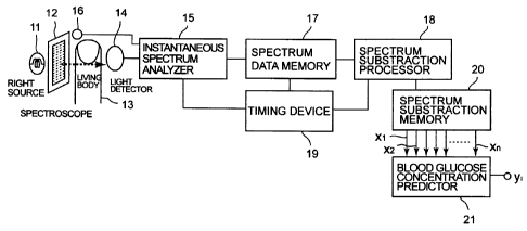
FIG. 1 is a block diagram showing a non-invasive blood
glucose concentration measuring instrument of the present
invention.
As shown in FIG. 1, the light source 11 to emit a light
having a near infrared wavelength range of, for example,
800 ~ 2400 nmwavelength has been installed in a non-invasive
blood glucose concentration measuring instrument. The
light emitted from the light source 11 is irradiated to
a living body 13 such as a fingertip, an ear lobule, etc.
through an active spectroscope 12. The active
spectroscope 12 separates light emitted from the light
source 11 sequentially over its whole wavelength range at
an interval of, for example, 3 nm and sequentially outputs
about 530 number of lights having a diferent wavelength.
The scanning of the wavelength by the active spectroscope
12 in the above-mentioned wavelength range is executed
repeatedly about 20 times or more in one cycle time of the
arterial volume waveform in the living body 13. In other
words, the active spectroscope 12 transmits the lights in
a near infrared range sequentially at an interval of about
50 ms or less and irradiates them to the living body 13.
The light passed through the living body 13 is detected
by a light detector arranged at the opposite side of the
8
CA 02481981 2004-09-24
light source 11 and is converted into an electric signal.
An output signal of the light detector 14 is supplied
to an instantaneous spectrum analyzer 15, wherein an
absorption spectrum obtained as an output of the light
detector 14 for each wavelength of the light source 11 is
produced. That is, the output from a sensor 16 that detects
an intensity of the light incident to the living body 13
from the light source 11, that is, an intensity of~the
incident light I ~ o with each wavelength ( ~. ) is supplied with
the output signal of the light detector 14 to the spectrum
analyzer 15. As described later, the intensity of light
(Iz) with each wavelength (,) passed through the living
body 13, that is, an absorbance(ODz) which is a ratio of
the logarithmic intensity of passed light Iz to that of
the incident light I z o (ODz =log I ~ o / I z ) is computed here
and an absorption spectrum is produced. Twenty(20) number
of the absorption spectrums are produced per second by
twenty (20) times of scanning per second of the active
spectroscope 12 as described above.
The absorption spectrum data obtained by the spectrum
analyzer 15 is stored in a spectrum data memory 17. The
spectrum data memory 17 stores and maintains output data
for several seconds of the spectrum analyzer 15
sequentially on the first-in first-out basis.
Spectrum data read from the spectrum data memory 17
is supplied to a subtraction processor 18 and a spectrum
9
CA 02481981 2004-09-24
subtraction (ODzti,-ODzti=~ODzti) . which is composed of a
difference in absorbance in corresponding wavelengths (,)
between the absorption spectrums (ODzti) at different times
(ti) is produced as described later. From this subtraction
technique, the subtraction data(0 ODzti) include only
informationof arterialblood withouttheotherbiological
tissue components such as skin, bones, muscles etc, as also
described later.
The spectrum analyzer 15, the spectrum data memory 17
and the subtraction processor 18 are operated in sync with
the 20 times scanning per second of the active analyzer
12. The synchronization between these units is made by
a timing device 19 to supply a synchronizing signal to them.
The spectrum subtraction data ( 00Dzti) produced by the
subtraction processor 18 is stored in a spectrum
subtraction memory 20. The spectrum subtraction memory
20 also stores the output data of the subtraction processor
18 for several seconds sequentially on the first-in
first-out basis. It is noted, as described later, that the
subtraction data0 ODzti is mathematically derived to be
equal to the change in the intensity of the transmitted
light ( I z ti--I z ti= 0 I ~ ci ) divided by the intensity of the
light at the time ti ( I z ti ) . provided that 0 I ~ ti is very sma ll,
that is, ~ODzti=D Izt; /Izti. This means that the light
intensity and its change are needed in practical use without
detection of the intensity of the incident light(Izo).
CA 02481981 2004-09-24
The spectrum subtraction data read out of this spectrum
subtraction memory 20 is input into a blood glucose
predictor 21. The blood glucose predictor 21 is a device
to predict blood glucose concentration through the
multi-regression analysis using the PLS (Partial Least
Squares Regression) methodthat is one of multi-regression
analyses from input spectrum subtraction data. That is,
the blood glucose predictor 21 is constructed as a software
modeltocomputethebloodglucoseconcentrationaccording
to the PLS method using whole blood samples that have many
known blood glucose concentrations.
FIG. 2 is a flow chart showing a method for constructing
the blood glucose predictor 21 as the software model shown
in FIG. 1. Known blood glucose concentration samples 31
are the whole blood samples filled in plural quartz
photo-cells whose glucoseconcentrationsare known and are
slightly differrnt from each other. These samples 31 were
taken directly from, for example, seven healthy adult males
and were made the plural whole blood samples having
different albumin or hematocrit concentrationsfrom other
blood samples by 18 mg/dl like 36, 54, ... 486 mg/dl in
the glucose concentration range 30 ~ 450 mg. These samples
31 are analyzed by a spectroscopic analyzer composed of
the light source 11, the spectroscope 12, the lightdetector
14 and the spectrum analyzer 15, and thus a absorption
spectrum 32 is prepared. A PLS regression analysis
11
CA 02481981 2004-09-24
prediction model 34 is determined by data X consisting of
these absorption spectrum 32,together with corresponding
known n number of blood glucose concentrations(yn) 33.
That is, data X consisting of the absorption spectrum 32
is an absorbance for different m (about 530 waves) number
of the spectroscopic waveforms. Expressing these
absorbance with xl, x2, . . . , xm, the known n number of blood
glucose concentrations yl, y2, ..., yn are approximated
by the following determinant using these variables:
yl all ......... aln X1
y2
- Formula 1
yn a In ......... ann Xn
A coefficient of this determinant is determined using
the PLS method by substituting the absorption spectrum data
using the above-mentioned sample solution into the
determinant. A blood glucose prediction model formula is
thus obtained. Here, the PLS method is a technique to
consider the correlation of potential variables TpLS as
explanatory variables and to utilize data contained in X
as many as possible.
y = Tq + f
X = TPt + E Formula 2
S - ytT
where, T: Potential variable
12
CA 02481981 2004-09-24
q: Potential variable regression coefficient
E,f: Residual of X, y
P: Loading matrix
S: Covariance of Y and T
P of the determinant 2 and regression coefficient q
of potential variable T are determined by inputting blood
glucose yl, Y2 , ...Yn of n known blood glucose samples
into a regressionanalyticalcomputerapplicationsoftware
(for example, Trade Name: MATLAB) according to the PLS
method available in the market. Thus, the regression
analysis prediction model (blood glucose computing model)
according to the PLS method is obtained. Then, a new T
is computed based on P that is determined when a model is
prepared, when new absorbance of respective spectroscopic
wavelengths xl, x2,..., xm obtained from blood of which
blood glucose concentration is unknown are input as data.
These new absorbance of respective spectroscopic
wavelengths xl, x2, ..., xm are input as spectrum
subtraction data read from the above-mentioned spectrum
subtraction memory 20. Using this new T and q determined
when a model was prepared, a blood glucose prediction value
yi is obtained.
Next, the operations of the non-invasive blood glucose
concentration measuring instrument thus constructed
according to the embodiment of the present invention and
the blood glucose measuring procedures will be explained
13
CA 02481981 2004-09-24
referring to FIG. 3 and FIG. 4.
As shown in FIG. 1, the light emitted from the light
sourcellisspectroscopicallyscanned overthewavelength
range by the active spectroscope 12 at a rate of, for example,
20 times per second and is irradiated to the living body
13. The light transmitted through the living body 13 is
detected by the light detector 14 and each absorption
spectrum is measured by the spectrum analyzer l5 at an
intervals of 40 ~ 50 ms. The spectrum data thus measured
is stored in the spectrum memory 20 until the next spectrum
measuring time. FIG. 3 shows the arterial pulsatile volume
waveform in the living body 13, the horizontal axis shows
time and the vertical axis shows arterial blood volume
change (pulsatile volume waveform) . Time tl, t2, . . . , tn
in FIG. 3 show the time when the scanning of the wavelength
starts by the active spectroscope 12, where n is 20 in this
case. Absorption spectrum at the time tl, t2, . . . , tn thus
obtained are shown in FIG. 4, where the horizontal axis
shows the wavelength (,) and the vertical axis shows
absorbance(ODzti: ti=tl, t2, ---, tn).
Next, the spectrum subtraction processor 18 shown in
FIG. 1 produces a spectrum subtraction from absorption
spectrums at two any optional times, for example, a time
tl and a peak time tm in the arterial pulsatile volume
waveform selected from the times tl, t2~ ..., tn.
FIG. 5 is a diagram for explaining an operation of the
14
CA 02481981 2004-09-24 '
blood glucose predictor 21 shown in FIG. 1. One example
of the above-mentioned spectrum subtraction is shown in
FIG. 5(a). The horizontal axis in FIG. 5 shows the
wavelength (,) and the vertical axis shows a difference
in the absorbance ( DODzti) . The curved line indicating the
spectrum'subtraction is a plotted difference in the
absorbance at respective wavelengths of absorption
spectrum, for example, at t3 and t6 in this case
Graphes ( S1 ) , ( S2 ) , . . . , ( Sm) in FIG . 5 show absorpt ion
spectrums of m number of whole blood samples of known blood
glucose concentration.
Spectrum subtraction data shown in FIG. 5 (a) are input
tothe blood glucoseconcentration predictor2l. Further,
a PLS regression analytical model is incorporated in the
blood glucose concentration predictor 21. The PLS
regression analytical model is a numerical expression
showing the relation between absorption spectrums of m
number of whole blood samples ( S1 ) , ( S2 ) , . . . , ( Sm) shown
in FIG. 5 each having known blood glucose concentration
and the known blood glucose concentrations corresponding
tothesamples. The blood glucose concentration predictor
21 compares the spectrum subtraction given from the
spectrum subtraction memory 20 as input data with each of
the absorption spectrums of the sample solutions and
outputs the blood glucose concentration of the sample
solution having the most similar absorption spectrum as
CA 02481981 2004-09-24
a predicted blood glucose concentration.
Thus, it is revealed that a blood glucose concentration
can be predicted at a high level of accuracy when spectrum
subtraction is used as input data to the blood glucose
concentration predictor2l. Thereason willbeexplained
referring to FIG. 6. FIG. 6 is a schematic diagram showing
the relation of the intensity of incident light I ~ o~ the
intensities of transmitted lights I z I, I z 2 and absorption
amount in the living body 13 at the wavelength ~,. The
arterial blood volume waveform P as shown in FIG. 3 is also
shown in FIG. 6. In FIG. 6, for example, the transmitted
light intensity Izl (Incident light intensity Izo) at t
- tl where the arterial blood volume waveform P becomes
minimal is (Incident light intensity Izo) - (Absorption
light intensity in the arterial blood layer at the minimum
volume change I~3) - (Absorption light intensity in the
venous blood layer Iz4~) - (Absorption light intensity in
the biological tissues excluding blood Iz5); that is, I
z 1= I z o - ( I ~ 3+ I z 4+ I z 5 ) at t=tl . Further, the transmitted
light intensity I ~ 2 at t = tm where the volume change in
the artery becomes maximal is (Incident light intensity
Izo) - (Absorption light intensity in the arterial blood
layer of the maximum volume change Iz 6) - (Absorption light
intensity in the venous blood layer I ~ 4 ) - (Absorption light
intensity in the biological tissues excluding blood Iz
5), that is, Iz2= Izo -( Iz6+ Izq+ Iz5) at t=tm. The
16
CA 02481981 2004-09-24
differences of these two transmitted light intensities(I
z 1-I z 2 ) extract the spectrum of pulsative element 0 I z that
is the pulsating absorption intensity of the artery(Iz
1-Iz2= Iz6-Iz3= DIz). Although the absorption light
spectrum in the spectrum analyzer 15 or the spectrum data
memory 17 shown in FIG. 1 contains the absorption light
element in the venous blood and biological tissues
excluding blood, the spectrum subtraction ( 0 ODz ) generated
in the spectrum subtraction processor 18 becomes the light
absorption spectrum depending on the light absorption
element of arterialblood absorption element only. Because
the subtraction( ODz) from the absorbance at t=tl(ODz
1=log Izo / Izl) to that at t=tm(ODz2=log Izo / Iz2) is
equal to log Iz2/ Izl(=Iog(Izl-DIz)/ Izl=log(1-~Iz/ I
z 1 ) , and thus 0 ODz is~ nearly equal to - 0 I z / I z 1 when D
I z O I z 1 ( 0 ODz= - D I z / I z 1) . Accordingly, this subtract ion
does not contain the absorption element by the venous blood
and biological tissues excluding blood. Therefore, it
becomes possible to eliminate influence of these
interfering factors and to put into practical use of a highly
precise non-invasive blood glucose concentration
measuring instrument.
By the way, in producing the spectrum subtraction ( D
ODD ) by the measurement,of living body 13 described above,
when a difference in arterial spectrum waveforms that
become the maximum and minimum volume changes in one heart
17
CA 02481981 2004-09-24
beat, the blood glucose concentration is computed at one
time per one heart beat and the blood glucose concentration
is output at one time per one heart beat. However, as
spectrum data is measured repetitively nearly 20 times in
one heart beat, it is possible to take out spectrum
subtraction at two adjacent times as continuous spectrum
subtractionswhileshifting timessequentiallyand compute
blood glucose concentrations using these continuous
spectrum subtractions . In this case, it is expected that
a change in spectrums at adjacent times is very little,
signal noise ratio of spectrum subtraction drops and a
fluctuation (a residual error) of the result of blood
glucose concentration computation may become large.
Accordingly, it is also possible to display the measured
result easy to look by inputting the result into the blood
glucose concentration predictor 21 by executing the time
series average of these spectrum subtractions or by
smoothing successively computed blood glucose
concentrations through the statistical procedure such as
the time average or moving average by the blood glucose
concentration predictor 21.
Further, in the embodiment described above, the
transmitted light spectrum from the living body 13 is
measured but the reflected light from the living body 13
may be measured other than the transmitted light.
FIG. 7 is a partial explanatory diagram showing this
18
CA 02481981 2004-09-24
embodiment, in which the same composed elements as those
in FIG. 1 are assigned with the same reference numerals
and the detailed explanation thereof will be omitted. In
this embodiment, the light detector 14 is arranged at the
same side as the light source 11 to the living body 13 as
illustrated and detects the reflected light from the living
body 13. By supplying the output signal of the light detector
14 to the spectrum analyzer 15 shown in FIG. 1, it is possible
to measure blood glucose concentration likewise the
embodiment described above.
Further, in the embodiments shown in FIG. 1 and FIG.
7, the light from the light source 11 is separated by the
active spectroscope 12 and then irradiated to the living
body 13. However, the transmitted light or reflected light
may be separated for spectrum analysis after the light from
the light source 11 is irradiated to the living body 13.
For example, the light can be separated by an array of plural
light detectors each having a sensitivity only for specific
wavelengths ( ~, ) .
Further, in the embodiments described above, a model
applied with the PLS method is used as the blood glucose
concentration predictor 21. However, a model according to
the principal constituents regression shown in Formula 3,
which is one of multi-regression analyses may be used. The
regression analysis blood glucose concentration computing
model that is constructed using the PCRmethod is expressed
19
CA 02481981 2004-09-24
by the following Formula 3.
Y - Tb + f ~ Formula 3
- tlbl+t2b2+ ... +tnbn
where, T: Principal constituents score
b: Principal constituents score
regression coefficient
That is, a multi-regression analysis blood glucose
concentration computing model is constructed by
corresponding a known blood glucose concentration of the
whole blood sample 31 to an obj ective variable y, applying
spectrum data of the whole blood sample 31 to an explanatory
variable x and deciding a multi-regression analysis blood
glucose concentration computing model. When spectrum
subtraction data of an unknown blood glucoseconcentration
is input into the blood glucose concentration predictor
21 in which this principal constituents score regression
coefficient b is set, a blood glucose concentration predict
value ya is computed and output.
Further, when developing a regression analysis
prediction model (a blood glucose concentration computing
model) in the above-mentioned embodiment, the sample 31
having a known blood glucose concentration is filled in
plural quartz cells and absorption spectrum data X1, X2, . . .
Xm are developed with a spectroscopic analyzer comprising
the light source 11, the spectroscope 12, the light receiver
14 and the spectrum analyzer 15. However, for these
CA 02481981 2004-09-24
absorption spectrum data X1, X2, ..., Xm, spectrum
subtraction data obtained with units ranging from the light
source 11 to the spectrum subtraction memory shown in FIG.
1 using plural living bodies of which blood glucose
concentrations are known can be used.
Further, in the embodiment mentioned above, the
measurement of blood glucose concentration is shown.
However, regarding the measurement of concentration of
another material having absorption characteristics and
scatter reflection characteristics existing in the
arterial blood, it is possible to predict and compute the
concentration of that material existing in the arterial
blood similarly. That is, it is_possible to predict and
compute the concentration by measuring spectrum of
wavelength band corresponding to the absorption
characteristics or the reflecting characteristics of the
material and deciding the regression coefficient of the
multi-regression analyzing model using the PLS method or
the PCR method referring to a concentration of a sample
of that is the standard of that material using the same
system and procedures shown in the above embodiment.
As described above, with the non-invasive blood
constituent measuring instrument and the method according
to the embodiment of the present invention, it is possible
to measure blood constituents in a living body by
irradiating near infrared light to a finger tip, etc.
21
CA 02481981 2004-09-24
quickly and highly precisely without feeling pain and
burden involved in the blood drawing.
Further, according to the embodiment of the present
invention, spectrum subtraction using the arterial blood
beat is used as described above. However, the spectrum
subtraction analysis may be made by generating the venous
blood volume change in the biological tissues using such
a method as the venous occlusion method, for example . Thus,
the adverse effect of other biologicaltissue constituents
is eliminated and blood constituent can be measured at a
highly precise and sensitive level.
22