Note: Descriptions are shown in the official language in which they were submitted.
CA 02482402 2004-10-08
WO 03/087787 PCT/GB03/01510
SEMICONDUCTOR DIODE LASER SPECTROMETER ARRANGEMENT AND METHOD
The present invention relates to a semiconductor
diode laser spectrometer arrangement and in particular an
infrared semiconductor diode laser spectrometer having
time resolved absorption, in which the wavenumber scale
calibration is based on a time to wavenumber/cm-'- mapping.
Infrared absorption spectrometers are used for
detecting and measuring gases. Infrared semiconductor
diode lasers are used extensively to provide the light to
be absorbed by the measurement species, as these lasers
are relatively small, spectrally well defined, bright and
tunable. Further advantages of these lasers over other
lasers exist, some of which can be seen in spectroscopic
monographs.
In remote locations and harsh environments, one of
the most effective and accurate methods of trace gas
sensing uses semiconductor diode laser based
spectrometers. Although gas sensing has been undertaken
for some decades, in many environments it remains
difficult to remotely monitor trace gas constituents.
Many previous instruments have slow response times, are
frequently bulky, unreliable, expensive, and require
constant maintenance.
In order to retrieve information with known
technology, remote sensing of 'gases usually takes place
in the near and mid-infrared region of the
electromagnetic spectrum, where the chemical fingerprints
of most chemical compounds lie. By near and mid-
infrared, it is meant radiation having a wavelength in
the range of l m to 14 m. This spectral region contains
highly transmitting windows, so-called "atmospheric
windows", which owe their transparency to the low density
of strong absorption lines of CO2 and H20 . These
atmospheric windows are of great interest for
CA 02482402 2004-10-08
WO 03/087787 PCT/GB03/01510
2
spectroscopy since the absorption lines of strongly
absorbing trace molecules have similar or greater
intensity than the weak lines of CO2 and H20.
Near-infrared diode lasers produce light in the
wavelength range of the vibrational overtones, about l m
to 3.0 m. Since the absorption coefficients of the
vibrational overtones are much smaller than those of the
fundamental bands, the sensitivity of spectrometers that
use such lasers remains limited. Thus, the sensitivity of
such gas sensing apparatus rarely achieves the sub-part
per billion (sub-ppb) range.
Mid-infrared diode lasers produce light in the
wavelength range of the fundamental rotation-vibration
bands, about 3 m to 14 m. These lasers have not been as
technologically developed as those in the near infrared
region, and hence have low single mode output power. Gas
sensing systems based on mid-infra-red diodes are capable
of achieving sub-ppb sensitivity. The development of such
light sources has, therefore, been wholly dedicated to
spectroscopic applications. Several disadvantages are
associated with conventional mid-infrared diode lasers,
principally lead salt lasers, such as low output power,
and their need to be cryogenically cooled to 77K or to
even lower temperature. Thus, they require a bulky and
expensive operating system to maintain this temperature.
Recently, room temperature and high light output
power operation has been achieved in the mid-infrared
using quantum cascade (QC) lasers. Unlike preceding
lasers, QC lasers are unipolar semiconductor lasers that
can be designed to emit at any desired wavelength in the
mid- infrared. Replacement of lead salt lasers by QC
lasers provides the potential to improve both the
CA 02482402 2004-10-08
WO 03/087787 PCT/GB03/01510
3
detection sensitivity and spectral resolution of mid-
infrared absorption spectrometers.
The QC laser based spectrometers developed so far
use two main approaches. The first uses a continuous
wave (CW) operating QC laser as a "drop-in" replacement
for a lead salt laser. The second approach is to use a
pulsed QC laser in a way that mimics the use of a
continuously operating laser. In some experiments
conducted by Webster et al (Applied Optics LP 40,
321(2001)), the first approach was used with one of the
lead salt diode lasers in an ALIAS II spectrometer being
replaced by a QC laser. Test measurements made using an
ER2 aircraft platform showed that the QC laser could
successfully replace a lead salt laser and was less
affected by temperature instability. However, for CW
operation the laser needed to be operated at 77K. The
second method was described originally by Whittaker at al
(Optics Letters 23,219 (1998)). In' this method a very
short current pulse is applied to a QC laser operating
near room temperature to provide a narrow wavelength
pulse. In this mode of operation the spectral resolution
is limited by the wavelength up-chirp. Thus, in this
type of spectrometer the wavelength up-chirp is regarded
as detrimental to the operation of the system.
The wavelength up-chirp ("effective emission
linewidth") is induced by the temporal duration of the
drive current/voltage pulse. By the term "effective
emission linewidth", it is meant the
observable/measurable spectral width (FWHM) of the
emission of a semiconductor diode laser induced by an
applied current/voltage pulse to its electrical contacts.
For example, if the duration of the pulse applied to a QC
laser were of the order of 10 ns, the effective emission
CA 02482402 2004-10-08
WO 03/087787 PCT/GB03/01510
4
linewidth would be of the order of 700 MHz (0.024 cm-1) in
the spectral domain (Optics Letters 23,219(1998)).
In order to scan samples using a pulsed QC laser
based spectrometer, the effective emission linewidth is
tuned across a spectral region using a slow DC current
ramp superimposed on the pulse train. This means that
the resultant spectral tuning is a quadratic function of
the DC current ramp injected to the laser [Optics Letter
23,219(1998); Applied Optics 39 6866 (2000); Applied
Optics 41,573(2002)]. A problem with this approach is,
however, that an additional step is needed in the data
processing stage, to correct for the quadratic effect. In
some cases, to improve the signal to noise ratio, (Optics
Letters 23,219(1998)), a small AC current modulation
signal is added to the DC ramp in order to use phase
sensitive detection of the detected optical signal.
Whilst adding this modulation may increase sensitivity,
it requires the use of demodulation in the detection
system, so rendering the system more complex. A further
problem with this is that the use of a modulation
inherently reduces the scan rate, since the high speed
detected signals are demodulated to low audio frequencies
signals. Hence, prior art arrangements of this type
allow scan rates only of the order of tens of Hertz. One
system proposed by Beyer et al (Third International
Conference on Tunable Diode Laser Spectroscopy July 8-12
2001, Zermatt Switzerland) uses the wavelength variation
of the intrinsic wavelength chirp. However, the
arrangement proposed is of limited use for chemical
finger printing.
In both the CW operated laser (first method)
described by Webster et al (Applied Optics LP 40,
321(2001)) and the short pulse (second method), described
originally by Whittaker et al (Optics Letters 23,219
(1998)), for a gas with a small absorption coefficient
the simplest way of achieving an observable change in the
transmitted signal is to use a long sample length. This
CA 02482402 2010-07-14
can be achieved by use of either resonant or non-resonant
optical cells. Resonant cell schemes are complicated and
require sophisticated techniques to minimise the effects
of back-reflected signals from the input mirror to the
5 cell disrupting the performance of the laser. Non-
resonant cells, such as the so-called Herriot cell or
astigmatic Herriot cell are attractive as they offer long
path lengths, without the penalty of back-reflected
signals. In addition, the path length is independent of
the concentration of the gas in the cell. A major
drawback associated with non-resonant cells is the
occurrence of "fringing" due to the partial overlap of
the beams that propagate around the cell. This decreases
significantly the system performance.
As can be seen, known spectrometers using
semiconductor diode lasers, in particular quantum cascade
(QC) lasers, have shortcomings, which limit their use for
absorption spectroscopy in pulsed operation. Specifically
prior art QC laser based spectrometers, where the light
sources have to be driven in pulsed mode operation to
achieve room temperature operation, have the resolution
of their effective emission linewidth determined by the
temporal duration of the drive voltage/current pulse
applied to its electrical contacts.
An object of an aspect of the present invention is
to overcome at least one of the aforementioned problems.
Various aspects of the invention are defined in the
independent claims. Some preferred features are defined
in the dependent claims.
According to one aspect of the invention there is
provided a fringe free method for sensing gases using
semiconductor diode laser spectrometer. This involves
introducing a sample gas into a non-resonant optical cell
and injecting light from a semiconductor laser into the
cell. This light is generated by applying a one or a
series of substantially step function electrical pulses
CA 02482402 2010-07-14
6
to a semiconductor diode laser to cause the laser to
output one or more pulses, each having a continuous
wavelength chirp, for injecting into the optical cell.
Preferably, each applied pulse has a duration that is
greater than 150ns, in particular greater than 200ns.
Preferably, each applied pulse has a duration that is in
the range of 150 to 300ns, preferably 200 to 300ns. This
can provide a tuning range of about 60GHz. The chirp rate
is selected so that there is a time delay between spots
on the reflecting elements of the non-resonant cell
sufficient to substantially prevent light interference
from occurring, wherein the spots define locations at
which the injected chirp is reflected from the cell
walls. The wavelength variation provided by the
wavelength chirp itself is used to provide a wavelength
scan. Hence, there is no need to tune the effective
emission linewidth across a spectral region using, for
example, a slow DC current ramp superimposed on the pulse
train. Light output from the optical cell is detected
using a suitable detector.
Preferably, each detected pulse has a duration that
is greater than 150ns, in particular greater than 200ns.
Preferably, each detected pulse has a duration that is in
the range of 150 to 300ns, preferably 200 to 300ns.
Alternatively, rather than using a non-resonant
cavity, the gas sample may be unconfined, and the method
for sensing may use an open path configuration to prevent
light interference from occurring. In either case, by
preventing light interference from occurring, fringing
effects can be avoided. This means that the sensitivity
of the method can be significantly improved.
According to another aspect of the present invention
there is provided a method for sensing gases using a
semiconductor diode laser spectrometer, the method
comprising: introducing a sample gas into a non-resonant
optical cell having reflecting elements; applying a step
CA 02482402 2010-07-14
6a
function electrical pulse to a semiconductor diode laser
to cause the laser to output a continuous wavelength
chirp for injecting into the optical cell; injecting the
wavelength chirp into the optical cell; using the
wavelength variation provided by the wavelength chirp as
a wavelength scan, and detecting light emitted from the
cell, wherein the method further involves using a chirp
rate such that there is a time delay between spots on the
reflecting elements sufficient to prevent light
interference occurring in the optical cell.
According to still another aspect of the present
invention there is provided a semiconductor diode laser
spectrometer for measuring radiation absorption by a
sample, the spectrometer comprising a semiconductor diode
laser; a non-resonant optical cell for containing a
sample gas and having reflecting elements at either end
thereof; an electric pulse generator adapted to apply a
substantially step function electrical pulse to the laser
to cause the laser to introduce a continuous wavelength
chirp into the sample cell, and a detector for detecting
light output from the cell and adapted to use the
wavelength variation of the wavelength chirp as a
wavelength scan, wherein the chirp rate used is such that
there is a time delay between spots on the reflecting
elements sufficient to prevent light interference
occurring in the optical cell.
Various aspects of the invention will now be
described by way of example only and with reference to
the accompanying drawings, of which:
Figures la to If show computer simulated plots of
emission versus wavenumber for various modes of operation
of a QC laser;
CA 02482402 2004-10-08
WO 03/087787 PCT/GB03/01510
7
Figure 1g shows a computer simulated plot of emission
versus time for a QC laser in a particular mode of
operation;
Figure lh shows an experimental plot of emission
versus time for a QC laser that is being operated so as
to generate a chirp;
Figure 2 is a schematic diagram of an arrangement for
characterising a semiconductor laser using a scanning
Fourier transform spectrometer;
Figure 3a shows plots of wavenumber versus pulse
duration at various different temperatures;
Figure 3b shows plots of wavenumber versus pulse
duration at various different current amplitudes;
Figure 4a shows a plot of dynamic impedance of a QC
laser;
Figures 4b and 4c show plots of dissipated power
versus current for a QC laser at -10C;
Figure 5 is a plot of voltage and power as a function
of current for a QC laser operating at a temperature of
-10C;
Figure 6a shows a plot of wavenumber versus
temperature;
Figure 6b shows a plot of wavenumber versus duty
cycle;
Figure 7 is a block diagram of a system for sensing
gases that includes a QC laser and a Fourier transform
spectrometer;
Figure 8 shows an absorption spectrum of 1,1
difluoroethylene, CF2CH2, recorded using the apparatus of
Figure 7;
Figure 9 is a block diagram of another spectrometer;
Figure 10 shows a schematic diagram of a method of
detecting optical pulses using the spectrometer of Figure
CA 02482402 2004-10-08
WO 03/087787 PCT/GB03/01510
8
9, and, for comparison a method used for a known
spectrometer;
Figure 11 is a block diagram of the prior art
spectrometer used for the comparative measurements shown
in Figure 10;
Figure 12 shows a reference transmission spectrum of
CF2CH2 and laser spectra with and without absorption by
CF2CH2 obtained using the spectrometer of Figure 9;
Figure 13 shows an absorption spectrum of CF2CH2,
recorded using the spectrometer of Figure 9 (upper trace)
and a recording of an etalon fringe pattern of a solid Ge
etalon (lower trace);
Figure 14 shows a comparison of the absorption
spectra of two different molecules (upper trace: CF2CH2;
lower trace: COF2) recorded using the arrangement of
Figure 9;
Figure 15 shows absorption spectra for a sample of
atmospheric gases, recorded using the arrangement of
Figure 9;
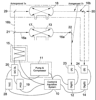
Figure 16 is a block diagram of a modified version of
the spectrometer of Figure 9;
Figure 17 is a block diagram of another spectrometer
in which the invention is embodied;
Figure 18 is a block diagram of a modified version of
the spectrometer of Figure 17;
Figure 19a shows simulated plots of part of a
transmission spectrum of a complex molecule over part of
the spectral range of a multi-longitudinal mode semi-
conductor laser, together with the laser profile;
Figure 19b shows the spectrometer output after
absorption;
1
CA 02482402 2004-10-08
WO 03/087787 PCT/GB03/01510
9
Figure 20 shows simulated plots of part of the
transmission spectrum of a complex molecule with a
spectral filter used, and
Figure 21 shows simulated plots of part of the
transmission spectrum of a complex molecule with a
spectral filter used and with temperature tuning.
The spectrometer in which the invention is embodied
advantageously uses the wavelength up-chirp exhibited by
pulsed QC and semiconductor lasers to provide a
wavelength scan. Each individual pulse output by the
laser provides a wavelength variation, i.e. a wavelength
scan, by virtue of the wavelength up-chirp. This
wavelength up-chirp is induced by a heating effect
occurring for the entire duration of the applied
current/voltage drive pulse. For these QC lasers, the
wavelength up-chirp has been shown to be continuous.
More specifically, under particular conditions of the
electrical drive pulse shape (Optics Communications
197,115(2001)), the spectral behaviour of pulsed QC
lasers is characterised by the fact that this wavelength
up-chirp is almost linear with respect to time. It has
further been shown that in pulsed operations the spectral
behaviour of QC lasers can be mapped to the temporal
definition of the applied drive current/voltage pulse to
its electrical contacts. In view of this, it is possible
to map the light output temporal behaviour of a QC laser
and to show it in the time domain with a photodetector.
Figures la to ig show computer simulated plots of
the temporal and spectral responses for single mode and
mult.imode semiconductor diode lasers when a square
current/voltage signal is applied to their electrical
contacts. For the purposes of this description, the term
temporal response means the time taken for the detection
CA 02482402 2004-10-08
WO 03/087787 PCT/GB03/01510
system to achieve a deflection on a range proportional to
an electrical signal, in the shape of a perfect step
function, applied to its input. The temporal response is
calculated using the usual equation for the relation
5 between the rise time and the bandwidth of a system, i.e.
temporal response = rise time = 0.35/bandwidth.
Figures la and lb show computer simulated results
for the spectral behaviour at a fixed moment in time- so
that no chirp is observed in the spectral domain and that
10 the represented emission linewidth is the intrinsic
emission linewidth. By the term "intrinsic emission
linewidth", it is meant the instantaneous
observable/measurable spectral width (FWHM) of the
emission. The intrinsic emission linewidth of a
semiconductor diode laser is usually much smaller than
the effective emission linewidth and can be difficult to
quantify under pulsed operation.
Figures lc and id show computer simulated results
achieved on the application of a well-defined rectangular
current/voltage drive pulse with a duration sufficiently
long so that a chirp - is observed towards longer
wavelength. As mentioned previously, this chirp arises
from heating effects induced by the drive pulse. The
amplitude decay that goes with this chirp. is caused by
the reduced efficiency of lasing action as the heating
increases. The effect of the wavelength chirp can be
seen more clearly in Figures le and if. A computer
simulation of the temporal behaviour of the emission is
shown in Figure 1g. Since the amplitude decay of the
chirp decreases with time, the temporal response is a
mirror image of that in the spectral domain. Figure lh
shows experimental results for a laser that is pulsed in
such a manner that a chirp is generated. From a
CA 02482402 2004-10-08
WO 03/087787 PCT/GB03/01510
11
comparison of Figures lg and lh, it can be seen that
there is a correlation between the theoretical and the
simulated plots.
Figure 2 shows an arrangement for characterising the
spectral output behaviour of semiconductor diode lasers
using a continuous scanning infrared Fourier transform
spectrometer. The results of experiments using this
arrangement are shown in Figures 3-6.
Figure 3a is a plot of wavenumber chirp as a function
of the temporal duration of the applied current pulse
(fixed amplitude 4.2 A) for a range of substrate
temperatures. The results indicate that the rate of
tuning, over the temperature range investigated, is
insensitive to temperature. From this plot the rate of
change of wavenumber as a function of time, (3, can be
determined empirically. To vary (3, the amplitude of the
current/voltage pulse must be altered, as illustrated in
Figure 3b. From this, it can be seen that irrespective
of the applied current, over the range of currents used,
13 is almost linear in nature.
P is related to the power dissipated inside the laser
diode and the almost linear variation in (3 arises from
the fact that the QC laser exhibits a dynamic impedance,
as shown in Figure 4a, which results in a almost linear
power dissipation over the current range used, see Figure
4b. It should be noted that the value of (3 is determined
over the temporal range for which the output shows no
transient behaviour, see Figure 4c. The limiting values
of 13 are defined, at the lower end, by the
current/voltage amplitude necessary to achieve a usable
output power and at the upper end, by the current/voltage
amplitude that induces a reduction in the output power,
CA 02482402 2004-10-08
WO 03/087787 PCT/GB03/01510
12
see Figure 5. The starting wavenumber of the wavenumber
chirp is influenced by both the substrate temperature of
the QC laser and the duty cycle of the applied
current/voltage pulses as shown in Figures 6a and 6b.
Hence, by varying the substrate temperature and/or the
duty cycle, the starting wavenumber can be altered.
As will be appreciated, the effectiveness of a gas
spectrometer that uses a wavelength-chirp to provide a
wavelength variation for scanning a sample depends on the
actual range of wavelengths over which the chirp extends.
This wavelength range may correspond to a frequency
variation of 60GHz. Figure 7 shows an arrangement for
measuring the upper limits of the effective line width of
a QC laser. This is based on a Fourier transform
spectrometer, which is adapted to generate spectra
representative of the output from a sample cell into
which light from a QC laser is injected. Fourier
transform based spectrometers are well known and use
Michelson interferometers. To measure accurately the
current supplied to the QC laser, a Rogowski coil is
provided. A typical spectrum measure using the
arrangement of Figure 7 is illustrated in Figure 8, which
shows a high resolution absorption spectrum of 1,1
difluoroethylene, CH2CF2. In this case, the resolution of
the spectrometer is 0.0015cm-1. The duration of the
electrical drive pulse applied to the QC laser was 200
ns, the pulse repetition frequency, 20 kHz, and the drive
current 4.8A. The substrate temperature was -1.5 OC. From
Figure 8, it can be inferred that the upper limit of the
laser linewidth is that set by the instrument resolution,
i.e. in this case 45MHz. Also, it can be seen that over
the wavelength scan range of the QC laser chirp three
groups, i.e. (i), (ii) and (iii), of lines of CH2CF2 can
CA 02482402 2004-10-08
WO 03/087787 PCT/GB03/01510
13
be easily identified. This demonstrates that the
effective resolution of a pulsed QC laser spectrometer is
sufficient to detect chemical fingerprints for at least
some chemicals.
Because of its controllable and predictable
characteristics, the almost linear wavenumber down-chirp
can be exploited to make spectral measurements. In
particular, the almost linearity of the wavenumber chirp
as a function'of time allows the construction of a high
speed, sub-microsecond, semiconductor diode laser
absorption spectrometer. Figure 9 shows two spectrometer
arrangements la and lb for measuring radiation absorbed
by a species, i.e a gas sample. In the low intensity
limit, the spectrometer determines the absorption
coefficient of a species by measuring the ratio of the
intensity of the light incident on the sample gas cell, Io
and that transmitted through a sample gas cell containing-
the absorbing species, Ia. In the low intensity limit, the
change in the intensity of light that passes through the
gas is described by the Beer-Lambert relationship, Ia =
I,,exp(-(XL), with a the absorption coefficient and L the
optical path length. It should be noted that a is a
function of wavenumber and is independent of the
intensity at low intensities of the incident' radiation.
The spectrometer of Figure 9 uses a closed non-
resonant optical cell (confined gas) configuration and
comprises a current/voltage drive pulse generator 19 that
is connected to an input of a laser 20. The pulse
generator 19 is operable to apply substantially
rectangular pulses to the laser 20. In this case, the
laser 20 is a single mode semiconductor diode quantum
cascade laser (QC laser) . The laser 20 is housed in a
Peltier temperature controlled enclosure (not shown). The
CA 02482402 2004-10-08
WO 03/087787 PCT/GB03/01510
14
Peltier element is controlled by a thermoelectric
controller 28. Connected to the laser enclosure is a
compressor and pump unit 11, which is used to cool/heat
fluid and circulate that fluid into the hollow housing of
the diode laser enclosure 20. This enables the laser
element to be operated over a wider temperature range
than is possible using solely the Peltier element.
On an optical path from the laser 20 output is- an
optional spectral filter 15, for example a small grating
monochromator, which may be used to provide a single mode
laser output if a multi-longitudinal mode laser is used.
On an optical path from the filter are two beam splitters
21 and 29 respectively. These could be, for example,
germanium beam splitters for laser radiation at
wavelengths close to 10 m. However, it will be
appreciated that any other suitable splitters could be
used. The first beam splitter 21 is positioned so as to
direct at least some of the light incident thereon into a
first optical sample cell 17, which contains the sample
that is to be sensed or characterised, and transmit the
rest of the light to the second beam splitter 29. The
second beam splitter is positioned so as to direct at
least some of the light incident thereon into a second
optical cell 18, which is a reference cell. The cells 17
and 18 have the same characteristics. Both are non-
resonant optical cells. The cells 17 and 18 may be
Herriot cells, either standard or astigmatic Herriot
cells.
In the arrangement of Figure 9, radiation emitted by
the QC laser can traverse two possible optical paths, 16a
and 16b, one through the sample cell 17 and one through
the reference cell 18. In order to detect radiation
transmitted through each of these cells, detectors 23 and
CA 02482402 2004-10-08
WO 03/087787 PCT/GB03/01510
24 are provided at the respective outputs. Connected to
each of these is a digitiser 12 and 14 respectively, each
of which in turn is connected to a control and
acquisition system 10, which provides overall control of
5 the spectrometer. In addition to the digitisers, the
control system 10 is connected to each of the
current/voltage drive pulse generator 19, the spectral
filter 15, and the pump and compressor 11. As part of
its functionality, the control system 10 is operable to
10 set the amplitude and duration of the pulse applied to
the laser input and monitor the resultant outputs
detected from the gas and reference cells 17 and 18
respectively. The control system 10 is also operable to
determine the ratio Ia/Ia. This could be done using, for
15 example, Beer-Lambert's Law, which may be written as Ia/Ia
= exp(-aL). Of course, as will be appreciated by the
skilled person, other techniques could be used.
The arrangement of Figure 9 can be adapted for use is
two separate modes: a single beam mode (SBM) or a double
beam mode (DBM). In the single beam mode only the sample
cell 17 is used, so that light only follows path 16a. In
this case the beam splitter 21 could be replaced by a
mirror. For the SBM both Io and Ia are measured using the
single optical absorption cell 17. To determine I0, the
cell 17 is evacuated and a series of chirped pulses from
the QC laser 20 are passed through it. The output from
the evacuated cell 17 is digitised by the digitiser 12,
and stored by the control and acquisition system 10. To
determine Ia, the cell 17 is filled with a sample of the
gas under study 13, and the sampling process is repeated.
For the dual beam method (DBM), measurement of Io and Ia
can be done simultaneously using both of paths 16a and
16b. In this case, the sample gas would be put in the
CA 02482402 2004-10-08
WO 03/087787 PCT/GB03/01510
16
sample cell 17 and the reference cell would be evacuated
and sealed. The beams output from the gas and reference
cells 17 and 18 respectively are directed to the
detectors 23 and 24. Detector 23 detects the absorbed
light pulse output from the gas cell 17 and detector 24
detects the background light pulse output from the
reference cell 18. An advantage of the DBM scheme is that
by taking simultaneous measurements, the effects of drift
can be minimised.
For SBM, the background light pulse with amplitude Io
and the absorbed light pulse with amplitude Ia, each has
the same distance to travel to the detection system.
Providing that the optical paths lengths associated with
paths 16a and 16b are identical, this is also the case
for DBM, and so both pulses arrive at the detectors 23
and 24 at the same time. In either case, the absorption
can be directly sensed via the use of the ratio Ia/Io.
For both modes of the spectrometer of Figure 9, that
is SBM and DBM, the current/voltage drive pulse generator
19 generates a plurality of substantially rectangular
pulses that are applied to the input of the laser 20.
More specifically, the generator 19 provides a train of
fixed amplitude sub-microsecond duration rectangular
current drive pulses. This causes a fast laser heating
effect and hence a continuous wavelength up-chirp of the
emitted semiconductor diode laser radiation at a rate in
time P. As discussed previously, the fast laser heating
caused by the sub-microsecond rectangular current pulses
is such that for each pulse emitted from the laser 20,
the chirp is a continuous almost linear spectral
variation from short to long wavelength. This is defined
as a continuous spectral or wavelength scan.
CA 02482402 2004-10-08
WO 03/087787 PCT/GB03/01510
17
As noted above, the spectrometer of Figure 9 uses a
non-resonant optical cell. As mentioned previously, the
use of non-resonant cells in conventional spectrometers
results in "fringing", which decreases significantly the
system performance. In order to prevent this, the chirped
laser spectrometer of Figure 9 is adapted to control the
light source with a chirp rate in such manner that the
laser wavelength of overlapping spots in the non-resonant
cell is sufficiently different to prevent interference
from occurring. For some QC lasers, this can be done by
dynamically varying the chirp rate. Otherwise, a laser
having a suitable chirp rate has to be selected. In
practice, this can be 'determined empirically by trial and
error. By spots, it is meant regions of the reflecting
elements of the cell, typically curved mirrors, of the
optical cell from which light in the cavity is reflected
as it bounces back and forward within the cavity. These
spots are distributed over the end walls of the cells.
The variation in the location of the spots arises because
light is injected into the cell at different angles, and
the mirrors of the cells can themselves cause a
transformation of the reflection angles. By ensuring
that the laser wavelength of overlapping spots is
sufficiently different, the effects of residual fringing
can be suppressed. The spectrometer of Figure 9 is
therefore a fringe free gas sensing system, with enhanced
absorption sensitivities. As a specific example, assuming
that neighbouring spots overlap and that the mirrors are
spaced by 0.5 m, and that the line width of the laser is
30 MHz, a chirp rate in excess of 10 MHz/ns would be
sufficient to prevent interference, and thereby provide
substantially fringe free performance.
CA 02482402 2004-10-08
WO 03/087787 PCT/GB03/01510
18
Figure 10 shows a schematic diagram of a data-
sampling scheme used in the spectrometer of Figure 9..
This is referred to as Method 1. For the sake of
comparison, a data-sampling scheme for a conventional QC
laser spectrometer is also shown. This is referred to as
Method 2. Figure 11 shows the prior art spectrometer
that ,was used to implement Method 2. For the purposes of
an accurate comparison the computer simulations of both
systems were made using the same pulse repetition
frequency (PRF) equal to 2 0KHz . The PRF is the f requency
at which the semiconductor diode laser has a
current/voltage pulse applied to its electrical contacts.
The value of 20KHz was chosen, since it is the maximum
rate at which the spectrometer of Figure 11 can be
operated (see: Applied Optics 41, 573 (2002)). It was
also assumed that the spectrometer of Figure 9 uses a
256ns duration current/voltage pulse to exploit the
wavelength up-chirp, and that the spectrometer of Figure
11 uses a 5ns duration current/voltage pulse (see:
Applied Optics 41, 573 (2002)). For the spectrometer of
Figure 11, the effective emission linewidth is
approximately 0.02cm-1. To provide a wavelength scan in
this case, the pulse has to be continuously tuned in a
non-linear manner over a 0.75cm`1 spectral range starting
from 992.3cm-1. For a current amplitude similar to that
used for spectrometer of Figure 11, the spectrometer, of
Figure 9 would have a parameter t of approximately -
5.9x10⁻³cm-1/ns. This would give rise to a total almost
linear wavelength up-chirp of 1.5cm-1 in 256ns. Each
chirp can therefore itself provide an entire scan.
. As can be seen from Figure 10, using the method in
which the invention is embodied, that is Method 1, allows
the entire spectral region to be recorded within each
CA 02482402 2004-10-08
WO 03/087787 PCT/GB03/01510
19
individual or single pulse. As shown in Figure 10, this
involves sampling the detected pulse along its entire
length, thereby to obtain a range of spectral elements
from that single pulse. In contrast, in Method 2 only a
single spectral element may be recorded during a single
pulse. Hence if the same number of sampling points, n, is
recorded, eg n=512 which is the maximum number possible
in Method 2 (see: Applied Optics 41, 573 (2002)), the
theoretical improvement in signal to noise achievable in
Method 1 should be 'n, which for 512 point is a factor of
about 22. An advantage of Method 1 is that it does not
suffer from pulse to pulse fluctuations (both amplitude
and temporal) inside a recorded scan since only one
optical pulse is necessary. In Method 2, it has been
shown that the system suffers from amplitude fluctuations
of the diode laser output from pulse to pulse (see:
Applied Optics 41, 573 (2002)).
Figures 12 and 13 show experimental results taken
using the spectrometer of Figure 9. In the spectrometer
arrangement used for Figures 12 and 13, a single mode
distributed feedback laser was used without a spectral
filter and I,, and Ia were recorded using the SBM method.
Figure 12 shows measurements for a sample of 1,1
difluoroethylene (CF2CH2). The CF2CH2 spectrum in the upper
trace was taken using the spectrometer of Figure 7, but
adapted to replace the QC laser with a black body source.
The two lower traces taken using the spectrometer of
Figure 9 show both Io with the cell evacuated and Ia with
a sample of 1,1 difluoroethylene (CF2CH2) within the cell.
Figure 13 shows results for 1,1 difluoroethylene (CF2CH2)
taken using the spectrometer of Figure 9. The absorbed
signal Ia was recorded using an average of 4096 scans.
The upper trace shows Ia. The lower trace is also Ia but
CA 02482402 2004-10-08
WO 03/087787 PCT/GB03/01510
with a solid Ge etalon in place of the sample gas cell
17. This lower trace shows the etalon fringe pattern
demonstrating an almost linear spectral variation from
short to long wavelength. As can be seen from a
5 comparison of the Fourier transform and diode laser
spectra in Figure 12, and the upper trace of Figure 13
with the Fourier transform spectrum of Figure 8, there is
a strong correlation between the fingerprint patterns, of
difluoroethylene recorded using the two types of
10 spectrometer. However the Fourier transform spectra in
Figures 8 and 12, recorded using the spectrometer of
Figure 7, took more than four hours to obtain, whereas
the diode laser spectra in Figures 12 and 13 required
less than two minutes.
15 The wavelength range over which the chirp-induced
scan occurs is sufficient to allow an identification of
the chemical fingerprint of the gas to be recorded, see
Figure 14. Figure 14 was recorded using the SBM method
of the arrangement of Figure 9. The upper trace in
20 Figure 14 is for 1,1, difluoroethylene (CH2CF2) and the
lower trace, of the same figure, is for carbonyl fluoride
(COF2) . Figure 14 shows the ease of pattern recognition
(identification of the chemical fingerprint) within a
200ns time window using the spectrometer of-Figure 9. For
the sake of clarity, the transmission spectra have been
offset. The wavenumber calibration used a Germanium (Ge)
etalon with fringe spacing 0.0483 cm-1, and reference
lines of 1,1, difluoroethylene taken from a high
resolution Fourier transform spectrum using the
arrangement shown in Figure 7, except with a black body
source.
In the spectrometer of Figure 9, the bandwidth-
duration product of a signal cannot be less than a
CA 02482402 2004-10-08
WO 03/087787 PCT/GB03/01510
21
certain minimum value found with the "uncertainty
relation". This relationship is described in detail by
Bracewell (The Fourier Transform and Its Applications,
McGraw-Hill (1965)), who has proved that the product of
the equivalent duration, At, and the equivalent
bandwidth, L1v, must exceed or be equal to C, a constant
that is determined by the pulse shape. For a rectangular
time window OtAv C = 0.886, and for a Gaussian time
window Atzv >_ C = 0.441. In a short pulse spectrometer
method, if the pulse duration were to be shortened there
would be a' Fourier transform limitation to the
resolution, whereas if it were to be lengthened the
wavelength chirp would be excessive. A similar analysis
may be carried out for the limitations of the time
resolved detection system in which the invention is
embodied, as outlined below. In a time window t the laser
frequency (2,v = c; X is the wavelength, V is the
frequency; c is the wave velocity) will chirp by the
amount dv/dt x 'C, so that if a smaller time window were to
be used the Fourier-limited frequency interval by would
increase, whereas the chirp limited frequency interval
would decrease. The best aperture time,ti, will therefore
be determined by C/r = dv/dt x T. Rewriting'this equation
in terms of Av gives, Av = dv/dt x C/Ov, from which AV =
4 ( C x dv/dt) . In the limiting case of C=1, and a chirp
rate of -0.0066 cm-1/ns, or 0.015 cm-1. This would fall to
0.014 cm-1 if the rectangular window function were used,
and to 0.01 cm-1 if a Gaussian time window were
appropriate.
Figure 15 shows the absorption spectra recorded using
the SBM method of Figure 9 for a sample of atmospheric
gas. An average of 64 thousand scans was used. Trace (a)
CA 02482402 2004-10-08
WO 03/087787 PCT/GB03/01510
22
shows the results for a cell pressure 50.5 Torr. Trace
(b) shows the results for a pressure of 04.5 Torr. Trace
(c) shows the results for a sample to which carbon
dioxide (C02) was added. In this case, the pressure was
103.2 Torr. The very low absorption co-efficient line,
which corresponds to H20, i.e. the peak on the left hand
side of Figure 15, has almost the same percentage
absorption in traces (b) and (c). However, it is evident
that a large increase in the percentage of absorption due
to carbon dioxide has occurred in trace (c) in comparison
to trace (b). Figures 14 and 15 show that it is possible
to do achieve simultaneous gas measurement of different
species and that it is possible to identify them
(compound identification).
Various modifications to the spectrometer of Figure
9 can be made. For example, for the double beam method,
rather than having a separate reference cell that is
evacuated, a reference signal could be passed through the
sample cell 17 itself. This is shown in Figure 16 as
arrangement lc. Here, the measurement path is 16a and
the reference path is 16b. For the purposes of clarity,
the paths 16a and 16b are shown separately in Figure 16,
but it will be appreciated that they both go through the
sample cell 17. If the optical path length of the signal
path, 16a, is La, and that of the reference path 16b is
Lb, then in order to minimise absorption in the reference
path 16b, La must be much greater than Lb (La >> Lb) . This
can be arranged by, for example ensuring that the
measurement beam makes many passes across the sample cell
17, whereas the reference beam either passes straight
through the cell, and so makes a single pass, or only
makes a limited number of passes.
CA 02482402 2004-10-08
WO 03/087787 PCT/GB03/01510
23
The modified Beer-Lambert expression required for
arrangement 1c may be derived as follows: for the signal
path Ia=Ioexp (-(xLa) and for the reference path Ib=Ioexp
(-OGLb) . Hence, ln(Ia/Ib)=-X(La-Lb) . In arrangement ic, the
transit time difference between both pulses is chosen to
be less than the wavelength up-chirp time or
current/voltage drive pulse duration. Therefore, the
background light pulse arrives at detector 24 in advance
of the arrival of the signal pulse at detector 23. The
outputs from the digitisers 12 and 14 are recorded, to
enable the control acquisition circuit 10 to ratio them
to provide Ia/Ib as detailed previously. An advantage of
the spectrometer of arrangement lc of Figure 16 is that
fewer optical elements are used than in the first
embodiment, arrangement lb of Figure 9, e.g. no reference
cell. This reduces the overall size and weight of the
spectrometer arrangement.
Arrangement id of Figure 16 is a modification of
arrangement lc. In this case, only a single detector is
used. To this end, instead of being directed into
detector 24, the reference beam is directed into detector
23. The absorption path difference is identical to that
of arrangement lc, namely AL = (La-Lb). When a pulse
train is incident on the beamsplitter of Figure 16, the
action of the beamsplitter is to split each individual
pulse in the pulse train into two components. Any one
pulse from the pulse train that follows optical path 16a
has a companion pulse that follows optical path 16c. This
has important consequences when considering the detection
of Ib and Ia by the single detector arrangement ld. To
compute the ratio of Ib to Ia the signals corresponding to
Ib and Ia must be recorded separately and then processed
in the manner described for the SB mode of operation in
CA 02482402 2004-10-08
WO 03/087787 PCT/GB03/01510
24
Figure 9, embodiment lb. This means that a pulse
corresponding to Ia cannot arrive at the detector until
its companion pulse corresponding to Ib has been digitised
by digitiser 12 and stored by the control and acquisition
system 10. The next pulse associated with Ib, however,
cannot arrive at the detector, before the previous Ia
pulse has been digitised by digitiser 12 and stored by
the control and acquisition system 10. Thus, the
difference in optical path length and hence transit time,
between optical path 16a and optical path 16c must be
greater than the distance defined by pulse temporal
duration (speed of light x tp) but less then the distance
defined by the pulse repetition time (speed of light x
trep) .
All of the spectrometers described so far are closed
systems, in which a sample gas is placed in a closed
optical cell. However, many measurements of atmospheric
trace gases have to be made using open path (unconfined
gas) arrangements, i.e. the spectrometer contains no gas
cell. Figure 17 a schematic diagram of an unconfined
spectrometer arrangement in which the invention is
embodied. Because no optical elements are used to contain
the sample gas this arrangement is fringe free. Such a
spectrometer could be used, for example, as shown in
arrangement le of Figure 17, for monitoring the exhaust
plume 40 of an engine. The arrangement of the optical
components up to and including the beamsplitter 21 is
identical to that of the previous embodiments la, lb
(Figure 9), and lc and ld (Figure 16) . Opposite the
filter 15 and on the optical path of beam 16a is a cube-
corner retro-reflector 39 that is positioned in use so
that the gas to be investigated is between the filter 15
and the reflector 39. Light reflected from the reflector
39 is directed back, through the gas towards the
detection system. In contrast, the reference beam 16b is
transmitted in a direction perpendicular to beam 16a
CA 02482402 2004-10-08
WO 03/087787 PCT/GB03/01510
through a much shorter optical path towards another
reflector, which reflects it toward the detection system.
In this case, the detection system is the same as for the
DBM arrangement of embodiments lb (Figure 9) and lc
5 (Figure 16).
In the use of the spectrometer of Figure 17, a
stream of current pulses is applied to the laser 20,
which emits light that is subsequently passed through the
filter 15, thereby to produce a suitable output, i.e.
10 that comprises a series pulses, each of which has a
wavelength up-chirp. The light pulse to be absorbed 16a
then travels through the exhaust plume 40 and is
reflected by the retro-reflector 39, returning through
the exhaust plume 40 to the spectrometer le. In this
15 way, the beam 16a makes two passes through the gas. The
reflected pulse 16a is then focussed onto detector 23.
The background pulse of light 16b, which is focussed onto
detector 24, travels via a much shorter optical path than
that of the signal pulse, 16a. Hence, the transit time
20 of the reference pulse 16b is less than that of the
signal pulse 16a, so that the background pulse 16b
arrives at the detector 24 before the signal pulse 16a at
detector 23, when both the time measurements are made
relative to that of an initial trigger pulse. Since the
25 digitisers 12 and 14 can each be delayed with respect to
one another, each of the detected pulse components 16a
and 16b are recorded such that the control and
acquisition system 10, which is incorporated in detection
system will ratio them to generate Ia/Io. In accordance
with the invention, detection and scan Method 1,
described with reference to Figure 10, is used.
Figure 18 shows a modified version of the
spectrometer of Figure 17, in which only a single
detector is used. This is similar to the closed path
CA 02482402 2004-10-08
WO 03/087787 PCT/GB03/01510
26
arrangements shown in Figure 16. In order to separate the
arrival of the signal pulse 16a and the background pulse
16b at the detector 23, the transit time difference
between the pulses must be greater than that of the
wavelength up-chirp time or current/voltage drive pulse
duration. As in embodiment id, since the digitiser 12
records both detected pulses 16a and 16b on the same
channel, they are then separated within the digitiser' 12
and processed such that the control and acquisition
system 10 can ratio them generating I,/I,.
So far, the spectrometers in which the invention is
embodied have been described with reference to a single
mode QC laser, such as a distributed feedback (DFB) QC
laser. This could, however be replaced with a multi-
longitudinal mode laser. Doing this brings both
advantages and disadvantages. The principal advantage is
that it widens the effective tuning range of the
spectrometer. Since the absorption spectra of many of
the gases of interest in sensing applications consist of
groups of absorption features separated by regular
intervals, the coincidences between emission and
absorption lines occur at regular but frequently widely
separated intervals (see Infrared Vibration-Rotation
Spectroscopy, Geoffrey Duxbury, Wiley 2000 Chapters 5 and
9, for a more detailed discussion of such coincidences) .
This can been seen in Figures 19a and 19b. In Figure
19a, the upper trace is an absorption spectrum for a
sample gas. As will be appreciated, this spectrum is
relatively complex. The lower trace of Figure 19a shows
the emission response of the chirped multi-mode QC laser,
which is used to sense the sample gas. Figure 19b shows
the detected signal, from which it can be seen that there
CA 02482402 2004-10-08
WO 03/087787 PCT/GB03/01510
27
are several coincidences between the sensing laser input
and the sample characteristics.
In the absence of a spectral filter 15 in the
spectrometer of any one of Figures 9, 16, 17 and 18, all
the spectra of Figure 19b would be superimposed. However,
the use of such a filter allows both the separation of
the spectra and also the identification of the
wavenumber/cm-' region in which they occur, as shown
schematically in Figure 20. Nevertheless, if the tuning
of each mode provided by the wavenumber down-chirp were
to be greater than the longitudinal mode spacing then
partial overlapping of the spectra would still occur. In
addition, if the spectrum of the multi-longitudinal mode
laser were to be contaminated by the occurrence of off
axis modes of the laser the spectral filtering method
described would become difficult to implement. This is
owing to the close wavenumber/cm-i spacing between off
axis (transverse) modes, which makes it extremely
difficult to design a suitable efficient broadband
spectral filter.
As well as widening the effective tuning range of
the spectrometer, another advantage of using a multimode
laser is the possibility of using a combination of mode
section and temperature tuning of individual modes to
achieve complete tuning within the usable intensity low
and high wavenumber modes (gain envelope) of the laser.
This is shown schematically in Figure 21.
The spectrometer in which the invention is embodied
exploits the almost linear wavelength up-chirp of the
intrinsic emission linewidth that occurs on a sub-
microsecond time scale and therefore is able to operate a
scan repetition frequency (PRF) of as high as 1MHz. This
potential gain of speed, which is an improvement of
CA 02482402 2004-10-08
WO 03/087787 PCT/GB03/01510
28
several orders of magnitude compared to prior art, would
allow the present system in which the invention is
embodied to fully exploit the multiplex capabilities
advantages by, for example, achieving real time
measurements to study processes such as fast chemical
reactions (i.e. such as Free Radicals or real time
atmospheric fluctuations).
The resolution of the, time-resolved spectrometer, in
which the invention is embodied is not determined by the
effective linewidth of the laser induced by the current
pulse, but by the chirp rate of the laser, that is the
uncertainty relation, and the temporal resolution of the
detection system. In terms of the temporal response of
the detection system, this is because the number of
pixels (a pixel corresponds to a given time interval)
into which the spectrum can be recorded within the
wavelength chirp is limited by this response. The rate of
this chirp is governed by the parameter (3. The two
parameters affecting wavenumber resolution are the rate
of tuning (3 of the intrinsic linewidth of the laser 20,
and the temporal response of the detection system. Since
the rate of wavenumber chirp is relatively insensitive to
the pulse amplitude for this laser (see Figure 3), the
only method for achieving increased spectral resolution
with the laser used here is to increase the detection
bandwidth (up to the limit of the uncertainty principle).
Thus the provision of a wide bandwidth detection system
(500MHz) can lead to very high spectral resolution as
seen in Figure 13.
Various modifications may be made to the arrangements
described without departing from the spirit and scope of
the invention. For example, it should be understood that
the spectrometer arrangement in which the invention is
CA 02482402 2004-10-08
WO 03/087787 PCT/GB03/01510
29
embodied is fully capable of using an even faster
detection system than that detailed or/and a
semiconductor diode laser exhibiting a slower chirp rate,
hence increasing further the available resolution. In a
further variation, the substrate temperature of the laser
could be changed. This could be done by varying the
repetition rate of the applied sub-microsecond
rectangular current pulse. In an alternative variation
the substrate temperature can be varied by varying the
base DC level of the sub-microsecond duration rectangular
current drive pulses applied to the electrical contacts
of the semiconductor diode laser. In addition, in the
embodiments detailed, the optical beam splitting means
have been described as being an optical beam splitter,
however, they may instead be a dichroic mirror or other
similar arrangement. It should be further understood that
several semiconductor diode lasers could be implemented
in the spectrometer arrangement in which the invention is
embodied to achieve simultaneous measurements of
different species. Further, the samples to be measured
are hereinbefore described as gases, but may
alternatively be aerosols.