Note: Descriptions are shown in the official language in which they were submitted.
CA 02482546 2004-10-13
WO 03/090249 PCT/US03/10835
SAMPLING PROBE FOR MICROARRAY READ OUT USING ELECTROSPRAY
MASS SPECTROMETRY
Field of the Invention
This invention relates generally to methods and apparatus for transport,
ionization and subsequent analysis of analytes, and more specifically to
methods
and apparatus for analyzing a plurality of sample spots on an array using
electrospray mass spectrometry.
Background of the Invention
The study of protein complements of cells, tissues or whole organisms is
referred to as proteomics. Proteomics is of great interest and much progress
has
been made in recent years in large part because of new enabling analytical
technologies. One theme in proteomics is to monitor the expression of proteins
in a
biological system as the system responds to a stimulus. Currently, two-
dimensional
gel electrophoresis (2-DE) is the most common and powerful platform for the
measurement of such protein complements. This approach can support expression
profiling of several thousand proteins in multiple samples.
However, 2-DE has several significant limitations. These limitations include,
for example, difficulty in running membrane proteins, complicated gel image
analysis
and manual preparation and running of the gels. Moreover, 2-DE requires spot
excising and clean-up to utilize the highly specific and sensitive mass
spectrometric-
based protein identification methods employed. Therefore, alternative
measurement
platforms for protein expression profiling within complex samples are being
explored.
Protein "arrays" or "chips" are one potential alternative. In addition to
protein
expression profiling, this technology has potential uses in identifying
protein-protein
interactions, protein substrates or potential candidates in drug discovery
processes.
This approach to screening protein activity benefits from the same advantages
as
commercially available DNA microarrays for mRNA expression analysis, namely
CA 02482546 2004-10-13
WO 03/090249 PCT/US03/10835
high-throughput parallel, quantitative microscale analysis. It also has
advantages
over DNA microarrays.
True expression analyses must be done at the protein level because the final
active product of most genes is the protein and protein expression and mRNA
expression are not necessarily quantitatively linked. Furthermore, proteins
can be
synthesized in both active and inactive forms. To understand the biological
function
of a gene, the amount of active gene product must generally be determined.
Analysis of nucleic acid chips is usually performed using a fluorescent probe
reporter attached to the analyte. A number of problems are associated with
using
this approach for protein array read out. First, and foremost, the
fluorescence
approach requires that only the analytes bind to the capture molecule and that
non-
specific binding is minimal. This is usually not the case with proteins,
especially
when the binding conditions cannot be optimized for each specific interaction.
A
second problem is that labeling of the proteins with a fluorescent probe can
change
their binding characteristics and can destroy protein complexes that exist in
solution.
Finally, the fluorescence approach cannot distinguish among the different
forms of a
given protein. This includes situations wherein the active and inactive form
of the
protein are captured and give equivalent signals. Both of these problems
plague the
current parallel standard for protein detection and quantitation, enzyme
linked
immunosorbant assay (ELISA), which operates on a capture/detection format.
Mass spectrometry (MS) techniques offer advantages for both detecting and
identifying proteins. At present there is no other technology that can rival
the
combination of speed of analysis, sensitivity, and high accuracy measurement
of
molecular mass afforded by mass spectrometry in protein analysis. High-
resolution,
accurate mass determination allows detection of post-translational
modification of
proteins, even in protein mixtures, which is difficult to assess by other
available
techniques. Peptide fragments of proteins generated enzymatically, and
analyzed
by mass spectrometry, are now routinely used to identify the whole molecule
via on-
line protein database searching (peptide mapping).
2
CA 02482546 2004-10-13
WO 03/090249 PCT/US03/10835
Alternatively, new methods allow protein identification from the fragments
generated from intact proteins in the gas-phase using tandem mass
spectrometry,
eliminating the need for enzymatic digestions. Tandem mass spectrometry uses
two
stages of mass analysis, one to preselect an ion and the second to analyze
fragments induced, for instance, by collision with an inert gas, such as argon
or
helium. This dual analysis can be tandem in time, or more commonly tandem in
space. Tandem in space is implemented using two mass spectrometers in series.
Mass spectrometry is now regarded as having great potential as a method for
protein microarray read out. There are currently two ionization methods
commonly
used to generate gas-phase ions from proteins for analysis by mass
spectrometry.
These methods are matrix-assisted laser desorption ionization (MALDI) and
electrospray (ES) ionization. Of these two methods, the most common choice for
protein array read out is MALDI-MS, which is a surFace analysis technique.
MALDI-MS approaches to protein chip read out are currently being exploited
by two different companies, Ciphergen Biosystems, Inc. (Fremont, CA) and
Intrinsic
Bioprobes, Inc. (Tucson, AZ). They each offer protein chips for MALDI-MS
containing from four to eight interaction sites. These commercial products can
be
obtained with particular general affinities for protein capture built-in,
e.g.,
hydrophobic or hydrophilic interaction, anion exchange, cation exchange, and
immobilized metal affinity substrates for capturing metal binding proteins.
Alternatively, special order chips can be obtained with immobilized receptor
species
of the investigator's choice. For example, the immobilized substrates can be a
specific antibody. While the commercial products are not true arrays, there
have
been laboratory demonstrations of the preparation and MALDI-MS analysis of
protein interaction arrays as large as ten by ten (100 spots).
The commercial availability of MALDI-MS protein chip products is an
indication of their utility. Nonetheless, the use of MALDI-MS for chip read
out
presents significant analytical limitations. There is a low number density of
analyte
at any small point on a particular array spot where the laser beam interacts
to
3
CA 02482546 2004-10-13
WO 03/090249 PCT/US03/10835
generate ions. This negatively impacts detection levels. Detection levels in
MALDI-
MS precipitously decline above a molecular mass of about 15 kDa. This can
severely
limit the range of proteins that can be analyzed directly. Time consuming
enzymatic
digestion methods are also needed for generating low mass peptides that are
more
amenable to detection when larger proteins are analyzed. These digestions are
also
needed to generate peptides for protein identification by peptide mapping.
Mass
accuracies in MALDI-MS are usually no better than about 0.01 % (e.g.,+ 6 Da
for
bovine albumin, ca. 66,000 Da). Finally, analysis of the arrays requires
removal of
the analyte from the native liquid environment within which the interactions
occur and
the application of a chemical matrix to facilitate desorption and ionization,
followed
by a drying step.
Electrospray is an alternative to MALDI. Electrospray generally involves
flowing a sample liquid into an electrospray ion source comprising a small
tube or
capillary which is maintained at a high voltage, in absolute value terms, with
respect
to a nearby surface. The nearby (e.g. 1 cm) surface is commonly referred to as
the
counter electrode. Conventional ES systems for mass spectrometry apply high
voltage (relative to a ground reference) to the emitter electrode while
holding the
counter electrode at a lower, near ground reference voltage. For the positive
ion
mode of operation, the voltage on the emitter is high positive, while for
negative ion
mode the emitter voltage is high negative.
The liquid introduced into the tube or capillary is dispersed and emitted as
fine
electrically charged droplets (plume) by the applied electrical field
generated
between the tube or capillary which is held at high voltage, referred to as
the working
electrode, and the nearby surface.
The ionization mechanism generally involves the desorption at atmospheric
pressure of ions from the fine electrically charged particles. The ions
created by the
electrospray process can then be used for a variety of applications, such as
mass
analyzed in a mass spectrometer.
4
CA 02482546 2004-10-13
WO 03/090249 PCT/US03/10835
In a typical ES-MS process, a solution containing analytes of interest is
directed to the ES emitter which is held at high voltage, resulting in a
charged
solvent droplet spray or plume. The droplets drift towards the counter
electrode
under the influence of the electric field. As the droplets travel, gas-phase
ions are
liberated from the droplets. This process produces a quasi-continuous steady-
state
current with the charged droplets and ions constituting the current and
completing
the series circuit.
Although ES-MS is known, the use of ES-MS for automatically reading out a
plurality of spots, such as from a protein chip array, has not been
demonstrated.
This is likely because of the technical challenges of sampling analytes from
small
spots on a sample surface with a liquid flow system in an automated way.
Specifically, electrospray normally operates by having a sample dissolved in
solution
flow through transfer tubing to the ion source of the mass spectrometer. When
trying
to analyze a surface with electrospray a significant challenge is presented in
producing a probe suitable for transporting a normally solid-state surface
sample into
solution and then into the transfer line. In addition, a sophisticated
structure is
needed to control the alignment of the probe with the surface, the structure
generally
providing fine resolution of the probe movement relative to the surface.
SUMMARY
A method for identifying analytes disposed on or in surface arrays includes
the step of providing a surface array including at least one spot. The spot
holds at
least one analyte. At least one eluting solvent is flowed across the spot .
The solvent
directs at least a portion of the analyte away from the spot. At least a
portion of the
analyte is ionized into a plurality of ion fragments using an electrospray ion
source.
The plurality of ion fragments are then analyzed permitting identification of
the
analyte. The analytes can include intact proteins, protein fragments,
pharmaceutical
agents and antibodies.
5
CA 02482546 2004-10-13
WO 03/090249 PCT/US03/10835
The method can include the step of automatically stepping to at least one of
the other spots and repeating the flowing, ionizing and analyzing steps. As
used
herein, the term "stepping" is used synonymously with the term scanning and
refers
to movement from one array spot to another array spot.
The analyzing step can include mass spectrometry. Mass spectrometry can
be tandem mass spectrometry.
The flowing step can include the step of flowing a wash solvent before flowing
the eluting solvent. The method can also include the step of flowing at least
one
reagent to the spot before flowing the eluting solvent.
The probe can be a multi-axial liquid junction probe, the liquid junction
probe
contacting the spot using a liquid bridge. The probe can be a multi-axial
surface
contact probe, the surface contact probe adapted for forming a sealed
enclosure
around the periphery of the spot. The surface contact probe can include an o-
ring
seal for forming the sealed enclosure. The surface contact probe can use
positive
pressure for the flowing step, wherein the eluting solvent and the analyte are
transmitted through the probe under influence of the applied positive
pressure.
In the embodiment which includes automatic stepping, the positioning device
can provide x, y and z positional control about a substantially flat surface
with at
least 1 nm resolution for each dimension. The positioning device can be a
piezoelectric positioner and controller of a scanning probe electrochemical
microscope (SECM).
The method is adapted to sample spot areas of less than about .04 mm2. The
surface array can be a protein array, thin-layer chromotography plates, SDS
polyacrylamide gel electrophoresis (SDS-PAGE), isoelectric focusing gels or
solid
phase extraction materials.
An automated sampling system is for obtaining samples from surface arrays
having a plurality of spots for analysis. The spots have at least one analyte
disposed
on or contained within. The system includes at least one probe. The probe
includes
an inlet for flowing at least one eluting solvent to the spots and further
includes an
6
CA 02482546 2004-10-13
WO 03/090249 PCT/US03/10835
outlet for directing the analyte away from the spot. An automatic positioning
system
is provided for translating the probe relative to the spots to permit sampling
of any of
the spots.
An electrospray ion source having an input fluidicly connected to the probe is
provided for receiving the analyte and generating ions from the analyte. The
system
includes a structure for analysis of the generated ions, the structure for
analysis
receiving the ions for the electrospray ion source. The structure for analysis
can
include a mass spectrometer or a tandem mass spectrometer.
The probe can be a multi-axial liquid junction probe, the liquid junction
probe
contacting the spots using a liquid bridge. The probe can also be multi-axial
surface
contact probe, the surface contact probe adapted for forming a sealed
enclosure
around a periphery of the spots.
The automatic positioning system can provide the ability to step from spot to
spot. For example, a piezoelectric positioner and controller of a scanning
probe
electrochemical microscope (SECM) can be used for this purpose.
BRIEF DESCRIPTION OF THE DRAWINGS
A fuller understanding of the present invention and the features and benefits
thereof will be accomplished upon review of the following detailed description
together with the accompanying drawings, in which:
FIG. 1 illustrates a sampling probe/ES-MS for surface array read out,
according to an embodiment of the invention.
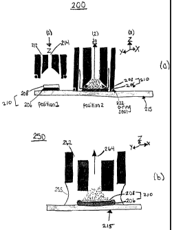
FIG. 2(a) illustrates a surface contact probe, according to an embodiment of
the invention.
FIG. 2(b) illustrates a liquid junction probe, according to an embodiment of
the
invention.
7
CA 02482546 2004-10-13
WO 03/090249 PCT/US03/10835
FIG. 3 illustrates a photograph of a prototype liquid junction probe and
associated system for ES-MS microarray read out.
FIG. 4 illustrates peak transient signals generated by an ES-MS system using
the sampling probe and system shown in FIG. 3 for 0.5 pmol per spot of
apomyoglobin (16951 Da) from 4 consecutive array spots on glass slide.
FIG. 5 illustrates a mass spectrum showing generation of a plurality of amino
acid "sequence tags" from a whole protein using tandem mass spectrometry.
8
CA 02482546 2004-10-13
WO 03/090249 PCT/US03/10835
DETAILED DESCRIPTION OF THE PREFERRED EMBODIMENTS
A system for detecting at least one analyte disposed in any of a plurality of
spots in a surface array includes at least one sampling probe. An automatic
positioning device is preferably provided for aligning the sampling probe
relative to
the array spots and for stepping to other array spots so that the detection
process
can be automatically ,repeated. The probe includes an inlet for flowing at
least one
eluting solvent to any of the array spots to carry the analyte and an outlet
for
directing the analyte away from the spot to an electrospray ion source for
ionizing the
analyte. A structure for analyzing ions, preferably being a mass spectrometer,
identifies the analyte by analysis of the ions generated by the ES source.
Accordingly, the invention does not require complicated and often unreliable
extrinsic
labeling methods for analysis generally required by previous methods for array
sampling.
The invention may be used to sari~ple virtually any surface of interest.
Accordingly, the invention has a broad range of potential applications.
Protein
microarrays is one such application. Protein microarray technology is a
rapidly
expanding market with a rapid projected growth rate. Some general uses of the
invention include protein purification, protein expression profiling and
protein
interaction profiling, including protein-protein interactions and drug
discovery. A road
block to growth in this area has been identification of sensitive molecular
specific
detection methods for arrays that do not required complicated and often
unreliable
extrinsic labeling methods. Significantly, the invention does not require
labeling for
analyte detection.
With regard to protein arrays, substrate surfaces can be coated with one or
more regions having immobilized capture material thereon. For example, protein
arrays can contain antibodies covalently immobilized onto the array surface to
capture corresponding antigens from a complex mixture. Different spots can
have
different capture material thereon. Many different types of capture material
substances can be bound to array substrates including antibodies, receptors,
9
CA 02482546 2004-10-13
WO 03/090249 PCT/US03/10835
ligands, nucleic acids (e.g. DNA), carbohydrates, gels (e.g. isolelectric
focusing
gels), and chromatographic surfaces, such as cationic, anionic, hydrophobic,
hydrophilic surfaces. Molecular imprinted materials may also be used as a
capture
material. Some surfaces can be designed to have broad specificity and bind
whole
classes of proteins, while others can be designed to be highly specific and
bind to
one or only a few proteins from a complex sample.
After the capture step, the analyte is bound to the capture material and
disposed on the array. The array is then preferably washed with a suitable
wash
solvent to reduce nonspecific binding. Rather than using a drying step
followed by
short bursts of high power laser light to uncouple the retained proteins from
a portion
of the array surface as in MALDI, the invention uses one or more solvents to
uncouple retained proteins, or other bound analytes generally from the entire
spot
area, without the need for application of a matrix and the associated drying
step.
Analyte is then ionized using electrospray ionization and the generated ions
analyzed using any suitable analysis technique, such as mass spectrometry.
Although mass spectrometry is generally preferred, ion mobility or a
combination of
ion mobility and mass spectrometry could be used. Light scattering detectors
may
also be used for analysis, such as the DUSTRAK model 8520 (ITI-044), provided
by
TSI Incorporated, St. Paul, Minnesota.
Figure 1 shows an ES-MS sampling system 100 for surface array read out,
according to an embodiment of the invention. Surface array 120 is disposed on
a
substrate (not shown), the substrate disposed on a stage 105. Surface array
120
includes a plurality of discrete array interaction spots (not shown). Each
spot has an
area generally being less than about 1 mm2. System 100 is adapted to sample
spot
areas as small as about .04 mm2, more preferably, sample spot areas as small
as
about .01 mm2.
Array 120 is preferably a protein array having capture material disposed
thereon, but can be any surface array, such as thin-layer chromotography
plates,
SDS polyacrylamide gel electrophoresis (SDS-PAGE), isoelectric focusing gels
and
CA 02482546 2004-10-13
WO 03/090249 PCT/US03/10835
solid phase extraction materials. For example, spots can include one or more
regions having immobilized capture material thereon, such as nucleic acids or
antibodies.
The array substrate (not shown) is typically an inert, non-porous material.
For
example, glass, a surface layer of Si02 disposed on a material such as
silicon,
various plastics or alumina may generally be used as substrate materials.
Although the system 100 is shown as having one sampling probe 130 and will
be described as generally being a serial readout system, the system can be
configured as a parallel, multiplexed system. A multiplexed probe system can
increase sample throughput. For example, commercially available ES systems
provide up to 8 indexed sprayers. Each sprayer can operate in parallel by
rapidly
sampling the sample stream from each sprayer in a cyclical fashion. Assuming a
single mass spectrometry analysis system is used, the amount of time the spray
from any one emitter is sampled is reduced by a multiple of the reciprocal of
the
number of sprayers.
In an alternate embodiment, discrete array positions of the surface array 120
can be provided with their own dedicated sprayers. This may be possible with
soon
to be commercialized microfabricated arrays of ES nozzles, providing faster,
fully
automated serial read out of surface arrays.
A translator and controller 125, preferably being a piezoelectric based
translator and controller integrated into a scanning electrochemical
microscope
(SECM) 165, is provided for aligning the sampling probe 130 relative to any of
the
array spots included on array 120 and stepping between individual spots. A
SECM
165 is a type of scanned probe microscope (SPM) related to scanning tunneling
and
atomic force microscopes. SPMs operate by scanning or "rastering" a small
probe tip
over the surface to be imaged. In SECM, imaging occurs in an electrolyte
solution
with an electrochemically active tip.
SECM systems are commercially available for providing reproducible x, y and
z positional control about a substantially flat surface with better than 1 nm
resolution
11
CA 02482546 2004-10-13
WO 03/090249 PCT/US03/10835
and an x and y travel distance of 5 cm. Systems are provided by CH
Instruments,
Inc., Austin, TX. This positional resolution and travel distance are
sufficient for
virtually all currently used surface devices and allows precise and complete
sampling
of large densely packed surface arrays with interaction locations with linear
dimensions as small as about 100 :m in size. Another positioning device of
similar
specifications could be used in place of the SECM.
SECM 165 preferably includes a video microscope 162 and video monitor
160, piezo based translator and controller 125 and SECM computer 135. SECM
computer 135 supervises the interaction between sampling probe 130 and chip
array
120 to spatially position the sampling probe 130 relative to the array surface
to
obtain and deliver captured material to ES ion source 145. Sampling probe 130
can
be moved relative to chip array 120, or chip array 120 can be moved relative
to
sampling probe 130 to provide contact between the same.
A solvent delivery system 115 is adapted to provide fluids including eluting
solvents. A pressure differential propels the fluids. In one embodiment
positive
pressure can be used to propel the fluids. However, a vacuum applied to the
output
can also generally be used in the absence of positive pressure. As used
herein, the
term "positive pressure" refers to a pressure above atmospheric pressure
necessary
to deliver the fluids through the system at the desired flow rate.
A syringe pump, gas pressure or other pumping systems may be used. As
noted above, a vacuum may also be used. In one embodiment using a vacuum,
flow caused by pressure can be matched to draw the liquid out by a venturi
vacuum
effect.
Fluids are delivered by delivery system 115 through a suitable fluid conduit
to
an inlet of sampling probe 130 which directs the eluting solvent to surface
array
spots to uncouple captured analyte. Analyte together with the eluting solvent
is then
directed by sampling probe 130 to an outlet of the probe and away from the
spot to
an electrospray ion source for ionizing the analyte.
12
CA 02482546 2004-10-13
WO 03/090249 PCT/US03/10835
Although the substrate (not shown) is typically formed from non-porous
material, the array substrate can be formed from porous materials such that
the
position of each interaction spot is on a porous medium. Thus, if the surface
where
the spot was placed is substantially porous, solvent can be alternatively
pushed
through the array by solvent delivery system 115 to transfer analyte into a
suitable
probe.
ES ion source 145 produces and supplies ions derived from analyte supplied
by sampling probe 130 to mass spectrometer 150. Mass spectrometer includes
electrospray interface 148. The mass spectrometer 150 is preferably selected
based
on required performance figures-of-merit, such as scan speed, mass accuracy,
ion/ion chemistry for the particular intended use. Mass spectrometer 150 is
preferably a tandem mass spectrometer.
The ES ion source 145 and mass spectrometer 150 are preferably computer
controlled, such as by ES-MS computer 155. ES-MS computer 155 can be separate
from SECM computer 135 or integrated with the same.
ES-MS sampling using the invention provides several significant advantages
as compared to MALDI-MS. ES-MS introduces the sample to the mass spectrometer
150 in a liquid solution, and therefore, the possibility exists for sampling
the
components interacting at each point on an array while the surface array 120
remains in a liquid environment. In MALDI-MS, analysis of array spots requires
the
removal from the native liquid environment within which the interactions
occur, the
application of a chemical matrix to facilitate desorption and ionization,
followed by a
drying step.
Using the invention, all the material on the interaction spot can potentially
be
collected and directed to the mass spectrometer 150, not just the small
fraction that
interacts with the laser beam in MALDI-MS. Moreover, ES-MS does not have the
same drop off in detection level as does MALDI-MS as molecular mass increases.
Furthermore, up to a mass of about 60 IcDa, even modest mass analyzers with
nominal mass resolution can obtain mass accuracies as good or better than
+0.002
13
CA 02482546 2004-10-13
WO 03/090249 PCT/US03/10835
(e.g., +1.3 Da for bovine albumin). Even better mass determinations can be
provided by selection of higher performance mass analyzers, such as an ES
equipped orthogonal ion injection time-of-flight (O-TOF) or Fourier Transform
Mass
Spectrometer (FTMS).
Thus, using the invention, proteins can be identified in two basic ways. The
first case is on the basis of high accuracy molecular mass determinations
which can
be performed even when mixtures of proteins are present. Suitable instruments
include the FTMS and O-TOF. lon/ion chemistry and instrumentation can also be
used to analyze relatively complicated protein mixtures. Mixture analysis is
regarded
as an advantage of the MALDI-MS method. In the second case, proteins are
identified on the basis of sequence tags generated from tandem mass
spectrometry
of the whole protein. FTMS, O-TOF, or ion/ion instrumentation might be used to
generate the sequence tags. This "top-down" approach to protein identification
eliminates the need for the time consuming enzymatic digestion methods
necessary
for protein identification with chip read out by MALDI-MS.
Sampling probe 130, which transfers the uncoupled captured analyte (e.g.
protein) from the surface array surface for identification, is preferably a
miniature
multi-conduit probe, such as a coaxial capillary probe. The probe includes at
least
one fluid conduit for receiving fluid flow of an eluting solvent or other
fluids. At least
one other fluid conduit provides fluid output from probe 130, the fluid output
including
the analyte and one or more other fluids. When positive pressure is use to
drive
fluids, the typical positive pressure ranges depend on the flow rate and the
tube
diameter and length. However, a few psi would generally be a minimum and 2000-
3000 psi would generally be a maximum.
Two basic types of sampling probe designs adapted for ES-MS are shown in
FIGs. 2(a) and 2(b), being a surface contact probe and a liquid junction
probe,
respectively. Advantageously, the surface contact probe permits spots to be
maintained in a liquid environment which is a preferred natural setting for
most
biological interactions. Since ES-MS introduces the sample to the mass
14
CA 02482546 2004-10-13
WO 03/090249 PCT/US03/10835
spectrometer in a liquid solution, the possibility exists for sampling the
components
interacting at each point on an array while the surface array 120 remains in a
liquid
environment. The surface contact probe also expands the range of eluting
solvents
that can be used as compared to other probe designs, such as the liquid
junction
probe described herein. For example, the liquid junction probe generally
requires a
higher surface tension liquid to maintain the meniscus at the surface. No such
requirement exists for the surface contact probe.
A suitable pressure differential can be used to propel fluids to the output of
probe 200. For example, the input of the probe may be held at ambient pressure
while a vacuum is pulled on the mode output. Alternatively, positive pressure
can be
used at the probe input.
A surface contact probe 200 is depicted in FIG. 2(a) in position 1 and
position
2 in relation to a single surface array spot 210, position 1 being an "up" and
position
2 being a "down" position. Spot 210 shown includes captured proteins 208 bound
to
immobilized capture proteins 206, the immobilized proteins disposed on a
miccroarray substrate material 215, such as glass substrate. Readout is
performed
while probe 200 is in position 2. Following readout, probe 200 is separated
(e.g.,
raised) from spot 210 to reach position 1, then a suitable automatic
positioning
device performs lateral translation to realign sampling probe 200 with another
array
spot. The probe 200 is again lowered into position 2 and the next spot is then
sampled.
Probe 200 is preferably sized such that it has sufficiently area to completely
surround an individual array spot but small enough to avoid reaching adjacent
spots
on the surface array. Thus, surface contact probe 200 isolates the spot being
sampled from the rest of the array spots on the surface array during read out
(position 2). An o-ring 222 or similar sealing device can be used to allow
probe 200
to only sample,a single spot during sampling, by isolating the fluid flow to a
single
spot.
CA 02482546 2004-10-13
WO 03/090249 PCT/US03/10835
Surface sampling probe 200 is shown as a coaxial probe, with the outer
conduit 212 for flowing fluids such as reagents, wash solvents and eluting
solvents
from a suitable solvent delivery and switching system, such as system 115 in
FIG. 1. More than two conduits can be used, such as three (3), one for a
reagent,
one for a wash solvent and one for an eluting solvent, with one (1 ) or more
fluid
conduits for flowing fluid from probe 200. Conduits can be in virtually any
shape.
A wash solvent and then an eluting solvent are preferably applied serially
onto
the spot from within the sampling probe, by flowing these fluids through an
outer
coaxial conduit 212. In this configuration, the eluting solvent flows onto and
over the
array spot surface to disrupt the affinity or other binding interactions,
eluting the
interacting components through the inner conduit 214 of sampling probe 200 to
an
electrospray ion source (not shown). Electrospray ion source (not shown) is
preferably interfaced with a mass spectrometer (not shown) for analyte
identification.
Surface sampling probe 200 has significant advantages over other probe
designs. Because the surface contact probe 200 can isolate discrete spots on
the
surface array from the outer environment before elution, the array readout can
be
performed while the surface array is in solution. This feature is generally
not
available for other probe designs because of mixing and dilution problems.
Also, this
design substantially avoids the introduction of foreign solvents into adjacent
array
spots while analyzing a given spot on the array.
A second probe embodiment, termed a liquid junction probe 250 is shown in
FIG. 2(b). This probe 250 uses a similar positive pressure solvent delivery
concept
as surface contact probe 200, but contact to a spot surface 210 is a liquid
bridge 255
or junction as shown in FIG. 2(b). Spot surface 210 includes captured proteins
208
bound to immobilized capture proteins 206, the immobilized proteins disposed
on a
microarray substrate material 215, such as glass substrate.
Like probe 200, liquid junction probe 250 is preferably sized such that it has
sufficiently large (including liquid bridge 255) so that it can be positioned
to surround
an individual array spot but small enough to avoid reaching adjacent spots on
the
16
CA 02482546 2004-10-13
WO 03/090249 PCT/US03/10835
surface array during sampling. Balancing the flow of solvent into the probe
250 and
pneumatic nebulization of the ES provides a self aspirating probe through
which
solvent can continuously flow if desired. Use of liquid junction robe 250 may
require
that the analysis be done with the array out of liquid solution because
introduction of
foreign solvents while analyzing one spot can affect the results obtained for
the other
spots by the time the other spots are analyzed. In addition, analysis in
solution can
dilute the eluting solvent with the solvent in which the array is immersed.
Sampling probe 250 is shown as a coaxial probe, with outer conduit 262 for
flowing fluids such as reagents, wash solvents and eluting solvents from a
suitable
solvent delivery and switching system, such as system 115 in FIG. 1. Probe 250
includes inner conduit 264 for delivering analyte to an electrospray ion
source (not
shown). As with probe 200, more than two conduits can be used and conduits can
be in virtually any shape. Electrospray ion source (not shown) is preferably
interfaced with a mass spectrometer (not shown) for analyte identification.
Following
read out from one spot (e.g. an individual interaction), sampling probe 200 or
250
can be separated from the array surface and stepped under computer control
(e.g.,
135 and 155 in FIG. 1 ) to the next spot and the process repeated. At a
suitable scan
rate, the liquid junction can be maintained to the surface and track along
with probe
200. This facilitates the reading out a thin layer chromatography (TLC) plate.
With either probe 200 or 250, the interacting proteins, wash, and interaction
disruption/elution steps can each take place at a particular array spot by
bringing the
respective reagents to the spot sequentially through the sampling probe. For
example, proteins can be first delivered for immobilization on a capture
material
disposed on the array, followed by a washing cycle, followed by the eluting
solvent
step.
Each spot could be tested more than once with the same or different
interacting species. Moreover, while the analysis is easily done in a liquid
environment over the chip, the analysis could take place on a "dry" array.
17
CA 02482546 2004-10-13
WO 03/090249 PCT/US03/10835
Referring again to FIG. 1, a mass spectrometer 150 including and
electrospray interface 148 is preferably used to identify the interacting
species
delivered to it from the ES ion source 145. This can be accomplished on the
basis of
molecular mass alone or by tandem mass spectrometric analysis. Tandem mass
spectrometry of whole proteins, which might be facilitated by high resolution,
accurate mass determinations or by ion/ion chemistry techniques, can be used
to
generate sequence tags for protein identification via on-line data-based
searching.
The combination of peptide identification from a proteolytic digest and
subsequent protein database searching can be a powerful tool for the
identification
of individual proteins from complex mixtures. This is the typical procedure
used for
positive protein identification with MALDI-MS chip read out. However, this
procedure
can involve one to several hours. The approach described here provides fast
(<1 s
analysis time), gas-phase approaches to acquire the protein identification
data.
Multiple charging of the proteins in ES-MS facilitates the dissociation of
high-
mass ions and.allows for the determination of structural information via the
analysis
of the dissociation products. Thus, enzymatic digestions are not needed.
Sequence-informative product ions derived from fragmentation of intact
proteins can
be identified in the product ion spectra that are analogous to the "sequence
tags"
described by Mann et al. [1], generated from the collisional activation of
proteolytic
digest fragments. Protein ions are almost exclusively singly charged in MALDI-
MS.
Therefore, the same procedures are not generally possible.
Multiple charging of the parent protein ions does complicate the tandem mass
spectrometry product ion spectrum, because product ion charge states may vary
from unity up to that of the parent ion. The product ion spectrum is therefore
typically composed of ions of varying mass and charge. The ability to overcome
this
complication is provided by measurement of the mass-to-charge spacings between
two or more ions. This can be done, for example, either by high resolution
accurate
mass capabilities like that provided with FTMS instrumentation or via ion/ion
proton
transfer chemistry. In the latter case, the entire product ion population is
subjected
18
CA 02482546 2004-10-13
WO 03/090249 PCT/US03/10835
to ion/ion reactions, thereby leading to a product ion spectrum where singly
charged
ions dominate and m/z spacing between peaks are more easily measured.
A prototype sampling probe using the liquid junction concept 250 was
constructed and tested. Pictures of the actual setup are shown in FIG. 3. As
proof-
s of-principle, the protein apomyoglobin was successfully sampled from the
surface of
a glass microscope slide. A sample of 0.5 pmol of the protein in solution was
spotted into square areas (1 mm x 1 mm) of a slide masked out by
polytetrafluoroethylene grids and left to dry. The protein sampled eluted to
the mass
spectrometer generated a peak transient. At the end of the peak transient, the
probe
was lifted from the surface moved to the next spot and the elution repeated.
Figure 4 shows peak transients recorded for elution of the protein from 4
different spots on the slide. The signal monitored to generate this signal was
that of
the multiply-charged protein carrying the charge of 15 protons, i.e., (M +
15H)+ at m/z
1131.2.
It is anticipated that using the described system 100, read out of an
individual
array position will require about 30 s on a typical mass spectrometer, or
about 50
minutes for a 10 x 10 (100 spot) chip array. This is approximately the
timeframe for
elution shown in FIG. 4. This read out time is relatively long compared to the
theoretical time to read out an array using MALDI-MS (8.3 min or about 5
s/spot).
However, when the time for the enzymatic digestion required by MALDI-MS is
considered, that being one to several hours, this read out time is very
competitive.
The read out time will differ depending on the electric circuitry of the ES
ion source.
A major portion of the read out time arises from the need to elute the sample
through
the transfer lines from the sample surface to the mass spectrometer and to
wash the
lines to prevent carryover between array position analyses. Sample flow rates
during read out will probably be a minimum of about 1.0 p,L/min.
As an example, for the optimization of the liquid junction probe 200 shown in
FIG. 2(b), will incorporate a 10 cm long capillary of 50 p,m i.d. providing a
low volume
of about 0.2 ~.L. At 1.0 ~,L/min this only about 12 s to flush the volume.
Several
19
CA 02482546 2004-10-13
WO 03/090249 PCT/US03/10835
elution volumes will generally be needed to totally elute the sample and to
clean the
sampling capillary. Cleanup time, and thus read out time, might be lowered by
increasing solvent flow rate once the proteins are eluted.
As noted above, in ion/ion proton transfer chemistry the entire product ion
population is subjected to ion/ion reactions, producing a product ion spectrum
where
singly charged ions generally dominate and m/z spacing between peaks are more
easily measured. Proof of the feasibility of the ion/ion proton transfer
chemistry
approach to protein identification has been recently reported for the
identification of
bacteriophage MS2 in an Escherichia coli lysate [2]. Using sequence tags
generated
via collisional activation of the multiply charged ions of the intact viral
coat protein in
a complex matrix (with subsequent ion/ion proton transfer reactions to produce
readily interpreted singly charged product ion mass spectra), the presence of
the
MS2 virus could be easily detected via database searching. This is illustrated
by the
data in FIG. 5. As shown in FIG. 5, a plurality of resolvable sequence tags in
the
form of amino acid fragments having differing m/z are shown. Using the
invention,
this same quality of data can be obtained with a transient protein signal that
can be
generated from the transport of the proteins from the chip surface to the mass
spectrometer.
The sampling probe/ES MS technique should be compatible with virtually any
type of protein capture array, such as those employing short peptides, intact
proteins
or protein fragments, candidate pharmaceutical agents, DNA or antibodies.
These
might be obtained commercially or prepared in house using this sampling
technology.
While the preferred embodiments of the invention have been illustrated and
described, it will be clear that the invention is not so limited. Numerous
modifications, changes, variations, substitutions and equivalents will occur
to those
skilled in the art without departing from the spirit and scope of the present
invention
as described in the claims.
CA 02482546 2004-10-13
WO 03/090249 PCT/US03/10835
REFERENCES CITED:
Mann, M.; Wilm. M. "Error-Tolerant Identification of Peptides in Sequence
Databases by Peptide Sequence Tags." Anal. Chem. 1994, 66, 4390-4399.
2. Cargile, B. J.; McLuckey, S. A.; Stephenson, Jr. J. L. "Identification of
Bacteriophage MS2 Coat Protein from E. Coli Lysates via Ion Trap Collisional
Activation of Intact Protein Ions." Anal. Chem. 2001, 73, 1277-1285.
21