Note: Descriptions are shown in the official language in which they were submitted.
CA 02482572 2004-09-27
1 -
.~ r
-1-
ENDOSCOPIC MUCOSAL RESECTION DEVICE WITH
OVERTUBE AND METHOD OF USE
[0001] Field of the Invention
[0002] The present invention is related generally to endoscopy and more
particularly to
endoscopic mucosal resection.
[0003] Background of the invention
[0004] Cancerous or benign lesions of the GI tract often start in the mucosal
layer of the stomach
or intestines. With improved diagnostics and screening, such lesions are being
identified
prior to extension into the wall of the stomach or intestines. Unfortunately,
definitive
therapy has historically involved invasive surgical resection of the lesion
and adjacent
bowel. Treatment of such early lesions by local excision of the mucosal, with
access via
natural -orifices, would represent:a far less invasive approach.
[0005] Existing approaches to local mucosal resection have utilized a variety
of endoscopic
instruments. Current methods can be described as "suck and cut" or "lift and
cut". In the
suck and cut method, a chamber attached to the end of the endoscope is placed
near the
lesion, suction is applied to draw the lesion into the chamber, an
electrosurgical snare
within the chamber is then activated to excise the entrapped tissue. This is
done
repeatedly to completely resect the affected tissue. In the lift and cut
method, a two-
channel endoscope is used. Through one channel of the endoscope a grasper is
passed to
lift the lesion. An electrosurgical snare, passed through the other endoscope
channel is
placed around the shaft of the grasper and advanced to encircle the lifted
tissue. The
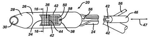
snare is then activated to excise the tissue. Both approaches are commonly
preceded by
injecting saline or other solutions under the mucosal to raise the lesion away
from the
CA 02482572 2012-08-17
-2-
underlying muscle wall in an effort to limit perforation. This lesion, common
in the art, is
known as a "bleb".
[0006] UK Patent Application GB 2365340A to Appleyard and Swain discloses a
tissue
resection device for removing tissue with a cavity of variable volume.
[0007] Other devices and methods have been proposed for providing resection of
tissue. Still,
scientists and engineers continue to seek improved methods for the resection
of tissue in
the gastro-intestinal tract.
[0008] Summary of the Invention
[0009] The present invention provides an apparatus which can employ suction to
engage
mucosal tissue for resection. In contrast to some existing devices which use
suction for
endoscopic mucosal resection, the suction chamber of the present device can
open
laterally, or on the side of apparatus corresponding to the long axis of the
endoscope.
Accordingly, the present invention can employ 'a suction opening which extends
generally parallel to the long axis of the endoscope. Existing devices which
employ an
opening which is at the distal end of the device have the plane of the suction
opening
being substantially perpendicular to the long axis of the endoscope.
[0010] Once tissue is drawn into the resection chamber, an electrosurgical
wire can be used for
transection. In contrast to the flexible electrosurgical snares used in
existing devices, the
present invention can employ a relatively rigid wire positioned within the
device to be
drawn across or pushed across the chamber opeing to excise the entrapped
tissue. The
wire is only electrically active over the portion, which is exposed, non
insulated, to the
chamber opening. The present invention can also include a flexible,
electrically
conductive tissue stop, which can function to limit the depth of tissue that
can enter the
suction chamber for resection. Such a tissue stop can provide for greater
safety of
resection by reducing risk of alimentary canal perforation and reducing
patient burns
CA 02482572 2004-09-27
-3
from monopolar ground pads. The tissue stop can also be perforated for
communicating
vacuum.
[0011] In one embodiment, the present invention provides a medical apparatus
comprising a
body with an outer surface having a side opening, the side opening for
receiving tissue
therethrough; a cutter adapted to receive energy for cutting tissue, the
cutter disposed
inward of the opening and adapted to traverse a length of the side opening for
cutting
tissue extending through the side opening; and a tissue stop disposed inward
of the side
opening and the cutter, the tissue stop having at least one opening
therethrough for
conveying vacuum to draw tissue through the side opening. The tissue stop can
comprise
a plurality of openings therethrough for conveying vacuum.
[0012] in another embodiment, the present invention provides a method
comprising the steps of
providing a source of vacuum; positioning, a perforated tissue stop in the
gastro-intestinal
tract; drawing tissue against the perforated tissue stop in the gastro-
intesinal tract; and
cutting a tissue sample from the tissue drawn against the perforated tissue
stop.
[0013] In another embodiment, the present invention provides a medical
apparatus comprising:
an overtube for receiving an endoscope therein, the overtube comprising a side
opening
for receiving tissue therethrough; and a tissue sample device disposed in the
overtube, the
tissue sample device comprising a tissue cutter adapted to traverse a length
of the side
opening for severing a tissue sample from tissue extending into the side
opening.
[0014] In another embodiment, the present invention provides a method for
obtaining a tissue
sample comprising: providing an endoscope; providing an overtube having a side
opening and a tissue cutter; inserting the overtube into a patient's body with
the
endoscope; receiving tissue into the side opening of the overtube; and cutting
tissue
extending into the side opening with the tissue cutter.
[0015] In another embodiment, the present invention provides a medical
apparatus comprising:
an outer surface having a side opening, the side opening for receiving tissue
therethrough;
a cutter adapted to receive RF energy for cutting tissue, the cutter supported
inward of the
CA 02482572 2012-08-17
-4-
side opening and adapted to traverse a length of the side opening for cutting
tissue
extending through the side opening; and a tissue stop disposed inward of the
cutter;
wherein the tissue stop comprises a pole of the RF circuit.
[0016] In another embodiment, the present invention provides a method of
cutting tissue
comprising the steps of: positioning an RF cutting device in the gastro-
intestinal tract of
a patient; positioning a tissue stop in the gastro-intestial tract;
positioning a tissue mass
against the tissue stop; energizing the RF cutting device; grounding the
tissue stop; and
cutting a tissue sample from the tissue mass.
[0016a] In a further aspect, there is provided a medical apparatus comprising:
an overtube for receiving an endoscope therein, the overtube comprising a side
opening for receiving tissue therethrough; and
a tissue sample device disposed in the overtube, the tissue sample device
comprising a tissue cutter adapted to traverse a length of the side opening
for severing a
tissue sample from tissue extending into the side opening and a deformable
tissue stop
disposed inwardly of the side opening;
wherein the tissue stop is deformable from a first position to permit passage
of
an endoscope thereby, to a second position to receive and support a tissue
mass drawn
through the side opening.
[0016b] In a further aspect, there is provided use of the medical apparatus
described herein for
obtaining a tissue sample.
[0017] Brief Description of the Figures
[0018] FIGURE 1 is a perspective view of a cutting device, showing a cutter
support attached
to a distal end of an endoscope, and features internal to the cutter support.
[0019] FIGURE 2 is a cross-sectioned end view of the cutter support of FIG. 1,
taken along
section line 16-16, showing a circular embodiment of the cutter support and
its internal
features.
CA 02482572 2012-08-17
-4a-
[0020] FIGURE 3 is a plan view of an alternative cutting element.
[00211 FIGURE 4 is a perspective view of an alternative cutting device,
showing a flexible
overtube slidable along and rotatable about an endoscope.
[0022] FIGURE 5 is a cross-sectioned end view of the flexible overtube of FIG.
4, taken along
section line 18-18, showing a circular embodiment of the cutter support and
its internal
features.
[0023] FIGURE 6 is a cross-sectioned end view similar to FIG. 5, showing
internal features in
a different position by virtue of a tissue bleb sucked into an aperture in the
overtube.
CA 02482572 2004-09-27
-5-
[0024] FIGURE 7 is a cross-sectioned top plan view of the cutter support of
FIG. 1, taken along
section line 17-17 of FIG. 2, showing a cutting mechanism extended forward of
an
aperture in the cutter support.
[0025] FIGURE 8 is a cross-sectioned side elevation view of the cutter support
of FIG. 1,
sectioned through the longitudinal axis thereof, showing a perpendicular view
of the
features of FIG. 7.
[0026] FIGURE 9 is a cross-sectioned side elevation view similar to FIG. 8,
showing a cutting
mechanism retracted rearwardly of the aperture into a shear slot.
[00271 FIGURE 10, is a cross-sectioned side elevation view similar to FIG. 8,
with the addition
of tissue shown adjacent the aperture, and a saline solution injection needle
extended to
enter the tissue to form a bleb.
[0028] FIGURE 11 is a cross-sectioned side elevation view similar to FIG. 10,
showing the
tissue bleb sucked into the aperture and against a stop plate, and the
injection needle
retracted.
[0029] FIGURE 12 is a cross-sectioned side elevation view similar to FIG. 11,
showing a cutting
element being retracted to cut through a first portion: of a bleb, wherein
mucosal and sub-
mucosal tissue are cut from muscularis tissue.
[0030] FIGURE 13 is a cross-sectioned side elevation view similar to FIG. 12,
showing
completion of cutting while vacuum holds the mucosal and sub-mucosal tissue to
the
underside of the stop plate.
[0031] FIGURE 14 is a cross-sectioned side elevation view similar to FIG. 13,
showing the
removal of the cutter support from the muscularis tissue after the cut has
been completed.
[0032] FIGURE 15 is a schematic view showing a monopolar arrangement of the
present
invention.
[0033] FIGURE 16 is a schematic view showing a bipolar arrangement of the
present invention.
CA 02482572 2004-09-27
{
-6-
[0034] FIGURE 17 is a schematic perspective illustration of a device of the
present invention
comprising a tissue stop having a foil conductor with rectangular openings
thererin, and
showing the tissue stop in an outwardly bowed, generally arcuate
configuration.
[0035] FIGURE 18 is a schematic perspective illustration of the device of
Figure 17 showing the
tissue stop deflected to a second configuration, such as by application of
vacuum, to
receive tissue and to permit passage of an endoscope thereby.
[0036] FIGURE 19 is a schematic illustration an end view of one embodiment of
the device of
the present invention having an overtube that has a flattened or oval non
circular cross-
section, and depicting a tissue stop plate in first and second configurations,
with the
second configuration shown in phantom.
[0037] FIGURE 20 is a schematic illustration of an embodiment of the device of
the present
invention including a transparent overtube, a transparent sleeve, and a
perforated stop
plate.
[0038] FIGURE 21 is a schematic illustration of an embodiment of the device of
the present
invention including a tissue receiving aperture having serrated side edges.
[0039J FIGURES 22A-22F illustrate various wire cutter configurations.
CA 02482572 2004-09-27
-7-
[0040] Detailed Description of the Invention
[0041] With reference to FIGS. 1, 2, 7 & 8, one embodiment of a cutting device
20 of the
present invention is shown attached to a distal end 22 of a commercially
available
endoscope. Endoscope 24 may be made by Olympus Optical, having an outside
diameter
of about 0.2 to 0.7 inches. Cutting device 20 can have a rigid or semi-rigid
cylindrical
cutter support 26 which is attached to the endoscope perimeter by any suitable
means,
such as by shrink wrap, adhesive, snap fit, press fit, threaded engagement, or
other
suitable means known in the art for connecting one generally hollow member to
another
along parallel longitudinal axes.
[0042] Distal end 22 of endoscope 24 can be located atone end of cutter
support 26. A flexible
conical member 28 can be attached to the opposite, distal end of cutter
support 26.
Conical member 28 can be employed to provide fora smooth entry of cutting
device 20
into the alimentary canal of a patient. Conical member 28 can have an open
distal end 30
of about 0.3 inches in diameter through which tooling, not shown, from a
working
channel 32 of endoscope 24 may extend, and through which unobstructed camera
vision
of the inside of the patient's alimentary canal is obtained. Conical member 28
can have
an open distal end 30 which permits passage of the distal end of the endoscope
24
therethrough.
[0043] Conical member 28 can be made of a flexible polymer, such as
polyvinylchloride (PVC),
polyethylene terephthalate (PET), or other suitable flexible materials.
Conical member
28 can be attached to cutter support 26 by threading it thereon, polymer
welding, press
fit, snap-fit, or other means well known in the art. Conical member 28 can be
coaxial
with cutter support 26, whereas a longitudinal axis of endoscope 24 can be
offset from a
longitudinal axis of cutter support 26.
CA 02482572 2004-09-27
-8-
[0044] Cutter support 26 can be generally cylindrical in shape, and can have
an outer diameter of
between about 0.50 and 0.75 inch, and an axial length of between about 1.0 and
about
1.50 inch. In one'embodiment, cutter support 26 can have an outer diameter of
about
0.60 inches and an axial length of about 1.25 inches. Cutter support 26 can be
formed of
a transparent polymer, such as polycarbonate or PVC.
[0045] Cutter Support 26 also can employ a lateral tissue receiving aperture
34. Aperture 34 can
have any suitable shape, and in the embodiment shown is generally rectangular
when
viewed straight on, and is positioned along one side of the cutter support 26.
The lateral
tissue receiving aperture 34 can be about 0.60 to 1.00 inches long (as
measured parallel to
the axial length of the cutter support 26), and about 0.30 to 0.50 inches wide
(as
measured around the circumference of the outside surface of the cutter support
26).
[0046] A perforated tissue stop plate 36 can be disposed radially inward from
tissue receiving
aperture 34, to be positioned inward of tissue receiving aperture 34. Tissue
stop plate 36
can be injection molded to the inner wall of cutter support 26, or
alternatively, made
separately and otherwise fixedly attached to the inner wall of cutter support
26. Stop
plate 36 can be semi-rigid, and can be deformable. In one embodiment, stop
plate 36 can
be formed and attached to cutter; support 26 so that stop plate 36 can take on
a first
configuration (such as an outwardly bowed, generally arcuate configuration),
and a
second configuration at least a portion of the tissue stop plate is drawn or
otherwise
deformed or deflected inward (such as by vacuum) to receive tissue through the
aperture
34. Stop plate 36 can be, in whole or in part, transparent, and can be made of
or comprise
a conductive material. For instance, stop plate 36 can be formed of a polymer
or
biocompatible metal which is conductive, or a polymer having a conductive ink
applied
thereto, or can include a generally transparent base layer with a conductive
outer layer
having openings therethrough, such as in the form of a grid pattern.
[0047] In Figure 1, stop plate 36 is shown having a plurality of perforations
therethrough.
Perforations in stop plate 36 can be employed to provide openings through the
thickness
CA 02482572 2004-09-27
of the stop plate 36, and to communicate vacuum from a source of vacuum to
draw tissue
into the tissue receiving aperture 34. In one embodiment, the perforations in
the stop
plate 36 can be about 0.03 to 0.10 inches in diameter and spaced about 0.10 to
0.30
inches apart. While circular perforations are shown, other suitable shapes,
including
rectangular, square, elliptical, or oval shapes. can be employed.
[0048] Cutter support 26 can have a support 38 molded therein, which contains
rectangular wire
guide slots 40 (Figure 2) which can be located parallel to the long edges of
aperture 34
on opposite sides of aperture 34. Guide slots 40 can be disposed outward of
stop plate
36, and inward of aperture 34. Wire guide slots 40 are sized for wire
insulating sleeves
42 to slide longitudinally therein. Insulating sleeves 42 surround two wires
that extend
from a heating source to distal ends of slots 40 near conical member 28, where
they are
attached to a heatable (such as by RF energy) cutting element 44. Cutting
element 44
extends from the sleeves 42 across aperture 34. As the wires and sleeves 42
are moved
parallel to the longitudinal axis of cutter support 26 within slots 40,
cutting element 44
passes across aperture 34 and cuts tissue drawn into aperture 34.
[0049] Cutting element 44 can be in the form of a straight wire filament about
0.01 to about 0.04
inches diameter, a flat blade about 0.01 inches thick and 0.03 inches deep, a
braided wire
about 0.01 to about 0.04 inches in diameter, or other suitable tissue cutting
devices. Such
cutting element configurations can be about 0.50 inches wide to in order to
span aperture
34, and can be made of a material capable of being heated, such as by radio
frequency
(RF) energy. Suitable materials from which cutting element 44 can be formed
when used
with RF energy include electrically conductive materials including without
limitation,
steel, steel alloys, titanium, or titanium alloys..;
[0050] Cutting element 44 may be heated by a number of heating means including
conduction
and RF heating, which are commonly known in the endoscopic cutting art. Wire
sleeves
42 can be formed of electrical insulating material such as teflon and can be
about 0.03
inches in diameter. Electrically conducting wires and their sleeves 42 can
extend along
CA 02482572 2004-09-27
-10-
the outside of endoscope 24 to an insulated slide block 46. Block 46 can be is
slidably
attached to a handle located alongside-an endoscope operating handle. Sleeves
42 can be
slidably attached at multiple places to endoscope 24 along its length. Slide
block 46 can
be supported to move longitudinally according to arrow 47 in Figure 1, to
extend and
retract sleeves 42 along endoscope 24 and through wire guide slots 40 so that
cutting
element 44 may be moved past the entire length of aperture 34. Moving block 46
in a
distal direction moves cutting element 44 across the length of aperture 34 in
a distal
direction, while moving block 46 in a proximal direction moves cutting element
across
the length of aperture 34 in a proximal direction.
[0051] For RF heating embodiments, an RF generator can be connected to the
wires attached to
the cutting element via a switching mechanism to deliver a wattage range of
from about
to about 150 watts at a suitable frequency, such as a frequency of between
about 300
kiloHertz to 3 megaHertz, thereby rapidly heating cutting element 44 to a
temperature
from about 60 C to about 120 C whenever heating is desired. In one
embodiment, an
Erbe 300 brand generator can be used with the following settings in monopolar
or bipolar
mode: pure cut, 40 Watts.
100521 In an RF heating embodiment an RF grounding plate or pad is typically
located outside a
patient's body. However, in the present invention an RF grounding plate may be
located
within cutting device 20, for example, by forming tissue stop plate 36 of a
conducting
material, or disposing a conductor on using tissue stop plate 36 as a metal or
metallized
electrical grounding plane. FIG. 2 shows an attachment of a ground wire 48 to
the edge
of stop plate 36. Ground wire 48 extends along side endoscope 24 to a ground,
not
shown, attached to the RF generator. Accordingly, the cutting device 20 can
provide an
electrical configuration which cutting element 44 provides one pole, and the
tissue stop
plate 36 provides the other pole.
CA 02482572 2004-09-27
[0053] Support 38 for wire slots 40 can also include at one ors both ends of
the wire slots a
cutting element shear slot 50, into which cutting element 44 moves at the end
of a cutting
stroke in order to strip tissue from the cutting element: With shear slots 50
located at
both ends of aperture 34 (as illustrated in Figure 7), cutting may occur in
either direction,
pushing or pulling cutting element 44 through tissue. The sizes of shear slot
50 and
cutting element 44 can be selected such that any removed tissue will not be
allowed to
adhere to the cutting element 44 due to the wiping action of the elements. For
example, a
cutting element 44 having a diameter of about 0.020 inch and fitting within a
shear slot
50 with a clearance spacing of about 0.005 inch is suitable.
[0054] FIG. 3 shows one of many possible configurations of an alternative
cutting element 52,
which includes a pointed portion 54. One or more points may be employed to
"bite into"
or initiate contact with tissue and begin cutting without deflecting the
tissue out of the
path of the cutting element. Also, an angled or pointed cutting element allows
for slicing
tissue parallel to aperture 34 in a progressive fashion to reduce resistance
of cutting.,
Cutting element 44 may also have a modified surface to be roughened or
otherwise
textured such as by being sand blasted, bead blasted, and/or machined
roughened, which
roughened profile can be useful to improve cutting efficiency by biting into
the tissue to
be resected.
[0055] Figures 22A-22F show various wire cutter configurations. Figure 22A
illustrates a
rectangular wire for providing intial cutting across the full width of the
wire. Figure 22B
illustrates an angled cutting wire for initiating cutting at one corner of the
wire, and for
progressively engaging more tissue as the cutting wire is advanced along the
length of the
aperture 34. Figure 22C illustrates a multiple point wire for providing
multiple points of
contact with tissue. Figure 22D illustrates a single point or notch for
providing single
point contact upon initial tissue engagement. Figure 22E illustrates a
relatively sharp
single point cutter for relatively high initial current density and mechanical
penetration.
Figure 22F illustrates a wire cutter having a flattened (as opposed to
circular cross-
CA 02482572 2004-09-27
-12-
section) blade which can have a sharpened edge and points for cutting tissue
with or
without RF energy.
[0056] Serrated edges can provided along the perimeter of a tissue receiving
aperture. The
textured surface provided by serrated aperture edges can provide for better
gripping of
the tissue during cutting. Figure 21 illustrates a tissue receiving aperture
having serrated
edges.
[0057] In order to cut a mucosal layer of tissue from the alimentary canal of
a patient for
external study, the mucosal layer and sub-mucosal layers are typically
separated
somewhat from a muscularis layer of tissue by injecting a saline solution
between them.
This is commonly done by extending an injection needle through working channel
32 of
endoscope 24 to contact and penetrate the target tissue.
[0058] In one embodiment, the present invention can provide an improved device
and method
for injecting saline solution. In the embodiments shown in FIGS. 1, 2, and 7-
14, support
38 has secured therein a flexible sheath 56 for an injection needle 58. Sheath
56 can
extend along side endoscope 24 to a handle, not shown, which is operated.to
deliver
saline solution thru a hollow cable connected to injection needle 58. The
hollow cable
can he slidable within sheath 56 so that needle 58 may be extended beyond the
fixed end
of sheath 56 to engage mucosal tissue adjacent aperture 34. Sheath 56, which
can be
fixedly attached to cutter support 26, serves as a needle guide that is
supported on the
cutter support 26. Sheath 56 can enable the operator of the injection needle
to control its
position more accurately (in order to avoid penetrating the muscularis tissue)
than when a
needle and a sheath are operated through an endoscope's working channel.
[0059] Injection needle 58 can be used to deliver saline solution 60, as shown
in FIGS. 10-13,
through mucosal tissue 62 and sub-mucosal tissue 64 only. These softer tissues
separate
from stiffer muscularis tissue 66.when saline solution 60 is introduced. After
injection,
the needle is withdrawn from the tissue. Needle 58 and sheath 56 are shown in
FIG. 2 in
CA 02482572 2004-09-27
-13-
a retracted position extending through support 38 and angled toward aperture
34, in a
plane which generally bisects aperture 34 and is centered between wire slots
40.
[0060] Tissue is drawn into aperture 34 by means of vacuum from a vacuum
source, not shown,
external to the patient's body. A suitable vacuum source can provide a vacuum
of about
50 to 250 mm hg. Vacuum can be drawn through working channel 32 in endoscope
24.
Air is drawn from the patient's alimentary canal, causing the canal to close
down around
cutter support 26 and bring tissue layer 62 in contact with the side of cutter
support 26
where the tissue engages aperture 34. Vacuum communicated through the working
channel 32 of the endoscope 24 and then through the openings in the stop plate
36 draws
tissue layer 62 against stop plate 36 as air flows through the openings in the
stop plate 36
to the opposite side of stop plate 36 where the distal end 22 of endoscope 24
can be
positioned.
[0061] Although FIG. 2 shows a circular cross-section for cutter support 26, a
flattened oval or
other shape may enable an aperture to be wider for cutting a larger sample of
tissue.
Similarly, while aperture 34 is shown as a generally rectangular shaped
opening on a
cylindrical surface, other aperture shapes can be employed, including without
limitation
oval, circular, and polygonal.
[0062] FIGS. 4-6 illustrates an alternative embodiment of a cutting device 80
of the present
invention. In Figures 4-6, an endoscope is not fixedly attached to a cutting
device 80.
Instead, the cutting device 80 can comprise an overtube 86. The overtube can
slide
along an endoscope and rotate about the endoscope. Such an embodiment can
permit
closer access by the distal end of the endoscope to target tissue for
examination and/or
manipulation before or after mucosal tissue cutting. Alternatively, the
cutting device and
overtube can employ integral vacuum lines and visualization means (e.g. ccd
camera) so
that the cutting device and overtube can be used independently of an
endoscope.
[0063] In Figure 4, cutting device 80 is shown having a distal end 82 of a
commercially
available endoscope 84 extended therethrough. Endoscope 24 may be made by
Olympus
CA 02482572 2004-09-27
-14-
Optical, having an outside diameter of about .2 to .7 inches. Cutting device
80 has a
flexible cylindrical overtube 86 slidably disposed along the length of the
endoscope
perimeter along parallel longitudinal axes. Overtube 86 can be relatively
short and rigid,
or can be flexible enough to conform to the articulations of flexible
endoscope 84.
Overtube 86 can have has at a distal end a flexible conical member 88, which
provides
for a smooth entry of cutting device 80 into the alimentary canal of a
patient. Conical
member 88 can be made of a flexible polymer such as PVC, PET, etc., and it has
an open
outer end 90 about 0.3 inches in diameter. The opening in the outer end can
expand or
be enlarged upon application of force so that endoscope 84 may extend
therethrough.
Conical member 88 can also be made of flexible polymer, and can be integral
with
overtube 86, or attached to overtube 86, such as by threading it onto overtube
86, by
polymer welding, by snap-fit, or by other means. Conical member 88 cab be
coaxial
with overtube 86, whereas a longitudinal axis of endoscope 84 may be offset
from a
longitudinal axis of overtube 86, as shown in Figure 2. The flexibility of the
conical
member 88 allows distal advancement of the endoscope to deflect the open end
so that
the endoscope is able to pass through the open end of member 88.
[0064] Overtube 86 can have a smooth outer diameter of about 0.40 to about
0.80 inches and a
length of about 0.7 to 2.0 inches. The overtube 86 can be disposed at the
distal end of a
elongated, flexible tube or sleeve. In Figure 4, the proximal end of the
overtube 86 is
molded or otherwise connected a flexible sleeve for receiving an endoscopic
therethrough, which sleeve can be in the form of an elongated, corrugated
tubular portion
92. Alternatively, the tubular portion 92 can be generally smooth. Tubular
portion 92
can have an internal diameter sized to receive an endoscope therethrough, and
tubular
portion 92 can have a length at least about the length of the portion of the
endoscope
which is inserted into the patient. Corrugated portion 92 can have generally
the same
outside diameter as overtube 86. It may be connected to the overtube similar
to the
conical member, and shrink wrap material may be added at the connection to
seal the
corrugated portion to the overtube. In one embodiment, the flexible, elongated
CA 02482572 2004-09-27
15-
corrugated portion 92 can have a length of between about 2.7 feet and about
4.0 feet. In
one embodiment, the internal diameter of the tubular portion 92 can be greater
than 0.15
inch and less than about 0.85 inch, and more particularly between about 0.30
to about
0.75 inch.
[0065] Overtube 86 has a rectangular tissue receiving aperture 94 along one
side, which is about
0.80 inches long and about 0.40 inches wide. A flexible stop plate 96 can be
disposed
just inside aperture 94. Stop plate 96 can be fastened to the inner wall of
overtube 86 or
otherwise disposed in aperture 94 such that stop plate 96 is able to toggle
between (or
otherwise assume) two different configurations. Two opposite edges of the stop
plate 96
can be joined directly or indirectly along their lengths to the overtube 86,
while the two
opposite end edges of the stop plate can remain free and unconnected to other
portions of
the device to facilitate movement of the stop plate from one configuration to
another. In
one embodiment Stop plate 96 can have a width greater than a chord length
across the
overtube where stop plate 96 is mounted so that stop plate 96 is bowed (or
otherwise
deflected or deformed) in a generally arcuate fashion toward aperture 94 or
away from
aperture 94. In one embodiment, the flexible stop plate 96 is biased to bow
toward
aperture 94 to enable endoscope 84 to pass over it on an opposite side. In
such an
embodiment, stop plate 96 can be formed of a thin flexible material, such as
PVC, PET
or other flexible polymer. Stop plate 96 can have a thickness of less than
about .05
inches, and can extend longitudinally beyond both ends of aperture 94.
[0066] The outwardly facing surface of stop plate 96 can include a portion
which is conductive
and which can serve as a ground or other pole of a electrical cutting circuit.
In one
embodiment, stop plate 96 has a conductive ink applied to one surface (e.g.
the outwardly
facing surface) so that it may serve as a grounding plate for RF heating of a
cutting
element as described for cutting device 20. Alternatively, an electrically
conductive
surface may be co-extruded on the stop plate 96, or the stop plate may be made
of thin
bio-compatible metal.
CA 02482572 2004-09-27
-16-
[0067] Overtube 86 can have a support 98 molded therein, which contains
rectangular wire guide
slots 100 between stop plate 96 and aperture 94. Wire guide slots 100 are
sized for
insulating sleeves 102 to slide longitudinally therein, just outside the width
of aperture
94. Insulating sleeves 102 surround two wires that extend from an RF heating
source
(not shown) to distal ends of slots 100 near conical member 88, where they are
attached
to a heatable cutting element 104. Cutting element 104 extends from the
sleeves 102
across aperture 94. As wires and sleeves 102 are slid parallel to the
longitudinal axis of
overtube 86 within slots 100, cutting element 104 passes across aperture 94 in
order to
cut tissue of a patient drawn into aperture 94, similar to the operation of
cutting device
20. Cutting element 104 can be the same as that described for cutting element
44 or
cutting element 52 above.
[0068) Cutting element 104 may be heated by a number of heating means
including conduction
and RF heating, which are commonly known in the endoscopic cutting art. Wire
sleeves
102 are made of electrical insulating material such as Teflon, similar to
insulating sleeves
42, and they extend along the outside of endoscope 84 to an insulated slide
block, not
shown. The slide block, similar to slide block 46, can be slidably attached to
a handle
located alongside an endoscope operating handle, such that the slide block is
moved
longitudinally to extend and retract sleeves 102 along endoscope 84 and
through wire
guide slots 100 so that cutting element 104 may be moved past the entire
length of
aperture 94 in overtube 86.
[00691 The heating of cutting element 104 may be the same as or similar to
cutting element 44.
In an RF heating embodiment, an RF grounding surface may be located within
cutting
device 80, for example by using a conductive tissue stop plate 96.
Alternatively, a
grounding plate separate from the stop plate 96 can be employed, but outside
of the path
of endoscope 84, so that the endoscope may freely pass through the overtube. A
ground
wire can be attached to the separate ground plate or to the stop plate, and
the ground wire
extends to a grounded location outside of the patient.
CA 02482572 2004-09-27
-17-
10070] Supports 98 can also include, at each end of the wire slots 100,
cutting element shear
slots 110. The cutting element 104 can move into the shear slots 110 at the
end of a
cutting stroke in order to strip tissue from the cutting element. Such shear
slots 110 can
be the same as or similar to slots 50 of cutting device 20, and cutting may
occur in two
directions, either by pushing cutting element distally, or by pulling cutting
element 104
proximally, through tissue.
[0071] FIGS. 4-6 show that overtube 86 has secured in support 98 a flexible
sheath 116 for an
injection needle 118. Sheath 116 extends along side endoscope 84 inside
corrugated
portion 92 to deliver saline solution thru a hollow cable connected to
injection needle 58,
in a similar manner to sheath 56 and needle 58, to engage mucosal tissue
adjacent
aperture 94. Needle 118 and sheath 116 are shown in FIGS. 4 and 5 in a
retracted
position extending through support 98 and angled toward aperture 94, in a
plane centered
within aperture 94, and between wire slots 100 and the aperture.
[0072] As shown in FIG. 6, when cutting device 80 is slid along endoscope 84
to a position
where endoscope 84 no longer interferes with the toggling of stop plate 96,
tissue may be
drawn into aperture 94 by means of vacuum from a vacuum source, not shown,
external
to the patient's body. Vacuum is drawn through working channel 112 in
endoscope 84.
Air is drawn from the patient's alimentary canal, causing the canal to close
down around
overtube 86 and bring tissue 114 in contact with the side of overtube 86 where
the tissue
engages aperture 94. Vacuum draws tissue 114 against stop plate 96 and causes
stop
plate 96 to toggle away from, or-otherwise deflect or deform away from, the
aperture 94.
[0073] Although FIGS. 5 and 6 show a circular cross-section for overtube 86, a
flattened oval or
other shape may be used to permit an aperture to be wider for cutting a larger
sample of
tissue.
[0074] Cutting devices 20 and 80 are operated in a similar manner to remove a
tissue sample.
FIGS. 10-14 describe one method of using cutting device 20. FIG. 10 shows
typical
alimentary canal tissue, with mucosal layer 62 atop sub-mucosal layer 64 atop
muscularis
CA 02482572 2004-09-27
-18-
layer 66 brought into contact with aperture 34, by placement of the cutting
device against
the tissue or by a low level of vacuum from the endoscope working channel to
close the
alimentary canal wall against cutter support 26. In this position, needle 58
is extended
from sheath 56 by pushing a hollow cable through the sheath, as described
hereinbefore.
Saline solution 60 is then injected into the tissue through the needle,
preferably at a
depth where sub-mucosal tissue and muscularis are separable, as is commonly
understood
in the endoscopic mucosal tissue cutting art. An amount of solution 60 is
injected which
separates the layers sufficient for cutting layers 62 and 64 without cutting
layer 66.
[0075] FIG. 11 shows needle 58 withdrawn from the tissue and a higher level of
vacuum sucking
the tissue into aperture 34 and against stop plate 36. Cutting element 44 in
this particular
method, is shown extended to shear slot 50. RF energy is now delivered via
wires
surrounded by insulating sleeves 42 to cutting element 44, using conductive
stop plate 36
as a ground for the RF energy path. Wire 48 connects stop plate 36 to an
external
ground, not shown. Cutting is ready to begin as cutting element 44 is rapidly
heated to
the desired temperature by controlling the level of RF energy.
[00761 FIG. 12 shows slide block 46 being moved along arrow 120 to pull
cutting element 44
into tissue layers 62 and 64 and solution 60. Solution 60 can be drawn out by
the
vacuum, which vacuum can also be employed to secure the cut portion of tissue
layers
62 and 64 against stop plate 36.
[0077] FIG. 13 shows slide block 46 being moved further along arrow 120 to
complete the cut
and shear tissue off cutting element 44 by pulling the cutting element into
shear slot 50.
Severed layers of tissue 62 and 64 continue to be held against perforated stop
plate 36 by
vacuum from endoscope 24 located on the opposite side of the stop plate. RF
power can
then be switched off. In Figures 12 and 13, stop plate 36 is not shown as
being
deformable. However, it will be understood that stop plate 36 can be made to
be
deformable as described above.
CA 02482572 2004-09-27
-19-
[0078] FIG. 14 shows cutting device lifted away from the remaining layers of
tissue so that the
cutting device may be withdrawn from the patient to examine the cut sample of
tissue. A
relatively lower level of vacuum can be employed to hold the cut tissue
against the stop
plate. The endoscope and cutting device may be rotated to a position such that
the tissue
sample is held against the stop plate by gravity when the vacuum is turned
off.
Alternatively, the cutting element (with not RF power applied) can be moved
forward to
a position similar to that of FIG. 12 to hold the cut tissue against the stop
plate when the
endoscope and tissue support 26 are manipulated to withdraw them from the
patient. In
another alternative, the cut tissue can be released from the stop plate and
allowed to exit
the aperture. Then the endoscope and cutting device can be partially withdrawn
to where
a gripper may be extended from a working channel of the endoscope through open
distal
end 30 to grasp the cut sample of tissue.
[0079] Fig 15 shows a monopolar arrangement of one embodiment of the present
invention. The
electrocautery generator 200 supplies the RF energy via a ground connected to
the
ground pad 203 at the patient's skin. The RF energy path 205 is connected to
the RF
cutting element 44/104. The vacuum pump 201 communicates with the cutter
support
26/overtube 86 via a vacuum channel 204 which can be integral to the endoscope
84
[0080] Fig 16 shows a bipolar arrangement of another embodiment of the present
invention. The
electrocautery generator 200 supplies the RF energy via energy paths 205. One
polarity
of the RF energy path 205 is connected to the RF cutting element 44/104 and
the other
polarity is connected to the stop plate 36/96 . The vacuum pump 201 is
connected to the
support 26/overtube 86 via a vacuum channel 204 which can be integral to the
endoscope
84.
[0081] Figures 17 and 18 illustrate an emobidment of the present invention
wherein the overtube
86 and elongated portion 92 can be transparent, and wherein the tissue stop
plate 96 can
be formed of a thin, transparent flexible polymeric material with a conductive
grid 97
disposed on a surface of the tissue stop 96 facing the tissue recieiving
aperture 94. Grid
CA 02482572 2004-09-27
20-
97 can define grid openings 99, which are generally rectangular in Figures 17
and 18.
One or more openings 99 can be perforated for communicating vacuum
therethrough if
desired. Grid 97 can be formed of a suitable conductive material, such as a
conductive
metallic foil, or be painted or printed on with a conductive ink or coating.
The
conductive surface of the grid 97 can be between about, 2 and about 10 times
the
conductive surface area of the cutter 104, and in one embodiment the
conductive surface
area of grid 97 can be about 4 times the conductive surface area of cutter
104.
[0082] The tissue stop 96 in Figures 17 and 18 can take on a first
configuration in Figure 17
(which permits passage of an endoscope thereby), and-a second configuration
shown in
Figure 18 when vacuum is applied (such as through endoscope 84) for limiting
the
amount of tissue drawn into aperture 94. The longitudinally extending sides 95
of tissue
stop 96 can be fixed, such as by being joined to overtube 86. The proximal and
distal
ends of the tissue stop 96 can be unsupported and free to deform. The first
and second
configurations can be bowed, generally arcuate shapes, as shown in Figures 17
and 18.
In one embodiment, the tissue stop 96 does not stretch or elongate in taking
on the first
and second configurations, but instead "toggles" or "snaps-through" from one
configuration to the other.
[0083] A suitable tissue stop 96 can be formed from a section of a clear PET
angioplasty
balloon. The tissue stop 96 can be an arcuate segment cut from a generally
cylindrical
angioplasty balloon formed of PET. The arcuate segment can be cut from an
angioplasty
balloon cylinder having a diameter between about 10 and about 16 mm and a wall
thickness of about .001 to about .002 inch. One suitable angioplasty balloon
from which
tissue stop 96 can be formed is a 10mm diameter angioplasty balloon having a
wall
thickness of 0.002 inch ( 0.05 mm) available from Advanced Polymers of Salem,
NH.
An arcuate segment can be cut from the angioplasty balloon to form the clear
tissue stop
96. A thin metallic foil having a thickness of about 0.005 inch or less, such
as a steel foil
having a thickness of about 0.001 inch can then be applied to the surface of
the stop 96
facing tissue receiving aperture 94, such as with an adhesive. Prior to
attaching the foil to
CA 02482572 2004-09-27
-21-
the stop 96, the foil can be cut to form a series of openings therethrough to
provide the
grid 97 shown in Figures 17 and 18.
[0084] Figure 19 illustrates a cross-sectional view of an overtube 86 having a
noncircular cross-
section, with a generally flattened outer surface portion in which tissue
receiving aperture
94 is formed. An endoscope 84 is shown positioned in the overtube 86. The
generally
flattened outer surface portion is located on a bottom half of the overtube 86
as viewed
in Figure 19. Providing the tissue receiving aperture 94 in such a generally
flattened
surface portion can be useful in positioning the aperture 94 relative to
tissue to be
resected. Figure 19 also shows first and second configurations of tissue stop
96, with the
second configuration shown in phantom. In one embodiment, the overtube 86 can
be
formed in two shell-like halves, such as a generally semi-circular upper half
and a non-
circular lower half. The tissue stop 96 can be formed from a nonplanar,
arcuate section
of thin polymeric film material (such as a section of an angioplasty balloon
described
above), and the side edges of the arcuate tissue stop can be captured between
the upper
and lower halves of the overtube as the upper and lower halves are joined
together, such
as by adhesive or other suitable means. The proximal and distal ends of the
tissue stop 96
can remain free and unsupported so that the tissue stop can snap through,
toggle, or other
wise deflect from the first configuration to the second configuration.
[0085] Figure 20 illustrates an embodiment of the present invention having a
transparent
overtube 86 and transparent elongated sleeve portion 92. The tissue stop 96 is
generally
planar, with generally circular shaped vacuum openings therethrough. Figure 21
illustrates an embodiment of the present invention wherein the overtube 86 has
a tissue
receiving aperture having serrated side edges 93 for assisting in grasping and
cutting
tissue with the cutting element 104. Tissue stop 96 is omitted from Figure 21
for
purposes of clarity in illustrating the side edges of aperture 94.
CA 02482572 2012-08-17
-22-
[00861 While the present invention has been illustrated by description of
several embodiments,
numerous other variations, changes, and substitutions will occur to those
skilled in the
art. For instance, but without limitation, RF energy has been described as the
tissue
cutting method in the illustrated embodiments, but it will be understood that
other
tissue cutting modes, such as ultrasonic energy modes, mechanical cutting, and
other
methods could be employed in various embodiments of the present invention.
Moreover, the structure of each element associated with the present invention
can be
alternatively described as a means for providing the function performed by the
element.
Accordingly, it is intended that the invention be limited only by the spirit
and scope of
the appended claims.