Note: Descriptions are shown in the official language in which they were submitted.
CA 02482686 2004-10-13
WO 03/087833 PCT/EP03/04041
A marker for measuring liver cirrhosis
Field of the invention
The invention provides methods and kits to detect liver cirrhosis in mammals.
The diagnostic
test is based on the profiling and identification of diagnostic carbohydrates
present in a body
fluid such as blood serum.
Background of the invention
Most of the common causes of liver injury result in cirrhosis. Cirrhosis is
the destruction of
normal liver tissue that leaves non-functioning scar tissue surrounding areas
of functioning
liver tissue, accompanied with the formation of regenerative liver nodules In
the United States,
the most common cause of cirrhosis is alcohol abuse. Among people ages 45 to
65, cirrhosis
is the third most common cause of death, after heart disease and cancer. In
many parts of
Asia and Africa, chronic hepatitis is a major cause of cirrhosis. Many people
with mild cirrhosis
have no symptoms and appear to be well for years. Others are weak, have a poor
appetite,
feel sick, and lose weight. If bile flow is chronically obstructed, the person
has jaundice, itching,
and small yellow skin nodules, especially around the eyelids. Malnutrition
commonly results
from a poor appetite and the impaired absorption of fats and fat-soluble
vitamins caused by the
reduced production of bile salts. Occasionally, the person may cough up or
vomit large
amounts of blood because of bleeding from varicose veins at the lower end of
the oesophagus
(oesophageal varices). These enlarged blood vessels result from high blood
pressure in the
veins that run from the intestine to the liver. Such high blood pressure,
called portal
hypertension along with poor liver function, may also lead to fluid
accumulation in the abdomen
(ascites). Kidney failure and liver encephalopathy also may develop. Other
symptoms of long-
standing liver disease may develop, such as muscle wasting, redness of the
palms (palmar
erythema), a curling up of the fingers (Dupuytren's contracture of the palms),
small spiderlike
veins in the skin, breast enlargement in men (gynecomastia), salivary gland
enlargement in the
cheeks, hair loss, shrinking of the testes (testicular atrophy), abnormal
nerve function, both in
the the periphery (peripheral neuropathy) and in the central nervous system
At present no cure exists for cirrhosis. The treatment includes withdrawing
toxic agents such
as alcohol, receiving proper nutrition including supplemental vitamins, and
treating
complications as they arise. Liver transplantation is presently the only cure
and may help a
person with advanced cirrhosis. Moreover, the presence of cirrhosis increases
the risk to
develop hepatocellular carcinoma about 40-fold over the risk in the general
population and, in
an etiological background of chronic hepatitis and alcoholism, the development
of cirrhosis
multiplies the already increased risk of the patient to develop hepatocellular
carcinoma from
34.4 to 119-fold and from 2.4 to 22.4-fold, respectively (Kuper et al., 2001).
Usually a number
1
CONFIRMATION COPY
CA 02482686 2004-10-13
WO 03/087833 PCT/EP03/04041
of blood tests are performed to measure liver function and to help determine
the severity and
cause of cirrhosis. One of the most important factors indicative of liver
damage is bilirubin, a
red-yellow pigment that is normally metabolised in the liver and then excreted
in the urine. In
patients with hepatitis, the liver cannot process bilirubin, and blood levels
of this substance
rise, sometimes causing jaundice. The levels of certain liver enzymes can also
be indicative for
cirrhosis (e.g. aspartate and alanine aminotransferase levels and several
clotting enzymes).
However, results of these liver function tests often are normal because only a
small
percentage of functioning liver cells are needed to carry out essential
chemical functions. In
addition a number of imaging tests are used to diagnose possible cirrhosis and
its
complications. For example, an ultrasound scan may show that the liver is
enlarged and that
particular lesions such as regenerative nodules are present. Other, much more
costly, imaging
techniques are magnetic resonance imaging (MRI) and computed tomography (CT).
In most of
the patients presenting with some form of chronic liver disorder, liver biopsy
is performed to
assess the degree of fibrosis and to detect the presence of cirrhosis
(Fracanzani et al., 2001).
As liver biopsy is an invasive procedure, it is generally difficult to perform
it on a regular follow-
up basis in the normal clinical setting. A specific serum marker for the
detection of liver
cirrhosis could thus have a very significant impact on the gastroenterology
practice, in allowing
regular follow-up of chronic liver disease patients and in providing early
warning for the onset
of cirrhosis. In the particular case of chronic alcoholism, a serum marker for
cirrhosis could
provide an important argument to convince the patient to stop drinking.
In the art the application of the measurement of diagnostic glycans in
carbohydrate metabolism
diseases is described (W09219975). In the field of hepatic disorders it is
also known that
single glycosylation enzyme activities are changed in liver disorders. For
example an
increased activity of the enzyme UDP-N-acetyl-glucosamine:glycoprotein N-
acetylglucosaminyltransferase (GnTIII) is correlated with the progression of
liver disease
(Ishibashi et al., 1989), a finding that has recently been elaborated upon in
a diagnostic setting
(Mori et al., 1998). However, these assays are complicated by the HPLC
separation of the
products of the enzymatic reaction. Moreover, the stability of the enzyme in
serum in storage
conditions is unknown and the values obtained for serum GnTIII activity had
large overlaps
between cirrhosis and chronic hepatitis. Glycosylation differences have also
been studied on a
purified protein, serum transferrin, and these differences are used for the
detection of chronic
alcoholism (Matsumoto K. et al (1994) Clin. Chim. Acta 224(1): 1-8).
Alterations in the
carbohydrate moiety of single purified proteins have also been described in
human cirrhotic
ascitic fluid (Biou, D. et al (1987) Biochimica et Biophysica Acta 913, 308-
312. Methods for the
detection of liver diseases are described in patents EP0503886 and DE3838718.
However, the
latter patents deal with the quantification of simple carbohydrates (fucose)
in urine. In view of
the prior art there is currently no easily measurable, reliable serum marker
for the
2
CA 02482686 2004-10-13
WO 03/087833 PCT/EP03/04041
differentiation of liver cirrhosis from other hepatic disorders. In the
present invention we have
identified multiple parameters of diagnostic carbohydrates derived from the
pool of proteins
present in the serum of cirrhosis patients. In serum, a complex mixture of
glycosylated and
unglycosylated proteins is present which are derived from liver and plasma
cells.
Unexpectedly, we have found that (relative) amounts of diagnostic
carbohydrates present on a
mixture of glycoproteins, that are present in the total serum, serve as a
diagnostic marker for
the differentiation of liver cirrhosis patients from chronic hepatitis
patients and for the
differentiation of liver cirrhosis from other non-malign and malign hepatic
disorders. An
advantage of analysing the pool of total serum glycoproteins is that the
amount of work
required for sample preparation is reduced to the minimum. This allows the
analysis of
clinically relevant numbers of patients.
Brief description of figures and tables
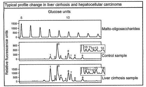
Fig. I Profile examples
The upper panel contains a dextran hydrolysate and can be used to assign a
glucose units
value to each peak. The second panel shows a typical electropherogram of the N-
glycans
derived from the proteins in a control serum sample. Nine peaks are clearly
visible in this
detection range and their height was used to obtain a numerical description of
the profiles of all
samples in this invention. The third panel shows the electropherogram obtained
from a
cirrhosis case. The extra peaks 10, 11, 12, 13 and 14 (see insert boxes) only
become visible in
the electropherogram after a ten-fold higher concentration of N-glycans
derived from the
proteins from the sera. Several profile alterations are evident and form the
basis for the
diagnostic marker.
Fig. 2 Boxplots summarizing the data.
The samples have been classified in 7 groups (Code 0 to 6) as shown in Table
1, fourth
column. For each of the 9 peaks, the median of the values in each of these 7
groups is
represented by the thicker black line and the interquartile ranges are
represented by the limits
of the boxes and errorbars. For peaks 1,2,3,7 and 8 the inner interquartile
ranges of sample
group 0 and 2 on the one hand and group I on the other hand do not overlap and
the relative
amount between peaks 1,2,7 and peak 8 was calculated to create three new
variables, the
properties of which are summarized in the right column of the figure. Note
that the ordinate
scale is logarithmic for these three new variables.
3
CA 02482686 2004-10-13
WO 03/087833 PCT/EP03/04041
Fig. 3 Evaluation of the diagnostic efficiency of the three variables using
Receiver
Operating Curve (ROC) analysis and binary logistic regression.
Section 1: ROC analysis was performed to evaluate the efficiency of the three
variables in
differentiating the sample group with cirrhosis and the group with chronic
viral hepatitis without
cirrhosis. The cut-off values determined from these ROC curves (optimal
combined sensitivity
and specificity) were used in the subsequent sections 2, 3, 4, 5 and 6 of this
figure to divide the
two-dimensional scatterplot fields in quadrants (the top right quadrant being
the positive
quadrant for liver cirrhosis, and the other three quadrants constituting the
negative area.) The
regression line obtained from binary logistic regression analysis with each
time two variables
as independents is also shown in these scatterplots. The cirrhosis sample
group is shown in
each scatterplot as black triangles. The 'negative' group in each section is
represented with
either circles or squares. Section 7: a comparison was made by ROC analysis of
the
classification efficiency between the sample group with cirrhosis and the
group with chronic
viral hepatitis without cirrhosis, for the variable Log(Peak 7/Peak 8) and
serum albumin
concentration and total serum bilirubin concentration. The result shows that
the serum N-
glycan profile-derived marker has an approximately 5-fold reduction in the
rate of misclassified
cases (approximately I in 25 versus approximately 1 in 4 (bilirubin) or 1 in 5
(albumin). Section
8: to validate the ROC-derived cut-off values for the serum N-glycan profile
derived markers,
these values were used to classify a second, independent group of chronic
hepatitis patients
with or without cirrhosis. Very similar classification efficiencies were
obtained in this second
group as were obtained in the optimization group (see Section 2 of the
figure).
Fig. 4 Partial structural analysis of the differentially regulated N-glycans.
The three columns in this figure represent the results of exoglycosidase array
sequencing on
the N-glycans derived from the glycoproteins in three serum samples. These
samples were
chosen to reflect the quantitative range of the observed alterations in this
study. The leftmost
sequencing column was obtained from analysis of a sample with chronic
hepatitis and is
indistinguishable from a healthy control's profile. The middle column
represents a mild
alteration, already trespassing the cut-off values for all three variables
described in the text.
The right column results from analysis of one of the worst affected samples.
It is useful to
compare the peaks described in the text over these three columns, and the
possibility for this
comparison greatly simplifies the peak tracking throughout the exoglycosidase
sequencing
panels. The peaks depicted in black do not bear a bisecting GIcNAc residue. In
this respect,
they can all be regarded as derivatives of the trim annosyl-GIcNAc2 core
oligosaccharide. The
peaks depicted in grey were found to be all modified with a bisecting GIcNAc
residue and thus
can all be considered as derivatives of the bisecting GIcNAc-substituted
trimannosyl-GIcNAc2
core oligosaccharide. The reference panel under the middle sequencing column
was
4
CA 02482686 2004-10-13
WO 03/087833 PCT/EP03/04041
assembled from 6 different electropherograms, each containing a specific
exoglycosidase
digest on reference glycans with known structure. The reference glycans used
were: 1) trisialo,
trigalacto triantennary; 2) bisialo, bigalacto biantennary with core-a-1,6-
linked fucose
(Reference panel under middle column) and 3) asialo, bigalacto biantennary
with core-a-1,6-
linked fucose and bisecting GIcNAc (Reference panel under rightmost column).
Fig. 5 Characteristics of the core-fucosylation variable.
To evaluate whether core-a-1,6-fucosylation was increased in the cirrhosis
group, a new
variable was created by summation of the normalised peak heights of all
identified peaks that
carry this modification. A boxplot visualisation analogous to those in Fig. 2
is shown, together
with the results of ANOVA and subsequent post hoc tests. Again, the cirrhosis
group is set
aside as a homogenous subgroup, thus confirming the increased core-
fucosylation in these
disorders.
Fig. 6 Discrimination between cirrhosis cases with and without hepatocellular
carcinoma.
Scatterplot of the cirrhosis cases with and without hepatocellular carcinoma,
plotting the height
of Peak 7 versus Peak 14 (see figure 1), normalized to the total measured peak
height in the
serum protein N-glycan profile. Cases with only cirrhosis are depicted as
empty triangles.
Cases with cirrhosis and HCC are depicted as black point-down triangles.
Binary logistic
regression allowed the identification of cases with HCC with a sensitivity of
71% and a
specificity of 90%. The logistic function of the model is: Z = -0.649[% Peak
7] + 5.722[% Peak
14] + 2.967. The diagonal line in the figure is the cut-off line where each
point on the line would
be a case with equal probability to belong to either of the two distinguished
groups.
Aims and detailed description of the invention
The study of complex glycans (glycobiology (Kobata, 2000; Roseman, 2001)) is
beginning to
evolve into a more general approach that might be called glycomics
(Hirabayashi et al., 2001;
Taniguchi et al., 2001). Analogously to proteomics, glycomics can be defined
as the study of
the realm of glycans present in all or a particular class of glycoconjugates
in a biological
sample on a quantitative basis and the comparison of the obtained profiles to
derive biological
information. On the basic research side, functional glyco(proteo)mics has the
ambition to
clarify the roles in a diverse range of physiological processes of the glycan
moieties of
glycoconjugates by detecting changes in the glycome and subsequently determine
the identity
of the non-glycan moieties (mostly proteins) that carry the altered glycans.
In this respect, it is
analogous in its approaches to basic proteomics research but the technologies
used are
CA 02482686 2009-12-17
29775-47
generally too complex and/or time-consuming to find applications outside the
basic research
laboratories. Especially in the clinical field, where analysis of hundreds of
samples is
necessary to derive meaningful information, the current glycomics approaches
have significant
shortcomings due to their complexity. In the present invention we have
developed a
technology platform for a clinical glycomics application in the detection of
liver cirrhosis. We
have profiled the carbohydrate structures derived from the glycoproteins
present in serum and
have identified statistically relevant differences in the glycan profiles
between patients suffering
from liver cirrhosis and patients free of liver cirrhosis. In other words,
amounts of diagnostic
carbohydrates or relative amounts between said carbohydrates have been
identified in the
present invention that are correlated with the presence of liver cirrhosis.
The profiling of
carbohydrates used in the diagnostic test for liver cirrhosis of the present
invention can for
example be carried out with an Applied Biosystems 377 gel-based DNA-sequencer
(Callewaert et al., 2001).
Thus, with the diagnostic method of the present invention it is possible to
reliably differentiate
patients with liver cirrhosis and liver cirrhosis complicated with
hepatocellular carcinoma from
(1) healthy donors, (2) patients suffering from chronic hepatitis B or C, (3)
patients suffering
from rheumatoid disorders, (4) patients with a suspected chronic alcohol abuse
and (5)
patients with non-HCC metastases to the liver. In addition, the diagnostic
method of the
invention can also diagnose a predisposition or presence of hepatocellular
carcinoma (HCC) in
a background of liver cirrhosis. We were able to differentiate patients with
cirrhosis from the
five groups mentioned above with sensitivities and specificities that were
never below 90 %
and in the most relevant diagnosis (distinction between cirrhosis and healthy
volunteers and
the distinction between cirrhosis and patients suffering from chronic
hepatitis) the values for
sensitivity and specificity were higher than 95 %.
As there is currently no easily measurable and specific serum diagnostic
marker for cirrhosis,
the usage of this non-invasive diagnostic test can include the guidance of
therapeutic
decisions in the treatment of cirrhosis (currently only liver transplantation
is a cure), the closer
monitoring of cirrhosis-positive patients for the development of
hepatocellular carcinoma and
the evaluation of the extent of liver damage in a patient presenting with
symptoms of chronic
alcohol consumption. This serum marker for cirrhosis can also be used to non-
invasively
measure the rate of conversion to cirrhosis of patients chronically infected
with the hepatitis C
virus in vaccine clinical trials, as a reduced incidence and/or a retardation
of the onset of liver
cirrhosis constitutes an important clinical endpoint which is currently very
difficult to assess
because of the necessity of invasive biopsy techniques.
In a first embodiment, the invention provides a method to detect liver
cirrhosis in a mammal,
comprising (a) generating a profile of carbohydrates or fragments derived
thereof, or labeled
derivatives of said carbohydrates or said fragments, or features of said
carbohydrates or said
*Trade-mark
6
CA 02482686 2004-10-13
WO 03/087833 PCT/EP03/04041
carbohydrate fragments that are determined by the structure of said
carbohydrates or said
carbohydrate fragments; said carbohydrates or said fragments being present on
or obtained
from a mixture of glycoconjugates that are present in or are isolated from a
sample of a body
fluid from said mammal, and (b) measuring in the profile generated in step a)
the amount of at
least one carbohydrate or a fragment derived thereof or a labeled derivative
of said
carbohydrate or said fragment, or a feature of at least one carbohydrate or
fragment derived
thereof present in said carbohydrate profile, and (c) comparing the measured
data obtained in
step b) with measured data obtained from profiles derived from mammals free of
liver cirrhosis,
and (d) attributing the deviation obtained in step c) to liver cirrhosis. The
wording 'a method to
detect liver cirrhosis' can be broadly understood as a method for screening, a
method for
diagnosis or a method for prognosing liver cirrhosis.
In another embodiment a carbohydrate profile is used for the manufacture of a
diagnostic
assay for the detection of liver cirrhosis, said diagnostic assay comprises
the following steps
(a) generating a profile of carbohydrates or fragments derived thereof, or
labeled derivatives of
said carbohydrates or said fragments, or features of said carbohydrates or
said carbohydrate
fragments that are determined by the structure of said carbohydrates or said
carbohydrate
fragments; said carbohydrates or said fragments being present on or obtained
from a mixture
of glycoconjugates that are present in or are isolated from a sample of a body
fluid from said
mammal, and (b) measuring in the profile of step a) the amount of at least one
carbohydrate or
a fragment derived thereof or a labeled derivative of said carbohydrate or
said fragment, or a
feature of at least one carbohydrate or fragment derived thereof present in
said carbohydrate
profile, and (c) comparing the measured data obtained in step b) with measured
data obtained
from profiles derived from mammals free of liver cirrhosis, and (d)
attributing the deviation
obtained in step c) to liver cirrhosis.
In another embodiment the invention provides a method to detect liver
cirrhosis in a mammal,
comprising (a) generating a profile of carbohydrates or fragments derived
thereof, or labeled
derivatives of said carbohydrates or said fragments, or features of said
carbohydrates or said
carbohydrate fragments that are determined by the structure of said
carbohydrates or said
carbohydrate fragments; said carbohydrates or said fragments being present on
or obtained
from a mixture of glycoconjugates that are present in or are isolated from a
sample of a body
fluid from said mammal, and comparing quantitative or qualitative aspects of
said profile to the
quantitative or qualitative aspects of such a said profile obtained from one
or more individuals
of said mammalian species.
The wording 'glycoconjugates that are present in' refers to carbohydrates
which are detected
on the glycoconjugates without any isolation step of said carbohydrates; thus
the sample is
used as such and does not imply any isolation step of the carbohydrates,
whereas the wording
7
CA 02482686 2004-10-13
WO 03/087833 PCT/EP03/04041
'or are isolated from a sample of a body fluid' refers to the fact that the
carbohydrates are
isolated from the glycoconjugates present in the sample.
In a particular embodiment the method of the invention can be used for
monitoring the effect of
therapy administered to a mammal suffering from liver cirrhosis. In another
particular
embodiment the method of the invention specifically detects liver cirrhosis.
The term
'specifically' refers to the fact that liver cirrhosis can be diagnosed
differently from other hepatic
disorders comprising mammals suffering from a hepatitits B or C infection.
The term 'carbohydrate' can be understood as glycans that are present in the
structure of or
that are derived from glycoconjugates, comprising the glycan categories known
in the art as
asparagine-linked glycans (also designated as N-glycans) or Serine/Threonine-
linked glycans
(also designated as 0-glycans) of proteins or glycosaminoglycans or
proteoglycan derived
glycans, glycans present in or derived from glycolipids and GPI-anchor derived
carbohydrates.
The words "glycan" and "carbohydrate" are interchangeable. A 'glycoconjugate'
means any
compound (e.g. protein or lipid) containing a carbohydrate moiety. With the
wording 'a mixture
of glycoconjugates', it is meant a composition containing at least two (at
least three, at least
four, at least five or more) of said glycoconjugates, potentially also
comprising non-
glycoconjugate materials such as proteins, lipids, salts and water. The
wording 'carbohydrates
or fragments derived thereof' means that carbohydrates can be fragmented to
yield at least
one oligosaccharide or a derivative thereof amongst the products of the
fragmentation process.
Other products of this fragmentation process might include monosaccharides and
oligosaccharides or derivatives thereof. An oligosaccharide is a carbohydrate
of which the
chemical structure consists of at least two chemically linked units known in
the art as
monosaccharide. The said fragmentation process can involve enzymatic, chemical
and
physical treatments. For example, carbohydrates can be treated (or digested)
with a
glycosidase enzyme (e.g. a sialidase to remove the sialic acid residues from
the
carbohydrates, or a fucosidase to remove fucose residues from the
carbohydrates) and
therefore the profile obtained consists of fragments of the carbohydrates.
Glycosidase
digestions can for example be carried out to obtain a more simple profile of
the carbohydrates.
Sialic acids may also be removed in a chemical way by mild acid hydrolysis of
the
carbohydrates. In mass spectrometric analysis methods, the word 'fragments'
refers to the fact
that carbohydrates are very often fragmented in the process of analysis (for
example in
collision induced dissociation), in which case the fragmentation products can
also yield an
oligosaccharide derivative made up of an oligosaccharide chemically linked to
the remnant of
one or more monosaccharides that were part of the structure of the
carbohydrate before
fragmentation took place. An example of such an oligosaccharide derivative
being the product
of a mass spectrometric fragmentation process is known in the art as a cross-
ring cleavage
product ion. A 'feature of said carbohydrate' refers to any measurable
parameter of which the
8
CA 02482686 2004-10-13
WO 03/087833 PCT/EP03/04041
properties and/or the quantity is determined by the structure of the
carbohydrate. Examples of
such measurable parameters are for example nuclear magnetic resonance
parameters such
as chemical shifts, homonuclear and heteronuclear coupling constants, Nuclear
Overhauser
Effects and residual dipolar couplings. Alternatively, such measurable
parameters might be the
extent of binding to said carbohydrate to other molecules such as lectins and
antibodies that
recognize specific structural determinants or combinations thereof in the
carbohydrate. Yet
other such measurable parameters might be the extent of the capacity of the
carbohydrate to
function as a substrate for an enzyme that specifically modifies certain
carbohydrates such as
glycosyltransferases and glycosidases.
The wording `said carbohydrates or said fragments being present on or obtained
from a
mixture of glycoconjugates' refers to the fact that a `profile of
carbohydrates of fragments
derived thereof or labeled derivatives of said carbohydrates or said
fragments, or features of
said carbohydrates or said carbohydrate fragments that are determined by the
structure of said
carbohydrates or said carbohydrate fragments' can be either obtained from
carbohydrates that
are still chemically linked to the glycoconjugates in the mixture, or from
carbohydrates that
have been released from the glycoconjugates by enzymatic, chemical or physical
means. In a
preferred embodiment, N-glycans are released from the glycoproteins in the
mixture by
enzymatic digestion with Peptide N-glycosidase F or other endoglycosidases
known in the art.
In another embodiment, N-and 0-glycans can be released using a procedure
involving
hydrazine, known to those skilled in the art. In yet another embodiment, 0-
glycans can be
selectively released using beta elimination in alkaline conditions according
to well-known
procedures. In case the profile is obtained on carbohydrates that are still
chemically linked to
the glycoconjugates in the mixture, one embodiment involves the use of enzymes
or chemical
procedures to modify the non-glycan part of the glycoconjugate prior to
obtaining the profile,
such as proteases or enzymes which modify the lipid part of glycolipids. The
wording `a profile
of carbohydrates' means any entity comprising qualitative and/or quantitative
information on
said carbohydrates. For example, this may mean an electrophoretic or
chromatographic profile
of said carbohydrates. In a particular case the profile is a mass spectrum of
said
carbohydrates. Alternatively, the profile can be information obtained by
Nuclear Magnetic
Resonance analysis. In yet another example, the profile can be information
describing
qualitative or quantitative aspects of lectin binding to the carbohydrates.
Alternatively, the
profile can be information describing the extent to which the carbohydrates
are substrates for
specific enzymes such as glycosyltransferases or glycosidases. Such
information can include
read-outs of measurements of by-products of such enzymatic reactions, such as
nucleotides
set free in equimolar amounts in glycosyltransferase reactions. In a
particular embodiment the
wording 'generating a profile of carbohydrates' or 'profiling of
carbohydrates' also can imply
that the glycan structures are separated and subsequently detected. Usually a
number of
9
CA 02482686 2004-10-13
WO 03/087833 PCT/EP03/04041
carbohydrates are identified in a profile of carbohydrates. Usually the
carbohydrates are
present in a complex mixture and separation is necessary for an efficient
detection. Separation
can be carried out with methods comprising electrophoretic and chromatographic
methods.
Detection can be carried out with methods comprising antibody detection,
lectin detection,
NMR, mass spectrometry and fluorescence. In a particular embodiment it is
necessary to
chemically and/or enzymatically remove the glycans from the glycoproteins
before the glycans
can be profiled. Methods to prepare glycans from glycoproteins are well known
in the art. In
another particular embodiment it is necessary to derivatize the glycans before
the separation
and the detection. In one approach the method of the present invention for the
profiling
(includes separation and detection) of glycans can be carried out in
combination with a DNA-
sequencer. However, it is clear for the person skilled in the art that this
method can also be
applied in connection with capillary electrophoresis systems adaptable to a
laser induced
fluorescence detector. Such systems for instance include the P/ACE series
Capillary
Electrophoresis Systems (Beckman Instruments, Inc., Fullerton, Calif.). The
invention can also
be applied with any electrophoresis system which is adaptable with a laser
induced
fluorescence detector. In another embodiment also mass spectrometric detection
methods can
be used such as MALDI-TOF-MS for the measurement of the amount of at least one
carbohydrate or a fragment derived thereof. In mass spectrometric methods very
often the
carbohydrates are fragmented and therefore in said methods fragments of
carbohydrates are
detected.
In yet another embodiment the profiling can be carried out with a
microfluidics method.
Microfluidics is a rapidly growing field and is based on fluid migration
through narrow-bore
channels created in a solid medium (mostly silica wafers or high-purity glass
plates) via
techniques borrowed from the microchip industry (photolithography and chemical
wet etching).
Fluids can migrate through these channels via capillary action or active
pumping, and analytes
can migrate in fluid-filled channels through electrophoresis (Schmalzing et al
(2001) Methods
MoL Biol. 163, 163-173). In yet another embodiment the separation of
carbohydrates can be
carried out via a chromatographic separation with methods including thin layer
chromatography (TLC), high performance liquid chromatography or gas
chromatography.
The term 'at least one carbohydrate' refers to the measurement of the amount
of at least one
carbohydrate present in the carbohydrate profile that is diagnostically
relevant for the detection
of liver cirrhosis (said at least one carbohydrate can therefore be designated
as an at least one
diagnostic carbohydrate). In one embodiment the measurement of one
carbohydrate is
sufficient to diagnose liver cirrhosis. This means that in one particular case
one carbohydrate
is present in a mammal suffering from cirrhosis and is absent in a mammal free
of cirrhosis, in
another particular case one carbohydrate is present in a mammal free of
cirrhosis and absent
in a mammal suffering from cirrhosis. In another particular example a
different amount of one
CA 02482686 2004-10-13
WO 03/087833 PCT/EP03/04041
carbohydrate is sufficient to differentiate between a mammal suffering from
cirrhosis and a
mammal free of cirrhosis. In a preferred embodiment the amount of one, two or
even more
(diagnostic) carbohydrates is measured. In a profiling method the amount of
the (diagnostic)
carbohydrate can for example be measured by calculating the peak height or the
peak surface.
By comparing the amount of at least one (diagnostic) carbohydrate, present in
patient
samples, with corresponding diagnostic carbohydrate levels present in an
individual free of
liver cirrhosis, the presence or absence of liver cirrhosis can be diagnosed.
The invention can
be used on samples obtained from mammals such as humans. Diagnostic
carbohydrates may
be oligosccharides, or polysaccharides. Diagnostic carbohydrates may be
'branched or
unbranched. Diagnostic carbohydrates in a sample from an afflicted individual
with liver
cirrhosis are present with an abundance (amount) that is either consistently
higher or
consistently lower than in a sample from an unafflicted individual (not having
liver cirrhosis).
The term "labeled derivatives of said carbohydrates or said fragments" refers
to carbohydrates
that have been labeled with an agent that leads to an efficient detection of
the carbohydrates.
Said labeled carbohydrates are also called derivatized carbohydrates. As an
example, a
fluorescing compound can be used for the labelling of the carbohydrates. Said
fluorescing
compounds are also preferentially charged such that the derivatized compounds
can migrate
under electrophoretic conditions. When the fluorophore label is uncharged, it
can be coupled
with a charge imparting species. Said fluorophore label also permits the
quantitative
measurement of the derivatized carbohydrates by fluorescence. Fluorescing
compounds such
as 9-aminopyrene-1,4,6-trisulfonic acid (APTS) and 8-aminonaphthalene-1,3,6-
trisulfonic acid
(ANTS) are particularly suitable for electrophoretic separation of derivatized
carbohydrates.
Other compounds for fluorescent labelling of carbohydrates include 2-
aminopyridine (AP), 5-
aminonaphthalene-2-sulfonate (ANA), 1-amino-4-napthalene sulfonic acid (ANSA),
1-amino-
6,8-disulphonic acid (ANDA), 3-(4-carboxybenzoyl)-2-quinolinecarboxaldehyde
(CBQCA),
lucifer yellow, 2-aminoacridone and 4-aminobenzonitrile (ABN).
In a particular embodiment, regarding the detection of the fluorescently
labeled carbohydrates,
any detection method known in the art can be applied, but preferably the
detection is carried
out with a laser such as a diode laser, a He/Cd laser or an argon-ion laser.
In a particular
embodiment, the profile of labeled carbohydrate bands produced by the
electrophoretic
separation is visualized using an imaging system based on a charge-coupled
device (CCD)
camera. Information from the CCD camera may subsequently be stored in digital
form and
analyzed by various computer programs for comparing diagnostic carbohydrate
patterns
between individuals and between reference standards. In another particular
embodiment the
gel separated diagnostic carbohydrates may be transferred to an immobilizing
membrane, i.e.,
blotted and then probed with various diagnostic carbohydrate-specific reagents
such as lectins
or monoclonal or polyclonal antibodies specific for said diagnostic
carbohydrates. In a specific
11
CA 02482686 2004-10-13
WO 03/087833 PCT/EP03/04041
embodiment the invention provides a method to detect liver cirrhosis in a
mammal comprising
measuring and detecting at least one glycan structure and/or glycoconjugate
that has a
different abundance in samples derived from individuals with and without
cirrhosis by using
ligands that specifically bind to said at least one glycan structure and/or
glycoconjugate.
Ligands comprise lectins and antibodies. For example, the increased abundance
of the N-
glycan structures (or their conjugates) with a 'bisecting GIcNAc' residue (GnT-
III product) in a
body fluid sample can be detected with a lectin that specifically recognizes
glycans (or their
conjugates) that are modified with a bisecting GIcNAc, such as the erythro-
agglutinating lectin
from Phaseolus vulgaris (E-PHA). Alternatively, the increased abundance of the
N-glycan
structures with a 'bisecting GIcNAc' residue (or their conjugates) can be
detected by a
reduction in the binding to the N-glycans (or their conjugates) to lectins
that only bind N-
glycans (or their conjugates) if they are not substituted with a bisecting
GIcNAc residue. An
example of such a lectin is the lectin from Canavalia ensiformis (Con A). The
observed
undergalactosylation of the serum glycoprotein N-glycans can be detected by a
terminal-
GIcNAc binding lectin such as the Griffonia simplicifolia II (GS-II) lectin.
Alternatively, the
undergalactosylation can be measured by a reduction in the binding of a
terminal-galactose
binding lectin such as the lectin from Erythrina crystagelli.
In a particular embodiment, the 'profile of a feature determined by the
structure of the
carbohydrates' is obtained by measuring the property of the carbohydrates that
is constituted
by being a substrate for a specific glycosyltransferase. In a preferred
embodiment, this
glycosyltransferase is beta- 1,4-g alactosyltransferase and the carbohydrates
are those present
on the total mixture of serum or plasma proteins. An additional substrate for
this reaction is
UDP-Galactose, and the reaction yields UDP in a stoechiometric amount. Thus,
the profile can
be obtained by measuring the difference between the extent of galactosylation
of the
desialylated proteins before and after the reaction, for example by a method
involving binding
of the glycoproteins to a lectin specific for terminal beta-galactose (such as
the lectins known
in the art derived from Ricinus communis and from Erythrina crystagalli, or
the galectins such
as the one derived from Coprinus cinereus). Alternatively, the profile can be
obtained by
measuring the amount of UDP generated in the beta-l,4-galactosyltransferase
reaction on the
mixture of serum or plasma proteins, for example by HPLC. The amount of UDP
can also be
measured using a coupled enzyme reaction with one or more enzymes known from
nucleotide
metabolism, such as for example a nucleotide diphosphatase such as the yeast
Golgi
GDPase, which also shows significant hydrolytic activity towards UDP. In this
latter case, the
profile can be obtained by measuring either UMP or phosphate, using well-known
techniques.
Still another example of a measurement of UDP involves the use of
supramolecular membrane
pores with differential affinity for UDP-Gal and UDP, as known in the art. The
profiles thus
obtained can for example be standardized for the total amount of protein or
carbohydrate
12
CA 02482686 2004-10-13
WO 03/087833 PCT/EP03/04041
present in the serum or plasma sample. In yet another embodiment, the profile
can be
obtained by using the carbohydrates present on the mixture of serum or plasma
proteins as
substrate for both beta- l,4-galactosyltransferase and a sialyltransferase,
with UDP-Galactose
and CMP-N-acetylneuraminic acid as sugar donor substrates. In this embodiment,
the profile
can either consist of the difference in binding of a sialic-acid binding
lectin (such as the lectin
well known in the art derived from Maackia amurensis or Sambucus nigra) to the
glycoproteins
before and after the reaction, or can consist of measuring the amount of UDP
and/or CMP
released during the reaction, using methods known in the art.
In another embodiment the carbohydrate profiling method can be supplemented
pre-
electrophoretically with one or more internal standards labeled with a
chromophore or
fluorophore different from the label attached to the carbohydrate analytes.
The internal
standard allows for accurate and reproducible determination of the
electrophoretic mobilities of
the derivatized carbohydrate by referencing these mobilities to the mobilities
of the
components in the internal standard mixture. For example, a rhodamine-labeled
oligonucleotide standard GenescanTM 500 (Applied Biosystems, Foster City, CA,
USA) or a
mixture of rhodamine-labeled 6-,18-,30-,and 42-meric oligonucleotides may be
added to the
derivatised glycans before profiling. Diagnostics standards may be labeled
prior to the labeling
of the samples for analysis; however diagnostic standards are preferably
labeled concomitantly
with the labeling for the standards for analysis. Furthermore, the diagnostic
carbohydrates in
the standards are preferably quantitated so as to provide for quantitative or
qualitative
comparisons with the amount of diagnostic carbohydrates in the samples for
analysis.
The term `body fluid' includes blood, blood serum, blood plasma, saliva,
urine, bone marrow
fluid, cerebrospinal fluid, synovial fluid, lymphatic fluid, amniotic fluid,
nipple aspiration fluid and
the like. Preferred body fluids for analysis are those that are conveniently
obtained from
patients, particularly preferred body fluids include blood serum and blood
plasma.
Although the present invention can be carried out without pre-treatment of the
sample prior to
the profiling of the (derivatized) glycans, in a particular embodiment,
samples for analysis may
require processing prior to the separation and quantification of the
diagnostic carbohydrates.
The precise method of sample processing employed may vary in accordance with a
number of
factors attributable to the choice of sample fluid and the identity of the
diagnostic
carbohydrates; these factors include: the abundance of the diagnostic
carbohydrate, the
concentration of background carbohydrates, the presence of interfering
molecules, for
example, molecules that adversely affect diagnostic carbohydrate band mobility
or the
fluorescent labeling of the diagnostic carbohydrates, and whether the
fluorescent label has to
be separated from the derivatized diagnostic carbohydrates. Suitable methods
for this
processing or pre-treatment of samples include: dialysis, to remove
interfering molecules (e.g.
salt for an efficient mass spectrometric detection); ultrafiltration, to
concentrate diagnostic
13
CA 02482686 2004-10-13
WO 03/087833 PCT/EP03/04041
carbohydrates and remove interfer,ng molecules; centrifugation, to remove
interfering
particulates or concentrate cells; precipitation, to remove interfering
molecules, removal of
albumin from the serum to enrich for glycosylated proteins and hence for lower
abundance
glycans, deglycosylation with a glycosidase (e.g. a sialidase digest of the
glycans) to generate
a more simple glycan profile; chromatography such as affinity chromatography
to remove for
example albumin from the serum
In yet another embodiment the invention provides a method to detect liver
cirrhosis in a
mammal, said method comprising (a) generating a profile of carbohydrates or
fragments
derived thereof, or labeled derivatives of said carbohydrates or said
fragments, or features of
said carbohydrates or said carbohydrate fragments that are determined by the
structure of said
carbohydrates or said carbohydrate fragments; said carbohydrates or said
fragments being
present on or obtained from a mixture of glycoconjugates that are present in
or are derived
from a sample of a body fluid from said mammal and (b) measuring the relative
amount of at
least one carbohydrate or a fragment derived thereof or a labeled derivative
of said
carbohydrate or said fragment, present in said carbohydrate profile. The term
`measuring the
relative amount' refers to the aspect that the amount of at least one
carbohydrate or fragment
(e.g. one particular carbohydrate or fragment) can be measured between two
profiles, one
profile being derived from a mammal free of liver cirrhosis and another
profile derived from a
mammal possibly suffering from liver cirrhosis and to be diagnosed for liver
cirrhosis.
Alternatively, the amount of one particular carbohydrate can be compared
between an average
reference range taken from mammals free of liver cirrhosis and the measured
amount of said
particular carbohydrate in a mammal to be diagnosed for liver cirrhosis. In
yet another
embodiment the `measuring of the relative amount' refers to measuring the
relative amount of
at least two carbohydrates or fragments derived thereof or labelled
derivatives of said
carbohydrates or said fragments, or features of said carbohydrates or said
carbohydrate
fragments present in one carbohydrate profile derived from a sample of a body
fluid from an
animal.
In another embodiment of the invention, in order to be able to measure
relative amounts of the
carbohydrates, diagnostic standards are included on the gels used to analyze
the diagnostic
carbohydrates in the subject samples; however, the information embodied by the
diagnostic
standard, e.g., band migration distance and intensity, may also be obtained
from comparison
with stored records made from diagnostic standards previously subjected to
fluorophore
assisted carbohydrate electrophoresis under conditions similar to the
conditions to which the
samples for analysis are exposed. Diagnostic standards may be both positive,
i.e.,
corresponding to the complete carbohydrate pattern in an afflicted individual,
or negative, i.e.,
corresponding to unafflicted individual. Diagnostic standards may have a
composition similar to
that of samples for analysis in that they may contain both diagnostic
carbohydrates and
14
CA 02482686 2004-10-13
WO 03/087833 PCT/EP03/04041
background carbohydrates with composition similar to that found in actual
samples. Diagnostic
standards may be derived from samples obtained from afflicted and non-
afflicted individuals.
Alternatively, diagnostic standards may contain one or more diagnostic
carbohydrates free of
background carbohydrates.
In a particular embodiment the diagnostic technique to measure liver cirrhosis
does not require
an a priori detailed knowledge of the structure of the carbohydrates.
In another particular embodiment the diagnostic technique to measure liver
cirrhosis uses the
knowledge of the structure of the carbohydrates. The results of the structural
analysis of the
differentially regulated glycans can be summarized as an increased abundance
of N-
acetylglucosaminyltransferase III products (bisecting GIcNAc, glycan
structures of peaks 2, 4
and 7 depicted in figure 1), a decreased galactosylation of the biantennary
glycans (increased
intensity of glycan structures of peaks 1 and 2 depicted in figure 1), and a
decrease in the
abundance of the bi-and triantennary fully galactosylated glycan structures
(glycan structures
of peaks 3 and 8 depicted in figure 1).
In another embodiment the invention provides a method for the detection of
liver cirrhosis in a
mammal, said method comprising generating a profile of carbohydrates or
fragments derived
thereof, or labeled derivatives of said carbohydrates or said fragments, or
features of said
carbohydrates or said carbohydrate fragments that are determined by the
structure of said
carbohydrates or said carbohydrate fragments; said carbohydrates or said
fragments being
present on or obtained from a mixture of glycoconjugates that are present in
or are derived
from a sample of a body fluid from said mammal, and measuring the amount of at
least one
carbohydrate or a fragment derived thereof or a labeled derivative of said
carbohydrate or said
fragment, or a feature of at least one carbohydrate or fragment derived
thereof present in said
carbohydrate profile, wherein said at least one carbohydrate is selected from
the group
consisting of:
i) GIcNAc(R-1,2)Man(a-1,3)[GIcNAc([3-1,2)Man(a-1,6)]Man([3-1,4)GIcNAc((3-
1,4)[Fuc(a-1, 6)]GIcNAc (glycan 1),
ii) GIcNAc([3-1, 2)Man(a-1, 3)[GIcNAc([i-1,4)][GIcNAc([3-1, 2)Man(a-1,
6)]Man((3-
1,4)GIcNAc((3-1,4)[Fuc(a-1,6)]GIcNAc (glycan 2),
iii) Gal([3-1,4)GIcNAc(R-1, 2)Man(a-1, 3)[Gal((3-1,4)GIcNAc([i-1, 2)Man(a-
1,6)]Man([3-
1,4)GIcNAc(R-1,4)GIcNAc (glycan 3),
iv) Gal([3-1,4)GIcNAc((3-1,2)Man(a-1,3)[GIcNAc([i-1,4)][Gal((3-1,4)GIcNAc((3-
1,2)Man(a-
1,6)]Man((3-1,4)GIcNAc([3-1,4)[Fuc(a-1,6)]GIcNAc (glycan 7),
v) Gal([3-1,4)GIcNAc((3-1,2)[Gal(R-1,4)GIcNAc([3-1,4)]Man(a-1,3)[Gal([3-
1,4)GIcNAc(R-
1,2)Man(a-1,6)]Man((3-1,4)GIcNAc([3-1,4)GIcNAc (glycan 8),
vi) a fragment derived of glycan 1, 2, 3, 7 or 8,
CA 02482686 2004-10-13
WO 03/087833 PCT/EP03/04041
vii) a sialylated derivative of glycan 1,2, 3, 7 or 8,
viii) a feature of glycan 1, 2, 3, 7 or 8 or derivative or fragment thereof.
For the sake of clarity the structures of the peaks 1, 2, 3, 7 and 8
correspond with the
carbohydrate profile depicted in figure 1 and with the graphic representation
of these structures
in Figure 4. Said carbohydrate profile is a desialylated profile (without
sialic acid on the
glycans), meaning that the structures of peaks 1, 2, 3, 7 and 8 are strictly
spoken carbohydrate
fragments (missing the sialic acid structures). The carbohydrates are herein
presented with the
IUPAC rules for nomenclature
(httr)://www.chem.gmul.ac.uk/iupac/2carb/38.html), the peaks
according to figure 1 have been identified in the present invention and are
represented by their
condensed and extended nomenclature. In the claims the condensed nomenclature
is used.
The name of the four structures is summarized here below.
Desialylated glycan structure of peak 1 from figure 1:
Condensed nomenclature:
GIcNAc((3-1,2)Man(a-1,3)[GIcNAc((3-1,2)Man(a-1,6)]Man((3-1,4)GIcNAc((3 -
1,4)[Fuc(a-
1, 6)]GIcNAc
Extended nomenclature:
(3-D-GlcpNAc-(1->2)-a-D-Manp-(1-),3)-[[3-D-GlcpNAc-(1-*2)-a-D-Manp-(1->6)]-(3-
D-Manp-
(1-->4)-(3-D-GIcpNAc-(1 ->4)-[a-L-Fucp-(1-).6)]-D-GlcpNAc
Desialylated glycan structure of peak 2 from figure 1:
Condensed nomenclature:
GIcNAc((3-1, 2)Man(a-1, 3)[GIcNAc((3-1,4)][GIcNAc((3-1, 2)Man(a-1, 6)]Man((3-
1,4)GIcNAc(R-
I , 4)[Fuc(a-1, 6)]G IcNAc
Extended nomenclature:
3-D-GIcpNAc-(1-a2)-a-D-Manp-(1-->3)-[(3-D-GIcpNAc-(1-*4)][ 3-D-GIcpNAc-(1-*2)-
a-D-Manp-
(1-->6)]-[3-D-Manp-(1--*4)-[3-D-GIcpNAc-(1-a4)-[a-L-Fucp-(1- >6)]-D-GIcpNAc
Desialylated glycan structure of peak 3 from figure 1:
Condensed nomenclature:
Gal((3-1,4)GIcNAc((3-1,2)Man(a-1,3)[Gal([3-1,4)GIcNAc(R-1,2)Man(a-1,6)]Man((3-
1,4)GIcNAc(f3-
1,4)GIcNAc
Extended nomenclature:
R-D-G alp-(1- *4)-[3-D-GIcpNAc-(1 -->2)-a-D-Manp-(1-->3)-[R-D-Galp- (1-->4)-R-
D-GIcpNAc-(1-32)-
a-D-Manp-(1--*6)]-[3-D-Manp-(1 ->4)-[3-D-GIcpNAc-(1--34)-D-GIcpNAc
Desialylated glycan structure of peak 7 from figure 1:
Condensed nomenclature:
Gal([3-1,4)GIcNAc((3-1,2)Man(a-1,3)[GIcNAc((3-1,4)][Gal((3-1,4)GIcNAc((3-
1,2)Man(a-
1,6)]Man((3-1,4)GIcNAc((3-1,4)[Fuc(a-1,6)]GIcNAc
16
CA 02482686 2004-10-13
WO 03/087833 PCT/EP03/04041
Extended nomenclature:
[3-D-Galp-(1 ->4)-[3-D-GIcpNAc-(1 ->2)-a-D-Manp-(1->3)-[R-D-GIcpNAc-(1--
>4)][(3-D-Galp-
(1-4)-(3-D-GIcpNAc-(1-*2)-a-D-Manp-(1->6)]-[i-D-Manp-(1-4)-(3-D-GIcpNAc-(1-*4)-
[a-L-
Fucp-(1 ->6)]-D-GIcpNAc
Desialylated glycan structure of peak 8 from figure 1:
Condensed nomenclature:
Gal([3-1,4)GIcNAc((3-1,2)[Gal((3-1,4)GIcNAc(13-1,4)]Man(a-1,3)[Gal([3-
1,4)GIcNAc(R-1,2)Man(a-
1,6)]Man((3-1,4)GIcNAc((3-1,4)GIcNAc
Extended nomenclature:
[3-D-Galp-(1-*4)-(3-D-GIcpNAc-(1-->2)-[(3-D-Galp-(1-*4)-(3-D-GIcpNAc-(1->4)]-a-
D-Manp-
(1-),3)-[(3-D-Galp-(1 --- 4)-[3-D-GIcpNAc-(1-*2)-a-D-Manp-(1-*6)]-[3-D-Manp-(1-
4)-(3-D-
GlcpNAc-(1-->4)-D-GIcpNAc
In another embodiment the invention provides a method to detect liver
cirrhosis comprising the
steps of (a) generating a profile of carbohydrates or fragments derived
thereof, or labeled
derivatives of said carbohydrates or said fragments, or features of said
carbohydrates or said
carbohydrate fragments that are determined by the structure of said
carbohydrates or said
carbohydrate fragments; said carbohydrates or said fragments being present on
or obtained
from a mixture of glycoconjugates that are present in or are derived from a
sample of a body
fluid from said mammal and (b) measuring the relative amount the glycan
structure 1 or a
fragment thereof and the glycan structure 8 or a fragment thereof and/or the
glycan structure 2
or a fragment thereof and the glycan structure 8 or a fragment thereof and/or
the glycan
structure 7 or a fragment thereof and the glycan structure 8 or a fragment
thereof and/or the
glycan structure 1 or a fragment thereof and the glycan structure 3 or a
fragment thereof
and/or glycan structure 2 or a fragment thereof and the glycan structure 3 or
a fragment
thereof and/or the glycan structure 7 or a fragment thereof and the glycan
structure 3 or a
fragment thereof.
The average peak heights for glycan structures 1, 2, 7 and 8 were calculated
in the different
patient groups. The average relative amounts between these glycan structures
for the cirrhosis
group (n = 37) are: peakl/peak 8: 2.444, peak2/peak 8: 0.590 and peak?/peak 8:
1.479.
Relative amounts for the healthy control group (n=60) are peak 1/peak 8:
0.809, peak 2/peak
8: 0.081 and peak 7/peak 8: 0.7234. This means that a sample is diagnosed as
having
cirrhosis when the relative amounts between peak 1/peak 8 is 3.02 times higher
than the
average within this healthy population and/or when the relative amounts
between peak 2/peak
8 is 7.28 times higher than the average within this healthy population and/or
when the relative
amounts between peak 7/peak 8 is 2.04 times higher than the average within
this healthy
population.
17
CA 02482686 2004-10-13
WO 03/087833 PCT/EP03/04041
Relative amounts between these glycan structures for the chronic hepatitis
group (n = 27) are:
peakl/peak 8: 1.21, peak2/peak 8: 0.25 and peak?/peak 8: 0.95. This means that
a sample is
diagnosed as having cirrhosis when the relative amounts between peak 1/peak 8
is 2.01 times
higher than the average within this chronic hepatitis group and/or when the
relative amounts
between peak 2/peak 8 is 2.36 times higher than the average within this
chronic hepatitis
group and/or when the relative amounts between peak 7/peak 8 is 1.56 times
higher than the
average within this chronic hepatitis group.
Relative amounts between these glycan structures for the complete control
population (n =
153, consisting of healthy individuals, individuals suffering from chronic
hepatitis and
individuals suffering from chronic alcoholism) are: peakl/peak 8: 0.98,
peak2/peak 8: 0.115
and peak7/peak 8: 0.87. This means that a sample is diagnosed as having
cirrhosis when the
relative amounts between peak 1/peak 8 is 2.49 times higher than the average
within this
complete control population and/or when the relative amounts between peak
2/peak 8 is 5.13
times higher than the average within this complete control population and/or
when the relative
amounts between peak 7/peak 8 is 1.7 times higher than the average within this
complete
control population.
Thus in another embodiment, when the ratio (relative amounts) between peak
1/peak 8 is
higher than at least 20 %, at least 30 %, at least 40 %, at least 50 %, at
least 60%, at least 70
% or at least 80 % of the relative amount of the average within the control
population, a sample
is diagnosed as being derived from a mammal suffering from liver cirrhosis.
In a specific embodiment, the invention provides a method to detect liver
cirrhosis comprising
the steps of (a) generating a profile of carbohydrates or fragments derived
thereof, or labeled
derivatives of said carbohydrates or said fragments, or features of said
carbohydrates or said
carbohydrate fragments that are determined by the structure of said
carbohydrates or said
carbohydrate fragments; said carbohydrates or said fragments being present on
or obtained
from a mixture of glycoconjugates that are present in or are derived from a
sample of a body
fluid from said mammal, and (b) measuring the relative amount of the glycan
structure 1 or a
fragment thereof and the glycan structure 8 and wherein said relative amount
between said
glycan structures or fragments thereof is at least 80% higher than the average
of said relative
amount in mammals free of liver cirrhosis.
In another embodiment, when the relative amount between peak 2/peak 8 is
higher than at
least 20 %, at least 30 %, at least 40 %, at least 50 %, at least 60%, at
least 70 %, at least 80
%, at least 90 % or at least 100 % of the relative amount of the average
within the control
population, a sample is diagnosed as being derived from a mammal suffering
from liver
cirrhosis.
In a specific embodiment, the invention provides a method to detect liver
cirrhosis comprising
the steps of (a) generating a profile of carbohydrates or fragments derived
thereof, or labeled
18
CA 02482686 2004-10-13
WO 03/087833 PCT/EP03/04041
derivatives of said carbohydrates or said fragments, or features of said
carbohydrates or said
carbohydrate fragments that are determined by the structure of said
carbohydrates or said
carbohydrate fragments; said carbohydrates or said fragments being present on
or obtained
from a mixture of glycoconjugates that are present in or are derived from a
sample of a body
fluid from said mammal, and (b) measuring the relative amount of the glycan
structure 2 or a
fragment thereof and the glycan structure 8 and wherein said relative amount
between said
glycan structures or fragments thereof is at least 100% higher than the
average of said relative
amount in mammals free of liver cirrhosis.
In yet another embodiment when the relative amount between peak 7/peak 8 is
higher than at
least 20 %, at least 30 % or at least 40 % of the relative amount of the
average within the
control population, a sample is diagnosed as being derived from a mammal
suffering from liver
cirrhosis.
In another specific embodiment, the invention provides a method to detect
liver cirrhosis
comprising the steps of (a) generating a profile of carbohydrates or fragments
derived thereof,
or labeled derivatives of said carbohydrates or said fragments, or features of
said
carbohydrates or said carbohydrate fragments that are determined by the
structure of said
carbohydrates or said carbohydrate fragments; said carbohydrates or said
fragments being
present on or obtained from a mixture of glycoconjugates that are present in
or are derived
from a sample of a body fluid from said mammal, and (b) measuring the relative
amount of the
glycan structure 7 or a fragment thereof and the glycan structure 8 and
wherein said relative
amount between said glycan structures or fragments thereof is at least 40%
higher than the
average of said relative amount in mammals free of liver cirrhosis.
In another embodiment, the invention also includes a kit for performing
diagnosis of liver
cirrhosis. For example a kit can be made for performing fluorophore assisted
carbohydrate
electrophoresis diagnosis of liver cirrhosis. As another example a kit can be
made for
performing mass spectrometric diagnosis of liver cirrhosis. Fluorophore
assisted carbohydrate
electrophoresis diagnosis kits provide collections of reagents required for
performing the
diagnosis of liver cirrhosis. Suitable kits enable laboratories to
conveniently perform
fluorophore assisted carbohydrate electrophoresis diagnosis. Kits may include
reagents for
performing tests to identify liver cirrhosis. Kits may include diagnostic
standards, fluorescent
label, blotting and binding materials, e.g., membranes, carbohydrate specific
binding reagents,
lectins, instructions, sample containers, and polyacrylamide gel reagents,
precast gels,
enzyme buffers, reducing agents (for use in the fluorophore labelling of
carbohydrates), and
glycosidase enzymes (e.g. sialidase, galactosidase, fucosidase) capable of
catalyzing
reactions that structurally alter diagnostic carbohydrates. More complete kits
may include
equipment for performing fluorophore assisted carbohydrate electrophoresis,
such as
19
CA 02482686 2004-10-13
WO 03/087833 PCT/EP03/04041
polyacrylamide gel apparatus, CCDs, laser, DNA sequencer, computers, software,
and the
like. Reagents included in fluorophore assisted carbohydrate electrophoresis
diagnosis kits are
preferably provided in pre-measured amounts. The kits preferably include the
instructions for
carrying out the fluorophore assisted carbohydrate electrophoresis method of
the present
invention.
The diagnostic test is useful in practice because it is sufficiently easy to
apply on a large scale
by normally trained laboratory staff. Furthermore, since electrophoresis-based
high-resolution
and high-sensitivity analysers for DNA sequencing and mutation detection are
already present
in a rapidly increasing number of clinical laboratories or are affordable for
most clinical
laboratories, the novel diagnostic glycomics test for liver cirrhosis can be
run on them.
Moreover, the available range of DNA-analysers allows for the sample
throughput to be easily
scaled from just a few to hundreds of samples per day per machine, depending
on the demand
of each laboratory. This DNA-analysis equipment offers the added advantage of
automation,
reducing the complexity of the overall analytical process. The profiling on
the total mixture of
glycoproteins increases the tolerance of the test for small inter-individual
variations of the
abundance and the glycosylation pattern of each individual glycoprotein in the
mixture and
thus allows more robust testing than the current classical approaches where
the glycosylation
is studied on purified glycoproteins.
In another embodiment the method for the detection of liver cirrhosis further
comprises clinical
chemistry parameters and/or histological data. Thus, the present invention can
also be
conveniently carried out in combination with clinical chemistry parameters
and/or histology
and/or imaging parameters. Measurement of clinical chemistry parameters
comprises
measurement of levels of bilirubin, albumin, prothrombin time, C-reactive
protein, IgA
abundance, serum hyaluronic acid concentration, aminotransferases and several
liver
metabolism test known in the art. Histology comprises liver biopsies. Imaging
comprises
ultrasound, CT-scan, MRI-scan and imaging of radioactive tracers specific for
the liver.
In yet another embodiment the invention provides a method to detect the
presence or the
predisposition of hepatocellular carcinoma in a mammal suffering from liver
cirrhosis
comprising a) generating a profile of carbohydrates or fragments derived
thereof, or labeled
derivatives of said carbohydrates or said fragments, or features of said
carbohydrates or said
carbohydrate fragments that are determined by the structure of said
carbohydrates or said
carbohydrate fragments; said carbohydrates or said fragments being present on
or obtained
from a mixture of glycoconjugates that are present in or are isolated from a
sample of a body
fluid from said mammal, and b) measuring in the profile of step a) the amount
of at least one
carbohydrate or a fragment derived thereof or a labeled derivative of said
carbohydrate or said
fragment, or a feature of at least one carbohydrate or fragment derived
thereof present in said
CA 02482686 2011-06-17
29775-47
carbohydrate profile, and c) comparing the measured data obtained in step b)
with
measured data obtained from profiles derived from mammals suffering from liver
cirrhosis but free of hepatocellular carcinoma, and d) attributing the
deviation
obtained in step c) to the presence of hepatocellular carcinoma. In yet
another
embodiment in the method to detect the presence or the predisposition of
hepatocellular carcinoma in step c) a two-parameter analysis is carried out
with
glycans 7 and 14 derived from a human serum carbohydrate profile. In yet
another
embodiment said two-parameter analysis is a two-parameter binary logistic
regression analysis.
According to one aspect of the present invention, there is provided a method
for
obtaining a diagnostic indicator of liver cirrhosis in a mammal, comprising:
a)
generating a profile of: total asparagine-linked carbohydrates or fragments
derived
thereof, from a pool of total glycoproteins that are present in or are
isolated from a
sample of a body fluid from said mammal, labeled derivatives of said total
asparagine-linked carbohydrates or said fragments, or features of said total
asparagine-linked carbohydrates or said fragments wherein said features are
determined by the structure of said asparagine-linked carbohydrates or said
fragments; b) measuring in the profile of step a) the amount present in said
asparagine-linked carbohydrate profile of: at least one asparagine-linked
carbohydrate or a fragment derived thereof, a labeled derivative of said
asparagine-
linked carbohydrate or said fragment, or a feature of said at least one
asparagine-
linked carbohydrate or said fragment; c) comparing the measured data obtained
in
step b) with said same measured data obtained from other profiles derived from
other
mammals of the same species suffering from a hepatic disorder but free of
liver
cirrhosis; wherein a deviation obtained from the comparison in step c)
indicates that
the at least one asparagine-linked carbohydrate or fragment, the labeled
derivative,
or the feature, is a diagnostic indicator of liver cirrhosis.
According to another aspect of the present invention, there is provided a
method for
obtaining an indicator that a mammal suffering from liver cirrhosis has, or is
21
CA 02482686 2011-06-17
29775-47
predisposed to having, hepatocellular carcinoma, the method comprising: a)
generating a profile of: total asparagine-linked carbohydrates or fragments
derived
thereof, from a pool of total glycoproteins that are present in or are
isolated from a
sample of a body fluid from said mammal; labeled derivatives of said total
asparagine-linked carbohydrates or said fragments, or features of said total
asparagine-linked carbohydrates or said fragments wherein said features are
determined by the structure of said asparagine-linked carbohydrates or said
fragments; b) measuring in the profile of step a) the amount present in said
asparagine-linked carbohydrate profile of: at least one asparagine-linked
carbohydrate or a fragment derived thereof, a labeled derivative of said at
least one
asparagine-linked carbohydrate or said fragment, or a feature of said at least
one
asparagine-linked carbohydrate or said fragment; c) comparing the measured
data
obtained in step b) with said same measured data obtained from other profiles
derived from other mammals of the same species suffering from liver cirrhosis
but
free of hepatocellular carcinoma; wherein a deviation obtained from the
comparison
in step c) indicates that the at least one asparagine-linked carbohydrate or
fragment,
the labeled derivative, or the feature, is an indicator of the presence of, or
the
predisposition to, hepatocellular carcinoma in a mammal suffering from liver
cirrhosis
21a
CA 02482686 2009-12-17
29775-47
The examples which follow are offered as descriptive of certain embodiments.
As such they
are exemplary only and are not limiting in their nature.
Examples
Data collection and glycomic serum profile characteristics
Profiles of the N-glycan pool present on the complete collection of proteins
present in the 214
human sera were obtained, starting from 5 p1 serum, without any pre-treatment.
The Applied
Biosystems 377 DNA-sequencer was used for this study. The peak height of 14
peaks was
quantified in every analysed serum sample, accounting for >99% of the total
observed signal
intensity. We limit the discussion here to the 9 peaks in the mobility range
of 8 to 12 glucose
units (Fig. 1). Their intensity is sufficiently high to allow easy routine
quantitation.
Statistical processing
Staying true to the 'omics' setup of our study, we approached data analysis in
a purely
statistical way, without bias towards the identity of the measured peaks.
In Fig. 2, an overview is given of the data characteristics (median and
interquartile ranges) for
these 9 peaks over 7 sample groups. Early in the analysis, we observed that a
very similar
profile change occurred in samples from patients suffering from hepatocellular
carcinoma and
from patients with liver cirrhosis (the general characteristics of this
profile change are evident
from comparison of the ,2 lower panels in Fig. 1). Since all of the patients
with hepatocellular
carcinoma in this study also had liver cirrhosis (16), we decided to regard
these samples as
one group (n=37, group I in Table I) in the further statistical analysis,
designated as the
cirrhosis group. Group 0 consists of the 60 control samples from Red Cross
blood donors. All
samples from patients with chronic hepatitis B or C, without cirrhosis, were
taken together in
group 2 (n=27). Group 3 consists of 8 samples from patients with non-HCC
metastases to the,
liver. In group 4, all samples were assembled from patients with suspected
chronic alcohol
abuse and a positive carbohydrate deficient transferrin (CDT) test result. The
glycosylation
degree of serum transferrin is responsive to recent (2-3 weeks) alcohol intake
and is currently
21b
CA 02482686 2004-10-13
WO 03/087833 PCT/EP03/04041
the best serum marker for the detection of chronic alcohol abuse (Anton, 2001;
Wuyts et aL,
2001). It is obvious that the presence of CDT could influence the
glycosylation profile of total
serum glycoprotein measured here. For comparison purposes, group 5 consists of
samples
(n=33) of chronic alcohol abusers with a negative CDT. Group 6, finally,
consists of samples
from patients with auto-immune disorders (n=24). Glycosylation changes
(especially
undergalactosylation and increased presence of a-1,6-linked core fucose) of
IgG have been
well documented in rheumatoid arthritis and ankylosing spondylitis (Martin et
al., 2001;
Watson et al., 1999) and could also influence the glycan pattern of total
serum glycoprotein, as
IgG is a very abundant serum protein.
From the data in Fig. 2, it is apparent that in the cirrhosis group, peaks 1,2
and 7 are
upregulated and peaks 3 and 8 are down-regulated sufficiently so that the
inner interquartile
range of their values for the cirrhosis group do not overlap with the inner
interquartile ranges of
the control group (Group 0). Moreover, these changes are highly correlated, as
shown by
Spearman correlation analysis (Table 2). Therefore, we created new variables
by scaling the
upregulated peaks 1,2 and 7 to the down-regulated peak 8 and subsequently log-
transformed
these new variables to normalize the distributions. An analysis that took the
differential amount
of Peak 4 into account did not improve the classification efficiency (see
below) and neither did
incorporation of the downregulation of Peak 3. The values for the resulting
three new variables
are summarized in the right panel of Fig. 2. To detect significant differences
in the means of
the values for these variables between the 7 sample groups, one-way analysis
of variance was
performed, followed by two different multiple comparison tests (Tukey's
Honestly Significant
Difference test and Scheffe's comparison) to pinpoint the specific intergroup
differences. At a
familywise error rate of 0.0001, for all three variables, the Cirrhosis group
was significantly
different from all other sample groups (Table 3). The classification
efficiency of this difference
was evaluated with non-parametric Receiver Operating Curve (ROC) analysis and
with binary
logistic regression. For optimal clinical relevance and after consultation
with the collaborating
hepatologist (Dr. H. Van Vlierberghe, UZGent), these classification algorithms
were used in a
comparison of the Cirrhosis group and the group of patients with chronic
hepatitis B or C. In a
typical gastro-enterology clinical setting, the detection of cirrhosis is the
most relevant in
patients with chronic hepatitis (and in patients with chronic alcohol abuse).
The group of
samples from patients with chronic alcohol abuse that was at our disposition
was insufficiently
well characterized as to the presence of cirrhosis to include this collection
in this cut-off value
determination. The results of the ROC analysis are shown in Fig. 3.1, and
allow very
satisfactory classification efficiencies of around 95%. The cut-off values
obtained from these
ROC curves (value at which highest combined sensitivity and specificity is
attained) are
represented as the lines dividing the two-dimensional scatterplots in the rest
of Fig. 3 in
quadrants. The skewed line dividing the scatterplots in two halves are the
regression lines
22
CA 02482686 2004-10-13
WO 03/087833 PCT/EP03/04041
calculated from a linear binary logistic regression model for each two
variables. It is instructive
to compare the efficiency of the calculated cut-off values and regression
lines in the distinction
between the Cirrhosis group and all the other sample groups (Fig. 3.2-6). It
is a general
observation that the 'double ROC' classification has a higher specificity and
a lower sensitivity
than the regression approach. In the distinction of the Cirrhosis group and
the chronic hepatitis
group, sensitivity and specificity are both in the range of 90-98%. As can be
seen in Fig. 3.3,
the sample group of healthy Red Cross blood donors has no (for left and right
plot) or only 1
(on 60, for the center plot) false positives for the double ROC classification
and no (for right
plot) or 1 (center and left) false positives for the regression approach. This
close to 100%
specificity at around 95% sensitivity is very similar to the values obtained
in the distinction with
chronic hepatitis and indicates again that the measured variables indicate a
pathological
change that is specific for cirrhosis and does not change significantly with
the onset of chronic
hepatitis (as can also be derived from ANOVA results). A particular
observation is that there
are more false positives in the sample group from chronic alcoholics (Fig.
3.4) in regression
analyses that incorporate the parameter Log(Peak 7/Peak 8). However, double
ROC
classification with one of the other two parameters still keeps these cases
out of the 'positive
area'. In the group of auto-immune diseases (Fig. 3.5), the false positives
were mainly due to
increased values of Log(Peak 1/Peak 8) in 25% of the cases and due to
increased values of
Log(Peak 7/Peak 8) in 17% of the cases. Log(Peak 2/Peak 8) was only
trespassing the cut-off
value in 1 case (4%). Again, double ROC analysis was more successful than
binary logistic
regression in keeping these cases out of the positive area. Fig. 3.6 gives a
total picture of all of
these analyses. Overall, the combination of the parameters in the leftmost
plot yields the best
result for this assembly of 'samples of convenience', with a sensitivity and
specificity of 95%
via binary logistic regression and a sensitivity of 92% and specificity of 98%
via double ROC
analysis.
Structural analysis of the differentially regulated N-glycans
The results as presented in the last paragraph have value in their own for the
differentiation of
patients with cirrhosis from patients with less far advanced chronic liver
disorder. However, to
increase confidence in the information obtained from an 'omics' approach to a
biological
problem, an attempt is usually made to relate the outcome of the statistical
data processing to
already known aspects of the problem under study. For glycomics, a
prerequisite to be able to
evaluate such a relations is the structural analysis of the glycans that are
differentially
'expressed'. By making use of exoglycosidase arrays, it was possible for us to
obtain sufficient
structural information on the differentially regulated glycans. Several
additional elements made
this possible: first, the N-glycans present in 'healthy' human serum have been
reportedly
mapped in a three-dimensional HPLC approach (Nakagawa et al., 1995). This
study
23
CA 02482686 2004-10-13
WO 03/087833 PCT/EP03/04041
constitutes a true 'catalogue' of the N-glycans that are present on the
glycoproteins in normal
serum, and thus is a resource that, for our study, is comparable in value as a
fully annotated
proteome in a proteomics study. Second, from our diagnostic studies, samples
were available
with a broad quantitative range in the changes of interest. This was very
helpful in 'tracing' the
peaks of interest through the post- exoglycosidase array profiles. The
exoglycosidase
sequencing of three of these samples is shown in Fig. 4. The structure of five
of the major
peaks in the profile of healthy serum (structures and their exoglycosidase
products are shown
as black peaks in Fig. 4.The position of reference glycans with these
structures and some of
their exoglycosidase products are shown in the bottom panel of the middle
sequence. This
panel was assembled from individual panels,' each containing one of these
peaks). The
availability of these structures considerably simplified the task of tracing
the remaining,
differentially regulated peaks through the profiles.
Glycan structure corresponding with peak 1 in figure 1
Peak 1 shifts 1.2 glucose units upon fucosidase digestion, to the position of
the a-galacto
biantennary reference glycan (first peak in the reference panel at the bottom
of the central
sequencing column). Moreover, upon galactosidase digestion, the peak at this
position
becomes very intense because Peak 6 shifts to this position (structure of Peak
6: bigalacto
core-fucosylated biantennary glycan. Taken together, these data demonstrate
that Peak 1 is
the biantennary, agalacto, core-a-1,6-fucosylated glycan. Its upregulation in
the Cirrhosis
sample group thus signals a combination of undergalactosylation and increased
core
fucosylation of the serum glycoproteins.
Glycan structure corresponding with peak 2 in figure 1
The identification of this peak is more difficult due to its relatively low
abundance.
Nevertheless, sufficient information can be derived to positively identify its
structure: in the
profile resulting from double digestion with sialidase and (3-1,4-
galactosidase, the product of
peak 7 exactly co-migrates with peak 2 in the sialidase panel. Subsequently,
as peak 2 is not
observed in the left sequencing column (hepatitis sample), we can identify its
exoglycosidase
products in the sialidase + fucosidase double digestion pattern of the other
two samples,
where peak 2 is detectable. No peak is present anymore at the position of Peak
2 upon
fucosidase digestion, and there is only one new peak that can be the digestion
product
(highlighted in gray, first arrow in the sialidase + fucosidase profile of the
middle sequencing
column). In the triple digestion profile (supplementary R-1,4-galactosidase),
this peak becomes
more intense because the digestion product of Peak 7 comigrates with it.
Supplementary
digestion with hexosaminidase leaves no trace of a peak at this position. This
leads us to the
conclusion that Peak 2 represents the bisected, agalacto core-a-1,6-
fucosylated structure.
24
CA 02482686 2004-10-13
WO 03/087833 PCT/EP03/04041
Thus, this peak bears a combination of the structural alterations of peak 3
and peak 7, i.e. it is
non-galactosylated and it has a bisecting GIcNAc residue.
Glycan structure corresponding with peak 3 in figure 1
This is the most abundant N-glycan on total serum glycoproteins. After
sialidase digestion, its
size is estimated to be 9 monosaccharide units by comparison to the malto-
oligosaccharide
reference ladder. After sialidase/(3-1,4-galactosidase digestion, this glycan
loses two galactose
residues and it further loses two GIcNAc residues upon digestion with (3-N-
acetylhexosaminidase. The residual glycan migrates at the position of the
Man3GIcNAc2 core
0
N-glycan structure. We conclude that the structure of glycan nr.3 is
biantennary, bi-(3-1,4-
galactosylated. This conclusion is corroborated by its exact comigration with
a reference
glycan of this structure, both undigested and digested with (3-1,4-
galactosidase. Peak 3 is
down-regulated in cirrhosis with the increased abundance of its
undergalactosylated product
(or precursor) and the increased presence of other biantennary glycans, for
which this basis
biantennary substrate is the precursor.
Glycan structure corresponding with peak 7 in figure 1
This peak is also present at relatively low abundance in the profile of serum
from a patient with
chronic hepatitis (third arrow in second panel of the left sequencing column
in Fig. 4) and in
normal serum (not shown). Its sequencing can most easily be followed in the
third sequencing
column, representing one of the most severely affected sera in our collection.
Peak 7 is the
third most abundant glycan in this profile and this peak, nor any of its
digestion products co-
migrate with one of the reference glycans mentioned above or their digestion
products. As no
comigration is seen down to the quadruple exoglycosidase digest, this most
likely means that a
substituent is present that is absent from these reference glycans. After the
quadruple digest,
the Peak 7 digestion product migrates 1 glucose unit slower than the
trimannose core
oligosaccharide, which means that a substituent is present on this trimannose
core with a size
of one monosaccharide. From the mapping study of total serum glycoprotein N-
glycans, this
can only be a so-called bisecting GIcNAc. That this substituent is resistant
to digestion in these
conditions with Jack Bean (3-N-acetylhexosaminidase is not unexpected, as
bisecting GIcNAc
residues are known to be particularly resistant to enzymatic removal
(confirmed by the
resistance of this residue on a reference glycan to the same exoglycosidase
treatment as used
here, see Fig. 4, reference panel under the third sequencing column).
Fucosidase digest
induces a shift of 1.2 glucose units, which signals the presence of a core-a-
1,6-linked fucose
residue. Supplementary (3-1,4-galactosidase digestion shifts the peak another
2 glucose units,
which indicates the presence of two (3-1,4-galactose residues. Thus, we
conclude that Peak 7
represents the bisected biantennary, bi-(3-1,4-galactosylated, core-a-1,6-
fucosylated glycan
CA 02482686 2004-10-13
WO 03/087833 PCT/EP03/04041
structure. This result is corroborated by the co-elution of all Peak 7's
digestion products with
the corresponding digestion products of a reference glycan with this structure
(bottom panel of
the right sequencing column, the panel was assembled from the
electropherograms of different
lanes, each containing the digestion products of one specific exoglycosidase
array on the
reference glycan with structure 7) This bisecting GlcNAc substituent is the
product of N-
acetylglucosaminyltransferase III (GnTIII) activity on the structure
represented by Peak 6, the
core-a-1,6-fucosylated variant of Peak 3.
Glycan structure corresponding with peak 8 in figure 1
The glycan corresponding to peak 8 is about 2 monosaccharide units longer than
glycan 3, is
not digestable by bovine kidney fucosidase and comigrates with a triantennary
fully (3-1,4-
galactosylated reference glycan. Moreover, (3-1,4-galactosidase removes 3
galactose residues
from the glycan), after which the glycan is 1 monosaccharide unit longer than
the remnant of
glycan 3, in accordance with the one extra GlcNAc residue that is expected for
a triantennary
glycan when compared to a biantennary structure. In conclusion, peak 8
represents a 2,4-
branched triantennary, tri-(3-1,4-galactosylated glycan structure.
Glycan structure corresponding with peak 9 in figure 1
This glycan is one glucose unit longer than the triantennary unfucosylated
glycan of peak 8
and is sensitive to both bovine kidney and almond meal fucosidase, after which
digestions the
glycan is converted to peak 8. Thus, the fucose residue present on this glycan
is a-1,3/4
linked. We conclude that the glycan of peak 9 is a branch-fucosylated
derivative of glycan 8.
The exact position of the branch fucose residue cannot be determined using
exoglycosidase
digestions.
Glycans corresponding with other peaks in figure 1
Peak 4 is also upregulated in the Cirrhosis group, albeit not sufficiently for
the inner
interquartile range not to overlap with the one of the control group. Its full
structure is difficult to
elucidate unambigously using only exoglycosidases as its position in the
sialidase-digested
profile overlaps with the one of a core-fucosylated monogalactosylated
biantennary glycan.
This monogalactosylated glycan is responsible for most of the Peak 4 intensity
in the sialidase-
digested profile of normal serum. That it is not this structure which is
present in increased
amounts in the sera of Cirrhosis patients is suggested by the following
observation: upon
supplementary 1i-1,4-galactosidase digestion, a peak appears at the same
position as
described above upon fucosidase digestion of Peak 2. This indicates that a
galactosylated
variant of Peak 2 must be present in the diseased sialidase-treated profiles,
which is one or
two galactose residues larger. The only differentially regulated peak that
fulfils these
26
CA 02482686 2004-10-13
WO 03/087833 PCT/EP03/04041
requirements and has not been identified up to here is Peak 4. The distance
between Peak 4
and its (3-1,4-galactosidase product is exactly the same as between the
biantennary,
bigalactosylated glycan and its P-1,4-galactosidase product, which is strong
evidence for the
presence of 2 P-1,4-linked galactose residues on Peak 4. Thus, we tentatively
assign at least
part of the increased peak intensity of Peak 4 in Cirrhosis to the bisected,
bigalactosylated
glycan (without fucosylation).
Mass spectrometry will be necessary to positively identify the other peaks,
many of which are
most probably monogalactosylated variants of the structures described above.
Analysis of the core-a-1,6-fucosylation.
An increased expression of the a-1,6-fucosyltransferase has been reported
(Noda et al.,
1998) in hepatocellular carcinoma. As several of the upregulated peaks (1,2
and 7) and the not
differentially regulated Peak 6 identified in the preceding paragraph bear an
a-1,6-linked
fucose residue on the proximal GIcNAc of the N-glycan core, it was interesting
to investigate
the characteristics of a variable composed of these four peak heights.
Therefore, the peak
heights of these 4 peaks were added and one-way ANOVA was performed over the
same
groups as described above for the other variables, followed by the same
multiple comparison
tests at the 0.0001 familywise error rate. The results are presented in Fig.
5. The mean of the
group with Cirrhosis was significantly different from the mean of all other
groups. When the
Cirrhosis group was split in cases with and without HCC, no significant
difference in the degree
of core fucosylation could be detected (t-test, P>0.1).
Development of a clinically useful high-throughput deglycosylation and
labeling protocol using
only a PCR thermocycler
The available deglycosylation and labeling protocols are still relatively
labour-intensive and
from serum to desialylated labeled glycans ready for analysis, they take about
two and a half
days, so that the complete analysis can be performed in three standard working
days. In a
basic research environment, the protocols are largely satisfactory, but would
be more
cumbersome in a clinical research laboratory because some pieces of equipment
are not in
general use (plate and vacuum centrifuges). Even more importantly, the
protocol involves
several tube-to tube transfers and manual procedures that are difficult to
validate as sample
tracking becomes relatively complicated. Also, the labour involved and the
quite lengthy time
from sample to results are not favourable for easy implementation in the
clinical lab. Therefore,
we decided to radically rethink the sample preparation procedure, from a truly
applied
viewpoint. As the serum glycoprotein concentration is very high (almost all
proteins except for
albumin are glycosylated, which means about 35 g glycoprotein per liter), a 5
l sample
contains about 175 g glycoprotein. Further calculating with an estimated
average MW for the
27
CA 02482686 2004-10-13
WO 03/087833 PCT/EP03/04041
serum glycoproteins of 50 kDa and three occupied N-glycosylation site per
molecule, a rough
estimate of 10 nmol N-glycan in this 5 l serum sample is obtained. Knowing
that a peak of 15
fmol is easily detected on DSA-FACE and that, to have a relatively complete
profile, one
should be able to detect peaks representing about 0.5% of the total glycan
pool, about 5 pmol
labeled glycan is desirable at the analysis stage of the protocol. This means
that there is a
broad 3000-fold margin between the amount of N-glycans that we have available
and the
amount we actually need for the analysis. This broad margin can be sacrificed
to making some
steps in the sample preparation protocol less efficient, but easier to apply.
Looking for
an affordable and familiar apparatus that is designed to handle small-volume
fluid samples
with minimal evaporation, we found that the thermocyclers with heated lid used
for PCR
seemed suitable for this. Then, by careful consideration of the subsequent
buffer systems
used, a procedure was developed that only involves fluid addition/removal and
dilution, from
serum to ready-to-analyse labeled N-glycans. Protocol for PCR-machine based
serum protein
N-glycan labelling:
1) Add 5 microliter serum to a PCR tube (or a tube of a 96-well PCR plate) and
add 1 microliter
of a 10% SDS-containing 20 mM NH4Ac buffer, pH=7. Mix and close the tube. Put
in a
thermocycler with heated lid and heat at 96 degrees for 5 min. Make the
thermocycler cool
down (This step denatures the glycoproteins to increase accessibility for
PNGase F).
2) Add 1 microliter 10% NP-40 solution to neutralize the SDS-denaturing effect
on PNGase F
(standard procedure for PNGase F digest). Add 2 microliter PNGase F solution
(1000
Biolabs units). Close the tube and heat in the thermocycler at 37 degrees for
3h.
3) Transfer 1 microliter of the solution to another PCR tube (or 96-well PCR
plate) and keep
this sample during the desialylation (step 4) of the rest. This sample will
give the profile of
the sialylated glycans, if required.
4) Add 8 microliter 50 mM NaAc buffer pH=5 and mix. (This amount of buffer was
calculated to
take into account the bicarbonate-based buffering capacity of serum and is
enough to
compensate for 2-fold variations in this buffering capacity, which is much
more than ever
observed clinically). Add 2 U (2 microliter) Arthrobacter ureafaciens
sialidase. Close the
tube and heat in the thermocycler at 37 C for 3h.
5) Take 1 microliter and transfer to a new PCR tube (or plate). Alternatively
(but less accurately
due to minimal evaporation effects), remove 19 microliter from the tube and
process the
remaining 1 l. Also take the tubes from step 3 (sialylated glycans) and
process both
samples similarly further on.
6) On the thermocycler, put the tubes open and the lid of the cycler open and
heat to 65
degrees. This evaporates the samples, which is complete in less than 5 minutes
due to the
very small volumes (1 RI).
28
CA 02482686 2004-10-13
WO 03/087833 PCT/EP03/04041
7) Prepare the labelling solution containing 10 mM APTS in 0,6 M citric acid
and 0,5 M sodium
cyanoborohydride in 50% DMSO. Add 1,5 microliter of this labelling solution to
the bottom of
the now dry PCR tubes. Close the tubes very well and with the heated lid on,
heat at 90
degrees for 1 h (This is the fast-kinetics labelling chemistry for
desialylated glycans. For the
sialylated glycans, the labelling solution is the same but the temperature
should be kept at
37 degrees and the reaction should be done overnight to avoid desialylation).
8) Add 150 microliter water to every tube to stop the reaction and dilute the
label to about 100
pmol/ l. The resulting solution can be directly used for loading on DNA
sequencers (after
addition of formamide and internal standard). In early experiments, we
determined that 100
pmol free label represents the maximum tolerable amount that can be loaded on
a
sequencing gel lane without extensively overloading the separation capacity of
the gel.
Overall, this protocol leads to the presence of 1/3000th of the original
amount of N-glycans in 1
l of the labeled glycan preparation.
Four serum samples of healthy donors were analysed as described above. The
signal intensity
and data quality are very satisfactory, proving the principle that serum N-
glycan sample
preparation can be achieved using only a relatively cheap thermocycler, with
very little tube
transfers. The potential for (semi)-automation, analogous to large-scale PCR
setup, will be
clear to the reader. Implementation of this protocol allows serum analysis for
cirrhosis to be
completed with a turn-around time of 24 h (or less if faster analysers based
on capillary arrays
can be used). We are convinced that this improvement in the sample preparation
procedure
brings the diagnostic test for liver cirrhosis much closer to actual use in
routine clinical
chemistry.
Validation of the diagnosis of liver cirrhosis in an independent patient group
To validate the cut-off values for cirrhosis detection obtained in the above
study, an
independent group of chronic hepatitis patient with (n=10) or without (n=13)
cirrhosis was
classified using these cut-off values. As can be seen in Fig. 3, section 8, a
classification
efficiency of 91% was obtained for all three parameter combinations. Thus,
although the size
of this second subject group is rather small, we conclude that the
classification model derived
from our optimization group is not over-fitted and that the >90%
classification efficiency is
conserved.
Comparison between classical clinical chemistry parameters and glycoprofiling
for liver
cirrhosis detection
In the present invention the efficiency of the derived parameters to
discriminate between HCC
and/or cirrhosis and non-cirrhotic chronic hepatitis B and C was evaluated
with Receiver
Operating Curve (ROC) analysis. The results of the ROC analysis indicate
classification
29
CA 02482686 2004-10-13
WO 03/087833 PCT/EP03/04041
efficiencies of around 95%. ROC analysis in the same sample group of only
classical clinical
chemistry parameters related to liver dysfunction (Fig. 3, section 7) yields
values of. 76 6 %
for total bilirubin and 80 5% for serum albumine, whereas AST, ALT, GGT and
CRP yielded
non-significant AUC values: P>0.05. In addition, for the subgroup of 14
`compensated' cirrhosis
cases (cirrhosis cases with normal bilirubin and albumin levels) in our
cirrhosis study group
with serum, total bilirubin and serum albumin concentrations within the
reference range (total
bilirubin <1.3 mg/dL and serum albumin >3.5 g/dL), we assessed how many of
these fell within
the positive region for each of the three markers derived from the glycan
profile of total serum
proteins (cut-offs determined by ROC analysis of the markers in the
distinction between
chronic hepatitis without cirrhosis and the total cirrhosis group (both
compensated and
decompensated for albumin and bilirubin). For log(Peak 1/Peak 8), this was
12/14 (85.7%); for
log(Peak 2/Peak 8): 11/14 (78.5%); for log(Peak 7/Peak 8): 11/14 (78.5%). So,
on average,
these markers can detect about 80% of cirrhosis cases that were missed by both
serum
albumin and serum total bilirubin measurement. This clearly demonstrates that
the
glycomarkers of this invention are more performant than these standard
clinical chemistry
markers used in the routine assessment of chronic liver patients. Thus, our
glycoprofiling
markers detect liver cirrhosis in an earlier stage of the disease.
Diagnosis of hepatocellular carcinoma (HCC) in patients suffering from liver
cirrhosis
So far, all cases with HCC were taken together with the cirrhosis group, as
all but one of them
had underlying cirrhosis. However, it is known in the art that the current
diagnosis of HCC on a
cirrhosis background is difficult and there is significant room for
improvement. Therefore, within
the HCC and/or cirrhosis group described above, we screened for alterations in
the serum N-
glycome pattern that could pinpoint the HCC cases. ROC analysis of all 14
detected peaks
(see figure 1) within the HCC and/or cirrhosis group indicated that the
abundance of Peaks 5,
7 and 14 had significant differentiating power (ROC; P<0.05). We found that a
two-parameter
analysis with Peak 7 and 14 had the best classification efficiency (Fig. 6).
This was assessed
with a two-parameter binary logistic regression model, the regression line of
which is shown in
Fig. 6 (a case falling on this line would have equal probability to fall in
either of the two
differentiated categories). The logistic function of this model was: Z = -
0.649[% Peak7] +
5.722[% Peak 14] + 2.967. Classification based on this model detected HCC
cases with a
sensitivity of 71 % and a specificity of 91 % (overall classification
efficiency: 82%). The structure
of Peak 14 could not be assigned due to its low abundance. Further mass
spectrometric
characterization is ongoing. We speculate that this glycan is a product of N-
> acetylglucosaminyltransferase V, as it runs in a position compatible with a
tetra-antennary
glycan and as increased GnT-V activity is a hallmark of many tumors.
CA 02482686 2004-10-13
WO 03/087833 PCT/EP03/04041
Materials and Methods
Study design
The clinical study was based on samples of convenience and was approved by the
local
ethical committee of the University Hospital Ghent. A sample of 73 patients
(presenting with
symptoms of chronic liver disorders at the Gastroenterology Department of the
University
Hospital Ghent over a one-year period (12/2000-12/2001) were included. The
diagnosis of
chronic hepatitis B (n=8) and C (n=39) was made by a raise in ALT level (above
the upper limit
of normal) in at least two blood samples in a time period of 6 months in the
presence of either
detectable hepBsAg and HBV DNA or detectable anti-HCV antibodies and HCV RNA.
In the
patients with no contra-indications for a liver biopsy (clotting disorders,
presence of ascites,...),
this was confirmed by a percutaneous liver biopsy. The diagnosis of cirrhosis
(n=37) was made
on clinical (presence of ascites, varices, encephalopathy) and biochemical
grounds (albumin
and bilirubin level, INR) in the patients with a decompensated cirrhosis. In
the other patients
(when contra-indications for a liver biopsy were absent), diagnosis was made
by a
percutaneous liver biopsy. The underlying etiology of the cirrhosis was
chronic alcohol abuse
(n=15) and chronic hepatitis (n= 20). In one case each, the etiology of the
cirrhosis was auto-
immune hepatitis or unknown (cryptogenic cirrhosis). In the patients with
cirrhosis, the
diagnosis of a hepatocellular carcinoma (HCC, n=16) was made by the presence
of a rise in
alfa-foetoprotein or the presence of a focal liver lesion on ultrasound, CT or
MRI with the
characteristics of a HCC; in some of the patients, both were present. In the
absence of
cirrhosis and in patients where there was doubt about the diagnosis, a true
cut needle biopsy
of the focal lesion was performed. The clinical center where the diagnosis was
performed is
the reference center for HCC for Flanders, a low-incidence region for HCC of
about 6 million
inhabitants, mainly Caucasion but with important Italian, Turkish and Morrocan
communities
(about 15% of the population). Also, a sample was included of 58 patients with
suspected chronic alcohol abuse and admitted for this reason to the Department
of Psychiatry,
Academic Hospital Stuivenberg in Antwerp (a major city in the Flanders
region). Recent heavy
alcohol consumption was evaluated by the Carbohydrate Deficient Transferrin
measurement
(%CDT-TIATM, Axis Biochemicals, Oslo, Norway) and the sample group was divided
in two
subpopulations, one being positive on the CDT test (more than 6% CDT, n=25)
and the other,
negative (n=33).
Twenty four samples were included of patients with either rheumatoid arthritis
(n=8),
ankylosing spondylitis (n=8) or Crohn's disease (n=8), diagnosed with these
disorders in the
Rheumatology Department of the University Hospital Ghent.
To establish reference values for the measured glycans, a control group of 60
blood
donors (of which 26 female, average age of women: 60y, of men: 59y) of which
the health
31
CA 02482686 2004-10-13
WO 03/087833 PCT/EP03/04041
situation was compliant with Red Cross standards was studied. These samples
were obtained
from the Transfusion Center of the Red Cross in Ghent, Belgium.
Etiologies, age and gender data are summarized in Table 1.
Serum diagnostic glycomics
Sample preparation procedure: 5 I of the sera (215 in total) were incubated
with 50 l
of RCM buffer (8M urea, 360 mM Tris, pH 8.6, 3.2 mM EDTA) at 50 C for 1 h to
denature the
serum proteins. Subsequently, these mixtures were loaded in the wells of a
Multiscreen-IP
plate (Millipore, Bedford, CA, USA), prepared as described previously (Papac
et al., 1998).
Reduction, iodoalkylation and deglycosylation steps were performed according
to reported
procedures (ibid.).
APTS derivatization reaction and cleanup: N-glycan derivatization with 8-amino-
1,3,6-
pyrenetrisulfonic acid and removal of excess free label were as described
recently (Callewaert
et at., 2001). Briefly, the deglycosylation mixture was evaporated to dryness
and a 1 l 1:1
mixture of 20 mM APTS (Molecular Probes, Eugene, CA, USA) in 1.2 M citric acid
and 1 M
NaCNBH3 in DMSO was added. The derivatization was allowed for 18h at 37 C.
After this, the
reaction was quenched by the addition of 10 l of DI water. Excess not reacted
APTS was
removed using a bed of Sephadex G10 packed in a Multiscreen filterplate
(Millipore, Bedford,
CA, USA). After sample application, the resin beds were eluted three times by
addition of 10 l
of water and a 10 second centrifugation at 750 x g in a table-top centrifuge
equiped for
handling 96-well plates (Eppendorf, Hamburg, Germany). The eluate was
evaporated to
dryness. After evaporation, the derivatized glycans were reconstituted in 5 gi
of water.
Exoglycosidase digestion: 1 l batches of the cleaned-up derivatized N-glycans
were
transferred to 250 gi PCR tubes or tapered-well microtiter plates for
treatment with
Arthrobacter ureafaciens sialidase (2 U/ml, Glyko, Novato, CA), overnight at
37 C in 10 l 20
mM sodium acetate pH 5Ø One unit of the sialidase is the amount of enzyme
that hydrolyzes
one mole of N-acetylneuraminosyl-D-lactose per minute at 25 C and pH 5Ø
After completion
of the digestion, the samples were evaporated to dryness and reconstituted in
1 l water.
Analysis by DNA-sequencer-adapted FACE: To each sample, 0.5 l of the
rhodamine-
labeled GenescanTM 500 standard mixture (Perkin Elmer, Foster City, CA, USA)
and 1 l of
deionized formamide was added for internal referencing and to facilitate
sample loading,
respectively.
All experiments were performed on an Applied Biosystems 377A DNA-sequencer
(Perkin
Elmer, Foster City, CA, USA), adapted for cooling as described (Callewaert et
al., 2001). The
36 cm gel contained 10% of a 19:1 mixture of acrylamide:bisacrylamide (89 mM
Tris, 89 mM
borate, 2.2 mM EDTA). Prerunning was done at 3000 V for 1h. The
electrophoresis voltage
32
CA 02482686 2004-10-13
WO 03/087833 PCT/EP03/04041
during separation was 3500 V and data were collected for 3h (separation of
glycans up to 15
glucose units in size).
Data processing: Data analysis was performed using the Genescan 3.1 software
(Applied Biosystems, Foster City, CA, USA). We chose to use the heights of 14
well-defined
peaks to obtain a numerical description of the profiles. This is a subset of
the total number of
observed peaks, because only those peaks were included that were quantifiable
in all
samples. We chose peak height rather than peak surface because the latter was
more difficult
to quantify in a routine way because of sometimes extensive peak overlap.
These data were
assembled in MS Excell and further processed with the SPSS 10.0 statistics
package (SPSS
Inc., Chicago, IL, USA). The assumption of normality of the variables over the
studied
populations was assessed using the Kolmogorov-Smirnov test at the 0.05
significance level.
One-way parametric analysis of variance was followed by Tukey's Honestly
Significant
Difference and Scheffe's multiple comparison tests at 0.0001 significance
level. Both Receiver
Operating Curve (ROC) analysis and binary logistic regression were used to
assess the
classification efficiency of the potential diagnostic variables.
For figure preparation, all lanes on the same gel were aligned with the lane
containing the
APTS-labelled malto-oligosaccharide standard, using the positions of the peaks
of the internal
rhodamine-oligonucleotide standard. For clarity, the peaks corresponding to
the rhodamine-
labeled internal standards have been omitted after the alignment procedure.
Exoglycosidase array sequencing of the N-glycan pool.
1 I batches of APTS-labeled N-glycans, prepared as described under paragraph
8.2.2 were
subjected to digestion with different mixtures of highly purified
exoglycosidases. The identity of
the mixtures is indicated in the Figures and were combinations of the
following: Arthrobacter
ureafaciens sialidase (2 U/ml, Glyko, Novato, CA); Diplococcus pneumoniae (3-
1,4-
galactosidase (1 U/mI, Boehringer, Mannheim, Germany); Jack bean (3-N-
acetylhexosaminidase (30 U/ml, Glyko, Novato, CA, USA) and bovine epididymis a-
fucosidase
(0.5 U/ml, Glyko, Novato, CA, USA). Unit definitions are as specified by the
enzyme suppliers.
After completion of the digestions, the samples were evaporated to dryness,
reconstituted in 1
l deionized water and analyzed on an AB1377 as described above.
33
CA 02482686 2004-10-13
WO 03/087833 PCT/EP03/04041
Tables
Table 1: Details N Group Age (years) Sex
number (%male)
Healthy control 60 0 59,5+/-12,1 57
HCC + cirrhosis Alcoholism 7
Chronic viral 9
hepatitis
Cirrhosis Alcoholism 8
Chronic viral 11 1 60,0+/-9,0 70
hepatitis
Autoimmune 1
hepatits
Cryptogenic 1
Chronic viral HBV 5
hepatitis 2 44,6+/-17,3 60
HCV 22
Non-HCC liver 8 3
71,3+/-6,5 38
metastases
Chronic alcoholism CDT+ 25 4
N.A. N.A.
CDT- 33 5
Autoimmune Crohn's 8
diseases disease
Ankylosing 8
6 44,4+/-13,5 46
spondylitis
Rheumatoid 8
arthritis
Table 2:
Pearson correlation analysis (n=214)
Peak 1 Peak 2 Peak 3 Peak 7 Peak 8
Peak 1 1 0,845 -0,802 0,418 -0,596
Peak 2 1 -0,81 0,551 -0,609
Peak 3 1 -0,759 0,533
Peak 7 1 -0,471
Peak 8 1
All correlations are significant at the 0.00001 level (2-tailed)
34
CA 02482686 2004-10-13
WO 03/087833 PCT/EP03/04041
Table 3:
One-way analysis of variance over the 7 sample groups
ANOVA Sum of df Mean F Sig.
Squares Square
Log (Peak Between groups 21,47 6 3,5787 49,3741 2,267E-37
1/Peak 8) Within groups 15 207 0,0725
Total 36,48 213
-------------------
Log (Peak Between groups 44,39 6 7,399 58,0935 9,212E-42
2/Peak 8) Within groups 26,36 207 0,1274
Total 70,76 213
Log (Peak Between groups 13,85 6 2,3084 46,7134 6,091 E-36
7/Peak 8) Within groups 10,23 207 0,0494
Total 24,08 213
Multiple comparison test results
Log (Peak 1/Peak 8)
N Subset for alpha =Ø001
Sample groups 1 2
Controls 60 -0,0918
CDT - alcoholism 33 -0,0184
CDT + alcoholism 25 -0,0045
Tukey's HSD Non-HCC liver metastases 8 0,053
Chronic hepatitis 27 0,0631
Auto-immune diseases 24 0,1381
cirrhosis 37 0,8128
---------------------- - -----------
Sig. 0,0688 1
Controls 60 -0,0918
CDT - alcoholism 33 -0,0184
CDT + alcoholism 25 -0,0045
Scheffe's comparison Non-HCC liver metastases 8 0,053
Chronic hepatitis 27 0,0631
Auto-immune diseases 24 0,1381
cirrhosis 37 0,8128
----------------------- - -----------
Sig. 0,2413 1
Means for groups in homogeneous subsets are displayed
CA 02482686 2004-10-13
WO 03/087833 PCT/EP03/04041
Log (Peak 2/Peak 8)
N Subset for alpha =Ø001
Sample groups 1 2 3
Controls 60 -1,0926
CDT - alcoholism 33 -0,9803 -0,9803
CDT + alcoholism 25 -0,9537 0,9537
Tukey's HSD Auto-immune diseases 24 -0,8616 -0,8616
Non-HCC liver metastases 8 -0,7223 -0,7223
Chronic hepatitis 27 -0,5947
cirrhosis 37 0,8128 0,204
- - - - - - - - - - - - - - - - - - - - -- - -- - - - - - -- - - - - -- - - -
Sig 0,0103 0,0062 1
Controls 60 -1,0926
CDT - alcoholism 33 -0,9803
CDT + alcoholism 25 -0,9537
Scheffe's
comparison Auto-immune diseases 24 -0,8616
Non-HCC liver metastases 8 -0,7223
Chronic hepatitis 27 -0,5947
cirrhosis 37 0,204
------------- Sig------ - -0,00-- --1-- ---
Means for groups in homogeneous subsets are displayed
36
CA 02482686 2004-10-13
WO 03/087833 PCT/EP03/04041
Log (Peak 7/Peak 8)
`TT Subset for alpha =Ø001
Sample groups 1 2
Auto-immune diseases 24 -0,1435
Controls 60 -0,1406
Non-HCC liver metastases 8 -0,0848
Tukey's HSD Chronic hepatitis 27 -0,0212
CDT - alcoholism 33 -0,0076
CDT + alcoholism 25 0,0258
cirrhosis 37 0,5798
------ ------------ -------- -----
Sig. 0,1491 1
Auto-immune diseases 24 -0,1435
Controls 60 -0,1406
Non-HCC liver metastases 8 -0,0848
Scheffe's
Chronic hepatitis 27 -0,0212
comparison
CDT - alcoholism 33 -0,0076
CDT + alcoholism 25 0,0258
cirrhosis 37 0,5798
----- ----- Sig------ ----0--- -- --
Means for groups in homogeneous subsets are displayed
37
CA 02482686 2004-10-13
WO 03/087833 PCT/EP03/04041
References
1. Adam, B. L., Vlahou, A., Semmes, O. J. and Wright, G. L., Jr. (2001)
Proteomic approaches
to biomarker discovery in prostate and bladder cancers, Proteomics, 1, 1264-
1270.
2. Anton, R. F. (2001) Carbohydrate-deficient transferrin for detection and
monitoring of
sustained heavy drinking. What have we learned? Where do we go from here?,
Alcohol, 25, 185-188.
3. Aoyagi, Y., Suzuki, Y., Igarashi, K., Saitoh, A., Oguro, M., Yokota, T.,
Mori, S., Suda, T.,
Isemura, M. and Asakura, H. (1993) Carbohydrate structures of human alpha-
fetoprotein of patients with hepatocellular carcinoma: presence of fucosylated
and non-
fucosylated triantennary glycans, Br J Cancer, 67, 486-492.
4. Ashwell, G. and Harford, J. (1982) Carbohydrate-specific receptors of the
liver, Annu Rev
Biochem, 51, 531-554.
5. Ashwell, G. and Steer, C. J. (1981) Hepatic recognition and catabolism of
serum
glycoproteins, Jama, 246, 2358-2364.
6. Burgess, J. B., Baenziger, J. U. and Brown, W. R. (1992) Abnormal surface
distribution of
the human asialoglycoprotein receptor in cirrhosis, Hepatology, 15, 702-706.
7. Byrn, R., Thomas, P., Medrek, P., Spigelman, Z. and Zamcheck, N. (1984)
Modified
radioassay for measuring asialoglycoprotein in serum, Clin Chem, 30, 1692-
1696.
8. Callewaert, N., Geysens, S., Molemans, F. and Contreras, R. (2001)
Ultrasensitive profiling
and sequencing of N-linked oligosaccharides using standard DNA-sequencing
equipment, Glycobiology, 11, 275-281.
9. Dell, A. and Morris, H. R. (2001) Glycoprotein structure determination by
mass
spectrometry, Science, 291, 2351-2356.
10. Eto, T. and Takahashi, H. (1999) Enhanced inhibition of hepatitis B virus
production by
asialoglycoprotein receptor-directed interferon, Nat Med, 5, 577-581.
11. Fracanzani, A. L., Burdick, L., Borzio, M., Roncalli, M., Bonelli, N.,
Borzio, F., Maraschi, A.,
Morelli, G. and Fargion, S. (2001) Contrast-enhanced Doppler ultrasonography
in the
diagnosis of hepatocellular carcinoma and premalignant lesions in patients
with
cirrhosis, Hepatology, 34, 1109-1112.
12. Fung, E. T., Wright, G. L., Jr. and Dalmasso, E. A. (2000) Proteomic
strategies for
biomarker identification: progress and challenges, Curr Opin Mol Ther, 2, 643-
650.
13. Granovsky, M., Fata, J., Pawling, J., Muller, W. J., Khokha, R. and
Dennis, J. W. (2000)
Suppression of tumor growth and metastasis in Mgat5-deficient mice, Nature
Medicine,
6, 306-312.
38
CA 02482686 2004-10-13
WO 03/087833 PCT/EP03/04041
14. Guile, G. R., Rudd, P. M., Wing, D. R., Prime, S. B. and Dwek, R. A.
(1996) A rapid high-
resolution high-performance liquid chromatographic method for separating
glycan
mixtures and analyzing oligosaccharide profiles, Anal Biochem, 240, 210-226.
15. Ha-Kawa, S. K., Tanaka, Y., Hasebe, S., Kuniyasu, Y., Koizumi, K., Ishii,
Y., Yamamoto,
K., Kashiwagi, T., Ito, A., Kudo, M., Ikekubo, K., Tsuda, T. and Murase, K.
(1997)
Compartmental analysis of asialoglycoprotein receptor scintigraphy for
quantitative
measurement of liver function: a multicentre study, Eur J Nucl Med, 24, 130-
137.
16. Hirabayashi, J., Arata, Y. and Kasai, K. (2001) Glycome project: concept,
strategy and
preliminary application to Caenorhabditis elegans, Proteomics, 1, 295-303.
17. Ikeda, Y. and Taniguchi, N. (2001) Enzymatic properties and biological
functions of beta
1,4-N- acetylglucosaminyltransferase III, Trends in Glycoscience and
Glycotechnology,
13, 167-176.
18. Ise, H., Sugihara, N., Negishi, N., Nikaido, T. and Akaike, T. (2001) Low
asialoglycoprotein
receptor expression as markers for highly proliferative potential hepatocytes,
Biochem
Biophys Res Commun, 285, 172-182.
19. Ishibashi, K., Nishikawa, A., Hayashi, N., Kasahara, A., Sato, N., Fujii,
S., Kamada, T. and
Taniguchi, N. (1989) N-Acetylglucosaminyltransferase-Iii in Human-Serum, and
Liver
and Hepatoma Tissues - Increased Activity in Liver-Cirrhosis and Hepatoma
Patients,
Clinica Chimica Acta, 185, 325-332.
20. Johnson, P. J., Poon, T. C., Hjelm, N. M., Ho, C. S., Blake, C. and Ho, S.
K. (2000)
Structures of disease-specific serum alpha-fetoprotein isoforms, Br J Cancer,
83, 1330-
1337.
21. Johnston-Wilson, N. L., Bouton, C. M., Pevsner, J., Breen, J. J., Torrey,
E. F. and Yolken,
R. H. (2001) Emerging technologies for large-scale screening of human tissues
and
fluids in the study of severe psychiatric disease, Int J Neuropsychopharmacol,
4, 83-92.
22. Kang, R., Ikeda, Y., Miyoshi, E., Wang, W., Li, W., Ihara, Y., Sheng, Y.
and Taniguchi, N.
(2000) Cell cycle-dependent regulation of N-acetylglucosaminyltransferase-III
in a
human colon cancer cell line, Colo201, Arch Biochem Biophys, 374, 52-58.
23. Kellner, R. (2000) Proteomics. Concepts and perspectives, Fresenius J Anal
Chem, 366,
517-524.
24. Kitada, T., Miyoshi, E., Noda, K., Higashiyama, S., Ihara, H., Matsuura,
N., Hayashi, N.,
Kawata, S., Matsuzawa, Y. and Taniguchi, N. (2001) The addition of bisecting N-
acetylglucosamine residues to E- cadherin down-regulates the tyrosine
phosphorylation
of beta- catenin, Journal of Biological Chemistry, 276, 475-480.
25. Kobata, A. (2000) A journey to the world of glycobiology, Glycoconj J, 17,
443-464.
39
CA 02482686 2004-10-13
WO 03/087833 PCT/EP03/04041
26. Kuper, H., Ye, W., Broome, U., Romelsjo, A., Mucci, L. A., Ekbom, A.,
Adami, H. 0.,
Trichopoulos, D. and Nyren, O. (2001) The risk of liver and bile duct cancer
in patients
with chronic viral hepatitis, alcoholism, or cirrhosis, Hepatology, 34, 714-
718.
27. Marshall, J. S., Green, A. M., Pensky, J., Williams, S., Zinn, A. and
Carlson, D. M. (1974)
Measurement of circulating desialylated glycoproteins and correlation with
hepatocellular damage, J Clin Invest, 54, 555-562.
28. Martin, K., Talukder, R., Hay, F. C. and Axford, J. S. (2001)
Characterization of changes in
IgG associated oligosaccharide profiles in rheumatoid arthritis, psoriatic
arthritis, and
ankylosing spondylitis using fluorophore linked carbohydrate electrophoresis,
J
Rheumatol, 28, 1531-1536.
29. Matsumoto, K., Maeda, Y., Kato, S. and Yuki, H. (1994) Alteration of
asparagine-linked
glycosylation in serum transferrin of patients with hepatocellular carcinoma,
Clin Chim
Acta, 224, 1-8.
30. Miki, K., Kubota, K., Inoue, Y., Vera, D. R. and Makuuchi, M. (2001)
Receptor
measurements via Tc-GSA kinetic modeling are proportional to functional
hepatocellular mass, J Nucl Med, 42, 733-737.
31. Miyoshi, E., Ihara, Y., Hayashi, N., Fusamoto, H., Kamada, T. and
Taniguchi, N. (1995)
Transfection of N-acetylglucosaminyltransferase III gene suppresses expression
of
hepatitis B virus in a human hepatoma cell line, HB611, J Biol Chem, 270,
28311-
28315.
32. Miyoshi, E., Ihara, Y., Nishikawa, A., Saito, H., Uozumi, N., Hayashi, N.,
Fusamoto, H.,
Kamada, T. and Taniguchi, N. (1995) Gene expression of N-
acetylglucosaminyltransferases III and V: a possible implication for liver
regeneration,
Hepatology, 22, 1847-1855.
33. Miyoshi, E., Nishikawa, A., Ihara, Y., Gu, J. G., Sugiyama, T., Hayashi,
N., Fusamoto, H.,
Kamada, T. and Taniguchi, N. (1993) N-Acetylglucosaminyltransferase-Iii and N-
Acetylglucosaminyltransferase-V Messenger-Rna Levels in Lec Rats During
Hepatocarcinogenesis, Cancer Research, 53, 3899-3902.
34. Miyoshi, E., Nishikawa, A., Ihara, Y., Hayashi, N., Fusamoto, H., Kamada,
T. and
Taniguchi, N. (1994) Selective Suppression of N-Acetylglucosaminyltransferase-
Iii
Activity in a Human Hepatoblastoma Cell-Line Transfected with Hepatitis-B
Virus,
Cancer Research, 54, 1854-1858.
35. Miyoshi, E., Noda, K., Ko, J. H., Ekuni, A., Kitada, T., Uozumi, N.,
Ikeda, Y., Matsuura, N.,
Sasaki, Y., Hayashi, N., Hori, M. and Taniguchi, N. (1999) Overexpression of
alpha 1-6
fucosyltransferase in hepatoma cells suppresses intrahepatic metastasis after
splenic
injection in athymic mice, Cancer Research, 59, 2237-2243.
CA 02482686 2004-10-13
WO 03/087833 PCT/EP03/04041
36. Mori, S., Aoyagi, Y., Yanagi, M., Suzuki, Y. and Asakura, H. (1998) Serum
N-
acetylglucosaminyltransferase III activities in hepatocellular carcinoma,
Journal of
Gastroenterology and Hepatology, 13, 610-619.
37. Nakagawa, H., Kawamura, Y., Kato, K., Shimada, I., Arata, Y. and
Takahashi, N. (1995)
Identification of neutral and sialyl N-linked oligosaccharide structures from
human
serum glycoproteins using three kinds of high-performance liquid
chromatography, Anal
Biochem, 226, 130-138.
38. Noda, K., Miyoshi, E., Uozumi, N., Yanagidani, S., Ikeda, Y., Gao, C.,
Suzuki, K.,
Yoshihara, H., Yoshikawa, K., Kawano, K., Hayashi, N., Hori, M., Taniguchi, N.
and
Yoshikawa, M. (1998) Gene expression of alphal-6 fucosyltransferase in human
hepatoma tissues: a possible implication for increased fucosylation of alpha-
fetoprotein,
Hepatology, 28, 944-952.
39. Noda, K., Miyoshi, E., Uozumi, N., Yanagidani, S., Ikeda, Y., Gao, C. X.,
Suzuki, K.,
Yoshihara, H., Yoshikawa, M., Kawano, K., Hayashi, N., Hori, M. and Taniguchi,
N.
(1998) Gene expression of alpha 1-6 fucosyltransferase in human hepatoma
tissues: A
possible implication for increased fucosylation of alpha-fetoprotein,
Hepatology, 28,
944-952.
40. Opanasopit, P., Shirashi, K., Nishikawa, M., Yamashita, F., Takakura, Y.
and Hashida, M.
(2001) In vivo recognition of mannosylated proteins by hepatic mannose
receptors and
mannan-binding protein, Am J Physiol Gastrointest Liver Physiol, 280, G879-
889.
41. Pandey, A. and Mann, M. (2000) Proteomics to study genes and genomes,
Nature, 405,
837-846.
42. Papac, D. I., Briggs, J. B., Chin, E. T. and Jones, A. J. (1998) A high-
throughput
microscale method to release N-linked oligosaccharides from glycoproteins for
matrix-
assisted laser desorption/ionization time-of-flight mass spectrometric
analysis,
Glycobiology, 8, 445-454.
43. Paweletz, C. P., Trock, B., Pennanen, M., Tsangaris, T., Magnant, C.,
Liotta, L. A. and
Petricoin, E. F., 3rd (2001) Proteomic patterns of nipple aspirate fluids
obtained by
SELDI-TOF: potential for new biomarkers to aid in the diagnosis of breast
cancer, Dis
Markers, 17, 301-307.
44. Petricoin, E. F., Ardekani, A. M., Hitt, B. A., Levine, P. J., Fusaro, V.
A., Steinberg, S. M.,
Mills, G. B., Simone, C., Fishman, D. A., Kohn, E. C. and Liotta, L. A. (2002)
Use of
proteomic patterns in serum to identify ovarian cancer, Lancet, 359, 572-577.
45. Roseman, S. (2001) Reflections on glycobiology, J Biol Chem, 276, 41527-
41542.
46. Saitoh, A., Aoyagi, Y. and Asakura, H. (1993) Structural analysis on the
sugar chains of
human alpha 1-antitrypsin: presence of fucosylated biantennary glycan in
hepatocellular carcinoma, Arch Biochem Biophys, 303, 281-287.
41
CA 02482686 2004-10-13
WO 03/087833 PCT/EP03/04041
47. Salas-Solano, 0., Schmalzing, D., Koutny, L., Buonocore, S., Adourian, A.,
Matsudaira, P.
and Ehrlich, D. (2000) Optimization of high-performance DNA sequencing on
short
microfabricated electrophoretic devices, Anal Chem, 72, 3129-3137.
48. Sawamura, T., Nakada, H., Hazama, H., Shiozaki, Y., Sameshima, Y. and
Tashiro, Y.
(1984) Hyperasialoglycoproteinemia in patients with chronic liver diseases
and/or liver
cell carcinoma. Asialoglycoprotein receptor in cirrhosis and liver cell
carcinoma,
Gastroenterology, 87, 1217-1221.
49. Taniguchi, N., Ekuni, A., Ko, J. H., Miyoshi, E., Ikeda, Y., Ihara, Y.,
Nishikawa, A., Honke,
K. and Takahashi, M. (2001) A glycomic approach to the identification and
characterization of glycoprotein function in cells transfected with
glycosyltransferase
genes, Proteomics, 1, 239-247.
50. Taniguchi, N., Miyoshi, E., Ko, J. H., Ikeda, Y. and Ihara, Y. (1999)
Implication of N-
acetylglucosaminyltransferases III and V in cancer: gene regulation and
signaling
mechanism, Biochimica Et Biophysica Acta-Molecular Basis of Disease, 1455, 287-
300.
51. Verma, M., Wright, G. L., Jr., Hanash, S. M., Gopal-Srivastava, R. and
Srivastava, S.
(2001) Proteomic approaches within the NCI early detection research network
for the
discovery and identification of cancer biomarkers, Ann N Y Acad Sci, 945, 103-
115.
52. Wang, W., Li, W., Ikeda, Y., Miyagawa, J. I., Taniguchi, M., Miyoshi, E.,
Sheng, Y., Ekuni,
A., Ko, J. H., Yamamoto, Y., Sugimoto, T., Yamashita, S., Matsuzawa, Y.,
Grabowski,
G. A., Honke, K. and Taniguchi, N. (2001) Ectopic expression of alpha 1,6
fucosyltransferase in mice causes steatosis in the liver and kidney
accompanied by a
modification of lysosomal acid lipase, Glycobiology, 11, 165-174.
53. Wassler, M. J., Foote, C. I., Gelman, I. H. and Shur, B. D. (2001)
Functional interaction
between the SSeCKS scaffolding protein and the cytoplasmic domain of betal,4-
galactosyltransferase, J Cell Sci, 114, 2291-2300.
54. Wassler, M. J. and Shur, B. D. (2000) Clustering of cell surface (beta)1,4-
galactosyltransferase I induces transient tyrosine phosphorylation of focal
adhesion
kinase and loss of stress fibers, J Cell Sci, 113, 237-245.
55. Watson, M., Rudd, P. M., Bland, M., Dwek, R. A. and Axford, J. S. (1999)
Sugar printing
rheumatic diseases: a potential method for disease differentiation using
immunoglobulin G oligosaccharides, Arthritis Rheum, 42, 1682-1690.
56. Wuyts, B., Delanghe, J. R., Kasvosve, I., Wauters, A., Neels, H. and
Janssens, J. (2001)
Determination of carbohydrate-deficient transferrin using capillary zone
electrophoresis,
Clin Chem, 47, 247-255.
42
CA 02482686 2004-10-13
WO 03/087833 PCT/EP03/04041
57. Yamashita, K., Koide, N., Endo, T., Iwaki, Y. and Kobata, A. (1989)
Altered glycosylation of
serum transferrin of patients with hepatocellular carcinoma, J Biol Chem, 264,
2415-
2423.
58. Yoshimura, M., Ihara, Y., Matsuzawa, Y. and Taniguchi, N. (1996) Aberrant
glycosylation
of E-cadherin enhances cell-cell binding to suppress metastasis, J Biol Chem,
271,
13811-13815.
59. Yoshimura, M., Nishikawa, A., Ihara, Y., Taniguchi, S. and Taniguchi, N.
(1995)
Suppression of Lung Metastasis of B16 Mouse Melanoma by N-
Acetylglucosaminyltransferase-Iii Gene Transfection, Proceedings of the
National
Academy of Sciences of the United States of America, 92, 8754-8758.
60. Zhang, S. W., Fu, X. Y., Cao, S. L., Shen, Z. H. and Gu, J. X. (1999) Down-
regulation of
betal,4-galactosyltransferase gene expression by cell-cycle suppressor gene
p16,
Biochim Biophys Acta, 1444, 49-54.
61. Zhang, S. W., Lin, W. S., Ying, X. L., Zhu, D., Guo, M. Y. and Gu, J. X.
(2000) Effect of
suppression of TGF-betal expression on cell-cycle and gene expression of beta-
1,4-
galactosyltransferase 1 in human hepatocarcinoma cells, Biochem Biophys Res
Commun, 273, 833-838.
43